Embed Size (px)
Citation preview

R E S U M O
A dissecção aguda da aorta é uma patologiarelativamente frequente, relacionada com aelevada incidência de hipertensão arterial,
em alguns grupos populacionais. Éacompanhada de uma mortalidade elevada,
quando não tratada de imediato. Odiagnóstico precoce é relativamente simples,
mas depende de um forte índice desuspeição. A etiopatogénese está relacionada
com a necrose quística da média. Na maiorparte dos casos, a aorta é anormal em toda a
sua extensão. A cirurgia está indicada emquase todas as dissecções de tipo A, comenvolvimento da aorta ascendente, e em
muitas do tipo B, caracterizada peloenvolvimento exclusivo da aorta descendente.
Na maior parte destas últimas, contudo, aterapêutica médica parece obter resultadossuperiores aos da cirurgia, mas, em tempos
recentes, o seu tratamento está a serrevolucionado pela utilização de stents. Acirurgia é raramente curativa e por isso, o
follow-up a longo prazo (provavelmente paratoda a vida) é essencial, e inclui o controlo
apertado da hipertensão arterial e a vigilânciados segmentos aórticos não excisados e, em
especial, do falso lúmen patente distalresidual, que se observa na maioria dos casos,numa tentativa de evitar a rotura e minimizar
as consequências da formação de falsosaneurismas.
ARTIGO DE REVISÃO
Dissecção Aguda da Aorta [41]
DAVID PRIETO, MANUEL J. ANTUNES
Centro de Cirurgia CardiotorácicaHospitais da Universidade de Coimbra, Coimbra, Portugal
583
A B S T R A C T
Acute Aortic Dissection
Acute aortic dissection is a relativelycommon pathology, which is related to thehigh incidence of arterial hypertensionobserved in some population subgroups. It isaccompanied by a high mortality rate if nottreated immediately. Early diagnosis isrelatively simple but depends on a high indexof suspicion. Its etiopathogenesis is related tocystic medial necrosis. In the majority ofcases, the aortic wall is abnormal along itsentire length. Surgery is indicated in virtuallyall type A dissections, with involvement ofthe ascending aorta, and in many type Bdissections, characterized by isolatedinvolvement of the descending aorta. In themajority of the latter, however, medicaltherapy appears to lead to better results thansurgery, but in recent times, treatment hasbeen revolutionized by the use of stents.Surgery is rarely curative. Hence, long-termfollow-up (probably for life) is essential. Thisincludes strict control of arterial hypertensionand monitoring of the untreated aorticsegments, and especially any residual patentfalse lumen, which is observed in the majorityof cases, in order to prevent rupture and tominimize the consequences of the formationof false aneurysms.
Recebido para publicação: Novembro de 2004 • Aceite para publicação: Dezembro de 2004Received for publication: November 2004 • Accepted for publication: December 2004
Rev Port Cardiol 2005;24(4) :583-604
Palavras-ChaveAorta; Dissecção aguda; Cirurgia
Key wordsAorta; Acute dissection; Surgery

Adissecção aguda da aorta é um aconteci-mento súbito, em que se produz a separa-
ção brusca, longitudinal e circunferencial daparede média da aorta, tendo como consequên-cia a saída do sangue do lúmen verdadeiro daaorta, para um falso lúmen através dum pontode rotura da íntima, geralmente discreto. Nobloco operatório observa-se um grande hema-toma da aorta ascendente, muitas das vezesquase em rotura e associando um derrame peri-cárdico hemático. Uma vez aberta a aorta, podeobservar-se o falso lúmen e, na maior parte doscasos, a zona de rotura da íntima (Fig. 1). Se adissecção progride retrogradamente poderáatingir as estruturas da raiz aórtica, afectando omecanismo de suspensão valvular, permitindoque a válvula se inverta e se torne incompe-tente. Por vezes, a dissecção pode produzircompressão ou oclusão dos vasos coronários,associando-se uma clínica similar à síndromecoronária aguda. Distalmente, nas aortas des-cendente e abdominal, o envolvimento dos ra-mos parietais ou viscerais pode originar isqué-mia de órgãos ou sistemas vitais (Fig. 2).
A dissecção aórtica foi descrita por váriosautores, durante a Idade Média, embora com al-guma confusão e conhecimento incompleto. Ve-salio, em 1557, e Sennertus (1), em 1650, fizerama primeira descrição e diagnóstico da doença.Posteriormente, vários autores fazem completasdescrições anatómicas e clínicas da doença,como foi o caso de Hunter (2), em 1757, e Mor-gagni (3), em 1761. René Laennëc (4), em 1819,
584
Acute aortic dissection is a sudden event in-volving the abrupt longitudinal and cir-
cumferential separation of the media of theaorta, resulting in blood leaking from the truelumen into a false lumen through a tear, usuallysmall, in the intima. In the operating room, alarge hematoma of the ascending aorta is usu-ally seen, often on the point of rupture andassociated with a bloody pericardial effusion.On opening the aorta, the false lumen and, inmost cases, the intimal tear can be seen (Fig. 1).If the dissection progresses retrogradely it canreach the structures of the aortic root, affectingthe valve suspension mechanism and causingthe valve to invert and become incompetent.The dissection may compress or occlude the co-ronary arteries, producing a similar clinical set-ting to acute coronary syndrome. Distally, in thedescending and abdominal aorta, involvementof the parietal or visceral branches can lead toischemia in vital organs or systems (Fig. 2).
Aortic dissection was described by variousauthors during the Middle Ages, although theirdescriptions are confused and lacking in fullunderstanding. Vesalius in 1557 and Sennertus (1)
in 1650 first described and diagnosed thedisease. Several authors, such as Hunter (2) in1757 and Morgagni (3) in 1761, then publishedcomplete anatomical and clinical descriptions.In 1819 René Laënnec (4) used the term ‘dissec-ting aneurysm’ to refer to the disease. Shekelton (5)
first described the double aortic lumen resul-ting from an intimal tear in 1822. Shennan (6) in
Rev Port CardiolVol. 24 Abril 05/April 05
Fig. 1 A - Fotografia intra-operatória da raiz da aorta, mostrando grande hematoma subepicárdico em pré-rotura. B – Após incisão daaorta ascendente, visualiza-se o falso lúmen (*). A dissecção estende-se até aos seios de Valsalva, resultando em perda de suspensão daválvula aórtica que assim se torna incompetente.
Fig. 1 A – Intraoperative photograph of the aortic root, showing a large subepicardial hematoma about to rupture. B – Following incisionof the ascending aorta, showing the false lumen (*). The dissection extends to the sinuses of Valsalva, resulting in loss of suspension ofthe aortic valve, which becomes incompetent.
A B

utiliza o termo de «aneurisma dissecante» aoreferir esta doença. Shekelton (5), em 1822, des-creve, pela primeira vez, o duplo lúmen aórtico,como resultado duma porta de entrada. Shenna-nem (6), em 1934, e Hirst (7), em 1958, correlacio-nam os sinais e sintomas clínicos com a enti-dade patológica, baseando-se em duas grandesséries de estudos necrópsicos.
As primeiras tentativas de tratamento cirúr-gico da dissecção aórtica foram efectuadas emcasos crónicos e limitados à aorta descendente,com a intenção de redirigir o sangue circulantepara o lúmen verdadeiro e acabar com os fenó-menos isquémicos por má perfusão. Em 1949,Abbot (8) realiza o primeiro tratamento paliativoda dilatação aórtica, nas dissecções crónicas,forrando-as com celofane. Em 1953, Johns (9) rea-liza a sutura do local de rotura duma dissecçãoaórtica abdominal. DeBakey (10), em 1955, ex-cisa a porção dilatada e restabelece a continui-dade numa dissecção aórtica tipo III. O mesmo,conjuntamente com Cooley (11), realiza a pri-meira substituição da aorta ascendente utili-zando um by-pass temporário. Finalmente, apartir de 1962, Hufnagel, Conrad, Rohman,Spencer, Blake e Gerbode, começam a repararas dissecções agudas da aorta ascendente, de-senvolvendo o engenhoso conceito de recons-truir e suturar as paredes aórticas apoiadas embandas de Teflon®, ao mesmo tempo que sus-pendiam as comissuras da válvula aórtica, cor-rigindo-lhe a disfunção (12-18). Em 1964, Wheat (19)
substitui a válvula aórtica e a aorta ascendente 585
1934 and Hirst (7) in 1958 correlated the clinicalsigns and symptoms with the pathological entityon the basis of two large series of autopsy studies.
The first attempts at surgical treatment ofaortic dissection were made in chronic dissec-tion of the descending aorta only, in order to re-direct the circulating blood back into the reallumen and to prevent ischemia caused by poorperfusion. In 1949, Abbot (8) performed the firstpalliative treatment of aortic dilatation in chron-ic dissections by lining them with cellophane.In 1953 Johns (9) sutured the tear in an abdomi-nal aortic dissection. DeBakey excised the dila-ted portion and re-established continuity in atype III aortic dissection in 1955, and togetherwith Cooley (11) performed the first ascendingaorta replacement, using a temporary bypass.Beginning in 1962, Hufnagel, Conrad, Rohman,Spencer, Blake and Gerbode began repairingacute dissections of the ascending aorta, basedon the ingenious idea of reconstructing and su-turing the aortic walls using strips of Teflon,while suspending the commissures of the aorticvalve and restoring its function (12-18). In 1964,Wheat (19) replaced the aortic valve and the as-cending aorta using a conventional supracoro-nary technique. In 1968, the Bentall-De Bonoprocedure was developed, completely replacingthe aortic valve and root. Since then, new tech-niques have been developed using extracorpo-real circulation and hypothermia, increasingthe safety of the treatment and improving out-comes.
DAVID PRIETO, et alRev Port Cardiol 2005; 24:583-604
Fig. 2 Fotografia postmorten daaorta descendente e abdominal,mostrando o falso lúmen, com ar-rancamento dos vasos intercostaise viscerais.
Fig. 2 Postmortem photograph ofthe descending and abdominalaorta, showing false lumen and in-tercostal and visceral vessels tornaway.
pela técnica convencional ou supracoronária.Finalmente, em 1968, realizou-se a primeiraoperação de Bentall-De Bono, com substituição,em bloco, da válvula e raiz da aorta. Desde en-tão, e fazendo uso das vantagens da circulaçãoextracorpórea e hipotermia, têm-se vindo a de-senvolver novas técnicas, elevando a segurançado tratamento e a melhoria nos resultados.
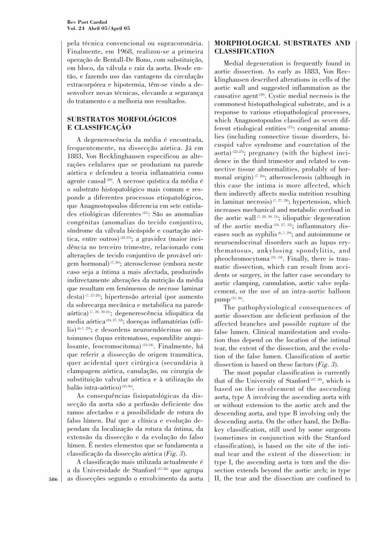
SUBSTRATOS MORFOLÓGICOS E CLASSIFICAÇÃO
A degenerescência da média é encontrada,frequentemente, na dissecção aórtica. Já em1883, Von Recklinghausen especificou as alte-rações celulares que se produziam na paredeaórtica e defendeu a teoria inflamatória comoagente causal (20). A necrose quística da média éo substrato histopatológico mais comum e res-ponde a diferentes processos etiopatológicos,que Anagnostopoulos diferencia em sete entida-des etiológicas diferentes (21): São as anomaliascongénitas (anomalias do tecido conjuntivo,síndrome da válvula bicúspide e coartação aór-tica, entre outros) (22-25); a gravidez (maior inci-dência no terceiro trimestre, relacionado comalterações de tecido conjuntivo de provável ori-gem hormonal) (7, 26); aterosclerose (embora nestecaso seja a íntima a mais afectada, produzindoindirectamente alterações da nutrição da médiaque resultam em fenómenos de necrose laminardesta) (7, 27-28); hipertensão arterial (por aumentoda sobrecarga mecânica e metabólica na paredeaórtica) (7, 28, 30-31); degenerescência idiopática damedia aórtica (24, 27, 32); doenças inflamatórias (sífi-lis) (6-7, 29); e desordens neuroendócrinas ou au-toimunes (lupus eritematoso, espondilite anqui-losante, feocromocitoma) (33-34). Finalmente, háque referir a dissecção de origem traumática,quer acidental quer cirúrgica (secundária àclampagem aórtica, canulação, ou cirurgia desubstituição valvular aórtica e à utilização dobalão intra-aórtico) (35-36).
As consequências fisiopatológicas da dis-secção da aorta são a perfusão deficiente dosramos afectados e a possibilidade de rotura dofalso lúmen. Daí que a clínica e evolução de-pendam da localização da rotura da íntima, daextensão da dissecção e da evolução do falsolúmen. É nestes elementos que se fundamenta aclassificação da dissecção aórtica (Fig. 3).
A classificação mais utilizada actualmente éa da Universidade de Stanford (37-38) que agrupaas dissecções segundo o envolvimento da aorta586
MORPHOLOGICAL SUBSTRATES ANDCLASSIFICATION
Medial degeneration is frequently found inaortic dissection. As early as 1883, Von Rec-klinghausen described alterations in cells of theaortic wall and suggested inflammation as thecausative agent (20). Cystic medial necrosis is thecommonest histopathological substrate, and is aresponse to various etiopathological processes,which Anagnostopoulos classified as seven dif-ferent etiological entities (21): congenital anoma-lies (including connective tissue disorders, bi-cuspid valve syndrome and coarctation of theaorta) (22-25); pregnancy (with the highest inci-dence in the third trimester and related to con-nective tissue abnormalities, probably of hor-monal origin) (7, 26); atherosclerosis (although inthis case the intima is more affected, whichthen indirectly affects media nutrition resultingin laminar necrosis) (7, 27, 28); hypertension, whichincreases mechanical and metabolic overload inthe aortic wall (7, 28, 30, 31); idiopathic degenerationof the aortic media (24, 27, 32); inflammatory dis-eases such as syphilis (6, 7, 29); and autoimmune orneuroendocrinal disorders such as lupus ery-thematosus, ankylosing spondylitis, andpheochromocytoma (33, 34). Finally, there is trau-matic dissection, which can result from acci-dents or surgery, in the latter case secondary toaortic clamping, cannulation, aortic valve repla-cement, or the use of an intra-aortic balloonpump (35, 36).
The pathophysiological consequences ofaortic dissection are deficient perfusion of theaffected branches and possible rupture of thefalse lumen. Clinical manifestation and evolu-tion thus depend on the location of the intimaltear, the extent of the dissection, and the evolu-tion of the false lumen. Classification of aorticdissection is based on these factors (Fig. 3).
The most popular classification is currentlythat of the University of Stanford (37, 38), which isbased on the involvement of the ascendingaorta, type A involving the ascending aorta withor without extension to the aortic arch and thedescending aorta, and type B involving only thedescending aorta. On the other hand, the DeBa-key classification, still used by some surgeons(sometimes in conjunction with the Stanfordclassification), is based on the site of the inti-mal tear and the extent of the dissection: intype I, the ascending aorta is torn and the dis-section extends beyond the aortic arch; in typeII, the tear and the dissection are confined to
Rev Port CardiolVol. 24 Abril 05/April 05
ascendente; assim, o tipo A envolve a aorta as-cendente, com ou sem extensão à crossa e aortadescendente, e o tipo B implica o envolvimentoexclusivo da aorta descendente. Pelo contrário,a classificação clássica de DeBakey, ainda uti-lizada por alguns cirurgiões, por vezes em asso-ciação com a de Stanford, baseia-se no local derotura da íntima e na extensão da dissecção: Notipo I, há rotura na aorta ascendente e a dissec-ção ultrapassa a crossa; no tipo II, rotura e dis-secção exclusivos da aorta ascendente; tipo III,rotura na aorta descendente e dissecção até odiafragma (III-A) até ao abdómen e artérias ilía-cas (III-B) (39). Em termos cirúrgicos faz maissentido utilizar a classificação de Stanford, por-que nos limitamos, essencialmente, a substituira aorta ascendente, quer no tipo I quer no tipoII de DeBakey (daí o seu agrupamento em tipoA). Embora seja mais discriminatória sob oponto de vista patológico, esta classificação teráimplicações prognósticas. Dois terços das dis-secções aórticas afectam a aorta ascendente eoutro terço só a aorta descendente (7).
Outras classificações, a mais actual propostapor Svensson et al, também consideram a he-morragia intramural, ou hematoma intramural ea úlcera aórtica como sinais de dissecção emevolução ou subtipos de dissecção (40- 41).
A história natural da doença é quase invaria-velmente fatal, especialmente no tipo A. Hirstem 1958, num estudo de 505 casos, verificouque 30 % dos doentes com dissecção aórtica fa-leceu nas primeiras 24 horas e 50 % nas pri-meiras 48 horas. Noutra grande série, verificou--se que 50 % dos doentes faleceram até às 48horas, 84 % no primeiro mês e 92 % no pri-meiro ano (6, 42). Praticamente todos os doentesestariam mortos ao fim de um ano se não fossemtratados médica ou cirurgicamente. Na maioriados casos, a morte ocorre por rotura da aorta nosaco pericárdico ou no mediastino, pelo que odiagnóstico precoce e/ou tratamento médico-ci-rúrgico imediato é crucial.
DIAGNÓSTICO E COMPLICAÇÕES
O factor principal para o diagnóstico clínicode dissecção aórtica é um elevado grau de sus-peita e a realização dum precoce e correctoexame físico (43). A primeira referência ao diag-nóstico pré-mortem de dissecção aórtica foi pu-blicada por Swaine e Latham em 1852 (44).
Os elementos clínicos mais frequente-mente associados à dissecção aguda da aorta 587
the ascending aorta; and in type III, the descen-ding aorta is split and dissected as far as thediaphragm (type III-A) or to the abdomen andthe iliac arteries (type III-B) (39). In surgicalterms it makes more sense to use the Stanfordsystem, because in both DeBakey type I and IIthe option is basically to replace the ascendingaorta, which is why the Stanford classificationgroups them as type A. Although the DeBakeysystem is more accurate in pathological terms,there are prognostic implications to the choiceof classification. Two-thirds of aortic dissectionsinvolve the ascending aorta, while only onethird affect the descending aorta (7).
Other classifications, the most recent propo-sed by Svensson et al., also consider intramuralhemorrhage or intramural hematoma and aorticulcer as signs of evolving dissection or assubtypes of dissection (40, 41).
The natural history of the disease is almostalways fatal, especially in type A. Hirst, in astudy of 505 cases in 1958, found that 30 % ofpatients with aortic dissection died in the first24 hours and 50 % in the first 48 hours. In an-other large series, it was found that 50 % died inthe first 48 hours, 84 % in the first month and92 % in the first year (7, 42). Virtually all patientswould die within a year if they did not receivemedical or surgical treatment. In most cases,death occurs because of rupture of the aortainto the pericardial sac or the mediastinum.Early diagnosis and immediate medical or sur-gical treatment are thus essential.
DIAGNOSIS AND COMPLICATIONS
The main factors in diagnosis of aortic dis-
DAVID PRIETO, et alRev Port Cardiol 2005; 24:583-604
Fig. 3 Classificação da dissecção da aorta; Stanford (A e B) e De-Bakey (I, II e III).
Fig. 3 Classification of aortic dissection: Stanford (A and B) andDeBakey (I, II and III).

são a história prévia de hipertensão arterial e ador.
A dor aparece em mais de 90 % dos doen-tes (39, 42, 45), é retroesternal ou interescapular, ca-racteristicamente de início súbito, severo, decarácter lancinante, pulsátil e migratória (se-gundo o sentido da dissecção) e com irradiaçãoanterior ou posterior (segundo a sua localiza-ção). É fácil de confundir com a dor anginosa,mas não é controlada com nitratos nem com an-tiálgicos não opiáceos.
A dissecção do tipo A é frequentementeacompanhada de vários tipos de complicaçõesassociando, na maioria das vezes, o shock comtensões arteriais conservadas ou altas (46). Em20 % dos casos pode aparecer acompanhada dehipotensão e shock, indicando a possibilidadede tamponamento, rotura de aorta, insuficiênciacardíaca secundária a regurgitação valvularaórtica severa ou compromisso das artérias co-ronárias (por oclusão ou dissecção) (47-48). No en-tanto, o primeiro sintoma pode ser a síncope,presente em 10 % dos doentes com dissecçãoaguda da aorta (45), sobretudo quando há envolvi-mento dos grandes vasos do pescoço ou roturaintrapericárdica (geralmente contida) com tam-ponamento cardíaco, representando uma situa-ção de franca emergência. A esta clínica podemassociar-se sinais de envolvimento do sistemanervoso central, perda de pulsos periféricos,sintomas gastrointestinais ou nefrológicos, con-soante o envolvimento dos vasos eferentes daaorta. O aparecimento destes sinais e sintomasfacilita o diagnóstico e tem impacto determi-nante no prognóstico.
A dissecção de tipo B aparece em idadesmais avançadas – idade média 63 anos – e comuma elevada incidência de hipertensão arterial.A dor torácica ocorre em 90 % dos casos, con-fundindo-se com a dor anginosa ou do trom-boembolismo pulmonar. Apresenta, por vezes,sintomatologia correlacionada com a oclusão deramos da aorta. Assim, com frequência, o diag-nóstico acontece pela perda de pulsos distais,preferencialmente à esquerda, confundindo-semuitas vezes com fenómenos embólicos (fre-quentemente os doentes chegam à cirurgia car-díaca só depois de uma exploração das artériasilíacas por pressuposto embolismo periférico).Outras vezes, manifestam-se insuficiência re-nal, por oclusão das artérias renais; dor abdo-minal, por isquémia mesentérica; ou paraple-gia, por oclusão das artérias intercostais.588
section are a high index of suspicion and aprompt and thorough physical examination (43).The first report of a premortem diagnosis waspublished by Swaine and Latham in 1852 (44).
The clinical aspects most commonly associ-ated with acute aortic dissection are history ofhypertension and pain.
Pain, which is experienced in over 90 % ofcases (39, 42, 45), is retrosternal or interscapular,typically of sudden onset, severe, stabbing, pul-sing and migratory (following the direction ofthe dissection) and radiating anteriorly or pos-teriorly depending on location. It can easily bemistaken for angina, but cannot be controlledwith nitrates or with non-opiate analgesics.
Type A dissection is frequently accompa-nied by various types of complication, mostcommonly shock with preserved or high bloodpressure (46). In 20 % of cases it is associatedwith hypotension and shock, which may indi-cate cardiac tamponade, aortic rupture, heartfailure secondary to severe aortic regurgitation,or occlusion or dissection of the coronary arte-ries (47, 48). However, the first symptom may besyncope, which is found in 10 % of patientswith acute aortic dissection (45), particularlywhen there is involvement of the large neckvessels or intrapericardial rupture (usually con-tained) with cardiac tamponade, which is clearly a medical emergency. This clinical settingmay be associated with signs of central nervoussystem involvement, loss of peripheral pulses,or gastrointestinal or nephrological symptoms,depending on which efferent vessels are in-volved. Such signs and symptoms aid diagnosisand are crucial to prognosis.
Type B dissection tends to occur at a moreadvanced age (mean 63 years) and in associ-ation with a high incidence of hypertension.Chest pain, experienced by 90 % of patients,can be confused with angina or pulmonary em-bolism. Symptoms are sometimes consistentwith occlusion of the aortic branches, and sodiagnosis is frequently by loss of distal pulses,particularly on the left, but this can be confusedwith embolism – patients often undergo cardiacsurgery only after exploration of the iliac arte-ries for presumed peripheral embolism. In othercases there is renal failure through occlusion ofthe renal arteries, abdominal pain due to mes-enteric ischemia, or paraplegia caused by oc-clusion of the intercostal arteries.
Diagnosis is confirmed by imaging studies(Fig. 4). In practical terms transesophageal
Rev Port CardiolVol. 24 Abril 05/April 05

O diagnóstico é confirmado por meios ima-giológicos (Fig. 4). Se do ponto de vista práticopodemos considerar a ecocardiografia transeso-fágica e a tomografia computorizada como osmétodos diagnósticos de eleição da patologiaaguda da aorta, a ressonância magnética é ométodo que dá a informação mais completa (49).São as limitações de carácter estratégico ou lo-gístico, tais como o isolamento do doente du-rante a exploração, a demora da técnica e emespecial a sua disponibilidade, que fazem quena prática seja apenas um método diagnósticocomplementar. Por outro lado, a ecocardiografia 589
echocardiography and computerized tomogra-phy are the methods of choice in acute aorticpathologies, but magnetic resonance imagingprovides the most complete information (49).However, the logistical limitations imposed bythe need to isolate the patient during the study,and the fact that it is time-consuming and notwidely available, mean that in practice it is onlyused as a complementary exam. Transesopha-geal echocardiography, on the other hand, is ef-ficient and reliable and is a more realisticchoice for diagnosis, particularly in unstablepatients, simply by detecting an intimal flap
DAVID PRIETO, et alRev Port Cardiol 2005; 24:583-604
Fig. 4 Diagnóstico imagiológico da dissecção da aorta (A e B, TAC; C, Ressonância magnética; D, Aortografia) com visualização do flapda íntima e do falso lúmen.
Fig. 4 Diagnosis of aortic dissection by imaging (A and B, computerized tomography; C, magnetic resonance; D, aortic angiography) showing the intimal flap and the false lumen.
A
C D
B

transesofágica tem demonstrado uma elevadarentabilidade e fiabilidade e é a alternativamais realista na hora do diagnóstico, sobretudonos doentes instáveis, pela simples identifica-ção do flap intimo-medial e do falso lúmen (50-53).
Estes métodos têm relegado a angiografia decontraste, outrora o método de diagnóstico defi-nitivo, a um papel secundário. No entanto,ainda é fundamental para a obtenção de infor-mação sobre a anatomia coronária, sobretudo seconsideramos que estamos perante um grupoetário que geralmente supera os 50 anos deidade. Estudos necrópsicos confirmam a suaimportância, detectando-se o envolvimento dacoronária direita em 10 % das dissecções aórti-cas tipo A e da coronária esquerda em 30 % (7, 54).Daí que se recomende a angiografia coronária,na avaliação pré-operatoria das dissecções agu-das distais e nas dissecções crónicas, sendomais discutível a realização por rotina nos casosde dissecção aguda proximal.
TRATAMENTO
A experiência demonstrou que os resultadoscirúrgicos são geralmente melhores que os daterapêutica médica na dissecção proximal, istoé, de tipo A (I ou II), enquanto a terapêuticamédica parece ter uma vantagem relativa sobrea cirurgia na maior parte dos casos na dissec-ção distal tipo B (III) não complicada. No en-tanto, ao longo do tempo foi-se modificando aopinião que temos sobre estes aspectos, a quevoltaremos a referir-nos mais tarde.
De qualquer modo, o tratamento médico éfundamental e deve ser a base da abordagemterapêutica inicial em todos os casos. O sucessodo tratamento médico já foi relatado por Wheatem 1965 (55). Temos agora a noção de que quasetodos os doentes podem esperar pela cirurgia,pelo menos algumas horas, para que, tantoquanto possível, possam chegar à sala de opera-ções estabilizados sob o ponto de vista médico emetabólico. Na nossa experiência, se os doenteschegam em choque a mortalidade é de quase100 %; se chegam estabilizados, não acidóticose a passar urina, a mortalidade é muitíssimo in-ferior, próxima da cirurgia electiva. Desde queimplementámos esta atitude de estabilizaçãoprévia, há 5 ou 6 anos, perdemos apenas umdoente neste período de espera e temos a cons-ciência de que salvámos muitíssimos mais. Dequalquer modo, há que ter em conta que, nagrande maioria dos casos, o diagnóstico é feito590
and false lumen (50-53).These techniques have relegated contrast
angiography, formerly the definitive diagnosticmethod, to a secondary role. However, angio-graphy is still essential to obtain information oncoronary anatomy, which is particularly impor-tant when dealing with a patient group gene-rally aged over 50. Autopsy studies confirm itsimportance by showing involvement of the rightcoronary artery in 10 % of type A dissectionsand of the left coronary artery in 30 % (7, 54). Co-ronary angiography is thus recommended inpreoperative assessment of acute distal dissec-tion and in chronic dissection, although its rou-tine use in cases of acute proximal dissection isdebatable.
TREATMENT
Experience has shown that surgical resultsare generally better than those of medical ther-apy in proximal dissections, i.e. type A (I or II),while medical therapy appears to be better thansurgery in most cases of uncomplicated distal(type B or III) dissection. However, opinions onthese questions have changed over the years, aswill be discussed below.
In any event, medical treatment is essentialand should be the basis of the initial therapeu-tic approach in all cases. Successful medicaltreatment was described by Wheat in 1965 (55).Current thinking is that virtually all patientscan wait at least a few hours before surgery andcan therefore be stabilized as far as possible inmedical and metabolic terms before arrival inthe operating room. In our experience, if pa-tients arrive in shock, mortality is almost 100 %,while if they are stable, not acidotic, and pas-sing urine, mortality is much lower, close to thatof elective surgery. Since implementation of theprior stabilization approach five or six yearsago, we have lost only one patient in the waitingperiod and have without doubt saved manymore. It should also be borne in mind that inthe great majority of cases diagnosis takes placein hospitals or health centers with no direct ac-cess to surgery.
Medical treatment includes immediate ad-mission to the ICU of patients with suspected ordiagnosed aortic dissection. The aim is to mini-mize the risk of the dissection spreading or theaorta rupturing by controlling the determiningfactors of hypertension and left ventricularejection wave amplitude (dp/dtmax). To this
Rev Port CardiolVol. 24 Abril 05/April 05

em hospitais ou centros de saúde onde não háacesso directo à cirurgia.
O tratamento médico inclui a admissão ime-diata na UCI do doente com suspeita ou diag-nóstico de dissecção aórtica. O objectivo é mi-nimizar o risco de propagação da dissecção ou arotura da aorta, mediante o controlo de dois fac-tores determinantes, a hipertensão arterial e aamplitude da onda de ejecção do ventrículo es-querdo (dp/dtmáx). Para o atingir utilizam-se va-sodilatadores endovenosos (nitroprussiato desódio, nitroglicerina) de modo a diminuir rapi-damente a pressões sistémicas arteriais média esistólica – o ideal estaria entre 90 e 100 mmHg– e β-bloqueadores endovenosos (esmolol, pro-panolol) para reduzir o impacto da onda depulso (56). Ao mesmo tempo procura-se aliviar ador (opiáceos), também fundamental no con-trolo da pressão arterial, e o grau de ansiedadee agitação (benzodiazepinas), tratando de man-ter o débito urinário acima dos 25 ml/h.
Nalguns casos, pode até estar indicado o tra-tamento médico definitivo, como na dissecçãodistal, que voltaremos a referir, e nalguns casosraros de dissecção isolada e estável do arco aór-tico, se não houver sinais de extensão proximalou distal. É, ainda, o tratamento de escolha nadissecção distal crónica estável não compli-cada, que se apresenta mais de duas semanasapós o seu início. Independentemente da apre-sentação, estamos na presença duma doençageneralizada que afecta toda a extensão da pa-rede da aorta, que não vamos curar definitiva-mente; se já está estável após duas semanas,provavelmente não há indicação para trata-mento cirúrgico. Contudo, este tema é contro-verso e alguns autores aconselham a cirurgiaem todas as dissecções distais e proximais (57-60).
Após a estabilização, há que completar odiagnóstico por angiografia, TAC, ou ecocardio-grama transesofágico, no que for consideradopor cardiologistas e cirurgiões como essencialpara obter uma classificação morfológica tãoexacta quanto possível.
CIRURGIA
A finalidade da cirurgia é evitar a rotura(que causaria a morte do doente), restabelecer ofluxo anatómico em áreas isquémicas potenciais(perante a isquémia de algum território, há queproceder à cirurgia, quer na dissecção proximalquer na distal) e corrigir a regurgitação valvularaórtica, se a houver. Isto significa que na dis- 591
end, intravenous vasodilators (sodium nitro-prusside or nitroglycerin) are used for rapid re-duction of mean and systolic systemic bloodpressure, ideally to between 90 and 100 mmHg,and intravenous beta-blockers (esmolol or pro-panolol) to reduce the impact of the pulse wave (56).At the same time, efforts are made to relieve thepain with opiates, which is also essential in re-ducing blood pressure, and to control anxietyand agitation with benzodiazepines, while aim-ing to maintain urine output above 25 ml/h.
In some cases, medical therapy may even bethe definitive treatment, particularly in distaldissection, as will be discussed below, and inrare cases of stable isolated dissection of theaortic arch, so long as there are no signs of prox-imal or distal extension. It is also the treat-ment of choice in uncomplicated chronic stabledistal dissection presenting more than two weeks after onset. Whatever the presentation, thedisease is a generalized one that affects the en-tire aortic wall and that cannot be permanentlycured; if the patient is stable after two weeks,there is probably no need for surgery. However,the question is a controversial one, and someauthors recommend surgery in all dissections,both distal and proximal (57-60).
After stabilization, diagnosis should be con-firmed by angiography, CT scan, or transeso-phageal echocardiography, as cardiologists andsurgeons consider it essential to have as accu-rate a morphological description as possible.
SURGERY
The purpose of surgery is to prevent rupture(which would result in the patient’s death), tore-establish anatomical flow in potentially isch-emic areas (when there is a threat of ischemiain any territory, surgery is essential in both prox-imal and distal dissection), and to correct anyaortic regurgitation. This means that in dissec-tion of the ascending aorta (type A, I or II), theindication is usually for immediate surgery, tosave the patient’s life rather than to cure thedisease. Even so, surgery may be contraindica-ted (or at least not indicated) in cases of veryadvanced age (patients aged over 80 and invery poor health), associated severe and incura-ble or terminal disease, or profound neurologi-cal damage related to the dissection. In our ex-perience, mortality in such cases is 100 %, andit is not worth carrying out “heroic” surgery on a patient who is incapable of recovery. It is
DAVID PRIETO, et alRev Port Cardiol 2005; 24:583-604

secção da aorta ascendente, isto é, no tipo A (Iou II), a indicação é geralmente imediata, poisse trata de salvar a vida do doente, não de curardefinitivamente a doença aórtica. Ainda assim,podemos considerar contraindicação (ou não in-dicação) para cirurgia, os casos de idade muitoavançada (doentes com 80 ou mais anos egrande fragilidade física), de doença grave e in-curável ou terminal associada e de afectaçãoneurológica profunda, relacionada com a dis-secção. Na nossa experiência, a mortalidadenestas situações é de 100 % e não vale a penaestar a fazer a chamada cirurgia heróica numdoente que não é sistemicamente recuperável.Por isso, é importante que cada equipa cirúr-gica saiba quais são os seus próprios resulta-dos, para pode decidir o que lhe é e o que nãolhe é possível fazer.
Por outro lado, na dissecção da aorta des-cendente, a cirurgia tem historicamente umaelevada incidência de mortalidade, podendoatingir 35 % a 75 %, e embora algumas séries jámostrem uma redução significativa destes valo-res, ela continua a ser muito elevada nos gruposcom isquemia renal ou visceral associada (57, 61).Daí, que a cirurgia esteja indicada apenas noscasos de rotura de aorta (grande hemotorax),dor persistente ou recorrente (que indica pro-gressão), na isquemia dos membros inferiores,visceral ou renal aguda (desde que não muitoprolongada, portanto irreversível) e a hiperten-são arterial intratável. Ainda assim, temos quecomparar os resultados da cirurgia com os dasnovas formas de tratamento da aorta distal,como seja o stent endoluminal, que referiremosmais tarde.
Uma das primeiras intervenções para trata-mento ‘definitivo’ da dissecção aguda do tipo Afoi a operação descrita por Bentall em 1968 (62).Esta cirurgia envolve a substituição total daaorta ascendente, incluindo a válvula aórtica,por um conduto composto por um tubo de Da-cron (fibra de polyester) e uma prótese valvular,com necessidade de re-implantação das duasartérias coronárias (Fig. 5). Este método aindahoje demonstra ser tecnicamente seguro e eficazno tratamento da patologia da raiz da aorta, comóptimos resultados a médio e longo prazo (63-65).
Na maior parte dos casos, contudo, apenas énecessário substituir um segmento da aorta e aválvula pode ser preservada. Não cabe aqui de-talhar os aspectos técnicos; contudo, alguns sãoimportantes. Sendo que apenas se substitui a592
therefore essential for each surgical team toanalyze its own results in order to decide whatcan and cannot be done.
At the same time, surgery in dissection ofthe descending aorta has historically had highmortality (between 35 % and 75 %), and al-though some series have shown significantly lo-wer rates, they remain high in patients withassociated renal or visceral ischemia (57, 61).Hence surgery is only indicated in cases of aor-tic rupture with a large hemothorax, persistentor recurrent pain (which indicates progression),acute lower limb, visceral or renal ischemia (solong as it is not prolonged and therefore irrever-sible), and untreatable hypertension. Even so,surgical results should be compared with newforms of treating the distal aorta, such as sten-ting, which will be discussed below.
One of the first interventions designed toprovide a definitive treatment of acute type Adissection was the operation described by Ben-tall et al. in 1968 (62), consisting of complete re-placement of the ascending aorta, including theaortic valve, with a conduit composed of a Da-cron polyester fiber tube and a prosthetic valve,and involving the reimplantation of the coro-nary arteries (Fig. 5). This procedure remains asafe and effective treatment for pathologies ofthe aortic root and gives excellent medium- andlong-term results (63-65).
In most cases, however, it is only necessaryto replace a segment of the aorta, and the valvecan be preserved. This is not the place to give adetailed description of all the technical aspects,but some are important. When only the ascen-ding aorta is to be replaced, in some cases asfar as the arch, it is important to try and elimi-nate the false lumen by sandwiching the twowall layers between two strips of Teflon®, oneoutside and the other inside. This method canalso be used to strengthen an extremely fragileaorta (Fig. 6). An alternative method, which iseasier and very popular, involves the use of bio-logical glue (resorcin-formalin or fibrin adhe-sive) (66, 67). It should be noted, however, that eli-mination of the false lumen is only achieved ina relatively small number of cases, whatevertechnique is used.
When the aortic arch is involved, it may benecessary to replace it completely and reim-plant the neck artery trunks, either as a blockor separately. In other cases, when the wall tearand the false lumen are in the concave part ofthe arch, only partial replacement may be re-
Rev Port CardiolVol. 24 Abril 05/April 05

aorta ascendente, nalguns casos com extensão àcrossa, é importante tentar obliterar o falso lú-men, fazendo uma sanduíche das duas camadasda parede com duas tiras de Teflon®, uma ex-terna e outra externa. Este método, tambémpermite reforçar a parede em aortas muito friá-veis (Fig. 6). Outra possibilidade, menos labo-riosa e com muitos adeptos, é a utilização decolas biológicas (resorcina-formol ou fibrinaadesiva) (66-67). Refira-se, no entanto, que a obli-teração só é conseguida numa porção relativa-mente menor dos casos, independentemente datécnica utilizada.
Quando o arco aórtico está envolvido, podehaver necessidade de substituí-lo totalmentecom reimplantação dos troncos arteriais do pes-coço, quer em botão, quer isoladamente. Nou-tros casos, isto poderá ser feito apenas com asubstituição parcial da crossa, quando a rotura 593
quired.With regard to the aortic valve, a decision
must be taken as to whether to replace or pre-serve it, and if the latter, how to do so. Mostsurgeons believe that it should be preservedwhenever possible, particularly when it is mor-phologically normal (68-72), but that a structurallyabnormal valve should be replaced. A bicuspidvalve is nowadays usually immediately replaced,even if functioning normally, since this willeventually have to be done.
As stated above, aortic regurgitation is cau-sed by loss of suspension, and when the wall isrepaired, the valve can be restored to its normalfunction. Resuspension of the commissures hasthe twofold effect of raising the valve again andmaking it competent, and helping to close theproximal false lumen.
Three types of intervention are used for thispurpose. The preferred option is to preserve theaortic wall at the sinuses of Valsalva, if they arenot dilated, so as not to have to reimplant thecoronary arteries. Everything below the aorticcommissures is preserved and strengthenedwith Teflon or biological glue and sutures to eli-
DAVID PRIETO, et alRev Port Cardiol 2005; 24:583-604
Fig. 5 Técnica de Bentall-De Bono: substituição da aorta ascen-dente e raiz da aorta por prótese de Dacron e prótese valvular,com reimplantação dos ostia coronários. (de: Surgical Treatmentof Aortic Dissection. Borst HG. Churchill Livingstone 1996).
Fig. 5 Bentall-De Bono operation: replacement of the ascendingaorta and aortic root with a Dacron tube and prosthetic valve, withreimplantation of the coronary ostia (from Surgical Treatment ofAortic Dissection, Borst HG, Churchill Livingstone 1996).
Fig. 6 Técnica de substituição da aorta ascendente, com reforçoda parede aórtica e encerramento do falso lúmen, utilizando duasbandas de Teflon, que envolvem as duas camadas da aorta disse-cada. Este método é utilizado tanto na anastomose proximal comona distal. (de: Surgical Treatment of Aortic Dissection. Borst HG.Churchill Livingstone 1996).
Fig. 6 Replacement of the ascending aorta, strengthening theaortic wall and closing the false lumen using two Teflon strips toenclose the two layers of the dissected aorta (from Surgical Treat-ment of Aortic Dissection, Borst HG, Churchill Livingstone 1996).

da parede e o falso lúmen ocorrem só na conca-vidade do arco aórtico.
No que diz respeito ao tratamento da válvulaaórtica, é necessário decidir se substituí-la ouconservá-la e, neste caso, como fazê-lo. A maiorparte dos cirurgiões pensa que, tanto quantopossível, se deva conservá-la, sobretudo quandoé morfologicamente normal (68-72). Mas, se a vál-vula for estruturalmente anormal deverá sersubstituída. No caso das válvulas bicúspides,mesmo que funcionalmente normais, há hojeuma tendência para substituí-las de imediato,porque necessitarão eventualmente de substi-tuição.
Como acima referimos, a regurgitação aór-tica é causada pela perda do mecanismo desuspensão e, ao reconstituir a parede, é possíveldevolver a válvula aórtica a uma função normal.A ressuspensão da zona das comissuras temdois efeitos, elevar de novo a válvula aórtica etorná-la competente e ajudar a encerrar o falsolúmen proximal. Com este fim, são hoje utiliza-dos três tipos de intervenções: Geralmente pre-fere-se a conservação da parede aórtica a níveldos seios de Valsalva, se estes não estão dilata-dos, para evitar ter que reimplantar as artériascoronárias. Neste tipo de intervenção tudo oque está para baixo das comissuras aórticas émantido, reforçado com Teflon® ou cola bioló-gica e suturas para obliterar o falso lúmen.
Outra técnica utilizada é a descrita por Ya-coub, que consiste numa remodelação dos seiosde Valsalva e pilares da válvula, por ressecçãoda maior parte da parede aórtica e substituiçãopor um tubo de Dacron, onde serão reimplanta-das as coronárias. Por outro lado, na operaçãode David, a válvula aórtica e pilares são reim-plantadas dentro do tubo protésico (Fig 7).
Nalguns casos em que há necessidade desubstituir a válvula temos utilizado um homoen-xerto porque é de muito mais fácil implantação.Trata-se de uma cirurgia muitíssimo mais rá-pida, mas tem o problema da durabilidade limi-tada do homoenxerto (73).
Há ainda alguns dilemas por resolver. Porexemplo, o que fazer em termos da fissura daíntima? Na maior parte dos casos esta está loca-lizada na aorta ascendente e a sua eliminaçãoestá, portanto, incluída na cirurgia clássica.Mas, nalguns doentes, a dissecção pode iniciar--se na crossa da aorta ou mesmo na aorta des-cendente e progredir retrogradamente. Nestescasos, deve ou não incluir-se a fissura na re-594
minate the false lumen. Another technique isthat described by Yacoub, consisting of remod-eling the sinuses of Valsalva and the valve sup-ports, resecting most of the aortic wall and re-placing it with a Dacron tube into which thecoronary arteries are reimplanted. In the Davidoperation, on the other hand, the aortic valveand supports are reimplanted inside the pros-thetic tube (Fig. 7).
In some cases when it was necessary to re-place the valve we have used an autograft, asthis is easier to implant and is much faster, butan autograft is of limited durability (73).
There are still unresolved questions, such ashow to deal with the tear in the intima. This isusually located in the ascending aorta and itselimination is thus part of the standard surgicalprocedure. However, in some patients the dis-section may begin at the aortic arch or even inthe descending aorta, and then progress retro-gradely. In such cases, should the tear be inclu-ded in the repair, which would mean more ex-tensive replacement (74)? There is still noagreement on this issue, although wheneverpossible we inspect the aortic arch and includeit in the repair if the intimal tear is locatedthere. This means that distal suturing of the Da-cron tube is now performed by open surgery,with circulatory arrest under moderate or pro-
Rev Port CardiolVol. 24 Abril 05/April 05
Fig. 7 Técnica de Sarsam-Yacoub de substituição da aorta as-cendente e raiz da aorta por tubo de Dacron, com preservação daválvula aórtica. A anastomose proximal é efectuada a nível supra-anular, com substituição da parede dos seios de Valsalva. Em al-ternativa, na técnica de David, todo o aparelho valvular é implan-tado no interior da prótese tubular. (de: Surgical Treatment ofAortic Dissection. Borst HG. Churchill Livingstone 1996).
Fig. 7 Sarsam-Yacoub technique to replace the ascending aortaand aortic root with a Dacron tube, preserving the aortic valve.Proximal anastomosis is supra-annular, replacing the wall of thesinuses of Valsalva. In the David procedure, the entire valve ap-paratus is implanted inside the prosthetic tube (from SurgicalTreatment of Aortic Dissection, Borst HG, Churchill Livingstone1996).

paração, isto é, proceder a uma substituiçãomais extensa da aorta (74)? Ainda não há con-senso sobre este aspecto mas, sempre que pos-sível, inspeccionamos o arco aórtico e incluí-mos esta zona da parede na reparação se aliencontrarmos a ferida da íntima. Significa istoque a sutura distal do tubo de Dacron é, hojeem dia, feita de modo aberto, isto é, com para-gem circulatória em hipotermia moderada ouprofunda (18º a 22º C).
RESULTADOS
Dissecções de tipo AOs resultados da cirurgia variam muitíssimo
e dependem, essencialmente, de como os doen-tes chegam à cirurgia, da idade e do tipo de pa-tologia. Na literatura encontramos mortalidadesoperatórias entre 5 e 30 %, com sobrevivênciasde cerca de 80 % a um ano, de 66 % a 5 anos, eda ordem dos 35 % a 10 -15 anos.
Recentemente, o grupo de Stanford publicoua sua experiência, comparando os casos de ope-ração de Bentall, se a válvula era anatómica efuncionalmente anormal, com a substituição su-pracomissural apenas da aorta ascendente, eainda com um terceiro grupo, com reparação daválvula aórtica, que esta equipe usa cada vezmais. Contudo, este grupo não encontrou dife-renças a longo prazo, até 20 anos de follow-up,em que apenas 20 % dos doentes estavam vivos(há que ter em conta que muitos destes doentestinham mais de 60 ou 70 anos, no momento dacirurgia, pelo que não podem sobreviver muitomais que 20 anos). Mas também não encontra-ram nenhuma diferença entre a conservação e asubstituição da válvula por qualquer dos doismétodos.
No entanto, outros autores demonstraram di-ferenças significativas entre quando se con-serva e quando não se conserva a válvula, a favor da primeira daquelas atitudes Adicional-mente, há que considerar a qualidade de vida.A necessidade de reoperação também não foipraticamente diferente, ainda que quandohouve a substituição separada da válvula e daaorta ascendente se tivessem registado algunscasos de recorrência de aneurismas proximaisque obrigaram a reoperação. O grupo de Turina,de Zurich, por exemplo, demonstrou que a ci-rurgia da dissecção aguda do tipo A mantêmriscos elevados, mas, apesar de uma incidênciamais elevada de reoperações, os resultados alongo prazo foram bastantes satisfatórios, com 595
found hypothermia (18 to 22 ºC).
RESULTS
Type A dissectionSurgical results vary greatly, and depend
mainly on the condition of patients on arrival,their age, and the type of pathology. Operativemortality in the literature is 5 to 30 %, with80 % survival at one year, 66 % at five years,and around 35 % at 10-15 years.
Recently the Stanford group published theirexperience and compared the Bentall operation,in cases of an anatomically and functionally ab-normal valve, with supracommissural replace-ment of the ascending aorta only and also withaortic valve repair, which is increasingly popu-lar with this group. They found no differencesin the long term, up to 20-year follow-up, afterwhich only 20 % of the patients were still alive(it should be borne in mind that many of thesepatients were aged over 60 or 70 at the time ofsurgery and were unlikely to live much morethan another 20 years). They also found no dif-ference between preservation and replacementof the valve by either method.
However, other authors have reported thatpreserving the valve gives significantly betterresults. Quality of life is also an issue here.There were no significant differences in theneed for reoperation, although when the valveand the ascending aorta were replaced separa-tely there were some cases of recurrence of pro-ximal aneurysm, necessitating reoperation. Tu-rina’s group in Zurich, for example, showed thatsurgery for acute type A dissection is still ahigh-risk procedure but that despite a high in-cidence of reoperation long-term results weresatisfactory, with around 60% survival at 7years (Fig. 8). Results also, of course, dependon the method of valve preservation (68-72).
Type B dissectionIt was established 20 years ago that there
was no advantage in surgical treatment of typeB dissections and most surgeons have come torecommend medical treatment when uncompli-cated by ischemia. Nevertheless, the debatecontinues. The Stanford group, which is amongthe most experienced in this pathology, haveshown that long-term prognosis, which is poorin these patients, is determined more by factorsrelated to the dissection and to the patient thanto the type of treatment, and that these factors
DAVID PRIETO, et alRev Port Cardiol 2005; 24:583-604

uma sobrevivência de cerca de 60 % aos 7 anos(Fig. 8). Os resultados dependem, ainda, na-turalmente, do método de conservação da vál-vula (68-72).
Dissecções de tipo BHá uns 20 anos atrás estabeleceu-se que,
nas dissecções de tipo B, não havia vantagemna cirurgia e a maior parte dos grupos cirúrgi-cos passou a considerar as dissecções de tipo Bnão complicadas com isquemia para tratamentomédico. Contudo, a discussão mantém-se. Ogrupo de Stanford, um dos que tem maior expe-riência neste tipo de patologia, demonstrou queo prognóstico a longo prazo, que é pobre nestesdoentes, é determinado, primariamente, pelosfactores relacionados com a dissecção e com odoente e não pelo tipo de tratamento, e que es-tes factores não podem geralmente ser modifi-cados (75-78). Naquela experiência, com umagrande preferência pelo tratamento médico naúltima década (cerca de 85 % dos casos), amortalidade foi bastante mais elevada no trata-mento cirúrgico. Nalguns casos, de doentesmais idosos, a mortalidade oscila entre 20 e596
cannot in general be modified (75-78). In their ex-perience, there has been a strong preference formedical therapy (used in 85 % of cases) in thelast ten years, while mortality is much higherwith surgery. In older patients, mortality can bebetween 20 and 35 %. However, their long-termresults are similar for both medical and surgicalapproaches (Fig. 9).
Unlike the Stanford group, whose expe-rience over more than a decade has inclinedthem towards the medical treatment of type Bdissections, Griepp’s group in New York havecontinued to opt for surgery in most cases (57, 60, 75).These authors have achieved a 16-year survivalof 55-60 %, with a curve that is the same as orclose to the survival curve for the general popu-lation. More importantly, most patients (over80 %) did not need reintervention after surgicaltreatment of type B dissection.
However, for the last ten years there hasbeen growing interest in the use of endovascu-lar stents to treat the descending aorta (79-81) and,more recently still, some surgeons have exten-ded this technique to the aortic arch and the as-cending aorta. Others have used a complex
Rev Port CardiolVol. 24 Abril 05/April 05
Fig. 8 Sobrevivência (actuarial) dos doentes submetidos a cirurgia de correcção da dissecção de tipo A, comparando os doentes comsubstituição (AV Replaced) e com conservação (AV Preserved) da válvula aórtica (de: Niederhäuser U et al. Eur J Cardiothorac Surg1999;15:557-563).
Fig. 8 Actuarial survival of patients undergoing surgery to correct type A dissection, comparing replacement (AV Replaced) and preser-vation (AV Preserved) of the aortic valve (from Niederhäuser U et al. Eur J Cardiothorac Surg 1999;15:557-563)
Postoperative Interval (Months)
Doentes com substituiçãoAV Replaced
Doentes com substituiçãoAV Replaced
Doentes com conservaçãoAV Preserved
Doentes com conservaçãoAV Preserved
Doentes em riscoPatients at Risk
p = 0,13746p = 0.13746
% d
e so
bre
vivê
ncia
/ %
Sur
vivi
ng

35 %. Apesar de tudo, os resultados a longoprazo são idênticos, quer para o tratamento mé-dico quer para o tratamento cirúrgico (Fig. 9).
Ao contrário da experiência de Stanford queevoluiu há mais de uma década para o trata-mento médico da dissecção de tipo B, o grupode Griep, de Nova Iorque, continuou a utilizar acirurgia na maior parte dos casos de tipo B (57, 60, 75).Estes autores obtiveram uma sobrevivência aos16 anos de cerca de 55 a 60 %, numa curva queé próxima ou que se foi afastando apenas ligei-ramente da curva de sobrevivência da popula-ção em geral. E, mais importante, a maior partedos doentes (mais de 80 %) não necessitaramde reintervenção após tratamento cirúrgico dadissecção de tipo B.
Contudo, há cerca de 10 anos atrás para cá,tem havido um crescente entusiasmo pela utili-zação dos stents endovasculares para tratar aaorta descendente (79-81) e, ainda mais recente-mente, alguns autores têm mesmo estendido autilização desta técnica ao arco aórtico e à aortaascendente. Outros, ainda, utilizaram uma téc-nica complexa de introdução de parte do stentna aorta descendente para obliterar o falso lú-men, acompanhada por cirurgia clássica daaorta ascendente e crossa (82-84).
Há cerca de 2 anos, o grupo de Buffolo, deSão Paulo, Brasil, publicou a sua experiênciade 191 doentes com dissecção de tipo B trata-dos com stents endoluminais, com resultadosque foram considerados excelentes e bastantemelhores que os do tratamento conservador (80)
(Fig. 10). É bem possível que todos os casos 597
technique to introduce part of the stent into thedescending aorta to eliminate the false lumen,combined with standard surgery of the ascen-ding aorta and aortic arch (82-84).
Around two years ago, Buffolo’s group in SãoPaulo, Brazil, published their experience of 191patients with type B dissection treated with en-doluminal stents, with excellent results thatwere considerably superior to those of conser-vative treatment (80) (Fig. 10). It may well be thatall cases of extensive type B dissection shouldnow be treated with stents, which are easy to in-troduce and relatively cheap; when the aorticmorphology is suitable, they are probably thebest option. We therefore believe that the dis-cussion in coming years will not be about surgi-cal versus medical treatment, but rather aboutmedical treatment versus stenting; and thisquestion in turn may no longer be asked be-cause all patients will be stented (77).
FOLLOW-UP
Long-term survival varies according to thetype of pathology. The Stanford series indicatesa 10-year survival of 40 % for type A dissec-tion, all of them treated surgically, and 60 % fortype B.
However, whatever the pathology or treat-ment, surgical or medical, the disease has notbeen cured. There remains a substantial part ofthe aortic wall that is abnormal, with or withoutdissection or a false lumen. Follow-up mustthus include constant and careful control of
DAVID PRIETO, et alRev Port Cardiol 2005; 24:583-604
Fig. 9 Sobrevivência global e sobrevivência sem complicações (actuarial) dos doentes submetidos a cirurgia de correcção da dissecçãode tipo B (de: Lansman SL et al: Ann Thorac Surg 2002;74:S1833-S1835).
Fig. 9 Overall actuarial survival and actuarial survival without complications of patients undergoing surgery to correct type B dissection(from Lansman SL et al. Ann Thorac Surg 2002;74:S1833-S1835).
So
bre
vivê
ncia
/ S
urvi
val
Anos / Years Anos / Years
5 anos / 5 Years: 80 %10 anos / 10 Years: 55 %
5 anos / 5 Years: 77 %10 anos / 10 Years: 77 %
Doentes em riscoPatients at risk
Doentes em riscoPatients at risk
–––• ???? / Survival––– ???? / Age/Sex matched survival

mais extensos de dissecção de tipo B devam,actualmente, ser tratados com um stent endolu-minal. O dispositivo é fácil de introduzir, a in-tervenção é relativamente barata e, quando aaorta tem uma morfologia aceitável, é provavel-mente a melhor forma de tratamento. Portanto,pensamos que na próxima década não vai maisdiscutir-se a cirurgia versus tratamento médico;vai discutir-se tratamento médico versus. stent e,eventualmente, essa discussão nem sequerexistirá porque todos os doentes acabarão porreceber um stent (77).
FOLLOW-UP
A sobrevivência a longo prazo varia con-soante o tipo de patologia. A série de Stanford,aponta para as dissecções de tipo A, sempre su-jeitos a intervenção cirúrgica, uma sobrevivên-cia a 10 anos de cerca de 40 % e na ordem dos60 % para o tipo B.
Mas seja qual for o tipo de patologia e o tipode tratamento utilizado, cirúrgico ou médico, a598
blood pressure, which has been shown to in-crease survival, reduce the incidence of aneu-rysms and minimize the recurrence of dissection.
At the same time, it is essential to monitorany false lumen to detect aneurysms early,using chest X-ray, computerized tomography(CT) or transesophageal echocardiography. Pa-tients should have a chest X-ray every 6 monthsto check whether the cardiac silhouette haschanged, in which case it should be followed bya CT scan and/or magnetic resonance imagingand echocardiography (Fig. 11).
The false lumen in fact remains patent inover 80 % of cases, and 20 % of these developaneurysms that require surgery. Redissectionoccurs in 10 % of cases, de novo aortic regurgi-tation in under 10 %, and 10-20 % need reope-ration within 5 years of surgery.
Other risk factors for late mortality includediabetes, functional class, the time of surgery(current results are much better than those ofseveral years ago), the institution and the sur-geon. Late mortality is also influenced by the
Rev Port CardiolVol. 24 Abril 05/April 05
Fig. 10 Ressonância Magnética antes (A) e após (B) a implantação de stent intraluminal para tratamento de dissecção de tipo B (de: Buf-folo E et al: Ann Thorac Surg 2002;74:S1815-S1817).
Fig. 10 Magnetic resonance imaging before (A) and after (B) implantation of intraluminal stent to treat type B dissection (from Buffolo Eet al. Ann Thorac Surg 2002;74:S1815-S1817).
A B

doença não foi curada. Mantém-se uma porçãosubstancial da parede aórtica que é anormal,com dissecção ou sem dissecção, com falso lú-men ou sem falso lúmen. Por isso, o seguimentoobriga a um controle permanente e rigoroso datensão arterial. Está demonstrado que este tipode intervenção terapêutica prolonga a sobrevi-vência, reduz a incidência de aneurismas e mi-nimiza a recorrência de uma nova dissecção.
Por outro lado, é essencial manter uma vigi-lância sobre o falso lúmen para detectar preco-cemente e minimizar a formação de aneurismas,o que se consegue com a radiografia do tórax, atomografia computorizada ou o ecocardiogramatransesofágico. Os doentes devem fazer uma ra-diografia do tórax pelo menos de 6 em 6 mesespara se ver se a silhueta cardíaca se modifi-cou e, se for esse o caso, proceder à tomografiae/ou ressonância magnética e ecocardiograma(Fig. 11).
De qualquer modo, o falso lúmen mantêm-sepatente em mais de 80 % dos casos e cerca de20 % destes desenvolvem aneurismas que ne-cessitam de cirurgia. A re-dissecção, ocorre em10 % dos doentes, a regurgitação aórtica denovo em menos de 10 %, e cerca de 10 a 20 %dos doentes necessitam de reoperação nos pri-meiros 5 anos após a cirurgia.
Outros factores de risco para a mortalidadetardia incluem a diabetes, a classe funcional, adata da cirurgia (os resultados actuais são muitomelhores que os de há alguns anos atrás), a ins-tituição e o cirurgião. A mortalidade tardia de-pende ainda do tipo de cirurgia efectuada (a in-clusão da crossa parece ser um factor importantena sobrevivência) e da presença ou não de doen-ça visceral isquémica concomitante.
EXPERIÊNCIA DE COIMBRA
A nossa experiência dos últimos 10 anos in-clui 78 casos com idades entre os 12 e os 76anos (média de 58 anos), 80 % do sexo mascu-lino, na grande maioria (87 %) do tipo A (compredominância do tipo I de DeBakey), corres-pondendo à experiência da maior parte dos gru-pos cirúrgicos. O Quadro I detalha as caracte-rísticas demográficas e clínicas destes doentes.
Como factores de risco predominantes ob-servaram-se a hipertensão arterial, a doença dotecido conjuntivo, o tabagismo e a insuficiênciarenal crónica. Os sintomas iniciais foram prin-cipalmente a dor em todos os tipos, especial-mente no tipo I e no tipo III. 599
type of surgery (the inclusion of the aortic archappears to be an important factor in survival)and concomitant visceral ischemic disease.
THE COIMBRA EXPERIENCE
Our experience over the last 10 years inclu-des 78 cases, aged between 12 and 76 years(mean 58), 80 % male, most (87 %) type A,most of them type I in the DeBakey classifica-tion; this is similar to the experience of mostsurgical groups. Table I lists the patients’ demo-graphic and clinical characteristics.
The main risk factors are hypertension, con-nective tissue disease, smoking, and chronic re-nal failure. The main initial symptom was painin all types, especially types I and III.
In a significant number of cases there wasischemia of the abdominal organs, including thekidneys, with acute renal failure. Paresis due tomedullary involvement occurred in at least a
DAVID PRIETO, et alRev Port Cardiol 2005; 24:583-604
Fig. 11 Ressonância magnética mostrando pseudo-aneurisma daraiz da aorta (*), 12 anos após cirurgia de dissecção de tipo A.
Fig. 11 Magnetic resonance imaging showing pseudoaneurysm ofthe aortic root (*), 12 years after surgery for type A dissection.

Num número significativo de casos havia is-quemia de órgãos abdominais, incluindo osrins, com insuficiência renal aguda. A parésia,por envolvimento medular, ocorreu em, pelomenos, 1/4 dos casos de dissecção de tipo A, enum terço dos doentes havia hemopericárdio,isto é, rotura contida da aorta ascendente. O en-volvimento da válvula aórtica foi observado emcerca de 50 % dos casos do tipo A. Nesta expe-riência, a doença coronária significativa estevepresente em apenas 6 % dos casos e em so-mente 2 a 4 % dos doentes foi necessário fazerbypass aortocoronário.
Nestas circunstâncias foi utilizado apenasum tubo protésico em cerca de 50% dos casosdo tipo A, tubo protésico mais comissuroplastiacom re-suspensão da válvula aórtica em cercade 15 % dos casos, operação de Bentall emcerca de 14 %, substituição da crossa em 13 %e, apenas num número pequeno de doentes, autilização de homoenxerto valvular (Quadro II).Onze por cento dos doentes tiveram cirurgia as-sociada, incluindo a cirurgia coronária. Doisdoentes com isquemia abdominal severa foramsubmetidos a laparatomia exploradora.
Nas dissecções de tipo I, a morbilidade pós--operatória incluiu a necessidade de re-explora-ção cirúrgica por hemorragia em 11 % e a insu-ficiência renal aguda em 13 % dos casos. A600
quarter of cases of type A dissection and a thirdhad hemopericardium, i.e. contained rupture ofthe ascending aorta. The aortic valve was in-volved in 50 % of type A cases. Significant co-ronary artery disease was present in only 6%and coronary bypass was necessary in only 2-4 %.
It was thus only necessary to use a prosthe-tic tube in half of type A cases, prosthetic tubeplus commissuroplasty with resuspension of theaortic valve in 15 %, the Bentall operation in14%, arch replacement in 13 %, and a valveautograft in a small number of patients (TableII). Other surgery, including coronary surgery,was required in 11% of cases. Two patients withsevere abdominal ischemia underwent explora-tory laparotomy.
In type I dissections, postoperative morbi-dity included the need for surgical re-explora-tion following hemorrhage in 11 % and acuterenal failure in 13 %. In-hospital mortality was15 %, around 20 % over the whole ten yearsand 10 % in the last five. It was lower in type Adissections than in type B, but there was a dif-ference (20 % versus 0 %) between DeBakeytype I and type II (in which only the ascendingaorta is involved). The difference in in-hospitalmortality between patients operated in theacute phase and in the chronic phase was sta-
Rev Port CardiolVol. 24 Abril 05/April 05
Quadro IDados clínicos e epidemiológicos
Tipo de DissecçãoStanford A BDeBakey I II IIITotal 58% 29% 13%Aguda 84% 52% 30%
Dados demográficosIdade média (anos) 65 55 53Sexo masculino 83% 78% 80%Sintoma inicialToracalgia 58 % 43% 70%Síncope 20% 13% –Dispneia – 17% 10%
Outros sintomasHemopericárdio 33% 30% –Hemotórax 2% 9% 20%Parésia de m. inferiores 27% – 10%Isquemia abdominal 11% – 30%
Patologia AssociadaValvulopatia aórtica 54% 43% 10%Valvulopatia mitral – 4% –Aneurisma aórtico 2% 22% 10%Doença coronária 6% 4% –Cirurgia coronária prévia 2% 4% –Substituição v. aórtica prévia 4% 4% –
Table IClinical and epidemiological data
Type of dissectionStanford A BDeBakey I II IIITotal 58% 29% 13%Acute 84% 52% 30%
Demographic characteristicsMean age (years) 65 55 53Male 83% 78% 80%Initial symptomChest pain 58 % 43% 70%Syncope 20% 13% –Dyspnea – 17% 10%
Other symptomsHemopericardium 33% 30% –Hemothorax 2% 9% 20%Lower limb paresis 27% – 10%Abdominal ischemia 11% – 30%
Associated pathologyAortic valve disease 54% 43% 10%Mitral valve disease – 4% –Aortic aneurysm 2% 22% 10%Coronary disease 6% 4% –Previous coronary surgery 2% 4% –Previous aortic valve 4% 4% –replacement

mortalidade hospitalar foi de 15 %, cerca de20 % nos primeiros 10 anos e de 10 % nos últi-mos 5. Foi muito menor no tipo A do que notipo B, mas se distinguirmos entre o tipo I e otipo II de DeBakey encontramos uma diferença,20 % versus 0 %, quando apenas estava envol-vida a aorta ascendente. A diferença de morta-lidade hospitalar entre os doentes operados nafase aguda e os operados na fase crónica foi es-tatisticamente significativa.
601
tistically significant.In-hospital mortality was mainly due to
ventricular dysfunction (in 50 % of cases), ab-dominal complications (33 %), stroke (5 %) andhemorrhage in the other 5 % (Table III).
Finally, in long-term follow-up of these pa-tients (two were lost to follow-up), late mortalitywas 16%, higher in type B, which had requiredsurgery because their cases were complex,again with a significant difference between
DAVID PRIETO, et alRev Port Cardiol 2005; 24:583-604
Quadro IICirurgia efectuada
CIRURGIA %
Dissecção I II IIITubo protésico apenas 53 35 100Valvuloplastia aórtica 17 17 –Remodelação da raiz aórtica 24 –Bentall-DeBono 6 35 –Starr 4 – –C/ Substituição do arco aórtico 13 4 –Homoenxerto valvulado 2 4 –
Table IIType of surgery
SURGERY %
Type of dissection I II IIIProsthetic tube only 53 35 100Aortic valvuloplasty 17 17 –Remodeling of aortic root 24 –Bentall-DeBono 6 35 –Starr 4 – –With replacement of aortic root 13 4 –Valve autograft 2 4 –
MORTALIDADE HOSPITALAR�IN-HOSPITAL MORTALITY
DISSECÇÃO AÓRTICA�AORTIC DISSECTION�
12/78 - 15%
TIPO I�TYPE I�20%
TIPO III�TYPE III�30%
TIPO II�TYPE II�
0%
AGUDA�ACUTE�21%
CRÓNICA�CHRONIC�
14%
AGUDA�ACUTE�33%
AGUDA�ACUTE�28%
MORTALIDADE TARDIA�LATE MORTALITY
DISSECÇÃO AÓRTICA�AORTIC DISSECTION�
13/66 - 20%
TIPO I�TYPE I�15%
TIPO III�TYPE III�35%
TIPO II�TYPE II�17%
AGUDA�ACUTE�18%
CRÓNICA�CHRONIC�
10%
AGUDA�ACUTE�35%
AGUDA�ACUTE�30%
AGUDA�ACUTE�10%
Fig. 12 Mortalidade peri-operató-ria e tardia após cirurgia da dis-secção (HUC).
Fig. 12 Perioperative and latemortality after surgery for aorticdissection (Coimbra UniversityHospitals).

A mortalidade hospitalar foi essencialmentedevida a disfunção ventricular, em 50 % dos ca-sos, a complicações abdominais em 33 %, oAVC em 5 % e a hemorragia noutros 5 % (Qua-dro III).
Finalmente, estes doentes foram seguidos alongo prazo (dois não foram encontrados) e amortalidade tardia foi de 16 %, mais elevadanos casos de tipo B (que tinham obrigado a ci-rurgia porque eram já complexos), mais umavez com uma diferença significativa entre os ca-sos crónicos e os casos agudos.
A morte súbita ocorreu em 6 % dos doentes,e resultou de enfarte agudo do miocárdio em5 %, de AVC em 1 %, e de neoplasia, acidentede viação e rotura de aneurisma em 1 % cada.
CONCLUSÃO
A dissecção aguda da aorta é uma entidadepotencialmente fatal. Na maior parte dos casos,a aorta está fragilizada em toda a sua extensão.O diagnóstico precoce depende de um forte ín-dice de suspeição. A cirurgia está indicada emquase todas as dissecções de tipo A e em mui-tas do tipo B. Na maior parte destas, contudo, aterapêutica médica parece obter resultados su-periores. A cirurgia é raramente, se é que al-guma vez, curativa; por isso, o controlo a longoprazo (provavelmente para toda a vida) é essen-cial e inclui o controlo apertado da hipertensãoarterial e a vigilância dos segmentos aórticosnão excisados e, em especial, do falso lúmenpatente distal, numa tentativa de evitar a roturae minimizar as consequências da formação defalsos aneurismas.
602
acute and chronic cases.Sudden death occurred in 6 % of patients,
resulting from myocardial infarction in 5 %,stroke in 1 %, and cancer, road accidents, andruptured aneurysm in 1 % each.
CONCLUSION
Acute aortic dissection is a potentially fatalcondition. In most cases, the aorta is weakenedalong its entire length. Early diagnosis dependson a high index of suspicion. Surgery is indica-ted in almost all cases of type A dissection andin many cases of type B. In most of the latter,however, medical therapy appears to give betterresults. Surgery is rarely if ever curative, hencelong-term follow-up (probably for life) is essen-tial, including strict control of hypertension andmonitoring of the untreated aortic segments,and especially any residual patent false lumen,in order to prevent rupture and to minimize theconsequences of the formation of falseaneurysms.
Rev Port CardiolVol. 24 Abril 05/April 05
Quadro IIICausas de morbilidade
MORBILIDADE %Dissecção Tipo I Tipo II Tipo IIIHemorragia 11 9 20Insufc. Renal Aguda 13 – –Sdr. Febril 8Arritmia SPV 7 4AVC 4 4Isquémia mesentérica 4 – 10Isquémia de MMII – – 10Deiscência de esterno 4 4 –Pneumonia – – 10TOTAL 46% 22% 50%
Table IIICauses of morbility
MORBIDITY %Type of dissection I II IIIHemorrhage 11 9 20Acute renal failure 13 – –Fever 8Supraventricular arrhythmia 7 4Stroke 4 4Mesenteric ischemia 4 – 10Lower limb ischemia – – 10Sternal dehiscence 4 4 –Pneumonia – – 10TOTAL 46% 22% 50%
Pedidos de separatas para:Address for reprints:
M. J. ANTUNESCirurgia CardiotorácicaHospitais da Universidade 3000-075 COIMBRA, PORTUGALe-mail: [email protected]

1. Sennertus D. Cap.42. Op Omn Lib 1650;5:306-15.
2. Hunter W. A history of aneurysms of the aorta with someremarks on aneurysms in general. Med Observ Inquiries1757;1:328-33.
3. Morgagni GB. De sedibus et causis morborum. Venetiis1761.
4. Laënnec RTH. De L’auscultation Mediate, ou Traité duDiagnostic des Maladies des Poumons et du Cœur, FondéPrincipalement sur ce Nouveau Moyen d’Esploration. Brosson& Chaudé, Paris, 1819.
5. Shekelton J. On healed aneurysms. Dublin Hosp Rep1822;3:231-3.
6. Shennan T. Dissecting aneurysms. Medical ResearchCouncil. London 1934 Special Report no. 193.138p.
7. Hirst AE, Johns VJ, Kime SW. Dissecting aneurysm of theaorta: a review of 505 cases. Medicine 1958;37:217-79.
8. Abbott OA. Clinical experiences with application of poly-thene cellophane upon aneurysms of thoracic vessels. J Tho-rac Surg 1949;18:435-6.
9. Johns TNP. Dissecting aneurysm of the abdominal aorta, areport of a case with repair of perforation. Ann Surg 1953;137(2):232-5.
10. DeBakey ME, Cooley DA, Creech O. Surgical considera-tions of dissecting aneurysm of the aorta. Ann Surg1955;142:586-9.
11. Cooley DA, DeBakey ME. Resection of the entire ascen-ding aorta in fusiform aneurysm using temporary cardiacbypass. J Am Med Assoc 1956;162:1158-9.
12. Hufnagel C, Conrad PW. Repair of dissecting aneurysmsof the aorta. Circulation 1962 25:568-72.
13. Hufnagel C, Conrad PW. Dissecting aneurysm of the as-cending aorta. Direct approach to repair. Surgery 1962;51:84-9.
14. Morris GC, Henly WS, DeBakey ME. Correction of acutedissecting aneurysm of aorta with valvular insufficiency. J AmMed Assoc 1963;184:185-90.
15. Spencer FC, Blake H. A report of the successful surgicaltreatment of aortic regurgitation from dissecting aorticaneurysm in a patient with the Marfan syndrome. J ThoracCardiovasc Surg 1962;44:238-45.
16. Rohman M, Goetz RH, State D. Surgical treatment of dis-secting aneurysms of the aorta with cardiac tamponade. JThorac Cardiovasc Surg 1963;46:498-508.
17. Stonesifer GL. Emergency correction of ascending aorticdissection. Surgery 1964;56:594-600.
18. Gerbode F, Semb GS, Hill JD. Aneurysms of the ascen-ding aorta. A method of reconstructing the aortic root. AnnThorac Surg 1966;2(4):525-31.
19. Wheat MW, Wilson JR, Bartley TD. Successful replace-ment of the entire ascending aorta and aortic valve. J Am MedAssoc 1964;188:717-9.
20. Von Recklinhausen FD. On multiple fibromas in the skinand their relationship to multiple neuromas. Berlin, A. Hirs-chwald, 1882.
21. Anagnostopoulos CE. Acute aortic dissections. UniversityPark Press, Baltimore, 1975.
22. McKusick VA. The cardiovascular aspects of Marfan’ssyndrome: a heritable disorder of connective tissue. Circula-tion 1955;11:321-4.
23. McKusick VA. Association of congenital bicuspid aorticvalve and Erdheim’s cystical medial necrosis. Lancet 1972;1:1026-7.
24. Gore I, Seiwert VJ. Dissecting aneurysm of the aorta in persons under forty years of age. Arch Pathol 1953;55:142-5. 603
25. Matthias Bechtel JF, Sievers HH. Histopathological gra-ding of ascending aortic aneurysm: Comparison of patientswith bicuspid versus tricuspid aortic valve. J Heart Valve DisJan 2003;12:1:55-59.
26. Konishi Y, Tatsuta N, Kumada K. Dissecting aneurysmduring pregnancy and the puerperium. Jpn Circ J 1980;44:726-33.
27. Hirst AE, Gore I. Is cystic medionecrosis the cause of dis-secting aortic aneurysm? Circulation 1976;53(6):915-16.
28. Roberts WC. Aortic dissection: anatomy, consequencesand causes. Am Heart J 1981;101:195-207.
29. Harris M. Dissecting aneurysm of the aorta due to giantcell arteritis. Br Heart J 1968; 30:840-4.
30. Burchell HB. Aortic dissection. Circulation 1955;12:1068-74.
31. Virgilio C, Nelson RJ, Milliken J. Ascending aortic dis-section in weight lifters with cystic medial degeneration. AnnThorac Surg 1990;49:638-42.
32. Schlatmann TJ, Becker AE. Pathogenesis of dissectinganeurysm of aorta. Comparative histopathologic study of sig-nificance of medial changes. Am J Cardiol 1977;39:21-6.
33. Walts AE, Dubois EL. Acute dissecting aneurysm of theaorta as the fatal event in systemic lupus erythematosus. AmHeart J 1977;93:378-81.
34. Takagi H, Kato T, Matsuno Y, Umeda Y, Fukumoto Y,Mori Y, Hirose H. Aortic dissection without Marfan’ssyndrome in ankylosing spondylitis. J Thorac Cardiovasc S.2004;127:600-2.
35. Eigel P, Hopp H, Sold M. Successful management of dis-section of the aortic root during aortic valve replacement.Thorac Cardiovasc Surg 1986;34:92-3.
36. Murphy DA, Crave JM, Jones EL. Recognition and mana-gement of ascending aorta aortic dissection complicating car-diac surgical operations. J Thorac Cardiovasc Surg 1983;85:247-56.
37. Crawford ES, Svensson LG, Coselli JS, Safi HJ, Hes KR.Surgical treatment of aneurysm and/or dissection of the as-cending aorta, transverse aortic arch, and ascending aorta andtransverse aortic arch. Factors influencing survival in 717 pa-tients. J Thorac Cardiovasc Surg 1989;98:659-74.
38. Daily PO, Trueblood HW, Stinson EB, Wuerflein RD,Shumway NE. Management of acute aortic dissections. AnnThorac Surg 1970;10:237-47.
39. DeBakey ME, McCollum CH, Crawford ES. Dissectionand dissecting aneurysm of the aorta. Twenty-year follow upof five hundred twenty-seven patients treated surgically. Sur-gery 1982;92:1118-33.
40. Svensson LG, Labib SB, Einsenhauser AC, Butterfly JR.Intimal tear without haematoma. Circulation 1999;99:1331-6.
41. Erbel R, Alfonso F, Boileau C, Zollikofer CH et al. Diag-nosis and management of aortic dissection. European HeartJournal 2001;22:1642-81.
42. Anagnostopoulos CE, Prabhakar MJ, Kittle CF. Aorticdissections and dissecting aneurysm. Am J Cardiol 1972;30:263-7.
43. Rosman HS, Patel S, Bozak S, Paone C, Retter K. Qualityof history taking in patients with aortic dissection. Chest1998;114:793-5.
44. Swaine K, Latham PM. A case of dissecting aneurysm ofthe aorta. Trans Pathol Soc Lond 1852;7:106-10.
45. Slater EE, De Sanctis RW. The clinical recognition of dis-secting aortic aneurysm. Am J Med 1976 ;60:625-33.
46. Wheat MW. Acute dissecting aneurysm of the aorta: diag-nosis and treatment. Am Heart J 1980;99:373-7.
DAVID PRIETO, et alRev Port Cardiol 2005; 24:583-604
REFERÊNCIAS / REFERENCES

47. Logue RB, Sikes C. New sign in dissecting aneurysm ofthe aorta: pulsation of sternoclavicular joint. J Am Med Assoc1952;148:1209-12.
48. Roberts WC. Aortic dissection: anatomy, consequencesand causes. Am Heart J 1981;101:195-207.
49. Hartnell GG, Finn JP, Zenni M, Cohen MC, Dupuy DE,Wheeler HG, Longmaid HE. MR imaging of the thoracicaorta: comparison of spin-echo, angiographic and breath-holdtechniques. Radiology 1994;191:697-704.
50. Mohr-Kahaly S, Erbel R, Meyer J et al. Aortic intramuralhemorrhage visualized by transesophageal echocardiography:Findings and prognostic implications. J Am Coll Cardiol1994;23:658-64.
51. Erbel R, Daniel W, and the European Cooperative StudyGroup for Echocardiography. Echocardiography in diagnosisof aortic dissection. Lancet 1989;1:457-61.52. Nienaber CA, Von Kodolitsch Y, Spielmann PP et al. Thediagnosis of thoracic aortic dissection by non-invasive imag-ing procedures. N Engl J Med 1993;328:1-9.53. Miller DC, Mitchell RS, Oyer PE et al. Independent de-terminants of operative mortality for patients with aortic dis-sections. Circulation 1984;70:153-64.54. Larson EW, Edwards WD. Risk factors for aortic dissec-tion: a necropsy study of 161 cases. Am J Cardiol1984;53:849-55.55. Wheat MW Jr, Palmer RF, Bartley TD, Seelman RC.Treatment of dissecting aneurysms of the aorta without sur-gery. J Thorac Cardiovasc Surg 1965;50:364-73.56. Wheat MW. Drug therapy in acute aortic dissection. In-ternational Symposium on Diseases of the Aorta, Tokyo,1992.57. Glower DD, Fann JI, Speier RH, Morrison L, Wolfe WG.Comparison of medical and surgical therapy for uncomplica-ted descending aortic dissection. Circulation 1990;82:39-46.58. Appelbaum A, Karp RB, Kirklin JW. Ascending versusdescending aortic dissections. Ann Surg 1976;183:296-300.59. Myrmel T, Lai D, Miller D. Can the principles of evi-dence-based medicine be applied to the treatment of aorticdissection? Eur J Cardiothorac Surg 2004;25:243-5.60. Umaña JP, Lai D, Mitchell R, Moore K, Rodriguez F,Robbins R, Oyer P, Dake M, Shumway N, Reitz B, Miller D.Is medical therapy still the optimal treatment strategy for pa-tients with acute type B aortic dissections? J Thorac Cardio-vasc Surg 2002;124:896-910.61. Dalen JE, Alpert JS, Cohn LH et al. Dissection of the tho-racic aorta. Medical or surgery therapy? Am J Cardiol1974;34:803-7.62. Bentall, De Bono A. A technique for complete replace-ment of the ascending aorta. Thorax 1968;23:338-9.63. Svensson LG, Crawford S, Hess KR, Coselli JS, Safi HJ.Composite valve graft replacement of the proximal aorta:Comparison of techniques in 348 patients. Ann Thorac Surg1992;54:427-39.64. Bachet J, Termignon JL, Goudot B, Dreyfus G, Piquois A,Brodaty, Dobois C, Guilmet D. Aortic root replacement with acomposite graft. Factors influencing immediate and long-terms results. Eur J Cardiothorac Surg.1996;10:207-13.65. Hagl Ch, Strauch J, Spielvogel D, Galla J, Lansman ST,Squitieri R, Bodian C, Griepp R. Is the Bentall procedure forascending aorta valve replacement the best approach for long-term event-free survival? Thorac Surg 2003;76:698-703.66. Nguyen B, Müller M, Kipfer B, Berdat P, Walpoth B, Al-thaus U, Carrel T. Different techniques of distal aortic repairin acute type A dissection: impact on late aortic morphologyand reoperation. Eur J Cardiothorac Surg 1999;15:496-501.67. Hata M, Shiono M, Sezai A, Iida M, Negishi N, Sezai Y.Type A acute aortic dissection: Immediate and mid-term re-sults of emergency aortic replacement with the aid of gelatineresorcin formalin glue. Ann Thorac Surg 2004;78:853-7.604
68. Lai DT, Miller DC, Mitchell RS, Oyer PE, Moore KA,Robbins RC, Shumway NE, Reitz BA. Acute type A aorticdissection complicated by aortic regurgitation: compositevalve graft versus separate valve graft versus conservativevalve repair. J Thorac Cardiovasc Surg 2003;126:1978-85.
69. Mazzucotelli JP, Deleuze P, Baufreton Ch, Duval AM,Hillion ML, Loisance DY, Cachera JP. Preservation of theaortic valve in acute aortic dissection: Long-term echocardio-graphic assessment and clinical outcome. Ann Thorac Surg1993;55:1513-7.
70. Graeter TP, Langer F, Nikoloudakis N, Aicher D, Schä-fers HJ. Valve-preserving operation in acute aortic dissectiontype A. Ann Thorac Surg 2000;70:1460-5.
71. Niederhäuser U, Künzli A, Seifert B, Schmidli J, LachatM, Zünd G, Vogt P, Turina M. Conservative treatment of theaortic root in acute type A dissection. Eur J Cardiothorac Surg1999;15:557-63.
72. Gott V, Gillinov AM, Pyeritz RE, Cameron DE, Reitz BA,Greene PS, Stone CHD, Ferris RL, Alejo DE, McKusick VA.Aortic root replacement. Risk factor analysis of a seventeen-year experience with 270 patients. J Thorac Cardiovasc Surg1995;109:536-45.
73. Takkenberg JJM, Herwerden LA, Eijkemans MJC, Bek-kers JA, Bogers AJJC. Evolution of allograft aortic valve re-placement over 13 years: results of 275 procedures. Eur JCardiothorac Surg 2002;21:683-91.
74. Bachet J, Termignon JL, Goudot B, Dreyfus G, Piquois A,Brodaty, Dobois C, Delentdecker P, Guilmet D. Late reopera-tions in patients with aortic dissection. J Thorac CardiovascSurg 1994;9:740-7.
75. Lansman SL, Hagl Ch, Fink D, Galla JD, Spielvogel D,Ergin MA, Griepp RB. Acute type B aortic dissection: surgi-cal therapy. Ann Thorac Surg 2002;74:S1833-5.
76. Schor JS, Yerlioglu ME, Galla JD, Lansman SL, ErginMA, Griepp RB. Selective management of acute type B aorticdissection: long-term follow-up. Ann Thorac Surg 1996;61:1339-41.
77. Umaña JP, Miller DC, Mitchell RS. What is the best treat-ment for patients with acute type B aortic dissections - medi-cal, surgical, or endovascular stent-grafting? Ann Thorac Surg2002;74:S1840-3.
78. Metha RH, Bossone E, Evangelista A, O’Gara PT, SmithDE, Cooper JV, Oh JK, Januzzi JL, Hutchison S , NienaberCA, Isselbacher EM, Eagle KA. Acute type B aortic dissec-tion in elderly patients: clinical features, outcomes, and sim-ple risk stratification rule. Ann Thorac Surg 2004;77:1622-8.
79. Dake MD, Craig Miller D, Semba CP, Scott Michell R,Walker PJ, Lidell RP. Transluminal placement of endovascu-lar stent-grafts for the treatment of descending thoracic aorticaneurysms. N Eng J Med 1994;331:1729-34.
80. Buffolo E, Fonseca JP, Souza JA, Alves C. Revolutionarytreatment of aneurysms and dissections of descending aorta:the endovascular approach. Ann Thorac Surg 2002;74:S1815-7.
81. Totaro M, Mazzesi G, Marullo AGM, Neri E, Fanelli F,Miraldi F. Endoluminal stent grafting of the descending tho-racic aorta. Ital Heart J 2002;3:366-9.
82. Kato M, Kuratani T, Kaneko M, Kyo S, Ohnishi K. The re-sults of total arch graft implantation with open stent-graft pla-cement for type A aortic dissection. J Thorac Cardiovasc Surg2002;124:531-40.
83. Saccani S, Nicolini F, Borrello B, Agostinelli A, Larini P,Marcato C, Gherli T. New technologies for the treatment oftype A aortic dissection: A case report. J Thorac CardiovascSurg. 2004;127:1821-3.
84. Ihnken K, Sze D, Dake M, Fleischmann D, Starre P, Robbins R. Successful treatment of a Stanford type A dissec-tion by percutaneous placement of a covered stent graft inthe ascending aorta. J Thorac sCardiovasc Surg 2004; 127:1810-2.
Rev Port CardiolVol. 24 Janeiro 05/January 05


![A força curativa da respiração [Marietta Till]](https://img.dokumen.tips/doc/110x75/548033e2b4af9fa5158b5c3c/a-forca-curativa-da-respiracao-marietta-till.jpg)


