Embed Size (px)
Citation preview

Article J. Braz. Chem. Soc., Vol. 29, No. 3, 515-527, 2018.Printed in Brazil - ©2018 Sociedade Brasileira de Química
http://dx.doi.org/10.21577/0103-5053.20170164
*e-mail: [email protected]
An in silico Study of Benzophenone Derivatives as Potential Non-Competitive Inhibitors of Trypanosoma cruzi and Leishmania Amazonensis Cysteine Proteinases
Poliany G. Freitas,*,a Thiago E. Castilho,a Leticia de Almeida,b Claudia M. Maciel‑Rezende,c Luciano T. Costa,d Claudio Viegas Junior,c Marcos J. Marques,b Marcelo H. dos Santose and
Nelson J. F. da Silveiraa
aLaboratório de Modelagem Molecular e Simulação Computacional, Instituto de Química, bLaboratório de Biologia Molecular de Microorganismos, Instituto de Ciências Biomédicas,
cLaboratório de Fitoquímica e Química Medicinal, Instituto de Química, Universidade Federal de Alfenas, Rua Gabriel Monteiro da Silva, 700, 37130-000 Alfenas-MG, Brazil
dInstituto de Química, Departamento de Química Física, Universidade Federal Fluminense, Outeiro de São João Batista, 24020-150 Niterói-RJ, Brazil
eDepartamento de Química, Universidade Federal de Viçosa, Avenida PH Rolfs, 36571-000 Viçosa-MG, Brazil
This study investigates the mechanisms of interaction between benzophenone derivatives and cruzain and Llacys1 (the protein expressed by cysteine protease gene isoform 1 of L. amazonensis) by homology modelling, docking and molecular dynamics simulation. The results predict that the same binding site in cruzain and Llacys1 is involved in complexes with benzophenone derivatives that cause non-competitive inhibition of the enzymes. The Gln residue is conserved among the enzymes, and is shown to be a key residue in the allosteric site of these cysteine proteases and in the interaction with benzophenone derivatives. The binding free energies highlight that the main energetic term contributing to the cruzain- and Llacys1-benzophenone compound interactions is the van der Waals term. Experimental results showed that benzophenone derivatives are promising potential inhibitors of cysteine proteases. Moreover, we found that two benzophenone derivatives are the most effective inhibitors of cruzain and L. amazonensis cysteine protease.
Keywords: benzophenone, Trypanosoma cruzi, L. amazonensis, cysteine protease, computational methods
Introduction
Neglected tropical diseases (NTDs) are characterized as a wide range of infectious diseases that remain a leading cause of morbidity and mortality in the developing world and have a clear economic burden on the affected countries.1-3 Despite the global morbidity of those diseases, only 1.3% of the 1556 drugs approved for their treatment was specifically developed to address NTDs.4 This situation reflects the lack of interest among pharmaceutical companies in developing drugs to treat NTDs. Despite the high demand for new treatments, the potential consumers have no money to pay for medicines. Among the 17 medical conditions listed by the World Health Organization (WHO) as NTDs, there are two Trypanosomiasis conditions: leishmaniasis and Chagas disease.3
Classified as one of the most neglected diseases, leishmaniasis comprises several clinical manifestations caused by different species of the protozoan parasites from the genus Leishmania.5,6 This complex disease has emerged in 98 countries and has a high rate of incidence, affecting 12 million people worldwide, with 350 million people at risk of infection.7 Pentavalent antimonials remain the first line of treatment for all clinical forms of leishmaniasis, despite the variable therapeutic response and growing concerns about treatment failure and drug administration by parents.8 Should these treatments fail, a number of other drugs may be employed, depending upon the species of Leishmania concerned and the resources available to the health professionals involved. Recommended secondary treatments employ a variety of drugs such as amphotericin B, pentamidine, paromomycin and miltefosine.9 Some new strategies involve newer formulations of amphotericin B; however, they are too expensive to use in the majority

An in silico Study of Benzophenone Derivatives as Potential Non-Competitive Inhibitors J. Braz. Chem. Soc.516
of endemic countries.10 Nonetheless, extensive toxicity complications are associated with the available drugs, and the emergence of drug resistant parasite strains is another problem related to leishmaniasis therapeutics.
American trypanosomiasis, also known as Chagas disease (CD), is a parasitic illness that results from infection by the hemoflagellate protozoan Trypanosoma cruzi (T. cruzi).11 CD is an important public health problem not only in Latin America, where it is endemic in 21 countries, but also increasingly in other areas such as Europe, North America, Japan and Australia, mainly due to migration.12,13
Around 6 million people are affected worldwide and approximately 7000 deaths occur annually, making CD the major cause of death from parasitic disease in Latin America and a significant contributor to the global burden of cardiovascular disease, as CD is the most important cause of infectious cardiomyopathy in the world.14,15 Concerning therapeutics for CD, two almost 100-year old drugs are the only treatments used, namely the heterocyclic derivatives benzonidazole (1) and nifurtimox (2). However, neither of these drugs is effective during the chronic phase of the disease, and both of them cause numerous toxic side effects.11
In this context, both of the mentioned neglected Trypanosomiasis diseases present an urgent and continuous need for safe and effective new drugs. In the development of new treatments, it is important to identify the molecular targets of a new drug in order to avoid potential side effects. Parasite proteases have been described as promising chemotherapeutic targets.16-18 The interest in cysteine proteases (CP) as targets derives from the recognition that they are critical to the life cycle or pathogenicity of many parasites, including Leishmania and T. cruzi. Parasite CPs play key roles in immunoevasion, enzyme activation, virulence, and tissue and cellular invasion.19 Thus, CPs are an interesting target for the design and development of new antileishmanial and trypanocidal drug candidates. CPs are putative virulence factors of Leishmania,20,21 and indeed Leishmania parasites that lack the multicopy cathepsin L-like genes in the CPB array (Δcpb) are considerably less effective at invading macrophages in vitro.22 In 2003, the gene which expresses the L. amazonensis CP isoform 1 (Llacys1) was described, and its sequence is similar to CPs from other Leishmania species.23 In T. cruzi the best characterized CP is cruzipain,24 also known as cruzain.25 The enzyme is expressed in the four main stages of the parasite, and is present in lysosome-related organelles. Its highest concentration is found in an epimastigote-specific prelysosomal organelle called the “reservosome”.26,27
We have recently described the antileishmanial activity of nine alkyl-substituted benzophenones (1a-c, 2a-c and
3a-c) against promastigotes28 and intracellular amastigotes of L. amazonensis. Moreover, we also highlighted the potential of benzophenones as non-competitive cysteine protease inhibitors against isoforms of cathepsin-L such as CPB and cruzain.29 The aim of this study is to understand the interactions involved in the binding of these compounds to T. cruzi and L. amazonensis CPs using computational methods and to gain experimental insights into their pattern of inhibition. Thus, to predict the possible binding site responsible for the non-competitive inhibition of cysteine proteases by benzophenone derivatives, an in silico study of cruzain and Llacys1 interacting with the compounds was performed using homology, docking and molecular dynamics (MD). We identified a common binding site in cruzain and Llacys1 for compounds 1c, 2c and 3a. The information in this study is expected to provide a better understanding of the mechanism of inhibition of T. cruzi and L. amazonensis cysteine proteases and to enable the design of safe and potent new drugs against neglected Trypanosomiasis diseases.
Methodology
Inhibitory activity against cruzain
Since the benzophenone derivatives LFQM-117 (1c), LFQM-120 (2c) and LFQM-121 (3a) (Figure 1), which were further evaluated in vitro against the amastigote form of L. amazonensis, showed the best relationship between potency against the parasite (50.0% inhibitory growth concentration (IC50) = 74.4 ± 3.4; 27.4 ± 3.5 and 66.1 ± 2.4 µM) and safety (selectivity index (SI) = 6.7; 11.9 and 7.3), these compounds were selected for the cruzain inhibition assay.29
The concentration of cruzain obtained from Escherichia coli, strain DH5a containing the T. cruzi cruzain expression plasmid (kindly supplied by McKerrow, University of California, San Francisco) was 1.7 µM. Hydrolysis of the fluorogenic peptide substrate (10 µM carbobenzoxy-Phe-Arg-7-amino-4-methylcoumarin (Z-FR-MCA)) by the enzyme was assessed in 50 mM sodium acetate, pH 6.2 at 37 °C and 10 mM ethylenediaminetetraacetic acid (EDTA), with previous activation of the enzyme by the addition of 5 mM dithiothreitol (DTT) for 5 min. The inhibitors assayed were 1c, 2c and 3a, with concentrations ranging from 1 to 200 µM. Substrate hydrolysis was monitored by measuring the fluorescence at λem = 460 nm with excitation at λex = 380 nm using a Cary Varian fluorescence spectrophotometer. The IC50 values were determined by non-linear regression using GraFit 5.0 software (Erithacus Software Ltd.).30

Freitas et al. 517Vol. 29, No. 3, 2018
Homology modelling
Since there are no crystal structures of the cysteine protease encoded by the L. amazonensis Llacys1 gene deposited in the Protein Data Bank31 (PDB), a homology model was generated using the MODELLER32 9v11 program. The primary sequence of the cysteine proteinase target [Protein: AAP21894] consisting of 209 residues belonging to the peptidase_C1 region (residues 132-340) was obtained from the National Center for Biotechnology Information33 (NCBI). We selected cruzain from T. cruzi comprising 215 residues as a template [PDB: 1EWP, resolution: 1.75 Å], with the aid of the basic local alignment search tool program34 (BLAST). The sequence alignment between the target and the template protein was performed using the ClustalW235 program. We generated 100 models, which were submitted to a stereochemical analysis using the PROCHECK36 program. The model with the highest percentage of amino acid residues in the most favorable regions of the Ramachandran plot was selected.37,38 The best model was subjected to molecular dynamics simulation with 10 ns duration to examine relaxation of the atoms. The final structure obtained from the molecular dynamics simulation was used for binding site prediction and molecular docking analysis.
Binding site prediction and druggability
DoGSite39 finds active sites in proteins and evaluates their druggability. The binding pockets, volume and drug score were calculated using the DoGSiteScorer for the Llacys1 model and cruzain. DoGSite predicts druggability, reporting a value between 0 and 1. A score of zero indicates that a site is nondrugable and a score greater than zero indicates druggability; the closer the score is to 1 the greater the druggability.
Molecular docking
Docking analysis was performed for all of the pockets found for the Llacys1 and cruzain models with the two
benzophenone derivatives, using the AutoDock440 and Vina41 programs. The protonated states of ionizable residues were determined based on the pKa values predicted by H++ server42 for receptors at pH 7. Molecules were drawn in the software ChemSketch43 and their geometries were optimized with the Gaussian0944 program using a hybrid density functional method (B3LYP)45 and basis set 6-31+G(d,p).46 The molecular descriptors of compounds such as molecular weight, hydrogen donors and acceptors, log P, total polar surface area (TSPA), rotatable bonds, Lipinski violations, and oral bioavailability were obtained using the FAF-Drugs347 web server. The structures of cruzain, Llacys1, 1c, 2c and 3a were prepared using the MGLTools48 suite of programs. For docking simulations, a grid box of 60 × 60 × 60 and 22.5 × 22.5 × 22.5 points was constructed, with a grid spacing of 0.375 and 1.0 Å for AutoDock4 and Vina, respectively, in the center of the box, covering the entirety of all of the cavities found for the Llacys1 and cruzain. Details of the configuration parameters and the centers of the search space are in Table S1 (in the Supplementary Information (SI) section). Molecular docking was performed using flexible ligands and rigid receptors.
Molecular dynamics
The Llacys1 model, Llacys1- and cruzain-compound complexes with the best docking poses were placed in a cubic periodic box and solvated by about 32000 water molecules, respectively, using a transferable intermolecular potential 3P49 (TIP3P) model. The AMBER99sb50 force field was used for the Llacys1 and cruzain templates. Force field parameters for the small molecules were obtained using the generalized amber force field (GAFF).51 Amber topologies for the compounds were converted to GROMACS format using acpype.52 The system was neutralized and the ionic strength (0.15 mol L-1) of the medium was adjusted by adding Na+ and Cl-. The molecular dynamics simulations were performed using the GROMACS53-55 software package, keeping the number of particles constant. The energy minimization of the systems was performed with the steepest descent method and number of steps of 100
Figure 1. Chemical structures of benzophenones.

An in silico Study of Benzophenone Derivatives as Potential Non-Competitive Inhibitors J. Braz. Chem. Soc.518
with cycles until the energy converged. The pressure and temperature (NpT ensemble) were kept constant at 300 K and 1.0 bar using the velocity-rescaling56 and Parrinello-Rahman57 methods for temperature and pressure control, with 0.1 and 2.0 ps time coupling, respectively. Equilibration periods were 1.0 ns, and production runs were of 10 ns duration. A cutoff distance of 1.2 nm was used for Lennard-Jones interactions and particle mesh Ewald58 (PME) was used for long-range electrostatic interactions. The bond lengths of hydrogen atoms were controlled with the LINCS59 algorithm, and the SETTLE60 algorithm was used to constrain the geometry of the water molecules.
Binding free energy estimation for cruzain- and Llacys1-compound complexes, based on molecular mechanics/Poisson-Boltzmann solvent-accessible surface area (MM-PBSA),61 was calculated using the g_mmpbsa tool implemented as a subroutine of the GROMACS and APBS62 packages. The binding free energy was estimated from 1000 snapshots extracted every 10 ps during the MD trajectories and does not include the entropy term. LigPlot+63 was used to evaluate the interaction complexes.
Results and Discussion
Inhibitory activity against cruzain
The inhibition assay against cruzain was performed with the benzophenone derivatives 1c, 2c and 3a. The results show potential inhibition of the enzyme (Table 1) by 1c and 2c, but 3a showed no inhibition. These same compounds were also tested against papain, in which 1c and 2c were more active than 3a with IC50 values of 42.8,
72.1 and 253.6 µM, respectively.29 Therefore, the methoxyl group in 3a may not be favorable for cruzain inhibition. In addition, it was observed that 1c and 2c are more lipophilic than 3a according to log P (Table 2). Thus, lipophilicity may influence the characteristics of the interactions of the compounds with the binding site. Compounds 1c, 2c and 3a were used for an in silico assay to predict binding modes on the surface of the enzymes cruzain and Llacys1, based on the non-competitive inhibition mechanism of 1c against cruzain and two isoforms of Leishmania CP, rCPB2.8 and rCPB3.0, as previously described.29
Homology modelling
A 3D structure for Llacys1 (PDB coordinate file can be found in SI) was constructed by homology modelling based on the X-ray crystal structure of cruzain from T. cruzi (PDB: 1EWP, resolution: 1.75 Å). No homology was available for the N and C terminal regions, so only the 209 residues (amino acids 132-340 of the complete enzyme) corresponding to the peptidase_C1 region were modeled. A sequence alignment between the template and the peptidase_C1 region of Llacys1 computed by EMBOSS Needle and shown in Figure 2 exhibits 60.0% identity, 74.0% sequence similarity and 2.8% gaps.64 The matching score computed by ClustalW2 was 61.24, being sufficient for structure prediction.65
One hundred models were generated and model number 82 was selected, which presented 87.9% of the residues in the most favorable regions of the Ramachandran plot,
Table 1. IC50 values for the in vitro inhibitory effects of compounds 1c, 2c and 3a against cruzain
Compound IC50 / (µg mL-1)
1c 9.51
2c 10.86
3a no inhibition
Figure 2. Alignment in the ClustalW2 program between the primary sequences of the enzyme cruzain of T. cruzi (1EWP) and cysteine protease isoform 1 from L. amazonensis (AAP21894.1).
Table 2. Molecular properties of the compounds obtained from FAF-Drugs3a
Compound MW log P tPSA (A2) Donor HB Accept HB Rotatable bond Lipinski violation (max. 4)
Oral bioavailability
(Veber)
1c 281.33 4.93 46.53 1 3 6 0 good
2c 266.33 4.73 26.30 0 2 5 0 good
3a 242.27 3.21 35.53 0 3 4 0 good
aMW: Molecular weight; log P: logarithm of the partition coefficient between n-octanol and water, characterizing lipophilicity; tPSA: topological polar surface area; donor HB: hydrogen bond donors; accept HB: hydrogen bond acceptors.

Freitas et al. 519Vol. 29, No. 3, 2018
11.6% of residues in additional allowed regions and 0.6% in the generously allowed and 0.0% disallowed regions (Figure 3). ERRAT was used to check the protein structure prediction based on known crystallographic structures, and the score found for the Llacys1 model was 87.047%. Verify 3D evaluates the profile of the three-dimensional structure; for the Llacys1 model, we found 89.0% of the residues had an average 3D-1D score ≥ 0.2. The calculated RMSD (root mean square deviation) was 0.324 Å (computed by the UCSF Chimera software MatchMaker tool)66 taking all atoms into account. The ERRAT, Verify3D and RMSD analysis confirmed that the model was of good quality. This model was submitted for refinement using MD simulations. The relaxed model was evaluated by Procheck (Figure 2) and 93.1% of the residues were located in favorable regions of the Ramachandran plot, while 6.9% of residues were in additional allowed regions and 0.0% in the generously allowed and disallowed regions. Thus, we can conclude that the structural relaxation of the protein produced by the MD simulations was important for the refinement of the Llacys1 model.
Binding site prediction and druggability
Binding site detection is used in structure-based drug design to identify where a potential drug may bind. Figure 4
shows the location of the binding sites on the surface of the protein structure and Table 3 shows the volume, depth and drug score for cruzain and Llacys1. Three pockets were found for cruzain and four pockets for Llacys1; the first two pockets presented larger volume, depth and drugability. The P_1, P_2, and P_3 pockets of cruzain and Llacys1 are similar, while the Llacys1 P_4 pocket had a lower drug score. In the docking step, all of the pockets predicted for cruzain and Llacys1 were selected.
Molecular docking
An in silico study was performed with cruzain and Llacys1 against compounds 1c, 2c and 3a. Molecular docking analysis was performed for several pockets in both cruzain and Llacys1 against compounds using the AutoDock4 and Vina programs (Figure 5). Table 4 shows the binding energy values for the enzymes. The binding energies are better for pocket P_2 in both enzymes (Figure 5) calculated by both docking programs. Thus, we can infer that pocket P_2 is a possible allosteric site for benzophenone derivatives in cruzain and Llacys1. As well as having the best binding energies, there are also conserved residues between the two enzymes in this pocket. Thus, these two programs confirm the same fit for the compounds in pocket P_2. The binding energy
Figure 3. Model validation: (a) overlap between the Llacys1 model and cruzain of T. cruzi used as template; (b) Ramachandran plot of the Llacys1 model; (c) Ramachandran plot of the Llacys1 model after molecular dynamics and (d) ERRAT analysis of the Llacys1 model. *On the error axis, two lines are drawn to indicate the confidence with which it is possible to reject regions that exceed that error value. **Expressed as the percentage of the protein for which the calculated error value falls bellow the 95% rejection limit. Good high resolution structures generally produce values around 95% or higher. For lower resolutions (2.5 to 3 Å) the average overall quality factor is around 91%.
An in silico Study of Benzophenone Derivatives as Potential Non-Competitive Inhibitors J. Braz. Chem. Soc.520
values for cruzain in the P_2 pocket are -6.27, -6.26 and -5.17 kcal mol-1 as calculated by the AutoDock4 program and -6.3, -6.1 and -5.4 kcal mol-1 using Vina for compounds 1c, 2c and 3a, respectively. The results are very similar for both programs, and correspond with the IC50 values against cruzain (Table 1). However, 1c is a little more active and 3a presents the highest energy and no activity. In Llacys1 the binding energy values are -5.72, -5.77 and -4.58 kcal mol-1 calculated using AutoDock4 and -6.4, -6.4
and -4.5 kcal mol-1 using Vina for compounds 1c, 2c and 3a. The energies being very close using both programs for compounds 1c and 2c, except for 3a which shows higher binding energy values. The binding energies in the pocket P_2 of cruzain and Llacys1 complexes are very close when calculated by either program. The binding energies of the compounds 1c and 2c are probably so similar because the molecules are highly similar, being differentiated only by the presence of a hydroxyl group in the ring of compound 1c.
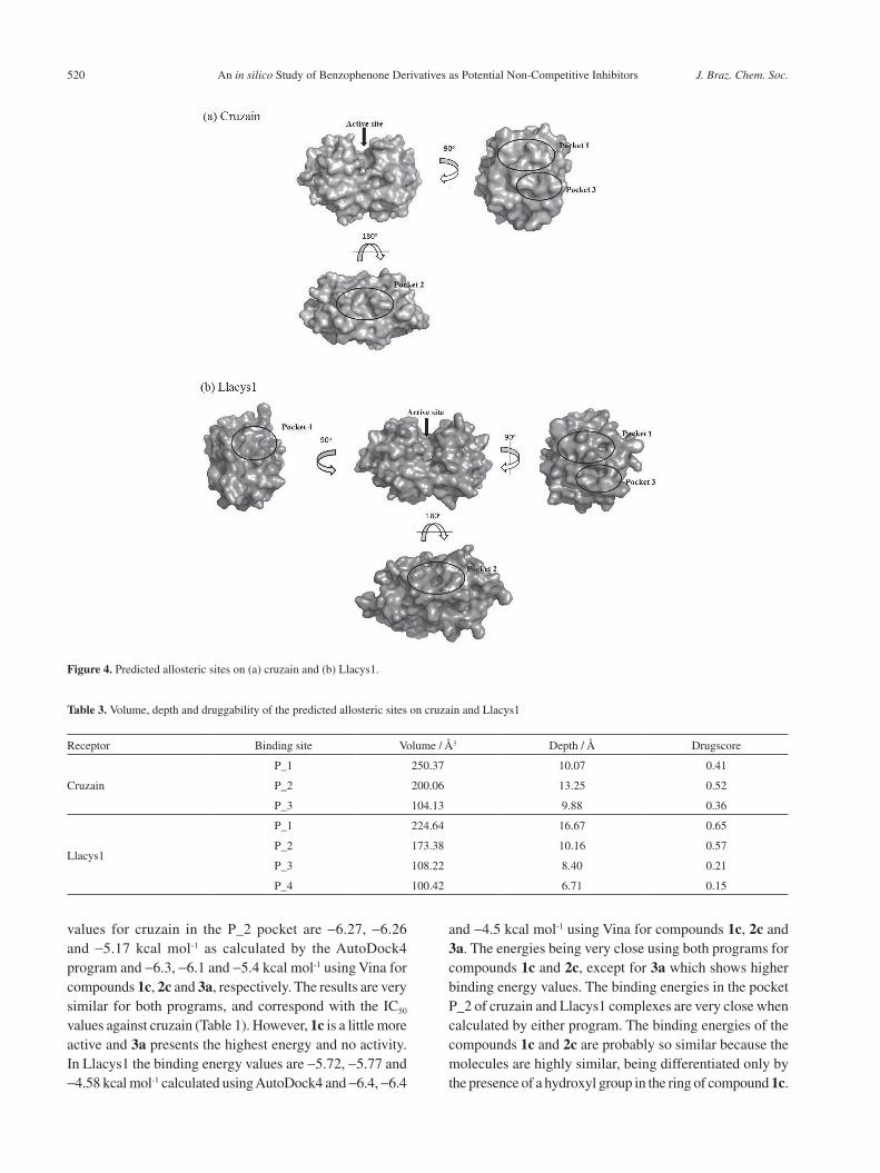
Table 3. Volume, depth and druggability of the predicted allosteric sites on cruzain and Llacys1
Receptor Binding site Volume / Å3 Depth / Å Drugscore
Cruzain
P_1 250.37 10.07 0.41
P_2 200.06 13.25 0.52
P_3 104.13 9.88 0.36
Llacys1
P_1 224.64 16.67 0.65
P_2 173.38 10.16 0.57
P_3 108.22 8.40 0.21
P_4 100.42 6.71 0.15
Figure 4. Predicted allosteric sites on (a) cruzain and (b) Llacys1.

Freitas et al. 521Vol. 29, No. 3, 2018
Compound 3a shows the greatest difference both in the binding energy values and in its chemical structure. This compound does not show good interaction with the site, nor does it inhibit cruzain. Therefore, compound 3a may not be a good Leishmania inhibitor promising. Thus, pocket P_2 with compounds 1c and 2c was selected for molecular dynamics simulation, in order to refine the docking results taking into account possible conformational changes in the interaction site and the effects of the solvent, as well as to possibly improve the results of the binding energy
calculations taking into account the flexibility of both proteins and benzophenones.
Absorption, distribution, metabolism and excretion (ADME) properties
For a new drug to be successfully developed, its ADME properties are very important, since unfavorable properties can cause a drug to be rejected.67 The web server FAF-Drugs3 was used to determine the ADME properties
Figure 5. Binding poses of cruzain (gray) and Llacys (green) calculated by the AutoDock4 and Vina programs: (a) overlap of cruzain and Llacys with 1c, 2c and 3a by AutoDock4 and Vina; (b) residues conserved between cruzain and Llacys1 in the P_2 pocket; (c) cruzain surface with 1c, 2c and 3a by AutoDock4 and Vina; (d) Llacys1 surface with 1c, 2c and 3a by AutoDock4 and Vina; (e) pose of 1c with cruzain AutoDock4 (green) and Vina (cyan); (f) pose of 2c with cruzain AutoDock4 (magenta) and Vina (yellow); (g) pose of 3a with cruzain AutoDock4 (blue) and Vina (pink); (h) pose of 1c with Llacys1 AutoDock4 (green) and Vina (cyan); (i) pose of 2c with Llacys1 AutoDock4 (magenta) and Vina (yellow) and (j) pose of 3a with Llacys1 AutoDock4 (orange) and Vina (brown).
Table 4. Binding energies as calculated by the AutoDock4 and Vina programs
Receptor Binding site
Compound
1c 2c 3a
ΔGAutoDock4 / (kcal mol-1)
ΔGVina / (kcal mol-1)
ΔGAutoDock4 / (kcal mol-1)
ΔGVina / (kcal mol-1)
ΔGAutoDock4 / (kcal mol-1)
ΔGVina / (kcal mol-1)
Cruzain
P_1 -5.49 -5.4 -5.31 -5.6 -4.88 -4.9
P_2 -6.26 -6.3 -6.27 -6.1 -5.17 -5.4
P_3 -4.93 -5.3 -5.23 -5.4 -4.21 -5.0
Llacys1
P_1 -5.42 -5.1 -5.07 -5.0 -4.49 -4.9
P_2 -5.72 -6.4 -5.77 -6.4 -4.58 -4.5
P_3 -5.04 -5.6 -5.32 -5.4 -4.18 -4.5
P_4 -5.24 -6.1 -5.58 -5.7 -4.33 -4.6
ΔGAutoDock4: binding energy calculated by AutoDock4; ΔGVina: binding energy calculated by Vina.

An in silico Study of Benzophenone Derivatives as Potential Non-Competitive Inhibitors J. Braz. Chem. Soc.522
(Table 2) of the compounds. All of the compounds showed good oral bioavailability, and conformed to Lipinski’s rule of five by meeting the following criteria (RO5): molecular weight ≤ 500; calculated log P (n-octanol/water) ≤ 5; number of hydrogen bond donors (HBD) ≤ 5; number of hydrogen bond acceptors (HBA) ≤ 10.68 Therefore, compounds 1c, 2c, and 3a have favorable ADME properties.
Molecular dynamics
To understand the interactions between the compounds and their targets, MD simulations were performed for a 10 ns duration. First, we evaluated the structural stability of the protease-inhibitor complex using the RMSD of the carbon alpha receptor relative to the initial structure, whose pose was obtained by better energy docking. The RMSD calculation is an important measure for the analysis of molecular dynamics simulations in biological systems. Using the RMSD it is possible to assess the structural stability of the protein, providing a measure of the deviation of a given molecular structure over time. The RMSD values (Figure 6) of cruzain in complex with 1c and 2c reached values up to 0.168 and 0.161 nm, with mean values of RMSD of
0.162 ± 0.001 and 0.163 ± 0.001 nm, respectively. Llacys1 in complex with compounds 1c and 2c reached values up to 0.159 and 0.150 nm, with mean values of 0.090 ± 0.010 and 0.100 ± 0.020. The mean values of RMSD indicate that there was little movement of the enzymes. However, the complexes with cruzain rapidly reached equilibrium and were slightly more stable than the complexes with Llacys1.
In addition, the radius of gyration of the proteins was evaluated, which means that the hydrodynamic radius of the molecular system was estimated. Using this calculation, it is possible to show processes of compression or expansion, i.e., folding or unfolding of the polypeptide chain, indicating a conformational change in the protein. Figure 6 shows that cruzain presents good stability in complex with compounds 1c and 2c, but after 4 ns in complex with 1c, the enzyme becomes more compact than when in complex with 2c, with average values of 1.630 ± 0.010 and 1.640 ± 0.010, respectively. In Llacys1, the complexes were stable after 5 ns. The enzyme complexed with 1c became more compact than with 2c with average values of 1.620 ± 0.010 and 1.630 ± 0.010, respectively. These results indicate that both enzymes in complex with 1c and 2c are more stable.
Figure 6. RMSD, radius of gyration and RMSF values of Llcys1-compound complexes were generated against the initial structures of the complexes during a 10 ns MD simulation period. The three-dimensional structures show the RMSF values in the regions of smallest to largest movement (blue to red).

Freitas et al. 523Vol. 29, No. 3, 2018
RMSF is the root mean square deviation based on the coordinates of the amino acid residues atoms of the protein. Figure 6 shows the RMSF values of the enzymes and the areas of the protein where major fluctuations occur during the simulation. Cruzain presents mean values of RMSF of 0.085 ± 0.053 and 0.082 ± 0.042 nm for complexes with compounds 1c and 2c, respectively, the values are very close to each other. Llacys1 presents mean values of RMSF of 0.089 ± 0.045 and 0.102 ± 0.068 nm for complexes with compounds 1c and 2c, respectively. Thus, the Llacys1-2c complex presents regions with greater movement than in the Llacys1-1c complex. It is also observed that in the region of the binding site, the movement of the residues is very small, which suggests a strong affinity of this region for the ligands.
Hydrogen bonds
Hydrogen bonding was determined at a distance of 0.35 nm and at an angle of 30o using the g_hbond tool of Gromacs (Figure 7). For cruzain in complex with 1c the average value of hydrogen bonding was 2, with 53.5% occupancy for the pair 1c (H3-hydroxyl group), Gln37 (OE1) and 74.1% occupancy for Val214 (H), 1c (O2). For 2c the mean binding value was zero. For Llacys1 in complex with molecule 1c the average value of hydrogen bonds was 1, with binding between the pair 1c (H8-hydroxyl group), Thr209 (OC2) having 57% occupancy. The value was zero for hydrogen bonding with 2c. In both enzymes the complexes with compound 1c have hydrogen bonds, which may be favorable for the stability of the complex. The
differences in hydrogen bonding between molecules present valuable information, because of the hydroxyl group in the ring of molecule 1c. Its presence may explain the greater affinity of interaction resulting in the lower IC50 value for cruzain. However, hydrogen bonding in the Llacys1-1c complex is not as stable as in cruzain-1c.
Binding energy
The molecular mechanics Poisson-Boltzmann surface area (MM-PBSA) method has been used to estimate binding free energies both in the study of biomolecular interactions as well as for scoring functions in drug design.60 This method, combined with MD simulations to rescore the docked complexes, lent significant support to the choice of the best ligand. The calculated free energies of the cruzain and Llacys1 complexes with the benzophenone derivatives ranged from -7.54 to -13.50 kcal mol-1. Table 5 shows the energy components that contribute to the binding free energy, the most favorable components being the van der Waals, electrostatic and nonpolar solvation terms, while polar solvation was unfavorable. In the two compounds 1c and 2c, GvdW is much stronger than Geletrostatic, Gpolar and GSASA with values greater than 20.00 kcal mol-1. It is evident that this energy governs the strength of the interactions of the compounds with the cysteine proteases. However, Geletrostatic is more favorable for compound 1c, because its hydrogen bonding is more stable than for molecule 2c due to the chemical nature of the ligands. The binding free energy for the cruzain complexes follows the same pattern as the IC50 values (Table 1), where compound 1c presents lower values.
Figure 7. Hydrogen bond number over time in simulations of complexes with (a) cruzain and (b) Llacys1.

An in silico Study of Benzophenone Derivatives as Potential Non-Competitive Inhibitors J. Braz. Chem. Soc.524
The lower binding energy can be explained by the presence of two hydrogen bonds that can better stabilize the complex in the pocket on the surface of the protein. In complexes with Llacys1, the binding free energy follows the ranking of the IC50 values (1c = 74.4 and 2c = 27.4 µM) against the amastigote form of L. amazonensis. In this case, it is observed that there is only one hydrogen bond, with 57% occupancy, in the Llacys-1c complex, which is not as stable as the cruzain-1c complex. In addition, the contribution of van der Waals energy is greater for Llacys1-2c compared with the other complexes. Thus, although the compounds bind to the same site on the surface of the enzymes and have conserved amino acid residues, small differences in other residues can result in differences in the binding free energy.
The energy contribution of each residue was calculated to verify the difference between the complexes. This energy decomposition analysis (Figure 8) shows major contributions from the hydrophobic residues Leu40, Val213 and Val214
for cruzain in complexes with 1c and 2c. The polar residue Trp35, and the hydrophobic residues Ile108 and Ser38 make major contributions in Llacys1 in complexes with 1c and 2c. The energy contribution is unfavorable for the hydrophobic residue Ala212 in cruzain, and for the His40 and Lys107 residues for Llacys1. These results verify that the Val residues are important for interactions in the cruzain-1c complex. These interactions in combination with hydrogen bonds may account for the greater activity of 1c against cruzain, as the Val residues are not conserved in Llacys1.
Figure 9 shows interactions in the cruzain- and Llacys1- complexes calculated by LIGPLOT from the final structure derived from the MD simulations. In all poses, van der Waals contacts were observed between protein residues, except for complexes with ligand 1c where hydrogen bonding was also significant. In addition, there was a high affinity of the conserved residue Gln34/Gln37 for compounds 1c and 2c in both enzymes, showing it to be a key residue in the interaction.
Table 5. Binding free energy results for different compounds bound with cruzain and Llacys1
Compound GvdW / (kcal mol-1) Geletrostatic / (kcal mol-1) Gpolar solv / (kcal mol-1) GSASA / (kcal mol-1) Gtotal / (kcal mol-1)
Cruzain1c -22.57 ± 3.75 -6.24 ± 0.31 17.78 ± 2.45 -2.46 ± 0.29 -13.50 ± 2.78
2c -21.11 ± 2.88 -1.06 ± 0.36 23.56 ± 2.55 -2.58 ± 0.25 -12.96 ± 2.10
Llacys11c -22.80 ± 3.07 -8.02 ± 1.21 26.16 ± 5.29 -2.88 ± 0.34 -7.54 ± 2.05
2c -27.45 ± 3.08 -2.09 ± 0.28 19.77 ± 3.82 -2.94 ± 0.22 -12.72 ± 2.02
GvdW: van der Waals energy; Geletrostatic: electrostatic energy; Gpolar solv: polar solvation energy; GSASA: solvent accessible surface area energy and Gtotal: binding energy.
Figure 8. Per-residue decomposition free energy of cruzain and Llacys1 complexes with compounds (a, c) 1c and (b, d) 2c in simulations.

Freitas et al. 525Vol. 29, No. 3, 2018
Figure 9. (Left) Binding poses of (a) cruzain- and (b) Llacys1-complexes obtained after 10 ns MD simulations. 1c (cyan) and 2c (green) are rendered in stick form. Schematic representation of the final structure of MD. (Center, right) The interaction of cruzain and Llacys1 with the compounds calculated by LIGPLOT. Thatched semi-circles indicate van der Waals contacts between hydrophobic protein residues and the compounds 1c and 2c. Hydrogen bonds are shown as green dashed lines.
Conclusions
We investigated the mechanism of interaction of Llacys1 and cruzain with benzophenones by homology modelling, docking and molecular dynamics analysis. These enzymes belong to the C1 family of cysteine proteases and present a high degree of sequence identity with each other. An enzymatic inhibition assay of cruzain showed that compounds 1c and 2c act as potent inhibitors, however, 3a showed no inhibition. To better understand how these derivatives bind to the proteases, we searched for binding sites on cruzain and Llacys1. We found a similar binding site on the surface of cruzain and Llacys1 by computational methods. The in silico results corroborated the experimental data, with compounds 1c and 2c being the strongest inhibitors of cruzain and the amastigote form of L. amazonensis, respectively. These results may contribute to the development of novel inhibitors for the treatment of leishmaniasis and Chagas disease, with the benzophenone derivatives 1c and 2c as potential candidates for prototype drugs.
Supplementary Information
Supplementary information is available free of charge at http://jbcs.org.br as a PDF file.
Acknowledgments
This work was supported by FAPEMIG, CNPq, CAPES, Rede Mineira de Química (RQ-MG) (CEX-RED-00010-14) and INCT-INOFAR.
References
1. Conteh, L.; Engels, T.; Molyneux, D. H.; Lancet 2010, 375,
239.
2. Mathers, C. D.; Ezzati, M.; Lopez, A. D.; PLoS Neglected Trop.
Dis. 2007, 1, e114.
3. Njoroge, M.; Njuguna, N. M.; Mutai, P.; Ongarora, D. S.; Smith,
P. W.; Chibale, K.; Chem. Rev. 2014, 114, 11138.
4. Chirac, P.; Torreele, E.; Lancet 2006, 367, 1560.
5. Alvar, J.; Yactayo, S.; Bern, C.; Trends Parasitol. 2006, 22, 552.
6. Kedzierski, L.; Hum. Vaccines 2011, 7, 1204.
7. Moreno, J.; Alvar, J.; Trends Parasitol. 2002, 18, 399.
8. Chakravarty, J.; Sundar, S.; J. Global Infect. Dis. 2010, 2, 167.
9. Croft, S. L.; Yardley, V.; Curr. Pharm. Des. 2002, 8, 319.
10. Mohamed-Ahmed, A. H. A.; Brocchini, S.; Croft, S. L.; Curr.
Opin. Infect. Dis. 2012, 25, 695.
11. Sueth-Santiago, V.; Decote-Ricardo, D.; Morrot, A.; Freire-de-
Lima, C. G.; Lima, M. E.; World J. Biol. Chem. 2017, 8, 57.
12. Gascon, J.; Bern, C.; Pinazo, M. J.; Acta Trop. 2010, 115, 22.
13. Jackson, Y.; Pinto, A.; Pett, S.; Trop. Med. Int. Health 2014, 19,
212.

An in silico Study of Benzophenone Derivatives as Potential Non-Competitive Inhibitors J. Braz. Chem. Soc.526
14. Cucunuba, Z. M.; Okuwoga, O.; Basanez, M. G.; Nouvellet,
P.; Parasites Vectors 2016, 9, 42.
15. Martins-Melo, F. R.; Ramos Jr., A. N.; Alencar, C. H.;
Heukelbach, J.; J. Infect. Dev. Countries 2012, 6, 817.
16. McKerrow, J. H.; Engel, J. C.; Caffrey, C. R.; Bioorg. Med.
Chem. 1999, 7, 639.
17. McKerrow, J. H.; Int. J. Parasitol. 1999, 29, 833.
18. Selzer, P. M.; Pingel, S.; Hsieh, I.; Ugele, B.; Chan, V. J.; Engel,
J. C.; Bogyo, M.; Russell, D. G.; Sakanari, J. A.; McKerrow, J.
H.; Proc. Natl. Acad. Sci. U. S. A. 1999, 96, 11015.
19. Sajid, M.; McKerrow, J. H.; Mol. Biochem. Parasitol. 2002,
120, 1.
20. Mottram, J. C.; Coombs, G. H.; Alexander, J.; Curr. Opin.
Microbiol. 2004, 7, 375.
21. Mahmoudzadeh-Niknam, H.; McKerrow, J. H.; Exp. Parasitol.
2004, 106, 158.
22. Frame, M. J.; Mottram, J. C.; Coombs, G. H.; Parasitology
2000, 121, 367.
23. Lasakosvitsch, F.; Gentil, L. G.; dos Santos, M. R.; da Silveira,
J. F.; Barbieri, C. L.; Int. J. Parasitol. 2003, 33, 445.
24. Cazzulo, J. J.; Franke, M. C. C.; Martinez, J.; de Cazzulo, B.
M. F.; Biochim. Biophys. Acta 1990, 1037, 186.
25. Eakin, A. E.; Mills, A. A.; Harth, G.; McKerrow, J. H.; Craik,
C. S.; J. Biol. Chem. 1992, 267, 7411.
26. Soares, M. J.; Souto-Padron, T.; de Souza, W.; J. Cell Sci. 1992,
102, 157.
27. Souto-Padron, T.; Campetella, O. E.; Cazzulo, J. J.; de Souza,
W.; J. Cell Sci. 1990, 96, 485.
28. Maciel-Rezende, C. M.; de Almeida, L.; Costa, E. D.; Pires, F.
R.; Alves, K. F.; Viegas Jr., C.; Dias, D. F.; Doriguetto, A. C.;
Marques, M. J.; dos Santos, M. H.; Bioorg. Med. Chem. 2013,
21, 3114.
29. de Almeida, L.; Alves, K. F.; Maciel-Rezende, C. M.; Jesus, L.
D. O. P.; Pires, F. R.; Junior, C. V.; Izidoro, M. A.; Júdice, W. A.
S.; dos Santos, M. H.; Marques, M. J.; Biomed. Pharmacother.
2015, 75, 93.
30. Leatherbarrow, R. J.; GraFit Version 5.0, Erithacus Software
Ltd., Horley, UK, 2002.
31. Berman, H. M.; Westbrook, J.; Feng, Z.; Gilliland, G.; Bhat, T.
N.; Weissig, H.; Shindyalov, I. N.; Bourne, P. E.; Nucleic Acids
Res. 2000, 28, 235.
32. Šali, A.; Blundell, T. L.; J. Mol. Biol. 1993, 234, 779.
33. http://www.ncbi.nlm.nih.gov/protein/30142572?report=graph
&sat=4&itemID=8, accessed in February 2015.
34. Altschul, S. F.; Madden, T. L.; Schäffer, A. A.; Zhang, J.; Zhang,
Z.; Miller, W.; Lipman, D. J.; Nucleic Acids Res. 1997, 25,
3389.
35. Larkin, M. A.; Blackshields, G.; Brown, N. P.; Chenna, R.;
McGettigan, P. A.; McWilliam, H.; Valentin, F.; Wallace, I. M.;
Wilm, A.; Lopez, R.; Thompson, J. D.; Gibson, T. J.; Higgins,
D. G.; Bioinformatics 2007, 23, 2947.
36. Laskowski, R. A.; MacArthur, M. W.; Moss, D. S.; Thornton,
J. M.; J. Appl. Crystallogr. 1993, 26, 283.
37. Saraiva, L. A.; Veloso, M. P.; Camps, I.; da Silveira, N. J. F.;
Mol. Inf. 2011, 30, 219.
38. da Silveira, N. J.; Arcuri, H. A.; Bonalumi, C. E.; de Souza,
F. P.; Mello, I. M.; Rahal, P.; Pinho, J. R.; de Azevedo, W. F.;
BMC Struct. Biol. 2005, 5, 1.
39. Volkamer, A.; Kuhn, D.; Rippmann, F.; Rarey, M.; Bioinformatics
2012, 28, 2074.
40. Morris, G. M.; Huey, R.; Lindstrom, W.; Sanner, M. F.; Belew,
R. K.; Goodsell, D. S.; Olson, A. J.; J. Comput. Chem. 2009,
30, 2785.
41. Trott, O.; Olson, A. J.; J. Comput. Chem. 2010, 31, 455.
42. Gordon, J. C.; Myers, J. B.; Folta, T.; Shoja, V.; Heath, L. S.;
Onufriev, A.; Nucleic Acids Res. 2005, 33, W368.
43. Spessard, G. O.; J. Chem. Inf. Model. 1998, 38, 1250.
44. Frisch, M. J.; Trucks, G. W.; Schlegel, H. B.; Scuseria, G. E.;
Robb, M. A.; Cheeseman, J. R.; Scalmani, G.; Barone, V.;
Mennucci, B.; Petersson, G. A.; Nakatsuji, H.; Caricato, M.;
Li, X.; Hratchian, H. P.; Izmaylov, A. F.; Bloino, J.; Zheng, G.;
Sonnenberg, J. L.; Hada, M.; Ehara, M.; Toyota, K.; Fukuda,
R.; Hasegawa, J.; Ishida, M.; Nakajima, T.; Honda, Y.; Kitao,
O.; Nakai, H.; Vreven, T.; Montgomery Jr., J. A.; Peralta, J.
E.; Ogliaro, F.; Bearpark, M. J.; Heyd, J.; Brothers, E. N.;
Kudin, K. N.; Staroverov, V. N.; Kobayashi, R.; Normand,
J.; Raghavachari, K.; Rendell, A. P.; Burant, J. C.; Iyengar, S.
S.; Tomasi, J.; Cossi, M.; Rega, N.; Millam, N. J.; Klene, M.;
Knox, J. E.; Cross, J. B.; Bakken, V.; Adamo, C.; Jaramillo,
J.; Gomperts, R.; Stratmann, R. E.; Yazyev, O.; Austin, A.
J.; Cammi, R.; Pomelli, C.; Ochterski, J. W.; Martin, R. L.;
Morokuma, K.; Zakrzewski, V. G.; Voth, G. A.; Salvador, P.;
Dannenberg, J. J.; Dapprich, S.; Daniels, A. D.; Farkas, Ö.;
Foresman, J. B.; Ortiz, J. V.; Cioslowski, J.; Fox, D. J.; Gaussian
09, Gaussian, Inc., Wallingford, CT, USA, 2009.
45. Lee, C.; Yang, W.; Parr, R. G.; Phys. Rev. B 1988, 37, 785.
46. Ditchfield, R.; Hehre, W. J.; Pople, J. A.; J. Chem. Phys. 1971,
54, 724.
47. Lagorce, D.; Sperandio, O.; Baell, J. B.; Miteva, M. A.;
Villoutreix, B. O.; Nucleic Acids Res. 2015, 43, W200.
48. Sanner, M. F.; J. Mol. Graphics Modell. 1999, 17, 57.
49. Jorgensen, W. L.; Chandrasekhar, J.; Madura, J. D.; Impey, R.
W.; Klein, M. L.; J. Chem. Phys. 1983, 79, 926.
50. Hornak, V.; Abel, R.; Okur, A.; Strockbine, B.; Roitberg, A.;
Simmerling, C.; Proteins: Struct., Funct., Bioinf. 2006, 65, 712.
51. Wang, J.; Wolf, R. M.; Caldwell, J. W.; Kollman, P. A.; Case,
D. A.; J. Comput. Chem. 2004, 25, 1157.
52. da Silva, A. W. S.; Vranken, W. F.; BMC Res. Notes 2012, 5, 1.
53. Hess, B.; Kutzner, C.; van der Spoel, D.; Lindahl, E.; J. Chem.
Theory Comput. 2008, 4, 435.
54. van der Spoel, D.; Hess, B.; Wiley Interdiscip. Rev.: Comput.
Mol. Sci. 2011, 1, 710.
Freitas et al. 527Vol. 29, No. 3, 2018
55. Pronk, S.; Pall, S.; Schulz, R.; Larsson, P.; Bjelkmar, P.;
Apostolov, R.; Shirts, M. R.; Smith, J. C.; Kasson, P. M.; van
der Spoel, D.; Hess, B.; Lindahl, E.; Bioinformatics 2013, 29,
845.
56. Bussi, G.; Donadio, D.; Parrinello, M.; J. Chem. Phys. 2007,
126, 014101.
57. Parrinello, M.; Rahman, A.; J. Appl. Phys. 1981, 52, 7182.
58. Essmann, U.; Perera, L.; Berkowitz, M. L.; Darden, T.; Lee,
H.; Pedersen, L. G.; J. Chem. Phys. 1995, 103, 8577.
59. Hess, B.; Bekker, H.; Berendsen, H. J. C.; Fraaije, J. G. E. M.;
J. Comput. Chem. 1997, 18, 1463.
60. Miyamoto, S.; Kollman, P. A.; J. Comput. Chem. 1992, 13, 952.
61. Kumari, R.; Kumar, R.; Lynn, A.; Consort, O. S. D. D.; J. Chem.
Inf. Model. 2014, 54, 1951.
62. Baker, N. A.; Sept, D.; Joseph, S.; Holst, M. J.; McCammon,
J. A.; Proc. Natl. Acad. Sci. U. S. A. 2001, 98, 10037.
63. Laskowski, R. A.; Swindells, M. B.; J. Chem. Inf. Model. 2011,
51, 2778.
64. Rice, P.; Longden, I.; Bleasby, A.; Trends Genet. 2000, 16, 276.
65. Méndez-Cuesta, C. A.; Méndez-Lucio, O.; Castillo, R.; J. Mol.
Graphics Modell. 2012, 38, 50.
66. Pettersen, E. F.; Goddard, T. D.; Huang, C. C.; Couch, G. S.;
Greenblatt, D. M.; Meng, E. C.; Ferrin, T. E.; J. Comput. Chem.
2004, 25, 1605.
67. Hughes, J. P.; Rees, S.; Kalindjian, S. B.; Philpott, K. L.; Br. J.
Pharmacol. 2011, 162, 1239.
68. Lipinski, C. A.; Lombardo, F.; Dominy, B. W.; Feeney, P. J.;
Adv. Drug Delivery Rev. 2001, 46, 3.
Submitted: July 3, 2017
Published online: September 14, 2017