Embed Size (px)
Citation preview
Extracorporeal shockwave treatment: A novel tool to improve Schwanncell isolation and culture
CHRISTINA M.A.P. SCHUH1,2, DAVID HERCHER1,2, MICHAELA STAINER1,2,RUDOLF HOPF1,2, ANDREAS H. TEUSCHL2,3, ROBERT SCHMIDHAMMER1,2 &HEINZ REDL1,2
1AUVA Research Center, Ludwig Boltzmann Institute for Experimental and Clinical Traumatology,Vienna,Austria,2Austrian Cluster for Tissue Regeneration,Vienna,Austria, and 3Department of Biochemical Engineering, University ofApplied Sciences TechnikumWien,Vienna,Austria
AbstractBackground aims. As new approaches for peripheral nerve regeneration are sought, there is an increasing demand for nativeSchwann cells for in vitro testing and/or reimplantation. Extracorporeal shockwave treatment (ESWT) is an emergent tech-nology in the field of regenerative medicine that has also recently been shown to improve peripheral nerve regeneration.Methods. In this study, we elucidate the effects of ESWT on Schwann cell isolation and culture. Rat sciatic nerves weredissected and treated with ESWT, and Schwann cells were isolated and cultured for 15 passages. Results. Single treatmentof the whole nerve ex vivo led to significantly increased extracellular adenosinetriphosphate as an immediate consequence,and subsequently a number of effects on the culture were observed, starting with a significantly increased Schwann cellyield after isolation. In the ESWT group, the quality of culture, reflected in consistently higher purity (S100b, morpholo-gy), proliferation rate (5-bromo-2-deoxyuridine, population doublings per passage) and expression of regenerative phenotype-associated markers (P75, glial fibrillary acidic protein, c-Jun), was significantly improved. In contrast, the control groupexhibited progressively senescent behavior, reflected in a decrease of proliferation, loss of specific markers and increase inP16INK4A expression. Conclusions. ESWT has beneficial effects on Schwann cell isolation and culture.
Key Words: extracorporeal shockwave treatment, peripheral nerve regeneration, Schwann cells
Background
Peripheral nerve lesions occur with an incidence ofapproximately 300 000 cases annually in Europe, rep-resenting a frequent cause of hospitalization anddisplaying a major burden to patients and social healthcare [1].
Although the peripheral nerve system has a re-markable regenerative potential, regeneration over nervegaps or over long distances (e.g., after proximal lesions)presents several difficulties. In this regard, nerveautografts are the gold standard to treat peripheralnerve injuries with tissue loss but often do notresult in a satisfactory outcome [2]. In particular,long-distance gaps or severe injuries affecting severalnerves push autografting to its limits regarding the avail-ability of donor material. Alternatives to facilitate nerveregeneration, such as artificial nerve guidance tubesor other types of scaffolds or application of neuro-trophic substances, are sought. Some of these
approaches are currently used in clinical nerve repair,although there is an ongoing debate concerning theirappropriate use, effectiveness and side effects [3,4].One of the major reasons for the unsatisfactoryoutcome after repair of long-distance gaps is the limitedproliferative capacity of Schwann cells [5]. Schwanncells play a key role in peripheral nerve regenera-tion: they participate in the removal of myelin andaxonal remnants, start proliferation and align to buildthe so-called bands of Büngner [6]. After the axon haselongated along the bands of Büngner, the Schwanncells start to remyelinate the newly formed axon tocomplete the regenerative process.
A novel strategy to improve the functional outcomeof peripheral nerve regeneration is the therapy ofinjured nerves with extracorporeal shockwave treat-ment (ESWT). ESWT has its origin in the field ofurology, in which it is used to destroy kidney stones[7], but it has also been proven to be an effective ther-apeutic tool in the field of regenerative medicine. In
Correspondence: Christina M.A.P. Schuh, PhD, AUVA Research Center, Ludwig Boltzmann Institute for Experimental and Clinical Traumatology,Donaueschingenstraße 13, A-1200 Vienna, Austria. E-mail: [email protected]
ARTICLE IN PRESS
(Received 18 January 2016; accepted 5 March 2016)
ISSN 1465-3249 Copyright © 2016 International Society for Cellular Therapy. Published by Elsevier Inc. All rights reserved.http://dx.doi.org/10.1016/j.jcyt.2016.03.002
Cytotherapy, 2016; ■■: ■■–■■
preclinical and clinical trials, beneficial effects have beenreported in treatment of various medical indicationssuch as non-union fractures [8–10], ischemia-inducedtissue necrosis [11], or chronic wounds [12,13].Theshockwave generated is a sonic pulse and is charac-terized by an initial rise, reaching a positive peak ofup to 100 MPa within 10 ns, followed by a negativeamplitude of up to −10 MPa and a total life cycle ofless than 10 μs. Biological responses are thoughtto be triggered by the high initial pressure, followedby a tensile force and the resulting mechanicalstimulation [14].
Recently, Hausner et al. [15] showed a novel ap-proach of accelerating regeneration after peripheralnerve injury, bridged with an autologous nerve graft.After dissecting and bridging the sciatic nerve of aSprague-Dawley rat, extracorporeal shockwaves wereapplied at the site of injury. Six weeks after surgery,animals of the ESWT group exhibited a significantlyimproved functional recovery relative to controls. Onthe basis of this study, we investigated in vitro Schwanncell behavior after ESWT treatment with focus on theirregenerative capacity.
Methods
Shockwave treatment of nerve tissue and Schwanncell isolation
All animals were euthanized according to estab-lished protocols, which were approved by the CityGovernment of Vienna, Austria, in accordance withthe Austrian Law and Guide for the Care and Useof Laboratory Animals as defined by the NationalInstitutes of Health. Animals and treatment/controlgroups were randomly chosen and analyzed withoutpre- or post-selection of the respective nerves orcultures.
For ex vivo shockwave treatment an unfocusedelectro-hydraulic device was used (Dermagold 100,MTS Medical.The applicator was attached to a waterbath as described in other studies [16–18], ensuringdirect contact to the pre-warmed (37°C) water, al-lowing reproducible physical propagation andapplication of shockwaves in vitro. Sciatic nerves ofadult male Sprague-Dawley rats were dissected, andeach nerve was transferred into a 15-mL conical cen-trifuge tube (PAA Laboratories) containing phosphatebuffered saline (PBS; PAA Laboratories) pre-chilledon ice. Nerves were kept on ice until further use butnot longer than 1 h. For ESWT application, tubes wereplaced 5 cm in front of the applicator inside the watercontainer. Subsequently, unfocused shockwaves wereapplied using the parameters chosen according to pre-vious experiments [15] to maximize the effect of theESWT treatment, while minimizing possible nega-tive effects: 300 pulses at an energy level of 0.10 mJ/
mm2 with a frequency of 3 Hz. The correspondingsecond nerve from the same animal served as controland was placed in a water bath (37°C) for the timeof treatment to avoid the creation of artifacts due todifferent sample treatments.
After ESWT treatment, Schwann cells were iso-lated from the treated and non-treated sciatic nervetissues according to a method adapted from Kaewkhawet al. [19]. Briefly, the epineurium was removed andnerves were weighed on a fine scale to assess nervewet weight (Sartorius). Nerves were subsequentlystrained and minced. Nerve fragments were incu-bated with 0.05% collagenase (Sigma-Aldrich) for 1 hat 37°C and then filtered through a 40-μm cell strain-er and centrifuged at 400g for 6 min. After washingthe cell pellet with Dulbecco’s Modified Eagle Medium(DMEM; PAA Laboratories) containing 10% fetal calfserum (FCS; PAA Laboratories), the pellet was re-suspended in DMEM-D-valine (PAA Laboratories),supplemented with 10% FCS, 2 mmol/L L-glutamine(PAA, Austria), 1% antibiotics (PAA Laboratories),N2 supplement (Invitrogen), 10 μg/mL bovine pitu-itary extract (Sigma-Aldrich), 5 μmol/L forskolin(Sigma-Aldrich). This medium is subsequently re-ferred to as “Schwann cell medium.” Cell suspensionwas seeded on six-well plates (PAA Laboratories)coated with poly-L-lysin (Sigma-Aldrich) and laminin(Sigma-Aldrich).
Cell culture and experimental setup
Cells were subcultured for the first time after 19 days,to establish a proliferative phenotype and keep themin a proliferative state. Schwann cell medium was addedon day 5 after isolation (1 mL) and was partially (50%)changed on days 9, 13 and 17. Subsequent splittingof cells was performed for 15 passages as follows: cellswere detached with a cell scraper, centrifuged at1200 rpm for 5 min and seeded at a density of 4 × 10⁴
cells/cm2 on plates previously coated with poly-L-lysin. Residual cells were used for flow cytometricanalysis, 5-bromo-2-deoxyuridine uptake (BrdU) assayand protein isolation. Medium was partially (50%)changed every third day, and cells were split every sixthday.
Evaluation of cell yield
To evaluate cell yield, representative phase contrastpictures were taken from each culture using a LeicaDMI6000B microscope (Leica), and cells were countedusing a Bio-Rad TC20 automated cell counter (Bio-Rad Laboratories). Non-viable cells were identifiedand excluded by trypan blue staining. Cell count wasnormalized to 10-mg nerve wet weight assessed beforeisolation.
ARTICLE IN PRESS
2 C. M. A. P. Schuh et al.

Proliferation assay
Proliferation assay using a BrdU (Cell ProliferationELISA Assay Kit; Roche Diagnostics) for quantita-tive evaluation of Schwann cell proliferation wasperformed according to manufacturer’s instructions.Briefly, poly-L-lysine coated 96-well plates were seededwith cells at a density of 2.5 × 10⁴ cells/cm2. After 48 h,medium was changed to Schwann cell medium con-taining 100 μmol/L BrdU, and cells were incubatedfor 24 h at standard cell culture conditions (37°C and5% CO2). The culture plates were fixated withFixDenat solution and subsequently incubated withanti-BrdU POD antibody solution for 60 min at roomtemperature. After washing the plate with PBS,tetramethyl benzidine was added for 30 min as a sub-strate.The reaction was stopped with 1 mol/L H2SO4
and absorption was measured at 450 nm with 690 nmas reference wavelength on an automatic microplatereader (Tecan Sunrise).
Flow cytometric analysis
Purity of the Schwann cell cultures was evaluatedwith flow cytometry for common Schwann cellmarkers: anti-S100b (rabbit polyclonal; Dako),anti-P75 NGFR (goat polyclonal; Santa Cruz Bio-technology) and anti-P0 (rabbit polyclonal; SantaCruz Biotechnology). Antibodies were labeled withallophycocyanin (Lynx Rapid Conjugation Kit, ABDSerotec). For analysis, cells were detached with acell scraper and incubated with the antibodies (1:200)on ice and in the dark for 20 min. Cell pellets werewashed twice and resuspended in 200 μL PBS. Flowcytometric analysis (10 000 events) was performedwith a BD FACS Canto II (Becton Dickinson),and data were evaluated with Flowjo Version 8.8(Tree Star).
Immunoblotting
Total protein of cells was extracted using Trizol(peqGold TriFast, Peqlab) according to manufactu-rer’s instructions. Briefly, proteins were precipitatedfrom organic phase with ethanol and pelleted by cen-trifugation (12 000g, 10 min, 4°C). Protein pellet waswashed three times with 0.3 mol/L guanidine hydro-chloride (Sigma-Aldrich) in 95% ethanol and oncewith 100% ethanol (Merck), with each washing stepfollowed by centrifugation (7500g, 5 min, 4°C). Su-pernatants were discarded and dry protein pelletssolubilized in 1% SDS (Sigma-Aldrich) in analyticalgrade water.
Equal amounts of protein (up to 3 μg/lane; onedonor per gel: passage 2, passage 7, passage 15) wereseparated on a 12% SDS-polyacrylamide gel andblotted onto a nitrocellulose membrane. Membranes
were blocked with 5% skim milk inTris buffered salinecontaining 1% Triton-X100 (TBS-T; Sigma-Aldrich)for 120 min and incubated with primary antibodiesS100b (Dako), c-Jun (Abcam), glial fibrillary acidicprotein (GFAP; Bioss USA), P16INK4A (Abcam),α-tubulin (Calbiochem) diluted in 5% bovine serumalbumin (Sigma-Aldrich) in TBS-T at 4°C on a rollmixer for 12 h. Membranes were washed twice withTBS-T and incubated with the secondary antibodyin 5% milk-TBS-T. Signals were detected using anOdyssey Fc infrared imaging system (LI-COR Bio-sciences). After membranes were incubated in1 × NewBlot IR Stripping Buffer (LI-COR Biosci-ences) on a shaker at room temperature for 5 min andwashed three times in PBS, membranes were reprobedwith total antibodies. Ratio of analyzed protein tohousekeeping gene α-tubulin was densitometricallyanalysed using Image Studio Version 5.0.21 (LI-COR Biosciences).
Activation switch
In passages 4, 9 and 15, the activation status and thecapacity to switch activation status (proliferating to pro-myelinating) were assessed. Cells were split (2 × 10⁴
cells/cm2, 24 h adherence time) in two groups: one wascultured in Schwann cell medium and the other inbasic medium (DMEM-D-valine; PAA Laborato-ries), supplemented with 10% FCS, 2 mmol/LL-glutamine, 1% antibiotics) without supplements fa-voring the proliferating or the pro-myelinating status,respectively. Proliferation behaviour (BrdU enzyme-linked immunosorbent assay) and marker expression(flow cytometry) were assessed 5 days after mediumswitch.
Adenosinetriphosphate release and lactatedehydrogenase release
The amount of adenosinetriphosphate (ATP) re-leased into the supernatant from nerve tissue treatedwith ESWT was determined using the CellTiter-Glo assay (Promega). Sciatic nerves were dissected andkept in PBS on ice until further treatment. After re-moving the epineurium and teasing of the nerve fiberswith a mounted needle (15 times in the direction ofthe fiber), remaining nerve tissue was placed in 500 μLDMEM. Shockwave treatment was performed at 37°Cand with following parameters: 300 pulses with 3 Hzand 0.03 mJ/mm2, 0.10 mJ/mm2 or 0.19 mJ/mm2.Thecontrol group was placed in a water bath (37°C). Nervetissue was incubated for 5 min on ice and subse-quently centrifuged at 1500 rpm for 5 min at 4°C.Supernatant was transferred to a micronic tube(150 μL) for lactate dehydrogenase (LDH) measure-ment (Cobas C111; Roche Diagnostics) and a 96-well plate (triplicate, 100 μL) for ATP measurement.
ARTICLE IN PRESS
ESWT improves Schwann cell isolation/culture 3
An equal amount of CellTiter-Glo reagent was added,the plate was horizontally shaken for 2 min, and afterincubation for 10 min at room temperature, the re-sulting luminescence was measured. ATP standardswere used for calibration of the measured lumines-cence. After sampling of the initial supernatant,fresh, ice-chilled DMEM was added on nerve tissueand was incubated on ice for another 30 min. ATPand LDH concentration in supernatants was quan-tified as before.
Statistics
All data in this study are shown as mean ± SD andwere tested for normal distribution. Depending ongroups analyzed, statistical analysis was performed byusing Student’s t-test or one-way analysis of vari-ance followed by the Tukey range test for significantdifferences between the means. Significance was con-sidered for P < 0.05. For statistical calculations,GraphPad Prism 5 for Mac OS X, Version 5.0b(GraphPad Software) was used.
Results
Increased cell yield by incorporation of ESWT in theisolation process
To evaluate effects on the isolation efficacy, cellswere counted after 19 days in culture, and cellnumber was normalized to 100 mg nerve wet weight.
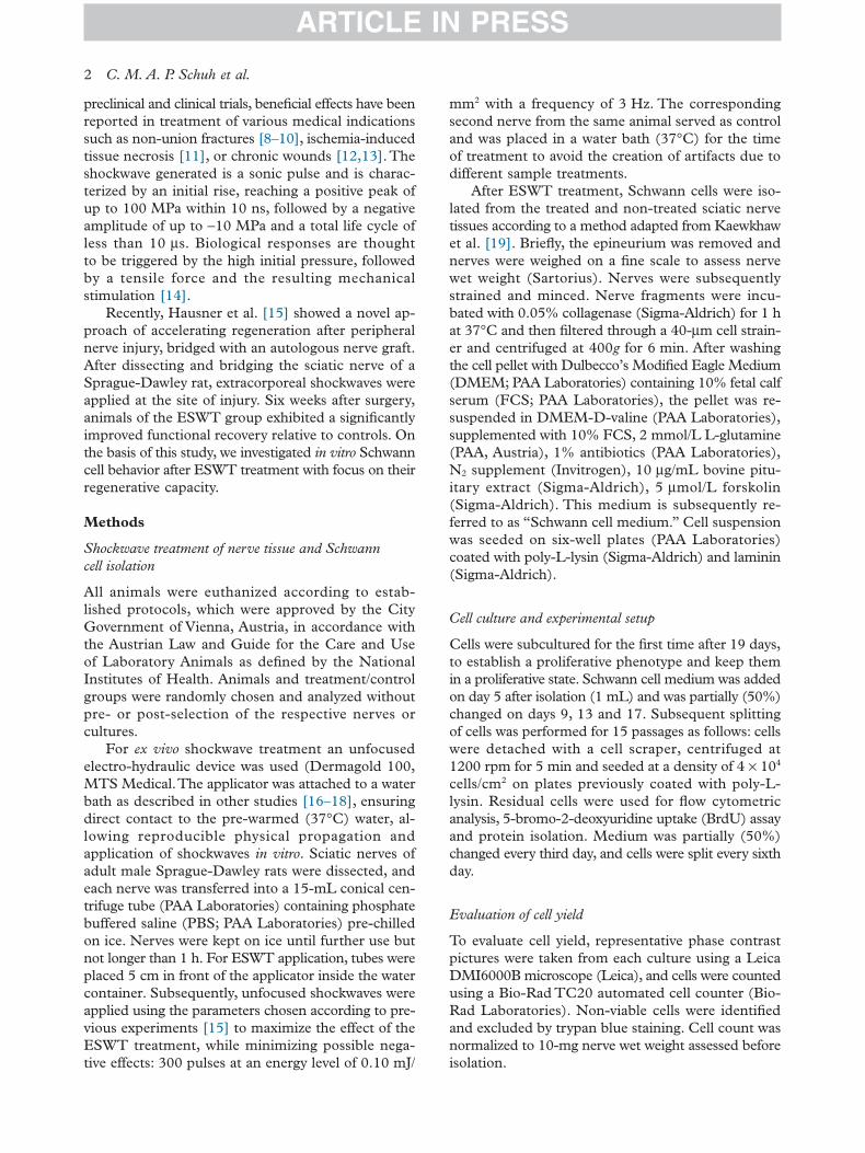
Sciatic nerve weights ranged between 66.2 and88.5 mg, and there was no significant differencebetween the groups (Figure 1B). As shown inFigure 1A, cell yield after 19 days was significantlyincreased in the ESW treated group. Although initialcell count revealed 1.62 × 10⁶ ± 9.3% cells per 100 mgnerve wet weight in the control group, cell yield inthe ESW-treated group was, on average, 52.3%higher (3.10 × 10⁶ ± 8.2% cells per 100 mg nervewet weight). Furthermore, Figure 2A illustrates aconsistent improvement of the cell yield for everyculture assessed.
BrdU assay and population doublings per passage reveala significantly higher proliferation after ESWT for15 passages
The cell proliferation was quantified using a BrdU assayand assessment of population doublings per passage.In all passages examined (passage 1, 4, 7, 10, 13 and15), Schwann cells treated with ESWT showed a higherproliferative behavior than the Schwann cells in thecontrol group, respectively (Figure 2). Furthermoreproliferation decreased steadily in the control groupstarting in passage 4, whereas proliferation in theESWT group further increased until passage 7 andremained at a similar level until passage 15. Westernblots in passage 2, 7 and 15 also exhibited a steadyincrease in the cell-cycle-arrest/senescence markerP16INK4a in the control group, while ESWT group
Figure 1. ESWT resulted in an increased cell yield. (A) Intra-animal comparison of Schwann cells counted after 19 days in culture,normalized on 100 mg nerve wet weight, n = 12. (B) nerve wet weight of the respective nerves, assessed with a fine scale beforeisolation; n = 12. (C) Phase contrast micrographs of SCs in passage 0, untreated control (CTRL) and treated with extracorporeal shockwaves(ESWT).
ARTICLE IN PRESS
4 C. M. A. P. Schuh et al.

remained at the same low level for the tested pas-sages (Figure 4).
Sustained purity and P75 expression after ESWT forextended culture period
To determine purity and phenotype of these culti-vated cells, expression of Schwann cell specific markerS100b, as well as the markers P75 (proliferative/regenerative phenotype) and P0 (myelinatingphenotype), were assessed in vitro over 15 passages withflow cytometry (Figure 3). In passage 1, 24.2% morecells expressed S100b in the ESW-treated group com-pared with the control group (62.6% compared with38.4%). In both groups, Schwann cell purity in-creased in passage 2 to 70.0% (ESWT) and 57.3%(CTRL). However, starting in passage 3, S100b ex-pression in the control group decreased steadily overthe following passages. In contrast, in the ESW treatedgroup expression increased until passage 5 (85.3%)and remained at this level until passage 15 (Figure 3,S100b). A similar temporal pattern was applied to theexpression of the proliferation-associated marker P75:while control group reached a peak expression of36.7% in passage 3, steadily decreasing over time,ESW-treated cells increased from 57.4% P75 expres-sion to 83.1% positive cells and remained at this leveluntil passage 15 (Figure 3, P75). Myelin marker P0was down-regulated in both groups; however, it wasstronger in the ESWT treated group, exhibiting a sig-nificant difference in passage 3 where control groupshowed an increase of 9.0% compared to passage 2(Figure 3, P0). Protein expression levels usingWesternblot densitometric analysis revealed a similar differ-ence between the groups in the expression of S100bas found with flow cytometry in the respective pas-sages passage 2, 7 and 15 (Figure 4, S100). Furtheranalysis of the markers c-Jun and GFAP, both asso-ciated with the proliferative Schwann cell phenotype,exhibited an increase of both markers in the ESWTgroup and a decrease in the control group (Figure 4).
Figure 2. BrdU assay and population doublings per passage revealed a significantly higher proliferation of the ESW treated Schwann cellsover 15 passages. BrdU: n = 6 rats, values were obtained in quadruplicates in passage 1, 4, 7, 10, 13 and 15. Population doublings perpassage: population doublings were obtained by cell count in passage 1, 4, 7, 10, 13 and 15; n = 6 rats. Data are mean ± SD, and signif-icance was tested with Student’s t test. *P < 0.05; **P < 0.01; ***P < 0.001. CTRL, untreated control.
Figure 3. Schwann cells treated with ESW show an increased purity(100b) over 15 passages, along with increased expression of pro-liferation associated marker P75 and a decreased expression of myelinmarker P0. Flow cytometric immunophenotype analysis (S100b,P75, P0) of Schwann cells treated with ESWT (black), comparedwith control (CTRL; gray) over 15 passages (P1–P15); one passagerepresents the detachment, counting, and seeding of the cells with4 × 104 cells/cm2 for 5 days; n = 10 rats. Data are mean of per-centage marker-positive cells ± SD, and significance was tested withStudent’s t test. *P < 0.05; **P < 0.01; ***P < 0.001.
ARTICLE IN PRESS
ESWT improves Schwann cell isolation/culture 5

Schwann cells treated with ESWT display aconsistent morphology
Schwann cell morphology was assessed in passage 0,5, 9 and 15 using phase contrast microscopy (Figure 5).Schwann cells of both groups showed a spindle ortripolar shape in passage 0. Starting in passage 5, asecond cell morphology was found in the control group,similar to the classic fibroblastic phenotype. Cells ex-hibiting this morphology increased in number frompassage 5 to passage 15 in the control group (Figure 5,indicated with arrows), whereas the ESWT-treated
group revealed a homogenous Schwann cell morphol-ogy over 15 passages.
Schwann cells revert faster to myelinating phenotypeafter ESWT
To demonstrate that the proliferative activation of SCsis not permanent and reversible the Schwann cell abilityto switch phenotype from proliferating to pro-myelinating was tested.Therefore, in passages 4, 9 and15, the medium was changed to one lacking pro-proliferative growth factors, giving the cells a minimal
Figure 4. Western blot analysis of Schwann cell protein lysate demonstrates a decrease of Schwann cell markers in the control group alongwith a simultaneous increase of senescence marker P16. Western blot analysis of Schwann cells in passage 2, passage 7 and passage 15,treated with ESW compared with untreated control (CTRL). Blots were densitometrically analyzed, and data are presented as mean ± SD(n = 6). Statistical significance was tested with one-way analysis of variance and Tukey range test. *P < 0.05; **P < 0.01; ***P < 0.001.
Figure 5. Morphology of Schwann cells changes over time in the control group, whereas it remained consistent in the group treated withextracorporeal shockwaves (ESWT). Phase contrast micrographs depicting Schwann cells in passage 0, 5, 9 and 15 of control group (CTRL,upper bar) and ESW-treated group (lower bar); arrows mark cells displaying non-typical morphology (tripolar, fibroblast-like); size barindicates 200 μm.
ARTICLE IN PRESS
6 C. M. A. P. Schuh et al.
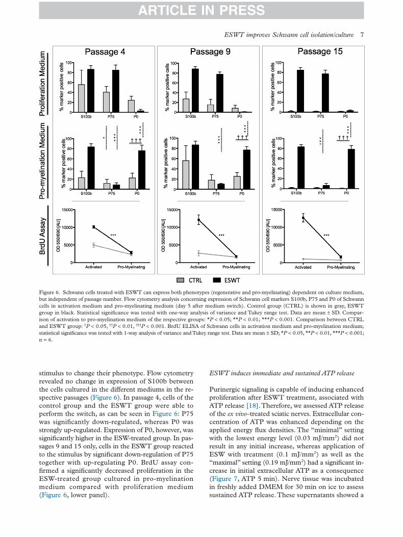
stimulus to change their phenotype. Flow cytometryrevealed no change in expression of S100b betweenthe cells cultured in the different mediums in the re-spective passages (Figure 6). In passage 4, cells of thecontrol group and the ESWT group were able toperform the switch, as can be seen in Figure 6: P75was significantly down-regulated, whereas P0 wasstrongly up-regulated. Expression of P0, however, wassignificantly higher in the ESW-treated group. In pas-sages 9 and 15 only, cells in the ESWT group reactedto the stimulus by significant down-regulation of P75together with up-regulating P0. BrdU assay con-firmed a significantly decreased proliferation in theESW-treated group cultured in pro-myelinationmedium compared with proliferation medium(Figure 6, lower panel).
ESWT induces immediate and sustained ATP release
Purinergic signaling is capable of inducing enhancedproliferation after ESWT treatment, associated withATP release [18].Therefore, we assessed ATP releaseof the ex vivo–treated sciatic nerves. Extracellular con-centration of ATP was enhanced depending on theapplied energy flux densities. The “minimal” settingwith the lowest energy level (0.03 mJ/mm2) did notresult in any initial increase, whereas application ofESW with treatment (0.1 mJ/mm2) as well as the“maximal” setting (0.19 mJ/mm2) had a significant in-crease in initial extracellular ATP as a consequence(Figure 7, ATP 5 min). Nerve tissue was incubatedin freshly added DMEM for 30 min on ice to assesssustained ATP release.These supernatants showed a
Figure 6. Schwann cells treated with ESWT can express both phenotypes (regenerative and pro-myelinating) dependent on culture medium,but independent of passage number. Flow cytometry analysis concerning expression of Schwann cell markers S100b, P75 and P0 of Schwanncells in activation medium and pro-myelinating medium (day 5 after medium switch). Control group (CTRL) is shown in gray, ESWTgroup in black. Statistical significance was tested with one-way analysis of variance and Tukey range test. Data are mean ± SD. Compar-ison of activation to pro-myelination medium of the respective groups: *P < 0.05; **P < 0.01; ***P < 0.001. Comparison between CTRLand ESWT group: †P < 0.05, ††P < 0.01, †††P < 0.001. BrdU ELISA of Schwann cells in activation medium and pro-myelination medium;statistical significance was tested with 1-way analysis of variance andTukey range test. Data are mean ± SD; *P < 0.05, **P < 0.01, ***P < 0.001;n = 6.
ARTICLE IN PRESS
ESWT improves Schwann cell isolation/culture 7

generally lower concentration of ATP in all groups;however, treatment (0.1 mJ/mm2) and maximal setting(0.19 mJ/mm2) again showed higher levels than controland minimal setting (0.03 mJ/mm2; Figure 7, ATP30 min). Among other triggers, ATP can be releasedinto the extracellular space due to cell membranedamage.To exclude membrane damage as the reasonfor increased extracellular ATP after ESW treatmentwe assessed extracellular LDH concentration as an in-dicator for cell damage. As seen in Figure 7 (LDH),supernatants of nerve tissue that received the highestenergy (0.19 mJ/mm2) were the only ones that showedsignificantly enhanced LDH concentrations. At bothtime points, our selected treatment setting (0.1 mJ/mm2) did not increase levels of extracellular LDH whencompared with the control group.
Discussion
After peripheral nerve injury, Schwann cells are trig-gered to change their phenotype from myelinating toactivated and proliferating and building bands ofBüngner, the substrate for outgrowing axons. Thereare myriad studies describing the importance ofSchwann cells during peripheral nerve regeneration[20–22], but the difficulties and issues associated arealso well known [23–25]. Particularly in long-distanceinjuries, the demand for supportive Schwann cells ex-panded in vitro (e.g., seeded on a tubular graft) wouldbe high because autologous Schwann cells lack the pro-
liferative capacity to provide bands of Büngnerthroughout a tube longer than 40 mm [5].This limitedproliferative capacity is also reflected in vitro, display-ing together with insufficient purity the main issue ofSchwann cell cultures.To our knowledge, we are thefirst to demonstrate an increase in proliferation, pro-liferative capacity and purity in in vitro Schwann cellcultures with ESWT.
The basis for our study was found in two preced-ing shockwave studies. Hausner et al. [15] showed thatshockwave treatment after nerve dissection leads tosignificantly accelerated regeneration in a rat model.It was hypothesized that this effect may result fromimproved macrophage infiltration and an earlier onsetof regeneration. However, Weihs et al. [18] elucidatein their study how ESWT stimulates cell prolifera-tion in several cell types (e.g., mesenchymal stem cells)by ATP release-coupled extracellular signal-regulatedkinase (ERK) activation. Applying ESWT on the wholenerve before isolation of Schwann cells, our resultsdemonstrate that ESWT is capable of enhancing theextracellular levels of ATP without causing any cellmembrane damage. Furthermore, the sustained releaseover 30 min suggests an active mechanism of ATPrelease.
Analyzing the cultures in passage 0, we assesseda significantly higher cell yield in all cultures treatedwith ESWT (Figure 1), alongside a significantlyincreased proliferation rate in passage 1 (Figure 2).In the shockwave-treated group, quality of culture,
Figure 7. Extracellular ATP was significantly increased in nerves treated with 0.1 mJ/mm2 (Treat) over a period of 30 min, with no in-crease in cell damage (LDH); whole nerves without epineurium were treated with ESW of different energy levels: 0.03 (Min), 0.10 (Treat)and 0.19 mJ/mm2 (Max) and compared with the untreated control (CTRL). ATP concentration in the supernatant was determined after5 min and 30 min on ice with luminescence, together with assessment of LDH. Amount of ATP was calculated using ATP standards; LDHwas normalized to untreated control; n = 5. Statistical significance was tested with one-way analysis of variance andTukey range test. *P < 0.05;**P < 0.01; ***P < 0.001.
ARTICLE IN PRESS
8 C. M. A. P. Schuh et al.
reflected in purity and proliferation rate, improved overthe first passages: S100b, a marker indicating purityof Schwann cells, increased over the first five pas-sages and remained at the level for the period ofanalysis; this purity was also observed in morpholo-gy of Schwann cells (Figure 4); moreover, P75increased over time, whereas P0 in the same mannerdecreased until passage 4 and subsequently was notexpressed (Figure 3); P0 and P75 are known to coun-teract each other [26]: P0 is a myelin component andtherefore solely expressed in myelinating Schwann cells[27], whereas P75 is a marker associated with the re-generative, proliferating phenotype of Schwann cells[28,29]. Western blot analysis at three time pointsthroughout the culture period revealed a similar ex-pression pattern for S100b, acting as a control markerbetween the methods (Figure 4, compared with Figure5 S100b). GFAP and c-Jun were also up-regulated inpassage 7 (compared with passage 2) and showed acomparable expression in passage 15. Both markersare associated with the regenerative phenotype. GFAPis suppressed in myelinated axons and becomesupregulated after injury, initiating proliferation bybinding of integrin αvβ8 [30].The transcription factorc-Jun however is an antagonist to Krox20, a proteincontrolling the myelination in the peripheral nervoussystem, and displays one of the key regulators to ini-tiate and maintain the regenerative phenotype [31,32].Together with the strongly increased proliferation (as-sessed with BrdU assay and population doubling perpassage), the Schwann cells treated with ESWT rep-resent a highly regenerative phenotype for an extendedculture period of 15 passages (103 days).
As the sum of these observations leads inevitablyto the question whether hyper-proliferating Schwanncells may lead to adverse effects such as post-stimulusproliferation and subsequently Schwannoma forma-tion, we conducted a functionality experiment.Schwann cells were cultured for 5 days in basicmedium lacking any kind of proliferation stimulat-ing growth factors (forskolin, pituitary extract).Schwann cells treated with ESWT reacted even moreto the change of stimulus, independent of the passage:they not only stopped proliferating (Figure 6, BrdU)but also significantly down-regulated the proliferation-associated marker P75 and up-regulated the myelincomponent P0. This prompt and consistent reactionto the absence of mitogenic growth factors demon-strates their capacity to switch to the myelinatingphenotype. However, the functionality of the SCs inan in vivo defect model has to be shown.
In contrast to the ESWT group, the untreatedSchwann cells did not display a consistent Schwanncell phenotype over the culture period of 15 pas-sages. Starting with passage 5 to 7, purity andproliferation significantly decreased. Abated purity was
observed in lower expression of S100b (Figures 3 and6), as well as in increased appearance of cells display-ing an atypical morphology (Figure 5). Reducedproliferation was primarily assessed in a decline of pop-ulation doublings and additionally by reduced BrdUOD values.The entire marker expression levels asso-ciated with the regenerative phenotype (P75, GFAP,c-Jun) diminished over time, whereas the senes-cence marker P16 significantly increased. Therefore,our in vitro study reflects the before mentioned in vivoproblem and represents a possible solution: the limitedproliferative capacity of Schwann cells can be im-proved with ESWT. With treated Schwann cellsbuilding a growth substrate faster and for a longerperiod of time, the in vivo effect can be twofold: ESWTnot only results in an acceleration of regenerationby activating autologous Schwann cells, as has beenshown by Hausner et al. [15], but would also allowreimplantation of a high number of autologousSchwann cells expanded ex vivo in a decidedly regen-erative state.
A partial explanation for the underlying mecha-nisms of the observed effects could be the sustainedATP release. A wide range of mechanisms from ve-sicular release over connexins/pannexins to ABCtransporters are thought to conduct an active releaseof ATP [33–35]. Subsequent purinergic signalingplays a crucial role not only as a danger-associated mo-lecular pattern but also in a variety of cellular functionssuch as proliferation, chemotaxis, differentiationand amplification of other signals [18,36]. This in-cludes Schwann cell–axon interactions. Immature/unmyelinating Schwann cells in particular signal viaextracellular ATP in a paracrine manner with axons[37,38]. It was proposed that ATP and glutamate arebuilding a positive feedback loop enhancing their ac-tivities [39]. The fate of Schwann cells is influencedby neuronal activity, by the activation of purinergicmetabotropic P2Y receptors and direct actions of ATPand its metabolite adenosine [40–42], as well as bythe activation of metabotropic glutamate receptors [43].Furthermore, purinergic signaling is thought to be bothan autocrine and a paracrine amplifier for other sig-naling inputs. This is a further explanation for theenhanced proliferative activity of ESWT-treatedSchwann cells in medium-containing proliferation-inducing factors such as forskolin and pituitary extract.Adenosine, a metabolic product of ATP hydrolization,is hypothesized to play a role in learning by affectinghistone-modifying proteins resulting in epigeneticchanges [44].Therefore, the prolonged phenotypic sta-bility and increased susceptibility to external stimulias observed in the phenotype switch experiments inthis study may be explained by epigenetic changes.
In conclusion, we observed a higher proliferativeactivity without phenotype commitment, increased
ARTICLE IN PRESS
ESWT improves Schwann cell isolation/culture 9
purity of culture and reduced expression of senescence-associated markers even after long cultivation periods.These positive effects of ESWT on Schwann cell iso-lation and cultivation may partly be explained by theactions of extracellular ATP. However, to gain a deeperunderstanding of the effects of ESWT on Schwanncells and their natural habitat, the nerve, additionalin vitro and in vivo studies focusing on purinergic sig-naling, mechanotransduction and epigenetic processeshave to be performed.
Acknowledgments
The financial support by the City of Vienna Project(MA23#14-06) is gratefully acknowledged.We thankAnna Khadem for assistance with the LDH measure-ment and Susanne Wolbank for proofreading themanuscript.
Disclosure of interest: The authors have no com-mercial, proprietary, or financial interest in the productsor companies described in this article.
References
[1] Mukhatyar V, Karumbaiah L, Yeh J, Bellamkonda R. Tissueengineering strategies designed to realize the endogenousregenerative potential of peripheral nerves. Adv Mater2009;21:4670–9.
[2] Siemionow M, Brzezicki G. Chapter 8: current techniques andconcepts in peripheral nerve repair. Int Rev Neurobiol2009;87:141–72.
[3] Arino H, Brandt J, Dahlin LB. Implantation of Schwann cellsin rat tendon autografts as a model for peripheral nerve repair:long term effects on functional recovery. Scand J PlastReconstr Surg Hand Surg 2008;42:281–5.
[4] Johnson EO, Soucacos PN. Nerve repair: experimental andclinical evaluation of biodegradable artificial nerve guides.Injury 2008;39:29–33.
[5] Saheb-Al-Zamani M, Yan Y, Farber SJ, Hunter DA, NewtonP, Wood MD, et al. Limited regeneration in long acellularnerve allografts is associated with increased Schwann cellsenescence. Exp Neurol 2013;247:165–77.
[6] Chen Z-L, Yu W-M, Strickland S. Peripheral regeneration.Annu Rev Neurosci 2007;30:209–33.
[7] Kaude JV, Williams CM, Millner MR, Scott KN, FinlaysonB. Renal morphology and function immediately afterextracorporeal shock-wave lithotripsy. AJR Am J Roentgenol1985;145:305–13.
[8] Schaden W, Fischer A, Sailler A. Extracorporeal shock wavetherapy of nonunion or delayed osseous union. Clin OrthopRelat Res 2001;90–4.
[9] Furia JP, Juliano PJ, Wade AM, Schaden W, Mittermayr R.Shock wave therapy compared with intramedullaryscrew fixation for nonunion of proximal fifth metatarsalmetaphyseal-diaphyseal fractures. J Bone Jt Surg Am2010;92:846–54.
[10] Elster EA, Stojadinovic A, Forsberg J, Shawen S, AndersenRC, Schaden W. Extracorporeal shock wave therapy fornonunion of the tibia. J Orthop Trauma 2010;24:133–41.
[11] Mittermayr R, Hartinger J, Antonic V, Meinl A, Pfeifer S,Stojadinovic A, et al. Extracorporeal shock wave therapy(ESWT) minimizes ischemic tissue necrosis irrespective of
application time and promotes tissue revascularization bystimulating angiogenesis. Ann Surg 2011;253:1024–32.
[12] Schaden W, Thiele R, Kölpl C, Pusch M, Nissan A, AttingerCE, et al. Shock wave therapy for acute and chronic soft tissuewounds: a feasibility study. J Surg Res 2007;143:1–12.
[13] Saggini R, Figus A, Troccola A, Cocco V, Saggini A, ScuderiN. Extracorporeal shock wave therapy for management ofchronic ulcers in the lower extremities. Ultrasound Med Biol2008;34:1261–71.
[14] Ogden JA, Tóth-Kischkat A, Schultheiss R. Principles of shockwave therapy. Clin Orthop Relat Res 2001;8–17.
[15] Hausner T, Pajer K, Halat G, Hopf R, Schmidhammer R,Redl H, et al. Improved rate of peripheral nerve regenerationinduced by extracorporeal shock wave treatment in the rat.Exp Neurol 2012;236:363–70.
[16] Holfeld J, Tepeköylü C, Kozaryn R, Urbschat A, ZacharowskiK, Grimm M, et al. Shockwave therapy differentially stimulatesendothelial cells: implications on the control of inflammationvia toll-like receptor 3. Inflammation 2014;37:65–70.
[17] Schuh CMAP, Heher P, Weihs AM, Banerjee A, Fuchs C,Gabriel C, et al. In vitro extracorporeal shock wave treatmentenhances stemness and preserves multipotency of rat andhuman adipose-derived stem cells. Cytotherapy 2014;16:1666–78.
[18] Weihs A, Fuchs C, Teuschl A, Hartinger J, Slezak P,Mittermayr R, et al. Shock wave treatment enhances cellproliferation and improves wound healing by ATP release-coupled extracellular signal-regulated kinase (ERK) activation.J Biol Chem 2014;27090–104.
[19] Kaewkhaw R, Scutt AM, Haycock JW. Integrated culture andpurification of rat Schwann cells from freshly isolated adulttissue. Nat Protoc 2012;7:1996–2004.
[20] Frostick SP, Yin Q, Kemp GJ. Schwann cells, neurotrophicfactors, and peripheral nerve regeneration. Microsurgery1998;18:397–405.
[21] Toy D, Namgung U. Role of glial cells in axonal regeneration.Exp Neurobiol 2013;22:68–76.
[22] Bhatheja K, Field J. Schwann cells: origins and role in axonalmaintenance and regeneration. Int J Biochem Cell Biol2006;38:1995–9.
[23] Casella GTB, Bunge RP, Wood PM. Improved method forharvesting human Schwann cells from mature peripheral nerveand expansion in vitro. Glia 1996;17:327–38.
[24] Thi AD, Evrard C, Rouget P. Proliferation and differentiationproperties of permanent Schwann cell lines immortalized witha temperature-sensitive oncogene. J Exp Biol 1998;201:851–60.
[25] Lehmann HC, Chen W, Mi R, Wang S, Liu Y, Rao M, et al.Human schwann cells retain essential phenotype characteristicsafter immortalization. Stem Cells Dev 2012;21:423–31.
[26] Lemke G, Chao M. Axons regulate Schwann cell expressionof the major myelin and NGF receptor genes. Development1988;102:499–504.
[27] Lemke G. Molecular biology of the major myelin genes.Trends Neurosci 1986;9:266–70.
[28] Jessen KR, Mirsky R. The origin and development ofglial cells in peripheral nerves. Nat Rev Neurosci 2005;6:671–82.
[29] Provenzano MJ, Minner SA, Zander K, Clark JJ, Kane CJ,Green SH, et al. p75(NTR) expression and nuclear localizationof p75(NTR) intracellular domain in spiral ganglion Schwanncells following deafness correlate with cell proliferation. MolCell Neurosci 2011;47:306–15.
[30] Triolo D, Dina G, Lorenzetti I, Malaguti M, Morana P,Del Carro U, et al. Loss of glial fibrillary acidic protein(GFAP) impairs Schwann cell proliferation and delays nerveregeneration after damage. J Cell Sci 2006;119:3981–93.
ARTICLE IN PRESS
10 C. M. A. P. Schuh et al.

[31] Arthur-Farraj PJ, Latouche M, Wilton DK, Quintes S, ChabrolE, Banerjee A, et al. c-Jun reprograms schwann cells of injurednerves to generate a repair cell essential for regeneration.Neuron 2012;75:633–47.
[32] Parkinson DB, Bhaskaran A, Arthur-Farraj P, Noon LA,Woodhoo A, Lloyd AC, et al. c-Jun is a negative regulator ofmyelination. J Cell Biol 2008;181:625–37.
[33] Schwiebert EM, Zsembery A. Extracellular ATP as a signalingmolecule for epithelial cells. Biochim Biophys Acta 2003;1615:7–32.
[34] Abbracchio MP, Burnstock G, Boeynaems J-M, Barnard EA,Boyer JL, Kennedy C, et al. International Union ofPharmacology LVIII: update on the P2Y G protein-couplednucleotide receptors: from molecular mechanisms andpathophysiology to therapy. Pharmacol Rev 2006;58:281–341.
[35] Lazarowski ER. Vesicular and conductive mechanisms ofnucleotide release. Purinergic Signal 2012;8:359–73.
[36] Junger WG. Immune cell regulation by autocrine purinergicsignalling. Nat Rev Immunol 2011;11:201–12.
[37] Liu GJ, Bennett MR. ATP secretion from nerve trunksand Schwann cells mediated by glutamate. Neuroreport2003;14:2079–83.
[38] Samara C, Poirot O, Domènech-Estévez E, Chrast R.Neuronal activity in the hub of extrasynaptic Schwanncell-axon interactions. Front Cell Neurosci 2013;7:228.
[39] Jeftinija SD, Jeftinija KV. ATP stimulates release of excitatoryamino acids from cultured Schwann cells. Neuroscience1997;82:927–34.
[40] Stevens B, Fields RD. Response of Schwann cells to actionpotentials in development. Science 2000;287:2267–71.
[41] Fields RD, Burnstock G. Purinergic signalling in neuron-gliainteractions. Nat Rev Neurosci 2006;7:423–36.
[42] Stevens B, Ishibashi T, Chen J-F, Fields RD. Adenosine: anactivity-dependent axonal signal regulating MAP kinase andproliferation in developing Schwann cells. Neuron Glia Biol2004;1.
[43] Saitoh F, Araki T. Proteasomal degradation of glutaminesynthetase regulates schwann cell differentiation. J Neurosci2010;30:1204–12.
[44] Boison D, Singer P, Shen H-Y, Feldon J, Yee BK. Adenosinehypothesis of schizophrenia—opportunities for pharmaco-therapy. Neuropharmacology 2012;62:1527–43.
ARTICLE IN PRESS
ESWT improves Schwann cell isolation/culture 11