Embed Size (px)
Citation preview
1
3
5
7
9
11
13
15
17
19
21
23
25
27
29
31
33
35
37
39
41
43
45
47
49
51
53
55
57
59
61
63
65
ARTICLE IN PRESSIB : 1935
Insect Biochemistry and Molecular Biology ] (]]]]) ]]]– ]]]
Contents lists available at ScienceDirect
Insect Biochemistry and Molecular Biology
0965-17
doi:10.1
� Corr
E-m1 Pr
Yale Un
PleasAmb
journal homepage: www.elsevier.com/locate/ibmb
Identification of two genes essential for sperm development in the male tickAmblyomma hebraeum Koch (Acari: Ixodidae)
Xiuyang Guo 1, W. Reuben Kaufman �
Department of Biological Sciences, Z 606 Biological Sciences Building, University of Alberta, Edmonton, Alberta T6G 2E9, Canada
a r t i c l e i n f o
Article history:
Received 4 December 2007
Received in revised form
4 April 2008
Accepted 18 April 2008
Keywords:
RNA interference
Ixodid ticks
Sperm
Testis
Amblyomma hebraeum
U
48/$ - see front matter & 2008 Elsevier Ltd. A
016/j.ibmb.2008.04.004
esponding author. Tel.: +1780 4921279; fax:
ail address: [email protected] (W.
esent address: Section of Rheumatology, Depa
iversity, New Haven, CT 06520-8031, USA.
e cite this article as: Guo, X., Reubenlyomma hebraeum Koch (Acari:.... Ins
OF
a b s t r a c t
In most ticks of the family Ixodidae, gonad maturation and spermatogenesis are stimulated by the
taking of a blood meal. Previous work from this laboratory identified 35 genes that are up-regulated by
feeding [Weiss, B.L., Stepczynski, J.M., Wong, P., Kaufman, W.R., 2002. Identification and characterization
of genes differentially expressed in the testis/vas deferens of the fed male tick, Amblyomma hebraeum.
Insect Biochemistry and Molecular Biology 32, 785–793]. The functions of most of these genes remain
unknown. We used RNA interference technology to investigate the consequences of blocking the
function of 13 of these genes. Attenuation of the expression of two of these in particular, AhT/VD 8 and
AhT/VD 10, correlated with deformities in the testis and abnormalities in spermiogenesis. Furthermore,
most females fed in the company of these males did not engorge properly and laid many fewer eggs,
most of which were infertile.
& 2008 Elsevier Ltd. All rights reserved.
O PR67
69
71
73
75
77
79
81
83
85
87
NCORRECTED1. IntroductionTicks are obligate hematophagous arthropods that are of greatmedical-veterinary importance worldwide (Jongejan and Uilen-berg, 2004). The human pathogens transmitted by ticks that arecurrently of greatest concern are Borrelia burgdorferi (sensu lato),the etiological agent of Lyme disease, and tick borne encephalitisvirus (Gern and Falco, 2000; Labuda and Nuttall, 2004).
Currently, the most widely used method for tick control is theapplication of chemical acaricides (George et al., 2004). Thenegative environmental impact of pesticides, and the eventualdevelopment of resistance in the target vector have, for decades,led to the search for alternative methods for tick control, inparticular, vaccination of the host against specific tick-derivedproteins (Willadsen, 2006; Sonenshine et al., 2006). For vaccina-tion to work, however, host-derived antibodies would have toremain active while in the gut of the tick, and would have toreadily cross the gut wall in active form, at least for any targetantigen that was not situated in the gut epithelium itself. It is afortunate circumstance that host-derived IgGs do meet thesecriteria in the case of ticks (Ackerman et al., 1981; Ben-Yakir et al.,1987; Chinzei and Minoura, 1987; Wang and Nuttall, 1994;
ll rights reserved.
+1780 492 9234.
Reuben Kaufman).
rtment of Internal Medicine,
Kaufman, W., Identificatioect Biochem. Mol. Biol. (20
Jasinskas et al., 2000), making vaccination a potentially excellentcontrol strategy.
Previous work in our lab identified 35 genes that are up-regulated in the testis/vas deferens of fed vs unfed ticks (Weiss etal., 2002). Twenty-eight of these genes with complete ORFs wereexpressed in an insect cell expression system, and two of them,AhT/VD 9 and AhT/VD 22, were identified as the tick engorgementfactor (Weiss and Kaufman, 2004). Two others, AhT/VD 16 andAhT/VD 146, showed significant homology to acylphosphatase ofnumerous organisms and a 9.0 kDa Drosophila melanogaster basicprotein, respectively (Weiss et al., 2002).2 None of the other genesshowed homologies to sequences in the Genbank at the time. Amore recent protein BLAST search (February 2008) reveals severalmore homologies that will be mentioned in the Discussion.
In this study we used RNA interference (RNAi) technology toexplore the importance of 13 AhT/VD genes to testiculardevelopment and sperm function. We demonstrate that whentwo of these genes were inhibited by RNAi, testicular develop-ment was impaired and sperm morphology altered. Femalesmated with such males3 did not fully engorge, and egg productionwas markedly reduced.
89
91
93
95
2 NB: AhT/VD 16 and 146 in Weiss et al. (2002) correspond to AhT/VD 7 and 8,
respectively in Weiss and Kaufman (2004), and in this paper; AhT/VD 8 shows
significant homology to ATP synthase E chain of several organisms.3 This study was not designed in such a way as to carry out extensive
observation on male behaviour. So, in this paper, the expression ‘‘females mated
with males’’ really means ‘‘females feeding in the company of males’’. In no case do
we know whether the resultant infertility of the females was due to failure: of
n of two genes essential for sperm development in the male tick08), doi:10.1016/j.ibmb.2008.04.004
1
3
5
7
9
11
13
15
17
19
21
23
25
27
29
31
33
35
37
39
41
43
45
47
49
51
53
55
57
59
61
63
65
67
69
71
73
75
77
79
81
83
85
87
89
91
93
95
97
99
101
103
105
107
109
111
112
113
114
115
116
117
118
119
ARTICLE IN PRESSIB : 1935
Table 1Primers used for amplifying genes in this study
Gene Forward primer (50-30) Reverse primer (50-30)
AhT/VD 1 taatacgactcactataggg
atgtccgcgccagagtcc
taatacgactcactataggg
ctgttgatctccaaggcggc
AhT/VD 2 taatacgactcactataggg
atgcagccgcacctagatcc
taatacgactcactataggg
agtttctagactgtttccagccagg
AhT/VD 3 taatacgactcactataggg
atgctcctaatcgtcaccg
taatacgactcactataggg
tccttttatcgcacgcc
AhT/VD 6 taatacgactcactataggg
atgcagctgacccaacc
taatacgactcactataggg
tcttgtgtttgcagtaaag
AhT/VD 7 taatacgactcactataggg
atggtgcgcagcgcaaa
taatacgactcactataggg
cagtctttttaatgctgaagtctt
AhT/VD 8 taatacgactcactataggg
atggtcgaattagcccctcc
taatacgactcactataggg
aagtttggaggaatcggaacg
AhT/VD 9 taatacgactcactataggg
atgttgatcaccaaggacctga
taatacgactcactataggg
tcgaccagtgtcaagctcgg
AhT/VD
10
taatacgactcactataggg
atgagcgcgtacaaggc
taatacgactcactataggg
tgggtgcggtaccactc
AhT/VD
13
taatacgactcactataggg
atgctggaacagacacatctc
taatacgactcactataggg
tttcctaacggttggcaag
AhT/VD
14
taatacgactcactataggg
atggatctgtcctcccagg
taatacgactcactataggg
aagggtgagaagttgagcag
AhT/VD
15
taatacgactcactataggg
atgaggttacgccccc
taatacgactcactataggg
aaagggcctagtttggc
AhT/VD
19
taatacgactcactataggg
atggtcaaatctcgaagccggc
taatacgactcactataggg
tcaatgatgaatagtcctatgagtgt
AhT/VD
22
taatacgactcactataggg
atggcgaaacagggactt
taatacgactcactataggg
ccgcaggctcccca
16S rRNA gacaagaagacccta atccaacatcgaggt
X. Guo, W. Reuben Kaufman / Insect Biochemistry and Molecular Biology ] (]]]]) ]]]–]]]2
UNCORRECTED
2. Materials and methods
2.1. Ticks and tissue preparation
The tick colony (Amblyomma hebraeum Koch) was reared at26 1C, high relative humidity and in darkness. The experimentalticks ranged in age from 3 to 12 months following the adult moult.Ticks were fed on NZ white rabbits. The dorsal side of the rabbitwas shaved and a foam rubber ‘backpack’ glued to the back with asoft latex glue (Roberts latex 8502; Bramlea, Ontario, Canada). Thebackpack was cut to the form of an oval with a foam partitionacross the center, thus forming two isolated compartments(anterior and posterior) once glued to the rabbit’s back. In thisway two experimental groups could be fed separately on a singlerabbit.
For dissection, male ticks were fixed, ventral side down, todisposable plastic Petri dishes using a cyanoacrylate adhesive(Locktite, Mississauga, Ontario, Canada) and cooled in therefrigerator for 20 min. They were then flooded in ice-coldphosphate buffered saline (PBS: 137 mM NaCl, 10 mM Na2HPO4,1.76 mM KH2PO4, 2.7 mM KCl, pH 7.4), and the testis/vas deferensand accessory glands dissected out.
The use of rabbits for this research project was reviewed andapproved by the Biosciences Animal Policy and Welfare Commit-tee, University of Alberta, which functions according to thecurrent guidelines established by the Canadian Council on AnimalCare.
2.2. RNA preparation and first strand cDNA synthesis
Each pair of testes was dissected out in diethyl pyrocarbonate(DEPC)-treated PBS, and kept at 4 1C in 100 ml RNAlater (Ambion,Austin, Texas, USA). RNA extraction was carried out with TRIzolreagent (Invitrogen, Burlington, Ontario, Canada) following themanufacturer’s protocol. Trace DNA in the total RNA was removedwith a TURBO DNA-free kit (Ambion, Austin, Texas, USA)according to the manufacturer’s protocol. Total RNA was quanti-fied by absorbance (260 nm) with a ND-1000 Spectrophotometer(NanoDrop Technologies, Wilmington, USA). Samples were thenstored at �70 1C. First strand cDNAs were synthesized withSuperScript III Reverse Transcriptase kit (Invitrogen, Burlington,Ontario, Canada) and Oligo(dT)12–18 Primer (Invitrogen, Burling-ton, Ontario, Canada) according to the manufacturer’s protocol.
2.3. Synthesis of dsRNA
Thirteen pairs of primers with T7 promoter sequences specificfor the 13 genes (Table 1) were synthesized by Integrated DNATechnologies (Coralville, Iowa, USA). Platinum Taq DNA Polymer-ase High Fidelity kit (Invitrogen, Burlington, Ontario, Canada) wasused to amplify the 13 cDNAs from the synthesized first strandcDNA. The amplified cDNAs were cloned into a pGEM-T vector(Promega, Madison, Wisconsin, USA), transformed into Top 10s
competent Escherichia coli cells (Invitrogen, Burlington, Ontario,Canada), and propagated. The sequence of each positive clone wasconfirmed by the Molecular Biology Service Unit of the BiologicalSciences Department at the University of Alberta, using an ABI3730 DNA analyzer (Foster City, California, USA). The templates fordouble-stranded RNA synthesis were amplified from the corre-sponding clones using the primer 50- ATA GAA TTC TCT AGA AGCTTA ATA CGA CTC ACT ATA GGG -30, which contains the T7
(footnote continued)
spermiogenesis, of spermatophore formation, of spermatophore transfer, of males
to attempt copulation, or of any combination of the latter.
Please cite this article as: Guo, X., Reuben Kaufman, W., IdentificatiAmblyomma hebraeum Koch (Acari:.... Insect Biochem. Mol. Biol. (2
PROOF
sequence to bind to both ends of the above 13 PCR products. To beused as templates for double-stranded RNA synthesis, the PCRproducts were purified from 1% agarose gels with a QIAquick GelExtraction Kit (QIAGEN, Mississauga, Ontario, Canada) afterelectrophoresis, and quantified spectrophotometrically at260 nm. Double-stranded RNAs of the 13 cDNAs were synthesizedand purified with MEGAscript RNAi Kit (Ambion, Austin, Texas,USA), quantified spectrophotometrically at 260 nm, and kept at�70 1C until injected into the male ticks.
120
2.4. Experimental protocol
For the first screen, groups of unfed male ticks (20 males pergroup) were injected as follows: group 1 received a mixture of all13 dsRNAs; group 2 received AhT/VD 1, 9, 19 and 22; group 3received AhT/VD 2, 3, 6, 10 and 13; group 4 received AhT/VD 7, 8,14 and 15. Control ticks (group 5) were injected with RNase-freeTE buffer (10 mM Tris, 1 mM EDTA, pH 7.0). After piercing theintegument using the tip of a 30 g needle (usually around thelower right quadrant of the ventral surface), the dsRNA (0.5–1mg,or about 2�1012 molecules of each dsRNA) was injected into thehemocoel via a 33 g needle fitted to a Hamiltons microlitresyringe. In all cases, injection volume was 5–7 ml per tick.
After dsRNA injection, the males were kept in the incubatorovernight and put on a rabbit the following day. Each rabbit with adouble-chambered backpack received two experimental groups.After 5 days, 12 females were put together with the males forfeeding until engorgement, or for up to 21 days, after which therabbits were terminated and all remaining ticks were removed.Engorged and removed females were weighed, and stored in thecolony incubator for laying eggs. Egg masses were weighed after50 days after detachment (by this time oviposition has essentiallyceased; Friesen and Kaufman, 2002) and observed for hatchingabout 30 days later. The male ticks were dissected immediatelyafter removal from the host (1) for isolation of RNA to assess thedegree of RNA inhibition, (2) for histology of the testis, (3) for
on of two genes essential for sperm development in the male tick008), doi:10.1016/j.ibmb.2008.04.004
P
1
3
5
7
9
11
13
15
17
19
21
23
25
27
29
31
33
35
37
39
41
43
45
47
49
51
53
55
57
59
61
63
65
67
69
71
73
75
77
79
81
83
85
87
89
91
93
95
97
99
101
103
105
107
109
111
112
113
114
115
116
117
118
119
120
ARTICLE IN PRESSIB : 1935
X. Guo, W. Reuben Kaufman / Insect Biochemistry and Molecular Biology ] (]]]]) ]]]–]]] 3
UNCORRECTED
observing whole mounts of testis and accessory glands and (4) forexamining gametes in testis squashes.
Results of the first screen suggested that groups 3 and 4possessed genes of particular interest to this study. Thus, for thesecond screen, each dsRNA from groups 3 and 4 was injected intoindividual male ticks (11–16 males per dsRNA) to identify thespecific dsRNA(s) that were responsible for the RNA interferenceeffect observed in the first screen. As in the first screen, theinjected males were kept in the colony incubator overnight andfed on a rabbit for either 5 days (AhT/VD 2, 3, 6, 10 and 13), or inone experiment (see Section 3.1) for a second 5-day period (AhT/VD 7, 8, 14 and 15), and unfed females added (one female permale). As before, females were allowed up to 21 days to engorge,after which all remaining ticks were removed. All females wereweighed, and stored in the colony incubator for monitoringoviposition and hatching. As for the first screen, a few males weredissected for histology/testis squashes or for measuring thedegree of RNA interference as described in Section 2.5.
2.5. Semi-quantative RT-PCR for assessing the degree of RNA
interference
RNA preparation and first-strand cDNA synthesis were carriedout as described above. In preparing the first-strand cDNA, equalamounts of total RNA from the treatment and control groups wereused as templates, with a 1:1 M mixture of Oligo(dT)12–18 and thereverse primer for the 16S rRNA gene, used as primers for reversetranscription, using a SuperScript III Reverse Transcriptase kit(Invitrogen, Burlington, Ontario, Canada). Serial dilutions (1/2, 1/4,1/8, 1/16,y, 1/512) of first-strand cDNA from the treatments andtheir controls were used as the template for 16S rRNA geneamplification. Dilutions amplifying equivalent amounts of the 16SrRNA amplicon in both the treatment and control samples werethen used as template for the amplification of AhT/VD 8 or AhT/VD 10. The primers for 16S rRNA gene are listed in Table 1. Theconditions for 16S rRNA gene amplification were 94 1C (2 min), 25cycles of 94 1C (30 s each), 50 1C (30 s), and 68 1C (1 min). Theprimers used for AhT/VD 8 and AhT/VD 10 were the same asdescribed above under ‘‘Synthesis of dsRNA’’. The conditions forAhT/VD 8 and AhT/VD 10 were 94 1C (2 min), 24–34 cycles of 94 1C(30 s each), 60 1C (30 s), and 68 1C (1 min). PCR products from thecycle number showing the most obvious difference between eachtreatment and its control were recorded for the results section.
2.6. RT-PCR analysis of the transcription of AhT/VD 8 and 10 in
various life stages of ticks
Total RNA was prepared from about 100 mg of the following lifestages of normal ticks: eggs close to the time of hatching, fedlarvae, fed nymphs, and a partially fed female adult (one that hadnot engorged by 21 days). The first-strand cDNA synthesis wasdone as described earlier. Primers for AhDV/T 8 and 10 were asdescribed under ‘‘Synthesis of dsRNA’’. RT-PCR products wereloaded on a 1% agarose gel containing ethidium bromide (0.5 mg/ml), electrophoresed (100 V, 30 min), and observed under UVlight.
2.7. Whole mounts and squash preparations of male gonads
Intact male gonads (testes plus accessory gland), two or threesamples for each treatment, were dissected out in cold PBS andplaced on a glass slide in a drop of the following buffer: glycerol:2�PBS (1:1), and covered with a cover slip. Images werephotographed under a Wild M8 dissecting microscope using aNikon LM CCD Digital Camera. To photograph spermatids, the
Please cite this article as: Guo, X., Reuben Kaufman, W., IdentificatioAmblyomma hebraeum Koch (Acari:.... Insect Biochem. Mol. Biol. (20
dissected testis (two or three samples per treatment) wassquashed on the slide by applying pressure to a coverslip.Unstained spermatids were observed by differential interferencecontrast microscopy and photographed with a Leica DMRXAUpright Microscope fitted with an Optronics MacroFire DigitalCamera. For observing the process of spermiogenesis in normalticks, a batch of unfed male ticks was put on a rabbit, and twowere removed on each of days 1–14 for monitoring spermatiddevelopment.
2.8. Histology of the testis
Dissected testes (three samples per treatment) were fixed in 4%PBS buffered paraformaldehyde, dehydrated in a series of alcohols,embedded in paraffin, sectioned at 7.5mm and stained withHarris’s hematoxylin and acidified eosin (Gurr, 1963). Images weretaken with a Leica DMRXA Upright Microscope fitted with anOptronics MacroFire Digital Camera.
2.9. Statistical analysis
Statistical analysis was performed on a personal computerusing Microsoft Excel software. Mean values between an experi-mental treatment and its corresponding control were analyzed bya two-tailed t-test.
ROOF3. Results
3.1. Feeding success of females mated with males injected with
dsRNA
For the first screen, we injected 13 dsRNAs in one group ofmales and the 13 divided arbitrarily into three groups, eachcomprising 4–5 dsRNAs. Table 2 shows the feeding success andsubsequent egg production of females mated with these males.
The mean weight of the group 1 females (398781 mg, 10) wasonly 20% that of the control, all of which fully engorged(20127266 mg, 8). The non-engorged group 1 females laid veryfew, if any, eggs; the eggs soon appeared shriveled and malformedand none of them hatched within 80 days. Control females all laida normal egg mass (4774.7% body weight (bw), 8) and themajority of these eggs hatched. The two spontaneous engorgedfemales of group 1 were fairly small (936 and 687 mg), but laid anegg mass (227 and 137 mg, respectively) that was normal for theirlow weights (Kaufman et al., 1986), and the majority of these eggshatched.
Group 2 females (mated with males injected with AhT/VD 1, 9,19 and 22) engorged more or less normally (18147220 mg, 9), laidnormal egg masses (4474.1% bw, 9) and the majority of theseeggs hatched.
Group 3 females (mated with males injected with AhT/VD 2, 3,6, 10, 13) fed to about 45% the weight of controls (9017289 mg, 10,cf. 20127266 mg, 8, p ¼ 0.012), and laid an egg mass (33% bw),that was not significantly lower than the control, perhaps becauseof the very small sample size (n ¼ 3).
Group 4 females (mated with males injected with AhT/VD 7, 8,14 and 15), at first sight, did not seem grossly abnormal. Theirfeeding success was not significantly below that of the control(15767258 mg, 10 cf. 20127266 mg, 8, p ¼ 0.257), and theaverage egg mass weight was normal. However, two of the 10females did not engorge within 21 days. Also, a squash prepara-tion of the testes of one of these males appeared to show someabnormality in sperm development. The sperm cells appearedlonger than normal prospermia and the sperm heads were bigger.
n of two genes essential for sperm development in the male tick08), doi:10.1016/j.ibmb.2008.04.004

RECTED PROOF
1
3
5
7
9
11
13
15
17
19
21
23
25
27
29
31
33
35
37
39
41
43
45
47
49
51
53
55
57
59
61
63
65
67
69
71
73
75
77
79
81
83
85
87
89
91
93
95
97
99
101
103
105
107
109
111
112
113
114
115
116
117
118
119
120
ARTICLE IN PRESSIB : 1935
Table 2Feeding success of females mated with males injected with groups of dsRNA corresponding to 13 genes up-regulated by feeding in the testis/vas deferens
Group1a Group 2a Group 3a Group 4a Controla
Number of males addedb 20 20 20 20 20
Number of females added 10 9 10 10 8
Feeding progress of females by day 21
Number engorged 2 9 3 8 8
Number still attached 8 0 7 2 0
Female body weight (mg)7SEM 398781 18147220 9017289 15767258 20127266
p-Value (t-test) for female weight compared to the control o0.001 0.574 0.012 0.257 –
Egg mass weight (mg)7SEMc 182d 7687113 8217528 9097163 10317210
p-Value (t-test) for egg mass compared to the control 0.014 0.356 0.791 0.784 –
Egg mass as % body weight (7SEM) 22e 4474.1 33713.7 4674.1 4774.7
a Group 1 received all 13 dsRNAs: AhT/VD 1, 2, 3, 6, 7, 8, 9, 10, 13, 14, 15, 19, 22; group 2 received: AhT/VD 1, 9, 19, 22; group 3 received: AhT/VD 2, 3, 6, 10, 13; group 4
received: AhT/VD 7, 8, 14, 15. The controls received RNase-free TE buffer (10 mM Tris, 1mM EDTA, pH 7.0).b Unfed males were injected with the indicated grouping of dsRNA, and fed for 5 days on rabbits in individual chambers. On day 6, the indicated number of unfed
females was added to each chamber. Feeding progress was monitored over the next 21 days. All female ticks, including those that had not engorged and detached
spontaneously by 21 days, were removed and weighed.c Sample size for each egg mass is the same as the number of engorged females in that group (i.e. only the engorged females laid eggs).d This mean comprises two values: 227 and 127 mg.e This mean comprises two values: 24% and 20%.
Table 3Feeding success of females mated with males injected with the individual dsRNAs from groups 3 and 4 of the first screen
Control Original group 3 Original group 4
AhT/VD
2
AhT/VD
3
AhT/VD
6
AhT/VD
10
AhT/VD
13
AhT/VD
7
AhT/VD
8
AhT/VD
14
AhT/VD
15
Number of males added 13 11 14 16 16 13 13 15 13 14
Number of females added 13 11 14 16 16 13 13 15 13 14
Feeding progress of females by day 21
Number engorged 13 11 14 16 2 12 13 3 12 13
Number still attached 0 0 0 0 14 1 0 12 1 1
Mean female weight (mg) 1275 1736 1350 1192 440 1270 1624 524 1415 1775
SEM of female weight 122 136 150 132 92 219 134 152 287 223
p-Value (t-test) for female weight
compared to control
– 0.020 0.702 0.647 o0.001 0.985 0.067 o0.001 0.660 0.063
Weight of egg mass (mg)b a 899 523 616 476d 629 668 572 681 743
SEM of egg mass weight a 92 88 82 – 126 93 308 165 140
p-Value (t-test) for egg mass weight
compared to AhT/VD 13c
a 0.111 0.496 0.929 0.247 – 0.804 0.875 0.806 0.552
Egg mass as % body weight (7SEM) – 5072.8 3673.4 4972.0 37e 4272.2 4073.6 3975.1 3674.3 3674.1
a These values are not available because these females were used for other purposes after recording the engorged body weight.b Sample size for each egg mass is the same as the number of engorged females in that group.c Because data for the controls were not available, the p-value here was calculated in reference to treatment AhT/VD 13, as these females were closest in mean weight to
the mean weight of the controls.d This mean comprises two values: 471 and 480 mg.e This mean comprises two values: 40% and 34%.
X. Guo, W. Reuben Kaufman / Insect Biochemistry and Molecular Biology ] (]]]]) ]]]–]]]4
UNCORWe then took nine of the group 4 males and fed them for 5days on a tick-naive rabbit and then added nine unfed females.This time only one of the nine females engorged normally(2479 mg), and laid a normal egg mass of 1331 mg. One femalefed to only 510 mg, and laid very few eggs, which did not hatch.The remaining seven females fed barely at all (3674 mg; 7). Theoverall feeding success of these nine females was 3607270 mg.
Because of the abnormalities just described for groups 3 and 4females, the dsRNAs tested together in these groups were nexttested individually in a second screen (Table 3). Of the group 3females, those mated with AhT/VD 2, 3, 6 and 13 males engorgednormally. However, females mated with males injected with AhT/VD 10 dsRNA fed to only 35% the weight of controls (440792 mg,16 vs. 12757122 mg, 13, po0.001).
Of the group 4 females, those mated with AhT/VD 7, 14 and 15males engorged normally and laid normal egg masses, most of theeggs ultimately hatching. However, those mated with males
Please cite this article as: Guo, X., Reuben Kaufman, W., IdentificatiAmblyomma hebraeum Koch (Acari:.... Insect Biochem. Mol. Biol. (2
injected with AhT/VD 8 dsRNA fed to only 41% the weight ofcontrols (5247152 mg, 15 cf. 12757122 mg, 13, po0.001).
The females mated with AhT/VD 8 and 10 males that were stillattached to the host after 21 days laid very few eggs, none ofwhich hatched within 80 days, whereas the ones that did engorgenormally laid normal egg masses, most of the eggs ultimatelyhatching.
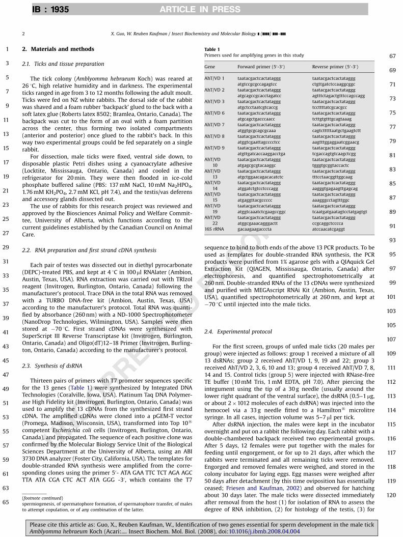
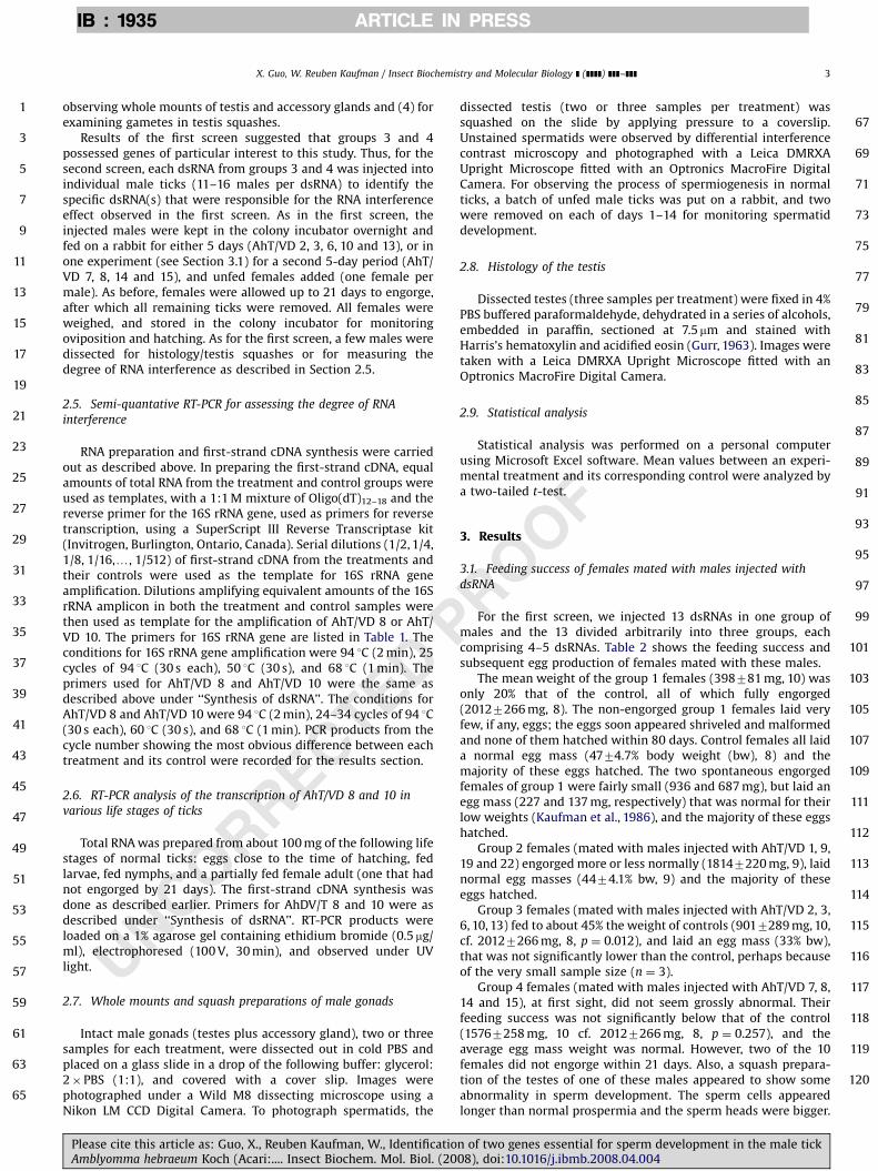
Fig. 1 demonstrates directly the inhibition of AhT/VD 8 and 10by semi-quantative RT-PCR.
3.2. Morphology/histology of the testis and progress of
spermiogenesis in feeding males
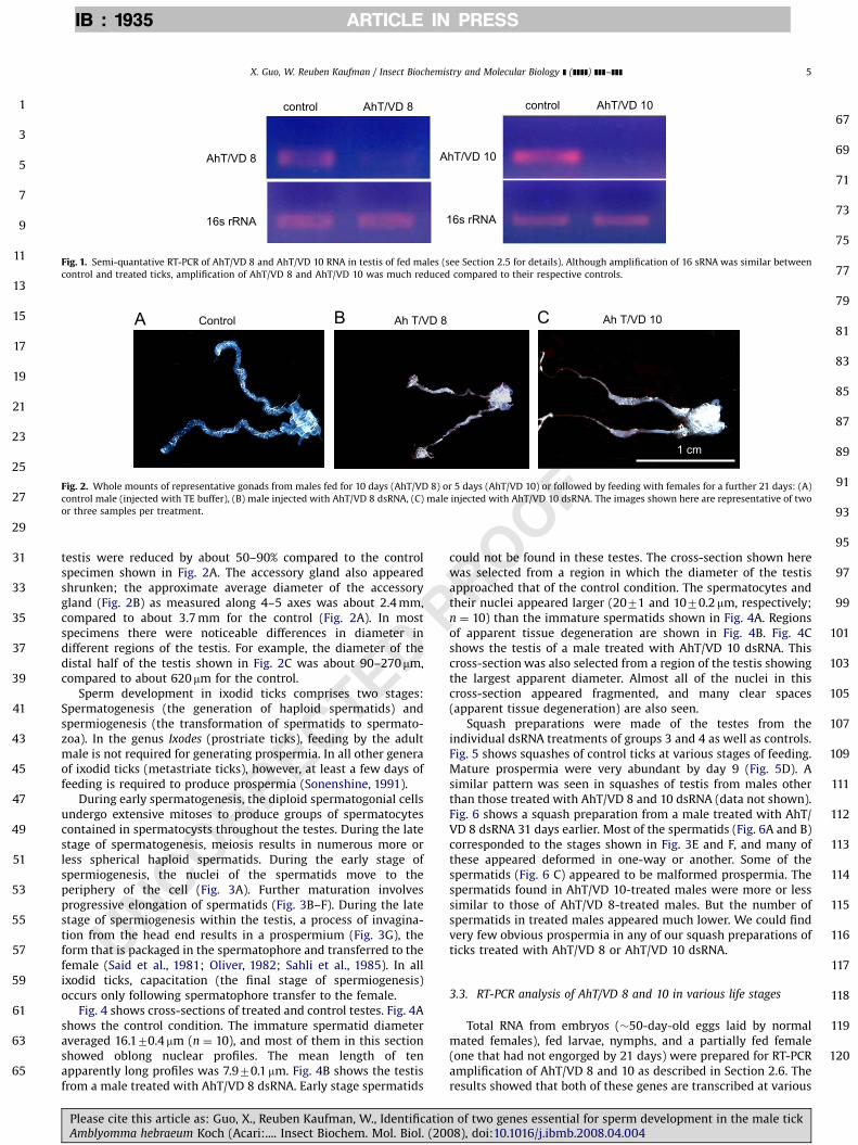
Representative whole mounts of the testes and accessory glandin control and treated ticks are shown in Fig. 2. The testis of ticksinjected with dsRNA of AhT/VD 8 or 10 appears significantlyshrunken. The diameters of the proximal and distal part of the
on of two genes essential for sperm development in the male tick008), doi:10.1016/j.ibmb.2008.04.004
P
F
1
3
5
7
9
11
13
15
17
19
21
23
25
27
29
31
33
35
37
39
41
43
45
47
49
51
53
55
57
59
61
63
65
67
69
71
73
75
77
79
81
83
85
87
89
91
93
95
97
99
101
103
105
107
109
111
112
113
114
115
116
117
118
119
120
ARTICLE IN PRESSIB : 1935
control
AhT/VD 8
16s rRNA
control AhT/VD 10
16s rRNA
AhT/VD 10
AhT/VD 8
Fig. 1. Semi-quantative RT-PCR of AhT/VD 8 and AhT/VD 10 RNA in testis of fed males (see Section 2.5 for details). Although amplification of 16 sRNA was similar between
control and treated ticks, amplification of AhT/VD 8 and AhT/VD 10 was much reduced compared to their respective controls.
Control Ah T/VD 8 Ah T/VD 10
1 cm
Fig. 2. Whole mounts of representative gonads from males fed for 10 days (AhT/VD 8) or 5 days (AhT/VD 10) or followed by feeding with females for a further 21 days: (A)
control male (injected with TE buffer), (B) male injected with AhT/VD 8 dsRNA, (C) male injected with AhT/VD 10 dsRNA. The images shown here are representative of two
or three samples per treatment.
X. Guo, W. Reuben Kaufman / Insect Biochemistry and Molecular Biology ] (]]]]) ]]]–]]] 5
UNCORRECTED
testis were reduced by about 50–90% compared to the controlspecimen shown in Fig. 2A. The accessory gland also appearedshrunken; the approximate average diameter of the accessorygland (Fig. 2B) as measured along 4–5 axes was about 2.4 mm,compared to about 3.7 mm for the control (Fig. 2A). In mostspecimens there were noticeable differences in diameter indifferent regions of the testis. For example, the diameter of thedistal half of the testis shown in Fig. 2C was about 90–270mm,compared to about 620mm for the control.
Sperm development in ixodid ticks comprises two stages:Spermatogenesis (the generation of haploid spermatids) andspermiogenesis (the transformation of spermatids to spermato-zoa). In the genus Ixodes (prostriate ticks), feeding by the adultmale is not required for generating prospermia. In all other generaof ixodid ticks (metastriate ticks), however, at least a few days offeeding is required to produce prospermia (Sonenshine, 1991).
During early spermatogenesis, the diploid spermatogonial cellsundergo extensive mitoses to produce groups of spermatocytescontained in spermatocysts throughout the testes. During the latestage of spermatogenesis, meiosis results in numerous more orless spherical haploid spermatids. During the early stage ofspermiogenesis, the nuclei of the spermatids move to theperiphery of the cell (Fig. 3A). Further maturation involvesprogressive elongation of spermatids (Fig. 3B–F). During the latestage of spermiogenesis within the testis, a process of invagina-tion from the head end results in a prospermium (Fig. 3G), theform that is packaged in the spermatophore and transferred to thefemale (Said et al., 1981; Oliver, 1982; Sahli et al., 1985). In allixodid ticks, capacitation (the final stage of spermiogenesis)occurs only following spermatophore transfer to the female.
Fig. 4 shows cross-sections of treated and control testes. Fig. 4Ashows the control condition. The immature spermatid diameteraveraged 16.170.4mm (n ¼ 10), and most of them in this sectionshowed oblong nuclear profiles. The mean length of tenapparently long profiles was 7.970.1mm. Fig. 4B shows the testisfrom a male treated with AhT/VD 8 dsRNA. Early stage spermatids
Please cite this article as: Guo, X., Reuben Kaufman, W., IdentificatioAmblyomma hebraeum Koch (Acari:.... Insect Biochem. Mol. Biol. (20
ROOcould not be found in these testes. The cross-section shown herewas selected from a region in which the diameter of the testisapproached that of the control condition. The spermatocytes andtheir nuclei appeared larger (2071 and 1070.2 mm, respectively;n ¼ 10) than the immature spermatids shown in Fig. 4A. Regionsof apparent tissue degeneration are shown in Fig. 4B. Fig. 4Cshows the testis of a male treated with AhT/VD 10 dsRNA. Thiscross-section was also selected from a region of the testis showingthe largest apparent diameter. Almost all of the nuclei in thiscross-section appeared fragmented, and many clear spaces(apparent tissue degeneration) are also seen.
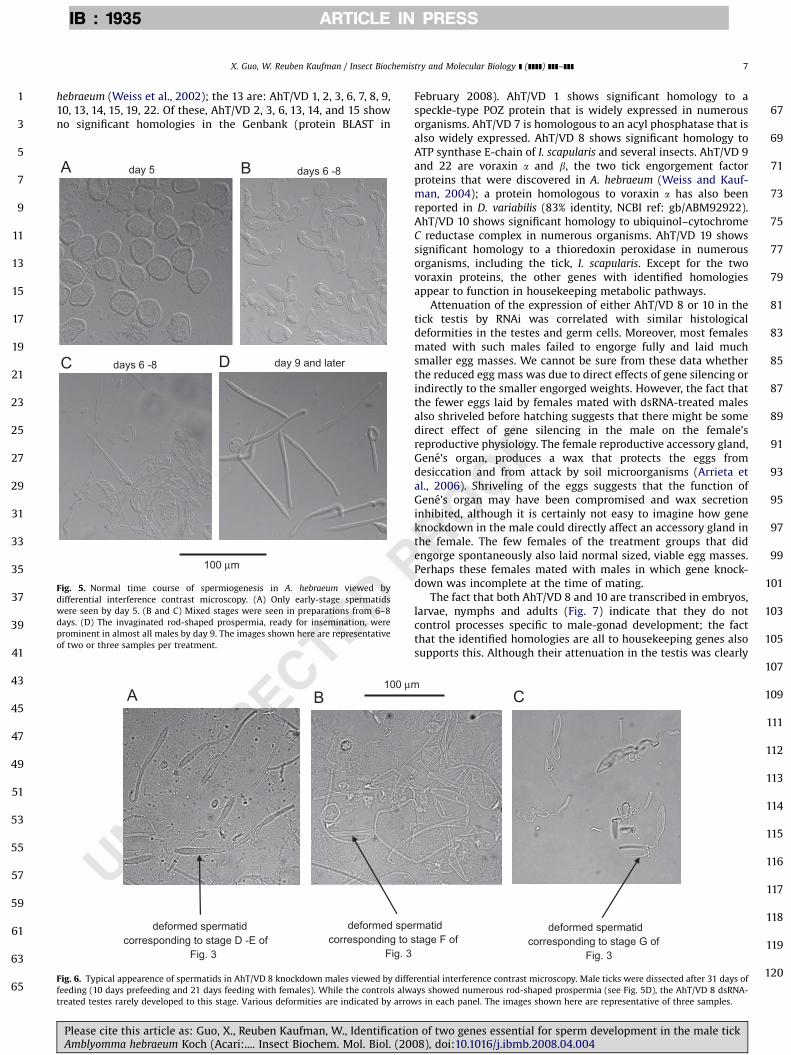
Squash preparations were made of the testes from theindividual dsRNA treatments of groups 3 and 4 as well as controls.Fig. 5 shows squashes of control ticks at various stages of feeding.Mature prospermia were very abundant by day 9 (Fig. 5D). Asimilar pattern was seen in squashes of testis from males otherthan those treated with AhT/VD 8 and 10 dsRNA (data not shown).Fig. 6 shows a squash preparation from a male treated with AhT/VD 8 dsRNA 31 days earlier. Most of the spermatids (Fig. 6A and B)corresponded to the stages shown in Fig. 3E and F, and many ofthese appeared deformed in one-way or another. Some of thespermatids (Fig. 6 C) appeared to be malformed prospermia. Thespermatids found in AhT/VD 10-treated males were more or lesssimilar to those of AhT/VD 8-treated males. But the number ofspermatids in treated males appeared much lower. We could findvery few obvious prospermia in any of our squash preparations ofticks treated with AhT/VD 8 or AhT/VD 10 dsRNA.
3.3. RT-PCR analysis of AhT/VD 8 and 10 in various life stages
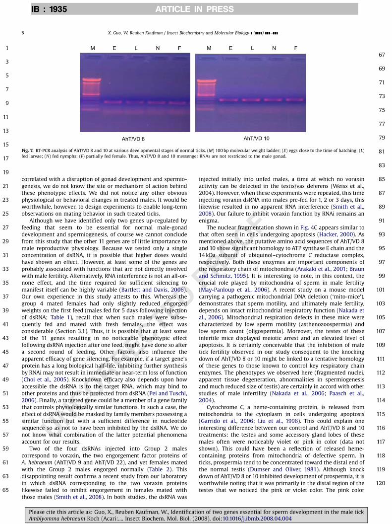
Total RNA from embryos (�50-day-old eggs laid by normalmated females), fed larvae, nymphs, and a partially fed female(one that had not engorged by 21 days) were prepared for RT-PCRamplification of AhT/VD 8 and 10 as described in Section 2.6. Theresults showed that both of these genes are transcribed at various
n of two genes essential for sperm development in the male tick08), doi:10.1016/j.ibmb.2008.04.004

UNCORRECTED PROOF
1
3
5
7
9
11
13
15
17
19
21
23
25
27
29
31
33
35
37
39
41
43
45
47
49
51
53
55
57
59
61
63
65
67
69
71
73
75
77
79
81
83
85
87
89
91
93
95
97
99
101
103
105
107
109
111
112
113
114
115
116
117
118
119
120
ARTICLE IN PRESSIB : 1935
Fig. 3. Selected germ cells seen in squash preparations of testis from normal males fed for various days and viewed by differential interference contrast microscopy. In this
study, germ cells reached the early spermatid stage by day 6 of feeding (A). Progressive elongation (B-F, selected from one preparation), and invagination to form the
prospermium (G, selected from another preparation), occurred between 6–8 days. By day 9, almost all testes contained numerous prospermia ready for packaging in a
spermatophore. The images shown here are representative of two or three samples per treatment.
nucleus of spermatocyte
area of tissue degeneration
fragmented nucleus
immature spermatid nucleus of spermatid
100 µm
Fig. 4. Histology of the AhT/VD 8 and AhT/VD 10 knock down testis stained with Harris’s hematoxylin and acidified eosin. Following prefeeding (5 or 10 days) and 21 days
feeding with females (as in Fig. 2), the spermatid profiles in the control testis (A) appeared spherical, and were evenly distributed across the testis profile. Many nuclei have
moved to the edge of the spermatid, characteristic of the early stage of spermiogenesis. In the two effective dsRNA treatments (B): AhT/VD 8, and (C): AhT/VD 10, germ cells
seemed to be arrested at the spermatocyte stage. There were also many clear spaces (apparent tissue degeneration) and apparent nuclear fragmentation. The images shown
here are representative of three samples per treatment. The scale bar shown is applicable to the three images.
X. Guo, W. Reuben Kaufman / Insect Biochemistry and Molecular Biology ] (]]]]) ]]]–]]]6
stages of tick development (Fig. 7), suggesting that these genescontrol more general functions than just male-gonad develop-ment.
Please cite this article as: Guo, X., Reuben Kaufman, W., IdentificatiAmblyomma hebraeum Koch (Acari:.... Insect Biochem. Mol. Biol. (2
4. Discussion
In this study, we tested the effect of RNAi on 13 of 28 genesthat are known to be up-regulated by feeding in the ixodid tick, A.
on of two genes essential for sperm development in the male tick008), doi:10.1016/j.ibmb.2008.04.004
1
3
5
7
9
11
13
15
17
19
21
23
25
27
29
31
33
35
37
39
41
43
45
47
49
51
53
55
57
59
61
63
65
67
ARTICLE IN PRESSIB : 1935
X. Guo, W. Reuben Kaufman / Insect Biochemistry and Molecular Biology ] (]]]]) ]]]–]]] 7
hebraeum (Weiss et al., 2002); the 13 are: AhT/VD 1, 2, 3, 6, 7, 8, 9,10, 13, 14, 15, 19, 22. Of these, AhT/VD 2, 3, 6, 13, 14, and 15 showno significant homologies in the Genbank (protein BLAST in
UNCORRECTED P
69
71
73
75
77
79
81
83
85
87
89
91
93
95
97
99
101
103
105
day 5 days 6 -8
day 9 and laterdays 6 -8
100 µm
Fig. 5. Normal time course of spermiogenesis in A. hebraeum viewed by
differential interference contrast microscopy. (A) Only early-stage spermatids
were seen by day 5. (B and C) Mixed stages were seen in preparations from 6–8
days. (D) The invaginated rod-shaped prospermia, ready for insemination, were
prominent in almost all males by day 9. The images shown here are representative
of two or three samples per treatment.
100 µm
deformed spermatid corresponding to stage D -E of
Fig. 3
deformed specorresponding to s
Fig. 3
Fig. 6. Typical appearence of spermatids in AhT/VD 8 knockdown males viewed by diffe
feeding (10 days prefeeding and 21 days feeding with females). While the controls alw
treated testes rarely developed to this stage. Various deformities are indicated by arrow
Please cite this article as: Guo, X., Reuben Kaufman, W., IdentificatioAmblyomma hebraeum Koch (Acari:.... Insect Biochem. Mol. Biol. (20
ROOF
February 2008). AhT/VD 1 shows significant homology to aspeckle-type POZ protein that is widely expressed in numerousorganisms. AhT/VD 7 is homologous to an acyl phosphatase that isalso widely expressed. AhT/VD 8 shows significant homology toATP synthase E-chain of I. scapularis and several insects. AhT/VD 9and 22 are voraxin a and b, the two tick engorgement factorproteins that were discovered in A. hebraeum (Weiss and Kauf-man, 2004); a protein homologous to voraxin a has also beenreported in D. variabilis (83% identity, NCBI ref: gb/ABM92922).AhT/VD 10 shows significant homology to ubiquinol–cytochromeC reductase complex in numerous organisms. AhT/VD 19 showssignificant homology to a thioredoxin peroxidase in numerousorganisms, including the tick, I. scapularis. Except for the twovoraxin proteins, the other genes with identified homologiesappear to function in housekeeping metabolic pathways.
Attenuation of the expression of either AhT/VD 8 or 10 in thetick testis by RNAi was correlated with similar histologicaldeformities in the testes and germ cells. Moreover, most femalesmated with such males failed to engorge fully and laid muchsmaller egg masses. We cannot be sure from these data whetherthe reduced egg mass was due to direct effects of gene silencing orindirectly to the smaller engorged weights. However, the fact thatthe fewer eggs laid by females mated with dsRNA-treated malesalso shriveled before hatching suggests that there might be somedirect effect of gene silencing in the male on the female’sreproductive physiology. The female reproductive accessory gland,Gene’s organ, produces a wax that protects the eggs fromdesiccation and from attack by soil microorganisms (Arrieta etal., 2006). Shriveling of the eggs suggests that the function ofGene’s organ may have been compromised and wax secretioninhibited, although it is certainly not easy to imagine how geneknockdown in the male could directly affect an accessory gland inthe female. The few females of the treatment groups that didengorge spontaneously also laid normal sized, viable egg masses.Perhaps these females mated with males in which gene knock-down was incomplete at the time of mating.
The fact that both AhT/VD 8 and 10 are transcribed in embryos,larvae, nymphs and adults (Fig. 7) indicate that they do notcontrol processes specific to male-gonad development; the factthat the identified homologies are all to housekeeping genes alsosupports this. Although their attenuation in the testis was clearly
107
109
111
112
113
114
115
116
117
118
119
120
rmatid tage F of
deformed spermatid corresponding to stage G of
Fig. 3
rential interference contrast microscopy. Male ticks were dissected after 31 days of
ays showed numerous rod-shaped prospermia (see Fig. 5D), the AhT/VD 8 dsRNA-
s in each panel. The images shown here are representative of three samples.
n of two genes essential for sperm development in the male tick08), doi:10.1016/j.ibmb.2008.04.004
1
3
5
7
9
11
13
15
17
19
21
23
25
27
29
31
33
35
37
39
41
43
45
47
49
51
53
55
57
59
61
63
65
67
69
71
73
75
77
79
81
83
85
87
89
91
93
95
97
99
101
103
105
107
109
111
112
113
114
115
116
117
118
119
120
ARTICLE IN PRESSIB : 1935
AhT/VD 8
M
AhT/VD 10
FNLE M FNLE
Fig. 7. RT-PCR analysis of AhT/VD 8 and 10 at various developmental stages of normal ticks. (M) 100 bp molecular weight ladder; (E) eggs close to the time of hatching; (L)
fed larvae; (N) fed nymphs; (F) partially fed female. Thus, AhT/VD 8 and 10 messenger RNAs are not restricted to the male gonad.
X. Guo, W. Reuben Kaufman / Insect Biochemistry and Molecular Biology ] (]]]]) ]]]–]]]8
UNCORRECTED
correlated with a disruption of gonad development and spermio-genesis, we do not know the site or mechanism of action behindthese phenotypic effects. We did not notice any other obviousphysiological or behavioral changes in treated males. It would beworthwhile, however, to design experiments to enable long-termobservations on mating behavior in such treated ticks.
Although we have identified only two genes up-regulated byfeeding that seem to be essential for normal male-gonaddevelopment and spermiogenesis, of course we cannot concludefrom this study that the other 11 genes are of little importance tomale reproductive physiology. Because we tested only a singleconcentration of dsRNA, it is possible that higher doses wouldhave shown an effect. However, at least some of the genes areprobably associated with functions that are not directly involvedwith male fertility. Alternatively, RNA interference is not an all-or-none effect, and the time required for sufficient silencing tomanifest itself can be highly variable (Bartlett and Davis, 2006).Our own experience in this study attests to this. Whereas thegroup 4 mated females had only slightly reduced engorgedweights on the first feed (males fed for 5 days following injectionof dsRNA; Table 1), recall that when such males were subse-quently fed and mated with fresh females, the effect wasconsiderable (Section 3.1). Thus, it is possible that at least someof the 11 genes resulting in no noticeable phenotypic effectfollowing dsRNA injection after one feed, might have done so aftera second round of feeding. Other factors also influence theapparent efficacy of gene silencing. For example, if a target gene’sprotein has a long biological half-life, inhibiting further synthesisby RNAi may not result in immediate or near-term loss of function(Choi et al., 2005). Knockdown efficacy also depends upon howaccessible the dsRNA is to the target RNA, which may bind toother proteins and thus be protected from dsRNA (Pei and Tuschl,2006). Finally, a targeted gene could be a member of a gene familythat controls physiologically similar functions. In such a case, theeffect of dsRNA would be masked by family members possessing asimilar function but with a sufficient difference in nucleotidesequence so as not to have been inhibited by the dsRNA. We donot know what combination of the latter potential phenomenaaccount for our results.
Two of the four dsRNAs injected into Group 2 malescorrespond to voraxin, the two engorgement factor proteins ofA. hebraeum (AhT/VD 9 and AhT/VD 22), and yet females matedwith the Group 2 males engorged normally (Table 2). Thisdisappointing result confirms a recent study from our laboratoryin which dsRNA corresponding to the two voraxin proteinslikewise failed to inhibit engorgement in females mated withthose males (Smith et al., 2008). In both studies, the dsRNA was
Please cite this article as: Guo, X., Reuben Kaufman, W., IdentificatiAmblyomma hebraeum Koch (Acari:.... Insect Biochem. Mol. Biol. (2
PROOF
injected initially into unfed males, a time at which no voraxinactivity can be detected in the testis/vas deferens (Weiss et al.,2004). However, when these experiments were repeated, this timeinjecting voraxin dsRNA into males pre-fed for 1, 2 or 3 days, thislikewise resulted in no apparent RNA interference (Smith et al.,2008). Our failure to inhibit voraxin function by RNAi remains anenigma.
The nuclear fragmentation shown in Fig. 4C appears similar tothat often seen in cells undergoing apoptosis (Hacker, 2000). Asmentioned above, the putative amino acid sequences of AhT/VD 8and 10 show significant homology to ATP synthase E chain and the14 kDa subunit of ubiquinol–cytochrome C reductase complex,respectively. Both these enzymes are important components ofthe respiratory chain of mitochondria (Arakaki et al., 2001; Braunand Schmitz, 1995). It is interesting to note, in this context, thecrucial role played by mitochondria of sperm in male fertility(May-Panloup et al., 2006). A recent study on a mouse modelcarrying a pathogenic mitochondrial DNA deletion (‘mito-mice’),demonstrates that sperm motility, and ultimately male fertility,depends on intact mitochondrial respiratory function (Nakada etal., 2006). Mitochondrial respiration defects in these mice werecharacterized by low sperm motility (asthenozoospermia) andlow sperm count (oligospermia). Moreover, the testes of theseinfertile mice displayed meiotic arrest and an elevated level ofapoptosis. It is certainly conceivable that the inhibition of maletick fertility observed in our study consequent to the knockingdown of AhT/VD 8 or 10 might be linked to a tentative homologyof these genes to those known to control key respiratory chainenzymes. The phenotypes we observed here (fragmented nuclei,apparent tissue degeneration, abnormalities in spermiogenesisand much reduced size of testis) are certainly in accord with otherstudies of male infertility (Nakada et al., 2006; Paasch et al.,2004).
Cytochrome C, a heme-containing protein, is released frommitochondria to the cytoplasm in cells undergoing apoptosis(Garrido et al., 2006; Liu et al., 1996). This could explain oneinteresting difference between our control and AhT/VD 8 and 10treatments: the testes and some accessory gland lobes of thesemales often were noticeably violet or pink in color (data notshown). This could have been a reflection of released heme-containing proteins from mitochondria of defective sperm. Inticks, prospermia tend to be concentrated toward the distal end ofthe normal testis (Dumser and Oliver, 1981). Although knockdown of AhT/VD 8 or 10 inhibited development of prospermia, it isworthwhile noting that it was primarily in the distal region of thetestes that we noticed the pink or violet color. The pink color
on of two genes essential for sperm development in the male tick008), doi:10.1016/j.ibmb.2008.04.004

Q1
1
3
5
7
9
11
13
15
17
19
21
23
25
27
29
31
33
35
37
39
41
43
45
47
49
51
53
55
57
59
61
ARTICLE IN PRESSIB : 1935
X. Guo, W. Reuben Kaufman / Insect Biochemistry and Molecular Biology ] (]]]]) ]]]–]]] 9
associated with the accessory gland lobes may reflect the high-energy requirement associated with production of seminal fluid.
We have shown here that the knockdown of AhT/VD 8 or 10 inmale A. hebraeum leads to major disruption of testis development,spermiogenesis and subsequent feeding success and fertility ofnormal females feeding with these males. This suggests that thesegenes are worthy of further consideration as candidate targets,alone or in conjunction with other genes of interest (e.g.subolesin: de la Fuente et al., 2006), for an anti-tick vaccine(Willadsen, 2006).
P Q2
63
65
67
69
71
73
75
77
79
81
83
85
87
89
91
93
95
97
99
101
RRECTEDAcknowledgments
This research was made possible by generous grants to W.R.K.from NSERC Canada (STPG program) and from the Alberta Scienceand Research Investments Program (ASRIP). The authors are mostgrateful to Mr. Alexander Smith for general assistance in handlingrabbits and ticks and reviewing the manuscript, to Mr. RandyMandryk for helping with the histology, and to Drs. Warren Gallin(University of Alberta), Kathy Kocan and Jose de la Fuente (both atOklahoma State University) for reviewing early drafts of themanuscript.
References
Ackerman, S., Clare, F.B., McGill, T.W., Sonenshine, D.E., 1981. Passage of host serumcomponents, including antibody, across the digestive tract of Dermacentorvariabilis (say). Parasitology 67, 737–740.
Arakaki, N., Ueyama, Y., Hirose, M., Himeda, T., Shibata, H., Futaki, S., Kitagawa, K.,Higuti, T., 2001. Stoichiometry of subunit e in rat liver mitochondrial H+-ATPsynthase and membrane topology of its putative Ca2+-dependent regulatoryregion. Biochim. Biophys. Acta 1504, 220–228.
Arrieta, M.C., Leskiw, B.K., Kaufman, W.R., 2006. Antimicrobial activity in the eggwax of the African cattle tick Amblyomma hebraeum (Acari: Ixodidae). Exp.Appl. Acarol. 39, 297–313.
Bartlett, D.W., Davis, M.E., 2006. Effect of siRNA nuclease stability on the in vitroand in vivo kinetics of siRNA-mediated gene silencing. Biotechnol. Bioeng., inpress, doi:10.1002/bit.21285.
Ben-Yakir, D., Fox, C.J., Homer, J.T., Barker, R.W., 1987. Quantification of hostimmunoglobulin in the hemolymph of ticks. J. Parasitol. 73, 669–671.
Braun, H.P., Schmitz, UK., 1995. Molecular features and mitochondrial LMPORTpathway of the 14-kilodalton subunit of cytochrome c reductase from potato.Plant Physiol. 107, 1217–1223.
Chinzei, Y., Minoura, H., 1987. Host immunoglobulin G titre and antibody activityin haemolymph of the tick, Ornithodoros moubata. Med. Vet. Entomol. 1,409–416.
Choi, I., Choa, B., Kima, D., Miyagawa, S., Kubo, T., Kim, J.Y., Park, C.G., Hwang, W.S.,Lee, J.S., Ahn, C., 2005. Choice of the adequate detection time for the accurateevaluation of the efficiency of siRNA-induced gene silencing. J. Biotechnol. 120,251–261.
de la Fuente, J., Almazan, C., Blas-Machado, U., Naranjo, V., Mangold, A.J., Blouin,E.F., Gortazar, C., Kocan, K.M., 2006. The tick protective antigen, 4D8, is aconserved protein involved in modulation of tick blood ingestion andreproduction. Vaccine 24, 4082–4095.
Dumser, J.B., Oliver Jr., J.H., 1981. Kinetics of spermatogenesis, cell-cycle analysisand testis development in nymphs of the tick, Dermacentor variabilis. J. InsectPhysiol. 27, 743–753.
UN
Please cite this article as: Guo, X., Reuben Kaufman, W., IdentificatioAmblyomma hebraeum Koch (Acari:.... Insect Biochem. Mol. Biol. (20
ROOF
Friesen, K., Kaufman, W.R., 2002. Quantification of vitellogenesis and its control by20-hydroxyecdysone in the ixodid tick, Ambyomma hebraeum. J. Insect Physiol.48, 773–782.
Garrido, C., Galluzzi, L., Brunet, M., Puig, P.E., Didelot, C., Kroemer, G., 2006.Mechanisms of cytochrome c release from mitochondria. Cell Death Differ. 13,1423–1433.
Gern, L., Falco, R.C., 2000. Lyme disease. Rev. Sci. Technol. 19, 121–135.George, J.E., Pound, J.M., Davey, R.B., 2004. Chemical control of ticks on cattle and
the resistance of these parasites to acaricides. Parasitology 129, S353–S366.Gurr, G.T., 1963. Biological Staining Methods, seventh ed. George T. Gurr Ltd,
London, pp. 22–23.Hacker, G., 2000. The morphology of apoptosis. Cell Tissue Res. 301, 5–17.Jasinskas, A., Jaworski, D.C., Barbour, A.G., 2000. Amblyomma americanum: specific
uptake of immunoglobulins into tick hemolymph during feeding. Exp.Parasitol. 96, 213–221.
Jongejan, F., Uilenberg, G., 2004. The global importance of ticks. Parasitology 129,S3–S14.
Kaufman, W.R., Ungarian, S.G., Noga, A.E., 1986. The effect of avermectins onfeeding, salivary fluid secretion, and fecundity in some ixodid ticks. Exp. App.Acarol. 2, 1–18.
Labuda, M., Nuttall, P.A., 2004. Tick-borne viruses. Parasitology 129, S221–S245.Liu, X.S., Kim, C.N., Yang, J., Jemmerson, R., Wang, X.D., 1996. Induction of apoptotic
program in cell-free extracts: requirement for dATP and cytochrome c. Cell 86,147–157.
May-Panloup, P., Chretien, M.F., Malthiery, Y., Reynier, P., 2006. Spermatozoonmitochondrial DNA. Gynecol. Obstet. Fertil. 34, 847–854.
Nakada, K., Sato, A., Yoshida, K., Morita, T., Tanaka, H., Inoue, S., Yonekawa, H.,Hayashi, J., 2006. Mitochondria-related male infertility. Proc. Natl. Acad. Sci.USA 103, 15148–15153.
Oliver, J.H., 1982. Tick reproduction: sperm development and cytogenetics. In:Obenchain, F.D., Galun, R. (Eds.), Physiology of Ticks. Pergamon Press, Oxford,pp. 245–275.
Paasch, U., Grunewald, S., Dathe, S., Glander, H.J., 2004. Mitochondria of humanspermatozoa are preferentially susceptible to apoptosis. Ann. N Y Acad. Sci.1030, 403–409.
Pei, Y., Tuschl, T., 2006. On the art of identifying effective and specific siRNAs. Nat.Methods 3, 670–676.
Sahli, R., Germond, J.E., Deihl, P.A., 1985. Ornithodoros moubata: spermateleosis andsecretory activity of the sperm. Exp. Parasitol. 60, 383–395.
Said, A.E., Swiderski, Z., Aeschlimann, A., Diehl, P., 1981. Fine structure ofspermiogenesis in the tick Amblyomma hebraeum (Acari: Ixodidae): late stagesof differentiation and structure of the mature spermatozoon. J. Med. Entomol.18, 464–476.
Smith, A.D., Guo, X.Y., de la Fuente, J., Naranjo, V., Kocan, K.M., Kaufman, W.R.,2008. The impact of RNA interference of the subolesin and voraxin genes inmale Amblyomma hebraeum (Acari: Ixodidae) on female engorgement andoviposition, manuscript submitted.
Sonenshine, D.E., 1991. Biology of Ticks. Oxford University Press, New York, Oxford,pp. 311–319.
Sonenshine, D.E., Kocan, K.M., de la Fuente, J., 2006. Tick control: further thoughtson a research agenda. Trends Parasitol. 22, 550–551.
Wang, H., Nuttall, P.A., 1994. Excretion of host immunoglobulin in tick saliva anddetection of IgG-binding proteins in tick haemolymph and salivary glands.Parasitology 109, 525–530.
Weiss, B.L., Kaufman, W.R., 2004. Two feeding-induced proteins from the malegonad trigger engorgement of the female tick Amblyomma hebraeum. Proc.Natl. Acad. Sci. USA 101, 5874–5879.
Weiss, B.L., Stepczynski, J.M., Wong, P., Kaufman, W.R., 2002. Identification andcharacterization of genes differentially expressed in the testis/vas deferens ofthe fed male tick, Amblyomma hebraeum. Insect Biochem. Mol. Biol. 32,785–793.
Willadsen, P., 2006. Tick control: thoughts on a research agenda. Vet. Parasitol. 138,161–168.
103
O Cn of two genes essential for sperm development in the male tick08), doi:10.1016/j.ibmb.2008.04.004