Embed Size (px)
Citation preview

Arthur Crutchfield PBL Case Overview of Educational Materials
Michael T. Fitch, M.D., Ph.D. Page 1 of 3
Arthur Crutchfield A Case for Problem Based Learning in Medical Education
Michael T. Fitch, M.D., Ph.D. Assistant Professor
Member, Core Teaching Faculty
Department of Emergency Medicine Wake Forest University
Medical Center Boulevard, Winston-Salem, North Carolina 27157 Phone: 336-716-4626 Fax: 336-716-5438
This Resource Successfully Peer Reviewed and Accepted with Acclamation by MedEdPORTAL on 04/05/2007. MedEdPORTAL Publication Number: 588 Alterations to this Resource Created After This Date Have Not Been Reviewed By MedEdPORTAL. Subsequent Revisions: None.
ABSTRACT
This educational resource provides the information and materials for a Problem Based Learning case suitable for medical students or physician assistant students during the pre-clinical portion of training. This case is currently in use at our institution as part of a small group course during the first year of medical education that focuses on group problem solving, self-directed learning, and clinical correlations to basic science learning. These materials were created by a faculty member who has served as a group facilitator in this course for several sessions, and it was successfully implemented this year during the lower extremity anatomy and physiology portion of the curriculum.
“Arthur Crutchfield” is a Problem Based Learning case that highlights the anatomy of the lower extremity. This case is appropriate for use in medical education during the curricular activities focusing on the musculoskeletal system. Using the Emergency Department as the clinical setting, students are encouraged to brainstorm about potential injuries to anatomic structures that they are studying in the framework of systematically evaluating a trauma patient with a knee dislocation. Elements of this case are based on a real patient encounter CONDENSED ABSTRACT
This educational resource provides the information and materials for a Problem Based Learning case suitable for medical students or physician assistant students during the pre-clinical portion of training. Using the Emergency Department as the clinical setting, students are encouraged to brainstorm about potential injuries to anatomic structures that they are studying in the framework of systematically evaluating a trauma patient with a knee dislocation.
Fitch PBL Case Materials Page 1 of 28
Arthur Crutchfield PBL Case Overview of Educational Materials
Michael T. Fitch, M.D., Ph.D. Page 2 of 3
CONTENT TOPICS
• Anatomic structures of the knee joint
• Correlation of knee examination with radiographic images and traumatic injuries
• Differential diagnosis of knee pain and injury • Vascular and peripheral nerve structures that traverse the popliteal fossa
• Cellular mechanisms of cartilage and ligament repair • Mechanism by which arterial injury may lead to thrombosis
STUDENT LEARNING OBJECTIVES
• Review the anatomy of the lower extremity, with emphasis on the ligamentous structures of the knee, vascular supply, and peripheral nerve structure and function.
• Demonstrate a systematic approach to the initial evaluation of a patient with a traumatic
injury. • Discuss possible causes of traumatic knee pain using an anatomic and mechanistic
perspective.
• Utilize physical examination findings to identify potential orthopedic and ligamentous injuries.
• Understand the relationship between vascular injury and thrombosis.
BACKGROUND AND SIGNIFICANCE
This Problem Based Learning case is used at our institution during the first year of medical school in a small group course which meets twice per week for two hours each session. Groups are comprised of 6 students with one clinical faculty facilitator and one basic science faculty facilitator. Facilitators are not expected to be content experts on the materials presented each week, but instead are present to assist the students in working through each case and determining their own group learning issues for individual student reading during the week. This case was designed for use during the portion of the curriculum where students are studying the anatomy and physiology of the lower extremity. Elements of the case are intended to stimulate discussion about basic anatomic topics and how they relate to clinical medicine. In addition, students are introduced to some basic concepts of clinical decision-making and patient care.
Fitch PBL Case Materials Page 2 of 28

Arthur Crutchfield PBL Case Overview of Educational Materials
Michael T. Fitch, M.D., Ph.D. Page 3 of 3
LESSONS LEARNED AND FUTURE DIRECTIONS
The materials provided in this PBL case were developed by a clinical faculty facilitator from our small group course, and were prepared as part of an ongoing project to revise and improve the cases used in this part of our curriculum. Based on student and facilitator feedback from other cases currently incorporated into the course, the author developed this clinical scenario based on elements of a real patient case. Lessons learned during the process of case revisions in this course and the recent implementation of this new case have led to improvements in the design and content of these materials. Inclusion of simulated patient photos for Session 1 and Session 2 are recent additions, and are intended to increase the personal connection of students with the patient in this case. Efforts were made to obtain high quality radiographic images to supplement the text in response to student feedback, and these images have been included in these materials.
Facilitator notes have been provided to assist faculty from many different disciplines who may participate in this type of course. Facilitators are not expected to be content experts on the clinical topic of this case, and therefore notes are provided to allow them to understand the highlights of this case with relevant background material.
Experience from the recent use of this case with 115 students in small groups has already led to editing of case components for increased clarity, and we anticipate that additional feedback from students and facilitators will allow other improvements to the case in the future. USE AND ADAPTATION OF CASE MATERIALS
This PBL case can be used or modified as needed for educational use at other institutions. Users are encouraged to tailor the materials for their own environment and course structure. The author requests citation of this work in any subsequent modified versions used in your curriculum, and he welcomes the feedback and suggestions for additional supplemental materials that will further assist in the design or implementation of this educational case. REFERENCES Barnes CJ, Pietrobon R, Higgins LD. Does the pulse examination in patients with traumatic knee dislocation predict a surgical arterial injury? A meta-analysis. 2002. J Trauma. 253(6):1109-14. Mills WJ, Barei DP, McNair P. 2004. The value of the ankle-brachial index for diagnosing arterial injury after knee dislocation: a prospective study. J Trauma. 56(6):1261-5. Perron AD, Brady WJ, Sing RF. 2001. Orthopedic pitfalls in the ED: vascular injury associated with knee dislocation. Am J Emerg Med. 19(7):583-8. Rihn JA, Groff YJ, Harner CD, Cha PS. 2004. The acutely dislocated knee: evaluation and management. J Am Acad Orthop Surg. 12(5):334-46. Stannard JP, Sheils TM, Lopez-Ben RR, McGwin G Jr, Robinson JT, Volgas DA. 2004. Vascular injuries in knee dislocations: the role of physical examination in determining the need for arteriography. J Bone Joint Surg Am. 86-A(5):910-5.
Fitch PBL Case Materials Page 3 of 28

Arthur Crutchfield PBL Case Session 1 & 2 Materials
Michael T. Fitch, M.D., Ph.D. Page 1 of 11
Arthur Crutchfield A Case for Problem Based Learning in Medical Education
Case author: Michael T. Fitch, M.D., Ph.D., Wake Forest University, Winston-Salem, NC
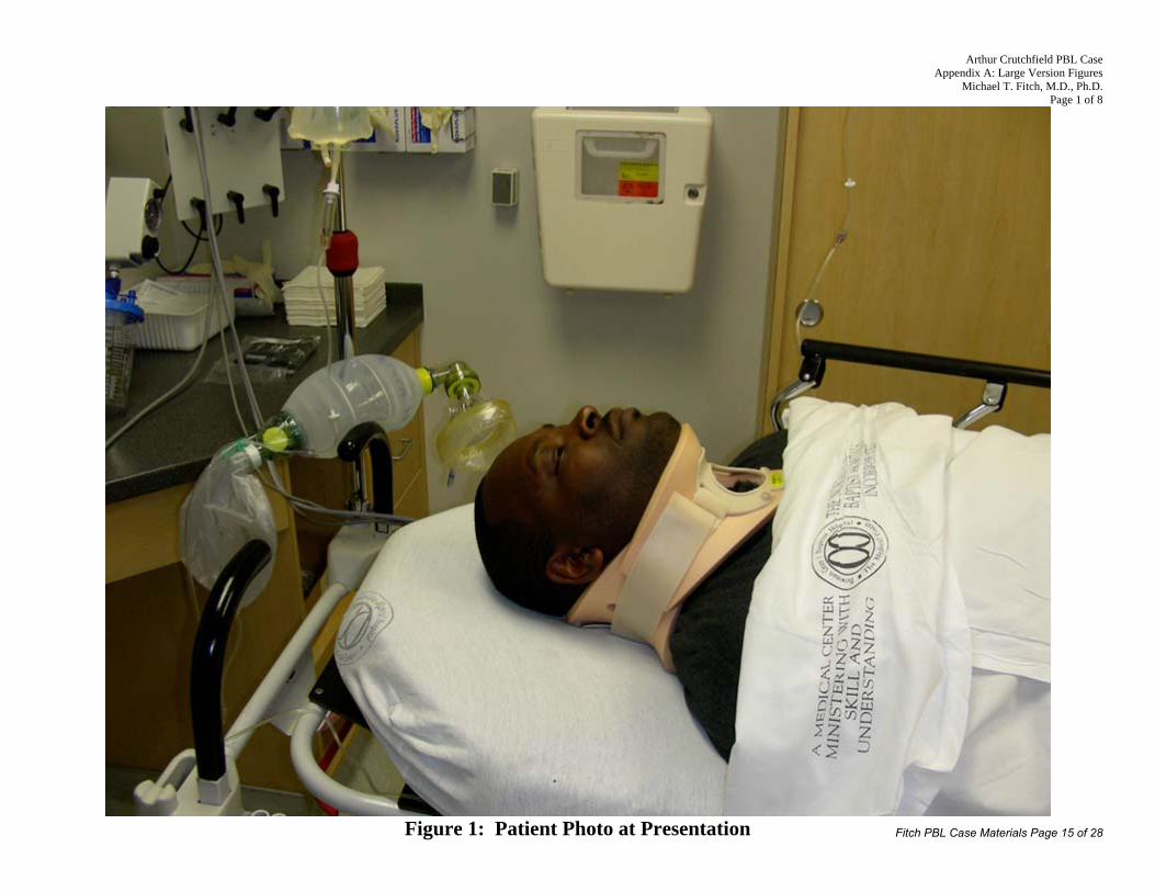
Figure 1: Patient photo at presentation
Session 1, Page 1: You are working in the Emergency Department at a local hospital on a busy Saturday night. County EMS Unit #14 contacts the ED by radio to give a patient report. “This is EMS Unit #14 calling with report. We are transporting a 35 year old male who has been involved in a single car motor vehicle accident. Patient reports swerving to avoid an animal in the road, then he lost control and drove into the ditch and hit a telephone pole. Significant damage to the vehicle with full airbag deployment. Patient was pinned in the vehicle for 30 minutes before he could be cut from the wreckage. At this time, patient is awake and alert and in full spinal immobilization on a back board. He is complaining of severe left lower extremity pain and his does have a visible deformity of that leg. We will arrive at your facility in 15 minutes. We would like to administer IV morphine for pain control if that is okay with you, and is there any other information we can provide at this time?” GROUP TASKS: Discuss any other additional information you would like to request from the EMS personnel. What factors will help you decide whether or not to approve IV morphine for this patient prior to his arrival in the Emergency Department? List any preparations or special medical equipment you would like to have available when the patient ambulance arrives with the patient.
Fitch PBL Case Materials Page 4 of 28

Arthur Crutchfield PBL Case Session 1 & 2 Materials
Michael T. Fitch, M.D., Ph.D. Page 2 of 11
Session 1, Page 2: You request additional information from the EMS personnel. You learn that his vital signs are: pulse 110, respirations 16, blood pressure 120/70, and pulse oxygenation 98% on nasal canula oxygen. The patient is not complaining of any other injuries besides his left leg, and the physical exam by EMS providers reveals no signs of head injury, no chest pain, no abdominal pain, and no open wounds. He is in severe pain and is having difficulty moving his left leg below the knee. Based on this information, you ask the EMS providers to place a large-bore IV line, start IV normal saline, and administer 6 mg of IV morphine for pain control. The patient arrives in the ED approximately 12 minutes later. You have prepared room #10 for a patient with a traumatic injury, and you have the following equipment immediately available: oxygen and masks, bag valve mask, larygoscopes and endotracheal tubes, IV catheters and fluids, central venous access catheters, and orthopedic splinting materials. You are present at the bedside when he is transferred from the Medic stretcher to the ED bed. The EMS providers report no change in his condition since the radio report, and that the patient has been awake and talking throughout transport. He is in full spinal immobilization with a cervical collar and backboard. The patient tells you that his name is Arthur Crutchfield. As Mr. Crutchfield is transferred from the EMS stretcher to the bed, he cries out in pain and attempts to reach for his left knee where you note a large amount of swelling and an obvious deformity. GROUP TASKS: Why is this patient in “full spinal immobilization” and how do you decide if this should be continued? Discuss your initial approach to this patient. What information do you need first, and how will you prioritize your evaluation? What elements of physical examination are most important? Are there any particular findings you will be specifically looking for?
Fitch PBL Case Materials Page 5 of 28
Arthur Crutchfield PBL Case Session 1 & 2 Materials
Michael T. Fitch, M.D., Ph.D. Page 3 of 11
Session 1, Page 3: You use the principles of Advanced Trauma Life Support (ATLS) to assess Mr. Crutchfield’s condition in an organized fashion as detailed below. In particular, you make sure to complete his primary survey before focusing attention on his painful knee injury. Primary Survey Airway: Intact. Patient is talking and answers questions. Breathing: Respirations 14/minute, clear breath sounds, pulse oxygenation 98%. Circulation: Strong radial pulse, rate 105. Blood pressure with a manual cuff 115/75. Additional history: Mr. Crutchfield has no allergies, takes no medications, and has no significant past medical history. His last meal was 6 hours ago and he denies alcohol or drug use. He reports no loss of consciousness and does remember the accident. His only complaint is knee pain. Secondary Survey HEENT: Pupils equal round and reactive with intact extraocular movements. No scalp lacerations or hematoma. Midface is stable and non-tender. Oral exam normal. On ear exam, no hemotympanum is found. Neck: Cervical collar is in place. He has mild neck pain on palpation of C5. Chest: No abrasions or ecchymoses. No tenderness. Clear breath sounds. CV: Regular rhythm with tachycardic rate. No murmurs. Abdomen: Obese, soft, nontender, and nondistended. No abdominal wall bruising. Pelvis: Nontender. Stable to AP, lateral, and symphyseal pressure. Back: Nontender thoracic and lumbar spine. No abrasions or ecchymoses. GU: Normal male genitalia. Rectal tone normal, no gross blood. Extremities: Patient has 5/5 strength and full range of motion of upper extremities bilaterally with no pain, swelling, or abrasions. Right lower extremity has no deformities, full range of motion with no tenderness to palpation, and distal sensation is intact. Right posterior tibial and dorsalis pedis pulses are 2+. Left lower extremity has a swollen knee that is extremely painful to touch, has limited range of motion, and his lower leg is angulated medially. Left posterior tibial and dorsalis pedis pulses are 1+. Patient reports decreased sensation on the dorsum of his foot near the great toe, and he is refusing to move his foot and toes because of severe pain. GROUP TASKS:
1. Discuss the current findings and their significance. 2. What injuries do you suspect Mr. Crutchfield has sustained? 3. Are there additional physical examination maneuvers should you consider to further
evaluate the patient’s painful swollen knee? 4. What further diagnostic testing will you order at this time?
Fitch PBL Case Materials Page 6 of 28
Arthur Crutchfield PBL Case Session 1 & 2 Materials
Michael T. Fitch, M.D., Ph.D. Page 4 of 11
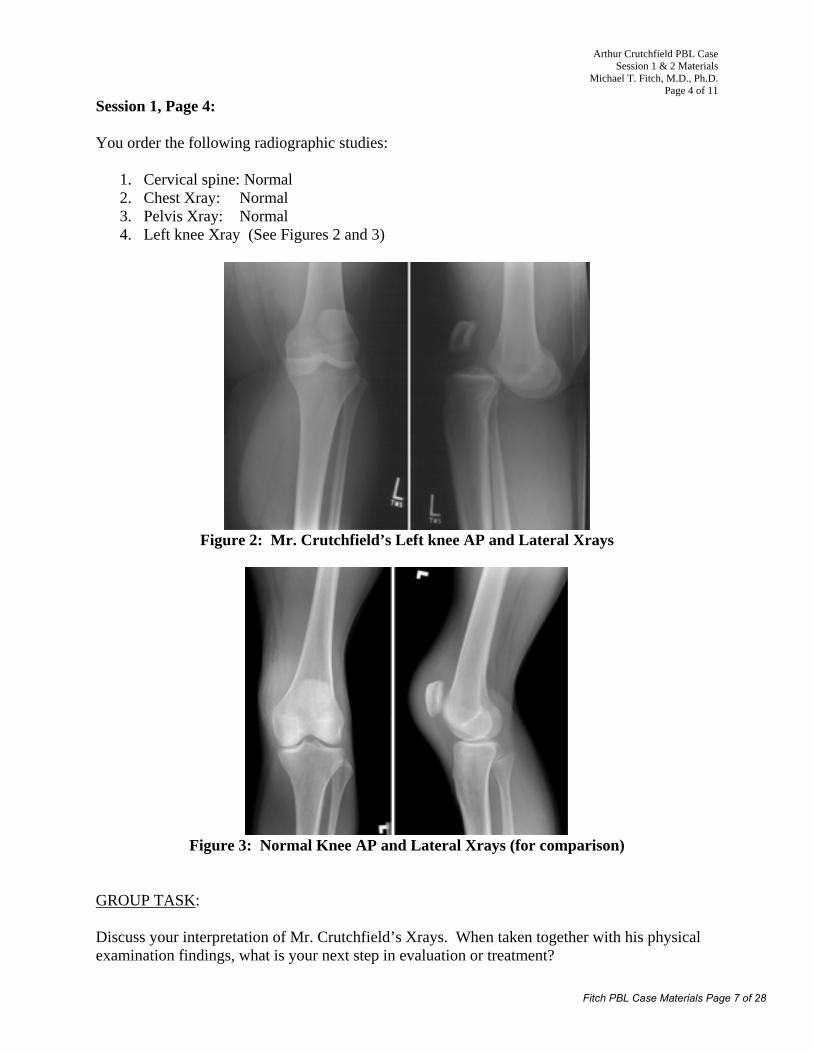
Session 1, Page 4: You order the following radiographic studies:
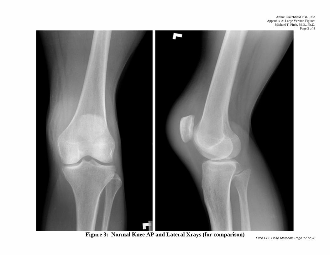
1. Cervical spine: Normal 2. Chest Xray: Normal 3. Pelvis Xray: Normal 4. Left knee Xray (See Figures 2 and 3)
Figure 2: Mr. Crutchfield’s Left knee AP and Lateral Xrays
Figure 3: Normal Knee AP and Lateral Xrays (for comparison)
GROUP TASK: Discuss your interpretation of Mr. Crutchfield’s Xrays. When taken together with his physical examination findings, what is your next step in evaluation or treatment?
Fitch PBL Case Materials Page 7 of 28

Arthur Crutchfield PBL Case Session 1 & 2 Materials
Michael T. Fitch, M.D., Ph.D. Page 5 of 11
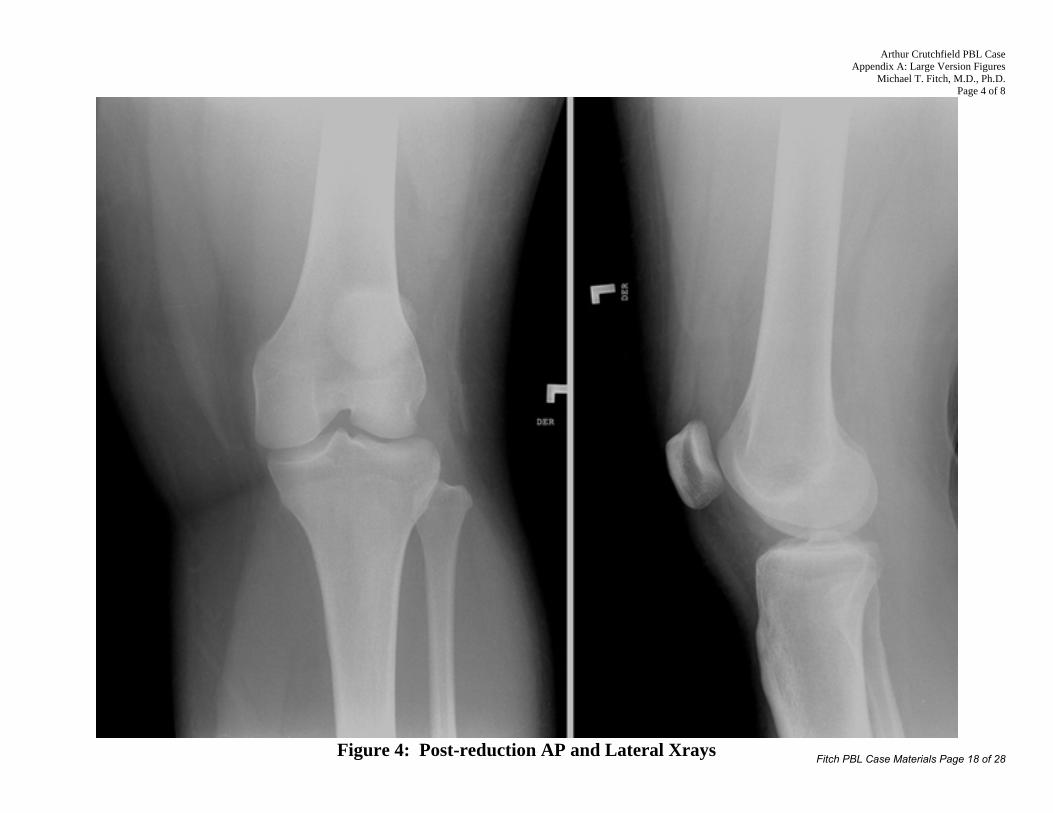
Session 1, Page 5: You realize that the Xray of Mr. Crutchfield’s knee demonstrates an acute anterior dislocation. With the decreased pulses in his foot, you decide to proceed with an emergent relocation procedure. You administer IV midazolam and IV morphine and manually relocate the knee. Post-procedure films demonstrate appropriate relocation of the joint with no associated fractures.
Figure 4: Post-reduction AP and Lateral Xrays
Repeat physical examination reveals that Mr. Crutchfield’s left knee is normal with varus stress testing but laxity to valgus stress testing. He has laxity to both anterior Lachman’s testing and posterior Drawer testing. His posterior tibial and dorsalis pedis pulses are now 2+. He has 5/5 strength with dorsiflexion and plantarflexion of his foot and can move all toes normally. Leg and foot sensation are normal. For specific information about these physical examination maneuvers for the knee, see this website: http://www.fpnotebook.com/ORT86.htm GROUP TASKS: What structures have apparently been damaged and which are intact based on your physical examination at this time? What would explain the patient’s decreased sensation over the dorsal surface of his distal foot that has now resolved after reduction? Why is the knee swollen and how do you explain the fact that the swelling is only around the knee joint itself?
Fitch PBL Case Materials Page 8 of 28

Arthur Crutchfield PBL Case Session 1 & 2 Materials
Michael T. Fitch, M.D., Ph.D. Page 6 of 11
Session 1, Page 5 (continued): Are there any concerns for occult injuries to other structures that are located near the knee? What physical examination findings would accompany such other injuries? Should you consider any additional diagnostic testing or examinations to evaluate for this possibility? After consultation with Orthopedic Surgery, you place the patient in a knee immobilizer. He is much more comfortable and his pain is well controlled. Repeat physical examination finds the patient now has no neck or back pain with full range of motion, and his cervical collar is removed and he is allowed to sit up. Abdomen remains soft and nontender. Because of the association of arterial injury with knee dislocation, you discuss with Vascular Surgery what options you have for evaluating for this possible complication. What possible testing could you do to evaluate for arterial injury of the lower extremity?
Fitch PBL Case Materials Page 9 of 28
Arthur Crutchfield PBL Case Session 1 & 2 Materials
Michael T. Fitch, M.D., Ph.D. Page 7 of 11
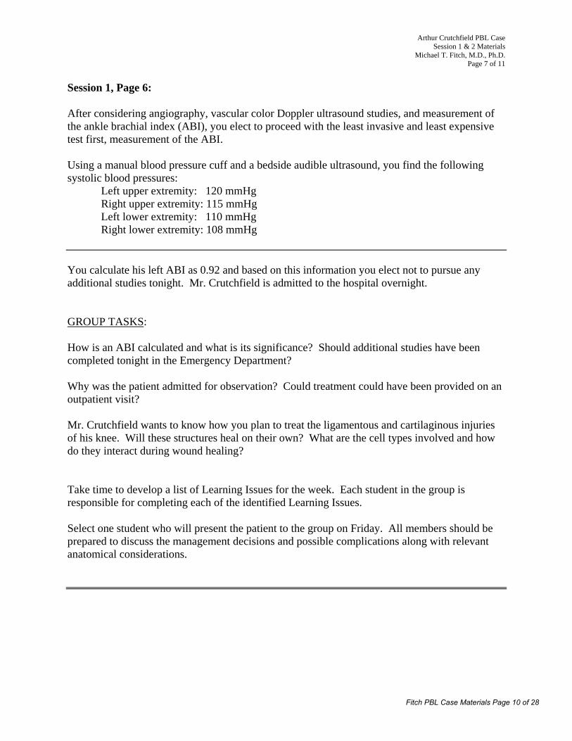
Session 1, Page 6: After considering angiography, vascular color Doppler ultrasound studies, and measurement of the ankle brachial index (ABI), you elect to proceed with the least invasive and least expensive test first, measurement of the ABI. Using a manual blood pressure cuff and a bedside audible ultrasound, you find the following systolic blood pressures:
Left upper extremity: 120 mmHg Right upper extremity: 115 mmHg Left lower extremity: 110 mmHg Right lower extremity: 108 mmHg
You calculate his left ABI as 0.92 and based on this information you elect not to pursue any additional studies tonight. Mr. Crutchfield is admitted to the hospital overnight. GROUP TASKS: How is an ABI calculated and what is its significance? Should additional studies have been completed tonight in the Emergency Department? Why was the patient admitted for observation? Could treatment could have been provided on an outpatient visit? Mr. Crutchfield wants to know how you plan to treat the ligamentous and cartilaginous injuries of his knee. Will these structures heal on their own? What are the cell types involved and how do they interact during wound healing? Take time to develop a list of Learning Issues for the week. Each student in the group is responsible for completing each of the identified Learning Issues. Select one student who will present the patient to the group on Friday. All members should be prepared to discuss the management decisions and possible complications along with relevant anatomical considerations.
Fitch PBL Case Materials Page 10 of 28
Arthur Crutchfield PBL Case Session 1 & 2 Materials
Michael T. Fitch, M.D., Ph.D. Page 8 of 11
Arthur Crutchfield
A Case for Problem Based Learning in Medical Education
Session 2
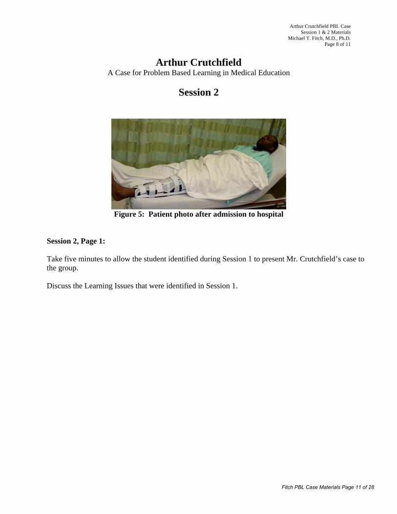
Figure 5: Patient photo after admission to hospital
Session 2, Page 1: Take five minutes to allow the student identified during Session 1 to present Mr. Crutchfield’s case to the group. Discuss the Learning Issues that were identified in Session 1.
Fitch PBL Case Materials Page 11 of 28

Arthur Crutchfield PBL Case Session 1 & 2 Materials
Michael T. Fitch, M.D., Ph.D. Page 9 of 11
Session 2, Page 2: Mr. Crutchfield was admitted overnight to the hospital for serial examinations and observation. Repeat physical examinations of his left leg revealed normal 2+ dorsalis pedis and posterior tibial pulses. The pain in his knee was well controlled with minimal amounts of pain medication. The strength and sensation of the leg and foot remain normal. GROUP TASK: What physical examination findings would you have seen if Mr. Crutchfield had an injury to his common peroneal nerve? The next morning on rounds, a medical student is the first person to see Mr. Crutchfield. At that time, he is complaining of severe lower leg pain. His left foot is pale and cool to the touch. His dorsalis pedis and posterior tibial pulses are not palpable. GROUP TASKS: Discuss your initial thoughts about Mr. Crutchfield’s apparent worsening condition. What might be going on here? What is the next step in your diagnostic or therapeutic plan?
Fitch PBL Case Materials Page 12 of 28

Arthur Crutchfield PBL Case Session 1 & 2 Materials
Michael T. Fitch, M.D., Ph.D. Page 10 of 11
Session 2, Page 3: You arrange for Mr. Crutchfield to go emergently to angiography, where the following images are obtained of his lower extremity:
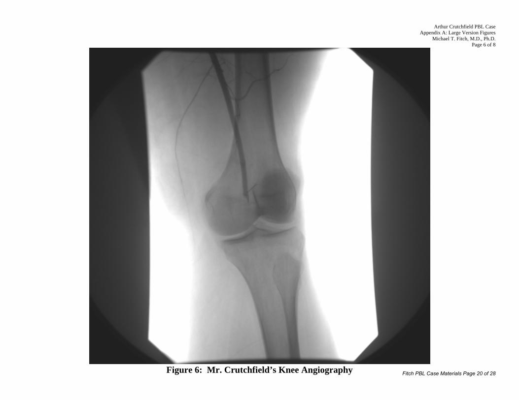
Figure 6: Mr. Crutchfield’s Knee Angiography
Figure 7: Normal Knee Angiography (for comparison)
GROUP TASKS: What is your interpretation of these findings? What is the next step in diagnosis or treatment?
Fitch PBL Case Materials Page 13 of 28

Arthur Crutchfield PBL Case Session 1 & 2 Materials
Michael T. Fitch, M.D., Ph.D. Page 11 of 11
Session 2, Page 4: Mr. Crutchfield undergoes emergent vascular surgery to repair the occult damage to his popliteal artery that led to the development of an acute thrombosis overnight. The surgeons are able to successfully remove the thrombosis, restore blood flow, and repair the injury to the intimal layer of the artery. After recovering from the emergency vascular surgery, an MRI is completed to evaluate the extent of ligamentous damage to his knee.
Figure 8: MRI of left knee
The MRI demonstrates: 1. Complete tear anterior cruciate ligament and posterior cruciate ligament. 2. Findings highly suspicious horizontal tear medial meniscus. 3. Joint effusion. 4. Grade II sprain medial collateral ligament. 5. Lateral collateral ligament is intact. Mr. Crutchfield has an uneventful remainder of his hospital course. He is subsequently discharged from the hospital with a knee immobilizer and crutches with plans for reconstructive knee surgery by an orthopedic surgeon in 2 weeks. GROUP TASK: Before his discharge, you are called to talk with Mr. Crutchfield. He is very concerned about his ability to work as a package delivery driver – particularly because his truck is a manual transmission. He has no disability coverage and no paid sick leave. He would like to continue working to support his wife and two small children. When do you anticipate he can return to work? How will you help him address this issue?
Fitch PBL Case Materials Page 14 of 28
Arthur Crutchfield PBL Case Appendix A: Large Version Figures
Michael T. Fitch, M.D., Ph.D. Page 1 of 8
Figure 1: Patient Photo at Presentation Fitch PBL Case Materials Page 15 of 28

Arthur Crutchfield PBL Case Appendix A: Large Version Figures
Michael T. Fitch, M.D., Ph.D. Page 2 of 8
Figure 2: Mr. Crutchfield’s Left knee AP and Lateral Xrays
Fitch PBL Case Materials Page 16 of 28
Arthur Crutchfield PBL Case Appendix A: Large Version Figures
Michael T. Fitch, M.D., Ph.D. Page 3 of 8
Figure 3: Normal Knee AP and Lateral Xrays (for comparison)
Fitch PBL Case Materials Page 17 of 28
Arthur Crutchfield PBL Case Appendix A: Large Version Figures
Michael T. Fitch, M.D., Ph.D. Page 4 of 8
Figure 4: Post-reduction AP and Lateral Xrays Fitch PBL Case Materials Page 18 of 28

Arthur Crutchfield PBL Case Appendix A: Large Version Figures
Michael T. Fitch, M.D., Ph.D. Page 5 of 8
Figure 5: Patient photo after admission to hospital
Fitch PBL Case Materials Page 19 of 28
Arthur Crutchfield PBL Case Appendix A: Large Version Figures
Michael T. Fitch, M.D., Ph.D. Page 6 of 8
Figure 6: Mr. Crutchfield’s Knee Angiography Fitch PBL Case Materials Page 20 of 28

Arthur Crutchfield PBL Case Appendix A: Large Version Figures
Michael T. Fitch, M.D., Ph.D. Page 7 of 8
Figure 7: Normal Knee Angiography (for comparison) Fitch PBL Case Materials Page 21 of 28

Arthur Crutchfield PBL Case Appendix A: Large Version Figures
Michael T. Fitch, M.D., Ph.D. Page 8 of 8
Figure 8: MRI of left knee
Fitch PBL Case Materials Page 22 of 28
Arthur Crutchfield PBL Case Appendix B: PBL Faculty Notes
Michael T. Fitch, M.D., Ph.D. Page 1 of 6
Appendix B: Problem Based Learning Faculty Notes
Arthur Crutchfield Case
Michael T. Fitch, M.D., Ph.D.
Wake Forest University Winston-Salem, North Carolina
Introduction:
“Arthur Crutchfield” is a Problem Based Learning case that highlights the anatomy of the lower extremity. This case is appropriate for use in medical education during the curricular activities focusing on the musculoskeletal system. Using the Emergency Department as the clinical setting, students are encouraged to brainstorm about potential injuries to anatomic structures that they are studying in the framework of systematically evaluating a trauma patient. Elements of this case are based on a real patient encounter. Case Synopsis:
Arthur Crutchfield is a 35 year old man who is involved in a motor vehicle accident
where he suffers a dislocated knee which requires emergent reduction. After admission to the hospital, he develops an acute thrombosis from an occult injury to the popliteal artery that requires emergency surgical repair.
The students are presented initially with the general problem of knee pain in a patient involved in a car accident. This is made more challenging when they are asked to make decisions based on information from the EMS radio report. When the patient arrives, a systematic approach to evaluation reveals a swollen knee with decreased distal pulses suspicious for a knee dislocation. Radiographs confirm an acute knee dislocation which is emergently reduced with return of normal pulses. Students are asked to discuss the anatomic structures of the knee that may be injured by such a dislocation. Ankle brachial index (ABI) testing as a screening test for occult vascular injury is introduced, and the patient is admitted to the hospital overnight.
In Session 2, the students will learn that the patient subsequently develops an acute thrombosis of the popliteal artery from an occult vascular injury that is demonstrated by angiography. Emergency surgery is successful in restoring blood flow and saving the patient’s leg. MRI demonstrates extensive ligamentous injury that will require further surgery in several weeks. The students are asked to help address some social issues as the patient will be unable to work while waiting for the surgery to repair his knee.
Fitch PBL Case Materials Page 23 of 28
Arthur Crutchfield PBL Case Appendix B: PBL Faculty Notes
Michael T. Fitch, M.D., Ph.D. Page 2 of 6
Content Topics:
• Anatomic structures of the knee joint • Correlation of knee examination with radiographic images and traumatic injuries • Differential diagnosis of knee pain and injury • Vascular and peripheral nerve structures that traverse the popliteal fossa • Cellular mechanisms of cartilage and ligament repair • Mechanism by which arterial injury may lead to thrombosis
Group Objectives:
• Review the anatomy of the lower extremity, with emphasis on the ligamentous structures of the knee, vascular supply, and peripheral nerve structure and function.
• Demonstrate a systematic approach to the initial evaluation of a patient with a traumatic
injury. • Discuss possible causes of traumatic knee pain using an anatomic and mechanistic
perspective.
• Utilize physical examination findings to identify potential orthopedic and ligamentous injuries.
• Understand the relationship between vascular injury and thrombosis.
Resources and References: Barnes CJ, Pietrobon R, Higgins LD. Does the pulse examination in patients with traumatic knee dislocation predict a surgical arterial injury? A meta-analysis. 2002. J Trauma. 253(6):1109-14. Mills WJ, Barei DP, McNair P. 2004. The value of the ankle-brachial index for diagnosing arterial injury after knee dislocation: a prospective study. J Trauma. 56(6):1261-5. Perron AD, Brady WJ, Sing RF. 2001. Orthopedic pitfalls in the ED: vascular injury associated with knee dislocation. Am J Emerg Med. 19(7):583-8. Rihn JA, Groff YJ, Harner CD, Cha PS. 2004. The acutely dislocated knee: evaluation and management. J Am Acad Orthop Surg. 12(5):334-46. Stannard JP, Sheils TM, Lopez-Ben RR, McGwin G Jr, Robinson JT, Volgas DA. 2004. Vascular injuries in knee dislocations: the role of physical examination in determining the need for arteriography. J Bone Joint Surg Am. 86-A(5):910-5.
Fitch PBL Case Materials Page 24 of 28

Arthur Crutchfield PBL Case Appendix B: PBL Faculty Notes
Michael T. Fitch, M.D., Ph.D. Page 3 of 6
Session 1, Page 1:
The initial case scenario is based on an EMS radio report, and is designed to get the students to begin thinking about how to prepare for a patient’s arrival with a possible serious emergency based only on a verbal report from a prehospital provider. Encourage them to discuss additional questions they would ask over the radio. The EMS providers are requesting narcotic pain medications for the patient, and students may identify the need to obtain vital signs or additional clinical information before making this kind of decision. Equipment important to consider having available includes: oxygen and masks, bag valve mask, larygoscopes and endotracheal tubes, IV catheters and fluids, central venous catheters, and orthopedic splinting materials. Session 1, Page 2:
The additional information obtained from EMS indicates that the patient has stable vital signs with no hypotension, but is somewhat tachycardic with a pulse of 110. Intravenous morphine is prescribed via the radio for the patient’s painful leg injury.
When the patient arrives, he is on an EMS backboard with a cervical collar in place (“full spinal immobilization”) based on the significant mechanism of injury (motor vehicle accident with entrapment). This will typically be continued in the Emergency Department until a full evaluation can be completed to determine whether clinical concern exists for a spinal injury.
Initially, it is evident that his knee injury is causing significant pain, and this may distract the students from doing an initial evaluation of the patient’s airway, breathing, and circulation. When discussing the initial approach to the patient, prioritization based on these principles is important to recognize and students should be encouraged to discuss what they feel is most important to address first and why.
Session 1, Page 3:
If the students were distracted on the previous page by the patient’s painful knee injury, this page will remind them about the need to complete the “Primary Survey” that includes assessment of Airway, Breathing, and Circulation before addressing any distracting painful injuries. ATLS is a protocol-driven approach to initial evaluation of a trauma patient, and the initial phase emphasizes the ABC approach prior to obtaining a basic history and completing the remainder of the physical examination.
Mr. Crutchfield’s primary survey is unremarkable. His secondary survey reveals only mild cervical spine tenderness and his painful knee injury. The left knee has limited range of motion, is tender to palpate, and he has decreased distal pulses with an abnormal sensory exam of the foot. These findings are highly suggestive of a knee dislocation with impingement of the popliteal artery and common peroneal nerve.
Encourage the students to brainstorm about the findings and their significance. Ask them to describe how they would evaluate the knee on physical examination, and what further diagnostic testing they would consider. Examination of the knee may be a point of discussion – allow this to continue for a short time but do not allow the students to become sidetracked by this issue as more details on this exam will be coming later in the case. Plain film xrays will be the most cost-effective and time-efficient radiographic studies to order first, and will be revealed on the next page.
Fitch PBL Case Materials Page 25 of 28

Arthur Crutchfield PBL Case Appendix B: PBL Faculty Notes
Michael T. Fitch, M.D., Ph.D. Page 4 of 6
Session 1, Page 4:
Xray films of the cervical spine, chest, and pelvis are all normal. Focus should be dedicated to the abnormal knee xrays.
The patient’s xrays demonstrate an anterior knee dislocation (anterior because the tibial is displaced anterior to the femur). Anterior dislocations are the most common and make up approximately 40% of all dislocations. When compared to the normal xrays that are provided, students should be able to identify that there are no broken bones (note the lack of bone fragments, smooth contour of the structures) but that there is an apparent misalignment of the distal femur and proximal tibia/fibula.
Students are asked about what the next step in treatment should be for this patient. In the setting of a dislocated joint with evidence of distal neurovascular compromise (decreased pulses and abnormal sensory and motor exam), this patient needs an emergent reduction of this dislocation. Session 1, Page 5:
The patient receives the recommended emergent reduction of the dislocated joint. Repeat xrays demonstrate appropriate alignment with no associated fractures. Additional examination is presented, which will allow for a detailed discussion of the joint anatomy (anterior and posterior cruciate ligaments, medial and lateral collateral ligaments). If students are unfamiliar with the testing methods for these ligaments, a web link is provided that has information about these physical examination maneuvers.
Lachman test: most sensitive for anterior cruciate ligament damage. Posterior drawer test: most sensitive for posterior cruciate ligament injury. Varus stress: Lateral collateral ligament testing Valgus stress: Medial collateral ligament testing
After reviewing this information, encourage the students to hypothesize about what
structures are damaged in Mr. Crutchfield’s knee. They should be able to identify the common peroneal nerve as a suspect for injury – the fact that these symptoms resolved after relocation suggest that it was a stretching or compression of this nerve during the dislocation and that there was no transection or permanent nerve damage.
Hemarthrosis and swelling of damaged tissues will lead to swelling around the knee joint itself, which is an opportunity to guide the students to discuss the nature of a synovial joint with its associated joint capsule.
Finally, students should identify the vascular structures that traverse the popliteal fossa and the concern for injury to the popliteal artery from stretching or shear forces during a dislocation. The popliteal artery is fixed to the adductor hiatus and the soleus muscles, which make it vulnerable to injury in a dislocation. Options for evaluation include vascular Doppler ultrasound, angiography, ankle brachial index measurement, and serial pulse examinations.
Fitch PBL Case Materials Page 26 of 28

Arthur Crutchfield PBL Case Appendix B: PBL Faculty Notes
Michael T. Fitch, M.D., Ph.D. Page 5 of 6
Session 1, Page 6:
This last page of the session begins the discussion about how to evaluate for occult injuries to the popliteal artery. This associated injury occurs in 30-40% of dislocations. Note that there is still significant ambiguity and controversy in the literature about how to appropriately manage this kind of patient who does not have an obvious vascular injury – which should lead to an interesting discussion on Friday about the decisions made in this case. Some sources will recommend mandatory angiography for all patients with knee dislocation, some suggest a “selective approach” using serial physical exams and ankle brachial index measurement, and others will use vascular Doppler examination in selected cases. There have been reports of patients with vascular injury even in the presence of normal pulses in up to 15% of patients.
There are several papers in the literature that have examined the ankle brachial index (ABI) as a potential screening test for occult vascular injuries (see References above). The students are given the information necessary to calculate the ABI (which is done by dividing the systolic blood pressure in the ankle by the systolic blood pressure in the arm on the side of interest). In this case, left leg pressure / left arm pressure = 110 mmHg / 120 mmHg = 0.92 which is a normal ABI, suggesting no injury to the popliteal artery. Based on this information, emergent angiography is not recommended. However, the patient is admitted to the hospital for serial physical exams to watch for the possibility of an occult vascular injury.
The tasks on this page center on the significance of the ABI and its use as a screening test for injury – and this is a good candidate for a Learning Issue. Note that the patient is admitted for observation because without definitive testing we cannot rule out an occult vascular injury even in the setting of normal ABI measurements. Encourage the students to discuss (and/or develop learning issues) that allow them to explore the cell types involved in ligamentous and cartilaginous injury repair and whether or not surgery is needed.
Note that a student should be selected to present the patient case to the group for Session 2. This is intended to allow practice with clinical presentations, and the student selected should be prepared to given a summary of the case as if the others in the group are hearing about the patient for the first time.
Fitch PBL Case Materials Page 27 of 28

Arthur Crutchfield PBL Case Appendix B: PBL Faculty Notes
Michael T. Fitch, M.D., Ph.D. Page 6 of 6
Session 2, Page 1:
Allow the selected student from Session 1 to present the patient case to the group. Spend time discussing and exploring the Learning Issues from Session 1. Session 2, Page 2:
Mr. Crutchfield was admitted overnight for serial examinations, which initially were unremarkable. Students are asked to review their understanding of the innervation of the lower extremity by discussing the physical examination findings expected from a common peroneal nerve injury (decreased sensation of the dorsal foot, loss of dorsiflexion leading to “foot drop”). This associated injury is found in 15-35% of knee dislocation injuries.
The next morning, the patient has severe leg pain with a pale and cool foot and decreased pulses – all suggestive of acute vascular insufficiency. The students are asked to discuss the possible etiology of this problem, such as an occult intimal injury to the popliteal artery that has now developed an acute thrombosis. This patient needs emergent arteriography and vascular intervention. Session 2, Page 3:
Arteriography is performed, and the images demonstrate an abrupt cut-off of dye flow in the popliteal fossa (consistent with an arterial disruption or clot). This can be compared to the normal angiography provided that has flow throughout the popliteal artery and distal vasculature. This patient needs surgical repair!
This is an opportunity for the students to consider what may have occurred to predispose Mr. Crutchfield to an arterial thrombosis. An intimal injury can lead to local turbulent blood flow and the deposition of fibrin and platelet aggregation leading to thrombus formation. While it is not necessary for students to explore the entire clotting cascade at this point, a basic understanding of why an intimal injury may lead to an increased risk of clot formation is a reasonable goal. Session 2, Page 4:
Surgery is successful at removing the acute thrombosis and repairing the intimal injury to the popliteal artery that led to this event.
An MRI is performed, and findings are detailed with multiple ligamentous injuries. These findings confirm those of the physical exam detailed in Session 1. Encourage the students to compare the MRI findings to the reported physical exam findings.
Mr. Crutchfield is discharged home in a knee immobilizer because of the significant ligamentous injuries. Repair of the ligaments has been delayed to allow some of the swelling to resolve prior to surgical repair.
Before leaving from the hospital, the patient inquires about his ability to work. He drives a truck with a manual transmission for a living, and wants to know about his options for employment during his recovery period. Encourage students to explore this issue and the financial hardship this may lead to for this patient.
Fitch PBL Case Materials Page 28 of 28