Embed Size (px)
Citation preview
Arthroscopically Pertinent Anatomy of theAnterolateral and Posteromedial Bundles of the
Posterior Cruciate LigamentColin J. Anderson, MD, Connor G. Ziegler, MD, Coen A. Wijdicks, PhD,
Lars Engebretsen, MD, PhD, and Robert F. LaPrade, MD, PhD
Investigation performed at the Department of BioMedical Engineering, Steadman Philippon Research Institute, Vail, Colorado
Background: The clock-face method to identify the femoral posterior cruciate ligament (PCL) attachment has pooraccuracy and reproducibility. Measurements of clinically relevant anatomic structures would provide more useful surgicalguidance. The purpose of the present study was to describe the attachments of the anterolateral and posteromedialbundles of the PCL relative to relevant landmarks to assist with arthroscopic anatomic PCL reconstructions.
Methods: Dissections were performed on twenty nonpaired fresh-frozen cadaveric knees.
Results: The distal articular cartilage margin of the intercondylar notch had a consistent shape conforming to theattachments of the anterolateral and posteromedial bundles. The mean distance (and standard deviation) between thefemoral centers of the anterolateral and posteromedial bundles was 12.1 ± 1.3 mm. The distal margins of the antero-lateral and posteromedial bundles were a mean of 1.5 ± 0.8 mm and 5.8 ± 1.7 mm proximal to the notch articularcartilage, respectively. On the tibia, the lateral plateau articular cartilage, the medial meniscus attachment, and anosseous ridge (‘‘bundle ridge’’) separating the anterolateral and posteromedial bundles were important arthroscopiclandmarks. The mean distance between the tibial centers of the anterolateral and posteromedial bundles was 8.9 ± 1.2mm.
Conclusions: The pertinent landmarks identified during arthroscopic PCL reconstruction consistently marked the bor-ders of the attachments of the anterolateral and posteromedial bundles. To guide femoral tunnel placement, the centersof both bundles should be triangulated relative to the reported landmarks. Furthermore, the distal edge of the femoralanterolateral bundle should be placed adjacent to the articular cartilage, whereas the posteromedial bundle should becentered, on average, 8.6 mm proximal to the cartilage margin, just distal to the medial intercondylar ridge. On the tibia,the PCL tunnel should be placed just anterosuperior to the bundle ridge, with use of the lateral articular cartilage andmedial meniscus attachment to guide placement.
Clinical Relevance: The results of the present study can assist with more anatomic tunnel placement during single anddouble-bundle PCL reconstructions. The results also suggest that two reconstruction tunnels are needed to reconstructthe broad femoral attachment, whereas one reconstruction tunnel should be investigated further for the compact tibialattachment.
A substantial number of patients have been reported toexperience declining knee function and early osteoar-thritis following the nonoperative treatment of isolated
and combined grade-III posterior cruciate ligament (PCL)
injuries1-5. The desire to restore knee function has drivengrowing interest in surgical reconstruction of the injured PCL.However, clinical studies have demonstrated that single-bundlePCL reconstructions have failed to restore normal kinematics
Disclosure: One or more of the authors received payments or services, either directly or indirectly (i.e., via his or her institution), from a third party insupport of an aspect of this work. In addition, one or more of the authors, or his or her institution, has had a financial relationship, in the thirty-six monthsprior to submission of this work, with an entity in the biomedical arena that could be perceived to influence or have the potential to influence what is writtenin this work. No author has had any other relationships, or has engaged in any other activities, that could be perceived to influence or have the potential toinfluence what is written in this work. The complete Disclosures of Potential Conflicts of Interest submitted by authors are always provided with theonline version of the article.
1936
COPYRIGHT ! 2012 BY THE JOURNAL OF BONE AND JOINT SURGERY, INCORPORATED
J Bone Joint Surg Am. 2012;94:1936-45 d http://dx.doi.org/10.2106/JBJS.K.01710
To learn more about this study, please click here: http://drlaprade.com

to the knee and have not prevented osteoarthritis in the longterm6,7. Double-bundle PCL reconstructions that restore theposteromedial bundle in addition to the biomechanically andfunctionally dominant anterolateral bundle have demonstratedsome improvement in knee stability in vitro8,9. However, clin-ical studies to date have not demonstrated improved subjectiveor objective outcomes for double-bundle compared with single-bundle reconstructions10-14.
There is a growing paradigm that cruciate ligament re-construction achieves improved results with restoration of thenative anatomy15-20. While this has been better supported in theanterior cruciate ligament (ACL) literature21,22, with the higherfrequency of ACL injuries having facilitated an accelerated pro-gression of research findings, similar evidence is mounting forthe treatment of PCL tears. Early in vitro studies evaluating iso-metric PCL reconstructions demonstrated inferior performancecompared with more anatomic tunnel placement because theisometric reconstructions resulted in posterior translational in-stability in flexion and knee overconstraint in extension9,17,23.Likewise, suboptimal clinical results have been reported fornon-anatomic, compared with more presumed anatomic,femoral tunnel placement19. However, an accurate and re-producible method to guide anatomic tunnel placement forPCL reconstructions has not been developed16.
The ‘‘clock-face’’ method is the most frequently re-ported method to describe PCL femoral tunnel placement16.However, anatomic and surgical studies have demonstratedwide disagreement in the reported locations of the PCLbundles with use of the clock-face method16. Furthermore,the clock-face method has been shown to have poor repro-ducibility between surgeons when used for ACL tunnelplacement24. A standardized method to guide tibial tunnelplacement also has not been described.
Our motivation for this study was to provide guidelinesfor femoral and tibial PCL tunnel placement based specificallyon a more detailed understanding of the surrounding anatomyso that recognition of key landmarks could more accuratelyguide arthroscopic localization of the PCL attachment centerson the femur and tibia. The purpose of the present study was toboth qualitatively and quantitatively describe the locations ofthe anterolateral and posteromedial bundles of the PCL relativeto arthroscopically relevant landmarks to assist with anatomictunnel placement during both single and double-bundle PCLreconstruction surgery.
Materials and MethodsSpecimens
Twelve femoral and twelve tibial bone specimens from the Human AnatomyProgram at the University of Colorado at Boulder were qualitatively ana-
lyzed for osseous prominences related to the position of the PCL. Three fresh-frozen nonpaired knees were then dissected as pilots to verify the presence ofthese osseous prominences and to define the optimal dissection approach.
Anatomic DissectionNext, dissections were performed on twenty nonpaired, fresh-frozen humancadaveric knees with no evidence of previous injury or degenerative change.The mean age of the donors at the time of death was 46.2 years (range, twenty-
one to forty-nine years). Standard anatomic nomenclature was used with theknee described in the extended position25. The PCLwas approached anteriorlythrough a medial parapatellar arthrotomy and posteriorly by means of carefuldissection of the posterior capsule and the oblique popliteal ligament, withblunt instrumentation being used to avoid damaging the distal fibers of the PCLor the posterior meniscofemoral ligament. If present, the anterior menisco-femoral ligament of Humphrey and the posterior meniscofemoral ligament ofWrisberg were separated from the PCL. Because the anterolateral bundle istightest at 90" of knee flexion and the posteromedial bundle is tightest at bothfull extension and flexion, the anterolateral bundle and posteromedial bundlewere individually identified following observation of their tensioning patternsas the knee was repeatedly cycled through its range of motion. The initial sepa-ration between the bundles was created posteriorly with a curved fine-tippedhemostat, close to the femoral attachment, where the separation between thebundles was best visualized. Next, the bundles were divided completely with useof fine dissecting scissors along the interfascicular connective tissue between thebundles. Nonabsorbable sutures were used to isolate each fiber bundle proximallyand distally, with the functional center of each individual bundle being isolatedaccording to a previously described technique25, and the knee was disarticulated.
Fig. 1
Illustration of the anterior view of a right knee flexed to 90" with the PCLintact, demonstrating the characteristicmorphology of the cartilagemarginof the femoral intercondylar notch. The illustration also shows the troch-lear, medial arch, and posterior points as well as the intercondylar notchapex and trochlear groove. ACL = anterior cruciate ligament, ALB = an-terolateral bundle, aMFL = anterior meniscofemoral ligament, and PMB =
posteromedial bundle.
1937
THE JOURNAL OF BONE & JOINT SURGERY d J B J S .ORG
VOLUME 94-A d NUMBER 21 d NOVEMBER 7, 2012ARTHROSCOP ICALLY PERT INENT POSTER IOR
CRUCIATE LIGAMENT ANATOMY
To learn more about this study, please click here: http://drlaprade.com

Finally, the proximal end of the femur and the distal end of the tibiawere potted toprovide secure fixation during the measurement process.
Quantitative Anatomic MeasurementsThe Liberty electromagnetic tracking system (Polhemus, Colchester, Vermont)was used to perform quantitative measurements. Three-dimensional positional
datawere collected with use of a calibrated stylus that sensed its position relativeto an electromagnetic field produced by the transmitter. Following secure fix-ation of the specimen to the testing apparatus, the PCL bundles, the anteriormeniscofemoral ligament, and the posterior meniscofemoral ligament weresharply dissected from their attachments. The outline of each attachment sitewas immediately recorded with the stylus. Then, distances were measuredbetween the center of the fiber bundles and the anatomic landmarks. All dis-sections were performed with the senior author (R.F.L.) present, and all re-ported measurements were performed by the same individual to decreaseinterobserver variability.
The MotionMonitor software package (version 8, Innovative Sports Train-ing, Chicago, Illinois) ran the Polhemus system during the data-collection processand exported the distances between landmarks and the three-dimensional coor-dinates of the outlines of the traced structures. Areas of traced structureswere calculated from the coordinate files of the tracing and are reported asthe area of the three-dimensional surface. Angular differences betweenstructures were calculated from normal vectors of the planes representingthe structures.
ValidationTo assess the accuracy of the electromagnetic tracking system, a calibrationdevice with a fixed linear distance accurate to within 0.074 mm and a circulararea accurate to within 0.000028 mm2 was utilized. This calibration device wasprecisely fabricated with a vertical endmill and lathe equipped with a highlyaccurate digital readout system (Sharp Industries, Torrance, California, andMitutoyo America, Aurora, Illinois). Measurement error for the Polhemussystem was calculated from twenty repeated measurements performed by twoseparate observers using this device. In order to establish the interobserverreproducibility of the measurements between the anatomic landmarks and thecenter of the fiber bundles, five of the cadaveric specimens were completelyremeasured by a second observer, and average-measure interobserver interclasscorrelation coefficients (ICCs) were calculated (SPSS, version 18; SPSS, Chi-cago, Illinois) on the basis of the repeated measurements.
Fig. 2
Anterior photograph of a right knee flexed to 90" with a probe (from pos-terior) separating the anterolateral bundle (ALB) and posteromedial bundle(PMB) of the PCL and demonstrating the landmarks surrounding thetrochlear and medial arch points along the cartilage margin of the femoralintercondylar notch.
Fig. 3-A Fig. 3-B
Fig. 3-A Illustration of the arthroscopic view of the femoral attachment of the PCL in a right knee, demonstrating pertinent landmarks. ALB = anterolateralbundle, aMFL = anterior meniscofemoral ligament, PMB = posteromedial bundle, and pMFL = posterior meniscofemoral ligament. Fig. 3-B Illustrationshowing the quantitative measurements for the femoral attachment of the PCL. The values are reported in millimeters.
1938
THE JOURNAL OF BONE & JOINT SURGERY d J B J S .ORG
VOLUME 94-A d NUMBER 21 d NOVEMBER 7, 2012ARTHROSCOP ICALLY PERT INENT POSTER IOR
CRUCIATE LIGAMENT ANATOMY
To learn more about this study, please click here: http://drlaprade.com

Source of FundingThis study was funded through a postdoctoral fellowship funded by the Nor-wegian South-East Regional Health Authority and by the Steadman PhilipponResearch Institute. The funds were used for salaries, supplies, and cadavericspecimens. The Steadman Philippon Research Institute is a 501(c)(3) nonprofitinstitution supported financially by private donations and corporate supportfrom the following entities: Smith & Nephew Endoscopy, Arthrex, SiemensMedical Solutions USA, OrthoRehab, ConMed Linvatec, Ossur Americas,Small Bone Innovations, and Opedix. One or more of the authors are paid/unpaid consultants for Arthrex.
ResultsMorphology of the Distal Articular Cartilage Margin of theFemoral Intercondylar Notch
Qualitative analysis of the femoral bone specimens revealeda consistent pattern of the shape of the distal articular
cartilage margin of the intercondylar notch. In correlating theseosseous landmarks on the femoral bone specimens to the freshcadavers, three distinct points along this margin were identifiedwhere the slope of the cartilage margin immediately changeddirection in a consistent and definable manner: the trochlear,medial arch, and posterior points (Figs. 1 and 2). On thelateral side of the intercondylar notch, the intersection ofthe sulcus terminalis with the cartilage edge created a con-vexity. Anterior to this convexity, the articular cartilage edgeformed the apex of the intercondylar notch, which was themost anterior extent of the notch. This apex was centered onthe trochlear groove, on the lateral side of the midline. On themedial side of the apex, a point was consistently found wherethe cartilage abruptly turned medially, which we termed the‘‘trochlear point’’ (Figs. 1 and 2). The cartilage margin con-tinued posteromedially in a smooth arch. At the end of thisarch, there was a point along the medial wall of the notchwhere the cartilage became oriented straight posteriorly. Wetermed this point the ‘‘medial arch point.’’ Continuing alongthe articular cartilage edge, the most posterior extent of thecartilage margin of the medial femoral condyle was termedthe ‘‘posterior point’’ (Fig. 3-A).
Osseous Morphology of the Roof and Walls of the FemoralIntercondylar NotchQualitative analysis of the femora revealed that the roof andwalls of the intercondylar notch had a consistent morphologythat was related to the attachments of the anterolateral andposteromedial bundles (Fig. 3-A). The previously describedmedial intercondylar ridge26 was found in all specimens. Thisridge was found to constitute the proximal borders of both theanterolateral and posteromedial bundles in every specimen. Onthe wall of the medial femoral condyle, where it marked theproximal border of the posteromedial bundle, this ridge wasoriented directly in the anteroposterior plane as it extendedanteriorly from the posterior point of the articular cartilage(Figs. 3-A and 4). The medial intercondylar ridge then con-tinued across the roof of the intercondylar notch, where itmarked the proximal border of the anterolateral bundle (Figs.3-A and 4). The medial bifurcate ridge has been previouslyreported to mark the separation between the anterolateral and
posteromedial bundles26. We identified a medial bifurcate ridgebetween the anterolateral and posteromedial bundles in onlythree specimens. However, in every specimen, there was a pal-pable and visible osseous prominence, previously not described,that was positioned adjacent to the medial intercondylar ridgeat the separation of the anterolateral and posteromedial bun-dles; we termed this structure the ‘‘medial bifurcate promi-nence’’ (Fig. 3-A).
Femoral Attachment of the PCLQuantitative data on the femoral attachment of the anterolat-eral and posteromedial bundles are reported in Table I. Whilethere was minor variability in the sizes of the footprints ofthe anterolateral and posteromedial bundles, the describedlandmarks were found to consistently reflect the borders ofthe footprint of each bundle in every specimen. Qualitatively,the center of the anterolateral bundle could be triangulatedbetween the trochlear point, the medial arch point, and the
Fig. 4
Photographsshowing the femoral attachment of thePCL in a right kneewiththe fibers intact (top) and sharply dissected (bottom). ALB = anterolateralbundle, aMFL = anterior meniscofemoral ligament, PMB = posteromedialbundle, and pMFL = posterior meniscofemoral ligament.
1939
THE JOURNAL OF BONE & JOINT SURGERY d J B J S .ORG
VOLUME 94-A d NUMBER 21 d NOVEMBER 7, 2012ARTHROSCOP ICALLY PERT INENT POSTER IOR
CRUCIATE LIGAMENT ANATOMY
To learn more about this study, please click here: http://drlaprade.com
medial bifurcate prominence (Fig. 3-A). The mean distance(and standard deviation) between the centers of the antero-lateral and posteromedial bundles was 12.1 ± 1.3 mm (Fig.3-B). The center of the anterolateral bundle was a mean of7.4 ± 1.2 mm from the trochlear point and 11.0 ± 2.4 mmfrom the medial arch point. The mean distance from thecenter of the anterolateral bundle to the articular cartilage dis-tally, parallel to the long axis of the femur, was 7.9 ± 1.5 mm.Along this same line, the distal edge of the anterolateral bundlefibers was 1.5 ± 0.8 mm proximal to the articular cartilagemargin.
The posteromedial bundle femoral attachment was morevariable in shape and size than the anterolateral bundle femoralattachment was. The posteromedial bundle was consistentlybordered by the medial intercondylar ridge proximally and theanterolateral bundle anteriorly. The distal border was usuallyshared with the anterior meniscofemoral ligament, if present.The posteromedial bundle was a mean of 11.1 ± 1.9 mm fromthe medial arch point and 10.8 ± 2.0 mm from the posteriorpoint (Fig. 3-B). The mean distance from the center of theposteromedial bundle to the articular cartilage distally, parallelto the long axis of the femur, was 8.6 ± 1.9 mm. Along thissame line, the distal edge of the posteromedial bundle fiberswas a mean of 5.8 ± 1.7 mm proximal to the articular cartilagemargin.
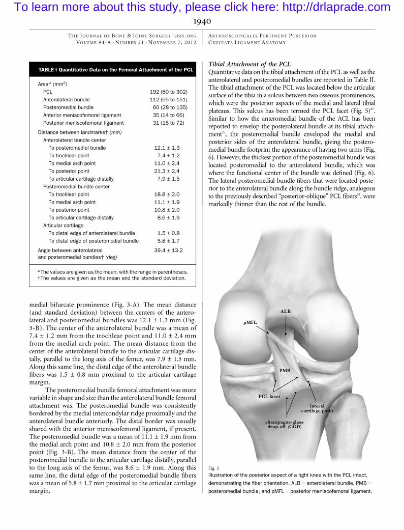
Tibial Attachment of the PCLQuantitative data on the tibial attachment of the PCL as well as theanterolateral and posteromedial bundles are reported in Table II.The tibial attachment of the PCL was located below the articularsurface of the tibia in a sulcus between two osseous prominences,which were the posterior aspects of the medial and lateral tibialplateaus. This sulcus has been termed the PCL facet (Fig. 5)27.Similar to how the anteromedial bundle of the ACL has beenreported to envelop the posterolateral bundle at its tibial attach-ment25, the posteromedial bundle enveloped the medial andposterior sides of the anterolateral bundle, giving the postero-medial bundle footprint the appearance of having two arms (Fig.6). However, the thickest portion of the posteromedial bundle waslocated posteromedial to the anterolateral bundle, which waswhere the functional center of the bundle was defined (Fig. 6).The lateral posteromedial bundle fibers that were located poste-rior to the anterolateral bundle along the bundle ridge, analogousto the previously described ‘‘posterior-oblique’’ PCL fibers28, weremarkedly thinner than the rest of the bundle.
Fig. 5
Illustration of the posterior aspect of a right knee with the PCL intact,demonstrating the fiber orientation. ALB = anterolateral bundle, PMB =
posteromedial bundle, and pMFL = posterior meniscofemoral ligament.
TABLE I Quantitative Data on the Femoral Attachment of the PCL
Area* (mm2)PCL 192 (80 to 302)Anterolateral bundle 112 (55 to 151)Posteromedial bundle 60 (28 to 135)Anterior meniscofemoral ligament 35 (14 to 66)Posterior meniscofemoral ligament 31 (15 to 72)
Distance between landmarks† (mm)Anterolateral bundle center
To posteromedial bundle 12.1 ± 1.3To trochlear point 7.4 ± 1.2To medial arch point 11.0 ± 2.4To posterior point 21.3 ± 2.4To articular cartilage distally 7.9 ± 1.5
Posteromedial bundle centerTo trochlear point 18.8 ± 2.0To medial arch point 11.1 ± 1.9To posterior point 10.8 ± 2.0To articular cartilage distally 8.6 ± 1.9
Articular cartilageTo distal edge of anterolateral bundle 1.5 ± 0.8To distal edge of posteromedial bundle 5.8 ± 1.7
Angle between anterolateraland posteromedial bundles† (deg)
39.4 ± 13.2
*The values are given as the mean, with the range in parentheses.†The values are given as the mean and the standard deviation.
1940
THE JOURNAL OF BONE & JOINT SURGERY d J B J S .ORG
VOLUME 94-A d NUMBER 21 d NOVEMBER 7, 2012ARTHROSCOP ICALLY PERT INENT POSTER IOR
CRUCIATE LIGAMENT ANATOMY
To learn more about this study, please click here: http://drlaprade.com

When viewed from an arthroscopic perspective, the PCLtibial footprint was divided into two planes by a horizontal ridgeof bone. This ridge was termed the ‘‘bundle ridge’’ becauseit occurred along the separation between the anterolateral andposteromedial bundles (Fig. 7-A). At the lateral limit of thebundle ridge, the articular cartilage of the lateral tibial plateauexhibited an abrupt distolateral turn, termed the ‘‘lateral car-tilage point’’ (Figs. 5 through 8). The lateral cartilage point wasreadily visible arthroscopically (Figs. 7-A, 7-B, and 8). Thebundle ridge extended medially to the medial limit of the
posterior border of the anterolateral bundle and did not extendthrough the attachment of the medial fibers of the postero-medial bundle. The shiny white fibers of the posterior horn ofthe medial meniscus were positioned anteromedial to the an-terolateral bundle (Figs. 6 and 7-A). These transversely ori-ented fibers, located distal to the posterior root attachmentfibers of the posterior horn of the medial meniscus, were one ofthe most notable landmarks in the PCL facet because of theirbrilliant-white arthroscopic appearance (Fig. 8). The disto-lateral corner of these fibers was termed the ‘‘shiny white fiberpoint.’’ The medial side of the PCLwas bordered by the osseouswall descending distal to the medial tibial plateau articularsurface. At the distal base of this wall, the medial edge of theposteromedial bundle footprint consistently coincided with agroove oriented in the anteroposterior direction, termed the‘‘medial groove’’ (Fig. 7-A). The distal border of the PCL isoften described relative to the prominent ridge at the mostposterior aspect of the tibial plateau27. This ridge has beentermed the ‘‘champagne-glass drop-off ’’ on the basis of itslateral radiographic and anatomic appearance27. This ridgecoincides with the inferior attachment of the joint capsule,below which the popliteus muscle fibers can be found ar-throscopically when the capsule is punctured (Fig. 8). Thechampagne-glass drop-off was a useful arthroscopic landmarkbecause it consistently marked the distal border of the pos-teromedial bundle of the PCL.
The center of the overall PCL tibial attachment was amean of 7.8 ± 1.4 mm from the shiny white fiber point, 9.8 ±1.6 mm from the lateral cartilage point, 5.0 ± 0.9 mm from themedial groove, and 1.3 ± 0.5 mm proximal to the bundle ridge(Fig. 7-B). The mean distance between the centers of the an-terolateral and posteromedial bundles was 8.9 ± 1.2 mm. The
Fig. 6
Posterosuperior photograph of the tibial attachment of the PCL in a rightknee. ALB = anterolateral bundle, and PMB = posteromedial bundle.
Fig. 7-A Fig. 7-B
Fig. 7-A Illustration of the arthroscopic view of the tibial attachment of the PCL of a right knee, demonstrating the pertinent landmarks. ALB = anterolateralbundle, PMB=posteromedial bundle, andm=muscle.Fig. 7-B Illustration showing thequantitativemeasurements for the tibial attachment of thePCL. Thevalues are reported in millimeters.
1941
THE JOURNAL OF BONE & JOINT SURGERY d J B J S .ORG
VOLUME 94-A d NUMBER 21 d NOVEMBER 7, 2012ARTHROSCOP ICALLY PERT INENT POSTER IOR
CRUCIATE LIGAMENT ANATOMY
To learn more about this study, please click here: http://drlaprade.com

center of the anterolateral bundle was a mean of 6.1 ± 1.0 mmfrom the shiny white fiber point, 8.6 ± 1.6 mm from the lateralcartilage point, 7.2 ± 1.1 mm from themedial groove, and 4.9 ±0.9 mm from the bundle ridge. The center of the posteromedialbundle was a mean of 11.1 ± 1.8 mm from the shiny white fiberpoint, 12.6 ± 1.7 mm from the lateral cartilage point, and 3.1 ±1.1 cm lateral to the medial groove.
Meniscofemoral LigamentsAt least one meniscofemoral ligament was present in nineteen(95%) of the twenty knees. The anterior meniscofemoral lig-ament was present in fifteen (75%) of the twenty knees, and theposterior meniscofemoral ligament was present in sixteen(80%) of the twenty knees. Both meniscofemoral ligamentswere present in twelve (60%) of the twenty knees. The mostcommon arrangement of the femoral footprints of the me-niscofemoral ligaments, found in ten of the twelve of kneeswith both meniscofemoral ligaments, was with the posteriormeniscofemoral ligament, posteromedial bundle, and anteriormeniscofemoral ligament aligned parallel to each other, prox-
imally to distally (Figs. 3-A and 4). The posterior meniscofem-oral ligaments of all specimens were located directly proximal tothe medial intercondylar ridge, proximal to the posteromedialbundle. In twelve of fifteen specimens, the anterior menisco-femoral ligament was distal to the posteromedial bundle. Theanterior meniscofemoral ligament was located distal to theanterolateral bundle fibers in only three of fifteen specimens. Inthree of the five specimens in which the anterior meniscofem-oral ligament was absent, the posteromedial bundle footprint ex-tended distally to the articular cartilage margin.
Validation AnalysisThe error of the electromagnetic tracking system was calcu-lated to be 0.64% and 0.51% for the measurement of the fixedlinear distance and the known circular area, respectively. Theinterobserver ICC for the repeated measurements on fiveof the cadaveric specimens by the two separate observers was0.995.
Discussion
Wefound that the locations of the PCL attachments on thefemur and tibia could be accurately described relative to
arthroscopically pertinent landmarks surrounding the attach-ment sites. The center of the femoral anterolateral bundletunnel should be triangulated on the basis of the trochlearpoint, the medial arch point, and the medial bifurcate promi-nence, with the distal edge placed adjacent to the articularcartilage. The femoral posteromedial bundle tunnel should beplaced equidistant from the posterior point and themedial archpoint and placed just distal to the medial intercondylar ridge,with the center an average of 8.6 mm proximal to the articularcartilage margin. The center of a single PCL tunnel on the tibiashould be placed just anterosuperior to the bundle ridge, on themedial side of the PCL facet and 9.8 mm from the lateralcartilage point and 5.0 mm from the medial groove.
Fig. 8
Arthroscopic view of the tibial attachment of the PCL in a right knee,demonstrating the shiny white fibers, bundle ridge, lateral cartilage point,medial groove, and popliteus muscle fibers. m = muscle.
TABLE II Quantitative Data on the Tibial Attachment of the PCL
Area* (mm2)PCL facet 281 (159 to 398)PCL 219 (108 to 351)Anterolateral bundle 88 (43 to 122)Posteromedial bundle 105 (45 to 226)
Distance between landmarks† (mm)PCL center
To shiny white fiber point 7.8 ± 1.4To lateral cartilage point 9.8 ± 1.6Horizontally to medial groove 5.0 ± 0.9To champagne-glass drop-off 7.4 ± 1.2Inferiorly to bundle ridge 1.3 ± 0.5
Anterolateral bundle centerTo posteromedial bundle 8.9 ± 1.2To shiny white fiber point 6.1 ± 1.0To lateral cartilage point 8.6 ± 1.6Horizontally to medial groove 7.2 ± 1.1To champagne-glass drop-off 10.7 ± 2.0Inferiorly to bundle ridge 4.9 ± 0.9
Posteromedial bundle centerTo shiny white fiber point 11.1 ± 1.8To lateral cartilage point 12.6 ± 1.7Horizontally to medial groove 3.1 ± 1.1To champagne-glass drop-off 4.4 ± 0.8
Angle† (deg)Between anterolateraland posteromedial bundles
31.5 ± 11.0
Between tibial plateau and PCL facet 44.9 ± 7.0
*The values are given as the mean, with the range in parentheses.†The values are given as the mean and the standard deviation.
1942
THE JOURNAL OF BONE & JOINT SURGERY d J B J S .ORG
VOLUME 94-A d NUMBER 21 d NOVEMBER 7, 2012ARTHROSCOP ICALLY PERT INENT POSTER IOR
CRUCIATE LIGAMENT ANATOMY
To learn more about this study, please click here: http://drlaprade.com

Previous measurements of the femoral attachment of thePCL have often been reported on the basis of lateral views withthe lateral femoral condyle removed26,29-32. However, thesemeasurements have limited arthroscopic applicability becausethey do not adequately illustrate the substantial attachment ofthe anterolateral bundle on the roof of the intercondylar notch.The anatomic descriptions in the present study are reportedfrom an arthroscopic perspective. The positions of the antero-lateral and posteromedial bundles have been previously reportedwith respect to an arthroscopic view of the knee33. However, wefound the center of the anterolateral bundle to be more distal,located at a mean of 7.9 mm proximal to the cartilage edge,rather than the 13 mm previously reported33. Furthermore, ourfinding that the center of the anterolateral bundle was 7.9 mmproximal to the articular cartilage is consistent with other an-atomic studies26,29,31,32. Likewise, our finding that the center ofthe posteromedial bundle was 8.6 mm from the cartilage edgeis also consistent with previous studies26,32,33. Measurementsbetween the distal edge of the PCL fibers and the articularcartilage have been previously reported parallel to the notchroof29. We report the distance between the distal edge of thePCL fibers and the articular cartilage parallel to the long axis ofthe femur because it is more arthroscopically applicable. Wefound that the anterolateral bundle fibers were only 1.5 mmproximal to the cartilage margin. Because it has been previouslyreported that distal femoral tunnel placement results in im-proved stability against posterior translation than moreproximal tunnel placement does17,23 and also that errors intunnel placement in the proximal-distal direction affectknee stability greater than in other directions23, we recom-mend that the distal edge of an anterolateral bundle re-construction tunnel be placed adjacent to the cartilage as thegraft fibers will be displaced away from the cartilage withgraft tensioning. An oblique tunnel orientation, rather thana horizontal tunnel orientation, is necessary to avoid vio-lating the subchondral bone, which could lead to osteone-crosis or an insufficiency fracture. Unlike the anterolateralbundle femoral attachment, which was adjacent to the ar-ticular cartilage margin, the anterior margin of the postero-medial bundle fibers was located 5.8 mm proximal to thecartilage margin. Thus, a femoral reconstruction tunnel of theposteromedial bundle should be placed more proximal fromthe articular cartilage margin to replicate its anatomic position.
The medial intercondylar ridge and the medial bifurcateridge are recently reported structures related to the positions ofthe anterolateral and posteromedial bundles on the femur26.Wefound that the medial intercondylar ridge was located on themedial wall of the notch, extending anteriorly from the pos-terior point, and that it consistently marked the proximalborders of the posteromedial and anterolateral bundles. As themedial bifurcate ridge was present in only 15% of specimens,its use as an arthroscopic landmark may be limited. However,as the medial bifurcate prominence was found in all specimens,this landmark should be sought to identify the posteroproximalend of the border between the anterolateral and posteromedialbundles.
Quantitative results are not reported in relation to anestimated center of the PCL on the femur because the attach-ment was so broad that center localization was not consistentlypractical. Furthermore, we do not believe that it would befeasible to place one reconstruction tunnel centered on thePCL attachment on the femur without excluding a substantialportion of the anterolateral bundle fibers or potentially dam-aging the meniscofemoral ligaments positioned in close prox-imity to the posteromedial bundle. With an average distance of12.1 mm between the anterolateral and posteromedial bundles,utilization of an 11-mm-diameter anterolateral bundle tunneland a 7-mm posteromedial bundle reconstruction tunnel, asreported clinically27, would still allow for a 3-mm bone bridgebetween the two femoral reconstruction tunnels. As single-bundle reconstructions typically aim to reconstruct the func-tionally dominant anterolateral bundle, our results for thefemoral location of the anterolateral bundle can also be used toguide tunnel placement in single-bundle PCL reconstructions.
The tibial attachment of the PCL was substantially morecompact than the femoral attachment.While the tibial attachmentof the PCL is often described relative to the prominent ridge at themost posterior aspect of the tibial plateau, termed the champagne-glass drop-off 27, few other landmarks have been described34-37. A‘‘shelf ’’ positioned through the PCL facet37 as well as a change inslope at the area of the bundle ridge have been previously re-ported35. These descriptions appear to be consistent with thestructure herein described as the bundle ridge. We further de-scribe the nature of this structure in relation to the attachment ofthe PCL in order to augment its use as a landmark for PCLreconstruction surgery. While the shiny white fibers of the pos-terior horn of the medial meniscus have been previously recog-nized to occur anteromedial to the anterolateral bundle37, we areunaware of any previous reports quantifyingmeasurements of thisstructure. A previously described depression at the superomedialcorner of the PCL attachment on the tibia34 may correlate with theanterior aspect of the medial groove that we have described.
Others have reported difficulty in identifying the tibialattachments of the anterolateral and posteromedial bundlesbecause of their close proximity at one insertion area38. We didnot find the anterolateral and posteromedial bundles to bereadily separable; however, careful dissection along interfas-cicular connective tissue after the initial separation had beenmade revealed the consistent shape of the anterolateral bundleand posteromedial bundle tibial footprints as reported in thepresent study. We found the mean distance between the centersof the anterolateral and posteromedial bundles to be only 8.9mm. The closer proximity of the anterolateral and postero-medial bundle attachments on the tibia would make it prudentto consider one tibial tunnel for both single and double-bundlePCL reconstructions. In addition, the two-armed shape of theposteromedial bundle could not be reproduced with one roundtunnel. As the functional center of the posteromedial bundlefibers was located directly posteromedial to the anterolateralbundle, a well-placed single tibial reconstruction tunnel couldencompass this fiber region. Also, as the bundle ridge was only1.3 mm distal to the overall PCL center, it would be justified to
1943
THE JOURNAL OF BONE & JOINT SURGERY d J B J S .ORG
VOLUME 94-A d NUMBER 21 d NOVEMBER 7, 2012ARTHROSCOP ICALLY PERT INENT POSTER IOR
CRUCIATE LIGAMENT ANATOMY
To learn more about this study, please click here: http://drlaprade.com
identify the bundle ridge with a tibial drill guide and then targetthe guide pin to protrude just proximal to this ridge. Likewise, aPCL inlay graft should also be centered at this site.
We recognize that the present study has some limitations.A limited number of specimens were used, which may not havedemonstrated the full spectrum of anatomic variation. How-ever, to our knowledge, this study is one of the largest anatomicstudies on the PCL, and the results were consistent betweenspecimens. In addition, we did not specifically vary the order ofmeasurements of the individual PCL bundles, which may haveintroduced some order bias. Instead, we measured each indi-vidual PCL bundle immediately after it was sharply dissectedfrom its attachment location.
The findings of the present study provide qualitativedescriptions and quantitative measurements of arthroscopiclandmarks for the femoral and tibial attachments of the PCL.The results presented in this study can assist with more ana-tomic tunnel placement in single and double-bundle PCL re-constructions. The results also suggest that two reconstructiontunnels are needed to reconstruct the broad PCL femoral at-tachment, whereas one reconstruction tunnel should be in-vestigated further for the compact tibial attachment. n
NOTE: The authors thank Steven Hobbs, PhD, from the Department of Integrative Physiology at theUniversity of Colorado at Boulder, for allowing the evaluation of their bone box specimens. Theyalso recognize Kyle Jansson for his mechanical expertise in developing the fixtures and for the highmeasurement accuracy in this study, and Daniel Westcott, MSc, for his technical assistance withthis project.
Colin J. Anderson, MDConnor G. Ziegler, MDCoen A. Wijdicks, PhDDepartment of BioMedical Engineering,Steadman Philippon Research Institute,181 West Meadow Drive, Suite 1000, Vail, Colorado 81657
Lars Engebretsen, MD, PhDDepartment of Orthopaedic Surgery,Oslo University Hospital and Faculty of Medicine,Kirkeveien 166, 0407 Oslo, Norway
Robert F. LaPrade, MD, PhDThe Steadman Clinic, 181 West Meadow Drive,Suite 400, Vail, Colorado 81657.E-mail address: [email protected]
References
1. Fanelli GC, Beck JD, Edson CJ. Current concepts review: the posterior cruciateligament. J Knee Surg. 2010 Jun;23(2):61-72.2. Boynton MD, Tietjens BR. Long-term followup of the untreated isolated posteriorcruciate ligament-deficient knee. Am J Sports Med. 1996 May-Jun;24(3):306-10.3. Covey CD, Sapega AA. Injuries of the posterior cruciate ligament. J Bone JointSurg Am. 1993 Sep;75(9):1376-86.4. Dejour H, Walch G, Peyrot J, Eberhard P. [The natural history of rupture of theposterior cruciate ligament]. Rev Chir Orthop Reparatrice ApparMot. 1988;74(1):35-43. French.5. Keller PM, Shelbourne KD, McCarroll JR, Rettig AC. Nonoperatively treated iso-lated posterior cruciate ligament injuries. Am J Sports Med. 1993 Jan-Feb;21(1):132-6.6. Kim SJ, Kim SH, Kim SG, Kung YP. Comparison of the clinical results of threeposterior cruciate ligament reconstruction techniques: surgical technique. J BoneJoint Surg Am. 2010 Sep;92 Suppl 1 Pt 2:145-57.7. Gill TJ, Van de Velde SK, Wing DW, Oh LS, Hosseini A, Li G. Tibiofemoral andpatellofemoral kinematics after reconstruction of an isolated posterior cruciate lig-ament injury: in vivo analysis during lunge. Am J Sports Med. 2009Dec;37(12):2377-85. Epub 2009 Sep 2.8. Harner CD, Janaushek MA, Kanamori A, Yagi M, Vogrin TM, Woo SL. Biome-chanical analysis of a double-bundle posterior cruciate ligament reconstruction. Am JSports Med. 2000 Mar-Apr;28(2):144-51.9. Race A, Amis AA. PCL reconstruction. In vitro biomechanical comparison of‘isometric’ versus single and double-bundled ‘anatomic’ grafts. J Bone Joint Surg Br.1998 Jan;80(1):173-9.10. Wang CJ, Weng LH, Hsu CC, Chan YS. Arthroscopic single- versus double-bundleposterior cruciate ligament reconstructions using hamstring autograft. Injury. 2004Dec;35(12):1293-9.11. Houe T, Jørgensen U. Arthroscopic posterior cruciate ligament reconstruction:one- vs. two-tunnel technique. Scand J Med Sci Sports. 2004 Apr;14(2):107-11.12. Hatayama K, Higuchi H, Kimura M, Kobayashi Y, Asagumo H, Takagishi K. Acomparison of arthroscopic single- and double-bundle posterior cruciate ligamentreconstruction: review of 20 cases. Am J Orthop (Belle Mead NJ). 2006 Dec;35(12):568-71.13. Kohen RB, Sekiya JK. Single-bundle versus double-bundle posterior cruciate lig-ament reconstruction. Arthroscopy. 2009 Dec;25(12):1470-7. Epub 2009 Jan 24.14. Kim YM, Lee CA, Matava MJ. Clinical results of arthroscopic single-bundletranstibial posterior cruciate ligament reconstruction: a systematic review. Am JSports Med. 2011 Feb;39(2):425-34. Epub 2010 Aug 11.15. Greiner P, Magnussen RA, Lustig S, Demey G, Neyret P, Servien E. Computedtomography evaluation of the femoral and tibial attachments of the posterior cruciate
ligament in vitro. Knee Surg Sports Traumatol Arthrosc. 2011 Nov;19(11):1876-83.Epub 2011 Apr 9.16. Apsingi S, Bull AM, Deehan DJ, Amis AA. Review: femoral tunnel placement forPCL reconstruction in relation to the PCL fibre bundle attachments. Knee Surg SportsTraumatol Arthrosc. 2009 Jun;17(6):652-9. Epub 2009 Mar 14.17. Galloway MT, Grood ES, Mehalik JN, Levy M, Saddler SC, Noyes FR. Posteriorcruciate ligament reconstruction. An in vitro study of femoral and tibial graft place-ment. Am J Sports Med. 1996 Jul-Aug;24(4):437-45.18. Mannor DA, Shearn JT, Grood ES, Noyes FR, Levy MS. Two-bundle posteriorcruciate ligament reconstruction. An in vitro analysis of graft placement and tension.Am J Sports Med. 2000 Nov-Dec;28(6):833-45.19. McGuire DA, Hendricks SD. Comparison of anatomic versus nonanatomicplacement of femoral tunnels in Achilles double-bundle posterior cruciate ligamentreconstruction. Arthroscopy. 2010 May;26(5):658-66. Epub 2010 Mar 3.20. Petersen W, Tretow H, Weimann A, Herbort M, Fu FH, Raschke M, Zantop T.Biomechanical evaluation of two techniques for double-bundle anterior cruciate lig-ament reconstruction: one tibial tunnel versus two tibial tunnels. Am J Sports Med.2007 Feb;35(2):228-34. Epub 2006 Nov 12.21. Zantop T, Wellmann M, Fu FH, Petersen W. Tunnel positioning of anteromedialand posterolateral bundles in anatomic anterior cruciate ligament reconstruction:anatomic and radiographic findings. Am J Sports Med. 2008 Jan;36(1):65-72. Epub2007 Oct 11.22. Musahl V, Ayeni OR, Citak M, Irrgang JJ, Pearle AD, Wickiewicz TL. The influenceof bony morphology on the magnitude of the pivot shift. Knee Surg Sports TraumatolArthrosc. 2010 Sep;18(9):1232-8. Epub 2010 Apr 8.23. Burns WC 2nd, Draganich LF, Pyevich M, Reider B. The effect of femoraltunnel position and graft tensioning technique on posterior laxity of the posteriorcruciate ligament-reconstructed knee. Am J Sports Med. 1995 Jul-Aug;23(4):424-30.24. Azzam MG, Lenarz CJ, Farrow LD, Israel HA, Kieffer DA, Kaar SG. Inter- andintraobserver reliability of the clock face representation as used to describe thefemoral intercondylar notch. Knee Surg Sports Traumatol Arthrosc. 2011 Aug;19(8):1265-70. Epub 2011 Jan 22.25. Ziegler CG, Pietrini SD, Westerhaus BD, Anderson CJ, Wijdicks CA, Johansen S,Engebretsen L, LaPrade RF. Arthroscopically pertinent landmarks for tunnel posi-tioning in single-bundle and double-bundle anterior cruciate ligament reconstruc-tions. Am J Sports Med. 2011 Apr;39(4):743-52. Epub 2010 Dec 20.26. Lopes OV Jr, Ferretti M, Shen W, Ekdahl M, Smolinski P, Fu FH. Topography ofthe femoral attachment of the posterior cruciate ligament. J Bone Joint Surg Am.2008 Feb;90(2):249-55.27. Spiridonov SI, Slinkard NJ, LaPrade RF. Isolated and combined grade-III pos-terior cruciate ligament tears treated with double-bundle reconstruction with use of
1944
THE JOURNAL OF BONE & JOINT SURGERY d J B J S .ORG
VOLUME 94-A d NUMBER 21 d NOVEMBER 7, 2012ARTHROSCOP ICALLY PERT INENT POSTER IOR
CRUCIATE LIGAMENT ANATOMY
To learn more about this study, please click here: http://drlaprade.com

endoscopically placed femoral tunnels and grafts: operative technique and clinicaloutcomes. J Bone Joint Surg Am. 2011 Oct 5;93(19):1773-80.28. Makris CA, Georgoulis AD, Papageorgiou CD, Moebius UG, Soucacos PN.Posterior cruciate ligament architecture: evaluation under microsurgical dissection.Arthroscopy. 2000 Sep;16(6):627-32.29. Takahashi M, Matsubara T, Doi M, Suzuki D, Nagano A. Anatomical study of thefemoral and tibial insertions of the anterolateral and posteromedial bundles of hu-man posterior cruciate ligament. Knee Surg Sports Traumatol Arthrosc. 2006Nov;14(11):1055-9. Epub 2006 Aug 4.30. Saddler SC, Noyes FR, Grood ES, Knochenmuss DR, Hefzy MS. Posterior cru-ciate ligament anatomy and length-tension behavior of PCL surface fibers. Am J KneeSurg. 1996 Fall;9(4):194-9.31. Mejia EA, Noyes FR, Grood ES. Posterior cruciate ligament femoral insertion sitecharacteristics. Importance for reconstructive procedures. Am J Sports Med. 2002Sep-Oct;30(5):643-51.32. Edwards A, Bull AM, Amis AA. The attachments of the fiber bundles of the pos-terior cruciate ligament: an anatomic study. Arthroscopy. 2007 Mar;23(3):284-90.
33. Morgan CD, Kalman VR, Grawl DM. The anatomic origin of the posterior cruciateligament: where is it? Reference landmarks for PCL reconstruction. Arthroscopy.1997 Jun;13(3):325-31.34. Sheps DM, Otto D, Fernhout M. The anatomic characteristics of the tibial in-sertion of the posterior cruciate ligament. Arthroscopy. 2005 Jul;21(7):820-5.35. Tajima G, Nozaki M, Iriuchishima T, Ingham SJ, Shen W, Smolinski P, Fu FH.Morphology of the tibial insertion of the posterior cruciate ligament. J Bone Joint SurgAm. 2009 Apr;91(4):859-66.36. Ramos LA, de Carvalho RT, CohenM, Abdalla RJ. Anatomic relation between theposterior cruciate ligament and the joint capsule. Arthroscopy. 2008 Dec;24(12):1367-72.37. Amis AA, Gupte CM, Bull AM, Edwards A. Anatomy of the posterior cruciateligament and the meniscofemoral ligaments. Knee Surg Sports Traumatol Arthrosc.2006 Mar;14(3):257-63. Epub 2005 Oct 14.38. Lorenz S, Elser F, Brucker PU, Obst T, Imhoff AB. Radiological evaluation of theanterolateral and posteromedial bundle insertion sites of the posterior cruciate lig-ament. Knee Surg Sports Traumatol Arthrosc. 2009 Jun;17(6):683-90. Epub 2009Mar 24.
1945
THE JOURNAL OF BONE & JOINT SURGERY d J B J S .ORG
VOLUME 94-A d NUMBER 21 d NOVEMBER 7, 2012ARTHROSCOP ICALLY PERT INENT POSTER IOR
CRUCIATE LIGAMENT ANATOMY
To learn more about this study, please click here: http://drlaprade.com