Embed Size (px)
Citation preview

ARSENIC IN DRINKING-WATER
pp33-40.qxd 11/10/2004 10:08 Page 39

pp33-40.qxd 11/10/2004 10:08 Page 40

ARSENIC IN DRINKING-WATER
1. Exposure Data
1.1 Chemical and physical data
Arsenic is the 20th most common element in the earth’s crust, and is associated withigneous and sedimentary rocks, particularly sulfidic ores. Arsenic compounds are found inrock, soil, water and air as well as in plant and animal tissues. Although elemental arsenicis not soluble in water, arsenic salts exhibit a wide range of solubilities depending on pHand the ionic environment. Arsenic can exist in four valency states: –3, 0, +3 and +5. Underreducing conditions, the +3 valency state as arsenite (AsIII) is the dominant form; the +5valency state as arsenate (AsV) is generally the more stable form in oxygenized environ-ments (Boyle & Jonasson, 1973; National Research Council, 1999; O’Neil, 2001; WHO,2001).
Arsenic species identified in water are listed in Table 1. Inorganic AsIII and AsV are themajor arsenic species in natural water, whereas minor amounts of monomethylarsonic acid(MMA) and dimethylarsinic acid (DMA) can also be present. The trivalent mono-methylated (MMAIII) and dimethylated (DMAIII) arsenic species have been detected in lakewater (Hasegawa et al., 1994, 1999). The presence of these trivalent methylated arsenicalspecies is possibly underestimated since only few water analyses include a solvent sepa-ration step required to identify these trivalent species independently from their respective
–41–
Table 1. Some arsenic species identified in watera
Name Abbreviation Chemical formula CAS No. pKa
Arsenous acid (arsenite) AsIII As(OH)3 13464-58-9 9.23, 12.13, 13.4 Arsenic acid (arsenate) AsV AsO(OH)3 7778-39-4 2.22, 6.98, 11.53 Monomethylarsonic acid MMAV CH3AsO(OH)2 124-58-3 4.1, 8.7 Monomethylarsonous acid MMAIII CH3As(OH)2 25400-23-1 Dimethylarsinic acid DMAV (CH3)2AsO(OH) 75-60-5 6.2 Dimethylarsinous acid DMAIII (CH3)2AsOH 55094-22-9 Trimethylarsine oxide TMAO (CH3)3AsO 4964-14-1
a From National Research Council (1999); Francesconi & Kuehnelt (2002); Le (2002)
pp41-96.qxd 11/10/2004 10:19 Page 41

pentavalent analogues. Other unidentified arsenic species have also been reported inseawater and fresh water, and could represent up to 20% of the total arsenic (Francesconi& Kuehnelt, 2002; Le, 2002).
1.2 Analysis
Studies of human exposure to arsenic and its consequences for human health requiretwo different kinds of arsenic analyses depending on whether quantitative or qualitativeresults are required. Several methods have been developed and improved for the measure-ment of total arsenic, and have been widely used for the evaluation of drinking-watercontamination and the resulting concentrations of arsenic in humans. On the other hand,analytical methods allowing arsenic speciation have gained increasing interest. Theenvironmental fate and behaviour, bioavailability and toxicity of arsenic vary dramaticallywith the chemical form (species) in which it exists, the inorganic AsIII and AsV being, forexample, far more toxic than MMA and DMA. Thus selective methods that determine therelative concentration of the different arsenic species in drinking-water are required whenmore precise assessments of their impact on human health are needed.
Analytical methods for arsenic have been reviewed (National Research Council,1999; WHO, 2001; Goessler & Kuehnelt, 2002).
The most commonly used methods for the analysis of arsenic and arsenic compoundsin water and biological samples are described below, and their characteristics aresummarized in Table 2.
1.2.1 Preservation of samples
Assessment of human exposure to arsenic through drinking-water relies on the analysisof arsenic in water and in biological samples. Biological markers may more accuratelyreflect total dose of exposure in populations exposed to low, but potentially carcinogeniclevels of arsenic in drinking-water. Many tissues contain arsenic following exposure to theelement, but not all represent useful biomarkers. For example, arsenic is removed fromblood within a few hours and excreted through the kidneys and urine within a few days.Determination of arsenic in urine is commonly used as a measure of recent exposure. Hairand nails have been shown to provide reliable biomarkers for long-term chronic exposure toarsenic in humans (Karagas et al., 1996, 2000). However, nails are preferred to hair sincetheir contamination with arsenic from the air is negligible, whereas hair can adsorb 9–16%exogenous inorganic arsenic (Mandal et al., 2003). Karagas et al. (2001a) found thatmeasurements of arsenic in both toenails and water were reproducible over a 3–5-yearperiod.
Depending on the sample studied and the type of analysis to be performed, particularcaution must be taken to overcome problems related to sample contamination and stabilityof the arsenic species. For determining total element concentrations, the main consi-derations for sample collection and storage are to prevent contamination and to minimize
IARC MONOGRAPHS VOLUME 8442
pp41-96.qxd 11/10/2004 10:19 Page 42

ARSEN
IC IN D
RINK
ING
-WATER
43
Table 2. Most commonly used analytical methods for arsenic and arsenic compounds in water and biological samples
Methodology Sample analysed
Detection Detection limit Advantages Disadvantages References
Colorimetric/spectro-photometric methods
Water Urine, serum Hair, nails
Total arsenic ∼ 40 µg/L Low cost, very simple, uses a simple spectrophotometer
Kingsley & Schaffert (1951); Vogel et al. (1954); Dahr et al. (1997); Pillai et al. (2000); Goessler & Kuehnelt (2002)
Inductively coupled plasma–atomic emission spectrometry (ICP–AES)
Water Total arsenic ∼ 30 µg/L SM 3120 (1999); Environmental Protection Agency (1994a); Goessler & Kuehnelt (2002)
Inductively coupled plasma–mass spectrometry (ICP–MS)
Water Nails
Total arsenic 0.1 µg/L Analytical method approved by US EPA
Spectral and matrix inter-ference
Environmental Protection Agency (1994b); Chen et al., 1999; Goessler & Kuehnelt (2002)
High resolution (HR)–ICP–MS
Water Urine Nails
Total arsenic 0.01 µg/L Solves spectral interferences in samples with complex matrices
Gallagher et al. (2001); Karagas et al. (2001, 2002)
Instrumental neutron activation analysis (INAA)
Hair, nails Tissues
Total arsenic ∼ 0.001 µg/g Reference method for detection of arsenic
Garland et al. (1993); Nichols et al. (1993); Pan et al. (1993); Pazirandeh et al. (1998); Karagas et al. (2001)
Electrothermal atomization laser–excited atomic fluorescence spectrometry (ETA–LEAFS)
Serum Total arsenic 0.065 µg/L Requires only minimal sample volume, sample pretreatment and measurement time
Swart & Simeonsson (1999)
Graphite furnace–atomic absorption spectrometry (GF–AAS)
Water, urine Hair, nails, tissues
Total arsenic ∼ 0.025 µg/g Analytical method approved by US EPA
Pre-atomization losses, requires the use of matrix modifyers
Agahian et al. (1990); SM 3113 (1999); WHO (2001)
pp41-96.qxd 11/10/2004 10:19 Page 43

IARC M
ON
OG
RAPH
S VO
LUM
E 8444
Table 2 (contd)
Methodology Sample analysed
Detection Detection limit Advantages Disadvantages References
Hydride generation–atomic absorption spectrometry (HG–AAS)
Water Urine Hair, nails
Total arsenic and arsenic speciation
0.6–6 µg/L Analytical method approved by US EPA
Braman & Foreback (1973); Crecelius (1978); Le et al. (1994a,b); Chatterjee et al. (1995); Lin et al. (1998); Ng et al. (1998); Wyatt et al. (1998a,b); Shraim et al. (1999, 2000); SM 3114 (1999)
Hydride generation–quartz furnace–atomic absorption spectro-metry (HG–QF–AAS)
Water Tissues
Total arsenic and arsenic speciation
0.003–0.015 µg/L
Inexpensive Environmental Protection Agency (1996c)
High-performance liquid chromatography (HPLC)–HG–AAS
Urine Total arsenic and arsenic speciation
1–47 µg/L Lamble & Hill (1996); Kurttio et al. (1998)
HPLC or solid-phase cartridge separation combined with hydride generation–atomic fluorescence spectrometry (HPLC–HG–AFS)
Water, urine Arsenic speciation
0.05–0.8 µg/L Rapid, inexpensive No need for sample pretreatment
Le & Ma (1997); Aposhian et al. (2000); Le et al. (2000a,b); Gong et al. (2001); Yalcin & Le (2001)
HPLC–ICP–MS Water Water, urine Hair, nails
Total arsenic 0.01 µg/L 0.14–0.33 µg/L
No need for sample pretreatment
Expensive and often time-consuming Spectral and matrix inter-ference
Shibata & Morita (1989); Londesborough et al. (1999); Chatterjee et al. (2000); Mandal et al. (2001); Shraim et al. (2001); Karagas et al. (2002); Mandal et al. (2003)
pp41-96.qxd 11/10/2004 10:19 Page 44

loss of trace amounts of analytes. High-density polyethylene containers are usuallypreferred to glass containers because they are less adsorptive for arsenic. These are pre-cleaned with nitric acid and then rinsed with distilled water.
Groundwater sampling is carried out by allowing the well-water to flow through thepumping pipe for approximately 10 min before collection.
Traditionally, water and urine samples are acidified with sulfuric or nitric acid to reducepotential adsorption of trace elements onto the surface of the sample container and toprevent bacterial proliferation. Samples can then be kept at +4 °C or at room temperatureand preferably measured within 7 days (Lin et al., 1998; Rahman et al., 2002). Pande et al.(2001) reported, however, that all the field kits they evaluated were subject to negative inter-ference if samples were acidified with nitric acid for preservation; they showed that acidifi-cation using 5% ascorbic acid instead of nitric acid eliminates interference.
In iron-rich waters, the stability of AsIII and AsV can be affected by the formation ofiron precipitates (iron oxides and/or hydroxides designated by ‘FeOOH’). These precipi-tates can form during transport to the laboratory for analysis of arsenic. Studies of labo-ratory reagent water containing both AsIII and FeIII indicated that, within 18 h at room tem-perature, the resulting FeOOH precipitates contained a mixture of AsIII and AsV with nearquantitative removal of aqueous arsenic. Addition of a chelating agent such as ethylene-diamine tetraacetic acid (EDTA), by sequestering FeIII, inhibits the formation of FeAsOHprecipitates and preserves the stability of arsenic species in iron-rich waters for more than10 days (Gallagher et al., 2001).
Reliable information from speciation analysis requires that the concentration of indi-vidual species of the element be unchanged by handling and treatment of the sample.Although traditionally used for their preservation, acidification of samples is not suitablesince it leads to changes in arsenic speciation.
For urine specimens, low temperature (4 °C and –20 °C) conditions are required if theyare to be stored up to 2 months without substantial changes in arsenic speciation (except forMMAIII and DMAIII species). For longer storage times, the stability of arsenic species varieswith the complex matrix and pH of the urine, and accurate measurement of inorganic AsIII
and AsV separately is more difficult since AsV is rapidly reduced to AsIII. MMAV and DMAV
are more stable (for up to 4.5 months). The trivalent arsenic species, monomethylarsonousacid (MMAIII) and dimethylarsinous acid (DMAIII), suspected to be key metabolic inter-mediates in human urine, are extremely unstable. It was shown that over 90% of MMAIII
was rapidly oxidized to MMAV in urine samples when stored at +4 °C or –20 °C over a 5-month period, while DMAIII was completely oxidized to DMAV within 1 day (Gong et al.,2001). In a recent review, these authors found that the use of a complexing agent, diethyl-ammonium diethyldithiocarbamate (DDDC), improved the stability of MMAIII and DMAIII
in urine samples. In the presence of DDDC (1–10 mM), MMAIII was found to be stable for4 months at –20 °C (with a recovery of 85–95%) and DMAIII was partially preserved.Approximately 80% of DMAIII remained after 3 weeks of storage and 10–24% remainedafter 4 months (Jiang et al., 2003). The use of other additives (such as hydrochloric acid,
ARSENIC IN DRINKING-WATER 45
pp41-96.qxd 11/10/2004 10:19 Page 45

sodium azide, benzoic acid, benzyltrimethylammonium chloride and cetylpyridinium chlo-ride) has no particular benefit (Feldman et al., 1999; Chen et al., 2002).
For arsenic speciation, well-water is usually filtered at the sampling site using a 0.45 µmfilter (Lin et al., 1998).
Methods for on-site separation of AsIII and AsV species immediately after water-sample collection using solid disposable cartridges can be efficiently used for speciationof particulate and soluble arsenic. A measured volume of the sample is passed through the0.45-µm membrane filter, then serially through a connected silica-based strong anion-exchange cartridge. The filter captures particulate arsenic, while the anion-exchangecartridge retains AsV. Arsenite is not retained and is detected in the effluent. Arsenate issubsequently eluted with 1 M hydrochloric acid (HCl) from the anion-exchange cartridgeand then analysed for concentration (Le et al., 2000a).
In hair and nail samples, the arsenic species are less prone to change. For analysis oftotal arsenic, as for speciation methods, these specimens are usually prepared accordingto the International Atomic Energy Agency (IAEA) procedure (Ryabukhin, 1978).
Following extensive washing to eliminate exogenous arsenic resulting from air conta-mination, approximately 100 mg of each hair sample are usually placed in a Teflon beaker,mixed with acetone and then washed with distilled water. Nails are treated similarly tohair following brushing. Samples are weighed prior to analysis (Lin et al., 1998; Mandalet al., 2003). More stringent washing procedures have also been described for completeremoval of surface contamination, by incubating nails for 20 min in 1% Triton X100before analysis (Chen et al., 1999).
1.2.2 Analytical methods for measurement of total arsenic
Determination of total arsenic in biological samples in most cases requires the com-plete destruction of the organic matrix. During this process, all the organic arsenic com-pounds should be converted into inorganic arsenic by oxidative digestion. Acid digestion(or wet ashing) (Kingsley & Schaffert, 1951) and dry ashing (George et al., 1973) are thetwo basic methods that have been widely employed for oxidative digestion of samplesprior to analysis. A microwave-assisted digestion technique has been developed recentlyand is currently used as a rapid preparation for sample analysis (Le et al., 1994c; Goessler& Kuehnelt, 2002). For analysis of soft biological tissues using inductively coupledplasma (ICP) techniques, a simple partial digestion in a closed vessel at low temperatureand pressure is often sufficient for the sample preparation and pretreatment step (WHO,2001).
Historically, colorimetric/spectrophotometric methods have been used to determinetotal arsenic concentration. Several commercial field kits have been based on thesemethods. At present, laboratories often prefer more sensitive methods such as atomicabsorption spectrometry (AAS), neutron activation analysis (NAA), atomic emissionspectrometry (AES), mass spectrometry (MS) or atomic fluorescence spectrometry (AFS).
IARC MONOGRAPHS VOLUME 8446
pp41-96.qxd 11/10/2004 10:19 Page 46

(a) Colorimetric/spectrometric methodsThese methods take advantage of the formation of volatile arsine (AsH3) gas to sepa-
rate arsenic from other possible interference with the sample matrix. The colorimetricmethods are easy to use and inexpensive in terms of equipment and operator cost. They areuseful for the semi-quantitative determination of high concentrations of arsenic in water.
The silver diethylthiocarbamate (AgDDTC) method is the most popular spectro-photometric method for the determination of arsenic in water. The method is based on thegeneration of arsine either with zinc and hydrochloric acid or sodium borohydride inacidic solutions. The arsine gas is then flushed through a solution of diethylthiocarbamatein pyridine or pyridine/chloroform. The red-coloured complex can be measured at 520nm. Using a modification of this method, Dhar et al. (1997) reported a detection limit of40 µg/L for arsenic in water samples, with a 95% confidence.
Pillai et al. (2000) reported a new simple and reliable spectrophotometric method todetermine total arsenic in environmental and biological samples. It involves bleaching thepinkish-red dye Rhodamine-B (measured at 553 nm) by the action of iodine released fromthe reaction between potassium iodate and arsenic in a slightly acidic medium.
The classic Gutzeit test (Vogel, 1954) is derived from the historical Marsh test. It isbased on the generation of arsine (AsH3) from arsenic compounds by the addition of zincgranules to concentrated sulfuric acid. The arsine can be detected by its reaction on a stripof filter moistened with silver nitrate or mercuric chloride, which produces a grey or ayellow to reddish-brown spot, respectively.
Field test kitsThe high concentrations of arsenic currently found in groundwater in many parts of
the world pose an important challenge because of the large number of wells that must betested. This is particularly true in Bangladesh and other Asian hot spots such as Myanmar,Nepal, Cambodia, Laos, Viet Nam and India. Although less accurate than laboratory-based methods, field kits that allow on-site semi-quantitative determination of arsenicconcentrations in well-water are of vital importance, since in these countries, the currentlaboratory capacity cannot cover the high level of analytical needs. Field testing hasseveral advantages. In Bangladesh and other hot climates, attempts to keep samples coolover a long period of transport to a laboratory can be difficult. With field kits, there is noneed for transport, no storage and therefore no need for preservation, which in additionreduces the cost of analysis and the time required for the well owner to be informed. Fieldkits are also simple to use after reasonable training of technicians.
These tests, however, must be accurate and sensitive enough to assess the level ofarsenic contamination.
Much concern about the reliability of field kits recently led to careful evaluations ofcommercially available kits (Pande et al., 2001; Rahman et al., 2002a,b; EnvironmentalProtection Agency-Battelle, 2002a,b; Erickson, 2003). The original field kit widely usedin Bangladesh had a stated minimal detectable concentration of 100 µg/L, which largelyexceeded the maximum permissible arsenic concentration defined by WHO (10 µg/L) and
ARSENIC IN DRINKING-WATER 47
pp41-96.qxd 11/10/2004 10:19 Page 47

even the maximum stated by most developing countries (50 µg/L). Fortunately, the newerfield test kits are more sensitive. Evaluations of these kits are summarized in Table 3.
A modification of the Gutzeit method using mercuric bromide is the basis of mostcommercial field kits. A test strip moistened with mercuric bromide is exposed to arsinegas derived from the sample solution, to form complex salts of arsenic and mercury. Thesereactions give a yellow [H(HgBr2)As] to brown [(HgBr)3As] to black [Hg3As2] stain. Theintensity of the yellowish-brown colour developed on the test strip is proportional to thearsenic concentration in the sample. When the reaction is completed, the test strip iscompared with a colour chart provided with the kit and allows semi-quantitative deter-mination of total arsenic concentration.
More recent field kits include digital measurement of arsenic levels without depen-ding on the judgement of the technician’s eyes to detect the difference between colourshades of the coloured strip (Arsenator, PeCo test). The improvement in reading results inhigher sensitivity and reliability (Environmental Protection Agency-Battelle, 2002a,b;Durham & Kosmus, 2003).
In addition, promising biological tools (bacterial biosensors) may lead to new kits forquantitative and qualitative measurement of arsenite and arsenate in aqueous solution(Flynn et al., 2002; Stocker et al., 2003).
(b) Inductively coupled plasma–atomic emission spectrometry(ICP–AES)
ICP–AES involves the use of plasma, usually argon, at temperatures between 6000 and8000 °K as the excitation source. The analyte is introduced into the plasma as an aerosol.A typical detection limit achievable for arsenic with this technique is 30 µg/L. Because ofthe rather high detection limits, ICP–AES is not frequently used for the determination ofarsenic in biological samples (Goessler & Kuehnelt, 2002).
In August 2002, ICP–AES was withdrawn from the US Environmental ProtectionAgency (US EPA)-approved analytical methods for arsenic since this technique is inade-quate to meet the requirements of the new EPA standard for arsenic in drinking-water of10 µg/L (10 ppb), effective since February 2002 (Environmental Protection Agency, 2002).
(c) Inductively coupled plasma–mass spectrometry (ICP–MS)ICP–MS is superior to ICP–AES with respect to detection limits, multi-element capa-
bilities and wide linear dynamic range. This technique combines the ICP as the ion sourcewith a mass analyser. Quadrupole mass filters are the most common mass analyser;double-focusing magnetic/electrostatic sector instruments and time-of-flight massanalysers are also used (Goessler & Kuehnelt, 2002).
ICP–MS is classified among the US EPA-approved analytical methods for arsenic(Environmental Protection Agency, 2002), with a detection limit of 0.1 µg/L.
The sensitivity can be further improved by the use of hydride generation (HG)techniques leading to a more efficient sample introduction and to matrix removal. The useof a high-resolution mode with HG–ICP–MS allows a 10-fold decrease in the detection
IARC MONOGRAPHS VOLUME 8448
pp41-96.qxd 11/10/2004 10:19 Page 48

ARSEN
IC IN D
RINK
ING
-WATER
49
Table 3. Evaluation of some field test kits for analysing arsenic in water
Field test kits Kit capability Minimum detection limit of arsenic
Detection range Rate of false positive/false negative
Effects of inter-ferences (sodium chloride, iron, sulfate, acidity)
Occupational hazard potential (OH)
Time required per test
Evaluation reference
QuickTM (industrial test kit, Rock Hill, USA)
Semi-quantitative ∼ 5–20 µg/L 5, 10, 20, 40, 60, 100, 200, … 500 µg/L
0–4%/5–16% ND Safe < 15 min Environmental Protection Agency-Battelle (2002a)
AS75 (PeCo test kit) (Peters Engineering, Graz, Austria)
Semi-quantitative ∼ 15–50 µg/L 10, 20, 30, … 100 µg/L 2.5, 5, 10, 20, … 60 µg/L
0–3%/0% None Safe ND Environmental Protection Agency-Battelle (2002b)
AAN (Asia Arsenic Network, Japan)
Semi-quantitative ∼ 20 µg/L 20, 50, 100, 200,… 700 µg/L
19%/71% Some with sulfide
Accidental escape of arsine gas may cause OH.
15 min Pande et al. (2001); Rahman et al. (2002)
E. Merck (Germany) Qualitative for arsenic concentration > 50 µg/L
∼ 50–100 µg/L 100, 500, 1000, 1700, 3000 µg/L
21%/60% Some with sulfide
Accidental spillage of acid and escape of arsine gas may cause OH.
30 min Pande et al. (2001); Rahman et al. (2002)
NIPSOM (National Institute of Preventive and Social Medicine, Bangladesh)
Qualitative for arsenic concentration > 50 µg/L
∼ 10–20 µg/L 10, 20, 50, 100, 200, 300 … 700 µg/L
21%/33% Some with sulfide
Accidental spillage of acid and escape of arsine gas may cause OH.
5 min Pande et al. (2001); Rahman et al. (2002)
AIIH-PH (All India Institute of Hygiene and Public Health, India)
Semi-quantitative ∼ 50 µg/L
> 50 µg/L 25%/1% Sulfide interference eliminated
Accidental spillage of acid and escape of arsine gas may cause OH.
30 min Pande et al. (2001); Rahman et al. (2002)
GPL (General Pharmaceuticals Ltd, USA)
Semi-quantative ∼ 10 µg/L 10, 50, 100, 200, 400, 500 … 1500 µg/L
10%/32% ND Accidental spillage of acid and escape of arsine gas may cause OH.
20 min Rahman et al. (2002)
Aqua (Aqua Consortium, Calcutta, India)
Semi-quantitative ∼ 100 µg/L > 50 µg/L ND Sulfide interference eliminated
Accidental spillage of acid and escape of arsine gas may cause OH. Contact with HgBr2 paper affects fingers of the user.
15 min Pande et al. (2001)
pp41-96.qxd 11/10/2004 10:19 Page 49
limit (0.01 µg/L) for arsenic in water samples. HG–ICP–MS can be used for biologicalsamples such as urine and nails (Chen et al., 1999; Gallagher et al., 2001; Karagas et al.,2001a, 2002).
(d) Neutron activation analysis (NAA)Instrumental NAA is an accurate and sensitive means to measure arsenic. The method
can analyse relatively small biological samples, and has been used efficiently to measuretotal arsenic in hair, nails and other tissues, with a detection limit of approximately0.001 µg/g (Pan et al., 1993; Garland et al., 1996; Nichols et al., 1998; Pazirandeh et al.,1998; Karagas et al., 2001a).
(e) Electro-thermal atomization laser–excited atomic fluorescencespectrometry (ETA–LEAFS)
ETA–LEAFS is a highly sensitive and selective method that has been developed bythe combination of laser-excited atomic fluorescence spectrometry with electro-thermalatomization in graphite cup or tube furnaces. The technique provides excellent analyticalperformance at ultra-trace levels, with a detection limit of 0.065 µg/L for arsenic inundiluted serum. This approach allows measurements to be taken directly on the serumsamples after a simple dilution step. It also minimizes the amounts of sample required andcan provide multiple measurements when only limited amounts of sample are available(Swart & Simeonsson, 1999).
(f) Atomic absorption spectrometry (AAS)AAS is one of the most common analytical procedures for measuring arsenic in both
environmental and biological materials. The main methods are flame AAS (FAAS),electro-thermal AAS (ET–AAS), also referred to as graphite furnace AAS (GF–AAS), andHG–AAS.
FAAS, with a relatively high detection limit (~1 mg/L), was never seriously consi-dered for determining arsenic in environmental and biological samples.
The principal difference among the various AAS techniques is the means and form ofpresentation and atomization of the sample.
In GF–AAS, a small aliquot, rather than a continuous flow of sample, is deposited ina graphite furnace in which it is completely dissolved and mineralized in situ. The analyteis vaporized to form volatile hybrids. Matrix modifiers, such as a mixture of palladiumand magnesium, must be used to protect the analyte from premature volatilization beforevaporization, and therefore loss of arsenic. GF–AAS is classified among the approved USEPA analytical methods for arsenic in water (Environmental Protection Agency, 2002). Ithas been used for the determination of total arsenic in water and many biological samples(Agahian et al., 1990).
HG–AAS uses the hydride generation technique, which can easily be connected tovarious detection systems and greatly improves the detection limit of all methods. The HG
IARC MONOGRAPHS VOLUME 8450
pp41-96.qxd 11/10/2004 10:19 Page 50

technique is based on the production of volatile arsines (by the addition of eitherzinc/hydrochloric acid or a sodium borohydrate/acid mixture) which are transported by aninert gas to the detection system. HG–AAS is probably the most widely used method todetermine total arsenic in water (Rahman et al., 2001; Chakraborti et al., 2002) andvarious matrices (Wyatt et al., 1998a; Das et al., 1995). HG–AAS is also classified amongthe US EPA-approved analytical methods for arsenic in water (Environmental ProtectionAgency, 2002). Detection limits for total arsenic in water achievable by this technique arearound 0.6 µg/L.
1.2.3 Analytical methods for arsenic speciation
The combination of high-performance separation methods with highly sensitiveinstrumental detection systems is necessary to determine arsenic species (arsenicspeciation) at trace levels. These combinations, referred to as hyphenated techniques,have been extensively described by Goessler and Kuehnelt (2002).
Three steps are required for arsenic speciation: the extraction of arsenic from thesample, the separation of the different arsenic species and their detection/quantification.The extraction procedure should be as mild and complete as possible. A combination ofvarious extractants is often necessary to remove all the arsenic; polar and organic solventsor water are commonly used for this purpose. In many cases (water or urine samples),extraction may not be necessary. In the next step, a combination of separation proceduresis usually required because of the different chemical properties of the arsenic compounds(anionic, neutral, cationic). Selective HG and high-performance liquid chromatography(HPLC) are the most commonly used. After the different arsenic compounds have beenseparated, they must be detected with a suitable detector. All the methods cited inSection 1.2.2 have been used more or less successfully to identify and determine arseniccompounds. Some efficient and sensitive hyphenated methods, commonly used orrecently developed, are described below and presented in Table 2.
(a) AAS–derived hyphenated methodsHydride generation quartz furnace atomic absorption spectrometry (HG–QF–AAS) is
an improved modification of GF–AAS, described by the US Environmental ProtectionAgency (Environmental Protection Agency, 1996c), in which the graphite furnace isreplaced by a quartz furnace. The method is designed to measure both total arsenic andarsenic species in water (range, 0.01–50 µg/L) and in tissue (range, 0.01–500 µg/g dryweight for arsenic and arsenic species). The detection limits for total inorganic arsenic,AsIII and AsV have been determined to be 3 ng/L and 15 ng/L for DMA and MMA, respec-tively, when no background element or interference is present.
Modifications of the HG–AAS method have also been described that allow the deter-mination of arsenic species (AsIII, AsV, MMA, DMA) in water and biological samples(Braman & Foreback, 1973; Crecelius, 1978; Le et al., 1994a,b,c; Hasegawa et al., 1994;Lin et al., 1998; Ng et al., 1998). These modifications, which involve trapping the arsine
ARSENIC IN DRINKING-WATER 51
pp41-96.qxd 11/10/2004 10:19 Page 51

species at liquid nitrogen temperature (–196 °C), allow the elution by chromatography ofeach compound at room temperature. Ng et al. (1998) described, for example, an opti-mized HG–cold trap–AAS procedure for the speciation of arsenic in urine, with detectionlimits of 0.25 µg/L, 0.325 µg/L and 0.75 µg/L for inorganic arsenic species, MMA andDMA, respectively. On the other hand, using the HG–AAS method after cold trapping andchromatographic separation, Hasegawa et al. (1994) were able, for the first time, to sepa-rate the trivalent MMAIII and DMAIII species from the pentavalent DMA and MMAspecies in natural water following solvent extraction using DDDC.
A system that can separate arsenic species using on-line HPLC prior to their on-linedecomposition by microwave digestion, prereduction with L-cysteine and analysis byHG–AAS (HPLC–HG–AAS) has been developed (Lamble & Hill, 1996), and enables thefull speciation of arsenobetaine, MMA, DMA, AsIII and AsV in biological samples. Asimple modification of the system can determine total arsenic in the sample. A comparablesystem was used to determine total arsenic and arsenic species in urine specimens, withdetection limits of 1.0, 1.6, 1.2 and 4.7 µg/L for AsIII, AsV, MMA and DMA, respectively(Kurttio et al., 1998).
(b) Atomic fluorescence spectrometry (AFS)-derived hyphenatedtechniques
AFS is an excellent detector of arsenic compounds; it is, in addition, rather simple andinexpensive. AFS has been used to detect arsenic hybrids in the ultraviolet spectral regionbecause of the small background emission produced by the relatively cool hydrogen diffu-sion flame. The use of cold vapour or HG, together with an intense light source, enablesvery low detection limits to be reached.
A rapid method for speciation of AsIII, AsV, MMA and DMA (and also arsenobetaine)has been developed based on the rapid separation of the target arsenic species on one ortwo 3-cm HPLC guard columns, followed by HG–AFS (Le & Ma, 1997). This simplemethod provides the complete speciation of arsenic present in water and urine sampleswithin 1.5 min with no need for treatment of the sample. Detection limits for the fourarsenic species in urine samples are 0.4–0.8 µg/L.
More recently, a solid-phase extraction cartridge linked to HG–AFS was described forspeciation of arsenic in water and urine, with detection limits of 0.05 µg/L in water. Thedisposable cartridges are inexpensive and specific for selective retention of arsenicspecies, and the method is suitable for routine determination of trace levels of arsenicspecies in drinking-water to comply with the more stringent environmental regulations(Yalcin & Le, 2001).
HPLC–HG–AFS has led to the speciation in urine of trace levels of trivalent MMAIII
and DMAIII together with the other arsenic species (Gong et al., 2001).
IARC MONOGRAPHS VOLUME 8452
pp41-96.qxd 11/10/2004 10:19 Page 52

(c) Inductively coupled plasma–mass spectrometry (ICP–MS)-derivedhyphenated methods
Among the detector methods, ICP–MS is certainly not the cheapest. The advantageof ICP–MS lies in its multi-element capabilities, excellent detection limits and wide linearrange. Moreover, low detection limits are not restricted to the hybrid-forming arseniccompounds (Goessler & Kuehnelt, 2002).
Numerous methods have been developed for the speciation of arsenic using the sepa-ration power of HPLC combined with the sensitivity of ICP–MS detection (Shibata &Morita, 1989; Le et al., 1998; Londesborough et al., 1999; Chen et al., 1999; Chatterjeeet al., 2000; Mandal et al., 2001, 2003).
High-temperature (column temperature at 70 °C) HPLC–ICP–MS was used to deter-mine 13 arsenic and selenium species in urine (Le et al., 1998). The high temperatureachieved an improved resolution and faster separation. The speciation of six arsenosugarmetabolites in urine can be completed in 19 min at 70 °C compared with 37 min at roomtemperature.
Londesborough et al. (1999) reported an improved HPLC–ICP–MS method for thespeciation of eight anionic, cationic or neutral arsenic species (AsIII, AsV, MMA, DMA,arsenobetaine, arsenocholine, trimethylarsine oxide (TMAO) and tetramethylarsoniumion (TMA)) using a single ion-exchange column, with detection limits of 0.19, 0.52, 0.29,0.16, 0.16, 0.58, 0.6 and 0.38 µg/L, respectively. In this method, the matrix of biologicalsamples noticeably affects the column efficiency.
High sensitivity was also obtained with the development of the HPLC–ultrasonicnebulizer high-power nitrogen-microwave–ICP–MS method, which could be particularlyuseful for arsenic speciation in samples with high chloride concentrations since nochloride interference (as40Ar35Cl) was observed in urine with a chloride matrix of up to10 000 mg/L (Chatterjee et al., 2000).
Using optimized HPLC–ICP–MS, Mandal et al. (2001) detected the trivalent MMAIII
and DMAIII species for the first time in urine samples, with no prechemical treatment,with detection limits in the range of 0.14–0.33 µg/L.
In conclusion, depending on the specific need, reliable results should be obtainableprovided that special care is taken in the preservation and preparation of samples and themethod of analysis is chosen carefully.
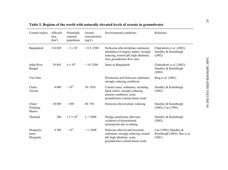
1.3 Natural occurrence
Arsenic is a metalloid that occurs naturally; it is the component of more than 245minerals. Examples of arsenic levels in some geological materials are given in Table 4.Arsenic is commonly concentrated in sulfide-bearing mineral deposits, especially thoseassociated with gold mineralization, and it has a strong affinity for pyrite, one of the moreubiquitous minerals in the earth’s crust. It is also concentrated in hydrous iron oxides.Arsenic and its compounds are mobile in the environment. Weathering of rocks converts
ARSENIC IN DRINKING-WATER 53
pp41-96.qxd 11/10/2004 10:19 Page 53

arsenic sulfides to arsenic trioxide, which enters the arsenic cycle as dust or by dissolutionin rain, rivers or groundwater. Arsenic can also enter the food chain, causing widespreaddistribution throughout the plant and animal kingdoms. The occurrence and behaviour ofarsenic in the environment have been extensively reviewed (Cullen & Reimer, 1989;Tamaki & Frankenberger, 1992; Matschullat, 2000; Mandal & Suzuki, 2002; Nordstrom,2002; Smedley & Kinniburgh, 2002).
A limited range of geological environments can result in significant natural elevationof arsenic in water supplies (Nordstrom, 2002). These include: organic rich (black) shales,Holocene alluvial sediments with slow flushing rates, mineralized and mined zones (most
IARC MONOGRAPHS VOLUME 8454
Table 4. Levels of arsenic in geological materials
Materials Concentration (mg/kg)
Source
Earth crust total 1–1.8 Matschullat (2000) Upper crust 1.5–2 Matschullat (2000) Igneous rocks Basic basalt 02–113 Mandal & Suzuki (2002); Smedley
& Kinniburgh (2002) Gabbro, dolorite 0.06–28 Mandal & Suzuki (2002); Smedley
& Kinniburgh (2002) Acidic granite 0.2–13.8 Mandal & Suzuki (2002); Smedley
& Kinniburgh (2002) Sedimentary rocks Phosphorites 0.4–188 Smedley & Kinniburgh (2002) Sandstones 0.6–120 WHO (1981); Mandal & Suzuki
(2002) Shale and argillite 0.3–500 Hale (1981) Schist and phyllite 0.5–143 Hale (1981) Carbonates 0.1–20 Matschullat (2000); Mandal &
Suzuki (2002) Coals 0.3–35 000 Smedley & Kinniburgh (2002) Sulfide minerals Pyrite 100–77 000 Smedley & Kinniburgh (2002) Pyrrhotite 5–100 Boyle & Jonasson (1973) Chalcopyrite 10–5000 Smedley & Kinniburgh (2002) Galena 5–10 000 Smedley & Kinniburgh (2002) Sphalerite 5–17 000 Smedley & Kinniburgh (2002) Marcasite 20–126 000 Smedley & Kinniburgh (2002) Oxide minerals Haematite up to 160 Smedley & Kinniburgh (2002) Iron oxide up to 2000 Smedley & Kinniburgh (2002) Iron(III) oxyhydroxide up to 76 000 Smedley & Kinniburgh (2002) Sulfate minerals Jarosite 34–1000 Smedley & Kinniburgh (2002)
pp41-96.qxd 11/10/2004 10:19 Page 54

often gold deposits), volcanogenic sources, thermal springs, closed basins in arid-to-semi-arid climates, particularly in volcanic regions, and strongly reducing aquifers with lowsulfate concentrations.
Depending on prevailing climatic and hydrological conditions, soils and sediments,surface waters, groundwaters and air can become enriched in arsenic where these geo-logical conditions prevail.
1.3.1 Arsenic speciation in natural materials
Mineral forms in which arsenic is present in soils are approximately 60% arsenatesand 20% sulfides and sulfosalts; the remaining 20% includes arsenides, arsenites, oxides,silicates and elemental arsenic.
These mineral forms are generally weathered to the inorganic water-soluble species,arsenate (AsV) and arsenite (AsIII), with arsenate dominating under oxidized conditionsand arsenite under reduced conditions (Cullen & Reimer, 1989). Under both aerobic andanaerobic conditions, micro-organisms can transform inorganic arsenic into organicforms such as MMA, DMA and volatile TMA. TMA in the air is then rapidly convertedinto water-soluble species, AsV and TMAO (Pongratz, 1998; Turpeinen et al., 1999,2002). These compounds can also be degraded by microflora. In certain materials, organicarsenic compounds naturally build up to high concentrations (Mandel & Suzuki, 2002;Smedley & Kinniburgh, 2002).
1.3.2 Abundance and distribution of arsenic
(a) Soils and sedimentsMeasurements of background arsenic levels in surface soil are all compromised by
atmospheric deposition of anthropogenically derived arsenic. Anthropogenic sources tosoil include use and resuspension of arsenic-based pesticides, mining, smelting, manufac-turing and waste-disposal activities. Shotyk et al. (1996) showed that arsenic levels were20-fold higher in surface horizons of ombrotrophic (rain-fed) peat bogs than in lowerhorizons. This high level was due to industrially derived inputs of arsenic. Centuries ofmining activities can result in an extremely high concentration of arsenic in soils. This isthe case in South-West England where arsenic concentrations in some old smelter and/ormine areas range from 24 to 161 000 mg/kg (Farago et al., 1997).
Koljonen (1992) estimated a global average level of arsenic in soils of 5 mg/kg, butconcentrations vary considerably among geographical regions. Arsenic concentrations insediments in lakes, rivers and streams in the USA ranged from 0.1 to 4000 mg/kg. Levelsof arsenic in a detailed survey of Finland, which has a low population density and isremote from major centres of pollution, ranged up to 60 mg/kg for the 1164 samplestested (Lahermo et al., 1998). Soils formed from arsenic-enriched geological substratescan have naturally higher levels than the ranges quoted. These ranges must therefore beconsidered as typical background levels rather than absolute ranges.
ARSENIC IN DRINKING-WATER 55
pp41-96.qxd 11/10/2004 10:19 Page 55

Soils formed on top of arsenic-rich bedrocks have elevated levels of this element.Colbourn et al. (1975) reported mean arsenic levels of 88 mg/kg (range, 24–250 mg/kg;n = 18) in soils formed naturally from parent material consisting of metamorphic aureolearound a granitic intrusion. The Strassegg area in Gasen (Styria, Austria) has extensivearsenopyrite (FeAsS) mineralization, with the ore body running close to the surface(Geiszinger et al., 2002). The soils formed on top of this ore vein are enriched in arsenic,with levels ranging from 700 to 4000 mg/kg, and are used for agronomic cultivation.
Soils formed in and around ancient and modern hot springs with elevated arsenic ingeothermal fluids have naturally elevated levels of arsenic due to enrichment of the parentmaterial of the soil (Ballantyne & Moore, 1988). The ancient hot-spring system at Rhynie,north-eastern Scotland, has cherts with arsenic levels ranging from 15 to 300 mg/kg (Riceet al., 1995). Sinter from active hot springs in the Taupo Volcanic Zone, New Zealand,have arsenic levels ranging from below detection limits to 1646 mg/kg (McKenzie et al.,2001). An area of at least 10 km2 in St Elizabeth, Jamaica, has a geochemical anomaly,whereby arsenic concentrations in soil reach 400 mg/kg (Lalor et al., 1999). The ano-malous values may result from an ancient hot-spring environment responsible for theintroduction and deposition of pyrite and arsenopyrite in the limestone bedrock, whichwere subsequently oxidized and weathered, leading to arsenic-rich soils.
Sediment levels of arsenic in the Waikato River, New Zealand, ranged from 7.9 to1520 mg/kg dry wt, resulting in high levels of arsenic in sediment living biota, such as thefreshwater mussel, Hyridella menziesi (Hickey et al., 1995).
In a number of delta environments in South-East Asia, deep fluvial and deltaicPleistocene-Holocene sediments have accumulated (up to 10 km thick in Bangladesh)(Nickson et al., 2000). During glaciation, river levels were 100 m lower than in inter-glacial times, and at this time of low sea level, the sediments were flushed and oxidized,leading to iron (FeIII) oxyhydroxide precipitation on sediment surfaces. These sedimentaryiron oxyhydroxides scavenge arsenic, with arsenic levels reaching up to 517 mg/kg inFeOOH phases (Nickson et al., 2000). Under reducing conditions caused by microbialmetabolism of sedimentary organic matter (present at up to 6% as C), in which sulfatelevels are low, insoluble FeIII is converted to soluble FeII, leading to the mobilization ofarsenic from the dissolved FeOOH phase. Although traces of arsenic-rich pyrites arefound in the sediments, they are present in quantities that are too small for pyrite oxidationto contribute significantly to arsenic in groundwaters.
Water percolating from hot-spring systems into the surrounding soil or sediment alsocauses a rise in arsenic concentrations (Langner et al., 2001; Koch et al., 1999).
The Antofagasta Region, northern Chile, is characterized by volcanism (Queiroloet al., 2000a). High levels of arsenic are found in soils and river sediments in this region(Caceres et al., 1992), and crops (maize and potato) grown on these soils have high levelsof arsenic, reaching 2 mg/kg in maize (Queirolo et al., 2000b).
Arsenic concentrations in mineralized zones rich in arsenic are further elevated, oftenseverely, by mineral extraction and processing (Smedley & Kinniburgh, 2002).
IARC MONOGRAPHS VOLUME 8456
pp41-96.qxd 11/10/2004 10:19 Page 56
(b) GroundwatersUnder natural conditions, the greatest range and the highest concentrations of arsenic
are found in groundwater as a result of the strong influence of the water–rock interactionsand the favourable physical and geochemical conditions in aquifers for the mobilizationand accumulation of arsenic. Arsenic is particularly mobile at pH values typically foundin groundwater (pH, 6.5–8.5) under both oxidizing and reducing conditions.
Background concentrations of arsenic in groundwater in most countries are less than10 µg/L and sometimes substantially lower. However, values quoted in the literature showa very wide range, from < 0.5 to 5000 µg/L. Most high levels of arsenic in groundwaterare the result of natural occurrences of arsenic. Cases of arsenic pollution caused bymining are numerous but tend to be localized.
Arsenic can occur in the environment in several oxidation states (–3, 0, +3 and +5)but, in natural waters, is mostly found in inorganic forms as oxyanions of trivalentarsenite (AsIII) or pentavalent arsenate (AsV). Redox potential (Eh) and pH are the mostimportant factors controlling arsenic speciation. Under oxidizing conditions, arsenate isdominant, as the H2AsO4
– form at low pH (less than approximately 6.9), or as theHAsO4
2– form at higher pH. Under reducing conditions at pH less than approximately 9.2,the uncharged arsenite species H3AsO3 predominates (Smedley et al., 2002).
In two recent reviews, Smedley and Kinniburgh (2002) and Smedley et al. (2002)focused extensively on the factors that control arsenic concentration in groundwater.
In relatively pristine habitats where anthropogenic activity can be excluded as acontributor to arsenic levels in aquifers, Lahermo et al. (1998) found that arsenic levelsin groundwaters in Finland reached up to 1040 µg/L, with a median of 0.65 µg/L(n = 472). The highest levels of arsenic were found in groundwaters from wells drilled inPrecambrian bedrock.
In an extensive groundwater survey in the USA, Welch et al. (2000) reported thatapproximately half of the 30 000 samples analysed had naturally occurring arsenic levels≤ 1 µg/L, with about 10% exceeding 10 µg/L. Geothermal water and high evaporationrates are associated with arsenic concentrations ≥ 10 µg/L in ground- and surface waters.
There are three major types of natural geological condition giving rise to high levelsof arsenic in groundwaters:
(i) aquifers composed of rocks or sediments enriched with arsenic-containingminerals of geogenic origin, such as sulfide mineralization;
(ii) aquifers containing sediments coated with iron oxyhydroxide (FeOOH) phasesenriched in arsenic through hydrological action, where arsenic is mobilized intoporewater by reducing conditions;
(iii) aquifers enriched in arsenic through high rates of evaporation in arid areas,leading to increased mineral concentration in groundwaters; the arsenic ismobile in such aquifers because of the high pH (> 8.5) caused by concentrationof alkali and alkali earth metals in solution.
ARSENIC IN DRINKING-WATER 57
pp41-96.qxd 11/10/2004 10:19 Page 57

Geochemical conditions similar to the alluvial sediments in Bangladesh exist in the RedRiver alluvial tract in the city of Hanoi, Viet Nam, where FeOOH reduction is thought tohave led to the high arsenic levels recorded in groundwaters (Berg et al., 2001). Smedleyand Kinniburgh (2002) outline that the reducing conditions observed in Bangladesh/WestBengal and Viet Nam aquifers are similar to those in the regions of Taiwan, China, northernChina and Hungary that suffer from high levels of arsenic in groundwaters.
Smedley et al. (2002) studied the geochemistry of arsenic in groundwaters fromQuaternary loess aquifers, which were high in arsenic, in an area thought to spread over106 km2 in La Pampa province, central Argentina. Dissolved arsenic ranged from 4 to5300 µg/L, with 73% of samples exceeding 50 µg/L. The conclusions drawn for LaPampa province may be applicable elsewhere in determining which regions are vulnerableto arsenic and related water-quality problems: “Under oxidising conditions, vulnerableaquifers potentially occur where several important criteria coincide: semi-arid climaticconditions with limited recharge where high-pH groundwater can be generated; young(Quaternary) sediments or volcanic sediments; and slow groundwater-flow conditions.Such aquifers are likely to have been poorly flushed over the geologically-short timescalesince deposition and hence will have had little opportunity for removal of trace elementssuch as arsenic from the aquifer.” Similar conditions exist in the Lagunera and Sonoraregions of Mexico and in the Atacama Desert, Chile (Smedley & Kinniburgh, 2002).
(c) Surface watersMatschullat (2000) collated measurements of arsenic in surface waters. Levels of
arsenic dissolved in uncontaminated stream waters ranged from 0.1 to 1.7 µg/L, and thosein seawaters were 1.5–1.7 µg/L. Concentrations in open seawater show little variationfrom the value of 1.5 µg/L (Smedley & Kinniburgh, 2002).
Arsenic in surface stream waters in Finland, which could be considered a pristineenvironment because of its low population density and remote geographical location,ranged from 0.06 to 1.6 µg/L (median, 0.36 µg/L; n = 1157) (Lahermo et al. 1998). Theselevels correlated well with arsenic levels in glacial till, with the highest stream water levelsoccurring in catchments with metamorphic, volcanic and sedimentary geologies. Levels inthe more geographically remote part of Finland were lower than those in the south, whichis nearer to continental Europe. Arsenic levels in Finnish water were lower than those forcontinental Europe, again emphasizing the pristine nature of the Finnish environment.
The Ciwidey River, West Java, drains a catchment dominated by the Quaternaryvolcano Patuha, which contains an acid crater lake (pH < 1) (Sriwana et al., 1998). Arsenicin the crater lake was recorded to be 279 µg/L, with the stream draining this lake havinglevels of 57 µg/L. In the tributary river of the stream, levels dropped to below 1 µg/L. In acrater lake with naturally elevated levels of arsenic, such as Lake Xolotlan in Nicaragua,mean arsenic concentrations ranged from 10.23 to 30.13 µg/L (Lacayo et al. 1992).
Takatsu and Uchiumi (1998) studied water from Lake Usoriko, Japan, which is acidi-fied by hot springs. The sediments of this lake contained 1.6% by mass of arsenic, witharsenic levels in the open lake waters ranging from 10 to 450 µg/L.
IARC MONOGRAPHS VOLUME 8458
pp41-96.qxd 11/10/2004 10:19 Page 58

Levels of arsenic in drinking-water extracted from the Waikato River, New Zealand, forthe city of Hamilton averaged 32 µg/L. Arsenic concentrations appear to follow a regularseasonal variation, being approximately 10–25 µg/L higher in the summer months, and fallto 6 µg/L after water treatment (McLaren & Kim, 1995). The elevated levels of arsenic inthe Waikato river are of natural origin, as its catchment is the volcanic region of the CentralPlains (Hickey et al., 1995).
Natural surface waters in the Antofagasta region of Chile, originating from springs,have very high levels of arsenic because of zones mineralization associated with volcanicactivity (eruptions, vents, geysers and thermal springs). Surface water is used as drinking-water and to irrigate crops (Queirolo et al., 2000a,b). Arsenic levels reached 3000 µg/L inrivers and canals in this region, with many rivers routinely having levels over 100 µg/L.
In an area with similar volcanic activity in the Salta Province, Argentina, high levelsof arsenic have been recorded in thermal springs, tap-water and river water (Vahter et al.,1995).
High levels of arsenic have been recorded in rivers in arid areas of Chile and Argentinawhere surface water is dominated by base-flow (whereby groundwater flows into the riverfrom surrounding rock) (Caceres et al., 1992; Lerda & Prosperi, 1996). Caceres et al.(1992) found concentrations in surface water up to 22 mg/L. The high degree of evapo-ration that occurs in these regions concentrates the arsenic leached from weathered rocks.Such surface waters have high pH, due again to high rates of evaporation that lead to con-centration of alkaline and alkaline earth cations leached from the rocks.
(d) AirConcentrations of arsenic in ambient air in remote locations range from < 1 to
3 ng/m3, but concentrations in cities may range up to 100 ng/m3. Arsenic in ambient air isusually a mixture of arsenite and arsenate, with organic species being of negligible impor-tance except in areas of arsenical pesticide application or other industrial activity (WHO,2001). Sources of arsenic to air include use and resuspension of arsenic-based pesticides,mining, smelting, manufacturing and waste-disposal activities. Arsenic may be intro-duced into the atmosphere directly from these processes, or it may be derived from sedi-ment and soil particles being entrained into the atmosphere or the production of volatilearsenic metabolites, such as arsines, from soils (Woolson, 1977; Turpeinen et al., 2002).Defining what constitutes natural levels is, therefore, difficult.
(e) OtherArsenic has been detected in rainwater at concentrations ranging from < 0.005 to
45 µg/L, with higher levels occurring in contaminated areas (WHO, 2001).Arsenic compounds are abundant in certain seafoods at concentrations as high as
several hundred milligrams per kilogram. Although marine animals contain many arseniccompounds, most species contain arsenobetaine as the major arsenical. Arsenobetaine isnot metabolized by humans and is believed to have low or negligible toxicity. Inorganic
ARSENIC IN DRINKING-WATER 59
pp41-96.qxd 11/10/2004 10:19 Page 59

arsenic and arsenosugars can, however, be present in some marine algae, seaweeds,oysters, mussels and clams (reviewed by Francesconi & Kuehnelt, 2002).
Dimethylarsinate is often the major arsenical constituent of species of fungi. Arseniteand arsenate are also commonly found in fungi (Francesconi & Kuehnelt, 2002).
Inorganic arsenic species are dominant in the chemistry of arsenic in terrestrial plants(Francesconi & Kuehnelt, 2002) and, although less studied, the concentration of arsenicin wheat and vegetables grown in countries highly contaminated with arsenic could berelevant to human health. Most of the vegetables cultivated in the Antofagasta Region(northern Chile), which is characterized by volcanic events (eruptions, thermal springs),are found at local markets of a population of approximately 4000 people. In this region,very high arsenic contents have been reported in Socaire and Talabre (1850 µg/kg in cornand 860 µg/kg in potatoes, including potato skins, respectively), two towns situated closeto the Lascar volcano (Queirolo et al., 2000b). These values exceed the national standardfor arsenic (500 µg/kg) by approximately 400% and 180%, respectively.
In Bangladesh, contamination of agricultural soils from long-term irrigation witharsenic-contaminated groundwater led to phyto-accumulation in food crops. Variousvegetables harvested in Samta village in the Jessore district have been reported to containhigh concentrations of arsenic (range, 306–489 µg/kg) (Alam et al., 2003). In WestBengal (India), high arsenic contents have also been reported in many vegetables andspices, especially in the skin of most vegetables, as a result of the dependence of the agri-cultural system on groundwater (Roychowdhury et al., 2002, 2003).
Moreover, high concentrations of arsenic have been reported in fruit, vegetables, grainand meat in regions contaminated by anthropogenic pollution; this is the case in the Moscowregion (Russia), which has been shown to be contaminated by fertilizer industry plants(Zakharova et al., 2002). High levels of arsenic have also been reported in plants, vegetablesand cow’s milk, as a consequence of heavy contamination of soils, surface and groundwatersby arsenic attributed to industrial sources (veterinary chemicals, pharmaceuticals, pesticideindustries) in the area of Patancheru, Andhra Pradesh (India) (Sekhar et al., 2003).
Interestingly, rare plants are able to accumulate exceedingly high concentrations ofarsenic (in the order of 1% dry mass). Brake fern (Pteris vittata) in particular is extremelyefficient at extracting arsenic from soils and translocating it into its fronds. Arsenic con-centrations in fern fronds, growing in soil spiked with 1500 mg/kg arsenic, increased from29.4 to 150 861 mg/kg within 2 weeks. Since it acts as an arsenic hyperaccumulator, brakefern could be used in the remediation of arsenic-contaminated soils (Ma et al., 2001).
1.4 Human exposure
The natural and anthropogenic occurrence of arsenic in drinking-water has been reco-gnized as a major public health issue in several regions of the world over the past two orthree decades. Areas affected by arsenic span the globe, and significant exposures havebeen identified in Bangladesh, India, Taiwan, China, Mexico, Argentina, Chile and the
IARC MONOGRAPHS VOLUME 8460
pp41-96.qxd 11/10/2004 10:19 Page 60

USA. Table 5 summarizes the geological characteristics of the regions of the world withnaturally elevated levels of arsenic in the drinking-water.
Recent reviews have outlined the worldwide problem of arsenic in drinking-water(WHO, 2001; Mandal & Suzuki, 2002; Nordstrom, 2002; Smedley & Kinniburgh, 2002;Chakraborti et al., 2003b).
1.4.1 Exposure in Bangladesh
In terms of the population exposed, the problem of arsenic contamination in much ofsouthern and eastern Bangladesh is the most serious in the world, and occurs in ground-water from the alluvial and deltaic sediments that make up much of the area. In addition,it is complicated by large variability in arsenic levels at both local and regional scales.
In Bangladesh, tubewells began to be used for drinking-water in the 1970s to controlthe problem of gastrointestinal disease linked to contamination of shallow wells andsurface waters. In the 1990s, it was discovered that the water from many of these wellswas contaminated with arsenic. Since then, extensive research has been carried out tocharacterize the extent of the problem. Figure 1 shows the districts in Bangladesh affectedby arsenic and Table 6 gives an overall picture of the database. Table 7 shows the distri-bution of concentrations of arsenic in hand tubewells, and Table 8 summarizes the levelsof arsenic measured in biological samples.
The level of contamination with arsenic of tubewells in Bangladesh exceeded both theWorld Health Organization guideline of 10 µg/L and the Bangladesh permissible limit of50 µg/L (Dhar et al., 1997; Smith et al., 2000a; Kinniburgh & Smedley, 2001: Alam et al.,2002).
A survey of 27 districts in Bangladesh up to January 1997 analysed over 3000 watersamples and revealed that 38% of them contained more than 50 µg/L arsenic (Dhar et al.,1997). In another survey examining 294 tubewells, 85 samples (29%) were contaminatedby arsenic at levels above 50 µg/L (Ahmad et al., 1997). Between September 1996 andJune 1997, all functioning wells (n = 265) in the village of Samta in the Jessore Districtwere tested for arsenic (Biswas et al., 1998). Approximately 91% of the wells containedarsenic at levels higher than 50 µg/L. Furthermore, 600 people were examined clinically,and a few hundred hair, nail and urine samples were tested using flow injection HG–AAS.The data obtained showed that 99% of urine samples and 98% of nail samples of thepopulation studied in Samta village contained levels of arsenic above normal and 78% ofhair samples above toxic levels. The arsenic problem of Bangladesh became highlightedwhen an international conference was held in Dhaka, Bangladesh, in 1998 (DhakaCommunity Hospital Trust and School of Environmental Studies, 1998).
By March 1998, it was reported that 4196 of 9024 wells in Bangladesh tested for arseniccontained levels higher than 50 µg/L and 884 wells had levels higher than 500 µg/L (Mandalet al., 1999). A Rapid Action Programme (RAP) was performed by field kit in a sample of500 villages with a total population of 469 424. Approximately 62% of the 32 651 tubewellssampled had levels of arsenic above 100 µg/L (Quamruzzaman et al., 1999).
ARSENIC IN DRINKING-WATER 61
pp41-96.qxd 11/10/2004 10:19 Page 61
IARC M
ON
OG
RAPH
S VO
LUM
E 8462
Table 5. Regions of the world with naturally elevated levels of arsenic in groundwater
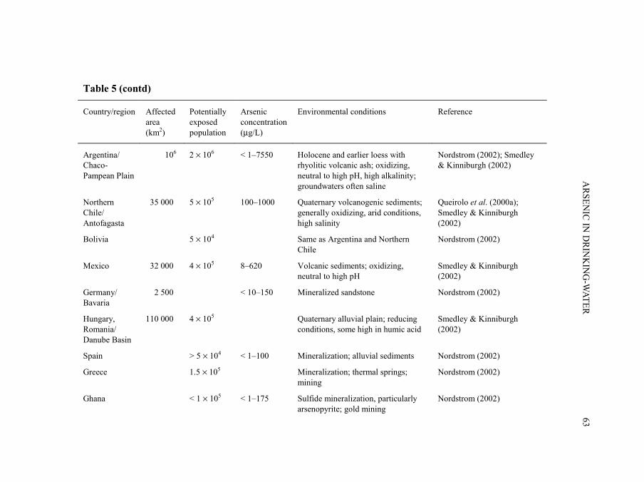
Country/region Affected area (km2)
Potentially exposed population
Arsenic concentration (µg/L)
Environmental conditions Reference
Bangladesh 118 849 ∼ 3 × 10⁷ < 0.5–2500 Hollocene alluvial/deltaic sediments; abundance of organic matter; strongly reducing, neutral pH, high alkalinity, slow groundwater flow rates
Chakraborti et al. (2002); Smedley & Kinniburgh (2002)
India/West Bengal
38 865 6 × 10⁶ < 10–3200 Same as Bangladesh Chakraborti et al. (2002); Smedley & Kinniburgh (2002)
Viet Nam Pleistocene and Holocene sediments; strongly reducing conditions
Berg et al. (2001)
China/ Taiwan
4 000 ∼ 105 10–1820 Coastal zones, sediments, including black shales; strongly reducing, artesian conditions, some groundwaters contain humic acids
Smedley & Kinniburgh (2002)
China/ Xinjiang, Shanxi
38 000 ∼ 500 40–750 Holocene alluvial plain; reducing Smedley & Kinniburgh (2002); Cao (1996)
Thailand 100 1.5 × 10⁴ 1–< 5000 Dredge quarternary alluvium; oxidation of disseminated arsenopyrite due to mining
Smedley & Kinniburgh (2002)
Mongolia/ Inner Mongolia
4 300 ∼ 105 < 1–2400 Holocene alluvial and lacustrine sediments; strongly reducing, neutral pH, high alkalinity, some groundwaters contain humic acids
Cao (1996); Smedley & Kinniburgh (2002); Sun et al. (2001)
pp41-96.qxd 11/10/2004 10:19 Page 62
ARSEN
IC IN D
RINK
ING
-WATER
63
Table 5 (contd)
Country/region Affected area (km2)
Potentially exposed population
Arsenic concentration (µg/L)
Environmental conditions Reference
Argentina/ Chaco-Pampean Plain
106 2 × 10⁶ < 1–7550 Holocene and earlier loess with rhyolitic volcanic ash; oxidizing, neutral to high pH, high alkalinity; groundwaters often saline
Nordstrom (2002); Smedley & Kinniburgh (2002)
Northern Chile/ Antofagasta
35 000 5 × 10⁵ 100–1000 Quaternary volcanogenic sediments; generally oxidizing, arid conditions, high salinity
Queirolo et al. (2000a); Smedley & Kinniburgh (2002)
Bolivia 5 × 10⁴ Same as Argentina and Northern Chile
Nordstrom (2002)
Mexico 32 000 4 × 10⁵ 8–620 Volcanic sediments; oxidizing, neutral to high pH
Smedley & Kinniburgh (2002)
Germany/ Bavaria
2 500 < 10–150 Mineralized sandstone Nordstrom (2002)
Hungary, Romania/ Danube Basin
110 000 4 × 10⁵ Quaternary alluvial plain; reducing conditions, some high in humic acid
Smedley & Kinniburgh (2002)
Spain > 5 × 10⁴ < 1–100 Mineralization; alluvial sediments Nordstrom (2002)
Greece 1.5 × 10⁵ Mineralization; thermal springs; mining
Nordstrom (2002)
Ghana < 1 × 10⁵ < 1–175 Sulfide mineralization, particularly arsenopyrite; gold mining
Nordstrom (2002)
pp41-96.qxd 11/10/2004 10:19 Page 63
IARC M
ON
OG
RAPH
S VO
LUM
E 8464
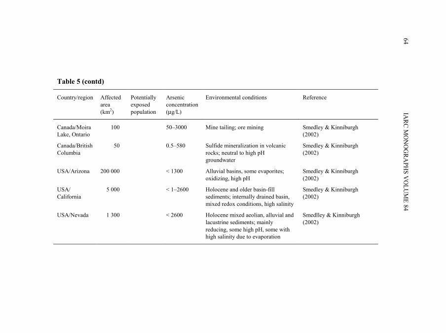
Table 5 (contd)
Country/region Affected area (km2)
Potentially exposed population
Arsenic concentration (µg/L)
Environmental conditions Reference
Canada/Moira Lake, Ontario
100 50–3000 Mine tailing; ore mining Smedley & Kinniburgh (2002)
Canada/British Columbia
50 0.5–580 Sulfide mineralization in volcanic rocks; neutral to high pH groundwater
Smedley & Kinniburgh (2002)
USA/Arizona 200 000 < 1300 Alluvial basins, some evaporites; oxidizing, high pH
Smedley & Kinniburgh (2002)
USA/ California
5 000 < 1–2600 Holocene and older basin-fill sediments; internally drained basin, mixed redox conditions, high salinity
Smedley & Kinniburgh (2002)
USA/Nevada 1 300 < 2600 Holocene mixed aeolian, alluvial and lacustrine sediments; mainly reducing, some high pH, some with high salinity due to evaporation
Smedlley & Kinniburgh (2002)
pp41-96.qxd 11/10/2004 10:19 Page 64

ARSENIC IN DRINKING-WATER 65
Figure 1. Degree of arsenic contamination in 64 districts in Bangladesh
From Chakraborti et al. (2002)
pp41-96.qxd 11/10/2004 10:19 Page 65

In continuing surveys of 42 districts affected by arsenic in Bangladesh, Chowdhuryet al. (2000a,b) reported the analysis of 10 991 water samples of which 59% containedarsenic levels above 50 µg/L.
Of the 34 000 drinking-water samples collected in Bangladesh up to August 2001, 272contained ≥ 1000 µg/L arsenic (Table 6; Chakraborti et al., 2002). The highest concen-tration of arsenic measured in drinking-water in Bangladesh was 4700 µg/L. In the Chiladivillage of Senbagh Thana in the Noakhali district, 100% of tubewell-water samplescontained arsenic concentrations ≥ 50 µg/L, 94% contained ≥ 300 µg/L and 28% contained≥ 1000 µg/L.
IARC MONOGRAPHS VOLUME 8466
Table 6. Status of contamination of groundwater by arsenic in Bangladesh
Bangladesh
Total area (km2) 148 393 Population (millions) 120 Total number of districts 64 Total number of water samples analysed 34000 Samples containing > 10 µg/L arsenic (%) 56.35 Samples containing > 50 µg/L arsenic (%) 37.38 Number of districts affected by arsenic (> 50 µg/L) 50 Population of districts affected by arsenic (millions) 104.9 Area of districts affected by arsenic (km2) 118 849 Number of villages affected by arsenic (arsenic in drinking-water > 50 µg/L)
2000
Number of people drinking arsenic-contaminated water > 50 µg/L (millions)
25
From Chakraborti et al. (2002)
Table 7. Distribution of arsenic concentrations in water samples from hand tubewells
Arsenic concentration range (µg/L) Total no. of water samples analysed
< 10 10–50 51–99 100–299 300–499 500–699 700–1000 > 1000
34 000 14 991
44.1%
6429
18.9%
2949
8.7%
5812
17.1%
2174
6.4%
894
2.6%
479
1.4%
272
0.8%
From Rahman et al. (2001)
pp41-96.qxd 11/10/2004 10:19 Page 66
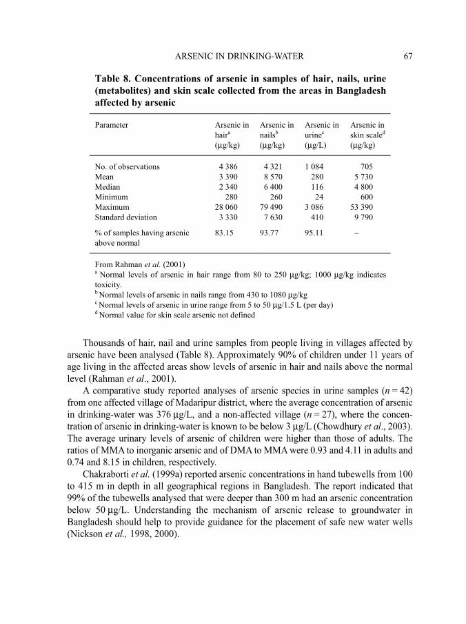
Thousands of hair, nail and urine samples from people living in villages affected byarsenic have been analysed (Table 8). Approximately 90% of children under 11 years ofage living in the affected areas show levels of arsenic in hair and nails above the normallevel (Rahman et al., 2001).
A comparative study reported analyses of arsenic species in urine samples (n = 42)from one affected village of Madaripur district, where the average concentration of arsenicin drinking-water was 376 µg/L, and a non-affected village (n = 27), where the concen-tration of arsenic in drinking-water is known to be below 3 µg/L (Chowdhury et al., 2003).The average urinary levels of arsenic of children were higher than those of adults. Theratios of MMA to inorganic arsenic and of DMA to MMA were 0.93 and 4.11 in adults and0.74 and 8.15 in children, respectively.
Chakraborti et al. (1999a) reported arsenic concentrations in hand tubewells from 100to 415 m in depth in all geographical regions in Bangladesh. The report indicated that99% of the tubewells analysed that were deeper than 300 m had an arsenic concentrationbelow 50 µg/L. Understanding the mechanism of arsenic release to groundwater inBangladesh should help to provide guidance for the placement of safe new water wells(Nickson et al., 1998, 2000).
ARSENIC IN DRINKING-WATER 67
Table 8. Concentrations of arsenic in samples of hair, nails, urine (metabolites) and skin scale collected from the areas in Bangladesh affected by arsenic
Parameter Arsenic in haira (µg/kg)
Arsenic in nailsb (µg/kg)
Arsenic in urinec (µg/L)
Arsenic in skin scaled (µg/kg)
No. of observations 4 386 4 321 1 084 705 Mean 3 390 8 570 280 5 730 Median 2 340 6 400 116 4 800 Minimum 280 260 24 600 Maximum 28 060 79 490 3 086 53 390 Standard deviation 3 330 7 630 410 9 790
% of samples having arsenic above normal
83.15 93.77 95.11 –
From Rahman et al. (2001) a Normal levels of arsenic in hair range from 80 to 250 µg/kg; 1000 µg/kg indicates toxicity. b Normal levels of arsenic in nails range from 430 to 1080 µg/kg c Normal levels of arsenic in urine range from 5 to 50 µg/1.5 L (per day) d Normal value for skin scale arsenic not defined
pp41-96.qxd 11/10/2004 10:19 Page 67

1.4.2 Exposure in India
(a) Contamination by arsenic of groundwater in northern IndiaA preliminary study was reported in 1976 on arsenic in dug wells, hand pumps and
spring water from Chandigarh and different villages of the Punjab, Haryana and HimachalPradesh in northern India (Datta & Kaul, 1976). A value as high as 545 µg/L arsenic wasobtained in one water sample from a hand pump. Datta (1976) further reported higharsenic content in the liver of five of nine patients with non-cirrhotic portal hypertensionwho had been drinking arsenic-contaminated water. To date no further information onarsenic poisoning in northern India is available.
(b) Contamination by arsenic of groundwater in West BengalSince 1984, extensive research in West Bengal has revealed that this region has one
of the most serious problems with groundwater contamination by arsenic in wells used fordrinking-water. Figure 2 shows the districts in West Bengal affected by arsenic andTable 9 gives an overall picture of the database and the extent of the problem. Table 10shows the distribution of concentrations of arsenic in hand tubewells in areas of WestBengal, and Table 11 summarizes the levels of arsenic measured in biological samples.
Contamination of groundwater by arsenic was first detected in the state of WestBengal, India, in 1983 (Garai et al., 1984). Sixteen people whose drinking-water camefrom two hand tubewells in one village in the district of 24-Parganas were identified ashaving arsenical skin lesions. Arsenic concentrations in these tubewells were 1250 and
IARC MONOGRAPHS VOLUME 8468
Table 9. Status of contamination of groundwater by arsenic in West Bengal, India
West Bengal
Total area (km2) 89 193 Population (millions; according to 1991 Census) 68 Total number of districts 18 Total number of water samples analysed 105 000 Samples containing > 10 µg/L arsenic (%) 51 Samples containing > 50 µg/L arsenic (%) 25 Number of districts affected by arsenic (> 50 µg/L) 9 Population of districts affected by arsenic (millions) 42.7 Area of districts affected by arsenic (km2) 38 865 Number of blocks/police stations affected by arsenic 74 Number of villages (approx.) affected by arsenic (arsenic in groundwater > 50 µg/L)
2700
Number of people drinking arsenic-contaminated water > 50 µg/L (millions)
6
From Chakraborti et al (2002)
pp41-96.qxd 11/10/2004 10:19 Page 68

ARSENIC IN DRINKING-WATER 69
Figure 2. Areas of West Bengal in which drinking-water is contaminatedwith arsenic
From Chakraborti et al. (2002)
pp41-96.qxd 11/10/2004 10:19 Page 69

700 µg/L. Saha and Poddar (1986) reported that 36 villages from 18 police stations/blocksof six districts were affected in 24-Parganas, Murshidabad, Nadia, Barddhaman, Midnapurand Maldah. Water samples from 207 hand tubewells were analysed and 105 (50.7%)showed arsenic concentrations above 50 µg/L; the highest concentration recorded was568 µg/L. Analysis of arsenic in hair, nails and skin-scale from people in the affectedvillages confirmed exposure to arsenic.
In 1987, an epidemiological survey in six villages of three districts (24-Parganas,Barddhaman and Nadia) revealed 197 patients with arsenical dermatosis in 48 families
IARC MONOGRAPHS VOLUME 8470
Table 10. Concentrations of arsenic in water samples from hand tubewells in West Bengal, India
Arsenic concentration range (µg/L) No. of water samples analysed
< 10 10–50 51–99 100–299 300–499 500–699 700–1000 > 1000
101 934 49 310
48.4%
27 309
26.8%
10 005
9.8%
11 782
11.6%
2354
2.3%
724
0.7%
334
0.3%
116
0.1%
From Rahman et al. (2001)
Table 11. Concentrations of arsenic in samples of hair, nails, urine (metabolites) and skin scale collected from the areas in West Bengal (India) affected by arsenic
Parameters Arsenic in haira (µg/kg)
Arsenic in nailsb (µg/kg)
Arsenic in urinec (µg/L)
Arsenic in skin scaled (µg/L)
No. of observations 7 135 7 381 9 795 165 Mean 1 480 4 560 180 6 820 Median 1 320 3 870 115 4 460 Minimum 180 380 10 1 280 Maximum 20 340 44 890 3 147 15 510 Standard deviation 1 550 3 980 268 4 750 % of samples having arsenic above normal
57 83 89 –
From Rahman et al. (2001) a Normal levels of arsenic in hair range from 80 to 250 µg/kg; 1000 µg/kg indicates toxicity. b Normal levels of arsenic in nails range from 430 to 1080 µg/kg c Normal excretion of arsenic in urine ranges from 5 to 40 µg/1.5 L (per day) d Normal value for skin scale arsenic not defined
pp41-96.qxd 11/10/2004 10:19 Page 70
(Chakraborty & Saha, 1987). Of 71 water samples collected from tubewells of the affectedvillages, the concentration of arsenic in 55 (77.5%) was higher than the permissible limit(50 µg/L) for arsenic in drinking-water in India. The mean arsenic concentration in 31water samples collected from tubewells of affected families was 640 µg/L and that in 40water samples collected from tubewells of unaffected families was 210 µg/L. Anotherepidemiological investigation (Guha Mazumder et al., 1988) in a village in 24-Parganasalso found evidence of effects of arsenic in 62 (92.5%) of 67 members of families whodrank contaminated tubewell-water (level of arsenic, 200–2000 µg/L). In contrast, only six(6.25%) of 96 persons from the same area who drank water with a level of arsenic< 50 µg/L showed any effects.
In 1991, a report from the government of West Bengal (Steering Committee, ArsenicInvestigation Project, 1991) concluded that water of the intermediate aquifer in areas ofWest Bengal was polluted with arsenic. Neither the shallow (first) nor the deep (third)aquifers had reported arsenic contamination. The sand grains in the arsenic-contaminatedaquifer were generally coated with iron and material rich in arsenic.
In October 1994, a committee constituted by the government of West Bengal(Committee Constituted by Government of West Bengal, 1994) reported arsenic contami-nation in 41 blocks in six districts of West Bengal. The committee analysed about 1200water samples from these six districts for arsenic and other common water-quality para-meters, and the highest concentration of arsenic reported was 3200 µg/L.
The expanding database on the problem of arsenic contamination in West Bengal hasbeen documented in a continuing series of publications. By December 1994, it wasreported that 312 villages from 37 blocks/police stations in six districts in West Bengalwere affected by contamination of groundwater with arsenic. From extrapolation of thedata, it was predicted that more than 800 000 people were drinking arsenic-contaminatedwater from these districts, and based on the analysis of several thousand water samples,average arsenic concentrations in the wells sampled ranged from 193 to 737 µg/L (Daset al., 1994; Chatterjee et al., 1995). The highest arsenic concentration of 3700 µg/L wasfound in a hand tubewell from a village in South 24-Parganas district. Groundwater andurine samples from affected villages were also analysed for arsenite, arsenate, MMA andDMA. Groundwater contained arsenate and arsenite in a ratio of approximately 1:1. Inurine, DMA and MMA were the predominant species, together with some arsenite andarsenate. Das et al. (1995) reported high arsenic levels in the hair, nails, urine, skin-scaleand a few liver tissues (biopsy) of people from arsenic-affected villages who had arsenicalskin lesions.
Based on the analysis of 20 000 water samples from areas of West Bengal, Mandalet al. (1996) reported that seven districts (North 24-Parganas, South 24-Parganas, Nadia,Barddhaman, Murshidabad, Maldah, Hugli) were affected by arsenic. Approximately45% of these samples had arsenic concentrations above 50 µg/L, and the averageconcentration was approximately 200 µg/L.
Groundwater contamination was reported in 985 villages from 69 police stations/blocks in nine districts of West Bengal on the basis of analyses of 58 166 water samples.
ARSENIC IN DRINKING-WATER 71
pp41-96.qxd 11/10/2004 10:19 Page 71

The nine districts were Maldah, Murshidabad, Barddhaman, Hugli, Howrah, Nadia, North24-Parganas, South 24-Parganas and Calcutta. After extrapolation of data from the wateranalyses and screening villagers for arsenical skin lesions, it was estimated that about 5million people were drinking-water contaminated with levels of arsenic above 50 µg/L.The total population in the nine districts of West Bengal affected by arsenic is about 43million (Chowdhury et al., 2000a,b).
On the basis of an analysis of 101 394 hand tubewells and approximately 25 000 bio-logical samples, and screening of 86 000 persons in affected villages of West Bengal,Rahman et al. (2001) reported that 2600 villages were affected by arsenic in groundwaterat levels of > 50 µg/L and that approximately 6 million people drank water contaminatedwith arsenic at levels above 50 µg/L. Mandal et al. (2001) identified DMAIII and MMAIII
for the first time in urine from the affected areas of West Bengal. Roychowdhury et al. (2002) reported total arsenic in food composites collected from
a few arsenic-affected villages in Murshidabad district, West Bengal, where arsenic-conta-minated groundwater was used for agricultural irrigation. The report showed averagedaily dietary intake of arsenic from foodstuffs for adults and children of 180 and 96.5 µg,respectively.
Rahman et al. (2003) studied North 24-Parganas, one of the nine affected districts ofWest Bengal, for 7 years. On the basis of analyses of 48 030 water samples and 21 000hair, nail and urine samples, and screening of 33 000 people in North 24-Parganas, it wasestimated that about 2 million and 1 million people are drinking water contaminated witharsenic at levels above 10 and 50 µg/L, respectively.
(i) Source of contamination of groundwater by arsenic inWest Bengal
When the contamination of drinking-water by arsenic was first discovered in WestBengal, tubewell strainers, pesticides, insecticides and other anthropogenic sources werefirst considered as possible origins of the groundwater contamination (Chakraborty & Saha,1987). However, Das et al. (1994) showed that a single deep tubewell supplying water to afew villages in Maldah, one of the nine arsenic-affected districts, was drawing nearly 150 kgarsenic per year, indicating that the source of arsenic was geological. Analyses of bore-holesediments showed high concentrations of arsenic in only a few soil layers and the arsenictherein was found to be associated with iron pyrites. Das et al. (1995, 1996) also confirmedanalytically the existence of arsenic-rich pyrites in bore-hole sediment. It was proposed thatheavy drawing of groundwater and aeration of the aquifer leads to the decomposition ofarsenic-rich pyrites and consequently contamination of groundwater with arsenic. Similarconclusions were reached by Mallick and Rajagopal (1995).
Bhattacharya et al. (1997, 1998) reported an association between arsenic and hydratedferric oxide (HFO) and its mobilization to the aquifer due to changes in redox conditionsduring the development of groundwater. Ahmed et al. (1998) and Nickson et al. (1998,2000) also suggested that reduction of HFO resulted in the mobilization of arsenic fromabsorbed HFO.
IARC MONOGRAPHS VOLUME 8472
pp41-96.qxd 11/10/2004 10:19 Page 72

(ii) Contamination of groundwater by arsenic in the residentialarea of Behala-Calcutta due to industrial pollution
In Calcutta, chronic arsenicosis was first reported by Guha Mazumder et al. (1992).The study of Chatterjee et al. (1993) on the source of arsenic and the magnitude of thecontamination revealed that a chemical factory producing several chemical compounds,including the insecticide Paris-Green (copper acetoarsenite), was responsible for thecontamination. This factory had been producing about 20 tonnes of Paris-Green per yearfor approximately 20 years. Analysis of soil surrounding the production waste-dumpingground showed very high concentrations of arsenic (as high as 10 000 µg/g). Nineteenhand tubewells, used for drinking and cooking in the immediate area, showed very highconcentrations of arsenic (up to 39 000 µg/L). The concentration of arsenic in hand tube-wells decreased the farther the wells were located from the dumping ground. A follow-upstudy in the affected areas (Chakraborti et al., 1998) showed that the total average concen-tration of arsenic in the 19 hand tubewells sampled previously had decreased by only10–15% from the levels observed 8 years before.
(c) Contamination of groundwater by arsenic in Chhattisgarh StateContamination of groundwater by arsenic was reported in a few villages of
Rajnandgaon district of Chhattisgarh by Chakraborti et al. (1999b). The present State ofChhattisgarh had been within the State of Madhya Pradesh 2 years previously. The sourceof arsenic in groundwater is natural and geological both for the alluvial Bengal Basin andthe rocky belt of Dongargarh-Kotri zone of Rajnandgaon district. The total population ofthe district is 1.5 million. Except for two towns — Rajnandgaon and Khairagarh — theentire district depends on tubewells and dugwells. Water samples (n = 146) were collectedfrom 22 villages of Chowki block, Rajnandgaon district, and levels of arsenic in ground-water were found to be above 10 µg/L in eight villages and above 50 µg/L in five villages,with the highest concentration being 880 µg/L. From 150 hair samples examined,approximately 75% of people were found to have levels of arsenic in hair above toxiclevels. Pandey et al. (1999) also reported contamination of groundwater by arsenic in theRajnandgaon district of Chhattisgarh. Of 390 samples analysed, 26 sites were found to becontaminated with arsenic, with the highest concentration being 1010 µg/L. The numberof people at risk was estimated at 10 000. Pandey et al. (2002) established that the extentof the arsenic contamination in this area is even greater; about 30 000 people residing in30 villages and towns are directly exposed to high levels of arsenic in drinking-water (upto 3050 µg/L arsenic) and more than 200 000 people are at risk. The source and mobi-lization process of arsenic from affected areas of Rajnandgaon district Chhattisgarh wasreported by Acharyya (2002).
ARSENIC IN DRINKING-WATER 73
pp41-96.qxd 11/10/2004 10:19 Page 73
(d) Contamination of groundwater by arsenic in Middle Ganga Plain,Bihar
In the Middle Ganga Plain, Bihar, tubewells replaced dugwells about 20 years ago.Analyses of the arsenic content of 206 tubewells from Semria Ojha Patti (95% of the totalin the village) showed that 56.8% exceeded concentrations of 50 µg/L, with 19.9%> 300 µg/L. The distribution indicated that, of the 5000 residents of Semria Ojha Patti,18.8% used safe water (< 10 µg/L arsenic), 24.7% used water containing 10–50 µg/Larsenic, 56.8% used water containing > 50 µg/L, and 19.9% used water containing> 300 µg/L. The concentrations of arsenic in urine, hair and nail correlated significantly(r = 0.72–0.77) with concentrations in drinking-water. Of the 51 urine samples analysed,98% had levels of arsenic above that of the normal secretion, with 47% > 500 mg/L, 33%> 1000 mg/L and 5.9% > 3000 mg/L; 57.6% of hair samples and 76.3% of nail sampleswere found to be above the normal range (Chakraborti et al., 2003a).
(e) Contamination of groundwater and surface water by arsenic in theindustrial region of Patancheru, Andhra Pradesh
Patancheru, in the Medak District of Andhra Pradesh, is one of the major industrialestates, situated 30 km from Hyderabad. The main source of arsenic has been identifiedas Park Trade Centre, Gaddapotharam Bulk Drug Factory, which makes veterinary drugsbased on arsonic acid, as well as other sources such as the pesticide and drug intermediateindustries. The solid wastes of these industries are dumped indiscriminately nearKazipally Lake, and represent a source of contamination of nearby waters and soils.Arsenic contamination was evaluated in 14 villages in this area. Very high levels ofarsenic were found in the range of 80–8960 µg/L and 140–7350 µg/L in surface water andgroundwater, respectively. In both surface water and groundwater, the average arsenite(AsIII) concentration was about 20% of total arsenic (Sekhar et al., 2003).
Samples of blood, urine, hair and nails from 193 inhabitants of these 14 contaminatedvillages were analysed. Arsenic levels in the biological samples were very high, rangingfrom 400 to 1400 µg/kg in blood (control, 6–10 µg/kg), from 60 to 160 µg/L in urine(control, 6–10 µg/L), from 300 to 940 µg/kg in hair (control, 10–130 µg/kg) and from 500to 1630 µg/kg in nails (control, 120–160 µg/kg). High concentrations of arsenic were alsodetected in vegetables, plants and cow’s milk in this area and represent a second possiblesource of exposure for the population (Sekhar et al., 2003).
1.4.3 Exposure in Central and South America
In South America, the main source of exposure to arsenic has been the natural conta-mination of drinking-water. In this area, arsenic originates from the geological formationsassociated with volcanoes, affecting Chile, Bolivia, Peru and Argentina in the Andeanregion (Queirolo et al., 2000a). The largest populations affected are the AntofagastaRegion in northern Chile, with approximately 400 000 exposed inhabitants, and theCórdoba Province in Argentina, with approximately 630 000 people exposed. Mexico also
IARC MONOGRAPHS VOLUME 8474
pp41-96.qxd 11/10/2004 10:19 Page 74

has naturally occurring arsenic in drinking-water, which is best characterized in theLagunera region, in central northern Mexico, where approximately 400 000 people areexposed. The mean levels in drinking-water for these populations range from 50 to500 µg/L; in isolated wells, levels reach as high as 6897 µg/L arsenic. Exposure has beenrecorded since the beginning of the last century. Currently, most areas in these regionsreceive water with levels of arsenic below 50 µg/L.
Exposures to arsenic due to contaminated air, soils and water as a result of copper,gold or silver mining have been described in Mexico (Díaz-Barriga et al., 1993; Calderón,1999; Mejía et al., 1999), Chile (Romo-Kroger & Llona, 1993; Romo-Kroger et al., 1994;Santolaya et al., 1995; Sancha, 1997; Flynn et al., 2002), Brazil (Romo-Kroeger & Llona,1993) and Nicaragua (Cruz et al., 1994). The area affected in central Mexico is San Luisde Potosí. In Chile, environmental and occupational exposures to arsenic in air have beenreported in the Andes Mountains in Regions II, III, V and VI and the MetropolitanRegion, where five major copper mining plants are located (Ministerio de Salud, 1986;Santolaya et al., 1995; Ferreccio et al., 1996), but no secondary contamination ofdrinking-water.
(a) Mexico In Mexico, most studies of arsenic in drinking-water have been conducted in the States
of Durango and Coahuila, which constitute the Lagunera Region (Table 12). Del Razoet al. (1990) studied 128 wells from 11 counties and found arsenic contents of 8–624 µg/L;50% of the wells had arsenic levels > 50 µg/L. They estimated that at least 400 000 people,mostly from the rural areas of the region, have been exposed to levels of arsenic > 50 µg/L.Since the 1960s, when arsenic contamination was first identified, the polluted wells havegradually been replaced and, by the end of 1989, most of the population was receivingwater with arsenic levels below 20 µg/L (Cebrián et al., 1994). Some contamination ofdrinking-water by arsenic has been reported in the State of Hidalgo in the Zimapan Valley,where the exposed population has been estimated at 35 000, and levels of arsenic in thedrinking-water ranged from 21 to 1070 µg/L (Armienta et al., 1997; Gomez-Arroyo et al.,1997).
In San Luis de Potosí, in central Mexico, exposure to arsenic associated with miningactivities arises from drinking-water, soil and dust, and the estimated exposed populationis 600 000. Mean arsenic concentrations in air have been measured at 0.48 µg/m3
(0.36–0.88 µg/m3) (Díaz-Barriga et al., 1993), and concentrations of arsenic in drinking-water vary from 9.9 to 20.9 µg/L (with some wells near the smelter having concentrationsthat range from 105 to 6897 µg/L). Studies of soil in San Luis de Potosí havedemonstrated extremely high levels of arsenic in the vicinity of the mines (188–944 µg/g,Díaz-Barriga et al., 1993; 2215–2675 µg/g, Mejía et al., 1999), and also in the dust of thenearby households (800–1182 µg/g, Díaz-Barriga et al., 1993; 1780–9950 µg/g, Mejíaet al., 1999). By 1991, the copper mining companies that caused the air, soil and watercontamination of the area implemented dust control technologies and other measures tocontrol soil pollution (Cebrián et al., 1994).
ARSENIC IN DRINKING-WATER 75
pp41-96.qxd 11/10/2004 10:19 Page 75

Levels of arsenic in urine and hair are presented in Tables 13–14. Levels of arsenic inhair were high in samples from subjects exposed to arsenic in water in Zimapán, and weretwice those in samples from subjects in Mexico City, which were also above the referencevalue, probably due to air pollution (Armienta et al., 1997).
(b) ArgentinaIn Argentina, the main source of arsenic in drinking-water has been from wells, with
concentrations ranging from 40 to > 4500 µg/L (Table 15), and arsenic was first reportedin well-water in 1917 (Arguello et al., 1938). In 1970 and 1980, aqueducts from riverswith low levels of arsenic were built to replace the use of well-water, but some popu-lations continued to be exposed (Hopenahyn-Rich et al., 1996a,b,c). The provinces withhigh levels of arsenic in their well-water are: Córdoba, Salta, La Pampa, Santa Fé,Tucuman, Santiago del Estero, San Luis and part of Buenos Aires. The best characterizedis Córdoba, a region in central Argentina, that extends over an area of 165 000 km2 andhas a population of 2 750 000, distributed in 26 counties. In some counties of Córdoba,high levels (between 100 and 2000 µg/L) of arsenic were recorded in drinking-waterduring the 1930s (Hopenhayn-Rich et al., 1996a).
In Córdoba, Hopenhayn-Rich et al. (1996a) obtained data from various sources, inclu-ding measurements of arsenic in drinking-water from official national health reports madein the 1930s, a survey in 1942, two studies reported in 1968 and 1985 and a water surveyreported in 1973. Based on the available measurements, average exposure of the popu-lation of each town was estimated, assuming that all people drank the same concentration
IARC MONOGRAPHS VOLUME 8476
Table 12. Exposure to arsenic in drinking-water in Mexico
Location Source of water No. of samples studied
Year Total arsenic in water (µg/L; range)
Reference
Sonora, Hermosillo
Wells of 29 cities 173 NR 2–305 Wyatt et al. (1998a)
Lagunera Region
Wells, different towns
171
1970s–1980s 7–624 Cebrían et al. (1983); Del Razo et al. (1990); García-Vargas et al. (1994); Gonsebatt et al. (1997); Hernández-Zabala et al. (1999)
Zimapán, Hidalgo
Aquifer, 6 different towns
(Since 1970) 21–1070 Gomez- Arroyo et al. (1997)
Tap-water, Morales and Graciano
19 NR 9.9–20.9 Díaz-Barriga et al. (1993)
San Luis de Potosí
Wells near smelter NR NR 106–6897 Meíja et al. (1999)
NR, not reported
pp41-96.qxd 11/10/2004 10:19 Page 76
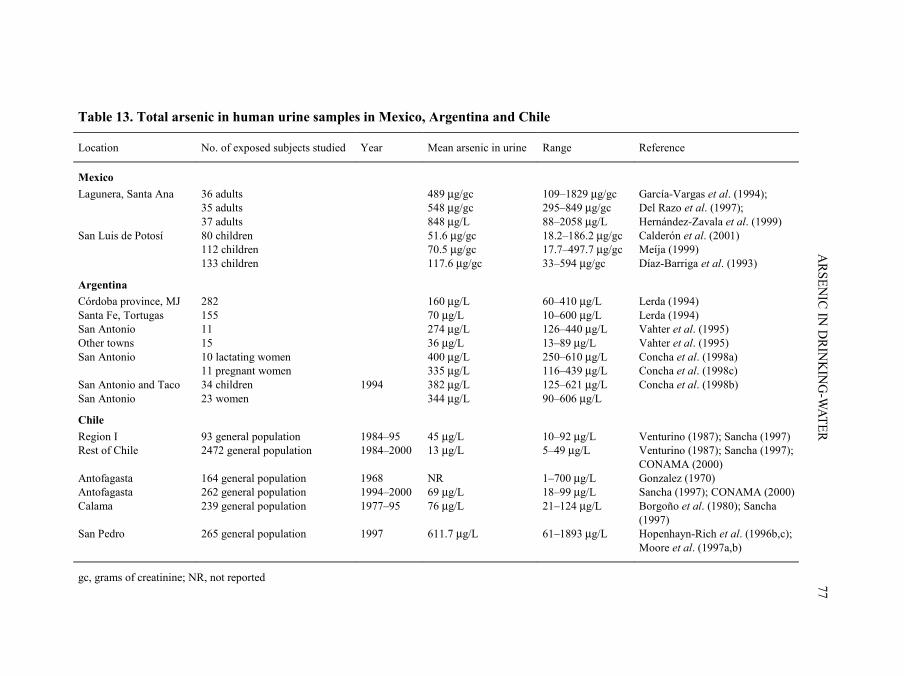
ARSEN
IC IN D
RINK
ING
-WATER
77
Table 13. Total arsenic in human urine samples in Mexico, Argentina and Chile
Location No. of exposed subjects studied Year Mean arsenic in urine Range Reference
Mexico Lagunera, Santa Ana 36 adults
35 adults 37 adults
489 µg/gc
548 µg/gc 848 µg/L
109–1829 µg/gc 295–849 µg/gc 88–2058 µg/L
García-Vargas et al. (1994); Del Razo et al. (1997); Hernández-Zavala et al. (1999)
San Luis de Potosí 80 children 112 children 133 children
51.6 µg/gc 70.5 µg/gc 117.6 µg/gc
18.2–186.2 µg/gc 17.7–497.7 µg/gc 33–594 µg/gc
Calderón et al. (2001) Meíja (1999) Díaz-Barriga et al. (1993)
Argentina Córdoba province, MJ 282 160 µg/L 60–410 µg/L Lerda (1994) Santa Fe, Tortugas 155 70 µg/L 10–600 µg/L Lerda (1994) San Antonio Other towns
11 15
274 µg/L 36 µg/L
126–440 µg/L 13–89 µg/L
Vahter et al. (1995) Vahter et al. (1995)
San Antonio 10 lactating women 11 pregnant women
400 µg/L 335 µg/L
250–610 µg/L 116–439 µg/L
Concha et al. (1998a) Concha et al. (1998c)
San Antonio and Taco San Antonio
34 children 23 women
1994
382 µg/L 344 µg/L
125–621 µg/L 90–606 µg/L
Concha et al. (1998b)
Chile Region I 93 general population 1984–95 45 µg/L 10–92 µg/L Venturino (1987); Sancha (1997) Rest of Chile 2472 general population 1984–2000 13 µg/L 5–49 µg/L Venturino (1987); Sancha (1997);
CONAMA (2000) Antofagasta 164 general population 1968 NR 1–700 µg/L Gonzalez (1970) Antofagasta 262 general population 1994–2000 69 µg/L 18–99 µg/L Sancha (1997); CONAMA (2000) Calama 239 general population 1977–95 76 µg/L 21–124 µg/L Borgoño et al. (1980); Sancha
(1997) San Pedro 265 general population 1997 611.7 µg/L 61–1893 µg/L Hopenhayn-Rich et al. (1996b,c);
Moore et al. (1997a,b)
gc, grams of creatinine; NR, not reported
pp41-96.qxd 11/10/2004 10:19 Page 77

of arsenic. It was estimated that 273 014 people had been exposed to an average of 178µg/L arsenic and another 406 000 people had been exposed to some arsenic (at least onemeasurement of 120 µg/L in water). A report available through CEPIS/PAHO (Penedo &Zigarán, 2002) described the arsenic content of 100 water samples from wells in Córdobaand confirmed Hopenhayn’s estimations: they estimated that 625 861 people wereexposed to arsenic, with regional averages ranging from 70 to 180 µg/L and individualwell measurements from 10 to 1900 µg/L.
The Salta Province is the only area where high levels of arsenic have also been foundin surface waters (Penedo & Zigarán, 2002). In the provinces of Salta and Jujui, in north-western Argentina, samples from five rivers had arsenic levels ranging from 52 to1045 µg/L, and samples from three surging thermal springs had arsenic levels of128–10 650 µg/L (de Sastre et al., 1992). The population of San Antonio de los Cobres isthe best studied in this province (Vahter et al., 1995; Concha et al., 1998a,b,c). SanAntonio de los Cobres is a village in the Salta Province, 3800 m above sea level, with
IARC MONOGRAPHS VOLUME 8478
Table 14. Arsenic in human hair samples in Mexico and Chile
Total arsenic in sample (µg/g)
Location (source of exposure)
Year of sample
No. of subjects
Mean SD (range)
Reference
Mexico Zimapán Mexico City (water)
NR 120 17
8.5 4.6
3.56 1.96
Armienta et al. (1997)
San Luis de Potosí (smelter (1.5 km)) (smelter (25 km))
NR 75 25
9.9 0.5
(1.4–57.3) (0.2–1.2)
Díaz-Barriga et al. (1993)
Lagunera (wells) NR 35 NR (0–23.3) Chávez et al. (1964)
Chile Iquique 1969 26 0.8 NR Borgoño & Greiber (1971) Antofagasta 1968–76 607 7.7 4.2–14.8 Gonzalez (1970); Borgoño
& Greiber (1971); Sandoval & Venturino (1987)
Antofagasta 1986–92 293 0.42 0.01–3.68 Jamett et al. (1992); Peña et al. (1992)
Calama 1977 203 3.75 0–10 Borgoño et al. (1980) Calama 1986–92 60 4.28 0.98–14.2 Jamett et al. (1992); Peña
et al. (1992) Chuquicamata 1986–92 60 17.19 3.03–54.77 Jamett et al. (1992); Peña
et al. (1992) Puchuncaví 1990 151 2.178 0.103–18.023 Chiang et al. (1990) Valparaíso 1990 NR 0.434 0.015–1.525 Chiang et al. (1990)
SD, standard deviation
pp41-96.qxd 11/10/2004 10:19 Page 78

approximately 5000 inhabitants (Vahter et al., 1995). Until recently, this population hadbeen drinking-water from wells with arsenic contents varying from < 1 to 440 µg/L, withone well reaching 9450 µg/L on average. Arsenic levels in urine are presented in Table 13and other biomarkers in Table 16.
There are no studies of arsenic in air in Argentina. High levels of arsenic have beenfound in prepared food and soups in San Antonio de los Cobres (soup, 259–427 µg/g;prepared food, 131–418 µg/g; Concha et al., 1998b).
(c) ChileNorthern Chile (Regions I–III) is an expanse of 250 000 km2, of which 35 000 are
quaternary volcanic rocks rich in arsenic (Queirolo et al., 2000a). Arsenic reaches thepopulation through drinking-water and through contamination of air and soil, as a resultof mining activities.
In Chile, the main sources of drinking-water are rivers that originate in Cordillera delos Andes and reach the Pacific Ocean. Rivers in northern Chile (Regions I and II) havehigh natural arsenic concentrations, particularly those from the Region of Antofagasta.Arsenic concentrations in rivers in Region II vary along its course, depending on thearsenic content of its tributaries, and range from 30 to 3310 µg/L but reach 14 250 µg/Lin some hot springs (Table 17) (Alonso, 1992; Queirolo et al., 2000a). Exposure of the
ARSENIC IN DRINKING-WATER 79
Table 15. Exposure to arsenic in drinking-water in Argentina
Total arsenic in water (µg/L)
Location Source of drinking-water wells
No. of samples (year)
Average Range
Reference
Córdoba Province Bell-Ville NR (1917–20) NR 1120–4500 Arguello et al. (1938)
Córdoba Province Marcos Juárez 282 (NR) 130 10–660 Lerda (1994)
Córdoba Province 2 counties 118 (1942) 178 40–533 Hopenhayn-Rich et al. (1996a)
Córdoba Province 5 counties 67 (NR) 70–180 10–1900 Penedo & Zigarán (2002)
Santa Fe Province Tortugas 155 (NR) 20 0–70 Lerda (1994)
Salta Province School pipes 18 (NR) 592 4–1490 Astolfi (1971)
San Antonio San Antonio
2 areas (NR) 1 well
NR NR
93–440 8250–10 650
de Sastre et al. (1992) Salta Province
San Antonio 10 (1994) 167 117–219 Vahter et al. (1995); Concha et al. (1998a); Del Razo et al. (1999)
NR, not reported
pp41-96.qxd 11/10/2004 10:19 Page 79

IARC M
ON
OG
RAPH
S VO
LUM
E 8480
Table 16. Arsenic in other biological samples in Salta Province, Argentina
Total arsenic in sample Location No. of subjects Type of sample (year)
Median Range
Reference
San Antonio de los Cobres 9 women
Breast milk Blood Urine
2.3 µg/kga 9.8 µg/L 390 µg/L
0.83–7.6 µg/kg 4.4–19 µg/L 250–610 µg/L
Concha et al. (1998a)
San Antonio Santa Rosa Olacapato Tolar Grande
15 5 5 5
Blood
µg/L 7.6 1.5 1.3 1.3
µg/L 2.7–18.3 1.1–2.0 1.2–2.4 1.0–1.3
Vahter et al. (1995)
San Antonio and T Pozo Rosario de Lerma
San Antonio and T Pozo Rosario de Lerma
Children 36 20
Women 27 11
Blood (1994) µg/L 9.1 0.8
9.3 0.95
µg/L 5.5–17 0.27–1.5
2.7–18 0.69–1.8
Concha et al. (1998b)
San Antonio de los Cobres Pregnant women 11 11 11 10
Blood Cord blood Placenta Maternal milk
11 µg/L 9.0 µg/L 34 µg/kg 3.0 µg/kga
5.6–13 µg/L 6.0–12 µg/L 17–54 µg/kg 2.3–4.8 µg/kg
Concha et al. (1998c)
a µg/kg fresh weight
pp41-96.qxd 11/10/2004 10:19 Page 80

population in this region has ranged from 40 to 860 µg/L, depending on the rivers usedfor its water supply; 1958–70 was the highest exposure period for the largest population(approximately 300 000) (Table 18).
Sancha (1997) estimated the total number of people exposed in 1996 to specific levelsof arsenic in the drinking-water in Chile: 7 million inhabitants (53.3%) were exposed toless than 10 µg/L; 5.5 million (41.9%) were exposed to 10–30 µg/L; 450 000 inhabitants(3.4%) were exposed to 30–50 µg/L; 170 000 inhabitants (1.3%) were exposed to 50–60 µg/L; and 1500 (0.01%) were exposed to 600–800 µg/L.
There are a few studies of arsenic in general environmental air in Chile (Romo-Kroger & Llona, 1993; Romo-Kroger et al., 1994; Sancha, 1997; COSUDE, 2000).Sancha (1997) and COSUDE (2000) covered a large part of the country from 1994 to1999. They found that cities not in the vicinity of copper smelting operations had arseniclevels in the air ranging from 0.001 to 0.057 µg/m3, with a population of approximately6 million people. The cities located 30–45 km from a copper smelter had arsenic levels inthe air ranging from 0.01 to 0.14 µg/m3 and had approximately 755 000 inhabitants. Thecities in the vicinity (within 10 km) of smelters had arsenic levels in air ranging from 0.03
ARSENIC IN DRINKING-WATER 81
Table 17. Concentration of arsenic in surface waters in Chile, Region II, 1983–86
River studied and location Mean total arsenic in water (µg/L)
Salado River Tatio Hot Springs 14 250 Codelco Mine Reservoir 7 500 Before Toconce River 3 310 Toconce River Before Salado 600 Before Curti 860 Ayquina 980 Before Loa River 760 Loa River Before Salado 270 Yalquincha 800 Finca 910 Before San Salvador River 1 380 San Salvador Before Loa 1 270 La Posada Bridge 1 500 Quillagua 1 440 Outlet of River 1 360 Upper Loa River Basin 210–330 Gorges south of Salado river 30–60 Spring north of Salado river 190–370
From Alonso (1992)
pp41-96.qxd 11/10/2004 10:19 Page 81

IARC M
ON
OG
RAPH
S VO
LUM
E 8482
Table 18. Average concentration (µµg/L) of arsenic in drinking-water in Regions II and I and the rest of Chile
Region Town Population (2002 census) living in exposed areas
1930–57 1958–70 1971–77 1978–80 1981–87 1988–94 1995–2002
Reference
II Tocopilla and Elena
31 175 250 250 636 110 110 40 – Ferreccio et al. (2000)
Calama 136 739 150 150 287 110 110 40 – 38
Ferreccio et al. (2000) Sancha (1997)
San Pedro 4 883 600 600 600 600 600 600 – Ferreccio et al. (2000)
Chiu Chiu 250 – – – – – – 753 Smith et al. (2000b)
Caspana 275 – – – – – – 13 Smith et al. (2000b)
Antofagasta and Mejillones
306 548 90 860 110 110 70 40 – 32
Ferreccio et al. (2000) Sancha (1997)
I Arica-Iquique 426 351 – – – – – – 32 Sancha (1997)
III–XIII Rest of Chile 14 213 266 – – – – – – 5 Sancha (1997)
Averages supplied by Empresa Servicios Sanitarios de Antofagasta for 1950–67 and Servicio de Salud Antofagasta for 1968–94 For 1995–2000, data and ranges published in studies
pp41-96.qxd 11/10/2004 10:19 Page 82
to 2.4 µg/m3, with an estimated 60 000 people exposed. The rest of the country that wasnot sampled has a population of approximately 6 million people whose exposure isestimated to be in the lowest range of < 0.010 µg/m3 (COSUDE, 2000).
Mean levels of arsenic in the air inside the Chuquicamata copper mine, the world’slargest open copper mine, for the period 1952–91 ranged from 1.6 µg/m3 in the adminis-trative areas to 201.72 µg/m3 in the smelting areas (Ferreccio et al., 1996). This exposurehas decreased in the last decade in correlation with the implementation of new techno-logies in smelting to avoid arsenic contamination. Workers exposed to arsenic had urinarylevels ranging from 40 to 490 µg/L in 1992; between 1987 and 1990, 32–58% of workersin exposed areas had levels of urinary arsenic above 300 µg/L.
In Region II, 1020 people were examined between 1987 and 2002; the mean totalarsenic in urine was 225 µg/L, ranging from 1 to 1893 µg/L. In comparison, in Region Iwhich has intermediate exposure to arsenic in water (40 µg/L), 91 people were examinedand their urinary arsenic averaged 45.5 µg/L, ranging from 10 to 92 µg/L. In the rest ofChile (arsenic in drinking-water, < 10 µg/L), 2472 people were sampled and mean urinaryarsenic was 13 µg/L, ranging from 5 to 49 µg/L (Table 13). Arsenic measured in hair frompeople in Chile is presented in Table 14.
(d) Other In Nicaragua, there has been concern regarding contamination with heavy metal of
Lake Asososca, which is a source of drinking-water for Managua. The level of arsenic insediment was found to be 4.1 µg/g, and that in water ranged from 0 to 18.07 µg/L, witha mean concentration of 5.86 µg/L, well below current water standards. Higher values of25 µg/L were found in Lake Monte Galán (Cruz et al., 1994). An earlier study conductedin Lake Xolotlán found arsenic levels in surface water ranging from 10.2 to 30.1 µg/L;wastewater from a thermal plant discharging into the river contained concentrations of5295–16 700 µg/L (Lacayo et al., 1992).
In Brazil, concerns have been raised regarding arsenic contamination as a result ofgold mining in the zone of Minas Gerais, in south-eastern Brazil. In 1998, urinary arsenicwas measured in 126 schoolchildren, and a mean concentration of 25.7 µg/L (range,2.2–106 µg/L) was found. Environmental studies in the surrounding areas found meanlevels of arsenic in surface water of 30.5 µg/L (range, 0.4–350 µg/L); levels of arsenic insoils ranged from 200 to 860 mg/kg; and sediments had a mean concentration of350 mg/kg, ranging from 22 to 3200 mg/kg (Matschullat et al., 2000).
1.4.4 Exposure in South-East Asia
There are many reports on the human exposure to arsenic in the drinking-water inSouth-East Asia. High concentrations of arsenic in drinking-water have been documentedin China (Cao, 1996), Taiwan, China (Tseng et al., 1968; Chiou et al., 1997a), Thailand(Choprapawon & Rodcline, 1997), and Viet Nam (Berg et al., 2001). The use of artesianwells, which were later shown to have high levels of arsenic in the water, began in the
ARSENIC IN DRINKING-WATER 83
pp41-96.qxd 11/10/2004 10:19 Page 83

early 1920s in southern Taiwan, China (Tseng et al., 1968), in the early 1950s in InnerMongolia (Ma et al., 1996), in the late 1950s in north-eastern Taiwan, China (Chiou et al.,1997a, 2001), in the early 1960s in Xinjiang, China (Wang, 1996), in the late 1980s inRonbipool, Thailand (Choprapawon & Rodcline, 1997), in the early 1990s in Shanxi,China (Cao, 1996) and in the mid-1990s in Viet Nam (Berg et al., 2001). There have beenseveral reports on industry-related exposure to arsenic through drinking-water contami-nation from tin mining in Ronpibool, Thailand (Choprapawon & Rodcline, 1997).
Table 19 summarizes data on arsenic contamination of drinking-water in variousregions of South-East Asia.
(a) ChinaSeveral geographical areas in mainland China have a high content of arsenic in the
drinking-water, including Xinjiang, Inner Mongolia and Shanxi (Cao, 1996). The villageswith high concentrations of arsenic in the drinking-water in Inner Mongolia are clusteredin Bamen and Huhehot. Ma et al. (1996) reported the arsenic concentration in the waterof 9733 wells in Bamen: 2465 had levels of arsenic > 50 µg/L; in five counties of Bamen,the percentage of wells with an arsenic concentration > 50 µg/L varied, ranging from 11to 59%; more than 500 villages had at least one well with an arsenic concentration> 50 µg/L; and the level of arsenic in drinking-water from all wells from the two areasranged from < 50 to 890 µg/L. The water from a total of 497 wells in Huhehot were testedfor arsenic: 111 had an arsenic level > 50 µg/L; 48 villages had at least one well witharsenic concentration > 50 µg/L; and the level of arsenic in drinking-water ranged from< 81 to 890 µg/L (Ma et al., 1996). Sun et al. (2001) reported a survey on the concen-tration of arsenic in 303 wells in a village in Inner Mongolia: 77 wells (25.4%) had a levelof arsenic < 10 µg/L, 85 (28.1%) had levels of 10–49 µg/L, 131 (43.2%) had levels of50–499 µg/L and 10 (3.3%) had levels of ≥ 500 µg/L.
In the highly contaminated area of Xinjiang, located in Tunguei, arsenic concen-trations in well-water in 15 villages of the area ranged from 50 to 850 µg/L, and weremostly between 100 and 500 µg/L (Wang, 1996).
Sun et al. (2001) reported a survey of 2373 wells in 129 villages in the Basin ofDatong and Jinzhong, Shanxi, in 1994–95. Levels of arsenic in drinking-water rangedfrom < 50 to 4440 µg/L and 833 wells had an arsenic concentration > 50 µg/L. The per-centage of wells with an arsenic concentration > 50 µg/L in seven counties of the areavaried from 6.3 to 54.7%.
(b) Taiwan, ChinaThere are two endemic areas of arseniasis in Taiwan, China. One is located in the
south-western coastal area where Blackfoot disease, a unique peripheral vascular diseaseassociated with long-term ingestion of arsenic from artesian well-water, is endemic. Thereare four townships in this area: Peimen, Hsuehchia, Putai and Ichu. High levels of arsenicin artesian wells and patients with Blackfoot disease have also been documented in twoneighbouring townships, Hsiayin and Yensui. Another endemic area of chronic arsenic
IARC MONOGRAPHS VOLUME 8484
pp41-96.qxd 11/10/2004 10:19 Page 84

ARSEN
IC IN D
RINK
ING
-WATER
85
Table 19. Contamination of drinking-water by arsenic in various regions of South-East Asia
Country Area/population Sample Level of arsenic (range; µg/L)
Source of arsenic
Reference
Taiwan (China)
South-western Blackfoot disease-endemic area (Peimen, Hsuehchia, Putai, Ichu)
13 artesian well-water 34 artesian well-water 11 artesian well-water 97 artesian well-water
240–960 350–1100 340–896 10–1100
Natural Natural Natural Natural
Blackwell et al. (1961) Chen et al. (1962) Yeh (1963) Kuo (1968)
North-eastern endemic area of chronic arsenic poisoning (Chuangwei, Wuchieh, Chiaohsi, Tungshan)
3901 well-water < 0.15–3590 Natural Chiou et al. (2001)
Taiwan (314 townships) 83 656 well-water < 10–> 1000 Natural Lo (1975)
Thailand Thammarat Province Surface water < 0.5–583 < 0.5–28.4 AsIII
Arsenopyrite wastes
Williams et al. (1996)
Shallow water 1.25–5114 < 0.5–125 AsIII
Surface water 4.8–583 Mining Choprapawon & Porapakkham (2001)
River 541–583
China Inner Mongolia 497 well-water (Huhhot) 9733 well-water (Bamen)
< 10–1860 < 50–890
Natural Natural
Ma et al. (1996); Luo et al. (1997) Ma et al. (1996)
Xinjiang Well-water in 15 villages (Tunguei)
50–850 Natural Wang (1996)
Shanxi 2373 well-water in 129 villages (Datong, Jinzhong)
< 50–4440 Natural Sun et al. (2001)
pp41-96.qxd 11/10/2004 10:19 Page 85

IARC M
ON
OG
RAPH
S VO
LUM
E 8486
Table 19 (contd)
Country Area/population Sample Level of arsenic (range; µg/L)
Source of arsenic
Reference
Japan Fukuoka 67 well-water 1–293 11–220 AsV 15–70 AsIII
Natural Kondo et al. (1999)
Sendai 1–35 Takatsuki 3–60 Kumamoto 5–66
Viet Nam Red River Basin 68 tubewells, 8 treatment plants
1–3050 (72% > 10 µg/L) 11–190
Natural Berg et al. (2001)
pp41-96.qxd 11/10/2004 10:19 Page 86

toxicity is located in the Lanyang Basin of north-eastern Taiwan, in which there are fourtownships: Chiaohsi, Chuangwei, Tungshan and Wuchieh (Table 19).
In the area of south-western Taiwan where Blackfoot disease is endemic, Blackwellet al. (1961) reported levels of arsenic of 240–960 µg/L in 13 artesian wells, Chen et al.(1962) reported levels ranging from 350 to 1100 µg/L in 34 artesian wells and Yeh (1963)found levels ranging from 340 to 900 µg/L in water samples from 11 wells. Kuo (1968)carried out a larger survey of 97 artesian wells in 42 villages of the six townships in theendemic area and found concentrations in well-water ranging from 10 to 1100 µg/L, witha median of 500 µg/L. In this south-western area, the arsenic concentration was higher inwater from deep artesian wells than in that from shallow wells, showing a correlationcoefficient of r = 0.627 (p < 0.01). Arsenate was the dominant species of arsenic in theartesian well-water.
Lo (1975) reported a nationwide survey of arsenic content in drinking-water from 83656 wells in 314 precincts and townships. In total, 15 649 (18.7%) wells had an arsenicconcentration ≥ 50 µg/L and 2224 (2.7%) had an arsenic concentration ≥ 350 µg/L. Mosttownships with high arsenic concentration in well-water were found to cluster in south-western and north-eastern Taiwan.
Chiou et al. (2001) tested the water from 3901 tubewells in 18 villages of fourtownships in the north-eastern endemic area of chronic arsenic toxicity by the HG–FAASmethod in 1991–94. The arsenic content ranged from undetectable (< 0.15 µg/L) to3590 µg/L.
(c) ThailandThe Ronpibool district is situated approximately 70 km south of Nakorn Sri
Thammarat Province, in the southern part of Thailand, and had a total population ofapproximately 23 000 in 1998. A geological survey found that the potential sources ofarsenic contamination in the mining areas were from high-grade arsenopyrite waste pilesin bedrock mining localities, sub-ore grade waste-rock piles, sulfide-rich wastes from ore-dressing plants, disseminated sulfide waste from small-scale prospecting and floatationactivities and alluvial tin workings (Choprapawon & Porapakkham, 2001).
In 1994, a collaborative study was initiated to establish the distribution and geo-chemical form of arsenic in surface drainage and aquifer systems in the area. Surfacewaters were sampled at 26 stations and groundwater samples were collected from 23shallow wells and 13 deep boreholes. Concentrations of arsenic in samples of surface waterranged from < 0.5 (limit of detection) to 583 µg/L and AsIII levels ranged from < 0.5 to28.4 µg/L. Concentrations in shallow groundwater samples ranged from 1.25 to 5114 µg/Land AsIII levels ranged from < 0.5 to 125 µg/L. Concentrations in deep borehole samplesranged from 1.25 to 1032 µg/L and AsIII levels ranged from < 0.5 to 53.6 µg/L (Williamset al., 1996).
In another study, significant concentrations of arsenite (AsIII) were detected in severalof the water samples with the highest levels of arsenic (28.4, 25.6 and 24.9 µg/L),
ARSENIC IN DRINKING-WATER 87
pp41-96.qxd 11/10/2004 10:19 Page 87

although arsenate (AsV) remained the dominant species (more than 92% of the total)(Choprapawon & Porapakkham, 2001).
(d) Viet NamBerg et al. (2001) reported arsenic contamination of the Red River alluvial tract in the
city of Hanoi, Viet Nam, and in the surrounding rural districts. Because of naturallyoccurring organic matter in the sediments, the groundwaters are anoxic and rich in iron.In rural groundwater samples from private small-scale tubewells, contamination levelsranged from 1 to 3050 µg/L, with an average concentration of 159 µg/L arsenic. In ahighly affected rural area, the groundwater that is used directly as drinking-water had anaverage concentration of 430 µg/L. Analysis of raw groundwater pumped from the loweraquifer for the Hanoi water supply yielded arsenic levels of 240–320 µg/L in three of eighttreatment plants and 37–82 µg/L in another five plants. Aeration and sand filtration thatare applied in the treatment plants for the removal of iron lowered the arsenic content tolevels of 25–91 µg/L, but 50% remained above 50 µg/L. The high arsenic concentrationsfound in tubewells (48% above 50 µg/L and 20% above 150 µg/L) indicate that severalmillion people consuming untreated groundwater might be at a considerable risk forchronic arsenic poisoning.
(e) JapanIn March 1994, high concentrations of arsenic (> 10 µg/L) were detected in 29 of 67
well-water samples in the southern region of the Fukuoka Prefecture, Japan. The range ofarsenic concentrations was 1–293 µg/L: AsV ranged from 11 to 220 µg/L; AsIII ranged from15 to 70 µg/L; and MMA and DMA were both < 1 µg/L. The maximum concentration waslower than the figures recorded in Taiwan, China, and India, but higher than those reportedin Sendai (range, 1–35 µg/L), Takatsuki (range, 3–60 µg/L) and Kumamoto (range,5–66 µg/L), Japan (Kondo et al., 1999).
Arsenic concentrations in water from 34 wells in the Niigata Prefecture were measuredbetween 1955 and 1959 as part of a historical cohort study using the Gutzeit method, andranged from non-detectable to 3000 µg/L: six wells had a non-detectable concentration; 17wells contained < 1000 µg/L; and 11 wells contained ≥ 1000 µg/L. All wells with arsenicconcentrations > 100 µg/L were located within a distance of 500 m from a factory that pro-duced arsenic trisulfide (Tsuda et al., 1995).
(f) OtherIn a recent United Nations Economic and Social Commission for Asia and the
Pacific–United Nations International Children’s Emergency Fund–World HealthOrganization (UNESCAP-UNICEF-WHO, 2001) expert group meeting, contamination ofgroundwater by arsenic was also reported from other countries including Lao People’sDemocratic Republic, Cambodia, Myanmar and Pakistan. It has also been reported fromNepal (Tandukar et al., 2001; Shreshta et al., 2002).
IARC MONOGRAPHS VOLUME 8488
pp41-96.qxd 11/10/2004 10:19 Page 88

1.4.5 Exposure in other countries
Exposure in other countries is summarized in Table 20.
(a) Africa (Egypt, Ghana) and the Middle East (Iran)In a 1999 study of 100 subjects in Cairo, Egypt, arsenic was measured by HG–AAS
(detection limit, 1 µg/L) in hair samples and drinking-water. Levels of arsenic in hairsamples ranged from 40 to 1040 µg/kg and levels in drinking-water samples were less than1 µg/L (Saad & Hassanien, 2001).
Concentrations of arsenic in groundwaters from two areas in Ghana — the Obuasi areain the Ashanti region and the Bolgatanga area of the Upper East region — ranged from < 1to 64 µg/L [AsIII range, 6–30 µg/L] and < 1 to 141 µg/L [AsIII range, < 1–9 µg/L], respec-tively. Sulfide minerals such as arsenopyrite and pyrite were present in the Birimian base-ment rocks of both areas and these constitute the dominant sources of arsenic. Concen-trations were lowest in the shallowest groundwaters, and increased at greater depths. Thelateral and vertical variations in dissolved arsenic concentrations were controlled byambient pH and redox conditions and by the relative influences of sulfide oxidation andsorption (Smedley, 1996).
Concentrations of arsenic were measured in the scalp hair of three groups of peoplefrom a village in western Iran using NAA. One group consisted of healthy subjects, thesecond of subjects with suspected arsenic poisoning, and the third of subjects with con-firmed arsenic poisoning. The arsenic content of water sources used by the inhabitantswas also measured. The average arsenic concentration in hair was 200 µg/kg in thehealthy group, 4900 µg/kg in the group with suspected poisoning and 5600 µg/kg in thegroup with arsenic poisoning; arsenic concentrations in water samples varied between30 µg/L and 1040 µg/L (Pazirandeh et al., 1998).
(b) AustraliaAustralia is a country rich in minerals that present a significant source of natural
arsenic contamination to the environment, in addition to anthropogenic sources such asmining activities and pesticide use. In 1991, survey data showed elevated levels of arsenicin the surface water and groundwater in Victoria, particularly around gold mining areas.Concentrations of arsenic in groundwater ranged from < 1 to 300 000 µg/L (n = 109) andthose in surface water ranged from < 1 to 28 300 µg/L (n = 590). In a follow-up study ofthe same region in the mid-1990s, arsenic concentrations ranged from 1 to 12 µg/L ingroundwater samples (n = 18), from 1 to 220 µg/L in surface water samples (n = 30) andfrom 1 to 73 µg/L in drinking-water samples (n = 170) (Hinwood et al., 1998).
In an investigation of the relationship between environmental exposure to arsenicfrom contaminated soil and drinking-water and the incidence of cancer in the Victoriaregion, median arsenic concentrations in groundwater ranged from 1 to 1077 µg/L (totalrange, 1–300 000 µg/L; n = 22 areas) (Hinwood et al., 1999).
ARSENIC IN DRINKING-WATER 89
pp41-96.qxd 11/10/2004 10:19 Page 89

IARC M
ON
OG
RAPH
S VO
LUM
E 8490
Table 20. Concentrations of arsenic (As) in drinking-water in other countries
Country Population Date Sample (no.) Levels (µg/L) Source of arsenic Reference
Africa Egypt Cairo 1999 Tap water (5 districts) 1 NR Saad & Hassanien (2001) Ghana Obuasi area
NR Groundwater < 1–64 (total As)
6–30 (AsIII) Natural Smedley (1996)
Bolgatanga area NR
1–141 (total As) 1–9 (AsIII)
Middle East Iran West Iran NR Spring water (20) 30–1040 Natural Pazirandeh et al. (1998)
Australia Victoria Victoria mid-1990s Groundwater (18)
Surface water (30) Drinking-water (170)
1–12 1–220 1–73
Natural anthro-pogenic (mining, pesticide)
Hinwood et al. (1998)
Ground- and surface water (22 geographical areas)
1–300 000 (1–1077 medians)
Hinwood et al. (1999)
Europe Finland 1993–94 Groundwater (69) 17–980 Kurttio et al. (1998) 1996 Wells (72)
Control population < 0.05–64 (median, 0.14)
Natural Kurttio et al. (1999)
Spain Madrid 1998 353 water supplies Wells (< 2% of population uses wells)
74% < 10 23% 10–50 3.7% > 50
Natural Aragones Sanz et al. (2001)
Romania (Transylvania)
Bihor and Arad counties
1992–95 Drinking-water 0–176 Natural Gurzau & Gurzau (2001)
Switzerland Grisons Canton Valais Canton
1998 Public water supplies (336) 14 000 people
< 10–170 12–50
Natural Pfeifer & Zobrist (2002)
United Kingdom South-west Private supplies (3) 11–80 Farago et al. (1997)
pp41-96.qxd 11/10/2004 10:19 Page 90
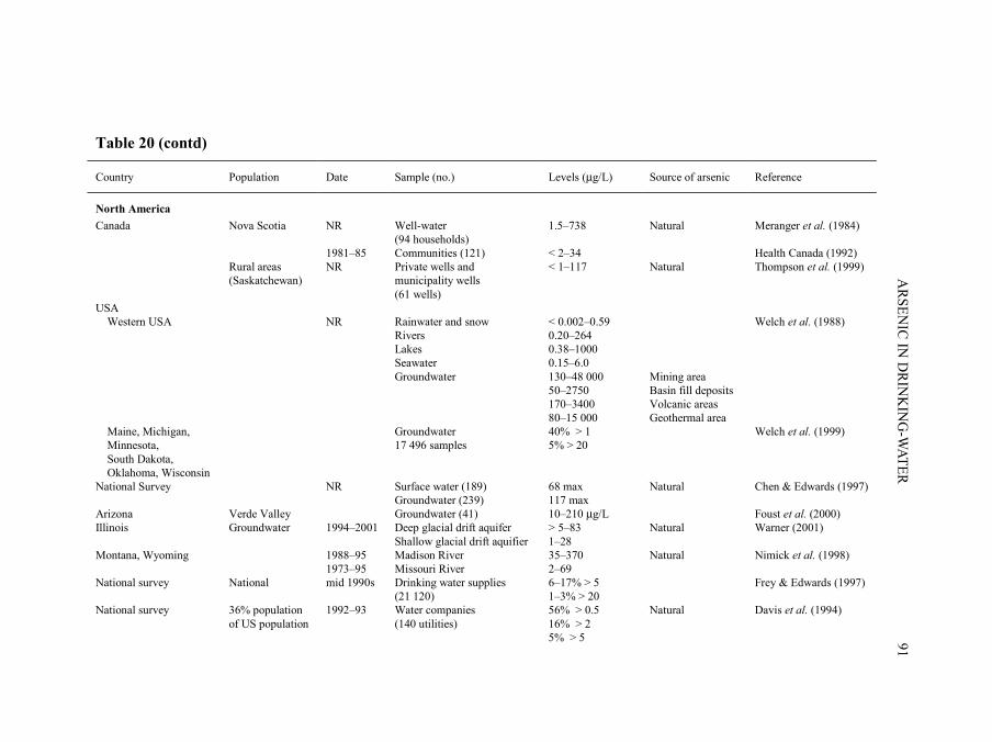
ARSEN
IC IN D
RINK
ING
-WATER
91
Table 20 (contd)
Country Population Date Sample (no.) Levels (µg/L) Source of arsenic Reference
North America Canada Nova Scotia NR Well-water
(94 households) 1.5–738 Natural Meranger et al. (1984)
1981–85 Communities (121) < 2–34 Health Canada (1992) Rural areas
(Saskatchewan) NR Private wells and
municipality wells (61 wells)
< 1–117 Natural Thompson et al. (1999)
USA Western USA NR Rainwater and snow < 0.002–0.59 Welch et al. (1988) Rivers 0.20–264 Lakes 0.38–1000 Seawater 0.15–6.0 Groundwater 130–48 000 Mining area 50–2750 Basin fill deposits 170–3400 Volcanic areas 80–15 000 Geothermal area Maine, Michigan, Minnesota, South Dakota, Oklahoma, Wisconsin
Groundwater 17 496 samples
40% > 1 5% > 20
Welch et al. (1999)
National Survey
NR Surface water (189) Groundwater (239)
68 max 117 max
Natural Chen & Edwards (1997)
Arizona Verde Valley Groundwater (41) 10–210 µg/L Foust et al. (2000) Illinois Groundwater 1994–2001 Deep glacial drift aquifer
Shallow glacial drift aquifier > 5–83 1–28
Natural Warner (2001)
Montana, Wyoming 1988–95 1973–95
Madison River Missouri River
35–370 2–69
Natural Nimick et al. (1998)
National survey National mid 1990s Drinking water supplies (21 120)
6–17% > 5 1–3% > 20
Frey & Edwards (1997)
National survey 36% population of US population
1992–93 Water companies (140 utilities)
56% > 0.5 16% > 2 5% > 5
Natural Davis et al. (1994)
pp41-96.qxd 11/10/2004 10:19 Page 91
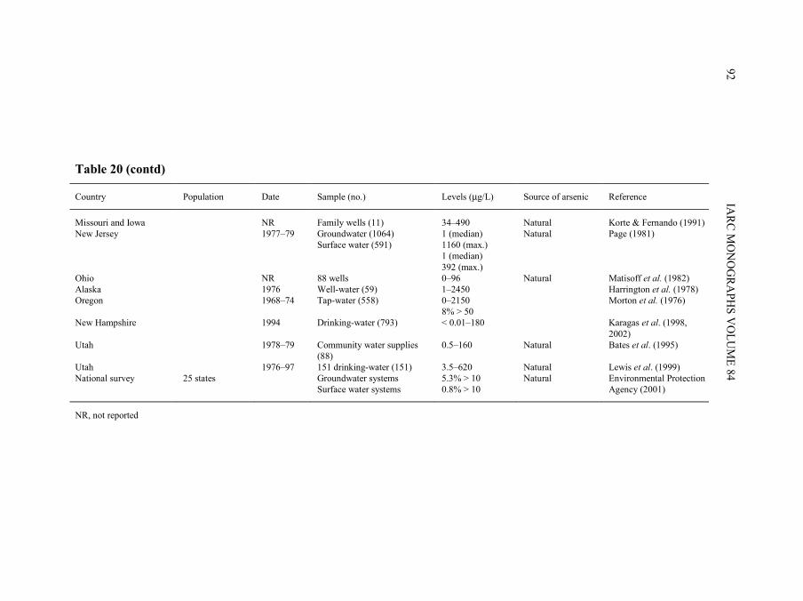
IARC M
ON
OG
RAPH
S VO
LUM
E 8492
Table 20 (contd)
Country Population Date Sample (no.) Levels (µg/L) Source of arsenic Reference
Missouri and Iowa NR Family wells (11) 34–490 Natural Korte & Fernando (1991) New Jersey 1977–79 Groundwater (1064)
Surface water (591) 1 (median) 1160 (max.) 1 (median) 392 (max.)
Natural Page (1981)
Ohio NR 88 wells 0–96 Natural Matisoff et al. (1982) Alaska 1976 Well-water (59) 1–2450 Harrington et al. (1978) Oregon 1968–74 Tap-water (558) 0–2150
8% > 50 Morton et al. (1976)
New Hampshire 1994 Drinking-water (793) < 0.01–180 Karagas et al. (1998, 2002)
Utah 1978–79 Community water supplies (88)
0.5–160 Natural Bates et al. (1995)
Utah 1976–97 151 drinking-water (151) 3.5–620 Natural Lewis et al. (1999) National survey 25 states Groundwater systems
Surface water systems 5.3% > 10 0.8% > 10
Natural Environmental Protection Agency (2001)
NR, not reported
pp41-96.qxd 11/10/2004 10:19 Page 92
(c) Europe (Finland, Romania, Spain, Switzerland, United Kingdom)Samples of well-water were collected in Finland between July and November 1996.
The final study population (144 627 from a register-based cohort) consisted of 61 bladdercancer cases and 49 kidney cancer cases diagnosed between 1981 and 1995, as well as anage- and sex-balanced random sample of 275 subjects (reference cohort). To evaluate thevalidity of water sampling, two water samples were taken from each of 36 randomlyselected wells at two different times (on average 31 days apart; range, 2 h–88 days). Thearsenic concentrations in the original samples and field duplicates were not significantlydifferent. The arsenic concentrations in the wells of the reference cohort ranged from< 0.05 to 64 µg/L (median, 0.14 µg/L). Five per cent of the reference cohort had arsenicconcentrations > 5 µg/L and 1% (4/275) had consumed well-water containing levels ofarsenic that exceed the WHO drinking-water quality guideline value of 10 µg/L (Kurttioet al., 1999). Locally in Finland, drinking-water from privately drilled wells contains highconcentrations of arsenic up to 980 µg/L (Kurttio et al., 1998). The arsenic is of geo-logical origin.
In the north-west region of Transylvania, Romania, drinking-water contains arsenicas a result of the geochemical characteristics of the land. The geographical distribution ofarsenic in drinking-water in this region, sampled between 1992 and 1995, was hetero-geneous, with a mixture of high (mostly in rural areas) and low concentrations in conti-guous areas (range, 0–176 µg/L arsenic). Estimates indicated that about 36 000 peoplewere exposed to concentrations of arsenic in the drinking-water ranging from 11 to48 µg/L, and about 14 000 inhabitants were exposed to arsenic levels exceeding 50 µg/L(Gurzau & Gurzau, 2001).
In 1998 in Madrid, Spain, arsenic concentrations of more than 50 µg/L, the maximumpermissible concentration for drinking-water in Spain, were detected in some drinking-water supplies from underground sources. In the initial phase, water samples from 353Madrid water supplies were analysed. In a second phase, 6 months later, analyses wererepeated on those 35 water supplies that were considered to pose a possible risk to publichealth. Seventy-four per cent of the water supplies studied in the initial phase had anarsenic concentration of less than 10 µg/L, 22.6% had levels of 10–50 µg/L and 3.7% hadover 50 µg/L. Most of the water supplies showing arsenic levels greater than 10 µg/Lwere located in the same geographical area. In the second phase, 26 of the 35 watersupplies were in the same range (10–50 µg/L arsenic) as in the first survey; nine hadchanged category, six of which had less than 10 µg/L and three had more than 50 µg/L.In Madrid, less than 2% of the population drinks water from underground sources(Aragones Sanz et al., 2001).
In Switzerland, areas with elevated levels of arsenic have been found primarily in theJura mountains and in the Alps. Weathering and erosion of rocks containing arsenicreleases this element into soils, sediments and natural waters. The limit for drinking-water(50 µg/L) in Switzerland is not generally exceeded but, in the cantons of Ticino, Grisonsand Valais, concentrations of arsenic above 10 µg/L have been found in the drinking-
ARSENIC IN DRINKING-WATER 93
pp41-96.qxd 11/10/2004 10:19 Page 93

water. The canton of Grisons tested all of the 336 public water supplies in 1998. In 312drinking-water supplies, arsenic concentrations were below 10 µg/L (93%), while 21samples had arsenic concentrations between 10 and 50 µg/L (6%). Three samplesexceeded the Swiss limit of 50 µg/L (0.9%); the maximum concentration found was170 µg/L. Ore deposits and sediments in the canton of Valais have also been known forsome time to contain arsenic. The drinking-water in this area was not tested for arsenicuntil 1999. Since then, it has been determined that in this canton approximately 14 000people live in areas where the drinking-water contains between 12 and 50 µg/L (Pfeifer &Zobrist, 2002).
Although levels of arsenic in public water supplies are low, there is concern about the20 000–30 000 private well-water supplies in South-West England, particularly those inold mining areas, which undergo limited or no treatment. From limited available data,three private supplies of those tested in Cornwall had arsenic levels above the 5-µg/Ldetection limit, and contained 11, 60 and 80 µg/L (Farago et al., 1997).
(d) North America(i) Canada
Samples from 61 groundwater sources, including 25 privately owned wells and 36wells operated by rural municipalities, in Saskatchewan, Canada, were tested for arsenic.For virtually all of the rural municipal wells, no chemical or physical water treatment wasperformed other than periodic chlorination, whereas approximately half of the privatewells underwent some form of water treatment. The most commonly used forms of watertreatment included water softening with an ion exchange device, filtration and removal ofiron. Arsenic was not detected in 25 samples (10 private wells and 15 rural municipalwells) using a method with a detection limit of 1 µg/L; 34 samples (13 private wells and21 rural municipal wells) had levels between 1 and 50 µg/L; only two wells (private) hadlevels greater than 50 µg/L (maximum concentration, 117 µg/L ) (Thompson et al., 1999).
In an earlier survey of water supplies from 121 communities in Saskatchewansampled between 1981 and 1985, arsenic levels were below 10 µg/L in 88% and below2 µg/L in 42% of the samples taken; the maximum level recorded was 34 µg/L (HealthCanada, 1992).
The concentration of total soluble inorganic arsenic (arsenate plus arsenite) wasmeasured in duplicate water samples from the wells of 94 residents in seven communitiesin Halifax County, Nova Scotia, where arsenic contamination of well-water wassuspected. Levels of arsenic exceeded 50 µg/L in 33–93% of wells in each of the commu-nities; in 10% of the wells sampled, concentrations were in the range of 500 µg/L. Thetotal measured levels ranged from 1.5 to 738.8 µg/L (Meranger et al., 1984).
(ii) USAThe occurrence of arsenic in groundwater has been reported in the USA for areas
within the states of Alaska, Arizona, California, Hawaii, Idaho, Nevada, Oregon andWashington. High concentrations are generally associated with one of the following geo-
IARC MONOGRAPHS VOLUME 8494
pp41-96.qxd 11/10/2004 10:19 Page 94

chemical environments: (a) basin-fill deposits of alluvial-lacustrine origin, particularly insemi-arid areas; (b) volcanic deposits; (c) geothermal systems; and (d) uranium and goldmining areas. Arsenic concentrations ranged from < 0.002 to 0.59 µg/L in rainwater andsnow, from 0.20 to 264 µg/L in rivers, from 0.38 to 1000 µg/L in lakes and from 0.15 to6.0 µg/L in seawater. Maximum observed concentrations of arsenic ranged from 130 to48 000 µg/L in groundwater from mining areas, from 50 to 2750 in basin-fill deposits,from 170 to 3400 in volcanic areas and from 80 to 15 000 µg/L in geothermal areas. Totalinorganic arsenic ranged from 1.1 to 6000 µg/L, arsenite ranged from 0.6 to 4600 µg/Land arsenate ranged from 0 to 4300 µg/L (Welch et al., 1988).
Within the last decade, high concentrations of arsenic exceeding 10 µg/L in ground-water have been documented in many other areas of the USA (Morton et al., 1976;Harrington et al., 1978; Page, 1981; Matisoff et al., 1982; Korte & Fernando, 1991; Daviset al., 1994; Bates et al., 1995; Chen & Edwards, 1997; Frey & Edwards, 1997; Karagaset al., 1998; Nimick et al., 1998; Lewis et al., 1999; Foust et al., 2000; Warner, 2001;Karagas et al., 2002) (Table 20). The US Geological Survey reported that these high con-centrations most commonly result from: (a) upflow of geothermal water; (b) dissolutionof, or desorption from, iron oxide; and (c) dissolution of sulfide minerals. Overall,analyses of approximately 17 000 groundwater samples in the USA suggest that about40% of both large and small regulated water supplies have arsenic concentrations greaterthan 1 µg/L. About 5% of regulated water systems are estimated to have arsenic concen-trations greater than 20 µg/L (Welch et al., 1999).
Using a 25-state database of compliance monitoring from community systems, theEnvironmental Protection Agency (2001) found that 5.3% of groundwater systems and0.8% of surface water systems had concentrations > 10 µg/L.
In a national retrospective groundwater study of 18 850 drinking-water samples (2262from community wells and 16 602 from private wells), the US Geological Survey foundthe 90th percentiles for community wells and private wells to be 8 µg/L and 13 µg/L,respectively (Focazio et al., 2000). A study in New Hampshire found that drinking-waterfrom private wells contained significantly more arsenic than that from community wells.In addition, this study found that deep wells had higher arsenic concentrations than super-ficial wells and that samples voluntarily submitted to the state for analysis had higherconcentrations than randomly selected household water samples (Peters et al., 1999).
1.5 Regulations and guidelines
Arsenic has been a contaminant of concern in drinking-water for several years. Forexample, in the USA in 1942, a maximum permissible concentration for arsenic was setat 50 µg/L by the Public Health Service. This standard was reaffirmed in 1946 and 1962;however, in 1962, the Public Health Service advised that concentrations in water shouldnot exceed 10 µg/L when “more suitable supplies are or can be made available” (Smithet al., 2002). In 2002, the maximum contaminant level for arsenic in the USA waslowered from 50 µg/L to 10 µg/L (Environmental Protection Agency, 2001). Table 21
ARSENIC IN DRINKING-WATER 95
pp41-96.qxd 11/10/2004 10:19 Page 95
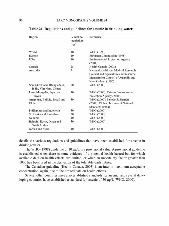
details the various regulations and guidelines that have been established for arsenic indrinking-water.
The WHO (1998) guideline of 10 µg/L is a provisional value. A provisional guidelineis established when there is some evidence of a potential health hazard but for whichavailable data on health effects are limited, or when an uncertainty factor greater than1000 has been used in the derivation of the tolerable daily intake.
The Canadian guideline (Health Canada, 2003) is an interim maximum acceptableconcentration, again, due to the limited data on health effects.
Several other countries have also established standards for arsenic, and several deve-loping countries have established a standard for arsenic of 50 µg/L (WHO, 2000).
IARC MONOGRAPHS VOLUME 8496
Table 21. Regulations and guidelines for arsenic in drinking-water
Region Guideline/ regulation (µg/L)
Reference
World 10 WHO (1998) Europe 10 European Commission (1998) USA 10 Environmental Protection Agency
(2001) Canada 25 Health Canada (2003) Australia 7 National Health and Medical Research
Council and Agriculture and Resource Management Council of Australia and New Zealand (1996)
South-East Asia (Bangladesh, India, Viet Nam, China)
50 WHO (2000)
Laos, Mongolia, Japan and Taiwan
10 WHO (2000); Taiwan Environmental Protection Agency (2000)
Argentina, Bolivia, Brazil and Chile
50 WHO (2000); Penedo & Zigarán (2002); Chilean Institute of National Standards (1984)
Philippines and Indonesia 50 WHO (2000) Sri Lanka and Zimbabwe 50 WHO (2000) Namibia 10 WHO (2000) Bahrain, Egypt, Oman and Saudi Arabia
50 WHO (2000)
Jordan and Syria 10 WHO (2000)
pp41-96.qxd 11/10/2004 10:19 Page 96
2. Studies of Cancer in Humans
Major epidemiological studies of cancer in relation to arsenic in drinking-waterinclude ecological studies and fewer case–control and cohort studies. For most otherknown human carcinogens, the major source of causal evidence arises from case–controland cohort studies, with little, if any, evidence from ecological studies. In contrast, forarsenic in drinking-water, ecological studies provide important information on causalinference, because of large exposure contrasts and limited population migration. As aconsequence of widespread exposure to local or regional water sources, ecologicalmeasures provide a strong indication of individual exposure. Moreover, in the case ofarsenic, the ecological estimates of relative risk are often so high that potential con-founding with known causal factors cannot explain the results. Hence, in the review thatfollows, ecological studies are presented in detail.
2.1 Cancer of the urinary bladder and kidney
The findings of epidemiological studies on arsenic in drinking-water and the risk forcancers of the urinary bladder and kidney are summarized in Table 22.
Historically, several case reports have related cancers of the urinary tract with medi-cinal arsenic treatments or arsenic-related diseases such as Bowen disease. In 1953, aseries of 27 cases with multiple skin cancers attributed to arsenical medicines wasreported (Sommers & McManus, 1953). Of these cases, 10 were diagnosed as also havinginternal cancers at various sites, three of which were urinary tract tumours. Graham andHelwig (1959) first investigated an association between Bowen disease and primaryinternal cancers. Twenty-eight (80%) of 35 cases had primary internal cancers, two ofwhich were malignant tumours of the bladder and one a tumour of the kidney. Cuzicket al. (1982) examined a cohort of subjects in the United Kingdom who had takenFowler’s solution (potassium arsenite) between 1945 and 1969. After further follow-up ofthe cohort through 1990 (Cuzick et al., 1992), a threefold increase in mortality frombladder cancer (standardized mortality ratio [SMR], 3.07; 95% confidence interval [CI],1.01–7.3) was reported, strengthening the evidence on bladder cancer reported previously(Cuzick et al., 1982).
Bergoglio (1964) published the first report of bladder cancer associated with arsenicin drinking-water in the Province of Cordóba in Argentina. He identified 2355 deathsbetween 1949 and 1959 in nine towns of a highly exposed region and found that cancerwas the cause of death of 24%; 11% of these cancer deaths involved cancers of the urinarytract. Biagini (1972) followed 116 patients with arsenic-related skin lesions in the sameregion and found that 12.5% of the cancer deaths were patients with urinary tract cancers.
ARSENIC IN DRINKING-WATER 97
pp97-166.qxd 11/10/2004 10:47 Page 97
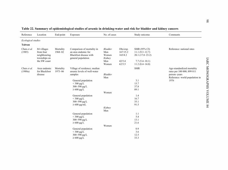
IARC M
ON
OG
RAPH
S VO
LUM
E 8498
Table 22. Summary of epidemiological studies of arsenic in drinking-water and risk for bladder and kidney cancers
Reference Location End-point Exposure No. of cases Study outcome Comments
Ecological studies
Taiwan
Chen et al. (1985)
84 villages from four neighbouring townships on the SW coast
Mortality 1968–82
Comparison of mortality in an area endemic for Blackfoot disease with general population
Bladder Men Women Kidney Men Women
Obs/exp. 167/15.2 165/8.2 42/5.4 62/5.5
SMR (95% CI) 11.1 (9.3–12.7) 20.1 (17.0–23.2) 7.7 (5.4–10.1) 11.2 (8.4–14.0)
Reference: national rates
Chen et al. (1988a)
Area endemic for Blackfoot disease
Mortality 1973–86
Village of residence; median arsenic levels of well-water samples General population < 300 µg/L 300–590 µg/L ≥ 600 µg/L General population < 300 µg/L 300–590 µg/L ≥ 600 µg/mL General population < 300 µg/L 300–590 µg/L ≥ 600 µg/L General population < 300 µg/L 300–590 µg/L ≥ 600 µg/L
Bladder Men Women Kidney Men Women
SMR 3.1 15.7 37.8 89.1 1.4 16.7 35.1 91.5 1.1 5.4 13.1 21.6 0.9 3.6 12.5 33.3
Age-standardized mortality rates per 100 000; 899 811 person–years Reference: world population in 1976
pp97-166.qxd 11/10/2004 10:47 Page 98
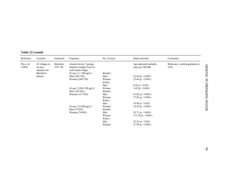
ARSEN
IC IN D
RINK
ING
-WATER
99
Table 22 (contd)
Reference Location End-point Exposure No. of cases Study outcome Comments
Wu et al. (1989)
42 villages in an area endemic for Blackfoot disease
Mortality 1973–86
Arsenic levels: 3 groups based on median level of well-water/village Group 1 (< 300 µg/L) Men (248 728) Women (248 728) Group 2 (300–590 µg/L) Men (138 562) Women (127 502) Group 3 (≥ 600 µg/L) Men (79 883) Women (74 083)
Bladder Men Women Kidney Men Women Bladder Men Women Kidney Men Women Bladder Men Women Kidney Men Women
Age-adjusted mortality rates per 100 000 22.64 (p < 0.001) 25.60 (p < 0.001) 8.42 (p < 0.05) 3.42 (p < 0.001) 61.02 (p < 0.001) 57.02 (p < 0.001) 18.90 (p < 0.05) 19.42 (p < 0.001) 92.71 (p < 0.001) 111.30 (p < 0.001) 25.26 (p < 0.05) 57.98 (p < 0.001)
Reference: world population in 1976
pp97-166.qxd 11/10/2004 10:47 Page 99

IARC M
ON
OG
RAPH
S VO
LUM
E 84100
Table 22 (contd)
Reference Location End-point Exposure No. of cases Study outcome Comments
Chen & Wang (1990)
314 geographical units (precincts and townships), including 4 townships in the endemic area of Blackfoot disease
Mortality 1972–83
Average arsenic levels in water samples of all geographical units. 73.9% of study precincts or townships had < 5% of wells with > 50 µg/L; 14.7% had 5–14%; 11.5% had ≥ 15%.
All 314 precincts and townships Bladder Men Women Kidney Men Women 170 south-western townships Bladder Men Women Kidney Men Women
3.9 (0.5) 4.2 (0.5) 1.1 (0.2) 1.7 (0.2) 3.7 (0.7) 4.5 (0.7) 1.2 (0.2) 1.7 (0.3)
Reference: world population in 1976. Analysis weighted by population in each group. Regression coefficients indicating an increase in age-adjusted mortality/100 000 person–years for every 0.1 µg/L increase in arsenic level (SE)
Chiang et al. (1993)
Incidence 1981–85
Exposure not evaluated Endemic area Neighbouring endemic area All Taiwan
Bladder Total Men Women Total Men Women Total Men Women
140 81 59 13 7 6 2135 1608 527
Incidence per 100 000 23.5 26.1 21.1 4.5 4.7 4.3 2.3 3.3 1.2
Adjusted for age
pp97-166.qxd 11/10/2004 10:47 Page 100
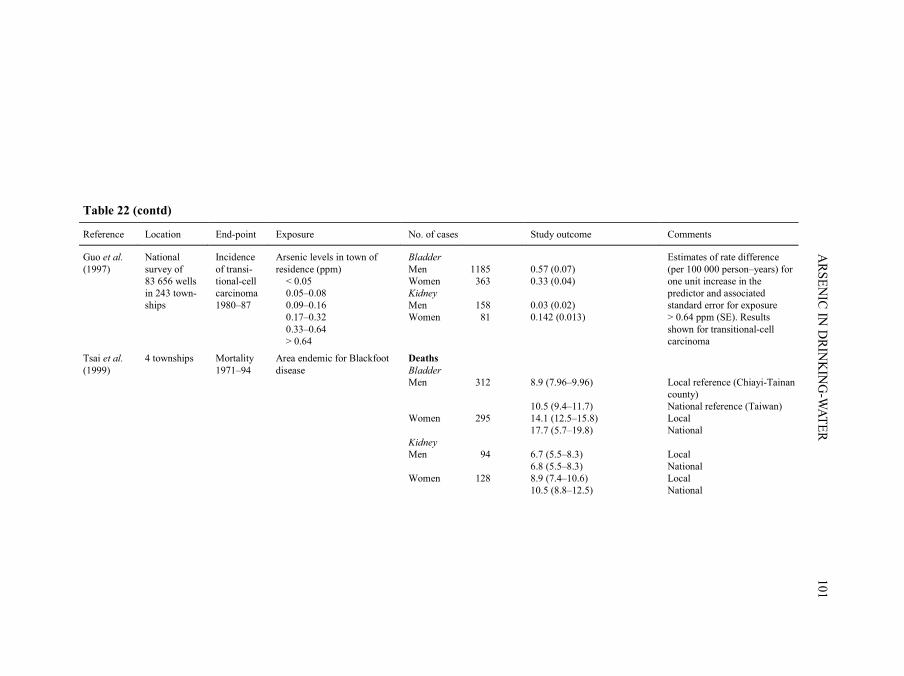
ARSEN
IC IN D
RINK
ING
-WATER
101
Table 22 (contd)
Reference Location End-point Exposure No. of cases Study outcome Comments
Guo et al. (1997)
National survey of 83 656 wells in 243 town-ships
Incidence of transi-tional-cell carcinoma 1980–87
Arsenic levels in town of residence (ppm) < 0.05 0.05–0.08 0.09–0.16 0.17–0.32 0.33–0.64 > 0.64
Bladder Men Women Kidney Men Women
1185 363 158 81
0.57 (0.07) 0.33 (0.04) 0.03 (0.02) 0.142 (0.013)
Estimates of rate difference (per 100 000 person–years) for one unit increase in the predictor and associated standard error for exposure > 0.64 ppm (SE). Results shown for transitional-cell carcinoma
Tsai et al. (1999)
4 townships Mortality 1971–94
Area endemic for Blackfoot disease
Deaths Bladder Men Women Kidney Men Women
312 295 94 128
8.9 (7.96–9.96) 10.5 (9.4–11.7) 14.1 (12.5–15.8) 17.7 (5.7–19.8) 6.7 (5.5–8.3) 6.8 (5.5–8.3) 8.9 (7.4–10.6) 10.5 (8.8–12.5)
Local reference (Chiayi-Tainan county) National reference (Taiwan) Local National Local National Local National
pp97-166.qxd 11/10/2004 10:47 Page 101

IARC M
ON
OG
RAPH
S VO
LUM
E 84102
Table 22 (contd)
Reference Location End-point Exposure No. of cases Study outcome Comments
South America
Hopenhayn-Rich et al. (1996a, 1998)
26 counties in the Province of Córdoba, Argentina
Mortality 1986–91
Exposure levels Low (690 421) Medium (406 000) High (mean arsenic level, 178 µg/L) (273 014) Low (690 421) Medium (406 000) High (273 014)
Deaths Bladder Men Women Men Women Men Women Kidney Men Women Men Women Men Women
113 39 116 29 131 27 66 38 66 34 53 27
SMR (95% CI) 0.8 (0.7–0.96) 1.2 (0.9–1.7) 1.3 (1.05–1.5) 1.4 (0.93–1.99) 2.1 (1.8–2.5) 1.8 (1.2–2.6) 0.9 (0.7–1.1) 1.0 (0.7–1.4) 1.3 (1.02–1.7) 1.4 (0.94–1.9) 1.6 (1.2–2.1) 1.8 (1.2–2.6)
Rivara et al. (1997)
Chile Mortality 1950–92
Arsenic-contaminated Region II of northern Chile versus non-contaminated region VIII
Bladder Kidney
SMR (95% CI) 10.2 (8.6–12.2) 3.8 (3.1–4.7)
SMR for period 1976–92
Smith et al. (1998)
Chile Mortality 1989–93
Region II of northern Chile with population-weighted average arsenic concentration up to 569 µg/L compared with rest of Chile; exposure generally < 10 µg/L
Bladder Men Women Kidney Men Women
SMR (95% CI) 6.0 (4.8–7.4) 8.2 (6.3–10.5) 1.6 (1.1–2.1) 2.7 (1.9–3.8)
Population of about 400 000
Australia
Hinwood et al. (1999)
Victoria Incidence 1982–91
Median arsenic concentration in drinking-water ranged 1–1077 µg/L
Bladder Kidney
303 134
SIR (95% CI) 0.9 (0.8–1.1) 1.2 (0.98–1.4)
State rates used as reference
pp97-166.qxd 11/10/2004 10:47 Page 102

ARSEN
IC IN D
RINK
ING
-WATER
103
Table 22 (contd)
Reference Location End-point Exposure No. of cases Study outcome Comments
Case–control studies
Taiwan
Chen et al. (1986)
4 neighbour-ing townships in endemic area of Black-foot disease
Mortality 1980–82
Median arsenic content of artesian well-water, 0.78 ppm Years of artesian water consumption 0 1–20 21–40 ≥ 40
Community controls 136 131 50 51
Bladder cancer cases 17 19 10 23
OR OR from multiple logistic regression analyses 1.0 1.0 1.2 1.3 1.6 1.7 3.9 4.1
Adjusted for age and sex
USA
Bates et al. (1995)
10 areas of the USA
Incident cases (aged 21–84 years) dia-gnosed in a 1-year period in the 1970s. Age-, sex- and area-matched controls
Cumulative dose (mg) < 19 19–32 33–52 ≥ 53 mg/L × years < 33 33–52 53–73 ≥ 74
Controls 47 36 39 38 42 42 40 36
Bladder cancer cases 14 21 17 19 18 16 16 21
OR (90% CI) All subjects 1.0 1.6 (0.8–3.2) 0.95 (0.4–2.0) 1.4 (0.7–2.9) 1.0 0.7 (0.3–1.5) 0.5 (0.3–1.2) 1.0 (0.5–2.1)
Adjusted for sex, age, smoking, years of exposure to chlorinated surface water, history of bladder infection, educational level, urbanization of the place of longest lifetime residence, ever employed in a high-risk occupation
pp97-166.qxd 11/10/2004 10:47 Page 103

IARC M
ON
OG
RAPH
S VO
LUM
E 84104
Table 22 (contd)
Reference Location End-point Exposure No. of cases Study outcome Comments
Bates et al. (1995) (contd)
Cumulative dose (mg) < 19 19–32 33–52 ≥ 53 mg/L × years < 33 33–52 53–73 ≥ 74 Cumulative dose (mg) < 19 19–32 33–52 ≥ 53 mg/L × years < 33 33–52 53–73 ≥ 74
Never smoked 1.0 1.1 (0.4–3.1) 0.7 (0.2–2.3) 0.5 (0.1–1.9) 1.0 0.2 (0.1–0.8) 0.3 (0.1–0.9) 0.9 (0.3–3.2) Ever smoked 1.0 3.3 (1.0–10.8) 1.9 (0.6–6.2) 3.3 (1.1–10.3) 1.0 1.95 (0.7–5.6) 1.2 (0.4–3.7) 1.4 (0.5–4.3)
pp97-166.qxd 11/10/2004 10:47 Page 104

ARSEN
IC IN D
RINK
ING
-WATER
105
Table 22 (contd)
Reference Location End-point Exposure No. of cases Study outcome Comments
Europe
Kurttio et al. (1999)
Areas in Finland in which < 10% of population belong to the municipal drinking-water system
Incidence 1981–95
Concentration of arsenic in water < 0.1 µg/L 0.1–0.5 µg/L ≥ 0.5 µg/L Total < 0.1 µg/L 0.1–0.5 µg/L ≥ 0.5 µg/L Total Concentration of arsenic in water < 0.1 µg/L 0.1–0.5 µg/L ≥ 0.5 µg/L Total < 0.1 µg/L 0.1–0.5 µg/L ≥ 0.5 µg/L Total
Short latency 23 19 19 61 Long latency 26 18 17 61 Short latency 23 12 14 49 Long latency 25 9 15 49
Bladder Kidney
Relative risk (95% CI) 1.0 1.5 (0.8–3.1) 2.4 (1.1–5.4) 1.4 (0.95–1.96) 1.0 0.8 (0.4–1.6) 1.5 (0.7–3.4) 0.96 (0.6–1.6) 1.0 0.8 (0.4–1.7) 1.5 (0.7–3.3) 1.2 (0.8–1.7) 1.0 0.3 (0.1–0.8) 1.07 (0.5–2.5) 0.7 (0.4–1.4)
Case–cohort design Adjusted for age, sex and smoking Short latency: exposure in the 3rd to 9th calendar year prior to the cancer diagnosis Long latency: exposure in the 10th calendar year and earlier prior to the cancer diagnosis
pp97-166.qxd 11/10/2004 10:47 Page 105

IARC M
ON
OG
RAPH
S VO
LUM
E 84106
Table 22 (contd)
Reference Location End-point Exposure No. of cases Study outcome Comments
Cohort studies
Taiwan
Chen et al. (1988b)
4 neighbour-ing townships in area endemic for Blackfoot disease
Mortality 1968–93
Comparison of mortality with general and endemic population
Cancer deaths Bladder Kidney Bladder Kidney
15 3 15 3
SMR 38.8 [21.7–64.0] 19.5 [4.0–57.0] SMR 2.6 [1.4–4.2] 1.6 [0.3–4.7]
95% CI calculated by the Working Group General population as reference Area endemic for Blackfoot disease as reference
Chiou et al. (1995)
4 neighbour-ing townships in area endemic for Blackfoot disease (BFD)
Cumulative index derived for each subject: Σ (Ci × Di). Cumulative exposure (mg/L × year) 0 0.1–19.9 ≥ 20 0 0.1–19.9 ≥ 20
BFD patients Healthy controls Bladder cancer cases
263 2293 29
Bladder cancer RR* (95% CI) 1.0 2.1 (0.6–7.2) 5.1 (1.5–17.3) RR** (95% CI) 1.0 1.6 (0.4–5.6) 3.6 (1.1–12.2)
*Relative risk after adjustment for age, sex and smoking **Relative risk after adjustment for age, sex, smoking and BFD status Ci, median concentration of arsenic in wells of village; Di, duration of drinking water in that village
pp97-166.qxd 11/10/2004 10:47 Page 106

ARSEN
IC IN D
RINK
ING
-WATER
107
Table 22 (contd)
Reference Location End-point Exposure No. of cases Study outcome Comments
Chiou et al. (2001)
North-eastern Taiwan
Incidence 1991–94 Person–years of observation 7978 6694 3013 5220 7978 6694 3013 5220
Area endemic for arseniasis Arsenic concentration in well-water (µg/L) < 10.0 10.1–50.0 50.1–100.0 > 100.0 < 10.0 10.1–50.0 50.1–100.0 > 100.0
3 3 2 7 1 1 2 6
RR (95% CI) Urinary tract 1.0 1.5 (0.3–8.0) 2.2 (0.4–13.7) 4.8 (1.2–19.4) Transitional-cell carcinoma 1.0 1.9 (0.1–32.5 8.2 (0.7–99.1) 15.3 (1.7–139.2)
Adjusted for age, sex, smoking and duration of drinking well-water p for trend < 0.01 p for trend < 0.05
Japan
Tsuda et al. (1995)
Niigata prefecture
1959–92 Arsenic concentration in well-water (ppm) from arsenic-polluted area < 0.05 0.05–0.99 ≥ 1 Total
No. of persons exposed at concen-tration level (1955–59) 254 76 113 443
Urinary Obs/exp. 0/0.3 0/0.08 3/0.10 3/0.48
SMR (95% CI) 0 (0–12.5) 0 (0–47.1) 31.2 (8.6–91.8) 6.3 (1.7–18.4)
pp97-166.qxd 11/10/2004 10:47 Page 107

IARC M
ON
OG
RAPH
S VO
LUM
E 84108
Table 22 (contd)
Reference Location End-point Exposure No. of cases Study outcome Comments
USA
Lewis et al. (1999)
Millard County, UT
Mortality Index of exposure to arsenic calculated for each cohort member and derived from number of years of residence and median arsenic concentration in the given community Low exposure (< 1000 ppb–years) Medium exposure (1000–4999 ppb–years) High exposure (≥ 5000 ppb–years) Total Low exposure (< 1000 ppb–years) Medium exposure (1000–4999 ppb–years) High exposure (≥ 5000 ppb–years) Total
Men Women Men Women Men Women Men Women Men Women Men Women Men Women Men Women
SMR (95% CI) Bladder/other urinary cancers 0.36 1.18 – – 0.95 1.10 0.4 (0.1–1.2) 0.8 (0.1–2.9) Kidney cancer 2.51 2.36 1.13 1.32 1.43 1.13 1.8 (0.8–3.3) 1.6 (0.4–4.1)
Confidence intervals not given for exposure categories 4058 members in cohort (2092 men and 1966 women)
SMR, standardized mortality ratio; CI, confidence interval; SIR, standardized incidence ratio; OR, odds ratio; RR, relative risk; BFD, Blackfoot disease
pp97-166.qxd 11/10/2004 10:47 Page 108

More systematic studies were conducted in various parts of the world, the most exten-sive being in Taiwan, China.
2.1.1 Studies in Taiwan, China
There are two areas in Taiwan, China, where exposure to arsenic is endemic. One islocated in the south-western coastal area where Blackfoot disease, a unique peripheralvascular disease induced by long-term ingestion of arsenic from artesian well-water, isendemic. Eighty-four villages constitute the four Blackfoot disease-endemic townships ofPeimen, Hsuehchia, Putai and Ichu, and artesian wells and patients with Blackfoot diseasewere also found in the two neighbouring townships of Yensui and Hsiaying (Wu et al.,1989). Residents in the south-western endemic areas in Taiwan drank artesian well-waterwith high concentrations of arsenic from the early 1910s to the late 1970s. The concen-trations of arsenic in artesian well-water, tested by Natelson’s method in 1964–66 in 42villages of the six townships, ranged from 10 to 1752 µg/L, and were mostly above100 µg/L (Kuo, 1968; Tseng et al., 1968). As well-water was the only source of drinking-water in the endemic area, all residents of a given village consumed the water from asmall number of shared wells in their daily home and working environments. Mostresidents were engaged in fishing, salt production and farming, and the migration rate waslow. More than 90% of residents had lived in the study area all their lives. As the studypopulation lived in a small area, they shared similar dietary patterns, lifestyle,socioeconomic status and health care facilities. The piped water supply system usingsurface water was first implemented in the south-western endemic area, but its coveragewas not complete until the late 1970s (Wu et al., 1989).
Another area with exposure to arsenic is located in the Lanyang Basin in north-eastern Taiwan (Chiou et al., 2001), and is comprised of four townships: Chiaohsi (fourvillages), Chuangwei (seven villages), Wuchieh (three villages) and Tungshan (fourvillages). Residents in this area of endemic arseniasis used river water for drinking andcooking before the Second World War, and started to use water from tubewells in theirhouses in the late 1940s. The concentrations of arsenic in the water of 3901 wells in theendemic area was tested from 1991 to 1994 by hydride generation combined with flameatomic absorption spectrometry, and ranged from undetectable (< 0.15 µg/L) to 3.59 ng/L,but were mostly between 10 and 100 µg/L, with a median of 27.3 µg/L. A piped watersupply system using surface water was first implemented in the endemic area in late 1997,and its coverage was almost complete in 2001. Residents in the endemic area wereengaged in farming, and most of them had lived in the area all their lives.
(a) Ecological studiesChen et al. (1985) reported an elevation in mortality from cancers of the urinary
bladder and kidney during the period 1968–82 in endemic areas of Blackfoot disease(four neighbouring townships comprising 84 villages) compared with the generalpopulation of Taiwan. The arsenic content of well-water ranged from 0.35 to 1.14 ppm
ARSENIC IN DRINKING-WATER 109
pp97-166.qxd 11/10/2004 10:47 Page 109
[mg/L] with a median of 0.78 ppm, while shallow well-water contained arsenic atconcentrations ranging from 0 to 0.3 ppm with a median of 0.04 ppm (Chen et al., 1962).The SMRs for bladder cancer and kidney cancer increased with the prevalence ofBlackfoot disease. Similarly, the SMRs for cancers of the bladder and kidney were highestin villages where only artesian wells were in use and lowest in those villages usingshallow wells. The high SMRs for bladder and kidney cancer were not readily explainedby the higher rate of cigarette smoking in the Blackfoot disease-endemic area comparedwith all of Taiwan (40% versus 32%).
Chen et al. (1988a) briefly described a dose–response relationship between medianarsenic levels in artesian well-water in the 84 villages studied by Chen et al. (1985) andrates of mortality from bladder cancer. The study period (1973–86) covered 899 811person–years of observation, and exposure was stratified into three categories (< 300,300–590 and ≥ 600 µg/L arsenic) based on concentrations from a survey of over 83 000wells, including 313 townships in all of Taiwan, conducted from 1962 to 1964.
Wu et al. (1989) examined age-adjusted mortality rates for various cancers in an areaof south-western Taiwan that comprised 42 villages in six townships (27 villages studiedby Chen et al. (1988a) and another 15 villages). The arsenic content of the 155 wellssampled, measured in 1964–66, ranged from 10 to 1750 µg/L. The villages were classifiedaccording to median arsenic levels in water into three exposure groups (< 300, 300–590and ≥ 600 µg/L). Death certificates were used to ascertain cause of death during the period1973–86. A dose–response relationship was found with concentration of arsenic in waterfor cancers of the bladder and kidney for both men and women.
Chen and Wang (1990) further investigated cancer mortality rates throughout Taiwanin 1972–83. Of 361 administrative areas, 314 were included in the study followingmeasurements of arsenic contents in well-water in 1974–76. Exposure measurementswere derived from a national water survey of over 83 000 wells throughout Taiwan. About19% of the wells contained levels of arsenic above 50 µg/L, and most of them were innorth-western and south-eastern Taiwan. Indices of urbanization and industrializationwere included in the analysis to adjust for the possible confounding effect of differingsocioeconomic characteristics between the 314 precincts and townships. Mortality datawere used to evaluate 21 malignant neoplasms, using population-weighted regression.Results were presented for increases in mortality per 100 000 that were calculated to occurfor every 0.1-mg/L increase in arsenic concentration in water.
Chiang et al. (1993) showed that the age-adjusted incidence of bladder cancer in theperiod 1981–85 in the Blackfoot disease-endemic area of Taiwan was higher than that ina neighbouring area of Taiwan and in the country as a whole.
Guo et al. (1997) used tumour registry data and the exposure data from the 1974–76nationwide water-quality survey used by Chen and Wang (1990), which included concen-trations of arsenic in drinking-water from 243 townships with about 11.4 million resi-dents. The annual incidence of cancers of the bladder and kidney for townships in1980–87 and subcategories of those cancer diagnoses were regressed against a model thatincluded six variables for the proportion of wells in each of six categories of arsenic
IARC MONOGRAPHS VOLUME 84110
pp97-166.qxd 11/10/2004 10:47 Page 110

concentration in each township. Sex-specific models were adjusted for age and includedan urbanization index and the annual number of cigarettes sold per capita. Regressionmodels were weighted by the total population of each township. A total of 1962 bladder,726 kidney, 170 ureter and 57 urethral cancers were included. The investigators foundassociations of high arsenic concentrations (more than 0.64 ppm) in both sexes withtransitional-cell carcinomas of the bladder, kidney and ureter, and all urethral cancerscombined, but they did not present relative risk estimates.
Tsai et al. (1999) compared mortality in people aged over 40 years in the Blackfootdisease-endemic area of Taiwan with both local and national references for the period1971–94. Greater mortality was found for men and women with cancers of the bladderand kidney.
(b) Case–control studyIn a retrospective case–control study using death certificates from 1980–82, Chen
et al. (1986) examined the relationship between exposure to high concentrations ofarsenic in artesian well-water and mortality from internal malignancies, includingtumours of the bladder (n = 69) in four townships from the Blackfoot disease-endemicarea. Controls (n = 368) were selected by random sampling from the same geographicalareas as the cases and were frequency-matched on age and sex. The response rate was93% for proxies of cases and 92% for matched controls. Adjustment for age, sex and othervariables (smoking, tea drinking, vegetarianism and frequency of consumption of vege-tables and fermented beans) was performed by logistic regression analysis. The resultsindicated increasing trends in odds ratios with increasing duration of intake of artesianwell-water containing arsenic. The highest risks were seen for over 40 years of exposure,with an odds ratio of 4.1 for bladder cancer in a multivariate analysis. Smoking, alcoholconsumption and other potential risk factors evaluated in the study did not confound theassociation between arsenic and cancer.
(c) Cohort studiesChen et al. (1988b) studied the association between arsenic in artesian well-water in
relation to Blackfoot disease and cancer from a multiple risk factor perspective. The studyarea included the four townships in south-western Taiwan where high rates of Blackfootdisease had been described. Levels of arsenic were reported to be high in water, soil andfood, and estimates of ingestion of arsenic by local residents were up to 1 mg per day. Thestudy examined mortality in a cohort of people who had or had since developed Blackfootdisease in 1968, totalling 789 patients and 7578 person–years of observation through1984. Follow-up started in 1968, since this was the year that registration of deaths inTaiwan was computerized and completeness and quality of death certificate registrationwas improved. Mortality of persons who had died (n = 457) and were not lost to follow-up (n = 84) was compared with that of the general population of Taiwan using age- andsex-specific mortality rates from 1968 through 1983. The SMRs for cancers of the bladderand kidney (men and women combined) were 38.8 [95% CI, 21.7–64.0] and 19.5
ARSENIC IN DRINKING-WATER 111
pp97-166.qxd 11/10/2004 10:47 Page 111

[95% CI, 4.0–57.0], respectively. The latter result, however, is based on only three deaths.SMRs were also calculated using all residents in the Blackfoot disease-endemic area,which includes people exposed to arsenic. These much lower SMRs were 2.6 [95% CI,1.4–4.2] for bladder cancer and 1.6 [95% CI, 0.3–4.7] for kidney cancer, indicating thatpatients with Blackfoot disease had somewhat higher rates for these cancers than theresidents in the arsenic-exposed region combined.
Chiou et al. (1995) investigated the relationship between incidence of internal cancersand arsenic in relation to Blackfoot disease in 2256 subjects from 1986 to 1993. Patientswith Blackfoot disease (n = 263) and a referent group of 2293 residents of the same regionwere followed for 7 years. In contrast to many other studies that evaluate mortality,incident cancer was the outcome of interest. Follow-up occurred many years afterexposure to elevated concentrations of arsenic in drinking-water had ended. Informationon exposure to other risk factors was gathered by individual interviews. Several measuresof exposure were evaluated, including average concentration of arsenic in artesian wellsand cumulative exposure to arsenic from drinking artesian well-water. Relative risks werecalculated using Cox’s proportional hazard regression analysis. After controlling for theeffects of age, sex and smoking in the regression analysis, a dose–response relationshipwas observed between exposure to arsenic from drinking well-water and the incidence ofbladder cancer. Patients with Blackfoot disease were found to be at increased risk evenafter adjustment for cumulative exposure to arsenic.
Chiou et al. (2001) studied the incidence of urinary tract cancers among 8102 residentsin the arsenic-endemic area in north-eastern Taiwan from 1991 to 1994. Levels of arsenicin the drinking-water ranged from less than 0.15 µg/L (undetectable) to 3590 µg/L.Exposure for each member of the cohort was assessed by measuring concentrations ofarsenic in the well associated with the individual’s household at one point in time only,although most households had used their current wells for at least 10 years (Chen & Chiou,2001). Using the general population as referent, the standardized incidence ratio (SIR) forbladder cancer was 1.96 (95% CI, 0.9–3.6), while that for kidney cancer was 2.8 (95% CI,1.3–5.4). These results were based on 10 subjects with bladder cancer and nine with kidneycancer. A dose–response relationship was observed between urinary tract cancers,particularly transitional-cell carcinoma, after adjusting for age, sex and smoking.
2.1.2 Studies in Japan
A retrospective cohort study was conducted in a small Japanese population, which,between 1955 and 1959, used well-water contaminated with arsenic from a factoryproducing King’s yellow (As2O3; Tsuda et al., 1995). The levels of arsenic measured in34 of 54 wells tested in the area around the factory ranged from undetectable to3000 µg/L, with 11 wells having levels exceeding 1000 µg/L. A total of 454 residentswere enlisted in the cohort. Death certificates, autopsy records and medical records wereobtained for the period 1 October 1959 to 30 September 1987. Smoking and occupationalhistories were ascertained from residents or close relatives. Expected numbers of deaths
IARC MONOGRAPHS VOLUME 84112
pp97-166.qxd 11/10/2004 10:47 Page 112

were based on sex-, age- and cause-specific mortality in Niigata Prefecture from 1960 to1989. Exposure was grouped into high (≥ 500 µg/L), medium (50–500 µg/L ) and low(< 50 µg/L), based on the arsenic content of the well-water. The SMR for urinary-tractcancer in the high-exposure group was 31.2 (95% CI, 8.6–91.8) based on three observeddeaths versus 0.1 expected. Two of these were deaths from bladder cancer and one fromcancer of the renal pelvis. [Excluding the cancer of the renal pelvis, the SMR for bladdercancer alone would be at least 20.]
2.1.3 Studies in South America
(a) ArgentinaAs early as the beginning of the twentieth century, physicians noted an increase in the
incidence of clinical skin alterations due to arsenic in well-water in certain areas of theProvince of Córdoba, Argentina (Hopenhayn-Rich et al., 1996a). In a study of 2355 deathsin 1949–59 in a highly exposed region, Bergoglio (1964) found that 24% of deaths in theexposed region were due to cancer compared with 15% in the Province of Córdoba. In a14-year follow-up of 116 patients diagnosed with arsenic-related skin lesions, 30.5% diedof cancer, and 12.5% of these deaths were due to cancers of the urinary tract (Biagini,1972). This was later contrasted with bladder cancer mortality in all of Argentina in 1980,with 2.9% of all cancer deaths attributable to bladder cancer (Hopenhayn-Rich et al.,1996a).
These early reports led to an ecological study on bladder cancer mortality for theperiod 1986–91 comparing counties categorized as previously having had high, mediumand low concentrations of arsenic in water in Córdoba, Argentina (Hopenhayn-Rich et al.,1996a). The majority of reported cases of arsenic-related skin lesions were residents oftwo counties that were classified as having high exposure since there were extensivereports of elevated concentrations of arsenic in the water there. The average concentrationof arsenic in water tested in these counties was 178 µg/L. The medium-exposure groupcomprised six counties based on some reports of elevated arsenic levels in the water andthe occurrence of a few cases of skin lesions. The remaining 16 rural counties wereclassified as having low exposure. Clear trends in mortality from bladder cancer wereobserved. Increasing trends were also observed for mortality from kidney cancer as expo-sure to arsenic increased (Hopenhayn-Rich et al., 1998). No differences were foundbetween the exposure groups for chronic obstructive pulmonary disease, suggesting thatthe trends for bladder and kidney cancer were not attributable to confounding by smoking.
(b) ChileChile is a long and narrow country divided into geopolitical units called regions (like
provinces in other countries), which are numbered sequentially from north to south,starting with Region I. Region II is thus located in the northern part of Chile, in an aridzone where the Atacama Desert is situated. At the time of the 1992 Census, the populationin Region II was about 420 000. About 90% of the population live in the cities and towns
ARSENIC IN DRINKING-WATER 113
pp97-166.qxd 11/10/2004 10:47 Page 113

in this Region, and more than half of the population lives in the city of Antofagasta (Smithet al., 1998). In view of the extremely low level of rainfall and the inability to obtain waterfrom wells, each city and town obtains drinking-water from rivers which originate in theAndes mountains, located on the eastern border of the Region. Many of these rivers arenaturally contaminated with inorganic arsenic, some at very high concentrations. This hasresulted in widespread exposure of the population of Region II to varying levels of arsenicin the drinking-water. In 1955–70, the majority of the population of Region II wasexposed to very high levels of arsenic in drinking-water (see Table 17). Prior to 1955, thedrinking-water supply in the main city of Antofagasta had an arsenic concentration ofabout 90 µg/L. A growing population, and the consequent increased need for water, led tosupplementation of the drinking-water supply at Antofagasta with water from the Toconceand Holajar Rivers, which, unknown at the time, had arsenic concentrations of 800 µg/Land 1300 µg/L, respectively. The concentration of arsenic in the drinking-water atAntofagasta, together with that of neighbouring Mejillones, which shared the samesupply, increased to an average of 870 µg/L. As shown in Table 17, the other towns in theregion, with the exception of Taltal, also had high concentrations of arsenic in thedrinking-water for variable periods. The population-weighted average concentration ofarsenic in drinking-water for the entire region was about 580 µg/L over a period ofapproximately 15 years (1955–70). With the introduction in 1970 of a water-treatmentplant, the concentration of arsenic in the water at Antofagasta initially dropped to260 µg/L, and further reductions occurred as a result of improvements to the treatmentplant. At present, levels of arsenic in water in Antofagasta are about 40 µg/L. Other citiesand towns also implemented water-treatment strategies or used alternative sources thatreduced arsenic levels. By the late 1980s, all of the towns with populations over 1000 hadconcentrations of arsenic in drinking-water of less than 100 µg/L, with the exception ofSan Pedro de Atacama (population about 3700, some of whom still drink the contami-nated water). In contrast, most water sources in the rest of Chile have had low concen-trations of arsenic (less than 10 µg/L) (Ferreccio et al., 2000).
Evidence of chronic arsenic toxicity in Region II was noted in the 1960s with theemergence of classic dermatological manifestations (Borgoño & Greiber, 1971; Zaldívar,1974; Zaldívar et al., 1978). In 1969, in a study of 180 residents in Antofagasta, abnormalskin pigmentation was found in 144 of the participants, 43.7% of whom also had hyper-keratoses (Borgoño et al., 1977). Evidence of effects on the respiratory and cardiovascularsystem, together with skin lesions, was also reported by Zaldívar et al. (1978), whoconducted a series of studies concerning the effects of arsenic in Antofagasta during thehigh-exposure period.
Two ecological mortality studies were conducted on kidney and bladder cancer inRegion II. Rivara et al. (1997) conducted a study comparing mortality for both sexes com-bined in Region II with that in Region VIII for the period 1950–92. SMRs for bladdercancer and kidney cancer were 10.2 (95% CI, 8.6–12.2) and 3.8 (95% CI, 3.1–4.7),respectively.
IARC MONOGRAPHS VOLUME 84114
pp97-166.qxd 11/10/2004 10:47 Page 114
Smith et al. (1998) also investigated cancer mortality in Region II for the years1989–93, using mortality rates in the rest of Chile (excluding Region II) in 1992, a censusyear, for reference. SMRs were calculated for men and women over the age of 30 years,using 10-year age groupings. The results indicated marked increases in mortality frombladder cancer and kidney cancer in Region II. Data on smoking obtained from a nationalsurvey of stratified random samples carried out in 1990, comparing the two largest citiesof Antofagasta and Calama with the rest of Chile, were included. No overall increases inmortality from chronic obstructive pulmonary disease were observed in Region II: theSMR for men was 1.0 (0.8–1.1) and mortality among women was lower than expected(SMR, 0.6; 0.4–0.7). In addition, not only did the national survey not find higher rates ofsmoking in Region II, but in Antofagasta, 76.4% of respondents reported being non-smokers at that time compared with 75.1% of respondents in the rest of Chile. The pro-portion of people who smoked more than one pack per day was lower in Antofagasta(0.8%) and Calama (1.1%) than in the rest of Chile (1.5%). The SMRs for other causesof death excluding cancers of the bladder, kidney, lung, liver and skin were 1.0 (95% CI,0.99–1.05) for men and 1.0 (95% CI, 0.97–1.03) for women.
2.1.4 Studies in the USA
Some ecological studies have been reported in the USA but they are not informativein view of the relatively small contrasts in exposure between counties.
(a) Case–control studyBates et al. (1995) linked 71 cases of bladder cancer and 160 controls from a sub-
sample of residents of Utah from the large national bladder cancer study conducted in1978 (Cantor et al., 1987) to levels of arsenic in water supplies. Exposures ranged from0.5 to 160 µg/L, but most concentrations were very low, with only 1.1% of sampleshaving concentrations greater than 50 µg/L. The findings did not provide evidence for anoverall increase in the incidence of bladder cancer with the two indices of exposure used.However, among smokers only, there was an increase in risk for the highest category ofcumulative dose of arsenic.
(b) Cohort studyLevels of arsenic in drinking-water and mortality were investigated in a cohort of
members of the Church of Jesus Christ of Latter-day Saints in Millard County, UT (Lewiset al., 1999). The cohort was assembled from an earlier study that consisted of 2073 parti-cipants (Southwick et al., 1983). Most of these individuals had a history of at least 20 yearsof exposure in their respective places of residence. The cohort was expanded to include allpersons who lived for any length of time in the study area, resulting in a total combinedcohort of 4058 members. More than 70% of the cohort had reached the age of 60 years atthe end of the follow-up period or by the time of their deaths. Approximately 7% of thecohort was lost to follow-up. Arsenic concentrations in the drinking-water supplies were
ARSENIC IN DRINKING-WATER 115
pp97-166.qxd 11/10/2004 10:47 Page 115

based on measurements maintained by the state of Utah dating back to 1964. The mediandrinking-water concentrations ranged from 14 to 166 ppb [µg/L], with wide variability ineach town. An index of exposure to arsenic was calculated from the number of years ofresidence and the median concentration of arsenic in drinking-water in a given community,and was categorized as low (< 1000 ppb–years), medium (1000–4999 ppb–years) and high(> 5000 ppb–years). Data on confounding factors were not available; however, Churchmembers are prohibited from using tobacco and consuming alcohol or caffeine. SMRs forkidney cancer were increased in the low- and high-exposure groups among men and in thelow- and medium-exposure groups among women. The overall SMRs for cancers of thebladder and other urinary organs were below unity for both sexes, but these results werebased on only three and two bladder cancers in men and women, respectively. [TheWorking Group noted that there were several problems in the interpretation of this study.The exposure assessment was ecological in nature because of relatively low exposures.There was widespread variability in water concentrations, which adds to the uncertainty ofthe study. Furthermore, the findings are influenced by lower rates of smoking for the cohortcompared with all of Utah. This is manifest in the SMRs for non-malignant respiratorydisease (SMR for men, 0.7; 95% CI, 0.5–0.9; SMR for women, 0.9; 95% CI, 0.7–1.2) andfor mortality from chronic bronchitis, emphysema and asthma (SMR for men, 0.6; 95% CI,0.4–0.9; SMR for women, 0.5; 95% CI, 0.1–1.2). For these reasons, the study is uninfor-mative with regard to the relationship between exposure to arsenic and mortality frombladder and kidney cancer.]
2.1.5 Studies in Europe
In a type of case–control study known as case–cohort, Kurttio et al. (1999) investigatedthe association between low exposure to arsenic in well-water in Finland and the risk forcancers of the bladder and kidney. Cases of bladder and kidney cancer were identified from1981 to 1995 within a registry-based cohort of the population who had lived at an addressoutside the municipal drinking-water system during 1967–80. The final study populationconsisted of 61 cases of bladder cancer and 49 cases of kidney cancer and an age- and sex-matched random sample of 275 subjects from the population register. The daily dose ofarsenic in drinking-water was calculated from the concentration of arsenic in well-waterand its reported consumption in the 1970s. Cumulative dose was defined as the integral ofduration and intensity of exposure to arsenic from well-water. For the shorter latencyperiod of 3–9 years prior to diagnosis of bladder cancer, cumulative dose was estimatedfrom the beginning of use of well-water until 2 years before the diagnosis of cancer. Forthe longer latency period, the cumulative dose was calculated until 10 years beforediagnosis. The concentrations of arsenic in the wells of the reference cohort ranged fromless than 0.05 to 64 µg/L (median, 0.14 µg/L). After adjusting for age, sex and smoking, anincreasing trend of arsenic in drinking-water and incidence of bladder cancer was observedwith shorter latency but not with longer latency, whereas no evidence of an associationbetween kidney cancer and arsenic in well-water was observed.
IARC MONOGRAPHS VOLUME 84116
pp97-166.qxd 11/10/2004 10:47 Page 116

2.1.6 Studies in Australia
Two geographical areas in Victoria, Australia, were selected for study because ofreports of concentrations of arsenic in the soil above 100 µg/g and/or concentrations inwater above 10 µg/L (Hinwood et al., 1999). Median concentrations of arsenic in waterwere reported for various towns and showed a wide range up to a median of 1077 µg/Lfor Ballarat, which had a population of 43 947 in 1986. However, the extent to which con-taminated water was used for drinking was not known. The authors noted that “highpercentages of the population may be relying on alternative drinking-water sources suchas bottled water and tank rain water”. Cancer incidence was assessed for the period1982–91 using the Victorian Cancer Registry. SIRs were estimated for the exposed popu-lation in 22 areas of Victoria, using cancer incidence rates for all of the State of Victoriaas reference. SIRs for both bladder and kidney cancers were close to unity. [The WorkingGroup noted that no information was presented on the actual use of water contaminatedwith arsenic for drinking by the population.]
2.2 Liver and lung cancer
2.2.1 Liver cancer
A previous IARC monograph on arsenic noted reports of liver angiosarcoma due tomedicinal exposure to Fowler’s solution (IARC, 1980). A summary of the findings of epi-demiological studies on arsenic in drinking-water and risk for liver cancer, mainly hepato-carcinoma, are shown in Table 23.
(a) Taiwan, China(i) Ecological studies
Chen et al. (1985) studied the mortality from liver cancer during the period 1968–82among residents in 84 villages exposed to arsenic of four townships in south-westerncoastal Taiwan. Increased mortality was observed among both men and women. There wasan exposure–response relationship between SMR and prevalence of Blackfoot disease. Anexposure–response gradient for mortality from liver cancer was noted in evaluating the riskin areas with shallow wells (presumably with low exposure to arsenic), both shallow andartesian wells (intermediate exposure) and artesian wells only (highest exposure). Invillages with artesian wells, the SMR was approximately 2.0 [CI not reported] for livercancer.
Chen et al. (1988a) and Wu et al. (1989) reported the age-adjusted mortality rates forliver cancer for men and women in 42 villages in south-western Taiwan and calculatedage-adjusted cancer mortality during the period 1973–86 within three groups of villagesstratified by exposure concentration (< 300 µg/L, 300–590 µg/L and ≥ 600 µg/L arsenic)tested in 1964–66. Age-adjusted mortality rates (per 100 000 person–years) from livercancer for residents of all ages in Taiwan (referent) increased with increasing concen-
ARSENIC IN DRINKING-WATER 117
pp97-166.qxd 11/10/2004 10:47 Page 117

IARC M
ON
OG
RAPH
S VO
LUM
E 84118
Table 23. Summary of epidemiological studies on arsenic in drinking-water and risk for liver cancer
Reference Location End-point Exposure No. of cases Study outcome Comments
Ecological studies
Taiwan
Chen et al. (1985)
84 villages on the SW coast
Mortality 1968–82, all ages
Endemic area for chronic arsenic toxicity (Blackfoot disease)
Men Women
305 146
Age- and sex-adjusted SMR (95% CI) 1.7 (1.5–1.9) 2.3 (1.9–2.7)
Mid-year population: 141 733 in 1968, 120 607 in 1982; national rate in 1968–82 used as the standard for estimation of SMR
Chen et al. (1988a)
42 villages on the SW coast
Mortality 1973–86, all ages
Average arsenic (1962–64) General population < 300 µg/L 300–590 µg/L ≥ 600 µg/mL General population < 300 µg/L 300–590 µg/L ≥ 600 µg/mL
Men Women
Age-adjusted SMR 28.0 32.6 42.7 68.8 8.9 14.2 18.8 31.8
899 811 person–years, rate per 100 000, age-standardized to 1976 world population
Wu et al. (1989)
42 villages on the SW coast
Mortality 1973–86, age ≥ 20
Average arsenic (1962–64) < 300 µg/L 300–590 µg/L ≥ 600 µg/mL < 300 µg/L 300–590 µg/L ≥ 600 µg/mL
Men Women
54 42 27 25 16 10
47.8 67.7 86.7 p for trend < 0.05 21.4 24.2 31.8 p for trend < 0.05
Men, 257 935 person–years; women, 234 519 person–years; rate per 100 000, age-standardized to 1976 world population
pp97-166.qxd 11/10/2004 10:47 Page 118

ARSEN
IC IN D
RINK
ING
-WATER
119
Table 23 (contd)
Reference Location End-point Exposure No. of cases Study outcome Comments
Chen & Wang (1990)
Taiwan Mortality 1972–83, all ages
National survey of 83 656 wells (1974–76); average arsenic for each of 314 precincts or townships
Men Women Men Women
β (SE) from regression 6.8 (1.3) 2.0 (0.5) Percentiles of age-adjusted mortality rate/100 000 person–years 25th 21.8 50th 27.0 75th 34.1 25th 7.0 50th 8.7 75th 11.6
Regression coefficient (β) estimates increase in age-adjusted mortality per 100 000 per 100 µg/L arsenic increase in water
Tsai et al. (1999)
SW Taiwan, 4 townships
Mortality 1971–94, all ages
Arsenic-exposed area Men Women
631 224
SMR (95% CI) 1.8 (1.7–1.98) 1.9 (1.6–2.1)
Men, 1 508 623 person–years; women, 1 404 759 person–years; national rates in 1971–94 used as the standard for estimation of SMR
South America
Rivara et al. (1997)
Region II and VIII, northern Chile
Mortality 1976–92
Arsenic-contaminated Region II
Relative risk 1.2 (0.99–1.6)
Population: 411 000 in Region II, 1 700 000 in Region VIII. Antofagasta (Region II) versus Region VIII.
Hopenhayn-Rich et al. (1998)
Córdoba Province, Argentina, 26 counties
Mortality 1986–91, age ≥ 20
County group Men Low exposure (341 547) Medium exposure (201 546) High exposure (135 209)
186 142 98
SMR 1.5 (1.3–1.8) 1.8 (1.5–2.1) 1.8 (1.5–2.2)
National rate in 1989 used as the standard for estimation of SMR
Women Low exposure (348 874) Medium exposure (204 454) High exposure (137 805)
173 125 90
1.7 (1.4–1.96) 1.9 (1.6–2.2) 1.9 (1.5–2.4)
pp97-166.qxd 11/10/2004 10:47 Page 119

IARC M
ON
OG
RAPH
S VO
LUM
E 84120
Table 23 (contd)
Reference Location End-point Exposure No. of cases Study outcome Comments
Smith et al. (1998)
Region II, Northern Chile
Mortality 1989–93, age ≥ 30
5-year intervals, 420 µg/L average
Men Women
48 37
SMR 1.1 (0.8–1.5) 1.1 (0.8–1.5)
National rates in 1991 used as the standard estimation of SMR; arsenic concentration is population-weighted average for major cities or towns in Region II, 1950–74
Australia
Hinwood et al. (1999)
Victoria Incidence 1982–91
Median concentration of arsenic in drinking-water ranged 1–1077 µg/L
749
SIR (95% CI) 0.5 (0.3–0.8)
State rates in 1982–91 used as the standard for estimation of SIR
Cohort studies
Chen et al. (1988b)
SW Taiwan Mortality Area endemic for Blackfoot disease
17 SMR: 4.66 (p < 0.001) compared with national standard; 2.48 (p < 0.01) compared with regional standard
789 patients with Blackfoot disease followed from 1968 to 1984. National and regional rates in 1968–83 used as the standard for estimation of SMR
Tsuda et al. (1995)
Niigata Prefecture, Japan
Mortality, 1959–92, all ages
Level of arsenic < 0.05 mg/L 0.05–0.99 mg/L ≥ 1.0 mg/L Total
0 0 2 2
SMR 0.0 (0–4.4) 0.0 (0–15.1) 7.2 (1.3–26.1) 1.5 (0.3–5.5)
113 persons who drank from industrially contaminated wells in 1955–59, then followed for 33 years; rates in Niigata Prefecture in 1960–89 used as the standard for estimation of SMR
Lewis et al. (1999)
Millard County, UT, USA
Mortality Arsenic in well-water ranged 3.5–620 µg/L
Men Women
3 7
SMR 0.9 (0.2–2.5) 1.4 (0.6–2.9)
State rates in 1950–92 used as the standard for estimation of SMR.
Nakadaira et al. (2002)
Niigata Prefecture, Japan
Mortality Industrially contaminated well-water in the town of Nakajo
1 O/E = 0.7 86 patients with chronic arsenic poisoning. National rates in 1959–92 used as the standard for estimation of SMR
pp97-166.qxd 11/10/2004 10:47 Page 120

ARSEN
IC IN D
RINK
ING
-WATER
121
Table 23 (contd)
Reference Location End-point Exposure No. of cases Study outcome Comments
Case–control study
Chen et al. (1986)
SW Taiwan, 4 townships
Mortality Duration of consumption of artesian well-water containing high levels of arsenic
65 cases 368 controls
Age- and gender-adjusted ORs by years of consuming high-arsenic artesian well-water: Never 1.00 1–20 years 0.85 21–40 years 1.24 > 40 years 2.67
ORs calculated using subjects who never consumed artesian well-water as referent Mantel-Haenszel χ2 value: 9.01 (p < 0.01)
SMR, standardized mortality ratio; CI, confidence interval; SIR, standardized incidence ratio; O/E, observed/expected; OR, odds ratio
pp97-166.qxd 11/10/2004 10:47 Page 121
trations of arsenic in water for both men and women in the first study (Chen et al., 1988a),as well as for residents aged 20 years or older in the second study (Wu et al., 1989).
Chen and Wang (1990) analysed nationwide cancer mortality in Taiwan usingmeasurements of arsenic concentrations in water from 83 656 wells located in 314precincts and townships from 1974 to 1976. Using a multiple regression approach, theauthors compared age-adjusted mortality for all ages during the period 1972–83 witharsenic concentrations in these locations. A significant association with concentration ofarsenic was found for liver cancer in both men and women. Using multiple linearregression models, a regression coefficient indicating the change in age-adjusted morta-lity per 100 000 person–years for every 0.1 µg/L increase in arsenic in well-water wascalculated, after adjusting for indices of industrialization and urbanization.
Tsai et al. (1999) studied mortality from liver cancer in four townships exposed toarsenic in south-western Taiwan during the period 1971–94. SMRs were calculated usingtwo comparison groups: mortality in the whole of Taiwan and mortality in the twocounties in which the four townships are located. Although differences in nutrition, socio-economic status or other factors between populations in south-western Taiwan and theremainder of the country may influence their respective cancer rates, Tsai et al. (1999)provided evidence that such differences are relatively unimportant. SMRs in both men andwomen, using both regional and national references, were all close to 1.8.
(ii) Case–control studyChen et al. (1986) carried out a case–control study on liver cancer and consumption
of artesian well-water with high concentrations of arsenic in four townships of south-western Taiwan. A total of 65 cases of liver cancer, identified from death certificates, and368 healthy controls were studied. Information on consumption of arsenic-contaminatedartesian well-water, cigarette smoking, habitual alcohol and tea drinking, and consump-tion of vegetables and fermented beans was obtained through interview using a standar-dized questionnaire. Unconditional logistic regression was used to estimate multivariate-adjusted odds ratios for developing liver cancer and various risk factors. There was anexposure–response relationship between the duration of consumption of artesian well-water with high arsenic content and risk for liver cancer.
(b) JapanCohort study
Tsuda et al. (1995) found excess mortality from liver cancer among a cohort of 113persons exposed to levels of arsenic above 1.0 mg/L from industrially contaminateddrinking-water in villages of Niigata Prefecture, Japan. The expected number of deaths wasbased on sex-, age- and cause-specific mortality in Niigata Prefecture in 1960–89. Basedon a subgroup of 86 study patients, Nakadaira et al. (2002) did not find excess mortalityfrom liver cancer (SMR, 0.7 [95% CI, 0.02–3.9]; one case observed and 1.42 expected).[The small number of liver cancer deaths limited further analysis by severity of chronicarsenic poisoning.] [See complete comment by the Working Group in Section 2.1.2.]
IARC MONOGRAPHS VOLUME 84122
pp97-166.qxd 11/10/2004 10:47 Page 122

(c) Australia Hinwood et al. (1999) investigated the association between arsenic in drinking-water
and liver cancer incidence in Victoria, Australia, in 1982–91. This study included 22 areaswhere the median arsenic concentration in drinking-water ranged from 14 to 166 µg/L.Using the incidence rate in Victoria, an SIR of 0.5 (95% CI, 0.3–0.8) was observed forliver cancer. [The small number of liver cancer deaths limited further analysis by severityof chronic arsenic poisoning.]
(d) South AmericaEcological studies
Rivara et al. (1997) compared the mortality from liver cancer in Antofagasta inRegion II with that in Region VIII of Chile in 1976–92. The relative risk for liver cancerwas 1.2 (95% CI, 0.99–1.6) in arsenic-exposed Region II compared with the control area,Region VIII. [The data source and statistical analysis were not clearly described.]
Smith et al. (1998) examined liver cancer mortality during the period 1989–93 amongpersons 30 years of age and over in Region II of northern Chile. Concentrations of arsenicin drinking-water were well documented and had been high in all major populationcentres of Region II, especially before 1975. The population-weighted average in theyears 1950–74 was 420 µg/L, with a maximum of 870 µg/L in Antofagasta, the largestcity, between 1955 and 1969. SMRs for Region II were calculated using the national rateas the standard, and for liver cancer, were 1.1 for both men and women.
Hopenhayn-Rich et al. (1998) examined SMRs for liver cancer during the period1986–91 among residents aged 20 years or older in the 26 counties of Córdoba Province,Argentina. They grouped counties into three strata according to the concentration ofarsenic in drinking-water. The low- and intermediate-exposure groups were defined quali-tatively. In the highest exposure group comprising two counties, the concentration ofarsenic in drinking-water ranged from 40 to 433 µg/L in the towns of one county and from50 to 353 µg/L in those of the other. Separate average concentrations in each county were181 and 174 µg/L. SMRs were calculated using sex- and age-specific rates for Argentinaas the referent. Increased mortality from liver cancer was observed for men and women,but SMRs were not related to exposure to arsenic.
(e) USACohort study
Lewis et al. (1999) reported the association between arsenic in drinking-water andmortality from liver cancer in a cohort of residents of Millard County, UT, where themedian concentration of arsenic in drinking-water ranged from 14 to 166 µg/L. [Thelimitations of this study are cited in Section 2.1.4.]
ARSENIC IN DRINKING-WATER 123
pp97-166.qxd 11/10/2004 10:47 Page 123

2.2.2 Lung cancer
A summary of the findings of epidemiological studies on arsenic in drinking-waterand risk for lung cancer are shown in Table 24.
(a) Taiwan, China(i) Ecological studies
In the study of Chen et al. (1985) (described in Section 2.1), increased mortality fromlung cancer was observed among men and women in 1968–82 in an area endemic forBlackfoot disease. There was an exposure–response relationship between the SMR andthe prevalence of Blackfoot disease. The exposure–response gradient for mortality fromlung cancer was noted in evaluating the risk in areas with shallow wells (presumably withlow exposure to arsenic), both shallow and artesian wells (intermediate exposure) andartesian wells only (highest exposure). In villages with artesian wells, SMRs wereapproximately 5.0 [CIs not reported] for lung cancer.
In the studies of Chen et al. (1988a) and Wu et al. (1989) (described in Section 2.1),age-adjusted mortality rates (per 100 000 person–years) from lung cancer increased withincreasing concentrations of arsenic in water for both men and women, for residents of allages in Taiwan (referent) (Chen et al., 1988a), as well as for residents aged 20 years orolder (Wu et al., 1989).
In the analysis of Chen and Wang (1990) (described in Section 2.1.1), regressioncoefficients (SE) for lung cancer showed a significant association with concentration ofarsenic for lung cancer in both men and women.
In the study of Tsai et al. (1999) (described in Section 2.1.1) using national andregional rates as standard, SMRs for lung cancer were also increased for both sexes.
(ii) Cohort studyChiou et al. (1995) (described in Section 2.1.1) followed 2556 subjects in an area
endemic for Blackfoot disease of south-western Taiwan for periods ranging up to approxi-mately 7 years from 1986 to 1993, including 263 patients with Blackfoot disease and 2293healthy individuals. Results, adjusted for cigarette smoking habits, showed an increasedrisk for lung cancer in relation to increasing average concentrations of arsenic and toincreasing cumulative exposure to arsenic.
(iii) Case–control studyChen et al. (1986) (described in Section 2.1.1) studied a total of 76 cases of lung
cancer and 368 healthy controls and observed a dose–response relationship between theduration of consumption of artesian well-water containing high levels of arsenic and riskfor lung cancer, showing the highest age- and gender-adjusted odds ratio for those whoconsumed artesian well-water for more than 40 years compared with those who neverconsumed artesian well-water.
IARC MONOGRAPHS VOLUME 84124
pp97-166.qxd 11/10/2004 10:47 Page 124

ARSEN
IC IN D
RINK
ING
-WATER
125
Table 24. Summary of epidemiological studies on arsenic in drinking-water and risk for lung cancer
Reference Location End-point Exposure No. of cases Study outcome Comments
Ecological studies
Taiwan
Chen et al. (1985)
84 villages on the SW coast
Mortality 1968–82, all ages
Area endemic for chronic arsenic toxicity (Blackfoot disease)
Men Women
332 233
Age- and sex-adjusted SMR (95% CI) 3.2 (2.9–3.5) 4.1 (3.6–4.7)
Mid-year population: 141 733 in 1968, 120 607 in 1982; national rate in 1968–82 used as the standard for SMR estimation
Chen et al. (1988a)
42 villages on the SW coast
Mortality 1973–1986, all ages
Average arsenic (1964–66) General population < 300 µg/L 300–600 µg/L ≥ 600 µg/L General population < 300 µg/L 300–600 µg/L ≥ 600 µg/L
Men Women
Age-adjusted SMR 19.4 35.1 64.7 87.9 9.5 26.5 40.9 83.8
899 811 person–years, rate per 100 000, age-standardized to 1976 world population
Wu et al. (1989)
42 villages on the SW coast
Mortality 1973–86, age ≥ 20
Average arsenic (1964–66) < 300 µg/L 300–600 µg/L ≥ 600 µg/L < 300 µg/L 300–600 µg/L ≥ 600 µg/L
Men Women
53 62 32 43 40 38
49.16 100.67 104.08 (p for trend < 0.001) 36.71 60.82 122.16 (p for trend < 0.001)
Men: 257 935 person–years; females, Women: 234 519 person–years; rate per 100 000 age-standardized to 1976 world population
pp97-166.qxd 11/10/2004 10:47 Page 125

IARC M
ON
OG
RAPH
S VO
LUM
E 84126
Table 24 (contd)
Reference Location End-point Exposure No. of cases Study outcome Comments
Chen & Wang (1990)
Taiwan Mortality 1972–83, all ages
National survey of 83 656 wells (1974–76); average arsenic for each of 314 precincts or townships
Men Women Men Women
β (SE) from regression 5.3 (0.9) 5.3 (0.7) Percentiles of age-adjusted mortality rate/100 000 person–years 25th 11.8 50th 16.2 75th 20.7 25th 5.2 50th 7.4 75th 10.4
Regression coefficient (β) estimates increase in age-adjusted mortality per 100 000 per 100 µg/L arsenic increase in water
Tsai et al. (1999)
SW Taiwan, 4 townships
Mortality 1971–94, all ages
Arsenic-exposed area Men Women Men Women Men Women
699 471
SMR (95% CI) 2.6 (2.5–2.8) 3.5 (3.2–3.8) 3.1 (2.9–3.3) 4.1 (3.8–4.5)
Men: 1 508 623 person–years; Women: 1 404 759 person–years National rates in 1971–94 used as the standard for estimation of SMR Regional rates in 1971–94
South America
Rivara et al. (1997)
Region II and VIII, northern Chile
Mortality 1976–92
Arsenic-contaminated Region II
Relative risk (95% CI) Region II versus region VIII 8.8 (8.1–9.5)
Population: 411 000 in Region II, 1 700 000 in Region VIII. Antofagasta (Region II) versus Region VIII.
pp97-166.qxd 11/10/2004 10:47 Page 126
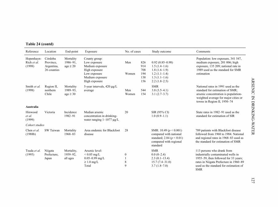
ARSEN
IC IN D
RINK
ING
-WATER
127
Table 24 (contd)
Reference Location End-point Exposure No. of cases Study outcome Comments
Hopenhayn-Rich et al. (1998)
Córdoba Province, Argentina, 26 counties
Mortality 1986–91, age ≥ 20
County group: Low exposure Medium exposure High exposure Low exposure Medium exposure High exposure
Men Women
826 914 708 194 138 156
0.92 (0.85–0.98) 1.5 (1.4–1.6) 1.8 (1.6–1.9) 1.2 (1.1–1.4) 1.3 (1.1–1.6) 2.2 (1.8–2.5)
Population: low exposure, 341 547, medium exposure, 201 006; high exposure, 135 209; national rate in 1989 used as the standard for SMR estimation
Smith et al. (1998)
Region II, northern Chile
Mortality 1989–93, age ≥ 30
5-year intervals, 420 µg/L average
Men Women
544 154
SMR 3.8 (3.5–4.1) 3.1 (2.7–3.7)
National rates in 1991 used as the standard for estimation of SMR; arsenic concentration is population- weighted average for major cities or towns in Region II, 1950–74
Australia
Hinwood et al. (1999)
Victoria Incidence 1982–91
Median arsenic concentration in drinking-water ranging 1–1077 µg/L
20 SIR (95% CI) 1.0 (0.9–1.1)
State rates in 1982–91 used as the standard for estimation of SIR
Cohort studies
Chen et al. (1988b)
SW Taiwan Mortality 1968–83
Area endemic for Blackfoot disease
28 SMR: 10.49 (p < 0.001) compared with national standard; 2.84 (p < 0.01) compared with regional standard
789 patients with Blackfoot disease followed from 1968 to 1984. National and regional rates in 1968–83 used as the standard for estimation of SMR
Tsuda et al. (1995)
Niigata Prefecture, Japan
Mortality, 1959–92, all ages
Arsenic level: < 0.05 mg/L 0.05–0.99 mg/L ≥ 1.0 mg/L Total
0 1 8 9
SMR 0.0 (0–2.4) 2.3 (0.1–13.4) 15.7 (7.4–31.0) 3.7 (1.8–7.0)
113 persons who drank from industrially contaminated wells in 1955–59, then followed for 33 years; rates in Niigata Prefecture in 1960–89 used as the standard for estimation of SMR
pp97-166.qxd 11/10/2004 10:47 Page 127

IARC M
ON
OG
RAPH
S VO
LUM
E 84128
Table 24 (contd)
Reference Location End-point Exposure No. of cases Study outcome Comments
Chiou et al. (1995)
SW Taiwan; 4 neigh-bouring townships
Incidence 1986–93
Cumulative arsenic exposure (mg/L × year) < 0.1 0.1–19.9 ≥ 20
3 7 7
Relative risk (95% CI) 1.0 3.1 (0.8–12.2) 4.7 (1.2–18.9)
Incidence among a cohort of 2556 subjects (263 Blackfoot disease patients and 2293 healthy individuals) followed for 7 years
Average arsenic concentration (mg/L) < 0.05 0.05–0.70 ≥ 0.71
5 7 7
1.0 2.1 (0.7–6.8) 2.7 (0.7–10.2)
Lewis et al. (1999)
Millard County, UT, USA
Mortality Arsenic in well-water, 3.5–620 µg/L
Men Women
28 6
SMR 0.6 (0.4–0.8) 0.4 (0.2–0.95)
State rates in 1950–92 used as the standard for SMR estimation.
Nakadaira et al. (2002)
Niigata Prefecture, Japan
Mortality Industrially contaminated well-water in the town of Nakajo
Men Women Total
7 1 8
Poisson probability distribution in men: 9.6 0/E = 11.01
86 patients with chronic arsenic poisoning. National rates in 1959–92 used as the standard for SMR estimation.
Case–control studies
Chen et al. (1986)
SW Taiwan, 4 townships
Mortality Duration of consumption of artesian well-water containing high levels of arsenic
76 cases 368 controls
Age- and sex-adjusted OR by years of consuming high-arsenic artesian well-water Never 1.00 1–20 years 1.26 21–40 years 1.52 > 40 years 3.39
OR calculated using subjects who never consumed artesian well-water as referent Mantel-Haenszel χ2 value: 8.49 (p < 0.01)
pp97-166.qxd 11/10/2004 10:47 Page 128

ARSEN
IC IN D
RINK
ING
-WATER
129
Table 24 (contd)
Reference Location End-point Exposure No. of cases Study outcome Comments
Ferreccio et al. (2000)
Northern Chile
Incidence 1994–96
Individual ≥ 40-year average arsenic concentration from public water supply records during 1930–94 0–10 µg/L 10–29 µg/L 30–49 µg/L 50–199 µg/L 200–400 µg/L
151 cases 419 matched hospital controls
Age- and sex-adjusted OR (95% CI) 1.0 1.6 (0.5–5.3) 3.9 (1.2–12.3) 5.2 (2.3–11.7) 8.9 (4.0–19.6)
OR calculated using subjects with average exposures of 0–10 µg/L as referent
SMR, standardized mortality ratio; CI, confidence interval; SIR, standardized incidence ratio; OR, odds ratio
pp97-166.qxd 11/10/2004 10:47 Page 129
(b) JapanCohort study
Tsuda et al. (1995) (described in Section 2.1.2) found excess mortality from lungcancer among a cohort of 113 persons exposed to levels of arsenic above 1.0 mg/L fromindustrially contaminated drinking-water in villages of Niigata Prefecture, Japan. Theexpected number of deaths was based on sex-, age- and cause-specific mortality in NiigataPrefecture in 1960–89. Based on a subgroup of 86 study patients, Nakadaira et al. (2002)did not find excess mortality from lung cancer.
(c) AustraliaHinwood et al. (1999) investigated the association between levels of arsenic in
drinking-water and lung cancer incidence in Victoria, Australia, during the period1982–91. This study included 22 areas where median concentrations of arsenic indrinking-water ranged from 14 to 166 µg/L. Using the incidence rate in Victoria, an SIRof 1.0 was observed for lung cancer.
(d) South America(i) Ecological studies
Rivara et al. (1997) compared the mortality from lung cancer in 1976–92 betweenAntofagasta in Region II with that in Region VIII of Chile. The relative risk for lungcancer was higher in Antofagasta compared with Region VIII. [The data source and statis-tical analysis were not clearly described.]
Smith et al. (1998) (described in Section 2.2.1) found elevated SMRs of about 3 forlung cancer for both sexes in Region II, using the national rate as standard.
In the study of Hopenhayn-Rich et al. (1998) (described in Section 2.2.1), significantincreases in the incidence of lung cancer associated with increasing exposure to arsenicwere observed for lung cancer.
(ii) Case–control studyFerreccio et al. (2000) conducted a case–control study of incident lung cancer cases in
northern Chile. Eligible cases included all lung cancer cases admitted to public hospitals inRegions I, II and III of Chile from November 1994 to July 1996. Eighty to ninety per centof all cancer patients in the north of Chile are admitted to public hospitals, and a total of151 cases participated. Controls were selected from all patients admitted to any publichospital in the study region and frequency-matched to cases by age and sex. Two controlseries were selected: cancers other than lung cancer and non-cancer controls [no responserates were indicated for cases and controls]. Potential biases in control selection wereassessed by several approaches including comparisons with geographical distribution ofthe general population based on census data. Information regarding residential history,socioeconomic status, occupational history (to ascertain employment in copper smelting)and smoking was obtained by questionnaire interview. Historical exposure to arsenic in
IARC MONOGRAPHS VOLUME 84130
pp97-166.qxd 11/10/2004 10:47 Page 130

drinking-water was estimated by linking information on residential history with a databaseof information on arsenic concentrations in public water supplies collected for the years1950–94. Arsenic concentrations in the year prior to 1950 were based on concentrations inthe 1950s. Average concentration of arsenic in the place of residence was assigned to eachsubject on a year-by-year basis for the period 1930–94. Population coverage of publicwater systems in the main cities in Regions I and II was over 90% and was between 80 and90% in the major cities of Region III. The coverage in smaller cities varied between 64 and91%. Odds ratios were calculated using unconditional logistic regression, adjusted for age,sex, socioeconomic status, smoking and working in a copper smelter. Results from theanalysis based on average exposures during 1930–94 and using all controls showed anincrease in the odds ratio with concentration of arsenic. Evidence for a synergistic effect ofarsenic in water and smoking was found for those who both smoked and had high concen-trations of arsenic in their drinking-water (results not shown).
(e) USACohort study
Lewis et al. (1999) reported the association between arsenic in drinking-water andlung cancer mortality in a cohort of residents of Millard County, UT, where the medianconcentration of arsenic in drinking-water ranged from 14 to 166 µg/L. The SMRs forlung cancer for both men and women were below unity. [Limitations of this study havebeen cited in Section 2.1.4.]
2.3 Skin cancer
The recognition of arsenic as a carcinogen originally came from case seriesdescribing skin cancers following ingestion of arsenical medicine, and exposure toarsenical pesticide residues and arsenic-contaminated drinking-water. Hutchinson (1888)noted skin cancers among patients treated for psoriasis and other ailments with arsenic-containing compounds (e.g. Fowler’s Solution containing 1% potassium arsenite).Neubauer (1947) summarized 143 skin cancer cases among arsenic-treated patients. Over50% of the skin cancers developed in patients treated for 10 years or less and lesionsdeveloped after 3–40 years, and on average after 18 years. Clinical reports have describedan association of chronic arsenicism with skin cancer in vineyard workers of the Moselleregion, Germany (Roth, 1957; Grobe, 1977). Numerous cases of skin cancer have beendocumented from communities with arsenic-contaminated drinking-water. These include,but are not limited to, case reports from Silesia (Neubauer, 1947), North America (Wagneret al., 1979), Taiwan, China (Yeh, 1973), Argentina (Bergoglio, 1964), Mexico (Cebríanet al., 1983), Chile (Zaldívar, 1974; Zaldívar et al., 1981) and, more recently, Bangladesh(Kurokawa et al., 2001), West Bengal, India (Saha, 2001) and Malaysia (Jaafar et al.,1993). The characteristic arsenic-associated skin tumours include squamous-cell carci-noma arising in keratoses (including Bowen disease) and multiple basal-cell carcinomas
ARSENIC IN DRINKING-WATER 131
pp97-166.qxd 11/10/2004 10:47 Page 131

(e.g. Neubauer, 1947; Neuman & Schwank, 1960; Yeh et al., 1968). Therefore, thissection focuses on these non-melanoma skin cancers. In addition, ecological studies ofskin cancer based on mortality rates in areas with low exposure to arsenic such as the USAor exposure imputed from soil levels are not considered here. Findings of epidemiologicalstudies on arsenic in drinking-water and risk for skin cancer are summarized in Table 25.
2.3.1 Taiwan, China
(a) Ecological studies(i) Study based on prevalence of skin cancer
In 1965, Tseng et al. (1968) completed a skin cancer prevalence survey on the south-west coast of Taiwan, a region known to have arsenic-contaminated artesian wells that wereintroduced into the region in 1910–20. A house-to-house examination was conducted offamily members from 37 villages: 10 in Chai-yi County, 25 in Tainan County and two in asuburb of Tainan City. The study covered a total population of 40 421 inhabitants. A total of428 skin cancers were identified, 238 of which were sent for histopathological review. Thestudy was based on clinical diagnoses. In the survey region, 142 water samples from 114wells were tested for arsenic. Arsenic concentrations ranged from 1 µg/L to 1820 µg/L, andthe majority of wells in the endemic region contained between 400 and 600 µg/L arsenic.Skin cancer prevalence was computed according to the median arsenic concentrations pervillage, categorized as < 300 µg/L, 300–600 µg/L and > 600 µg/L. Villages with eitherwide-ranging arsenic concentrations or residents who no longer drank the water weredeemed to be indeterminate in the analysis. Prevalence rates of skin cancer (based on clini-cal diagnosis) for inhabitants residing in low- (< 300 µg/L), medium- (300–600 µg/L) andhigh- (> 600 µg/L) arsenic areas represented over an eightfold difference from the highestto the lowest category.
(ii) Studies based on incidence of skin cancerGuo et al. (1998, 2001) correlated incidence rates of skin cancer with levels of arsenic
in well-water for 243 townships in Taiwan (with a total of about 11.4 million residents).Skin cancers were identified from the National Cancer Registry Program from 1980 to1987 by a hospital-based registry covering both clinical and pathological diagnoses ofskin cancers (reporting is not mandatory by law) (Guo et al., 1998). Arsenic concen-trations were provided by a national survey of wells conducted by the Taiwan ProvincialInstitute of Environmental Sanitation and published in 1977. The investigators modelledthe percentage of the population living in townships with drinking-water concentrationsof < 50, 50–89, 90–169, 170–329, 330–640 and > 640 µg/L arsenic. The models assumethat the same number of individuals drank from each well within a township. The resultsfor all skin cancers combined estimated a relative risk for the highest versus lowest expo-sure category of about 14 in men and 19 in women, with no excess detected in the othercategories (Guo et al., 1998).
IARC MONOGRAPHS VOLUME 84132
pp97-166.qxd 11/10/2004 10:47 Page 132

ARSEN
IC IN D
RINK
ING
-WATER
133
Table 25. Summary of epidemiological studies on arsenic in drinking-water and risk for skin cancer
Reference Location End-point Exposure No. of cases Study outcome Comments
Ecological studies
Taiwan
Tseng et al. (1968)
40 421 residents from 37 villages (SW)
Prevalence ≥ 20 years of age
Median arsenic concentrations of wells in village of residence (µg/L)
< 300 300–600 > 600
428 Prevalence (per 1000)
2.6 10.1 21.4
Prevalence based on clinical examination of all house-holds. Excludes villages with wells no longer in use or with variations in arsenic concentration (range, 1–1820 µg/L; most wells contained 400–600 µg/L arsenic)
Chen et al. (1985)
4 neighbouring townships on the SW coast
Mortality 1968–82
Areas hyperendemic (21 villages), endemic (25 villages) and not endemic (38 villages) for Blackfoot disease, corresponding to high, medium and low exposure
46 men 49 women
SMR (95% CI) 534 (379–689) 652 (469–835)
Mortality rates in all Taiwan as standard
Chen et al. (1988a)
Region endemic for Blackfoot disease (SW)
Mortality 1973–86
Median arsenic concentrations of well-water (µg/L)
< 300 300–600 > 600
Men Women Men Women Men Women
SMR 1.6 1.6 10.7 10.0 28.0 15.1
Age-standardized to the 1976 world standard population
pp97-166.qxd 11/10/2004 10:47 Page 133

IARC M
ON
OG
RAPH
S VO
LUM
E 84134
Table 25 (contd)
Reference Location End-point Exposure No. of cases Study outcome Comments
Wu et al. (1989)
42 villages in region endemic for Blackfoot disease (SW)
Mortality 1973–86
Median arsenic concentrations of well-water in village of residence (µg/L) in 1964–66 < 300 300–600 > 600 < 300 300–600 > 600
19 men 17 women Men Women
SMR 2.03 14.01 32.41 (p < 0.001) 1.73 14.75 18.66 (p < 0.001)
Age-standardized to the 1976 world standard population
Chen & Wang (1990)
314 precincts and townships
Mortality 1972–83
Average arsenic concentrations NS Men Women
Increase (β) in mortality rate per 100 000 per 0.1 µg/L increase: β (SE) = 0.9 (0.2) β (SE) = 1.0 (0.2)
Multiple regression adjusted for age and indices of urbanization and industrialization. Mortality rates standardized to the 1976 world standard population
Guo et al. (1998)
243 townships, 11.4 million residents
Incidence 1980–87
Arsenic concentration in wells Exposure categories: > 50, 50–89, 90–169, 170–329, 330–640 and > 640 µg/L
952 men 595 women
Risk difference of 0.34/100 000 (p < 0.01) associated with a 1% increase in arsenic concentrations > 640 µg/L Relative risk of highest versus lowest exposure category: 14.21 in men; 19.25 in women No excess risk for other categories
Rates standardized using the 1976 world standard population. Model assumes that same number of individuals use each well.
Tsai et al. (1999)
Four townships (SW)
Mortality 1971–94
Area endemic for Blackfoot disease 66 men 68 women
Age- and sex-adjusted SMR (95% CI) 4.8 (3.7–6.2) 5.97 (4.6–7.6) 5.7 (4.4–7.2) 6.8 (5.3–8.6)
Local standard National standard Local standard National standard
pp97-166.qxd 11/10/2004 10:47 Page 134

ARSEN
IC IN D
RINK
ING
-WATER
135
Table 25 (contd)
Reference Location End-point Exposure No. of cases Study outcome Comments
Guo et al. (2001)
243 townships, 11.4 million residents
Incidence 1980–89
Concentration of arsenic in well-water: Exposure categories (µg/L) arsenic Intercept 50–89 90–169 170–329 330–640 > 640 Intercept 50–89 90–169 170–329 330–640 > 640
2369 (1415 men, 954 women) 764 BCC Men Women Men Women Men Women Men Women Men Women Men Women Men 736 SCC Women Men Women Men Women Men Women Men Women Men Women 182 melanoma
Rate difference association with a 1% increase in residents with categories of arsenic (µg/L): 0.779 –0.002 0.004 –0.012 –0.017 0.018 0.006 0.004 –0.024 0.016 0.128** 0.027 0.821 1.488 0.024 –0.006 –0.026 0.006 0.073** 0.016 –0.100** –0.064* 0.155** 0.212** No increase associated with melanoma
Cancers identified through National Cancer Registry. Models include age and urbanization index. Models assume same number of individuals use each well. BCC, basal-cell carcinoma SCC, squamous-cell carcinoma *p < 0.05 **p < 0.01
pp97-166.qxd 11/10/2004 10:47 Page 135
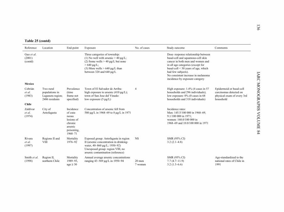
IARC M
ON
OG
RAPH
S VO
LUM
E 84136
Table 25 (contd)
Reference Location End-point Exposure No. of cases Study outcome Comments
Guo et al. (2001) (contd)
Three categories of township: (1) No well with arsenic > 40 µg/L; (2) Some wells > 40 µg/L but none > 640 µg/L; (3) More wells > 640 µg/L than between 320 and 640 µg/L
Dose–response relationship between basal-cell and squamous-cell skin cancer in both men and women and in all age categories (except for basal-cell < 30 years of age, which had few subjects). No consistent increase in melanoma incidence by exposure category
Mexico
Cebrián et al. (1983)
Two rural populations in Lagunera region; 2486 residents
Prevalence (time frame not specified)
Town of El Salvador de Arriba: high exposure to arsenic (410 µg/L); town of San Jose del Vinedo: low exposure (5 µg/L)
4 High exposure: 1.4% (4 cases in 57 households and 296 individuals); low exposure: 0% (0 cases in 68 households and 318 individuals)
Epidermoid or basal-cell carcinomas detected on physical exam of every 3rd household
Chile
Zaldívar et al. (1974)
City of Antofagasta
Incidence of cuta-neous lesions of chronic arsenic poisoning, 1968–71
Concentration of arsenic fell from 580 µg/L in 1968–69 to 8 µg/L in 1971
Incidence rates: Men: 145.5/100 000 in 1968–69, 9.1/100 000 in 1971; women: 168.0/100 000 in 1968–69 and 10.0/100 000 in 1971
Rivara et al. (1997)
Regions II and VIII
Mortality 1976–92
Exposed group: Antofagasta in region II (arsenic concentration in drinking-water, 40–860 µg/L; 1950–92) Unexposed group: region VIII, no arsenic contamination (reference)
NS SMR (95% CI) 3.2 (2.1–4.8)
Smith et al. (1998)
Region II, northern Chile
Mortality 1989–93, age ≥ 30
Annual average arsenic concentrations ranging 43–569 µg/L in 1950–94
20 men 7 women
SMR (95% CI) 7.7 (4.7–11.9) 3.2 (1.3–6.6)
Age-standardized to the national rates of Chile in 1991
pp97-166.qxd 11/10/2004 10:47 Page 136

ARSEN
IC IN D
RINK
ING
-WATER
137
Table 25 (contd)
Reference Location End-point Exposure No. of cases Study outcome Comments
USA
Berg & Burbank (1972)
Mortality 1950–67
Trace metals in water supplies from 10 basins throughout the USA; concentration of arsenic in water, Oct. 1962–Sept. 1967
No correlation
Morton et al. (1976)
Lane County, OR
Incidence 1958–71
Mean arsenic concentration in municipal water system and single-family systems
3039 Correlation of arsenic content in drinking-water: squamous-cell carcinoma: 0.151 for men and –0.20 for women; basal-cell carcinoma: –0.064 for men and 0.10 for women
Non-melanoma cases identified by review of pathology reports
Wong et al. (1992)
Four counties in Montana
Incidence 1980–86
Two contaminated counties (copper smelter and copper mines); two control counties
Around 2300 in the 4 counties
Age-adjusted skin cancer incidence higher in control counties
Overall incidence rates for exposed counties within range observed for other US locations
Cohort studies
Taiwan
Chen et al. (1988b)
Four townships (SW)
Mortality 1968–83, all ages
Diagnosis of Blackfoot disease as a surrogate for high exposure to arsenic
7 SMR 28.46 (p < 0.01) (national standard)
4.51 (p < 0.05) (local standard)
871 people who developed Blackfoot disease after 1968 were followed for 15 years. National standard used for the age- and sex-standardized rates of the general Taiwanese population.
Hsueh et al. (1997)
Three villages in Putai township (SW)
Incidence 1989–92, age, ≥ 30 years
Duration of residence in area endemic for Blackfoot disease (years) ≥ 33 34–43 44–53 > 53
1 4 8 20
Relative risk (95% CI) 1.0 5.01 (0.5–48.1) 4.9 (0.6–41.6) 6.8 (0.9–53.7) (p for trend = 0.07)
654 subjects (275 men and 379 women) without skin cancer followed with dermatological examinations. Total of 2239 person–years. Relative risk adjusted for age, sex and level of education
pp97-166.qxd 11/10/2004 10:47 Page 137

IARC M
ON
OG
RAPH
S VO
LUM
E 84138
Table 25 (contd)
Reference Location End-point Exposure No. of cases Study outcome Comments
Hsueh et al. (1997) (contd)
Duration of consumption of artesian well-water (years) 0 1–15 16–25 > 25
1 1 8 23
1.0 1.2 (0.4–19.7) 3.9 (0.5–32.1) 8.9 (1.1–72.9) (p for trend < 0.05)
Average concentration of arsenic in drinking-water (mg/L) 0 0.01–0.70 0.71–1.10 Unknown
1 12 13 7
1.0 3.3 (0.4–35.8) 8.7 (1.1–65.5) (p for trend < 0.05) 4.8 (0.6–40.4)
Cumulative exposure to arsenic (mg/L–years) 0 0.1–10.6 10.7–17.7 > 17.7 Unknown
1 2 5 18 7
1.0 2.8 (0.3–31.9) 2.6 (0.3–22.9) 7.6 (0.95–60.3) (p for trend = 0.06) 5.1 (0.6–44.4)
Level of serum β-carotene (µg/mL) ≤ 0.14 0.15–0.18 > 0.18
16 cases (61 controls) OR (95% CI) 1.0 0.4 (0.1–2.9) 0.01 (0.0–0.4) (p for trend < 0.01)
OR adjusted for age, sex, cumulative exposure to arsenic, serum cholesterol and triglyceride levels, cigarette smoking and alcohol drinking Incidence 14.74/1000 person–years
pp97-166.qxd 11/10/2004 10:47 Page 138

ARSEN
IC IN D
RINK
ING
-WATER
139
Table 25 (contd)
Reference Location End-point Exposure No. of cases Study outcome Comments
Case–control studies
USA
Karagas et al. (2001b, 2002)
New Hampshire Incidence 1993–96
Concentration of arsenic in toenails (µg/g) 0.009–0.089 0.090–0.133 0.134–0.211 0.212–0.280 0.281–0.344 0.345–0.81
155 64 33 14 5 13
OR (95% CI) Squamous-cell carcinoma 1.0 0.9 (0.6–1.3) 0.98 (0.6–1.6) 1.1 (0.55–2.2) 1.0 (0.3–3.0) 2.1 (0.9–4.7)
OR adjusted for age and sex 284 cases, 524 controls
0.009–0.089 0.090–0.133 0.134–0.211 0.212–0.280 0.281–0.344 0.345–0.81
281 156 92 22 10 26
Basal-cell carcinoma 1.0 1.01 (0.8–1.4) 1.06 (0.7–1.5) 0.7 (0.4–1.3) 0.8 (0.3–1.8) 1.4 (0.7–2.8)
587 cases, 524 controls
Nested case–control study
Taiwan
Hsueh et al. (1995)
Three villages in Putai Township (SW)
Prevalence Duration of residence in area endemic for Blackfoot disease (years) ≤ 45 46–49 ≥ 50
2 11 53
OR (95% CI) 1.0 5.2 (1.1–25.8) 8.5 (1.96–37.2) p for trend < 0.05
OR adjusted for age and sex; 1081 residents (468 men, 613 women) underwent a physical examination.
pp97-166.qxd 11/10/2004 10:47 Page 139

IARC M
ON
OG
RAPH
S VO
LUM
E 84140
Table 25 (contd)
Reference Location End-point Exposure No. of cases Study outcome Comments
Hsueh et al. (1995) (contd)
Duration of drinking artesian well-water (years) ≤ 13 14–25 ≥ 60
1.0 5.1 (1.03–24.98) 6.4 (1.4–27.9) p for trend < 0.05
Average exposure to arsenic (ppm) 0 0–0.70 > 0.71
2 15 52
2 20 30
1.0 3.5 (0.7–17.0) 5.0 (1.1–23.8)
Cumulative exposure to arsenic (ppm–years) ≤ 4 5–24 ≥ 25
1 22 28
1.0 8.9 (1.1–73.8) 13.7 (1.7–111.6)
Chronic hepatitis B carrier and liver function status: Non-carrier with normal liver function HBsAg carrier with normal liver function Non-carrier with liver dysfunction HBsAg carrier with liver dysfunction
41 13 3 4
1.0 1.1 (0.56–2.2) 2.1 (0.54–7.7) 8.4 (2.37–29.9) p for trend < 0.05
SMR, standardized mortality ratio; CI, confidence interval; NS, not specified; OR, odds ratio
pp97-166.qxd 11/10/2004 10:47 Page 140

In a second study, the incidence rates of each major histological type of skin cancer(basal-cell, squamous-cell and melanoma) were modelled using the previously definedexposure categories (Guo et al., 2001). The model included the percentage of residents incategories of age (30–49, 50–69 and > 69 years) and urbanization index as covariates. Inthis analysis, they found a statistically significant increase in squamous-cell skin cancerin the highest category (> 640 µg/L arsenic) in both men and women. Similarly, for basal-cell carcinoma, there was also a positive association in the highest exposure category.Melanoma was unrelated to any category of exposure to arsenic. In a post-hoc analysis,the investigators created three exposure groups: townships with no well containing levelsof arsenic above 40 µg/L, townships with some wells containing more than 40 µg/Larsenic but none above 640 µg/L and townships with more wells containing more than640 µg/L than between 320 and 640 µg/L. A dose–response relation between basal-celland squamous-cell skin cancer rates was observed in both men and women and in all agecategories (except for basal-cell cancer below age 30 years, which had few subjects).Again, no association was seen for melanoma. [The Working Group noted that the repor-ting of incident skin cancer may have been incomplete.]
(iii) Studies based on mortality from skin cancerA series of studies conducted in Taiwan, China (Chen et al., 1985, 1988a; Wu et al.,
1989; Chen & Wang, 1990; Tsai et al., 1999) (described in Section 2.1.1) analysed skincancer mortality in relation to levels of arsenic in well-water. The overall SMRs in thefour counties were 534 in men and 652 in women, with 100 as the referent value. Therewas also a gradient of increasing SMRs for skin cancer in areas not endemic or hyper-endemic for Blackfoot disease (Chen et al., 1985). A subsequent report (Chen et al.,1988a) presented SMRs for skin cancer grouped by the median levels of arsenic in well-water measured in 1962–64 into < 300, 300–600 and > 600 µg/L. The age-standardizedmortality rates for skin cancer increased in the three categories for both genders (Chenet al., 1988a). Wu et al. (1989) published age-adjusted mortality rates from skin cancerfor the years 1973–86 for the four townships endemic for Blackfoot disease plus 15villages in the townships of Yensui and Hsiaying. Using the same data on well-watercontents and the classification described above, skin cancer mortality rates also increasedin men and women with increasing median concentrations of arsenic. Subsequently, Chenand Wang (1990) used data on wells measured from 1974 to 1976 from 314 precincts andtownships throughout Taiwan. Based on a multiple regression analysis (adjusted forurbanization and age), a 0.1-µg/L increase in arsenic corresponded to an increase of 0.9(SE, 0.2) and 1.0 (SE, 0.2) per 100 000 in skin cancer mortality in men and women,respectively. More recently, Tsai et al. (1999) computed SMRs for the period 1971–94 forthe four townships endemic for Blackfoot disease by applying both local (Chiayi County)and national (Taiwan) rates as the standards. The SMRs for skin cancer were approxi-mately 5.0 for men and women, using both local and national standards.
ARSENIC IN DRINKING-WATER 141
pp97-166.qxd 11/10/2004 10:47 Page 141

(b) Cohort studies Chen et al. (1988b) conducted a retrospective cohort study of 871 patients who met
clinical criteria of Blackfoot disease after January 1968. From 1968 to 1983, seven deathcertificates identified the underlying cause of death as skin cancer. SMRs for skin cancerwere computed using both the area endemic for Blackfoot disease and the whole ofTaiwan as the population standards. The age-standardized observed versus expectednumber of skin cancer deaths was around 28 using Taiwan as the standard and 4.5 usingthe Blackfoot disease townships as the standard. [Use of the national population as thestandard may provide a better estimate of excess risk than use of the local population forwhom arsenic concentrations in well-water were elevated.]
Hsueh et al. (1997) established a cohort of residents living in three villages (Homei,Fuhsin and Hsinming) in Putai Township, one of the regions of the south-west coast ofTaiwan endemic for Blackfoot disease. Arsenic-contaminated well-water had existed inthese villages for over 50 years, with reported values ranging from 700 to 930 µg/L. Thegovernment introduced a new water system in the 1960s, with relatively low penetrationuntil the 1970s. Well-water remained in use for agriculture and aquaculture. The 1571residents aged 30 years or older who lived for at least 5 days a week in the villages wererecruited to take part in the study. Of these, 1081 (68.8%) participated in a physical exa-mination (468 men and 613 women). The 1015 study subjects who did not have aprevalent skin cancer comprised the cohort. Of these, 275 men and 379 women (64%)underwent regular examinations for dermatological conditions. Thirty-three incident skincancers developed during the follow-up period from September 1989 through December1992, with a total of 2239 person–years and a rate of 14.74/1000 person–years. Publichealth nurses interviewed cohort members on length of residence, history of drinkingwell-water and other potentially confounding factors. An index for cumulative exposureto arsenic was derived for each subject using concentrations of arsenic in well-water foreach village of residence measured in the 1960s. These data were multiplied by theduration of consumption of well-water in each village of residence and totalled for eachresidence. Dermatologists diagnosed clinically the skin cancers that occurred in thecohort, and 91% of clinically diagnosed carcinomas were confirmed histologically.Fasting blood samples and urine samples were collected at the time of interview. Serumwas analysed for β-carotene and urine for arsenic metabolites from 16 skin cancer cases[48%] and 61 age- and sex-matched controls. Cox proportional hazard regression wasused to analyse data on exposure to arsenic in relation to incidence of skin cancer adjustedfor age, sex and educational level. Conditional logistic regression analysis was used toassess the effects of β-carotene and urinary metabolites on the risk for skin cancer. Riskfor skin cancer was significantly related to duration of living in the area endemic forBlackfoot disease, duration of consumption of artesian well-water, average concentrationof arsenic and index for cumulative exposure to arsenic. There was evidence of a reducedrisk for skin cancer in the highest two tertiles of β-carotene versus the lowest tertile afteradjustment for multiple potential confounders. Also, compared with controls, cases of
IARC MONOGRAPHS VOLUME 84142
pp97-166.qxd 11/10/2004 10:47 Page 142

skin cancer had higher total urinary concentration of arsenic, percentage of MMA andratio of MMA to inorganic arsenic and a lower percentage of DMA and ratio of DMA toMMA (not shown in Table 25).
Hsueh et al. (1995) conducted a nested case–control study within the cohort study ofHsueh et al. (1997). A total of 66 prevalent skin cancers were identified after clinicalexamination. Age- and sex-adjusted prevalence odds ratios were computed for the samevariables of exposure to arsenic included in the cohort analysis: duration of living in thearea endemic for Blackfoot disease, duration of drinking artesian well-water, and averageand cumulative exposure to arsenic. Significant increases in risk for each exposure groupwere shown. In addition, a significantly elevated risk for skin cancer was observed amongchronic hepatitis B carriers with liver dysfunction.
2.3.2 Mexico
Ecological study based on prevalence of skin cancerCebrían et al. (1983) reported results from a household survey of two rural Mexican
towns in the Region of Lagunera. Towns were selected on the basis of levels of arsenic indrinking-water, one town with a high level of arsenic (average, 410 µg/L) and the otherwith a low level of arsenic (average, 5 µg/L). The towns are located 37 km apart, and theirpopulations were of comparable size (1488 and 998 inhabitants, respectively) and socio-economic and environmental conditions. A questionnaire and physical examination wereadministered to all family members of every third household in each community.Seventy-five per cent of the residents had lived in these communities since birth. Afterclinical diagnosis, prevalence of epidermoid or basal-cell carcinoma (referred to asulcerative lesions) was 1.4% in the exposed town, whereas no case was observed in thecontrol town. The 20 water samples tested from the exposed town between 1975 and 1978had arsenic concentrations ranging from 160 to 590 µg/L (standard deviation [SD],114 µg/L), indicating significant variability. The control town showed little variability(SD, 7 µg/L) in the 18 samples collected over the same period.
2.3.3 Chile
Ecological study based on incidence of cutaneous lesions Zaldívar et al. (1981) investigated the incidence of cutaneous lesions (leukoderma,
melanoderma, hyperkeratosis, and squamous-cell carcinoma) in residents of Antofagastain arsenic-contaminated Region II from 1968 to 1971. Among 457 patients, about 70%were children aged 0–15 years. Incidence rates decreased from 1968–69 to 1971 due to afilter plant which started operation in 1970.
Ecological studies based on mortality from skin cancer In an ecological analysis, Rivara et al. (1997) compared mortality rates from skin
cancer in 1976–92 between Antofagasta and the unexposed control Region VIII of Chile.
ARSENIC IN DRINKING-WATER 143
pp97-166.qxd 11/10/2004 10:47 Page 143

The SMR for skin cancer was 3.2 (95% CI, 2.1–4.8). In a later study, Smith et al. (1998)compared sex- and site-specific mortality for the years 1989–93 in Region II of Chile withnational mortality rates. The SMR for skin cancer was 7.7 (95% CI, 4.7–11.9) among menand 3.2 (95% CI, 1.3–6.6) among women.
2.3.4 USA
(a) Ecological studyBerg and Burbank (1972) showed no correlation between trace metals from 10 water
basins throughout the USA and mortality rates in 1950–67, using concentrations ofarsenic measured in 1962–67.
Morton et al. (1976) studied the incidence of histologically confirmed basal-cell andsquamous-cell skin cancers (in situ and invasive) for the period 1958–71 in Lane County,OR, USA. They identified skin cancers by reviewing the pathology records of facilitiesserving residents of the county and the biopsy files of two of five dermatologists. Watersamples were tested from selected points throughout the county from public watersystems and from a number of single-family systems. Single-family systems werereported to have been over-sampled in regions suspected of having a problem witharsenic. Within a given region, arsenic values ranged from undetectable to 2150 µg/L. Thecorrelation between census tract estimates of arsenic in drinking-water and incidence ofsquamous-cell carcinoma was 0.151 in men and –0.20 in women; for the incidence ofbasal-cell carcinoma, the correlation was –0.064 in men and 0.10 in women. [A majorweakness of the study is the misclassification of exposure because of widely varyingconcentrations of arsenic within a census tract.]
Wong et al. (1992) studied approximately 2300 incident cases of skin cancer in fourcounties in Montana (two contaminated and two controls) in 1980–86. Contaminationarose from copper smelters and mines. No difference in incidence rates was observedbetween exposed counties and the rest of the country.
(b) Case–control studies Karagas et al. (1998, 2001b, 2002) designed a case–control study of basal-cell and
squamous-cell skin cancers in the population of New Hampshire to evaluate the effects oflow to moderate levels of exposure to arsenic. About 40% of the population relied onprivate, unregulated water systems; over 10% of the private supplies contained levels ofarsenic above the WHO recommended level of 10 µg/L and 1% of supplies overall con-tained > 50 µg/L. A biomarker of internal dose was chosen to determine exposure levels.Earlier studies had indicated the reliability of concentrations of arsenic in toenails as ameasure of exposure > 1 µg/L arsenic through drinking-water (Karagas et al., 1996, 2000)and reproducibility of concentrations over a period of 3–6 years (Garland et al., 1993;Karagas et al., 2001a). Cases of basal-cell and squamous-cell skin cancer diagnosed from1 July 1993 to 30 June 1995 were identified through a statewide network of dermatologists,dermatopathologists and pathologists, with participation rates of over 90% (Karagas et al.,
IARC MONOGRAPHS VOLUME 84144
pp97-166.qxd 11/10/2004 10:47 Page 144

2001b). Because of the high incidence of basal-cell carcinoma, incident cases of basal-cellcarcinoma were randomly selected in a 2:1 ratio to cases of squamous-cell carcinoma. Tominimize detection bias, cases of squamous-cell carcinoma were restricted to invasivedisease only (cases of in-situ carcinomas were excluded). A 2:1 ratio of controls tosquamous-cell carcinoma cases was randomly selected from population lists (driver’slicence files for cases < 65 years and Medicare enrollment lists for cases ≥ 65 years),frequency-matched to the combined distribution of the basal-cell and squamous-cell carci-noma cases. To be eligible to participate, subjects were required to speak English and havea working telephone. Of the 1143 potential case subjects, 896 took part in the study (78%)and, of the 820 potential controls, 540 (66%) enrolled. The analysis included the 587 casesof basal-cell carcinoma, 284 cases of squamous-cell carcinoma and 524 controls (97% ofsubjects) who contributed a toenail sample for arsenic analysis. Study participants under-went a personal interview to obtain information on confounding factors such as exposureand sensitivity to sun, history of radiation treatment and other medical and lifestyle factors.Age- and sex-adjusted odds ratios were computed using logistic regression analysis accor-ding to percentiles of arsenic concentrations in toenails based on the control distribution. Inthis categorical analysis, concentrations of arsenic appeared to be unrelated to risk forsquamous-cell and basal-cell carcinomas except for the highest category (the top 97thpercentile; concentrations above 0.344 µg/g) versus concentrations below the median.
An analysis using continuous exposure variables was presented separately (Karagaset al., 2002). A quadratic and two-segment linear model fitted the data for both squamous-cell carcinoma and basal-cell carcinoma. The point at which the dose–response appearedto increase was at 0.105 µg/g (95% CI, 0.093–0.219 µg/g) for squamous-cell carcinomausing a maximum likelihood estimation of the change point for the two-segment linearmodel. After the change point, a 1% increase in arsenic concentration in toenails wasrelated to a 0.61% increase in risk for squamous-cell carcinoma. The quadratic model forboth squamous-cell carcinoma and basal-cell carcinoma produced a consistent nadir orchange point of 0.088 and 0.091, respectively. However, it was not possible to estimate atwo-segment model for basal-cell carcinoma because of sparse data at the extremes.Based on a regression analysis of concentrations of arsenic in water and toenails, a changepoint of 0.105 µg/g in toenails translated to 1–2 µg/L in water, with the 95% confidenceinterval ranging from < 1 to 10–20 µg/L.
2.4 Other organ sites
Studies on cancer at other organ sites are summarized in Table 26.Neubauer (1947) summarized cancers that had been reported in patients treated with
medicinal arsenic. His report of 143 published cases included patients who developedcutaneous tumours and other malignancies such as cancers of the stomach (one case),tongue (two cases, one who also had cancer of oral mucosa), oesophagus (two cases),uterus (one case) and urethra (two cases including one papilloma of the ureter). Amongpatients who had not developed cutaneous tumours, other reported malignancies included
ARSENIC IN DRINKING-WATER 145
pp97-166.qxd 11/10/2004 10:47 Page 145

IARC M
ON
OG
RAPH
S VO
LUM
E 84146
Table 26. Summary of epidemiological studies of arsenic in drinking-water and risk for other cancers
Reference Location End-point Exposure Site No. of cases Study outcome Comments
Ecological studies
Taiwan
Chen et al. (1985)
84 villages on the SW coast
Mortality 1968–82, all ages
Endemic area for chronic arsenic toxicity (Blackfoot disease)
Colon Small intestine Leukaemia Colon Small intestine Leukaemia
Men 54 17 45 Women 61 5 22
Age- and sex-adjusted SMR (95% CI) 1.6 (1.2–2.0) 2.98 (0.6–3.4) 1.4 (1.0–1.8) 1.7 (1.3–2.1) 0.97 (0.1–1.8) 0.9 (0.5–1.3)
SMRs for a total of 11 sites. Prostate not investigated; mid-year population: 141 733 in 1968, 120 607 in 1982; national rate in 1968–82 used as the standard for estimation of SMR
Chen et al. (1988a)
42 villages on the SW coast
Mortality 1973–86, all ages
Median level of arsenic in drinking-water grouped into 3 strata, 1962–64 General population < 300 µg/L 300–600 µg/L > 600 µg/L
Prostate Age-adjusted SMR 1.5 0.5 5.8 8.4
899 811 person–years, rate per 100 000, age-standardized to 1976 world population
pp97-166.qxd 11/10/2004 10:47 Page 146

ARSEN
IC IN D
RINK
ING
-WATER
147
Table 26 (contd)
Reference Location End-point Exposure Site No. of cases Study outcome Comments
Wu et al. (1989)
42 villages (SW)
Mortality 1973–86, age ≥ 20 years
Median level of arsenic in drinking-water grouped into 3 strata, 1962–64 < 30 µg/L 300–600 µg/L
Prostate Leukaemia Nasopharynx Oesophagus Stomach Colon Uterine cervix Prostate Leukaemia Nasopharynx Oesophagus Stomach Colon Uterine cervix
9 M 11 M 7 F 11 M 7 F 15 M 4 F 46 M 21 F 17 M 21 F 6 F 9 M 11 M 7 F 11 M 7 F 15 M 4 F 46 M 21 F 17 M 21 F 6 F
SMR 0.95 4.87 3.03 3.58 1.59 7.62 1.83 25.6 6.71 7.94 9.05 0.91 9.00 6.52 4.55 8.16 5.81 9.37 3.64 17.82 18.72 8.30 8.16 5.46
Age-adjusted mortality. All results are non-significant. Men, 257 935 person–years; women, 234 519 person–years; rate per 100 000, age-standardized to 1976 world population
pp97-166.qxd 11/10/2004 10:47 Page 147

IARC M
ON
OG
RAPH
S VO
LUM
E 84148
Table 26 (contd)
Reference Location End-point Exposure Site No. of cases Study outcome Comments
Wu et al. (1989) (contd)
> 600 µg/L Prostate Leukaemia Nasopharynx Oesophagus Stomach Colon Uterine cervix
9 M 11 M 7 F 11 M 7 F 15 M 4 F 46 M 21 F 17 M 21 F 6 F
9.18 2.69 0.00 8.58 4.89 6.55 0.00 56.42 5.98 12.5 17.21 3.92
Percentiles of age-adjusted mortality rate/100 000 person–years
Men 25th 50th 75th Oesophagus 3.6 6.0 9.2 Stomach 14.8 10.2 28.8 Small intestine 0.6 1.1 1.9 Colon 4.2 5.6 7.2
Chen & Wang (1990)
42 villages on the SW coast
Mortality 1972–83, all ages
National survey of 83 656 wells (1974–76); average arsenic content for each of 314 precincts or townships
Rectum 1.9 2.7 3.9 Pancreas 1.3 2.1 3.0 Nasal cavity 1.1 1.3 2.6 Larynx 1.1 1.7 2.8 Bone/cartilage 1.1 1.8 2.9
Prostate 0.9 1.4 2.3 Brain 0.7 1.1 1.8 Leukaemia 1.3 2.1 2.7 Women 25th 50th 75th Oesophagus 1.1 1.8 2.8 Stomach 7.2 10.0 13.7 Small intestine 0.6 0.9 1.6 Colon 3.6 5.5 6.9
pp97-166.qxd 11/10/2004 10:47 Page 148

ARSEN
IC IN D
RINK
ING
-WATER
149
Table 26 (contd)
Reference Location End-point Exposure Site No. of cases Study outcome Comments
Rectum 1.5 2.3 3.3 Pancreas 1.4 2.1 2.7 Nasal cavity 0.6 1.0 1.6
Chen & Wang (1990) (contd) Larynx 0.5 0.9 1.5 Bone/cartilage 1.0 1.7 2.5 Breast 2.7 4.4 6.2 Cervix uteri 3.8 6.2 8.3 Ovary 0.8 1.4 2.0 Brain 1.0 1.5 2.2 Leukaemia 1.1 1.7 2.4
Men SMR (95% CI) Tsai et al. (1999)
Endemic area for chronic arsenic toxicity Pharynx 24 1.1 (0.7–1.7)
Four townships (SW)
Mortality 1971–94, all ages Oesophagus 69 1.7* (1.3–2.1)
Stomach 195 1.4* (1.2–1.5) Intestine 15 2.1 (1.2–3.5) Colon 91 1.4 (1.1–1.7) Rectum 46 1.2 (0.9–1.7) Nasal cavity 40 3.7 (2.6–5.0)
SMRs with national reference, unless otherwise stated; *SMRs with local reference Men, 1 508 623 person–years; women, 1 404 759 person–years; national rates in 1971–94 used as the standard for SMR estimation
Larynx 30 1.8 (1.2–2.5) Bone 41 2.3 (1.7–3.2) Prostate 48 1.96 (1.4–2.6) Brain 19 1.1 (90.7–1.8) Lymphoma 56 1.4 (1.1–1.8) Leukaemia 67 1.3 (1.04–1.7) Women Pharynx 10 2.2 (1.1–4.1) Oesophagus 12 0.8 (0.4–1.4) Stomach 111 1.4* (1.2–1.7) Intestine 8 1.3 (0.5–2.5) Colon 83 1.4* (1.1–1.8) Rectum 33 1.5* (1.03–2.11) Nasal cavity 29 5.1 (3.4–7.3) Larynx 13 3.8 (2.0–6.4)
Bone 34 2.2 (1.5–3.1) Brain 21 1.8* (1.1–2.7)
Lymphoma 35 1.4 (1.0–2.0) Leukaemia 40 1.1 (0.8–1.4)
pp97-166.qxd 11/10/2004 10:47 Page 149

IARC M
ON
OG
RAPH
S VO
LUM
E 84150
Table 26 (contd)
Reference Location End-point Exposure Site No. of cases Study outcome Comments
Chile
Rivara et al. (1997)
Regions II and VIII, northern Chile
Mortality 1950–92
Arsenic-contaminated Region II
Larynx Relative risk (Region II versus Region VIII), 3.4 (95% CI, 1.3–8.6)
Population: 411 000 in Region II, 1 700 000 in Region VIII. Antofagasta (Region II) versus Region VIII.
USA
Berg & Burbank (1972)
10 water basins
Mortality 1950–67
Trace metals in water supplies (As, Be, Cd, Cr, Co, Fe, Pb, Ni)
Larynx Eye Myeloid leu- kaemia
Probability of a positive association 0.024 0.009 0.042
Australia
Hinwood et al. (1999)
Victoria Incidence 1982–91
Median arsenic concentration in drinking-water ranging 1–1077 µg/L
Prostate Melanoma Breast Chronic myeloid
Obs. no. 619 477 762 40
SIR (95% CI) 1.1 (1.05–1.2) 1.4 (1.2–1.5) 1.1 (1.03–1.2) 1.5 (1.1–2.1)
State rates in 1982–91 used as the standard for estimation of SIR
pp97-166.qxd 11/10/2004 10:47 Page 150
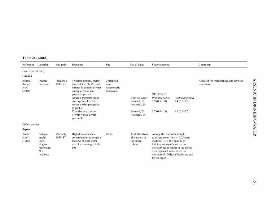
ARSEN
IC IN D
RINK
ING
-WATER
151
Table 26 (contd)
Reference Location End-point Exposure Site No. of cases Study outcome Comments
Case–control study
Canada
Infante-Rivard et al. (2001)
Québec province
Incidence 1980–93
Trihalomethanes, metals (As, Cd, Cr, Pb, Zn) and nitrates in drinking-water during prenatal and postnatal periods Arsenic exposure index Average level (> 95th versus ≤ 95th percentile [5 µg/L]) Cumulative exposure (> 95th versus ≤ 95th percentile
Childhood acute lymphocytic leukaemia
Exposed cases Prenatal, 18 Postnatal, 20 Prenatal, 20 Postnatal, 19
OR (95% CI) Prenatal period 0.9 (0.5–1.8) 0.7 (0.4–1.3)
Postnatal period 1.4 (0.7–2.8) 1.1 (0.6–2.2)
Adjusted for maternal age and level of education
Cohort studies
Japan
Tsuda et al. (1989)
Nakajo-machi town, Niigata Prefecture, 281 residents
Mortality 1959–87
High dose of arsenic contamination (through a factory) of well-water used for drinking (1955–59)
Uterus 17 deaths from all cancers in the entire cohort
Among the residents in high-exposure areas (low, < 0.05 ppm; medium, 0.05–0.5 ppm; high, ≥ 0.5 ppm), significant excess mortality from cancer of the uterus over expected value based on mortality for Niigata Prefecture and for all Japan
pp97-166.qxd 11/10/2004 10:47 Page 151

IARC M
ON
OG
RAPH
S VO
LUM
E 84152
Table 26 (contd)
Reference Location End-point Exposure Site No. of cases Study outcome Comments
Tsuda et al. (1995)
454 residents living in Namikicho and Nakajo-machi in Niigata Prefecture
Mortality 1959–92
High-dose arsenic contamination (through a factory) of well-water used for drinking (1955–59)
Uterus 0 0 2 2
Arsenic concentration (ppm) < 0.05 0.05–0.99 ≥ 1 Total
SMR (95% CI) 0.0 (0–8.0) 0.0 (0–37.6) 13.5 (2.4–48.6) 3.0 (0.5–11.1)
113 persons who drank from industrially contaminated wells in 1955–59, then followed for 33 years; rates in Niigata Prefecture in 1960–89 used as the standard for estimation of SMR
USA
Total, 433
Quin-tile
Cut-point (µg/g)
Odds ratio
95% CI
54 1 < 0.059 1.0 67 2 0.059–0.078 1.2 0.7–1.98 56 3 0.079–0.103 1.01 0.6–1.7 62 4 0.104–0.138 1.1 0.7–1.9
Garland et al. (1996)
Nested case–control in the Nurses’ Health Study Cohort, USA
Incidence 1984–86
Exposure to 5 metals (As, Cu, Cr, Fe, Zn) through any route measured
Breast, diagnosed from 1984 to 1986, 459 matched controls
69 5 > 0.138 1.1 0.7–1.9
Multivariate logistic regression models controlled for age, date of nail return, smoking, age at first birth, parity, history of benign breast disease, history of breast cancer in mother or sister, age at menarche, menopausal status, body mass index and alcohol consumption
Lewis et al. (1999)
Millard County, UT
Mortality Exposure to arsenic in drinking-water
Prostate 50 All men (µg/L–years) Low (< 1000) Medium (1000–4999) High (≥ 5000)
SMR 1.5 (95% CI, 1.07–1.9) 1.07 1.70 (p < 0.05) 1.65
Exposure index relies on ecological measures of arsenic concentration, median value for the community. State rates in 1950–92 used as the standard for estimation of SMR
SMR, standardized mortality ratio; CI, confidence interval; M, male; F, female; SIR, standardized incidence ratio; OR, odds ratio
pp97-166.qxd 11/10/2004 10:47 Page 152

cancers of the breast (two cases, one case who had keratoses present), pancreas (one casewho had keratoses present) and mouth (one patient who had treatment to the mouth forsyphilis). A report of seven cases of cancer following treatment with Fowler’s solution(Jackson & Grainge, 1975) included one case of bilateral breast cancer and one case ofcolon cancer. Multiple skin cancers were present in both cases. The literature alsoincludes cases of meningioma and intestinal malignancies associated with ingestion ofarsenic (IARC, 1987). [Case reports have helped to identify the role of ingestion ofarsenic in the occurrence of skin cancer and could provide leads to occurrences of othermalignancies. However, without an appropriate comparison group, it is unclear whetherany of the cases represent an excess over the norm.]
2.4.1 Taiwan, China
Ecological studiesChen et al. (1985) (described in Section 2.1.1) reported SMRs in the four-county
region in South-West Taiwan that is endemic for Blackfoot disease using mortality ratesfor the whole country as the standard. The analyses were based on mortality data obtainedfrom the Department of Health for the period 1968–82. To estimate dose, SMRs werecomputed for regions that were hyperendemic (21 villages), endemic (25 villages) and notendemic (38 villages) for Blackfoot disease. Age-standardized mortality rates for the fourcounties were elevated compared with national rates for cancer of the colon in both menand women. Age- and sex-standardized mortality rates for colon cancer were higher inendemic areas than in non-endemic areas, but were lower in the hyperendemic areascompared with other areas. SMRs for leukaemia were of borderline statisticalsignificance in men and close to unity in women. The SMR for cancer of the smallintestine also was increased in men but not in women, and was not statistically significantin either sex [Prostate cancer was not investigated in this report.] (Chen et al., 1985). Asubsequent report used the median concentrations of arsenic in well-water measured in1962–64 grouped into levels of < 300, 300–600 and ≥ 600 µg/L. A dose-related gradientin age-adjusted mortality was noted for prostate cancer. In the ≥ 600-µg/L group, the age-standardized mortality rate for prostate cancer was 5.6 times higher than that of thegeneral population of Taiwan (Chen et al., 1988a) (study described in Section 2.1.1).
Wu et al. (1989) (study described in Section 2.1.1) published age-adjusted mortalityrates for the years 1973–86 in 27 townships in the four counties endemic for Blackfootdisease together with 15 additional villages in the townships of Yensui and Hsiaying.Using the same data on well-water content and the classification scheme described above(Chen et al., 1988a), a dose-related trend in mortality from prostate cancer was againobserved (Mantel-Haenszel test for trend, p < 0.05). The authors also noted dose-relatedincreases in mortality rates from nasopharyngeal and colon cancer in men, which,however, were not statistically significant. In this analysis, leukaemia and oesophageal,stomach or uterine cancers did not appear to be related to levels of arsenic (Wu et al.,1989).
ARSENIC IN DRINKING-WATER 153
pp97-166.qxd 11/10/2004 10:47 Page 153

In a later study, Chen and Wang (1990) (described in Section 2.1.1) used data onarsenic in well-water measured from 1974 to 1976 in 314 precincts and townshipsthroughout Taiwan. Based on a multiple regression analysis (adjusted for urbanization andage), mortality rates from prostate cancer significantly increased with higher average levelof arsenic. Age-adjusted mortality rates for cancers of the nasal cavity also correlated withaverage arsenic concentration in men and women for all precincts and townships and forthe south-western townships. Cancers of the oesophagus, stomach, small intestine, colon,rectum, pancreas, larynx, bone and cartilage, breast, cervix, ovary and brain andleukaemia were not significantly correlated. [The regression estimates were onlypresented for statistically significant results.]
Based on data from death certificates for the period 1971–94, Tsai et al. (1999)(described in Section 2.1.1) computed SMRs for the four townships that are endemic forBlackfoot disease using local (Chiayi County) and national (Taiwan) rates as the standard.Applying national rates, SMRs were elevated for cancers of the intestine, colon andprostate in men, cancers of the nasal cavity and larynx in both men and women, lym-phoma in men and women, leukaemia in men and cancers of the pharynx and bone inwomen. These SMRs were similarly elevated using the local mortality rates as thestandard. In addition, using the local rates as the standard, higher SMRs were found forcancers of the oesophagus in men, cancers of the stomach in men and women and cancersof the colon, rectum and brain in women for the four townships.
[Data from South-West Taiwan indicate a consistent pattern of increases in mortalityfrom prostate cancer in areas with high contamination by arsenic, and there is evidence ofa dose-related effect. These studies do not specifically address the issue of dose of expo-sure, nor do they raise issues of latency and duration. These issues cannot be addressedusing mortality as an end-point for prostate cancer since the disease has a low case-fatalityrate. One possible source of bias is that prostate cancer often goes undetected, and ahigher mortality rate in regions with known exposure to arsenic could occur if screeningfor cancer deaths is enhanced in the region.]
2.4.2 Chile
Ecological studyRivara et al. (1997) compared the mortality rates for various cancers in 1976 between
Antofagasta in Region II and the non-contaminated Region VIII. An elevated risk ofcancer of the larynx was observed in the arsenic-exposed Region II. Among the variousother cancers (17 sites) investigated, no other elevated SMRs were reported.
2.4.3 Japan
Cohort studies From about 1945 to 1959, wells in the small town of Nakajo-machi in Niigata
Prefecture became contaminated with arsenic (up to 3000 µg/L) from a factory producing
IARC MONOGRAPHS VOLUME 84154
pp97-166.qxd 11/10/2004 10:47 Page 154

King’s yellow (As203). Two uterine cancer deaths occurred from 1959 to 1992 in thehighest exposure category (≥ 500 µg/L), with an SMR for uterine cancer of around 3.0using the age- and cause-specific mortality rates from Niigata Prefecture (Tsuda et al.,1989, 1995). [No other cancer sites were discussed.]
2.4.4 North America
(a) Cohort studiesGarland et al. (1996) conducted a nested case–control study in the USA to investigate
the relationship between concentrations of arsenic and other trace elements in toenails andthe incidence of breast cancer. Cases and controls were selected from the Nurses’ HealthStudy cohort comprising 121 700 nurses aged 30–55 years living in 11 states of the USA.Toenail samples were obtained from 72% of 94 115 cohort members in 1982. A total of62 641 women provided toenail clippings and did not have a diagnosis of breast canceras of 1982. The nested study was based on the 433 women who had a diagnosis of breastcancer reported in mailed questionnaires in 1984 and 1986 and an age-matched controlgroup. Compared with the quintile, the adjusted odds ratio for breast cancer was 1.1(95% CI, 0.7–1.9) for the highest quintile of arsenic.
Lewis et al. (1999) reported SMRs for various cancers using a retrospective cohort ofresidents from Millard County, UT, USA. Of the multiple types of cancers examined,mortality was only elevated for prostate cancer [The limitations of this study have beencited in Section 2.1.4.].
(b) Case–control studies Infante-Rivard et al. (2001) conducted a population-based case–control study of child-
hood leukaemia (occurring before the age of 9 years) in Québec Province, Canada. Casesincluded individuals who were newly diagnosed with childhood leukaemia from 1980 to1993. Of 510 eligible cases, 491 participated (96.3%) and, of 588 controls, 493 took part(83.8%). The investigators sought drinking-water test data from 1970 onward through apostal questionnaire that yielded usable data from 112 of 202 municipalities (55%). Addi-tional data was provided from the Ministry of Municipal Distribution Systems in 1986.Further analysis of tap-water was performed by the study investigators and covered 103 ofthe municipalities. These data were linked to information on residential history derivedfrom interviews with the subjects’ parents, and the value used for exposure to arsenic wasthat of the subjects’ municipalities of residence for the closest year when data wereavailable. Values from multiple test results were averaged over a given year, and indivi-duals with private water systems were assigned the arsenic value of their municipality ofresidence. Separate exposure variables were computed for subjects’ pre- and postnatalperiods and included average arsenic levels and cumulative exposure. A logistic regressionanalysis included maternal age and level of schooling. The odds ratio for childhoodleukaemia above versus less than or equal to the 95th percentile (5 µg/L arsenic) was 0.9(95% CI, 0.5–1.8) for prenatal exposure and 1.4 (95% CI, 0.7–2.7) for postnatal exposure.
ARSENIC IN DRINKING-WATER 155
pp97-166.qxd 11/10/2004 10:47 Page 155
For cumulative exposure (above versus less than or equal to the 95th percentile), the oddsratios were 0.7 (95% CI, 0.4–1.3) for prenatal exposure and 1.1 (95% CI, 0.6–2.2) for post-natal exposure. [The study does not provide evidence of a link between childhoodleukaemia and exposure to arsenic through drinking-water either prenatally or postnatally.However, the drinking-water concentrations were relatively low (95% were below 5 µg/L)and the estimates were imprecise. Furthermore, the exposure estimates are subject to mis-classification, but there is no discussion on the variability of arsenic concentrations withinmunicipalities and the fraction of residents that used private systems that were assumed tocontain the same concentrations of arsenic as the public systems.]
2.4.5 Australia
Ecological study
In an ecological study using incidence data from the Victoria Cancer Registry for theyears 1982–91, Hinwood et al. (1999) calculated SMRs for 14 cancer sites (in addition toliver, lung, bladder and kidney) in 22 postcode areas characterized by a level of arsenic inwater > 0.01 mg/L in most areas. Incidence rates for all of Victoria were used as thestandard. Cancer sites that showed elevated rates with 95% CIs that excluded 1.0 wereprostate, melanoma, breast and chronic myeloid leukaemia. [The Working Group notedthat no information was presented on the actual use of water contaminated with arsenicfor drinking by the population.]
3. Studies of Cancer in Experimental Animals
Previous evaluation
Various inorganic arsenic compounds were tested for carcinogenicity by oral adminis-tration, skin application, inhalation and/or intratracheal administration, subcutaneousand/or intramuscular administration, intravenous administration and other experimentalsystems in mice, rats, hamsters, dogs or rabbits. Arsenic trioxide produced lung adenomasin mice after perinatal treatment (Rudnay & Börzsönyi, 1981) and in hamsters after itsintratracheal instillation (Ishinishi et al., 1983; Pershagen et al., 1984). It induced a lowincidence of adenocarcinomas at the site of its implantation into the stomach of rats(Katsnelson et al., 1986). A higher incidence of lung carcinomas was induced in ratsfollowing a single intratracheal instillation of a Bordeaux pesticide mixture (coppersulfate and calcium oxide in a concentration of 1–2%) containing calcium arsenate(IARC, 1987). Intratracheal instillations of calcium arsenate into hamsters resulted in aborderline increase in the incidence of lung adenomas, while no such effect was observedwith arsenic trisulfide (Pershagen & Björklund, 1985). These studies provide limitedevidence for carcinogenicity of inorganic arsenics (IARC, 1980, 1987).
IARC MONOGRAPHS VOLUME 84156
pp97-166.qxd 11/10/2004 10:47 Page 156
No adequate data on the carcinogenicity of organic arsenic compounds were availableto the previous working group (IARC, 1980, 1987).
3.1 Oral administration
3.1.1 Mouse
Groups of 24 male A/J mice, 6 weeks of age, were given tap-water (control) or asolution of 50, 200 or 400 ppm [µg/mL] dimethylarsinic acid (DMAV) as drinking-waterfor 25 (10 mice per group) or 50 weeks (14 mice per group). The incidences of lungtumours were 2/10 (20%), 3/10 (30%), 4/10 (40%) and 3/10 (30%) in control, 50-, 200-and 400-ppm groups, after 25 weeks, with average numbers of tumours/mouse of0.2 ± 0.42, 0.3 ± 0.48, 0.5 ± 0.71 and 0.4 ± 0.70, respectively; no significant differenceswere apparent, nor did average tumour size vary significantly (0.9, 0.5, 1.4 and 1.1 mm,respectively). After 50 weeks, a non-significant increase in the incidence of lung tumours(50, 71.4, 64.3 and 78.6% in 14 animals per group, respectively), a significant increase inmultiplicity (0.5 ± 0.52, 1.07 ± 1.0, 1.07 ± 1.07 and 1.36 ± 1.01, respectively; p < 0.05for the 400-ppm group) and an increase in average diameter (1.0, 1.2, 1.4 and 1.5 mm,respectively) were observed. The numbers of mice with papillary lung adenoma and/oradenocarcinoma at 50 weeks were two, five, seven and 10 (p = 0.002 for the 200- and400-ppm groups) and increased with increasing dose of DMAV. In animals that received0, 50, 200 or 400 ppm DMAV, the number of alveolar adenomas ranged from 3/14 to 5/14per treatment group (Hayashi et al., 1998).
Groups of 90 female C57BL/6J mice and 140 female metallothionein heterozygousmice (MT–/–), aged 4–5 weeks, were given drinking-water containing sodium arsenate(500 µg/L arsenic) ad libitum for up to 26 months. Groups of 60 control mice were giventap-water. Preliminary findings indicate that tumours were observed in the lung(C57BL/6J, 17.8%; MT–/–, 7.1%), gastrointestinal tract (14.4%; 12.9%), liver (7.8%;5.0%), spleen (3.3%; 0.7%), reproductive organs (3.3%; 5.0%), skin (3.3%; 1.4%), bone(2.2%; 0%) and eye (1.1%; 0%) of treated animals. No tumours were observed in thecontrol groups (Ng et al., 1999). [The Working Group decided that this study was preli-minary because no histopathological findings were reported.]
Groups of 20–30 K6/ODC transgenic mice, 7 weeks of age, were administered DMAV
at either 10 or 100 ppm [µg/mL] in their drinking water or sodium arsenite at 10 ppm for5 months. The incidence of squamous skin tumours was 0% in the controls, 8 and 22% inthe 10- and 100-ppm DMA groups, respectively, and 15% in the arsenite groups (Chenet al., 2000).
Groups of 29 or 30 male p53+/– heterozygous or p53+/+ mice (C57BL/6J background)were exposed to 0, 50 or 200 ppm [µg/mL] DMAV in the drinking-water for 80 weeks. Inp53+/+ mice, a significant increase in the incidence (control, 10%; 50-ppm, 30%[p < 0.05]; and 200-ppm, 30% [p < 0.05] in 30 animals per group) and multiplicity (0.2,0.6 [p < 0.02] and 0.6 [p < 0.02] tumours per mouse, respectively) of total tumours was
ARSENIC IN DRINKING-WATER 157
pp97-166.qxd 11/10/2004 10:47 Page 157

observed at the terminal killing, but with no dose dependence. In the heterozygotes, a non-significant increase in incidence was observed (control, 14/29 [48.3%]; 50-ppm, 18/29[62.1%]; and 200-ppm, 19/30 [63.3%]), but the number of tumours per mouse was signi-ficantly increased at 200 ppm (p < 0.05) (control, 0.8; 50-ppm, 1.1; 200-ppm, 1.2). Noeffects were observed in either heterozygous or p53+/+ mice regarding the number oftumours per tumour-bearing animal (control, 1.6; 50-ppm, 1.8; 200-ppm, 1.9 in hetero-zygous mice; control, 2; 50 ppm, 1.9; 200 ppm, 2 in p53+/+ mice). No significant influenceon tumour development was noted in any particular organ or tissue site. The tumoursinduced in the p53+/– heterozygous mice were mainly malignant lymphomas or leukaemia(control, 8/29 [28%]; 50-ppm, 13/29 [45%]; 200-ppm, 10/30 [33%]), fibrosarcomas (5/29[17%], 8/29 [28%], 10/30 [33%]) and osteosarcomas (3/29 [10%], 2/29 [8%], 4/30[13%]), with lower incidences of other types of tumours such as hepatocellular carci-nomas, thyroid follicular carcinomas, squamous-cell carcinomas of the skin and lung ade-nomas. In p53+/+ mice, tumours were generally malignant lymphomas or leukaemia (2/30[7%], 9/30 [30%], 9/30 [30%]) with very low incidences of the other types of tumour. Nofibrosarcomas or osteosarcomas were detected in p53+/+ mice. Tumour latency curves inDMAV-treated p53+/– heterozygous and p53+/+ mice showed a dose-dependently signifi-cant shift towards early induction (p < 0.03) in comparison with untreated controls (Salimet al., 2003).
3.1.2 Rat
Groups of 36 male Fischer 344/DuCrj rats, 10 weeks of age, received 0, 12.5, 50 and200 ppm DMAV [µg/mL] (100% pure) in the drinking-water for 104 weeks. There was nosignificant difference in body weight or survival (25, 28, 28 and 24 animals) among thegroups at week 104. At week 97, the first tumour in the urinary bladder was observed inone animal of the 200-ppm DMAV group. Effective numbers were considered to be thenumbers of animals alive at week 97. Incidences of urinary bladder tumours were 0/28,0/33, 8/31 (26%; two papillomas and six carcinomas; p < 0.01, Fisher’s exact probabilitytest) and 12/31 (39%; two papillomas and 12 carcinomas; p < 0.001, Fisher’s exact proba-bility test), respectively, and were multiple in two animals given the highest dose. Histo-pathologically, the carcinomas were transitional-cell carcinomas. Urinary pH did not differsignificantly between groups during the experiment. Bladder calculi were not observed inany of the rats (Wei et al., 1999, 2002). In a more exhaustive examination of the urinarybladder in the same animals, preneoplastic lesions (papillary or nodular hyperplasia) wereobserved in 0/28, 0/33, 12/31 (39%; p < 0.01) and 14/31 (45%, p < 0.01) animals in the0-, 12.5-, 50- and 200-ppm groups, respectively. The incidences of tumours, other thanthose of the urinary bladder, in all DMAV-treated groups were not different from those ofcontrols (Wei et al., 2002).
IARC MONOGRAPHS VOLUME 84158
pp97-166.qxd 11/10/2004 10:47 Page 158

3.2 Transplacental exposure
Mouse: Groups of 10 pregnant C3H mice were given drinking-water containing 0, 42.5and 85 ppm [μg/mL] ad libitum from day 8 to 18 of gestation. Offspring were weaned at 4weeks and then divided into separate groups of 25 males and 25 females. The offspringreceived no additional treatment with arsenic for the next 74 (males) or 90 (females) weeks.Transplacental exposure to arsenic did not reduce body weight in any group of offspringover the course of the experiment. In male offspring, there was a marked increase in the inci-dence of hepatocellular carcinomas (control, 3/24 [12%]; 42.5-ppm, 8/21 [38%]; 85-ppm,14/23 [61%]; p for trend = 0.00006, two-sided chi-square test) and multiplicity per mouse(control, 0.13 ± 0.07; 42.5-ppm, 0.42 ± 0.13; 85-ppm, 1.30 ± 0.28; p for trend = 0.003) in adose-related fashion. There was also a dose-related increase in the incidence of adrenalcortical adenomas (control, 9/24 [37.5%]; 42.5-ppm, 14/21 [66.7%]; 85-ppm, 21/23[91.3%]; p for trend = 0.001) and multiplicity (control, 0.71 ± 0.20; 42.5-ppm, 1.10 ± 0.22;85-ppm, 1.57 ± 0.32; p for trend = 0.016). In female offspring, there was a strong, dose-related increase in the incidence of ovarian tumours. Total tumour (benign and malignant)incidence was control, 2/25 (8%); 42.5-ppm, 6/23 (26%); and 85-ppm, 9/24 (38%) (p fortrend = 0.015). Controls had one adenoma and one benign granulosa-cell tumour. The 42.5-ppm treatment group had three adenomas, one adenocarcinoma, one benign granulosa-celltumour and one malignant granulosa-cell tumour. The 85-ppm treatment group developedseven adenomas, one luteoma and one haemangiosarcoma. Lung carcinomas developed(control, 0/25 [0%]; 42.5-ppm, 1/23 [4%]; 85-ppm, 5/24 [21%]; p for trend = 0.0086) in adose-dependent manner. There were significant increases in the number of mice bearing atleast one tumour (control, 11/24; 42.5-ppm, 17/21; 85-ppm, 22/23; p for trend = 0.0006 inmales; control, 12/25; 42.5-ppm, 17/23; 85-ppm, 16/24; p < 0.172 in females) and in micebearing at least one malignant tumour (control, 3/24; 42.5-ppm, 9/21; 85-ppm, 14/23;p < 0.0001 in males; control, 2/25; 42.5-ppm, 9/23; 85-ppm, 8/24; p < 0.042 in females)with both doses of arsenic. Exposure to arsenic also increased the incidence of hyperplasiaof the uterus and oviduct. In this experiment, four of the organs that developed tumours orhyperplasia were endocrine-responsive organs: adrenal gland, liver, ovary and uterus(Waalkes et al., 2003).
3.3 Intratracheal administration
Hamster: Groups of 30 (20 for a control) male Syrian golden hamsters, 8 weeks ofage, were given arsenic trioxide, calcium arsenate or arsenic trisulfide by intratrachealinstillation once a week for 15 weeks. Each compound contained 0.25 mg arsenicsuspended in 0.1 mL phosphate buffer solution. The control group received buffersolution alone. All hamsters were kept during their entire lifespan. Numbers of survivorsafter 15 instillations were 18/30 (60%) in the arsenic trioxide-treated group, 27/30 (90%)in the calcium arsenate-treated group, 23/30 (77%) in the arsenic trisulfide-treated groupand 22/22 (100%) in the control group, showing a similar tendency in survival rates of all
ARSENIC IN DRINKING-WATER 159

groups. All hamsters had died by day 794 (arsenic trioxide group), day 806 (calciumarsenate group), day 821 (arsenic trisulfide group) and day 847 (control group) after theinitial instillation. Incidences of lung tumours were 1/17 (5.8%; adenocarcinoma) arsenictrioxide-treated, 7/25 (28.0%; one adenocarcinoma, six adenomas; p value, significantversus controls) calcium arsenate-treated, 1/22 (4.5%; adenoma) arsenic trisulfide-treatedand 1/21 (4.8%; adenosquamous carcinoma) control animals. No tumours of the upperrespiratory tract including the trachea were observed in any group. Besides lung tumours,one adrenal adenoma and one liver haemangiosarcoma in the arsenic trioxide-treatedgroup, two adrenal adenocarcinomas and one leukaemia in the calcium arsenate-treatedgroup, one nephroblastoma and one adrenal adenoma in the arsenic trisulfide-treatedgroup and one adrenal adenocarcinoma and one adrenal adenoma in the control groupwere found (Yamamoto et al., 1987).
3.4 Administration with known carcinogens
3.4.1 Mouse
Groups of 30–32 female Swiss mice, 21–24 days of age, received concentrations of 0,10, 50 or 100 µg/L sodium arsenate or sodium arsenite in the drinking-water for 15 weeks.At week 3, the animals were administered a single intraperitoneal injection of 1.5 mg/kg bwurethane in saline. When killed at week 15, the numbers of lung adenoma per mouse were29.0 ± 5.4, 21.4 ± 2.6, 15.7 ± 1.8 and 16.0 ± 2.1 (p for trend = 0.0185) in the 0-, 10-, 50-and 100-µg/mL arsenate-treated groups, respectively, and 20.1 ± 1.8, 25.7 ± 4.0, 19.5 ± 2.1and 10.8 ± 1.6 (p for trend = 0.00082) in 0-, 10-, 50- and 100-µg/mL arsenite-treated groups,respectively [suggesting that both forms of arsenic exerted an inhibitory effect]. Botharsenate and arsenite at 100 µg/mL caused a significant reduction in tumour size(0.64 ± 0.01, 0.65 ± 0.02; p < 0.05) compared with control animals (0.74 ± 0.01, 0.71 ± 0.02)(Blakley, 1987).
In a two-stage protocol, groups of 9–13 male ddY mice, 6 weeks of age, were givena single subcutaneous injection of 10 mg/kg bw 4-nitroquinoline 1-oxide (4NQO) andthen received tap-water, a 5% glycerol solution or a 200- or 400-ppm [µg/mL] DMAV
solution in drinking-water for 25 weeks. The incidences of lung tumour-bearing micewere 2/9 (22%), 5/10 (50%), 8/13 (62%) and 10/13 (77%), respectively, while thenumbers of tumours per mouse were 0.22 ± 0.15, 1.40 ± 0.62, 3.92 ± 1.79 and 4.38 ± 1.07(p < 0.05 Cochran-Cox t-test). Thus, DMAV promoted lung tumorigenesis initiated by4NQO (Yamanaka et al., 1996). [The authors described a shift from papillary-typeadenomas to adenosquamous carcinomas but no quantitative data were provided.]
Groups of 60 male and female C57BL6J outbred mice [sex distribution unspecified],2 months of age, were fed a diet containing 10% lipids and were given either 0.01%arsenic trioxide in the drinking-water for 28 weeks or 3 mg per mouse benzo[a]pyrene in0.2 mL corn oil by gavage once a week for 3 weeks or both arsenic trioxide andbenzo[a]pyrene. No significant differences between groups given benzo[a]pyrene with or
IARC MONOGRAPHS VOLUME 84160
pp97-166.qxd 11/10/2004 10:47 Page 160

without arsenic trioxide were observed at the end of the 28-week experimental periodwith regard to focal localized hyperplasia, ulcers, focal multiple hyperplasia, papilloma-tosis or papillomas of the forestomach (total number per mouse, 4.20 ± 0.39 in thebenzo[a]pyrene-treated group and 5.40 ± 0.89 in the benzo[a]pyrene–arsenic-treatedgroup) (Silva et al., 2000).
Groups of 10–11 female Hos:HR-1 hairless mice, 6 weeks of age, were administered0, 400 or 1000 ppm [µg/mL] DMAV in the drinking-water and irradiated twice weeklywith 2 kJ/m2 ultraviolet B (UVB) rays for 25 weeks. DMAV had no effect on body weightgain. The number of skin tumours per mouse was significantly increased by 1000 ppmDMAV compared with the 0-ppm value from weeks 13 to 19, and incidence of tumour-bearing mice was significantly increased at weeks 12 and 13 [exact data not clear as therewere no tabulations]. No differences were noted at later time points up to week 25 (100%incidence in all groups by week 16), showing a shift towards early tumour inductionfollowing treatment with DMAV. Malignant tumours were observed in only two animalsin the 1000-ppm group (Yamanaka et al., 2000).
Groups of 15 female Crl: SK1-hrBR hairless mice, 21 days of age, received 0 or 10mg/L sodium arsenite in the drinking-water and were irradiated with solar lamps at a doseof 1.7 J/m2 (lamp output: 85% in the UVB range, < 1% UVC and 4% UVA, and theremainder visible) three times weekly. The UVR dose was chosen to be approximatelyhalf of the minimal erythemic dose. Two control groups of five mice received sodiumarsenite only or no treatment. Sodium arsenite did not influence body weight gain. Noskin tumours were observed with arsenite alone or in the untreated controls. The firsttumours were noted after 8 weeks with arsenite and UVR but after 12 weeks with UVRalone (significantly earlier appearance). All UVR-treated animals had at least one tumourat 26 weeks; however, after 19 weeks of exposure to UVR, incidences were 100% forUVR and arsenite and 33% for UVR alone. The total number of tumours in the grouptreated with UVR alone (15 animals) was 53 and that in the group treated with UVR andarsenite (15 animals) was 127. In UVR and arsenite-treated animals, 64/127 (50.4%)tumours were highly invasive squamous-cell carcinomas, whereas in UVR alone-treatedanimals, 14/53 (26.4%) were highly invasive squamous-cell carcinoma (p = 0.003)(Rossman et al., 2001)
Groups of 10 female Hos:HR-1 hairless mice, 6 weeks of age, were treated with asingle topical application of 200 nmol 7,12-dimethylbenz[a]anthracene (DMBA)dissolved in acetone and were then administered 0, 400 or 1000 ppm [µg/mL] DMAV inthe drinking-water and/or irradiated twice weekly with 0.3 kJ/m2 UVB. All mice werekilled after 50 weeks. Skin tumours occurred faster in the DMAV-treated group. DMAV
without UVB increased the incidence of skin tumours (p < 0.05 at 20–22 weeks) but notdose-dependently. Greater effects were seen in combination with UVB, particularly at1000 ppm [exact data not clear as there was no tabulation]. Incidences of papillomas inanimals treated with DMAV without UVB were 1/10, 9/10 and 7/10 in the 0-, 400- and1000-ppm groups, respectively, and those of squamous-cell carcinomas were 2/10, 0/10and 0/10, respectively. Incidences of papillomas in animals treated with DMAV and UVB
ARSENIC IN DRINKING-WATER 161
pp97-166.qxd 11/10/2004 10:47 Page 161
were 0/10, 7/10 and 7/10, and those of squamous-cell carcinomas were 0/10, 3/10 and1/10 in the 0-, 400- and 1000-ppm groups, respectively (Yamanaka et al., 2001).
3.4.2 Transgenic mouse
Groups of 7–8 female keratin (K6)/ODC transgenic mice, 10–14 weeks of age, receivedtwo weekly applications of 3.6 mg DMAV in neutral cream or 5 µg 12-O-tetradecanoyl-phorbol 13-acetate (TPA) in 200 µL acetone 1 week after initiation with 50 µg DMBA in200 µL acetone. A significantly accelerated development of skin tumours (first tumour after8 weeks in DMAV-treated animals and after 11 weeks in controls) was observed followingtreatment with DMAV; 20 weeks after initiation, the numbers of tumours (average,19.4 ± 10.2 per mouse compared with 9.7 ± 3.5 in controls) were increased. Promotingactivity was similar to that achieved with application of 5 µg TPA (20.7 ± 8.4) twice weekly.Microscopically, most of the tumours were squamous papillomas, although squamouscarcinomas occurred in some DMAV- and some TPA-treated animals (Morikawa et al.,2000).
3.4.3 Rat
Sodium arsenite has been reported to enhance the incidence of renal tumours inducedin rats by intraperitoneal injection of N-nitrosodiethylamine (NDEA) (Shirachi et al.,1983). A subsequent re-evaluation of the study indicated that not only sodium arsenite butalso sodium arsenate enhanced NDEA-induced kidney tumours (Smith et al., 1992).
Groups of 20 male Fischer 344/DuCrj rats, 6 weeks of age, received a single intraperi-toneal injection of 100 mg/kg bw NDEA, followed by intraperitoneal injections of20 mg/kg bw N-methyl-N-nitrosourea on days 5, 8, 11 and 14 and subcutaneous injectionsof 50 mg/kg bw 1,2-dimethylhydrazine chloride on days 18, 22, 26 and 30. At the sametime, animals received 0.05% N-butyl-N-(4-hydroxybutyl)nitrosamine (BBN) in thedrinking-water for the first 2 weeks then 0.1% N-bis(2-hydroxypropyl)nitrosamine for thenext 2 weeks (so-called DMBDD model treatment). After a 2-week interval, the animalsreceived 0, 50, 100, 200 or 400 ppm [µg/mL] DMAV in the drinking-water from weeks 6to 30, at which time they were killed. DMAV significantly enhanced tumour induction inthe urinary bladder (both papillomas and transitional-cell carcinomas), kidney (bothadenomas and adenocarcinomas), liver (hepatocellular carcinomas) and thyroid (adeno-mas) (see Table 27). Values for preneoplastic lesions such as papillary or nodular hyper-plasia in the urinary bladder, atypical tubules in the kidney and altered hepatocyte foci inthe liver were also significantly increased. No promoting effects were noted in the lungs orthe nasal cavity (Yamamoto et al., 1995).
To confirm the above-mentioned results and to evaluate low-dose effects of DMA inurinary bladder and liver carcinogenesis, the following two studies were conducted.Groups of 20 male Fischer 344 rats, 6 weeks of age, received 0.05% BBN in drinking-water for 4 weeks followed by 0, 2, 10, 25, 50 or 100 ppm [µg/mL] DMAV for 32 weeks.
IARC MONOGRAPHS VOLUME 84162
pp97-166.qxd 11/10/2004 10:47 Page 162

ARSEN
IC IN D
RINK
ING
-WATER
163
Table 27. Incidence of preneoplastic and neoplastic lesions in various organs of Fischer 344/Du Crj rats treated with DMAV after initiation with DMBDD treatment
DMAV
0 ppm 50 ppm 100 ppm 200 ppm 400 ppm
Organ and finding
n = 20 (%) n = 20 (%) n = 19 (%) n = 20 (%) n = 20 (%)
Two-tailed Cochran-Armitage analysisa
Urinary bladder Papillary or nodular hyperplasia Papilloma Transitional-cell carcinoma No. of tumour-bearing animals
4 (20) 1 (5) 1 (5) 2 (10)
13 (65)c 12 (60)d 10 (50)c 17 (85)d
14 (73.7)d 12 (63.2)d 11 (57.9)d 16 (84.2)d
11 (55)b 11 (55)d 12 (60)d 17 (85)d
11 (55)b 7 (35)b 13 (65)d 16 (80)d
NE NE NE NE
Kidney Adenoma Adenocarcinoma Nephroblastoma No. of tumour-bearing animals
1 (5) 0 4 (20) 5 (25)
3 (15) 0 0 3 (15)
1 (5.2) 2 (10.5) 4 (12.1) 6 (31.6)
7 (35)b 1 (5) 6 (30) 13 (65)b
3 (15) 7 (35)c 9 (45) 13 (65)b
p < 0.01 p < 0.05 p < 0.001
Liver Altered cell foci Clear-cell foci Basophilic foci Eosinophilic foci Hyperplastic nodule Hepatocellular carcinoma Cholangioma Haemangioma No. of tumour-bearing animals
10 (50) 1 (5) 1 (5) 0 0 0 0 0
12 (60) 2 (10) 2 (10) 0 2 (10) 0 0 2 (10)
14 (73.7) 3 (15.8) 8 (42.1)c 2 (10.5) 0 0 0 2 (10.5)
19 (95)c
10 (50)c 15 (75)d 9 (45)d 8 (40)c 1 (5) 1 (5) 17 (85)d
20 (100)d
17 (85)d 16 (80)d 7 (35)c 8 (40)c 1 (5) 0 13 (65)d
p < 0.001 p < 0.001 p < 0.001 p < 0.001 p < 0.001 p < 0.001
pp97-166.qxd 11/10/2004 10:47 Page 163

IARC M
ON
OG
RAPH
S VO
LUM
E 84164
Table 27 (contd)
DMAV
0 ppm 50 ppm 100 ppm 200 ppm 400 ppm
Organ and finding
n = 20 (%) n = 20 (%) n = 19 (%) n = 20 (%) n = 20 (%)
Two-tailed Cochran-Armitage analysisa
Thyroid gland Hyperplasia Adenoma Adenocarcinoma No. of tumour-bearing animals
3 (15) 2 (10) 1 (5) 3 (15)
4 (20) 1 (5) 1 (5) 2 (10)
2 (10.5) 3 (15.8) 5 (26.3) 8 (42.1)
13 (65)c
1 (5) 5 (25) 6 (30)
13 (65)c
6 (30) 4 (20) 9 (45)b
p < 0.001 p < 0.05
From Yamamoto et al. (1995) DMAV, dimethylarsinic acid; DMBDD, N-nitrosodiethylamine + N-methyl-N-nitrosourea + 1,2-dimethylhydrazine chloride + N-butyl-N-(4-hydroxybutyl)nitrosamine + N-bis(2-hydroxypropyl)nitrosamine; NE, not examined
a The significance of differences in the incidence of lesions between groups was assessed using the Fisher’s exact probability test. To evaluate the dose–response relationships of the incidences in lesions in the kidney, liver and thyroid gland, two-tailed Cochran-Armitage analysis was used. Significantly different from 0 ppm at b p < 0.05, c p < 0.01 and d p < 0.001
pp97-166.qxd 11/10/2004 10:47 Page 164

Development of preneoplastic lesions and tumours of the bladder (papillary or nodularhyperplasia, papillomas and carcinomas) was enhanced in a dose-dependent manner (seeTable 28). Doses of 25, 50 and 100 ppm increased the incidences (%) and multiplicities(number per rat) of bladder papillomas and carcinomas. A significant increase inmultiplicity of total tumours (papillomas plus carcinomas) was observed with doses aslow as 10 ppm DMAV (p < 0.05): 0 ppm, 0.20; 2 ppm, 0.20; 10 ppm, 0.55; 25 ppm, 1.47;50 ppm, 2.30; 100 ppm, 2.40. Compared with controls, doses of 50 or 100 ppm signifi-cantly increased the incidence of papillary or nodular hyperplasia (Wanibuchi et al.,1996).
Groups of 10 male Fischer 344 rats, 6 weeks of age, were given a single intraperi-toneal injection of 0 (control) or 200 mg/kg bw NDEA in saline and 2 weeks laterreceived 0, 25, 50 or 100 ppm [µg/mL] DMAV in the drinking-water for 6 weeks. Partialhepatectomy was performed on all animals at the end of week 3. Final body weights weredecreased dose-dependently but not significantly. No significant variation in relative liverweights was noted. Dose-dependent significant increases in both numbers and areas ofglutathione S-transferase placental form (GST-P)-positive foci in the liver were observedafter initiation with NDEA; the significance was evident at doses of 50 and 100 ppm(p < 0.01) for numbers and at all three doses (p < 0.05 or p < 0.01) for areas [exact valueswere not listed because of figure]. No GST-P-positive foci were observed in groups notinitiated with NDEA (Wanibuchi et al., 1997).
Groups of eight male NCI-Black-Reiter rats (which lack α2u-globulin), 9–14 weeksof age, received 0.05% BBN in the drinking-water for 4 weeks followed by 0 or 100 ppm[µg/mL] DMAV for 32 weeks. When killed at the end of week 36, the incidence andmultiplicity of papillary or nodular hyperplasia in the bladder was significantly increasedin DMAV-treated rats (6/8 [75%]; p < 0.05; number per rat, 1.1 ± 1.0, p < 0.05) comparedwith rats receiving BBN alone (0/8 [0%]; number per rat, 0). A 38% incidence of bladderpapillomas or carcinomas was observed in DMAV-treated but not in control animals (Liet al., 1998).
Groups of 20 male Fischer 344 rats, 10 weeks of age, received a single intraperitonealinjection of 200 mg/kg bw NDEA followed 2 weeks later by DMAV, monomethylarsonicacid (MMA) or trimethylarsine oxide (TMAO) at a dose of 100 ppm in the drinking-waterfor 6 weeks. Numbers of GST-P-positive foci in the liver were significantly increased inrats treated with MMA, DMAV and TMAO compared with the controls. Areas of GST-positive foci were also significantly increased in rats treated with MMA, DMAV andTMAO compared with the controls (Nishikawa et al., 2002).
ARSENIC IN DRINKING-WATER 165
pp97-166.qxd 11/10/2004 10:47 Page 165

IARC M
ON
OG
RAPH
S VO
LUM
E 84166
Table 28. Induction of urinary bladder lesions in Fischer 344 rats treated with BBN followed by DMAV at various doses
Papillary or nodular hyperplasia Papilloma Carcinoma DMAV (ppm) No. of rats examined
Incidence (%) No./rat Incidence (%) No./rat Incidence (%) No./rat
0 (control) 20 14 (90) 1.05 ± 95a 3 (15) 0.15 ± 0.37 1 (5) 0.05 ± 0.22 2 20 13 (65) 1.30 ± 1.30 2 (10) 0.10 ± 0.31 2 (10) 0.10 ± 0.31 10 20 14 (70) 1.55 ± 1.47 7 (35) 0.40 ± 0.60 3 (15) 0.15 ± 0.37 25 19 18 (95) 2.37 ± 1.17b 11 (58)c 1.05 ± 1.18b 7 (37)d 0.42 ± 0.61e 50 20 20 (100)d 2.95 ± 1.88b 13 (65)f 1.50 ± 1.36b 10 (50)f 0.80 ± 0.95c 100 20 20 (100)d 4.10 ± 3.02b 17 (85)g 1.70 ± 1.17b 12 (60)g 0.70 ± 0.66b
From Wanibuchi et al. (1996) BBN, N-butyl-N-(4-hydroxybutyl)nitrosamine; DMAV, dimethylarsinic acid a Mean ± SD b p < 0.001 (significantly different from control, Student’s t-test); c p < 0.01; d p < 0.05; e p < 0.05; f p < 0.01; g p < 0.001 (significantly different from control, Fisher’s exact probability test)
pp97-166.qxd 11/10/2004 10:47 Page 166

4. Other Data Relevant to an Evaluation of Carcinogenicityand its Mechanisms
Extensive reviews of the metabolism of arsenic have been published recently (NationalResearch Council, 1999, 2001; WHO, 2001). This section focuses on data relevant for theevaluation of carcinogenic effects. Thus, it is not a complete review on all published data.The oxidation state of arsenic and its metabolites are given if reported. If speciation ofoxidation state has not been performed, the metabolites are given as monomethylarsonicacid (MMA) and dimethylarsinic acid (DMA).
4.1 Absorption, distribution, metabolism and excretion
4.1.1 Absorption
Arsenic in drinking-water is easily absorbed in the gastrointestinal tract. About70–90% of a single dose of dissolved arsenite (AsIII) or arsenate (AsV) was absorbed fromthe gastrointestinal tract of humans and experimental animals (Pomroy et al., 1980; Vahter& Norin, 1980; Freeman et al., 1995). A high rate of gastrointestinal absorption is alsosupported by the fact that people whose main fluid intake consists of drinking-water withelevated arsenic concentrations have very high concentrations of arsenic in their urine.
With regard to absorption of arsenic through the skin, a few experimental studies indi-cate a low degree of systemic absorption. Application of water solutions of radiolabelledarsenate in vivo to the skin of rhesus monkey and in vitro to human cadaver skin showedthat about 2–6% and 0.98% of the applied arsenic was absorbed within 24 h, respectively(Wester et al., 1993). Similar in-vitro studies using dorsal skin of mice showed a muchhigher absorption: 33–62% of the applied dose of radiolabelled arsenate in aqueoussolution was absorbed through the skin within 24 h. However, most of it, on average60–90%, was retained in the skin (Rahman et al., 1994). These authors suggested thatabsorption of arsenic through the skin may be species-specific. For many chemicals,mouse skin is more permeable in vitro than human cadaver skin (Bronaugh et al., 1982).In-vitro studies with human keratinocytes showed that 1–8% of the applied arsenic dosewas retained per hour (Bernstam et al., 2002). Morphological changes, cytotoxicity andinhibition of DNA and protein syntheses were found with in-vitro doses of AsIII as low as10 µg/L. Thus, it seems probable that inorganic arsenic can be absorbed from the exterior,leading to a breakdown in skin barrier function.
In-vitro studies of skin absorption of DMAV (10 µg in 20–100 µL water) by appli-cation to dorsal skin from adult mice mounted in flow-through diffusion cells showed that16–25% was retained in the receptor fluid (Hanks balanced salt solution), about 15% inthe skin and the remainder in the wash after 24 h (Hughes et al., 1995). After exposure
ARSENIC IN DRINKING-WATER 167
pp167-228.qxd 11/10/2004 10:53 Page 167

for 1 h only, essentially all of the applied dose was washed away. Less than 1% of theapplied dose was absorbed.
A low degree of systemic skin absorption of inorganic arsenic is supported by studiesshowing that people in Fairbanks, AK, who used tap-water containing about 345 µg/Larsenic for washing, but only bottled water (which did not contain arsenic) for drinking,had about the same low concentrations of arsenic metabolites in urine (on average about40 µg/L) as people with less than 50 µg/L in their tap-water (Harrington et al., 1978). Theconcentration of arsenic in hair was clearly elevated in the group drinking bottled water(5.74 µg/g compared with 0.43 µg/g in the low-arsenic group), which shows that arsenicis bound to hair and probably also to skin during washing with water rich in arsenic.
4.1.2 Distribution
Following its absorption, arsenate is rapidly reduced to AsIII; the distribution of its meta-bolites in the body are therefore very similar to that following exposure to AsIII. However,studies in mice given arsenite or arsenate (0.4 mg/kg bw) intravenously showed that the con-centrations in stomach and intestines were higher after exposure to AsIII than after exposureto AsV, while incorporation in bone was higher following exposure to AsV (Lindgren et al.,1982). The differences are less marked in the case of oral exposure, probably due to fastermethylation that occurs when the absorbed arsenic passes directly to the liver. After expo-sure to toxic doses at which methylation capacity is exceeded or inhibited, the differences indistribution patterns for the two forms are greater (Vahter & Norin, 1980).
Absorbed arsenic is transported, mainly bound to SH groups in proteins and low-mole-cular-weight compounds such as glutathione (GSH) and cysteine, to different organs in thebody (National Research Council, 1999, 2001). Complexation of trivalent arsenicalcompounds with GSH, probably mainly in the form of As(GS)3, has been demonstrated, butAsIII is easily transferred to binding sites of higher affinity, especially vicinal dithiols, suchas lipoic acid and dimercaptosuccinic acid (Cullen & Reimer, 1989; Delnomdedieu et al.,1993). Studies on serum arsenic in dialysis patients showed the presence of inorganicarsenic, partly bound to proteins, and DMA (Zhang et al., 1997, 1998a,b; De Kimpe et al.,1999). Transferrin was the main carrier protein, but the extent to which this occurs in healthyindividuals is not known. Most of the arsenic in blood is rapidly cleared, following a three-exponential clearance curve (Mealey et al., 1959; Pomroy et al., 1980). The majority ofarsenic in blood is cleared with a half-time of about 2 or 3 h. The half-times of the secondand third phases are about 168 and 240 h, respectively (Mealey et al., 1959; NationalResearch Council, 1999).
In experimental studies on mammals exposed to inorganic arsenic, the tissues with thelongest retention of arsenic, depending on species, were skin, hair, liver, kidney, blood,squamous epithelium of the upper gastrointestinal tract, epididymis, thyroid, skeleton andlens. Arsenic does not readily cross the blood–brain barrier, and concentrations in the brainare generally low compared with most other tissues (Lindgren et al., 1982; Vahter et al.,1982; Lindgren et al., 1984; Yamauchi & Yamamura, 1985).
IARC MONOGRAPHS VOLUME 84168
pp167-228.qxd 11/10/2004 10:53 Page 168

In human subjects exposed chronically to arsenic and also at background environmentalconcentrations, the hair and nails generally show the highest concentrations (0.02–10 mg/kgdry wt; Hindmarsh, 2002). Thus, arsenic appears to concentrate in tissues with a highcontent of cysteine-containing proteins. In areas of West Bengal and Bangladesh that havehigh concentrations of arsenic in the drinking-water, maximal concentrations of arsenic inhair, nail and skin exceeding 40 mg/kg have been reported (Guha Mazumder et al., 1988;Chowdhury et al., 2001; Basu et al., 2002). Very few studies have been carried out on thedistribution of arsenic in human tissues. Postmortem analysis of human tissues confirm thatarsenic is widely distributed in the body after long-term exposure, with highest concen-trations in the skin and lungs (0.01–1 mg/kg dry wt), as well as hair and nails (Liebscher &Smith, 1968; Cross et al., 1979; Dang et al., 1983). In people exposed to high concentrations(0.2–2 mg/L) of arsenic in drinking-water, the concentration in liver was 0.6–6 mg/kgdry wt compared with 0.16 mg/kg in unexposed people (Guha Mazumder et al., 1988). In acase of acute intoxication by arsenic, the liver and kidneys showed the highest concen-trations of total arsenic with values 350- and 63-fold higher than those in blood, respec-tively. In all organs, AsIII was the predominant species, and MMA occurred at higher con-centrations than DMA. MMA and DMA were more prevalent in lipid-rich organs (49% and45% of total arsenic in cerebellum and in brain, respectively) compared with other organs(∼ 20% of total arsenic). AsV was found in small quantities in the liver, kidneys and blood(2% of total arsenic) (Benramdane et al., 1999).
Dang et al. (1983) used neutron activation analysis (NAA) to measure total arsenic intissues of people [age and sex not specified] dying in accidents in Mumbai, India (Table 29).Concentrations in the brain were generally low compared with most other tissues. Thus, itappears that arsenic does not readily cross the blood–brain barrier. Notably, there was a largevariation in tissue concentrations of arsenic among individuals, similar to that reported inearlier studies (Liebscher & Smith, 1968; Larsen et al., 1974).
ARSENIC IN DRINKING-WATER 169
Table 29. Levels of arsenic in human tissues obtained from traffic accident victims in the Mumbai area of India
Tissue No. of samples
Mean concentration (± SD) of arsenic (mg/kg wet wt)
Brain 12 3.9 ± 1.0 Blood 8 5.9 ± 3.9 Kidney 13 12.4 ± 20.7 Liver 19 14.5 ± 6.9 Spleen 18 15.2 ± 16.6 Lung 13 19.9 ± 22.7
From Dang et al. (1983)
pp167-228.qxd 11/10/2004 10:53 Page 169

Few studies have examined the distribution of arsenic metabolites in tissues, owing toanalytical difficulties. Marafante et al. (1982) reported predominantly inorganic arsenic inultrafiltrates of rat and rabbit liver and kidney 1 h after intraperitoneal injection of50 µg/kg bw [74As] as sodium arsenite, using ion-exchange chromatography with radio-metric detection. The fraction present as MMA was generally less than one-tenth that ofinorganic arsenic. De Kimpe et al. (1996) studied the tissue distribution of arsenic meta-bolites up to 120 h after intraperitoneal injection of a trace amount of [74As]-arsenate inmale Flemish giant rabbits, also using ion-exchange chromatographic separation of ultra-filtrates with radiometric detection. The predominant metabolite present in tissues wasDMA, followed by inorganic arsenic species and low concentrations of MMA. Thepercentage of DMA increased steadily over time in bone marrow, heart, liver, muscle,pancreas, small intestine and spleen, but levelled off or declined in kidney and lung.
Yamauchi and Yamamura (1985) studied the tissue distribution over time of arsenicmetabolites in male Syrian golden hamsters given a single oral dose of 4.5 mg/kg bwarsenic trioxide. Speciation of arsenic metabolites was carried out by hybrid generation–atomic absorption spectrophotometry (HG–AAS) with a cold trap after alkaline digestion.The predominant form of arsenic present in all tissues up to 120 h after dosing wasinorganic arsenic. In contrast to other studies, the concentrations of MMA in tissue weretwo- to fourfold higher than those of DMA at all time-points, while much more DMA(22% of the dose in 5 days) than MMA (2.5% of the dose) was excreted in urine. Thehighest concentrations of MMA were found in lungs and spleen at 12–24 h, and those ofDMA in liver, lung and kidney at 24 h.
Yamauchi et al. (1988) reported data on the time-course tissue distribution in hamstersgiven a single oral dose of 50 mg/kg bw MMA. Peak MMA concentrations were achievedwithin 6–120 h after dosing and were highest in the kidney, followed by spleen, lung, skin,liver, muscle and brain. MMA itself accumulated in the kidney and levels declined veryslowly. DMA was also detected in several tissues, with highest levels occurring in thelung, followed by kidney and liver. Trimethylated arsenic was not detected in any tissues.
The fate of DMA has been studied in mice administered [74As] or [14C]DMA intra-venously. The highest levels of radioactivity were present in kidney at all time-points(5–60 min after injection). Tissues that retained arsenic for the longest time (24 h) werethe lungs, intestinal walls, thyroid and lens (Vahter et al., 1984; Hughes et al., 2000).Yamauchi and Yamamura (1984) studied the tissue distribution of DMA in hamsters admi-nistered a single oral dose of 50 mg/kg bw DMA. Concentrations were elevated in alltissues examined, including the brain, indicating that DMA is widely distributed in thebody and that it passes the blood–brain barrier, although not to a large extent. Concen-trations of DMA peaked at 6 h in all tissues examined except hair, with the highest levelsin lung, followed by kidney, spleen, liver, skin, muscle and brain. Part of the DMA wasfound to be methylated further to trimethylarsenic (TMA) in vivo. Concentrations of TMApeaked at 6 h in all tissues except skin and hair in which none was detected. The highestconcentrations of TMA were found in lung, and were equivalent to about half of those ofDMA. It is notable that the peak concentrations of DMA and TMA in the lung were over
IARC MONOGRAPHS VOLUME 84170
pp167-228.qxd 11/10/2004 10:53 Page 170

fourfold higher than those in the liver and kidney, but at 120 h after dosing, both haddeclined to control levels. The authors presumed that the TMA biosynthesized byhamsters is more likely to behave as an organic arsenic compound, such as arsenobetainewhich is present in seafoods, than to be a very toxic substance such as trimethylarsine.
4.1.3 Metabolism
(a) Methylation of arsenicFor several decades, it has been known that inorganic arsenic is metabolized via
methylation in microorganisms, aquatic organisms, birds and mammals. The methylationoccurs through alternating reductive and oxidative methylation reactions, that is, reductionof pentavalent to trivalent arsenic followed by addition of a methyl group (Figure 3). Incertain microorganisms, the methylation of inorganic arsenic may proceed to trimethylatedmetabolites. In humans, the relative amounts of species in urine are generally 10–30%inorganic arsenic, 10–20% MMAtotal and 60–80% DMAtotal (Hopenhayn-Rich et al., 1993;National Research Council, 1999; Vahter, 1999a) (see below for further discussions ofvariations). The main metabolites excreted in the urine of humans exposed to inorganicarsenic are mono- and dimethylated arsenic acids, together with some unmetabolized in-organic arsenic. The major urinary methylated metabolites of arsenic are MMAV andDMAV, with arsenic in its pentavalent oxidation state. However, recent studies havedemonstrated MMAV reductase activity in different tissues that gives rise to the presence ofboth monomethylarsonous acid (MMAIII) and/or dimethylarsinous acid (DMAIII) in hamsterliver (Sampayo-Reyes et al., 2000) and of MMAIII in the bile of various experimental
ARSENIC IN DRINKING-WATER 171
Adapted from Zakharyan et al. (2001)The conjugate acids and bases of the several forms of arsenic that are thought to predominate at physiological pHare shown.SAM, S-adenosyl-L-methionine; SAHC, S-adenosyl-L-homocysteine; hGSTO1-1, human glutathione-S-transferaseomega 1-1 (which is identical to MMAV-reductase)
(CH3)2AsO2–
MMAIII methyl- transferase
DMAV
MMAIII
SAM
CH3As(OH)2 GSH
MMAV
CH3As(OH)O2–
hGSTO1-1/MMAV-reductase
GSH
Arsenate reductase
H2AsO4–
AsV AsIII
As(OH)3
Arsenite methyl
transferase
SAM SAHC
SAHC
Figure 3. Biotransformation of inorganic arsenic
pp167-228.qxd 11/10/2004 10:53 Page 171

animal species (Csanaky & Gregus, 2002), as well as DMAIII and/or MMAIII in human urine(Aposhian et al., 2000a; Le et al., 2000b; Del Razo et al., 2001a; Mandal et al., 2001) afterexposure to inorganic arsenic.
Following exposure of mice, hamsters, rats and humans to DMAV, further methylationto trimethylarsine oxide (TMAO) has been observed (Marafante et al., 1987; Yoshida et al.,1997; Kenyon & Hughes, 2001). This probably occurs via DMAIII or the DMA-complexobserved in urine. About 5% of urinary arsenic was in the form of TMAO. Of the speciesstudied, demethylation of DMA to inorganic arsenic was detected only in rats, in whichinorganic arsenic constituted less than 10% of the total excreted (Yoshida et al., 1997).Inorganic arsenic appeared in urine during the first 24–48 h after administration, whereas thehighest rate of excretion of unchanged DMA had occurred by 6 h and that of TMAObetween 6 and 24 h after dosing. The authors suggested that demethylation of DMA waseffected by intestinal flora. TMAO was not found in an in-vitro study after incubation ofinorganic arsenic with rat liver cytosol. Indeed, it was shown that the methylation of MMAto DMA was inhibited by increasing concentrations of AsIII preventing the formation ofTMAO (Buchet & Lauwerys, 1985, 1988).
It should be noted that there are pronounced species differences in the metabolism ofarsenic (National Research Council, 1999; Vahter, 1999b). Most experimental animalsexcrete very little MMA in urine compared with humans (Vahter, 1999b), and some animals,in particular guinea-pigs and several species of non-human primates (Vahter, 1999b;Wildfang et al., 2001), are unable to methylate inorganic arsenic at all. In addition, rats showdifferent kinetics of arsenic metabolism with a pronounced accumulation of DMAIII in redblood cells (Shiobara et al., 2001) and greater biliary excretion of arsenic (Klaassen, 1974;Gregus et al., 2000), compared with humans. The unique disposition of arsenic in rats maybe due to the pronounced biliary excretion of MMAIII and blood cell uptake of DMAIII
(Gregus et al., 2000; Shiobara et al., 2001). Thus, it is difficult to evaluate human meta-bolism of arsenic based on much of the available experimental animal data. Studies inhamsters and rabbits seem to be the most useful because their metabolism is most similar tothat in humans (National Research Council, 1999). This phenomenon has been taken intoconsideration here, and data from rats are not included, except for some information fromin-vitro studies with rat hepatocytes, which has been used for the purpose of adding mecha-nistic information, where appropriate.
Compared with inorganic arsenic, the methylated metabolites containing pentavalentarsenic (MMAV and DMAV) are less cytotoxic, less reactive with tissue constituents andmore readily excreted in urine (for review see National Research Council, 1999, 2001;Vahter & Concha, 2001). This has been taken as evidence that methylation of arsenic isan efficient detoxification process. In general, trivalent arsenic is more toxic than thepentavalent form. Recent studies, however, show that the trivalent methylated metabolitesare considerably more toxic than inorganic AsIII (National Research Council, 1999, 2001;Thomas et al., 2001; Sections 4.2 and 4.4). Thus, their presence in tissues and body fluidsimplies that the metabolism of inorganic arsenic involves important bioactivationprocesses, and that the toxicity of inorganic arsenic probably depends on its metabolism,
IARC MONOGRAPHS VOLUME 84172
pp167-228.qxd 11/10/2004 10:53 Page 172

especially the capacity of cells to produce methylated intermediates that react with tissueconstituents. It should be noted that there may be other mechanisms of transport out oftissues to urine. Excretion of arsenic in chimpanzees was found to be more rapid than thatin humans, although methylation of arsenic does not occur in chimpanzees (Vahter et al.,1995a). A more complete understanding of the mechanisms of the metabolism of arsenicwill provide further insight into the factors determining susceptibility to its toxicity. Itshould be noted that it is difficult to evaluate the tissue concentrations of MMAIII andDMAIII based on the amounts detected in urine.
A few studies have indicated a slightly larger fraction of urinary MMA and a smallerfraction of DMA in people with arsenic-related health effects, including skin lesions(Del Razo et al., 1997; Yu et al., 2000) and chromosomal aberrations (Mäki-Paakkanenet al., 1998). Similarly, there are indications that a relatively large amount of MMA inurine is associated with greater retention of arsenic in the body. Evaluation of data froma number of experimental studies on humans receiving specified doses of inorganicarsenic indicates that a higher percentage of DMA in urine is associated with greateroverall excretion, while a higher percentage of inorganic arsenic and MMA is associatedwith slower excretion of total arsenic metabolites (Vahter, 2002). It should also be notedthat other mammals that excrete little (rat, rabbit, hamster, beagle and mouse) or no MMA(marmoset, chimpanzee and guinea-pig) in the urine, that is, most experimental animals,show a rapid overall excretion of arsenic (Vahter, 1999b). They also seem to be lesssusceptible than humans to arsenic-induced toxicity, including cancer (National ResearchCouncil, 1999).
(b) Mechanism of methylation of arsenicThe mechanism of methylation of arsenic in humans has not been elucidated, but
S-adenosylmethionine (SAM) seems to be the main methyl donor. In experimentalstudies, inhibition of SAM-dependent methylation pathways (by periodate-oxidized ade-nosine [PAD] or S-adenosylhomocysteine [SAH]) resulted in a marked decrease inmethylation of arsenic (Marafante et al., 1985; De Kimpe et al., 1999; Csanaky & Gregus,2001). Rabbits fed diets with a low content of methyl groups (low in methionine, proteinor choline) methylated arsenic to a lesser degree (Vahter & Marafante, 1987). In-vitrostudies using rat liver preparations have confirmed the requirement of SAM and thiols(reduced GSH) in the formation of MMA and DMA from AsIII (Buchet & Lauwerys,1988; Styblo et al., 1996; Healy et al., 1999; Thomas et al., 2001).
As shown in Figure 3, the methyl groups are transferred from SAM to arsenic in itstrivalent form. GSH or other thiols serve as reducing agents for AsV and MMAV (NationalResearch Council, 1999, 2001) and are required for the methylation of arsenic.Complexation of trivalent arsenic with GSH, probably mainly in the form of As(GS)3, hasbeen demonstrated. Depletion of hepatic GSH by buthionine sulfoximine in rats andhamsters has been shown to decrease the methylation of inorganic arsenic (Buchet &Lauwerys, 1988; Hirata et al., 1990). Although AsV may be reduced non-enzymaticallyby GSH, enzyme-catalysed reduction seems to predominate. Studies with mice, rabbits,
ARSENIC IN DRINKING-WATER 173
pp167-228.qxd 11/10/2004 10:53 Page 173

and marmoset monkeys showed that a substantial fraction of absorbed arsenate (AsV) israpidly reduced, probably mainly in the blood, to AsIII (Marafante et al., 1985; Vahter &Marafante, 1989; Vahter, 2002). Arsenate and pentavalent methylated metabolites mayalso be reduced to the corresponding trivalent form in tissues. Arsenate reductase activityhas been detected in human liver (Radabaugh & Aposhian, 2000) and MMAV reductasein human activity and rabbit liver (Zakharyan & Aposhian, 1999; Zakharyan et al., 2001)and various hamster tissues (Sampayo-Reyes et al., 2000). There is evidence that humanMMAV reductase is identical to glutathione-S-transferase class omega 1-1 (GSTO1-1)(Zakharyan et al., 2001). Based on studies on rabbit liver, it appears that reduction ofMMAV is the rate-limiting step in the metabolism of arsenic (Zakharyan & Aposhian,1999). In male hamsters, MMAV reductase activity was found to vary considerably amongtissues: the highest activity was found in the brain followed by urinary bladder, spleen,liver, lung, heart, skin, kidney and testis. The activity in the testis was only about 10% ofthat in the brain (Sampayo-Reyes et al., 2000).
Experimental studies conducted in rabbits have indicated that the liver is the main siteof arsenic methylation, especially following ingestion, when the absorbed arsenic initiallypasses through the liver, the only organ in which DMA is present 1 h after administrationof the parent compound (Marafante et al., 1985). This is supported by studies showing amarked improvement in the methylation of arsenic following liver transplantation inpatients with end-stage liver disease (Geubel et al., 1988). In-vitro studies have shown thatthe methylating capacity of different tissues may vary considerably. Investigation of themethylating activity of arsenic in male mice showed that the highest activity occurred inthe testes, followed by kidney, liver and lung (Healy et al., 1998). The situation in femaleanimals or humans remains to be elucidated. DMA was the main excretory metabolite ofrat and human hepatocytes exposed to inorganic arsenic in vitro (Styblo et al., 1999). Inaddition to arsenic methyltransferase activity, the tissue in which arsenic is methylated mayalso depend on the cellular uptake of its different forms. Experimental studies show aseveral-fold higher uptake of AsIII and MMAIII than of AsV and MMAV in liver cells(National Research Council, 1999; Styblo et al., 1999, 2000; National Research Council,2001). In contrast, arsenate is readily taken up in the kidneys, after which it can be reducedand excreted in the urine, partly in methylated form. Whether this also occurs with MMAV
and DMAV is not known. The uptake of MMAV and DMAV in most other tissues of miceseems to be low (Hughes & Kenyon, 1998; National Research Council, 2001).
The methyltransferases involved in arsenic methylation have not been fully charac-terized, although enzymes from liver cells of rats, rabbits, hamsters and rhesus monkeyshave been partially characterized (Zakharyan et al., 1995, 1996; Wildfang et al., 1998; Linet al., 2002) as cytosolic enzymes of 46–60 kDa, the activity of which requires both SAMand a thiol. AsIII and MMAV methylating activities seem to involve the same protein.Arsenate, selenate, selenite and selenide were not methylated by the purified enzymepreparations (Zakharyan et al., 1995). Arsenite and MAsIIIO (methylarsine oxide) methyl-transferase activities have been detected in primary cultures of human hepatocytes (Stybloet al., 1999). The mRNA for AsIII methyltransferase, purified from liver cytosol of male
IARC MONOGRAPHS VOLUME 84174
pp167-228.qxd 11/10/2004 10:53 Page 174

rats, was found to be similar to Cyt19, a putative methyltransferase expressed in humanand mouse tissues (Lin et al., 2002), and was detected in rat tissues and in HepG2 cells,a human cell line that was reported to methylate arsenic, but not in UROtsa cells, animmortalized human urothelial cell line that does not methylate arsenite.
(c) Variation in arsenic metabolismAlthough a number of studies have shown that the average relative distribution of
arsenic metabolites in the urine is 10–30% inorganic arsenic, 10–20% MMAtotal and60–70% DMAtotal (calculated percentages) (National Research Council, 1999), there is awide variation among individuals (Vahter, 1999a,b). In one study, interindividual variationwas found to exceed intra-individual variation considerably; the efficiency of arsenicmethylation of an individual is remarkably stable over time (Concha et al., 2002). Also, afew recent studies in which the trivalent and pentavalent metabolite forms have beenspeciated indicate a considerable variation in urinary excretion of MMAIII and DMAIII
(Aposhian et al., 2000a; Del Razo et al., 2001b; Mandal et al., 2001). Differences betweenpopulation groups have also been reported. In one study, indigenous people living in theAndes, mainly Atacameños, excreted less MMA in urine (often only a few per cent) (Vahteret al., 1995b); people living in certain areas of Taiwan, China, however, seem to have anunusually high percentage of MMA in urine (20–30% on average) (Chiou et al., 1997b;Hsueh et al., 1998). These findings indicate that the influence of genetic polymorphims ismore important than environmental factors for the variation in arsenic methylation.Recently, it was reported that the methylation pattern among 11 families in Chile correlatedmore strongly between siblings than between father–mother pairs (Chung et al., 2002),supporting a genetic basis for the variation in arsenic methylation.
A few human studies indicate that high doses of arsenic may influence its methylationin humans. In humans acutely intoxicated by inorganic arsenic, there is a marked delay inthe urinary excretion of DMA which exceeds all other metabolites (Mahieu et al., 1981;Foà et al., 1984). Only after 1 or 2 weeks did the fraction of DMA in urine reach 70–80%,a level commonly seen after lower exposures. In people exposed to arsenic via drinking-water, the ratio of DMA to MMA in urine decreased somewhat with increasing level ofexposure (Hopenhayn-Rich et al., 1993). This is probably related to inhibition of methyl-transferase, especially in the second methylation step, by high concentrations of arsenite,as demonstrated in experimental studies in vitro (Buchet & Lauwerys, 1988; Styblo et al.,1996). In people exposed to high concentrations of arsenic in drinking-water (severalhundred micrograms per litre), there is a slight decrease in the percentage of DMA and acorresponding decrease in the percentage of MMA (Hepenhayn-Rich et al., 1993). Insome studies, women tend to methylate arsenic more efficiently than men (Hopenhayn-Rich et al., 1996c), which may in part be related to the observed increase in arsenicmethylation during pregnancy (Concha et al., 1998c).
ARSENIC IN DRINKING-WATER 175
pp167-228.qxd 11/10/2004 10:53 Page 175

4.1.4 Placental transfer
Studies in experimental animals and humans show that both inorganic arsenic andmethylated metabolites cross the placenta to the fetus (Concha et al., 1998c). In womenexposed to arsenic in drinking-water (about 200 µg/L), the concentrations of arsenic inumbilical cord blood were about as high as those in maternal blood in late gestation (about10 µg/L) (Concha et al, 1998c). Placentas also had elevated concentrations of arsenic(median, 34 µg/kg wet wt; range, 17–54 µg/kg; n = 11). More than 90% of the arsenic inurine and plasma of both newborns and their mothers (at the time of delivery) was in theform of DMA (compared with about 70% in non-pregnant women), indicating an increasein arsenic methylation during pregnancy. The authors suggested that the DMA is muchless toxic to the embryo and fetus than inorganic arsenic; the increased arsenic methyl-ation during pregnancy could be highly protective for the developing organism.
Studies on women living in north-western Argentina indicated a low degree of arsenicexcretion in human breast milk (Concha et al., 1998a). The average concentration ofarsenic in milk was 2 µg/kg fresh wt, compared with 10 µg/L in maternal blood and320 µg/L in maternal urine. Breastfeeding of newborns from highly exposed areasdecreased the levels of arsenic in their urine (which were elevated directly after birth)because of the low concentration in maternal breast milk (compared with formula milkprepared from the local water, which would provide about 200 µg arsenic/day) (Conchaet al., 1998c). A study of 36 German women showed that the average concentration oftotal arsenic in breast milk was less than 0.3 µg/L (Sternowsky et al., 2002). The fewwomen who reported a high intake of seafood showed increased arsenic levels in breastmilk, indicating that organic arsenic compounds of marine origin, e.g. arsenobetaine, areexcreted in milk.
4.1.5 Excretion
In humans, the major route of excretion of most arsenic compounds is via the urine.The biological half-time of inorganic arsenic is about 4 days, but is slightly shorterfollowing exposure to arsenate than to arsenite (Crecilius, 1977; Yamauchi & Yamamura,1979; Tam et al., 1979; Pomroy et al., 1980; Buchet et al., 1981). In six human subjectswho ingested radiolabelled [74As]arsenate, 38% of the dose was excreted in the urinewithin 48 h and 58% within 5 days (Tam et al., 1979). The results of another study indi-cate that the data were best fit to a three-compartment exponential model, with 66%excreted with a half-time of 2.1 days, 30.4% with a half-time of 9.5 days and 3.7% with ahalf-time of 38.4 days (Pomroy et al., 1980). In three subjects who ingested 500 µg arsenicin the form of arsenite in water, about 33% of the dose was excreted in the urine within48 h and 45% within 4 days (Buchet et al., 1981).
The administration of sodium 2,3-dimercapto-1-propane sulfonate (DMPS), a chela-ting agent, to humans chronically exposed to inorganic arsenic in the drinking-waterresulted in increased urinary excretion of arsenic (Aposhian et al., 2000b; Guha Mazumderet al., 2001a). In particular, there was a marked increase in urinary excretion of MMAIII and
IARC MONOGRAPHS VOLUME 84176
pp167-228.qxd 11/10/2004 10:53 Page 176

MMAV, while the concentration and percentage of urinary DMA decreased (Aposhianet al., 2000b). Experimental studies supported the hypothesis that DMPS competes withendogenous ligands for MMAIII, forming a DMPS–MMA complex that is not a substratefor the MMAIII methyltransferase enzyme. This may explain the decrease in the conversionof the MMAIII to DMA. The DMPS–MMA complex is readily excreted in urine. Interes-tingly, MMAIII was excreted in the urine only after administration of DMPS (Aposhianet al., 2000b).
4.2 Toxic effects
4.2.1 Humans
(a) Acute and subacute toxicityAcute effects caused by the ingestion of inorganic arsenic compounds, mainly AsIII
oxide, are well documented. The major lesion is profound gastrointestinal damage, resul-ting in severe vomiting and diarrhoea, often with blood-tinged stools — symptoms thatresemble cholera. Other acute symptoms and signs include muscular cramps, facialoedema and cardiac abnormalities; shock can develop rapidly as a result of dehydration.Subacute effects mainly involve the respiratory, gastrointestinal, cardiovascular, nervousand haematopoietic systems (WHO, 1981).
(b) Chronic toxicityMost of the reports on chronic exposure to arsenic in humans focus attention on skin
manifestations because of their diagnostic specificity. However, data derived frompopulation-based studies and clinical case series and reports relating to intake of inorganicarsenic through drinking-water, medications or occupational and environmental exposureshow that chronic exposure to arsenic adversely affects multiorgan systems. The clinicalappearance of non-cancerous manifestations of arsenic intoxication in humans is insidiousin onset and is dependent on the magnitude of the dose and the time course of exposure.
(i) Cutaneous manifestationsThe specific cutaneous lesions of chronic arsenic toxicity are characterized by
pigmentation and keratosis. These have been reported from different regions of the worldincluding Argentina, Bangladesh, Chile, China, India (West Bengal), Japan, Mexico andTaiwan, China, where the content of arsenic in drinking-water is elevated (Zaldívar, 1974;Borgoño et al., 1977; Cebrián et al., 1983; Saha, 1984; Chakraborty & Saha, 1987; GuhaMazumder et al., 1988, 1992; Ahmad et al., 1997; Guha Mazumder et al., 1998b; Biswaset al., 1998; Mandal et al., 1998; Ahmad, S.A. et al., 1999; Milton & Rahman, 1999; Guoet al., 2001). The magnitude of dose and the time frame of exposure to arsenic needed toinduce the hyperpigmentation and hyperkeratosis characteristic of chronic arsenic intoxi-cation have been investigated to a limited extent.
ARSENIC IN DRINKING-WATER 177
pp167-228.qxd 11/10/2004 10:53 Page 177
Among the population exposed to arsenic in drinking-water in the Antofagasta region ofChile, where levels reached 0.8 mg/L, cases of cutaneous arsenicosis, including both hyper-pigmentation and hyperkeratosis, have been described in children as young as 2 years of age(Rosenberg, 1974). In a cohort of 40 421 inhabitants of south-western Taiwan, China, inves-tigated by Tseng et al. (1968), the youngest subjects found to have hyperpigmentation andhyperkeratosis were reported to be aged 3 and 4 years, respectively; in a later investigation,the youngest subjects were aged 5 and 15 years (Tseng, 1977). The amount of arsenicconsumed by these children was not specified. In a clinical evaluation conducted among 296residents of Region Lagunera in northern Mexico, where ingested groundwater contained amean arsenic concentration of approximately 0.4 mg/L, the shortest time of exposureassociated with hypopigmentation was 8 years, increasing to 12 years for hyperpigmen-tation and palmoplantar keratosis (Cebrián et al., 1983).
The hyperpigmentation of chronic arsenic poisoning commonly appears in a finelyfreckled, raindrop pattern that is particularly pronounced on the trunk and extremities, anddistributed bilaterally symmetrically, but can also involve mucous membranes such as theundersurface of the tongue or buccal mucosa (Yeh, 1973; Tay, 1974; Saha, 1984; GuhaMazumder, 1988, 1992; Saha, 1995; Guha Mazumder et al., 1998b; Ahmad, S.A. et al.,1999; Milton & Rahman, 1999). Although less common, other patterns include diffusehyperpigmentation (melanosis) (Tay, 1974; Saha, 1984), localized or patchy pigmen-tation, particularly affecting skinfolds (Tay, 1974; Szuler et al., 1979), and so-calledleukodermia or leukomelanosis (Saha, 1984; Mandal et al., 1996, 1997; Chowdhury et al.,2000a,b) in which the hypopigmented macules take a spotty, white appearance.
Arsenical hyperkeratosis appears predominantly on the palms of the hands and on theplantar aspect of the feet, although involvement of the dorsum of the extremities and thetrunk have also been described. Occasionally, lesions may be larger and have a nodular orhorny appearance. In severe cases, the hands and soles present diffuse verrucous lesions.Cracks and fissures may be severe on the soles (Sommers & McManus, 1953; Black,1967; Tseng et al., 1968; Yeh, 1973; Tay, 1974; Zaldívar, 1974; Borgoño et al., 1977;Cebrián et al., 1983; Saha, 1984; Guha Mazumder et al., 1988; Ahmad et al., 1997; GuhaMazumder et al., 1998b; Saha & Chakraborti, 2001). Histological examination of thelesions typically reveals hyperkeratosis with or without parakeratosis, acanthosis andenlargement of the rete ridges. In some cases, there may be evidence of cellular atypia(mitotic figure) in large vacuolated epidermal cells (Tay, 1974). Yeh (1973) classifiedarsenical keratosis into two types: a benign type A (further subgrouped into those with nocellular atypia and those with mild cellular atypia); and a malignant type B (intra-epidermal carcinoma or carcinoma in situ, basal-cell carcinoma or squamous-cell carci-noma). Type B arsenical keratosis is histologically similar to but not indistinguishablefrom Bowen disease. Skin cancer can arise in the hyperkeratotic areas or appear on non-keratotic areas of the trunk, extremities or hands (Sommers & McManus, 1953; Yeh,1973). In epidemiological studies in West Bengal (India) and Bangladesh, a higher pre-valence of arsenical skin lesions was observed in men compared with women, with a clear
IARC MONOGRAPHS VOLUME 84178
pp167-228.qxd 11/10/2004 10:53 Page 178

dose–response relationship (Guha Mazumder et al., 1998c; Rahman et al., 1999a; Tondelet al., 1999).
Early studies provided estimates for the dose–response relationship of arsenic-induced skin lesions. Tseng et al. (1968) and Yeh (1973) evaluated 40 421 inhabitants ofsouth-western Taiwan, China, where the drinking-water supply (artesian well-water) hadbeen contaminated with arsenic for more than 50 years. The concentration of arsenic inthe water supply varied from 0.01 to 1.82 mg/L, and most well-water in the endemic areahad a range of 0.4–0.6 mg/L. The entire population at risk numbered 103 154. Of thepeople surveyed and examined clinically, characteristic arsenic-induced hyperpigmen-tation was diagnosed in 18.4%, keratosis in 7.1%, skin cancer in 1.1% and invasive skincancer in 0.4%. Of the 428 people with clinically diagnosed skin cancer, 71.7% also hadkeratosis and 89.7% had hyperpigmentation. Ninety-nine per cent of the people with skincancer had multiple skin cancers; 74.5% of the malignant lesions were on unexposedareas (Tseng et al., 1968). Yeh (1973) studied 303 samples of skin cancers histologically:57 were squamous-cell carcinomas, 45 were basal-cell carcinomas, 176 were intra-epidermal carcinomas (including 23 type B arsenical keratoses and 153 Bowen disease)and 25 were combined forms. The study in Taiwan lacked individual data on exposure toarsenic, since the levels were reported by village. In general, however, the incidence ofhyperpigmentation, keratoses and skin cancer increased with increasing content of arsenicin the drinking-water and with age and length of exposure. The youngest patient with skincancer was 2 years old. No case of melanosis, keratosis or skin cancer was identified inthe nearby control population.
Guha Mazumder et al. (1998c) carried out the first population survey with individualdata on exposure to arsenic among 7683 participants in West Bengal, India, to ascertain theprevalence of keratoses and hyperpigmentation. The arsenic content of their current watersource ranged up to 3.4 mg/L, although 80% of participants consumed water containing< 0.5 mg/L arsenic. Of 4093 female and 3590 male participants, 48 and 108 had keratoticlesions and 127 and 234 had hyperpigmentation, respectively. Clear exposure–responserelationships were found for levels of arsenic in water and the prevalence of these arsenic-induced skin effects. Men were affected more than women. Subjects who had body weightsbelow 80% of the standard for their age and sex had a 1.6-fold and 1.2-fold increase in theprevalence of keratosis and hyperpigmentation, respectively. However, the survey examinedonly the participants’ primary current drinking-water source. A similar cross-sectional studywas conducted in Bangladesh by Tondel et al. (1999) who interviewed and examined 1481subjects ≥ 30 years of age. A total of 430 subjects had skin lesions. Individual exposureassessment could only be estimated by present levels. Concentrations of arsenic in waterranged from 0.01 to 2.04 mg/L and the crude overall prevalence rate for skin lesions was29/100. This study also showed a higher prevalence rate of arsenic-related skin lesions inmen than in women, with a clear dose–response relationship.
Haque et al. (2003) recently completed a nested case–control study of a previousstudy (Guha Mazumder et al., 1998c) to examine the dose–response relationship betweenconcentrations below 0.5 mg/L in drinking-water and arsenic-induced skin lesions using
ARSENIC IN DRINKING-WATER 179
pp167-228.qxd 11/10/2004 10:53 Page 179
a detailed exposure assessment that incorporated data on arsenic concentrations fromcurrent and past water sources used in households and work sites. A subset of 158 parti-cipants (69 cases and 89 controls) had complete histories of water concentrations. No caseof a skin lesion was found with peak water concentrations of arsenic less than 0.1 mg/L.All of the eight cases (four men aged 31–75 years, four women aged 21–66 years), whocurrently had skin lesions and had ingested peak arsenic concentrations between 0.1 and0.19 mg/L, had hyperpigmentation, and four also had keratoses.
Skin cancers are frequently associated with hyperkeratotic lesions (Yeh, 1973). Hyper-keratosis occurs more commonly and earlier in arsenic-exposed populations than skincancer. A dose–response analysis of hyperkeratotic lesions may therefore allow the obser-vation of a potential carcinogenic response to exposures lower than those used for skincancer.
(ii) Respiratory diseaseThe possible role of chronic ingestion of arsenic in the genesis of non-malignant
pulmonary disease has been suggested in a few case series describing medical problemsamong individuals chronically exposed to increased concentrations of arsenic in thedrinking-water. Among a total cohort of 180 residents of Antofagasta, Chile, exposed todrinking-water containing 0.8 mg/L arsenic, 38.8% of 144 subjects with abnormal skinpigmentation complained of chronic cough, compared with 3% of 36 subjects with normalskin (Borgoño et al., 1977). In autopsies of four children and one adolescent from theAntofagasta region with an antecedent history of cutaneous arsenicosis and postmortemfindings of extensive (non-pulmonary) vascular disease, two of the subjects were noted tohave chronic bronchitis, slight bronchiectasis and slight diffuse interstitial fibrosis of thelung (Rosenberg, 1974). Symptoms of chronic lung disease were present in 89 (57%) of156 cases of chronic arsenic toxicity caused by drinking arsenic-contaminated water inWest Bengal, India (Guha Mazumder et al., 1998b). Lung function tests carried out on 17patients showed features of restrictive lung disease in nine (53%) and combinedobstructive and restrictive lung disease in seven (41%) cases.
To investigate the relationship between non-malignant respiratory disease andingested arsenic, Guha Mazumder et al. (2000) analysed data from the cross-sectionalsurvey of 7683 participants who were examined clinically and interviewed, and measuredthe arsenic content in their current primary drinking-water source. Arsenic concentrationsranged from < 0.003 mg/L to 3.4 mg/L. Because there were few smokers, analyses wereconfined to nonsmokers (n = 6864 participants). Study subjects had arsenic-associatedskin lesions, such as hyperpigmentation and hyperkeratosis, and were also highly exposedat the time of the survey (concentration of arsenic in water ≥ 0.5 mg/L). Individuals withnormal skin and low concentration of arsenic in water (< 0.05 mg/L) were used as thereferent group. Participants with skin lesions had age-adjusted prevalence odds ratio esti-mates for cough, crepitations and shortness of breath of 7.8 (95% CI, 3.1–19.5), 9.6(95% CI, 4.0–22.9) and 23.2 (95% CI, 5.8–92.8) in women and 5.0 (95% CI, 2.6–9.9), 6.9(95% CI, 3.1–15.0) and 3.7 (95% CI, 1.3–10.6) in men, respectively.
IARC MONOGRAPHS VOLUME 84180
pp167-228.qxd 11/10/2004 10:53 Page 180

The effect of chronic exposure to arsenic on the respiratory system was studied in 218individuals (94 exposed to arsenic [0.136–1 mg/L] and 124 control cases), most of whomwere non-smokers, in Bangladesh (Milton et al., 2001). The overall crude prevalence (orrisk) of chronic cough and chronic bronchitis among exposed subjects was three timesthat in controls. Women were reported to be affected more than men.
The occurrence of chronic respiratory disease in the form of chronic cough or chronicbronchitis due to constant ingestion of arsenic through drinking-water has also beenreported (Hotta, 1989; Chowdhury et al., 1997; Kilburn, 1997; Chakraborti et al., 1998;Ahmad, S.A. et al., 1999; Ma et al., 1999; Chowdhury et al., 2000b).
(iii) Gastrointestinal systemChronic arsenic toxicity has been reported to produce various gastrointestinal
symptoms. Hotta (1989) reported gastrointestinal impairment in 76% of subjects exposedto environmental arsenic at Torku, Japan. The symptoms were not serious in most patients,as they had possibly been afflicted with the initial stage of disease. Gastroenteritis wasreported in a study of 1447 cases of chronic arsenicosis caused by drinking arsenic-conta-minated water (0.05–1.8 mg/L) in the Inner Mongolian Autonomous region of China (Maet al., 1999). Of patients suffering from chronic arsenicosis after drinking arsenic-contami-nated water (0.05–14.2 mg/L) in West Bengal, India, gastrointestinal symptoms charac-terized by dyspepsia were present in 60/156 (38.4%) cases studied (Guha Mazumder et al.,1998b). Many investigators variously reported symptoms such as nausea, diarrhoea, ano-rexia and abdominal pain in cases of chronic arsenic toxicity (Rosenberg, 1974; Zaldívar,1974; Borgoño et al., 1977; Cebrián et al., 1983; Guha Mazumder et al., 1988; Ahmadet al., 1997). However, in an epidemiological study carried out in the affected populationin West Bengal, there was no difference in the incidence of abdominal pain among peopledrinking arsenic-contaminated water (0.05–3.4 mg/L) and the control population(< 0.05 mg/L) (27.84% versus 31.81%) (Guha Mazumder et al., 2001b).
(iv) Liver and spleenExposure to inorganic arsenic compounds has been associated with the development
of chronic pathological changes in the liver. Several authors have reported cases of liverdamage following treatment with trivalent inorganic arsenic (Morris et al., 1974;Cowlishaw et al., 1979; Szuler et al., 1979; Nevens et al., 1990). A common finding inthese reports was portal hypertension without signs of liver cirrhosis. All patients hadbeen given arsenic as a medication, mostly Fowler’s solution, for several years. Typicalcutaneous signs of long-term exposure to arsenic were also observed in some of thepatients. In addition, there have been case reports on liver cirrhosis following medicationwith inorganic arsenic compounds (Franklin et al., 1950; Rosenberg, 1974).
Datta et al. (1979) reported portal hypertension associated with periportal fibrosis innine patients who were found to have high levels of arsenic in their liver in Chandigarh,India, two of whom had been drinking arsenic-contaminated water (0.549 and0.360 mg/L). Guha Mazumder et al. (1988) reported hepatomegaly in 62/67 (92.5%)
ARSENIC IN DRINKING-WATER 181
pp167-228.qxd 11/10/2004 10:53 Page 181

members of families who had drunk arsenic-contaminated water (0.2–2 mg/L) in WestBengal, India, but in only 6/96 (6.25%) people from the same area who had drunk un-contaminated water (< 0.05 mg/L). Thirteen arsenic-exposed patients who had hepato-megaly were further investigated in hospital. All showed varying degrees of portal zoneexpansion and liver fibrosis histologically. Four of the five patients who had spleno-megaly showed evidence of increased intrasplenic pressure (30–36 cm saline), suggestingportal hypertension. Splenoportography of these cases showed evidence of intrahepaticportal vein obstruction. Although routine liver function tests were normal in all thesecases, the bromosulphthalin retention test was abnormal in three. The level of arsenic inliver tissue, estimated by neutron activation analysis, was found to be elevated in 10/13cases (0.5–6 mg/kg dry wt versus 0.16 ± 0.04 mg/kg dry wt in controls). Santra et al.(1999) and Guha Mazumder (2001a) subsequently reported hepatomegaly in 190/248cases (76.6%) of chronic arsenicosis investigated in the same hospital. Evidence of non-cirrhotic portal zone fibrosis of the liver was found histologically in 63/69 cases (91.30%)of hepatomegaly. Liver function tests carried out on 93 such patients showed evidence ofelevated levels of alanine aminotransferase (ALT), aspartase aminotransferase andalkaline phosphatase in 25.8%, 61.3% and 29% of cases, respectively. Serum globulinwas found to be high (> 3.5 g/dL) in 19 (20.7%) cases.
Liver enlargement has been reported in cases of chronic arsenic toxicity caused bydrinking arsenic-contaminated water (Saha, 1984; Chakraborty & Saha, 1987; Ahmad,S.A. et al., 1997, 1999; Ma et al., 1999; Saha & Chakribori, 2001).
(v) Chronic cardiovascular effectsIngested inorganic arsenic has been related to an increased incidence of cardio-
vascular disease, especially ischaemic heart disease. This has been reviewed extensively(WHO, 1981; Engel & Smith, 1994; Chen et al., 1997; National Research Council, 1999,2001).
Arsenic has been well documented as one of the major risk factors for Blackfootdisease, a unique peripheral arterial disease characterized by severe systemic arterio-sclerosis, as well as dry gangrene and spontaneous loss of affected extremities at end-stages.Histologically, Blackfoot disease can be divided into two reaction groups: arteriosclerosisobliterans and thromboangiitis obliterans. The prevalence of Blackfoot disease was reportedto be 8.9/1000 among 40 421 inhabitants studied by Tseng et al. (1968) in Taiwan, China.The villages surveyed were arbitrarily divided according to the arsenic content of the well-water into low (< 0.3 mg/L), medium (0.3–0.6 mg/L) and high (> 0.6 mg/L) exposure. Theprevalence of Blackfoot disease revealed a clear-cut ascendency gradient from low tomedium to high exposure for both sexes and the three different age groups studied (Tseng,1977). Atherogenicity and carcinogenicity of high levels of arsenic in artesian well-waterwas examined by Chen et al. (1988b). The lifetable method used to analyse cancer mortalityof 789 patients with Blackfoot disease followed for 15 years showed a significantly highermortality from cardiovascular and peripheral vascular disease among these patients com-pared with the general population in Taiwan and residents in the area endemic for Blackfoot
IARC MONOGRAPHS VOLUME 84182
pp167-228.qxd 11/10/2004 10:53 Page 182

disease. Whether fluorescent humic substances isolated from artesian well-water play anetiological role in Blackfoot disease has not been ascertained by epidemiological or animalstudies (Van Duuren et al., 1986; Lu et al., 1990). A causal role for arsenic in the inductionof Blackfoot disease offers the best explanation for the observations in Taiwan (Engel et al.,1994).
Tsai et al. (1999) conducted a study in Taiwan, China, to analyse mortality from allcauses in areas endemic for Blackfoot disease. They calculated standardized mortalityratios (SMRs) for cancer and non-cancer diseases, by sex, during the period 1971–94 andcompared them with the local reference group (Chiayi-Tainan County) and the nationalreference group (population of Taiwan). The results revealed marked differences in SMRfor the two reference groups. With respect to non-cancer disease, mortality was greaterfor men and women in the endemic area who had vascular disease, ischaemic heartdisease, hypertension, diabetes mellitus and bronchitis than for the local reference groups.Mortality from other diseases including cancers of the rectum, stomach and oesophagusand cerebrovascular disease was higher among subjects in the study area than among thelocal reference group. These results indicated that the hazardous effect of arsenic wassystemic.
Comparable peripheral vascular disorders with varying degrees of severity includingRaynaud syndrome, acrocyanosis and gangrene of the feet have also been reported amongpeople drinking arsenic-contaminated water (Rosenberg, 1974; Zaldívar, 1974; Borgoñoet al., 1977; Tseng et al., 1996; Ahmad, S.A. et al., 1999; Ma et al., 1999; Guha Mazumderet al., 2001b). It should be emphasized that there are differences in the prevalence of peri-pheral vascular diseases that cause gangrene and limb loss among different populationsexposed to arsenic; the incidence is high in Taiwan, China, but low in Chile, India andBangladesh, and none has been reported from Mexico or Argentina (Engel et al., 1994).
An epidemiological study reported an increased prevalence of hypertension amongresidents in an area endemic for Blackfoot disease and a dose–response relationship withingested inorganic arsenic (Chen et al., 1995). A total of 382 men and 516 womenresiding in areas of Taiwan, China, endemic for arsenic were studied. A 1.5-fold increasein the age- and sex-adjusted prevalence of hypertension was observed compared withresidents in non-endemic areas, and was associated with higher cumulative exposure toarsenic. The dose–response relation remained significant after adjustment for age, sex,diabetes mellitus, proteinuria, body mass index and level of serum triglycerides.Increased prevalence of hypertension was also observed in 6.2% of patients affected witharsenic-induced skin lesions (144) compared with none of those with no skin lesion (36)in Antofagasta, Chile (Borgoño et al., 1977). Rahman et al. (1999b) conducted studies onarsenic-exposed people in Bangladesh and demonstrated an association between hyper-tension and cumulative exposure to arsenic in drinking-water (Rahman & Axelson, 2001;Rahman, 2002).
Significant dose–response relationships between the level of ingested inorganic arsenicand risk for ischaemic heart disease were observed in recent cohort and case–control studiesin Taiwan, China (Chen et al., 1994). In an ecological correlational study in Taiwan based
ARSENIC IN DRINKING-WATER 183
pp167-228.qxd 11/10/2004 10:53 Page 183

on 898 806 person–years and 172 deaths from ischaemic heart disease observed from 1973to 1986, a dose–response relationship between concentration of arsenic in drinking-waterand age-adjusted mortality from ischaemic heart disease was observed. A total of 257patients with Blackfoot disease and 753 matched healthy controls were recruited andfollowed-up for more than 7 years. Significantly increased mortality from ischaemic heartdisease was observed for patients with Blackfoot disease and matched controls showingSMRs (95% confidence interval [CI]) of 937 (536–1519) and 248 (139–409), respectively,compared with the general population of Taiwan (SMR, 100). Cox’s proportional hazardregression analysis also showed a dose–response relationship between mortality fromischaemic heart disease and cumulative exposure to arsenic after adjustment for age, sex,body mass index and disease status for hypertension and diabetes mellitus. A case–controlstudy including 78 patients with electrocardiogram-based ischaemic heart disease and 384healthy residents was carried out in three villages where Blackfoot disease was endemic.Based on a multiple logistic regression analysis, cumulative exposure to arsenic was foundto be associated with ischaemic heart disease in a dose-related manner after adjustment forage, sex, body mass index, disease status for hypertension and diabetes mellitus, ratiobetween total cholesterol and high-density lipoprotein cholesterol and cumulative alcoholconsumption. The occurrence of ischaemic heart disease due to chronic exposure to arsenichas also been reported by other investigators (Rosenberg, 1974; Zaldívar, 1974; Hotta, 1989;Chen et al., 1994, 1995; Ma et al., 1999), as has the occurrence of cardiac arrhythmia (Hotta,1989; Ma et al., 1999).
Mortality rates from 1973 through 1986 for ischaemic heart disease among residents in60 villages of an area in Taiwan, China, endemic for arsenicosis were analysed by Chenet al. (1996) to examine their association with the concentration of arsenic in drinking-water.Based on 1 355 915 person–years and 217 deaths from ischaemic heart disease, the cumu-lative mortality from birth to age 79 years as 3.4%, 3.5%, 4.7% and 6.6%, respectively, forresidents who lived in villages in which the median concentrations of arsenic in drinking-water were < 0.1, 0.1–0.34, 0.35–0.59 and ≥ 0.6 mg/L. A cohort of 263 patients withBlackfoot disease and 2293 residents in the endemic area of arsenicosis without Blackfootdisease were recruited and followed up for an average period of 5.0 years. There was amonotonic biological gradient relationship between cumulative exposure to arsenic throughdrinking artesian well-water and mortality from ischaemic heart disease. The relative risks(95% CI) were 2.5 (0.53–11.37), 4.0 (1.01–15.60) and 6.5 (1.88–22.24), respectively, forthose who had cumulative exposures to arsenic of 0.1–9.9, 10.0–19.9 and ≥ 20.0mg/L–years, compared with those with no known exposure to arsenic, after adjustment forage, sex, cigarette smoking, body mass index, serum cholesterol and triglyceride levels, anddisease status for hypertension and diabetes through proportional hazard regression analysis.Patients with Blackfoot disease were found to have a significantly higher mortality fromischaemic heart disease than residents without Blackfoot disease, showing a multivariate-adjusted relative risk of 2.5 (95% CI, 1.14–5.40).
Wang et al. (2002) reported evidence of a dose–response relationship between long-term exposure to arsenic in drinking-water and prevalence of carotid atherosclerosis in the
IARC MONOGRAPHS VOLUME 84184
pp167-228.qxd 11/10/2004 10:53 Page 184

arsenic-exposed area of south-western Taiwan, China. The extent of carotid athero-sclerosis was assessed by duplex ultrasonography among 199 male cases and 264 resi-dents who participated in the study. Three indices of exposure, duration of consumptionof artesian well-water, average concentration of arsenic in consumed well-water andcumulative exposure to arsenic, were all significantly associated with an increased pre-valence of carotid atherosclerosis with a dose–response relationship. The biologicalgradient remained significant after adjustment for age, sex, hypertension, diabetesmellitus, cigarette smoking, alcohol consumption, waist-to-hip ratio and serum levels oftotal and low-density lipoprotein cholesterol. The multivariate-adjusted prevalence oddsratio was 1.8 (95% CI, 0.8–3.8) and 3.1 (95% CI, 1.3–3.4) for those who had a cumulativeexposure to arsenic of 0.1–19.9 and ≥ 20 mg/L–years, respectively, compared with thosewithout exposure to arsenic from drinking artesian well-water.
(vi) Nervous systemAbnormal electromyographic (EMG) findings suggestive mostly of sensory neuropathy
were reported in 10/32 (31.25%) subjects exposed to arsenic by drinking contaminated well-water (range, 0.06–1.4 mg/L) in Canada (Hindmarsh et al., 1977). Paresthaesia was presentin 74/156 (47.43%) patients with chronic arsenicosis caused by drinking arsenic-contami-nated water (0.05–14.2 mg/L) in West Bengal, India. Objective evaluation of neuronalinvolvement carried out in 29 patients showed abnormal EMGs in 10 (34.5%) and alterednerve conduction velocity and EMGs in 11 (38%) cases (Guha Mazumder et al., 1997).Evidence of parasthaesia or peripheral neuropathy due to chronic exposure to arsenicthrough drinking-water has also been reported (Saha, 1984; Hotta, 1989; Kilburn, 1997;Ahmad, S.A. et al., 1999; Ma et al., 1999; Chowdhury et al., 2000a; Rahman et al., 2001,2003). More sensory than motor neuropathy has also been reported among arsenicosispatients in West Bengal (Basu et al., 1996; Mukherjee et al., 2003).
The relationship between the prevalence of cerebrovascular disease and ingestion ofinorganic arsenic in drinking-water was reported by Chiou et al. (1997a) in a cross-sectional study in Taiwan, China, that recruited a total of 8102 men and women from 3901households. The status of cerebrovascular disease of study subjects was identified throughpersonal home interviews and ascertained by review of hospital medical records accordingto WHO criteria. Information on consumption of well-water, sociodemographic charac-teristics, cigarette smoking habits and alcohol consumption, as well as personal and familyhistory of disease, was also obtained. The concentration of arsenic in the well-water ofeach household was determined by HG–AAS. A significant dose–response relationshipwas observed between concentration of arsenic in well-water and the prevalence of cere-brovascular disease after adjustement for age, sex, hypertension, diabetes mellitus, ciga-rette smoking and alcohol consumption. The biological gradient was even more prominentfor cerebral infarction, showing multivariate-adjusted odds ratios of 1.0, 3.21 (95% CI,1.51–6.88), 4.37 (95% CI, 1.99–9.60) and 6.58 (95% CI, 2.82–15.28), respectively, forthose who consumed well-water with an arsenic content of 0, 0.001–0.05, 0.051–0.299 and
ARSENIC IN DRINKING-WATER 185
pp167-228.qxd 11/10/2004 10:53 Page 185

> 0.3 mg/L. Increased incidences of cerebrovascular disease in cases of chronic arsenicosishave been reported elsewhere (Hotta, 1989; Chen et al., 1997; Ma et al., 1999).
Kilburn (1997) reported the occurrence of peripheral neuritis, sleep disturbances,weakness and cognitive and memory impairment in residents of Bryan-College Station,TX, USA, exposed to arsenic in air and water from the use of arsenic trioxide to producedefoliants for cotton at an Atochem plant. Siripitayakunkit et al. (1999) reported retarda-tion of intelligence among 529 children (6–9 years of age) living in Thailand who hadchronic exposure to arsenic from the environment.
(vii) Diabetes mellitusTo examine the association between ingested inorganic arsenic and the prevalence of
diabetes mellitus, Lai et al. (1994) studied 891 adults residing in villages in southernTaiwan, China. The status of diabetes mellitus was determined by an oral glucose tole-rance test and a history of diabetes regularly treated with sulfonylureas or insulin. Theyobserved a dose–response relationship between cumulative exposure to arsenic and pre-valence of diabetes mellitus. The relationship remained significant after adjustment forage, sex, body mass index and physical activity level at work by a multiple logisticregression analysis, giving multivariate-adjusted odds ratios of 6.61 (95% CI, 0.86–51.0)and 10.05 (95% CI, 1.30–77.9), respectively, for those who had a cumulative exposure toarsenic of 0.1–15.0 and > 15.1 mg/L–years compared with those who were unexposed.
Rahman et al. (1998) reported a significantly increased prevalence of diabetesmellitus in Bangladesh caused by drinking arsenic-contaminated water among subjectswith keratosis compared with subjects who did not have keratosis. A significant trend inrisk between an approximate, time-weighted exposure to arsenic and the prevalence ofdiabetes mellitus strengthened the possibility of a causal association. [The lack of com-prehensive, systematic, long-term sampling of the water supplies in the study area is alimitation of the study and data on individual exposures measured directly over timewould have been more informative. However, these results suggest that chronic exposureto arsenic may induce diabetes mellitus in humans.] A further study regarding glucosuriapatients with and without skin lesions in relation to exposure to arsenic in drinking-waterreported that the prevalence ratios among the subjects without skin lesions were 0.8(95% CI, 0.4–1.3), 1.4 (95% CI, 0.8–2.3) and 1.4 (95% CI, 0.7–2.4), after ajustment forage and sex compared with unexposed subjects as reference. The exposure categorieswere < 0.5, 0.5–1 and > 1 mg/L, respectively. For those with skin lesions, the prevalenceratios were slightly higher; 1.1 (95% CI, 0.5–2.0), 2.2 (95% CI, 1.3–3.8) and 2.6 (95% CI,1.5–4.6), respectively, in comparison with unexposed subjects (Rahman et al., 1999a). [Inthis study also, a lack of systematic sampling of water supplies in the study area is a limi-tation. Furthermore, although glucosuria is a primary indicator of diabetes mellitus, iden-tification of the hyperglycaemic patients among those with glucosuria would have beenmore informative.]
Tseng et al. (2000) reported a cohort study on 446 non-diabetic residents from anarsenic-contaminated area in south-western Taiwan, China. Diabetes mellitus was deter-
IARC MONOGRAPHS VOLUME 84186
pp167-228.qxd 11/10/2004 10:53 Page 186

mined by an oral glucose tolerance test. The age-specific incidence density ratio ofdiabetes mellitus was between two- and five-fold higher in the exposed cohort than in theunexposed cohort. An exposure–response relationship was observed between incidence ofdiabetes mellitus and long-term exposure to ingested arsenic from artesian well-water,showing a relative risk of 2.1 (95% CI, 1.1–4.2) for those who had a cumulative exposureto arsenic ≥ 17 mg/L–years compared with those who had a lower cumulative exposure(< 17 mg/L–years).
(viii) Study of oxidative stress in humans8-Hydroxy-2′-deoxyguanosine (8-OHdG) is generated by the hydroxyl radical (Kasai
& Nishimura, 1984) or singlet oxygen (Devasagayam et al., 1991) or by direct electrontransfer, which does not involve the participation of any reactive oxygen species (Kasaiet al., 1992). 8-OHdG is considered to be one of the main indicators of oxidative damageto DNA and may cause mutation (G:C→T:A) during DNA replication (Shibutani et al.,1991).
Matsui et al. (1999) investigated whether neoplastic and precancerous skin lesions ofarsenic-exposed individuals are under oxidative stress using 8-OHdG as a marker. Biopsysamples of arsenic keratosis, arsenic-induced Bowen disease and arsenic-induced Bowencarcinoma arising in areas not exposed or less exposed to the sun were obtained from 28individuals (aged 26–83 years) living in areas where chronic arsenicism was endemic ineither Taiwan, China, Thailand or Japan. The presence of 8-OHdG was studied by immuno-histochemistry using N45.1 monoclonal antibody in the 28 cases of arsenic-related skinneoplasm and arsenic keratosis as well as in 11 cases of Bowen disease unrelated to arsenic.The frequency of 8-OHdG-positive cases was significantly higher in arsenic-related skinneoplasms (22/28; 78%) than in Bowen disease unrelated to arsenic (1/11; 9%) (p < 0.001by chi-square test). 8-OHdG was also detected in normal tissue adjacent to the arsenic-related Bowen disease lesion. Furthermore, arsenic was detected by neutron activationanalysis in deparaffined skin tumour samples of arsenic-related disease (four of five, 80%),whereas it was not detected in control samples. The results may suggest the involvement ofreactive oxygen species in arsenic-induced human skin cancer.
Wu et al. (2001) reported an association of blood arsenic levels with increased reactiveoxidants and decreased antioxidant capacity among 64 subjects aged 42–75 years from anarsenic-contaminated area in north-eastern Taiwan, China. The blood level of arsenicdetermined by HG–AAS ranged from undetectable to 46.5 µg/L. The capacity of subjectsto methylate arsenic was determined by speciation of inorganic arsenic and its metabolitesin urine using high-performance liquid chromatography (HPLC) linked with HG–AAS.The plasma level of reactive oxidants was determined by a chemiluminescence methodusing lucigenin as an amplifier for measuring superoxide anion (O2
–), while the plasmalevel of antioxidant capacity was measured by the 2,2′-azino-di[3-ethylbenzthiazoline]-sulfonate method. There was a positive association between blood arsenic level and plasmalevel of reactive oxidants (r = 0.41; p = 0.001) and a negative association with plasma levelof antioxidant capacity (r = –0.30; p = 0.014). Categorical analysis showed that greater
ARSENIC IN DRINKING-WATER 187
pp167-228.qxd 11/10/2004 10:53 Page 187

primary capacity for arsenic methylation was correlated with a higher plasma level of anti-oxidant capacity (p = 0.029).
(ix) Other Generalized weakness and fatigue have been reported in people chronically exposed
to arsenic-contaminated drinking-water (Zaldívar & Guillier, 1977; Saha, 1984; GuhaMazumder et al., 1988, 1992; Kilburn, 1997; Guha Mazumder et al., 1998; GuhaMazumder, 2001b; Guha Mazumder et al., 2001b). Conjunctival congestion and non-pitting oedema of the legs and hands have also been reported in patients with chronicarsenic toxicity in West Bengal, India, and Bangladesh (Ahmad et al., 1997; Chowdhuryet al., 1997; Guha Mazumder et al., 1998b; Ahmad, S.A. et al., 1999).
García Vargas et al. (1994) carried out a detailed study of the urinary excretion patternof porphyrins in humans chronically exposed to arsenic via drinking-water in Mexico usingHPLC. Thirty-six individuals (15 men and 21 women) were selected from a town whichhad 0.40 mg/L arsenic in the drinking-water. The control group consisted of 31 individuals(13 men and 18 women) whose concentration of arsenic in the drinking-water was 0.02mg/L. Major abnormalities in the urinary porphyrin excretion pattern observed in arsenic-exposed individuals were (a) significant reductions in coproporphyrin III excretionresulting in decreases in the coproporphyrin III/coproporphyrin I ratio and (b) significantincreases in uroporphyrin excretion. Both alterations were responsible for the decrease inthe urinary coproporphyrin/uroporphyrin ratio. No porphyrinogenic response was found inindividuals with urinary concentrations below 1 mg arsenic/g creatinine. However, asarsenic concentrations exceeded this value, the excretion of porphyrins (except copropor-phyrin III) increased proportionally, and most of the individuals with high urinary arsenicconcentrations had alterations in porphyrin ratios and also presented cutaneous signs ofchronic arsenic poisoning. The prevalence of clinical signs of arsenicism showed a directrelationship with both concentration of arsenic in urine and time-weighted exposure toarsenic. A direct relationship between time-weighted exposure to arsenic and alterations inurinary porphyrin excretion ratios was also observed. These alterations in arsenic-exposedindividuals are compatible with a lower activity of uroporphyrinogen decarboxylase, theenzyme that converts the substrate uroporphyrinogen to a coproporphyrinogen product.
Except for anaemia, no haematological abnormality (in differential lymphocyte countor in the levels of blood sugar, urea or creatinine) has been described in cases of chronictoxicity caused by drinking arsenic-contaminated water (Guha Mazumder et al., 1988,1997, 1998b, 1999). Haematological consequences of subacute and chronic arsenic toxi-city have been reviewed extensively (National Research Council, 1999).
4.2.2 Experimental systems
(a) Acute toxicityThe acute toxicity of arsenic is related to its chemical form and oxidation state. The
LD50 (50% lethal dose) values of several arsenicals in laboratory animals have been
IARC MONOGRAPHS VOLUME 84188
pp167-228.qxd 11/10/2004 10:53 Page 188

reviewed (Hughes, 2002). In mice, the oral lethal dose of arsenic trioxide varies from 15to 48 mg/kg bw. In contrast, the lethal dose range of inorganic arsenic in adult humans isestimated at 1–3 mg/kg bw (Hughes, 2002). A basic tenet is that the acute toxicity oftrivalent arsenic is greater than that of pentavalent arsenic. For example, in mice, the oralLD50 of arsenic trioxide is more than 36-fold lower than that of MMAV. However,MMAIII has been found to be more toxic than trivalent arsenic. When MMAIII or sodiumarsenite was administered intraperitoneally to hamsters, the LD50s were found to be 29.3and 112.0 µmol/kg bw, respectively (Petrick et al., 2001).
(b) Chronic toxicityMany different systems within the body are affected by chronic exposure to inorganic
arsenic. Arsenates can replace phosphate in many biochemical reactions because theyhave similar structure and properties. Arsenate uncouples in-vitro formation of adenosine-5′-triphosphate (ATP) by a mechanism termed arsenolysis. In the substrate, arsenolysismay occur during glycolysis. ATP is generated during glycolysis in the presence ofphosphate (substrate phosphorylation), but not arsenate. In the mitochondria, arsenolysismay occur during oxidative phosphorylation. Adenosine-5′-diphosphate (ADP)-arsenateis synthesized by submitochondrial particles from ADP and arsenate, in the presence ofsuccinate. ADP-arsenate hydrolyses easily compared with ADP-phosphate, which isformed during oxidative phosphorylation. In both the substrate and the mitochondria,arsenolysis diminishes in-vitro formation of ATP by the replacement of phosphate witharsenate in the enzymatic reactions. Depletion of ATP by arsenate has been observed incellular systems: ATP levels are reduced in rabbit and human erythrocytes after in-vitroexposure to arsenate (rabbits, 0.8 mM; humans, 0.01–10 mM) (Delnomdedieu et al.,1994a; Winski & Carter, 1998; Hughes, 2002).
Trivalent arsenic reacts readily in vitro with thiol-containing molecules such as GSHand cysteine (Scott et al., 1993; Delnomdedieu et al., 1994b). In rat red blood cells, AsIII
forms mixed complexes with protein and GSH, and the main protein-binding species ishaemoglobin (Winski & Carter, 1995). Binding of MMAIII and DMAIII to protein in vitrooccurs to a greater extent than with the pentavalent organic forms (Styblo et al., 1995).Arsenite has a higher affinity for dithiols than monothiols, as shown by the highly favouredtransfer of arsenite from a (GSH)3–arsenic complex to the dithiol 2,3-dimercaptosuccinicacid (Delnomdedieu et al., 1993). The binding of trivalent arsenic to critical thiol groupsmay inhibit important biochemical events that could lead to toxicity (Hughes, 2002).
(i) Mitochondrial damageHepatic phosphate resonances were evaluated by Chen et al. (1986) in vivo by 31P
nuclear magnetic resonance spectroscopy following a single intravenous dose of sodiumarsenite (10 mg/kg bw) in rats. Acute in-vivo administration of arsenite rapidly decreasedintracellular pools of ATP with concomitant increases in inorganic phosphate andphosphomonoesters (phosphocholine and adenosine monophosphate). Glycerolphospho-rylcholine and glycerolphosphorylethanolamine were also increased. The data suggest
ARSENIC IN DRINKING-WATER 189
pp167-228.qxd 11/10/2004 10:53 Page 189
that liver cannot compensate for the rapid loss of nicotinamide-adenine dinucleotide(NAD)-linked substrate oxidation via other metabolic pathways, such as glycolysis, forthe production of ATP.
Arsenic fed to laboratory animals is known to accumulate in the mitochondria and hasbeen related to the swelling of this subcellular organelle in a number of tissues, especiallythe liver (Fowler et al., 1977, 1979). It has been suggested that the effects of arsenic onmitochondrial utilization of pyruvate results from arsenic binding to the lipoic acid anddithiol moieties of the pyruvate dehydrogenase (PDH) complex. The initial step in themitochondrial metabolism of pyruvate, which is catalysed by the PDH–enzyme complex,involves the formation of acetyl-coenzyme A (CoA) and the generation of CO2 and hydro-genated NAD (NADH). This complex is composed of three enzymes: pyruvate decarboxy-lase (PDH), dehydrolipoate transacetylase and dihydrolipoate dehydrogenase. The lattertwo enzymes contain dithiol moieties. Pyruvate decarboxylase is regulated by inactivationand activation reactions, which are controlled by phosphorylation/dephosphorylation.Phosphorylation and the concomitant inactivation of PDH is catalysed by a Mg-ATP-requiring kinase, and dephosphorylation and concomitant reactivation is catalysed by aMg2+- and Ca2+-requiring phosphatase. In order to examine whether this phosphorylation/dephosphorylation is a mechanism of action for arsenic, PDH activities, before and afterin-vitro activation with Mg2+, were measured in tissue from animals fed arsenic (Schilleret al., 1977). Adult male Charles River CD rats were given deionized drinking-water con-taining 0, 20, 40, and 85 mg/L arsenic as sodium arsenate (AsV) for 3 and 6 weeks. PDHactivity was assayed in the liver tissue obtained from the animals. After 3 weeks, the effectsof arsenic at the highest dose were pronounced compared with the basal activity (beforeactivation), with up to 47.5% inhibition of the control values. The total PDH activity (afteractivation) was inhibited by 13.5, 15.3 and 27.6% of the control values at 20, 40 and85 mg/L sodium arsenate, respectively. A similar pattern of inhibition of PDH activity wasobserved at 6 weeks, although the inhibition was lower at the highest dose. This patternmay be indicative of mitochondrial regeneration at the highest dose after 6 weeks (Schilleret al., 1977). The possible metabolic effects of this inhibition are a decrease in acetyl-CoAformation, which leads to a decrease in carbon flow through the tricarboxylic acid cycle,and a decrease in the citrate available to allow mitochondria to supply acetyl-CoA for fattyacid synthesis, which in turn results in fewer storage triglycerides.
Petrick et al. (2001) have compared the in-vivo toxicity of MMAIII and arsenite inhamsters. Groups of six male golden Syrian hamsters, 11–12 weeks old and weighing100–130 g, were injected intraperitoneally with MMAIII oxide or sodium arsenite. Inhibitionof PDH activity of the hamster kidney or purified porcine heart by MMAIII or arsenite wasdetermined. To inhibit PDH activity of hamster kidney by 50%, concentrations (mean ± SE)of 59.9 ± 6.5 µM MMAIII as methylarsine oxide, 62.0 ± 1.8 µM MMAIII as diiodomethyl-arsine and 115.7 ± 2.3 µM arsenite were needed. To inhibit in-vitro PDH activity of thepurified porcine heart by 50%, concentrations (mean ± SE) of 17.6 ± 4.1 µM MMAIII asmethylarsine oxide and 106.1 ± 19.8 µM arsenite were needed. These data demonstrate thatMMAIII is more toxic than inorganic arsenite, both in vivo and in vitro.
IARC MONOGRAPHS VOLUME 84190
pp167-228.qxd 11/10/2004 10:53 Page 190

Brown et al. (1976) demonstrated alteration of normal ultrastructure and respiratoryability of proximal renal tubules following administration of arsenate. Groups of maleSprague-Dawley rats weighing 70–150 g were fed laboratory chow. The control groupsreceived deionized water while the experimental groups received 40, 85 or 125 mg/Larsenic as sodium arsenate in deionized water (n = 28, 11, 7 and 10 in each group, respec-tively). After 6 weeks, the rats were killed in pairs of one experimental rat and a controlrat matched by weight. The kidneys were excised and the capsule removed; combinedoxygen electrode and electron microscopic studies were conducted. Decreased state 3respiration and respiratory control ratios were observed in kidneys of rats given the 85-and 125-mg/L dose levels. Ultrastructural alterations, which consisted of swollen mito-chondria and an increased number of dense autophagic lysosome-like bodies, wereconfined to proximal tubule cells of animals at all dose levels of arsenic.
Fowler et al. (1977) carried out investigations to delineate the subcellular mani-festations of arsenic toxicity following chronic exposure using combined ultrastructuraland biochemical techniques. Four groups of 18 male Charles River CD rats were fed acasein-based purified diet and had access to deionized drinking-water containing 0, 20, 40or 85 mg/L arsenic as sodium arsenate (Asv) for 6 weeks. At the end of this period, threeanimals from each group were killed and the livers removed. Mitochondrial respirationstudies were conducted. Extensive in-situ swelling of liver mitochondria and matrix rari-fication with lipidic vacuolation were the most prominent ultrastructural changesobserved at 40- and 80-mg/L AsV dose levels. Mitochondrial respiration studies indicateddecreased state 3 respiration and respiratory control ratios for pyruvate/malate- but notsuccinate-coupled respiration. Specific activity of monoamine oxidase, which is localizedon the outer mitochondrial membrane, showed increases of up to 150% of control, andcytochrome c oxidase, which is localized on the inner mitochondrial membrane, showedan increase in specific activity of 150–200%. Activity of malate dehydrogenase, which islocalized in the mitochondrial matrix, remained unchanged at all dose levels. Thesestudies indicate that decreased mitochondrial respiration is only one aspect of arsenictoxicity to this organelle. Marked arsenic-mediated perturbation of important enzymesystems localized in mitochondria, which participate in the control of respiration andother normal mitochondrial functions (such as haeme synthesis, carbohydrate metabolismand fatty acid synthesis), are also important manifestations of cellular dysfunction.
A positive, quantitative in-vivo correlation between mitochondrial structure andfunction and their alteration following administration of sodium arsenate has further beendemonstrated (Fowler et al., 1979). Two groups of male Charles River CD rats were feda casein-based semipurified diet for 6 weeks and had access to deionized drinking-watercontaining 0 or 40 mg/L arsenic as sodium arsenate. Ultrastructural morphometric andbiochemical studies were conducted on hepatic mitochondria. Morphometric analysisdisclosed an overall 1.2-fold increase in the relative mitochondrial volume density and a1.4-fold increase in the surface density of the inner mitochondrial membrane plus cristaeof arsenate-exposed rats. These observations suggest that arsenate-mediated perturbationof mitochondrial membrane integrity compromises the mechanisms of normal ion
ARSENIC IN DRINKING-WATER 191
pp167-228.qxd 11/10/2004 10:53 Page 191

transport. These structural changes were associated with a perturbation of mitochondrialprotein synthesis as expressed by a 1.5-fold increase in [14C]leucine incorporation into allmitochondrial proteins, which was primarily associated with the acid-insoluble mem-branous fraction. Mitochondria from arsenate-treated rats showed a marked disruption ofnormal conformational behaviour with depression of NAD+-linked substrate oxidationand a subsequent approximately two-fold in-vivo increase in the mitochondrial NAD+ toNADH+ ratio. Observed changes in mitochondrial membranes from arsenate exposurealso resulted in 1.5–2-fold increases in the specific activities of the membrane markerenzymes monoamine oxidase, cytochrome c oxidase and Mg2+-ATPase, which arelocalized in both inner and outer mitochondrial membranes. Activity of malate dehydro-genase, which is localized in the mitochondrial matrix, was unchanged.
Larochette et al. (1999) investigated whether arsenic compounds act on mitochondriato induce apoptosis. The mechanisms by which arsenic induces apoptosis are not clear.U937 cells transfected with an SFFV.neo-vector containing the human bcl-2 gene codingfor apoptosis-inhibitory protein or the neomycin-resistance gene (Neo) only, or 2B4.11Tcell hybridoma cells (1–5 × 10⁵/mL) were incubated with variable doses of sodium arse-nite, sodium arsenate, phenylarsine oxide, para-arsanilic acid or 1-(2-chlorophenyl)-N-methyl-N-(1-methylpropyl)-3-isoquinolinecarboxamide (PK11195) and 100 µM of thecaspase inhibitors N-benzyloxycarbonyl-Val-Ala-Asp.fluoromethylketone (Z-VAD.fmk)and tert-butyloxycarbonyl-Asp.fluoromethylketone (Boc-D.fmk) and 100 µM of thecathepsin inhibitor N-benzyloxycarbonyl-Phe-Ala.fluoromethylketone (Z-FA.fmk). Arse-nite induced apoptosis accompanied by a loss of mitochondrial transmembrane potential(∆Ψm). Inhibition of caspases by Z-VAD.fmk and Boc-D.fmk prevented arsenite-inducednuclear DNA loss, but had no effect on the ∆Ψm dissipation and cytolysis induced byarsenite, suggesting that arsenite might cause necrosis when the caspase pathway isblocked. In contrast, bcl-2 expression induced by gene transfer prevented all hallmarks ofarsenite-induced cell death (such as generation of reactive oxygen species, hypoploidyand loss of viability), including the collapse of ∆Ψm. PK11195, a ligand of the mitochon-drial benzodiazepine receptor, neutralized the bcl-2-mediated resistance to arsenite. Mito-chondria were required in a cell-free system (isolated nuclei in vitro) to mediate arsenite-induced nuclear apoptosis. Arsenite caused the release of an apoptosis-inducing factorfrom the mitochondrial intermembrane space. This effect was prevented by the permea-bility transition (PT) pore inhibitor cyclosporin A, as well as by bcl-2, which is known tofunction as an endogenous PT pore antagonist. Arsenite-permeabilized liposomal mem-branes contained the purified, reconstituted PT pore complex. Bcl-2 also inhibited thearsenite-triggered opening of the PT pore in the reconstituted system. As a control, amutant bcl-2 ∆α5/6 protein, which had lost its anti-apoptotic function as a result of thedeletion of a putative membrane insertion domain, failed to prevent arsenite-induced PTpore opening. Together these data suggest that arsenite could induce apoptosis via a directeffect on the mitochondrial PT pore.
IARC MONOGRAPHS VOLUME 84192
pp167-228.qxd 11/10/2004 10:53 Page 192

(ii) Urinary porphyrins, haeme biosynthetic enzyme activities,haeme metabolism and arsenic
Arsenic can modify the urinary excretion of porphyrins in animals and humans. It alsointerferes with the activities of several enzymes of the haeme biosynthetic pathway, suchas δ-aminolevulinate (ALA) synthase (ALA-S), porphobilinogen deaminase, uroporphy-rinogen III synthase, uroporphyrinogen decarboxylase, coproporphyrinogen oxidase,ferrochelatase and haeme oxygenase (H-O). The urinary porphyrins and several haemeenzymes can be used as early biomarkers of arsenic toxicity (García-Vargas &Hernández-Zavala, 1996).
Rodents exposed for 6 weeks to sodium arsenate in drinking-water showed asubstantial increase in the urinary excretion of porphyrins, with excretion of uroporphyrinexceeding that of coproporphyrin (Woods & Fowler, 1978). Groups of 12 male Sprague-Dawley rats (CD strain) (150–200 g) or male C57 BL mice (20–30 g) were given accessto laboratory chow and deionized drinking-water containing 0, 20, 40 or 85 mg/L arsenicas sodium arsenate (Asv) for up to 6 weeks. Livers of animals were homogenized andmitochondria and microsomal fractions were then prepared. Continuous prolonged expo-sure to sodium arsenate resulted in depression of hepatic δ-aminolevulinate synthase andhaeme synthase, the first and last enzymes in haeme biosynthesis, respectively, in bothrats and mice. ALA-S was maximally depressed to approximately 80% of control valuesat 40 mg/L in both species, whereas haeme synthase activity was maximally decreased to63 and 75% of control at 85 mg/L in rats and mice, respectively. Uroporphyrinogen Isynthase, the third enzyme in haeme biosynthesis, was increased at all doses in mice,whereas ALA dehydratase, the second haeme biosynthetic pathway enzyme, wasunaltered in either species. Concomitantly, urinary uroporphyrin concentrations wereincreased up to 12-fold and coproporphyrin levels up to 9-fold the control values in rats.Similar patterns of increased porphyrin excretion were seen in mice. In contrast, nochanges were observed in the activities of cytochrome oxidase or cytochrome P450, indi-cators of mitochondrial and microsomal haemoprotein function, respectively. Theseresults demonstrate that prolonged exposure to low levels of arsenic results in selectivealteration of hepatic haeme biosynthetic pathway enzymes, with concomitant increases inurinary porphyrin concentrations.
Cebrián et al. (1988) demonstrated that sodium arsenite is a potent inducer of H-O,which is the rate-limiting enzyme of haeme degradation. Male Wistar albino rats werefasted for 24 h before treatment and until they were killed. Animals received 0.1 mLsodium chloride (0.9%, w/v), or arsenic salts by subcutaneous injection. The doses of AsIII
were 12.5, 25, 50, 75 and 100 µmol/kg bw and those of AsV were 25, 50, 100, 150 and200 µmol/kg bw. Animals were killed 16 h after injection. In a subchronic study, animalswere exposed to AsIII in the drinking-water at a concentration of 50 mg/L for periods of5, 10, 20 or 30 days, and food was withheld for 24 h before sacrifice. The livers wereexcised, perfused and homogenized, and tryptophan pyrrolase (TP), ALA-S and H-O acti-vities were measured. Cytochrome P450 and b5 contents were also measured. Acuteadministration of arsenic produced a decrease in the haeme saturation of TP in rat liver,
ARSENIC IN DRINKING-WATER 193
pp167-228.qxd 11/10/2004 10:53 Page 193

accompanied by dose-related increased ALA-S and H-O activities, and a correspondingdecrease in cytochrome P450 concentration. The decrease in the haeme saturation of TPindicates that arsenic reduced the content of cytosolic haeme in liver cells and that theincrease in hepatic ALA-S activity appears to be in response to a reduction in haemeavailability. The alteration in the relationship between haeme synthesis and degradation isa result of treatment with arsenic. The magnitude of these effects was related to the oxi-dation state of arsenic: sodium arsenite (AsIII) was more potent than sodium arsenate(AsV). These results support the suggestion that haeme saturation of TP is sensitive totreatments that modify liver haeme concentration. The increase in H-O activity producedby arsenic appears to be mediated by a mechanism largely or entirely independent ofhaeme. Indeed, there were no indications of an increase in the free haeme pool that couldtrigger a positive feedback on H-O. On the contrary, it appears that one reason for thereduction in haeme saturation was the increase in H-O activity. Moreover, the concomitantincrease in ALA-S activity was a further indication of cellular depletion of haeme. Themain effects of continuous exposure to AsIII were an initial decrease in the haeme satu-ration of TP, which remained constant during the period of treatment, and an initialincrease in ALA-S activity, which after 10 days of exposure dropped somewhat butremained above control values. No significant effects on H-O or P450 activity wereobserved. These results were interpreted as being indicative that a new balance betweenhaeme synthesis and degradation had been reached and that an adaptive response to thesubchronic effects of AsIII was taking place.
(iii) Arsenic and oxidative stressAmong the various proposed mechanisms by which arsenic induces cancer, oxidative
damage may play a role in arsenic-induced carcinogenesis. Exposure to arsenite, arsenictrioxide or arsenate has been reported to result in the generation of reactive oxygenspecies in laboratory animals or in cultured animal and human cells by many investigators(Wang et al., 1996; Chen et al., 1998; Hei et al., 1998; Ahmad et al., 2000; Lynn et al.,2000; Chouchane & Snow, 2001; Liu, S.X. et al., 2001). The topic has been reviewed byDel Razo et al. (2001b), Ercal et al. (2001), and Thomas et al. (2001).
Arsenic-induced free-radical formation was indicated by Yamanaka et al. (1991), whostudied cellular response in the lung induced by the administration of DMAV, and in parti-cular the enzymes that participate directly in protective reactions against active oxygenspecies, superoxide dismutase, catalase and GSH peroxidase (GPx). Male ICR mice,weighing approximately 25 g, were given an oral dose of 1500 mg/kg bw DMAV afterfasting for several hours. The activities of mitochondrial superoxide dismutase, GPx andglucose-6-phosphate dehydrogenase (G6PDH) significantly increased at 6 h or longerafter dosing, whereas cytosolic superoxide dismutase and catalase were not. Furthermore,the NADPH levels were markedly decreased at 6–9 h after treatment with DMAV whileNADP+ levels increased, resulting in a marked reduction in the NADPH/NADP+ ratio.This change, accompanied by an increase in G6PDH activity, indicates that the pentose-phosphate pathway is activated by the oxidation of reduced GSH with hydrogen peroxide
IARC MONOGRAPHS VOLUME 84194
pp167-228.qxd 11/10/2004 10:53 Page 194

(H2O2). With regard to cellular sulfhydryls, after treatment with DMAV, levels of GSHand non-protein sulfhydryls were decreased and levels of oxidized GSH (GSSG)remained constant, whereas those of mixed disulfides were significantly increased. Thesecellular variations suggest that mouse pulmonary cells produced reduced oxygen species,that is, superoxide anion radical, hydrogen peroxide and subsequent radicals in the meta-bolism of DMAV, and that these and the dimethylarsenic peroxyl radical were responsiblefor pulmonary DNA damage, the diethylarsenic peroxyl radical probably being producedfrom the reaction of molecular oxygene and dimethylarsine (Yamanaka et al., 1990,2001). The same investigators had demonstrated previously that oral administration ofDMAV, the main metabolite of inorganic arsenic, induces lung-specific DNA damage inmice. An in-vitro experiment indicated that the breaks were not caused directly by DMAV
but by DMAIII, a further metabolite of DMA. They hypothesized that this damage waspartially due to the active oxygen species produced in the metabolism of DMAV
(Yamanaka et al., 1989). In a further study, the authors had shown that oral administrationof DMA to mice significantly enhanced the amounts of 8-oxo-2′-deoxyguanosine (8-oxodG) specifically in target organs of arsenic carcinogenesis (skin, lung, liver and uri-nary bladder) and in urine. The dimethyl arsenics thus may play an important role in thecarcinogenesis of arsenic through the induction of oxidative damage, particularly of base-oxidation (Yamanaka et al., 2001).
Ahmad, S. et al. (1999) investigated the biochemical effects of exposure to DMA inB6C3F1 mice using six biochemical parameters: DNA damage, GSH and GSSG content,cytochrome P450 content, ornithine decarboxylase (ODC) activity in liver and/or lungand ALT activity in serum. GSH was selected as an important constituent for cellular pro-tection against oxidative damage by free radicals and the three enzymes were employedas biological markers of cell proliferation and promotion of carcinogenesis. Groups of 10or 12 adult female B6C3F1 mice received DMAV at a dose of 720 mg/kg bw by oralgavage at one of three times (2 h, 15 h or at both 21 and 24 h) before sacrifice. Four orfive control mice were run on each of 5 experimental days and received distilled wateralone. Significant (p < 0.05) decreases in liver GSH and GSSG contents (15–37%) wereobserved. Pulmonary and hepatic ODC activities were reduced (19–59%) by treatmentwith DMAV. A significant decrease in hepatic cytochrome P450 content (21%) wasobserved only in the group treated at both 21 and 24 h before sacrifice. The mouse serumALT activity was not reduced after in-vivo administration of DMAV but the addition of2.8, 28 and 280 mM DMAV in vitro reduced ALT activity by 0, 8 and 6.5%, respectively.
Santra et al. (2000) examined the hepatic effects of chronic ingestion (for up to 15months) of drinking-water containing arsenic (1:1 arsenite to arsenate) at 3.2 mg/L inmale BALB/c mice (5–14 experimental animals, 5–10 control animals). Groups ofarsenic-exposed mice and unexposed controls were killed at 3, 6, 9, 12 and 15 months forexamination of hepatic histology and certain biochemical parameters of oxidative stress.Statistically significant decrements in body weight were observed in the exposed animalsat 12 months and 15 months, without significant differences in the amount of food orwater consumption between exposed and control groups. No abnormal hepatic morpho-
ARSENIC IN DRINKING-WATER 195
pp167-228.qxd 11/10/2004 10:53 Page 195

logy was observed by light microscopy during the first 9 months of exposure to arsenic,but at 12 months, 11/14 mice in the experimental group exhibited hepatocellular degene-ration and focal mononuclear cell collection. After 15 months, exposed mice displayedevidence of hepatocellular necrosis, intralobular mononuclear cell infiltration, Kupffercell proliferation and portal fibrosis. Hepatic morphology was normal in all control mice.Biochemical changes, consistent with oxidative stress, preceded the overt histologicalpathology. Hepatic GSH was significantly reduced after 6 months, in a time-relatedmanner; the hepatic activities of enzymes related to GSH homeostasis, namely G6PDH,GST, GSH reductase, GPx and catalase were also reduced in a time-related manner (at 9,12 and 15 months). There was a progressive, time-dependent increase in lipid peroxi-dation, as demonstrated by increased production of malondialdehyde, and concomitanttime-dependent damage to hepatocellular plasma membranes, as demonstrated bydecreases in membrane Na+/K+ ATPase activity. Depletion of GSH may result in the accu-mulation of free radicals that initiate lipid peroxidation and biochemical damage bycovalent binding to macromolecules. Biochemical changes observed in this long-termin-vivo animal feeding experiment suggest that the adverse histological effects of arsenicon the liver may be mediated through oxidative stress.
Ishinishi et al. (1980) studied the chronic toxicity of arsenic trioxide in rats withspecial reference to liver damage. Four groups of male adult Wistar rats were givendistilled water containing 0, 0.125, 12.5 or 62.5 ppm arsenic trioxide orally for 7 monthsand were thereafter given distilled water with no arsenic trioxide for 4 months. Despite nodifference in growth among the four groups of rats, chronic exposure to arsenic trioxideinduced not only liver injury but also dose-dependent proliferation of the bile duct withchronic angitis. The liver injury was characterized by degenerative changes inhepatocytes, such as cloudy swelling, disordered trabeculae or irregularity of hepatocytetracts, and spotty coagulative necrosis with infiltration of round cells. Sarin et al. (1999)demonstrated hepatic fibrosis and fibrogenesis following chronic ingestion of arsenic inSwiss albino mice fed arsenic daily (120, 240, 360 or 500 mg/L). A significant increase inhepatic collagen and its deposition in the extracellular matrix, an expression of hepaticfibrosis, were seen in arsenic-treated mice compared with controls. Hepatic 4-hydroxy-proline levels, indicative of fibrogenesis, were increased four- to 14-fold with differentdoses of arsenic compared with controls.
Effects on levels of GSH and some related enzymes in tissues after acute exposure toarsenic were studied in rats by Maiti and Chatterjee (2001). Male Wistar rats, maintainedon either an 18% or 6% protein (casein) diet, received an intraperitoneal injection ofsodium arsenite at its LD50 dose (15.86 mg/kg bw). One hour after exposure to arsenic, theGSH concentration was significantly depleted and lipid peroxidation was increased in boththe high- and low-protein diet groups. Acute exposure to arsenic significantly increasedGPx activity in the liver in both groups. GST activity was significantly decreased in theliver of the animals fed 18% protein, whereas it increased in the kidneys of both groups.No significant change in GSH reductase or G6PDH activity in the liver and kidneys wasobserved. In this study, liver as a whole seemed to be more affected in terms of level of
IARC MONOGRAPHS VOLUME 84196
pp167-228.qxd 11/10/2004 10:53 Page 196

GSH and GST activity. The animals fed 6% protein appeared to be less affected in termsof tissue arsenic concentration, level of GSH, level of lipid peroxidation and GST activitycompared with those fed 18% protein. This might be due to the deficiency in tissues ofpossible target proteins for arsenic binding and a lesser availability of specific amino acidto synthesize different stress proteins in the animals fed 6% protein.
As reviewed by Del Razo et al. (2001b), a variety of genes related to base excisionrepair and oxidative stress are commensurately up-regulated by nanomolar concentrationsof inorganic arsenic. Reactive oxygen species induced by low levels of AsIII or AsV
increase the DNA-binding activity of activator protein 1 (AP-1) and nuclear factor-κB(NF-κB) in cultured aortic endothelial cells (Barchowsky et al., 1996), human MDA-MB-435 breast cancer and rat H411E hepatoma cells (Kaltreider et al., 1999) and precision-cutlung slices (Wijeweera et al., 2001). This results in stimulation of cell proliferation and up-regulation of gene expression including that of mdm2 protein, which is a key regulator ofthe critical tumour-suppressor gene p53 (Germolec et al., 1996; Hamadeh et al., 1999). Incontrast, high levels of inorganic arsenic inhibit the activation of NF-κB and cell proli-feration and induce apoptosis in human acute myelogenous leukaemia cells, humanembryonic kidney (HEK 293) cells and human bronchial epithelial (BEAS 2B) cells(Estrov et al., 1999; Roussel & Barchowsky, 2000). Based on results obtained in NIH 3T3cells exposed to arsenic, Chen et al. (1998) have suggested that apoptosis is triggered bygeneration of H2O2 through the activation of flavoprotein-dependent superoxide-producingenzymes (e.g. NADPH oxidase) and the increase in superoxide levels in cells. The eventprobably acts as a mediator to induce apoptosis through the release of cytochrome c fromthe mitochondria to cytosol, the activation of caspase 3 and the degradation of poly(ADP-ribose) polymerase (PARP) leading to DNA fragmentation (Chen et al., 1998).
(iv) Stress proteinsExposure to arsenicals either in vitro or in vivo in a variety of model systems has been
shown to induce a number of the major stress protein families such as heat-shock proteins.Among them are members with a low molecular weight, such as metallothionein andubiquitin, and others with masses of 27, 32, 60, 70, 90 and 110 kDa. In most cases, theinduction of stress proteins depends on the capacity of the arsenic compound to reach thetarget, its valence and the type of exposure, with arsenite being the strongest inducer ofmost heat-shock proteins in several organs and systems. Induction of heat-shock proteinsis a rapid dose-dependent response (1–8 h) to acute exposure to arsenite. Thus, the stressresponse appears to be useful for monitoring toxicity resulting from a single exposure toarsenite. The capacity of arsenic compounds to modulate the expression and/or accumu-lation of stress proteins has been studied in normal and transformed cell lines byCaltabiano et al. (1986), Keyse and Tyrrell (1989), van Wijk et al. (1993), Wu and Welsh(1996) and Wijeweera et al. (2001) and has been reviewed by Bernstam and Nriagu(2000) and Del Razo et al. (2001b).
Metallothionein is a low-molecular-weight, cysteine-rich, metal-binding protein thathas been propounded to play an important role in the homeostasis of essential metals, in
ARSENIC IN DRINKING-WATER 197
pp167-228.qxd 11/10/2004 10:53 Page 197

the detoxication of heavy metals and in the scavenging of free radicals. Moreover, it is asmall protein easily induced by heavy metals, hormones, acute stress and a variety ofchemicals. Twenty of the 61 amino acid residues in mettalothionein molecules arecysteinyl residues, all of which are involved in metal binding (Sato & Bremner, 1993;National Research Council, 1999).
The induction of metallothionein is observed following oral administration; the dosesof organic arsenic compounds (MMA and DMA) required for its induction are one orderhigher than those of inorganic arsenic compounds (AsIII and AsV). Only a small portion ofthe arsenic dose was found to be associated with the metallothionein fraction, which therefore does not protect against arsenic toxicity by binding the metal (Maitani et al., 1987).Rather, because of its high sulfhydryl content, it has also been suggested that metallothio-nein reacts with organic free radicals and electrophiles (Klaassen & Cagen, 1981). Indeed,metallothionein can serve as a sacrificial scavenger for superoxide and hydroxyl radicalsin vitro (Thornalley & Vašák, 1985). It is induced by metal chemicals that produce oxi-dative stress (Bauman et al., 1993) and has been shown to protect against oxidativedamage (Sato & Bremner, 1993).
The effect of various arsenic forms on the tissue concentrations of metallothioneinwas determined in male CF-1 mice (25–30 g) injected subcutaneously with various dosesof AsIII (55–145 µmol/kg bw), AsV (165–435 µmol/kg bw), MMA (100–7250 µmol/kgbw) or DMA (2750–10 250 µmol/kg bw) (Kreppel et al., 1993). Controls were injectedwith an equal volume (0.01 mL/g bw) of saline. Metallothionein content in hepatic cytosolwas quantified by the cadmium–haemoglobin assay. AsIII was found to be a potent inducerof hepatic metallothionein, producing a 30-fold increase at a dose of 85 µmol/kg. Incomparison, it took three-, 50- and 120-fold higher molar amounts of AsV, MMA andDMA, respectively, to produce a similar effect. MMA produced the largest increase inhepatic metallothionein (80-fold), followed by AsIII (30-fold), AsV (25-fold) and DMA(10-fold). However, none of the compounds induced metallothionein in mouse primaryhepatocyte cultures, suggesting that arsenicals may be considered as indirect inducers ofmetallothionein. Both metallothionein-I (MT-I) and metallothionein-II (MT-II) proteinisoforms were commensurately induced by AsIII, AsV and MMA. Induction of metallo-thionein by AsIII was further characterized following subcutaneous administration ofarsenite (85 µmol/kg). Induction of hepatic metallothionein peaked at 24 h. AsIII alsoincreased metallothionein in kidney, spleen, stomach, intestine, heart and lung and themost marked increase occurred in the liver. MT-I mRNA increased 24-, 52- and 11-fold at3, 6 and 15 h after administration of AsIII, respectively. This induction profile is similar tothat observed after exposure to zinc or cadmium. This study showed that arsenic com-pounds are effective inducers of metallothionein in vivo and that their potency andefficacy are dependent on the chemical form of arsenic. AsIII is a potent inducer of hepaticmetallothionein for both MT-I and MT-II and this effect is associated with an increase inmetallothionein mRNA, suggesting that the mechanism of this induction appears to bedue, at least in part, to increased metallothionein gene transcription.
IARC MONOGRAPHS VOLUME 84198
pp167-228.qxd 11/10/2004 10:53 Page 198

In a recent study, Liu et al. (2000) demonstrated that MT-I/II-null mice are more sensi-tive than wild-type mice to the hepatotoxic and nephrotoxic effects of chronic oral adminis-tration or injection of inorganic arsenicals. Groups of 4–6 male and female MT-I/II-nullmice and corresponding wild-type mice, aged 6–8 weeks, were provided drinking-watercontaining AsIII at concentrations of 7.5, 22.5 or 45 mg/L, or AsV at concentrations of 37.5or 75 mg/L, or were injected subcutaneously in the dorsal thoracic midline with 10 mL/kgbw saline containing AsIII at doses of 10 and 30 µmol/kg bw or AsV at a dose of 100 µmol/kgbw once daily on 5 days per week for 15 weeks. Control mice received tap-water or wereinjected with the same volume of saline. Chronic exposure to arsenic produced only modestincreased tissue concentrations of metallothionein (two- to fivefold) in wild-type but not inMT-null mice, either following repeated injections or following oral administration. Arsenicby both routes produced damage to the liver (fatty infiltration, inflammation and focalnecrosis) and kidney (tubular cell vacuolization, inflammatory cell infiltration, glomerularswelling, tubular atrophy and interstitial fibrosis) in both MT-null and wild-type mice.However, in MT-null mice, the pathological lesions were more frequent and severe com-pared with those in wild-type mice in either liver or kidney. This was confirmed bio-chemically, in that, at the higher oral doses of AsV, the levels of blood urea nitrogen, an indi-cator of kidney injury, were increased to a greater extent in MT-null mice (60%) than inwild-type mice (30%). However, AsIII resulted in elevated levels of blood urea nitrogen inMT-null mice only. Chronic exposure to arsenic produced a two- to 10-fold increase inlevels of serum interleukin-1β (IL-1β), interleukin-6 (IL-6) and tumour necrosis factor-α(TNF-α), with greater increases seen after repeated injections than after oral exposure;again, MT-null mice had higher levels of serum cytokines than wild-type mice, followingrepeated injections of arsenic, but not after oral exposure. Repeated injections of arsenicalso decreased hepatic GSH up to 35% but had no effect on hepatic GPx or GSH reductaseactivities. MT-null mice were more sensitive than wild-type mice to the effect of GSHdepletion by AsV. Hepatic caspase 3 activity was increased (two- to threefold) in both wild-type and MT-null mice, indicating apoptotic cell death. The study demonstrated that chronicexposure to inorganic arsenic produced injuries to multiple organs, and that MT-null miceare generally more susceptible than wild-type mice to arsenic-induced toxicity regardless ofroute of exposure, suggesting that metallothionein could be a cellular factor in protectingagainst chronic arsenic toxicity.
Kato et al. (2000) reported from earlier studies the induction and accumulation ofheat-shock protein-72 (Hsp72) in the cell nuclei of human alveolar type II (L-132) cellsand DNA damage following exposure to DMAV (Kato et al., 1997). They also found thatthe accumulation of Hsp72 in cell nuclei was related to the suppression of apoptosis (Katoet al., 1999). Referring to reports indicating that Hsp72 might be involved in the tumori-genic process through the function of apoptosis (Jäättelä, 1999), they assumed that Hsp72induced by dimethylarsenics may play an important role in DNA damage and tumori-genesis. They therefore investigated whether Hsp72 was induced and accumulated in thelung, a target organ for tumorigenesis, following administration of DMAV to mice. Five-week-old male A/J mice were injected intraperitoneally with DMAV (100–600 mg/kg) or
ARSENIC IN DRINKING-WATER 199
pp167-228.qxd 11/10/2004 10:53 Page 199

arsenite (5 mg/kg bw) and then killed. Lung, kidney, liver and spleen were excised, homo-genized and immunoblotting analysis was performed with anti-Hsp72 monoclonal anti-body. Hsp72 in lung was also investigated immunohistochemically. Forty-eight hoursafter exposure to DMA, Hsp72 was observed in the lung and in the kidney, but not in theliver or spleen. Hsp72 was also detected by immunohistochemical analysis in the nucleiof alveolar flat cells containing capillary endothelium, in the lungs of DMA-treated mice.This result may be consistent with those observed in previous studies showing that oraladministration of DMA to mice induces a preferential increase in heterochromatin in thevesicular endothelium of the lung, an early morphological change in the development ofpulmonary carcinomas (Nakano et al., 1992; Hayashi et al., 1998). Kato et al. (2000)suggested that the increase and accumulation of Hsp72 following administration of DMAto mice occur specifically in target organs for the carcinogenesis of arsenic. It appears thatarsenic compounds regulate the expression of the major families of heat-shock proteinsand that inorganic AsIII is the most potent inducer of these proteins (Del Razo et al.,2001b).
Stress-related gene expression in mice treated with inorganic arsenic has been studiedby Liu, J. et al. (2001). Adult male 129/Sv mice, aged 6–8 weeks, were injected subcuta-neously in the dorsal thoracic midline with 100 µmol/kg bw AsIII, 300 µmol/kg bw AsV orthe same volume of saline (10 mL/kg bw). To examine stress-related gene expression,livers were removed 3 h after injection of arsenic to extract RNA and protein. The AtlasMouse Stress/Toxicology array revealed that the expression of genes related to stress —DNA damage and repair-responsive genes — and metabolism were altered by acuteexposure to arsenic. Expression of H-O-1, a hallmark for arsenic-induced stress, wasincreased 10-fold, together with increases in heat shock protein-60 (Hsp60), the DNAdamage-inducible protein GADD45 and the DNA excision repair protein ERCC1 andgrowth arrest. Down-regulation of certain cytochrome P450 drug-metabolizing enzymesoccurred after treatment with arsenic. Because the AP-1 complex is associated with stress-related gene activation, the effect of arsenic on AP-1 complex activation was examined.A multiprobe RNAse protection assay revealed the activation of the c-Jun–AP-1 trans-cription complex after treatment with arsenic. Western blot analysis further confirmed theenhanced production of arsenic-induced stress proteins such as H-O-1, Hsp70, Hsp90,metallothionein, metal-responsive transcription factor, NF-κB and c-Jun–AP-1. Increasesin caspase 1 and cytokines such as TNF-α and macrophage inflammatory protein-2 werealso evident. Activation of caspase has been propounded to play a role in arsenic-inducedapoptosis (Chen et al., 1998), and induction of inflammatory cytokines is another impor-tant aspect of arsenic toxicity. The results of this study profiled gene expression patternsin mice treated with inorganic arsenicals. The altered gene expressions following acuteexposure to arsenic in vivo include stress-related components —DNA damage and repair-responsive genes — activation of transcription factors such as the AP-1 complex and anincrease in proinflammatory cytokines.
Expression of shock proteins is regulated by a complex mechanism that requires theintegration of multiple signal pathways. The inter-relationships among stress signalling,
IARC MONOGRAPHS VOLUME 84200
pp167-228.qxd 11/10/2004 10:53 Page 200

cell death and oncogenesis after exposure to arsenic need further research (Del Razoet al., 2001b).
(v) ImmunotoxicityAlthough many studies have evaluated the immunological effects of environmental
toxic substances such as lead, cadmium and mercury, only a few studies on arsenic havebeen reported.
Yoshida et al. (1986) reported immunological effects of arsenic compounds on mousespleen cells in vitro. Spleens from male C57BL/6N mice were removed aseptically, andsterile viable spleen cells were cultured with 20 µL/mL of an arsenic solution (at concen-trations of 1–500 ng/mL, 0.01–50 µg/mL and 0.1–500 µg/mL, for sodium arsenite,sodium arsenate and DMA, respectively). Saline (0.9% NaCl) was added to controlcultures. For plaque-forming cell (PFC) response, spleen cells (2.5–3.0 × 10⁵ cells/mL)were cultured in triplicate and incubated with 8 × 10⁶ sheep erythrocytes, and the numberof direct (immunoglobulin M) PFCs were enumerated. Spleen cells (2.5 × 10⁶ cells/mL)were also cultured for 48 h with or without the mitogens, phytohaemagglutinin P (PHA)or lipopolysaccharide ω. At high doses, the three arsenic compounds (sodium arsenite,sodium arsenate and DMA) suppressed the PFC response to sheep erythrocytes and theproliferative response to mitogens whereas, at low doses, they enhanced both responses.In other studies, the authors have demonstrated that this immunoenhancing effect ofarsenic on PFC response to sheep erythrocyte is not attributable to the augmentation oflymphocyte function, but to the cytotoxicity of arsenic against precursors of suppressor Tcells (Yoshida et al., 1987). The concentration at which each arsenic compound exertedthe modulatory effects on both responses differed, and was correlated to the general toxi-city of each compound.
A pilot study on arsenic-exposed humans was carried out by Ostrosky-Wegman et al.(1991) to determine the lymphocyte proliferation of kinetics and genotoxic effects. Theexposed group comprised 11 individuals (nine women and two men) from Santa Ana,State of Coahuila, Mexico, where the drinking-water contained 0.39 mg/L arsenic (98%in pentavalent form and the rest in trivalent form). The non-exposed group (13 indivi-duals; 11 women and two men) was chosen from Nuevo Leon, State of Coahuila, wherelevels of arsenic in the drinking-water ranged from 0.019 to 0.026 mg/L during 1987–89;while sampling was performed, the levels rose to 0.060 mg/L because of new piping thatlinked several towns in the area. Venous blood samples were taken and lymphocytecultures were rapidly processed. The analysis of chromosomal aberrations and sister chro-matid exchange was performed in 100 consecutive first-division metaphases and in 30consecutive second-division metaphases, respectively, all with 46 centromeres. The pro-portion of first, second, third and subsequent metaphases was determined in 100 conse-cutive mitoses to study the kinetics of proliferation. The highly exposed group excretedgreater amounts of arsenic in urine; neverthelesss, the Bacillus subtilis rec-assay forgenetic damage induced by urine samples showed negative results. There was a signi-ficant difference in cell-cycle kinetics between the groups: the average generation time
ARSENIC IN DRINKING-WATER 201
pp167-228.qxd 11/10/2004 10:53 Page 201

was longer in the highly exposed group. The lag in lymphocyte proliferation could meanan impairment of the cellular immune response due to exposure to arsenic.
Because inhibition of lymphocyte proliferation has been used to identify agents thatdepress the cellular immune response, Gonsebatt et al. (1992) investigated in vitro theeffect of arsenic on human lymphocyte stimulation and proliferation using concentrationsof arsenic similar to those found in blood. When human lymphocytes collected fromhealthy donors (two men, two women) were exposed to arsenite and arsenate (10–7, 10–8
and 10–9 M) during culture and harvested after 24 h, a dose-related inhibition of proli-feration was observed. Cultures were also treated with 10–7 M arsenite and arsenate for 2,6 and 24 h at the beginning of culture in the presence or absence of PHA. Inhibition ofPHA stimulation and proliferation was directly related to the length of treatment witharsenic. The results show that, at the concentrations tested, arsenite and arsenate impairedlymphocyte stimulation and proliferation and confirm that chronic exposure to arsenic canaffect the proliferation of whole-blood lymphocytes.
A human monitoring study was subsequently carried out by Gonsebatt et al. (1994) toexplore the effect on lymphocyte proliferation of chronic exposure to arsenic via drinking-water. Blood and urine samples were taken from 33 volunteers from a town where levelsof arsenic in the drinking-water averaged 412 µg/L and from 30 subjects from a matchedgroup with similar socioeconomic status, who drank water with an average level of 37.2µg/L arsenic. Exposure was assessed by questionnaire and by determining the levels ofarsenic in urine and water samples. Peripheral blood lymphocyte proliferation wasevaluated at different culture times using labelling (radioactive thymidine incorporation),mitotic and replication indexes as end-points. No significant differences were seen foreither labelling or mitotic indices, except for mitotic index in 72-h cultures (higher in theexposed group) and for labelling index (lower) in men and women with skin lesionsversus those without lesions. Significant decreases in replication index were seen forexposed women but not for men. Correlations between labelling and mitotic indicesshowed that progression from the initial S- to M-phase is altered in exposed individuals.The results obtained corroborate the slower cell kinetics found previously in the pilotstudy by Ostrosky-Wegman et al. (1991).
From the preceding reports, it appears that inorganic arsenic is immunotoxic, but themechanism of immune suppression is not clear. Harrison and McCoy (2001) showed thatarsenite inhibits the enzymatic activity of lysosomal protease cathepsin L (CathL) incultures of the murine antigen-presenting B-cell line TA3 and in lysates from unexposedTA3 cells in vitro. Arsenite also significantly inhibits purified CathL. This enzyme playsan important role in antigen processing, the mechanism by which antigen-presenting cellscleave foreign protein antigens to peptides to stimulate a T-cell response. Deficientproteolysis may lead to diminished immune responses. Arsenite suppressed enzymaticactivity within TA3 cells after 4 h of exposure without affecting cell viability. Kineticanalyses revealed that arsenite was a reversible, partially noncompetitive inhibitor ofCathL with a Ki of 90 µM for TA3-derived and 120 µM for the purified enzyme. Indeed,upon addition of excess dithiotheitol, the enzyme activity of CathL was restored; the value
IARC MONOGRAPHS VOLUME 84202
pp167-228.qxd 11/10/2004 10:53 Page 202

of Ki was comparable to that of the arsenite concentration that maximally decreasedCathL in viable TA3 cells after 4 h of exposure. However, an 18-h exposure to arsenitetriggered massive cell death at concentrations that were substantially lower than thoserequired for enzymatic inhibition. Morphological analysis (chromatin condensation, cellshrinkage) and annexin V staining showed that arsenite-exposed TA3 cells underwentapoptosis within 18 h and early stages of apoptosis began within 4 h, indicating thatarsenic causes apoptosis independent of CathL. Although whether in-vivo exposure toarsenic causes apoptosis in lymphoid organs has not been assessed, these findings suggestthat apoptosis could be a major mechanism of arsenic-induced immunosuppression.
4.3 Reproductive and developmental effects
4.3.1 Humans
In a case–control study, Zierler et al. (1988) compared 270 cases of infants born withcongenital heart disease and 665 controls from Massachusetts (USA). The proportionalodds ratio, adjusted for all measured contaminants, source of water and maternal edu-cation, was not elevated for any congenital heart disease in relation to exposure to arsenicabove the detection limit of 0.8 µg/L. However, for a specific malformation, coarctationof the aorta, there was a significant proportional odds ratio of 3.4 (95% CI, 1.3–8.9). Theexposure was low, the 90th percentile level being 1 µg/L.
In a case–control study, Aschengrau et al. (1989) examined 286 women who expe-rienced spontaneous abortions and 1391 controls from Boston, MA (USA), in relation tothe content of their water supplies. An adjusted odds ratio of 1.5 was found for the groupwith the highest arsenic concentrations. [However, this exposure group had low levels ofarsenic in water (1.4–1.9 µg/L), close to or lower than laboratory analytical detectionlimits, and the possibility of chance or unaccounted confounders could not bediscounted.]
An ecological study in an area of south-east Hungary with exposure to arsenic fromdrinking-water examined the rates of spontaneous abortions and stillbirths for the period1980–87. Two populations were compared: one from an area with levels of arsenic indrinking-water > 100 µg/L (n = 25 648 people) and one control area with low levels ofarsenic (n = 20 836). [No information on analytical method, timing or frequency ofsampling was available.] The incidences of both outcomes were significantly higher in theexposed groups, with a 1.4-fold increase in spontaneous abortions (p = 0.007) and a 2.8-foldincrease in stillbirths (p = 0.028) (Borzsonyi et al., 1992). [Although both populations werestated to have several similar characteristics, such as smoking, lifestyle, occupation andsocioeconomic status, no data were provided, and other important factors such as maternalage were not considered. Furthermore, no mention was made of other potential environ-mental exposures.]
An ecological study conducted in the USA investigated mortality from vasculardiseases in the 30 counties with the highest average levels of arsenic in drinking-water for
ARSENIC IN DRINKING-WATER 203
pp167-228.qxd 11/10/2004 10:53 Page 203

the period 1968–84. The arsenic levels ranged up to 92 µg/L in Churchill County, NV.SMRs were based on comparison with the population of the USA. When counties weregrouped into three arsenic-exposure categories, defined as 5–10, 10–20 and > 20 µg/L,there appeared to be an increase in mortality from congenital anomalies of the heart onlyfor females in the highest exposure group (SMR, 1.3; 95% CI, 1.0–1.8) and for both sexesfor congenital anomalies of the circulatory system (female SMR, 2.0; 95% CI, 1.0–3.4;male SMR, 1.3; 95% CI, 0.7–2.4) (Engel & Smith, 1994).
A retrospective ecological study examined infant mortality rates in three Chilean citiesover a 46-year period (1950–96). Antofagasta, in northern Chile, experienced very highlevels of arsenic in drinking-water for a period of 12 years. In 1958, a new water source,which contained arsenic concentrations of around 800 µg/L, was introduced as the mainsupply of public water. In 1970, because of the overt signs of arsenicism observed inseveral studies, a plant for the removal of arsenic was installed, and levels decreased ini-tially to around 110 µg/L, and then gradually over time to around 40 µg/L (see Table 18).The changes in late fetal, neonatal and post-neonatal mortality rates over time inAntofagasta were compared with those in Valparaiso, another Chilean city with similardemographic characteristics but with low levels of arsenic. A temporal relationship wasobserved between the period of high arsenic contamination and a rise in neonatal mortalityrates, in particular in Antofagasta, whereas the other city had a fairly steady decline ininfant mortality (Hopenhayn-Rich et al., 2000). [Data on other contaminants or factorsrelated to infant mortality were not presented, but the temporal relationship suggests a rolefor exposure to arsenic.]
A retrospective survey in Bangladesh compared several outcomes in women exposed tohigh (mean, 240 µg/L; n = 96) and low (< 20 µg/L; n = 96) concentrations of arsenic indrinking-water. Rates of spontaneous abortions, stillbirths and pre-term births were 2.9(p = 0.08), 2.24 (p = 0.046) and 2.54 (p = 0.018) times higher, respectively, in the high-expo-sure group than in the low-exposure group. The groups were comparable in terms of age,socioeconomic status, level of education and age at marriage (Ahmad et al., 2001). [Thisstudy was based on recall of previous pregnancies, however, and ascertainment of theoutcomes was not clearly defined.]
4.3.2 Experimental systems
(a) Developmental toxicity(i) In vivo
Inorganic arsenic is toxic to mouse and hamster embryos and fetuses after oral orintraperitoneal administration to the dams, with arsenite being three- to 10-fold morepotent than arsenate. The embryos and fetuses of hamsters are more sensitive to this effectthan those of mice. The toxicity is characterized by decreases in fetal weight, crown–rumplength, embryo protein content and the number of somites and by growth retardation andlethality (Baxley et al., 1981; Hood & Harrison, 1982; Hood & Vedel-Macrander, 1984;Carpenter, 1987; Domingo et al., 1991; Wlodarczyk et al., 1996).
IARC MONOGRAPHS VOLUME 84204
pp167-228.qxd 11/10/2004 10:53 Page 204
Sodium arsenite was given by gavage to CD-1 mice on one of days 8–15 of gestationat doses of 20, 40 or 45 mg/kg bw. The lowest dose had no effect. The two highest dosesproduced 19 and 36% incidences of maternal deaths, respectively, and also decreasedfetal weight and increased the incidence of resorptions. Arsenite-induced lethality wasdependent on dose and day of gestation (Baxley et al., 1981).
In hamsters, sodium arsenite administered orally (20–25 mg/kg) caused less fetalmortality than parenteral dosing (2.5–5 mg/kg) (Hood & Harrison, 1982).
Nemec et al. (1998) evaluated the developmental toxicity of arsenate administered byoral gavage to CD-1 mice and New Zealand white rabbits. Rabbits received doses of 0,0.19, 0.75 or 3.0 mg/kg bw per day on gestation days 6–18 and mice received 0, 7.5, 24or 48 mg/kg per day on gestation days 6–15. Increased fetal resorptions and decreasedfetal weight were observed only at exposure levels resulting in maternal toxicity (severelydecreased weight gain, mortality).
A single intravenous administration of MMAV (disodium salt) or DMAV (sodium salt)on day 8 of gestation at dose levels of 20–100 mg/kg elicited a low resorption rate (≤10%) in pregnant hamsters (Willhite, 1981). Higher doses of DMA (sodium salt, 900–1000 mg/kg) administered intraperitoneally to pregnant hamsters on one of days 8–12 ofgestation induced higher resorption rates, ranging from 30–100% of the litters. MMAV
(500 mg/kg) was less toxic than DMA after intraperitoneal administration, with 6–21% ofthe litters resorbed. Fetal growth was retarded after administration of MMA on days 9, 10or 12 of gestation (Hood et al., 1982).
DMAV administered orally to pregnant mice (200–600 mg/kg per day) and rats(7.5–60 mg/kg per day) on days 7–16 of gestation resulted in significant fetal mortality inmice at 600 mg/kg per day and rats at 50–60 mg/kg per day. A significant decrease in fetalweight gain was observed in mice at 400–600 mg/kg and rats at 40–60 mg/kg (Rogerset al., 1981).
Inorganic arsenic elicits teratogenic effects in mice (Hood & Bishop, 1972; Baxleyet al., 1981; Morrissey & Mottet, 1983; Wlodarczyk et al., 1996), rats (Fisher, 1982) andhamsters (Hood & Harrison, 1982; Carpenter, 1987) at levels of tens of milligrams perkilogram body weight after oral or intraperitoneal administration. In these studies, themajor teratogenic effect induced is cephalic axial dysraphic disorder or neural tube defect.The defect is characterized by exencephaly and encephalocele, which are characterizedby non-closure and partial closure of the cephalic neural folds, respectively. Other malfor-mations that occur to a minor extent include fused ribs, renal agenesis, micromelia, facialmalformations, twisted hindlimb, microphthalmia and anophthalmia. The malformationsare dose- and gestational age-dependent. Sodium arsenite is more potent than sodiumarsenate in inducing a teratogenic response, and intraperitoneal administration of arsenicis more effective than oral administration.
Histological studies of the developing urogenital system in rat embryos after intra-peritoneal administration of arsenate to pregnant rats revealed that the first observablechange is a retardation in the growth of the mesonephric duct. This retardation led to theabsence of the ureteric bud (which arises from the mesonephric duct) and resulted in the
ARSENIC IN DRINKING-WATER 205
pp167-228.qxd 11/10/2004 10:53 Page 205

absence of the vas deferens, seminal vesicle and part of the epididymis (Burk & Beaudoin,1977).
Administration of inorganic arsenic to mice on days 7–9 of gestation results in neuraltube defects in the developing organism. The time most sensitive to arsenate in mouseembryos is when the dams are administered the chemical on day 8. Of the fetuses thatsurvived a single dose of sodium arsenate (45 mg/kg) administered intraperitoneally todams on day 8, 65% or more were exencephalic. After administration of a similar dose ofarsenate on day 7 or 9, 3% or less of the surviving fetuses were exencephalic (Morrissey& Mottet, 1983).
The neural tube defects seem to result from an apparent arsenic-induced arrest ordelay in neural-fold apposition. Takeuchi (1979) examined the changes induced by anembryo-lethal dose of arsenate (30 mg/kg) administered to pregnant rats intraperitoneallyon day 9 of gestation. At 4 h after exposure, some cellular necrosis was seen in the neuro-ectoderm and mesoderm of the embryos. By 12 h, abnormal mitotic and interphase cellswere observed in both tissues, and necrotic cells and debris from these cells were alsopresent. By 24 h, neurulation had stopped, as evidenced by the presence of the V-shapedneural fold that is normally closed by this time.
In studies by Morrissey and Mottet (1983), pregnant mice were killed 6–21 h afterintraperitoneal administration of sodium arsenate (45 mg/kg) on day 8 of gestation.Neural folds were widely separated and not positioned for closure in the prospectivehindbrain. Necrotic debris was also found primarily in the neuroepithelium of the pros-pective forebrain and sometimes in the mesenchyme, but it was not clear if this was themain lesion associated with exencephaly.
Fisher (1982) examined the effect on the development of embryos of sodium arsenate(45 mg/kg bw) administered intraperitonally to pregnant rats on day 10 of gestation. Theserats were killed 4 h or 24 h after injection. The embryos were removed and the macro-molecule levels were determined immediately, or at 24 h or 42 h after being placed inculture media. In-utero exposure to arsenate for 4 h did not affect the macromoleculelevels. A 24-h in-utero exposure to arsenate resulted in a significant decrease in DNA,RNA and protein accumulation at the beginning of cultivation and after 24 h in culture.However, after 42 h in culture, protein levels had recovered. After 24 h in culture,morphological changes in the 24-h exposed embryos included a failure to rotate to aventroflexed position, failure of closure of the anterior neuropore, no establishment ofvisceral yolk sac circulation, and no fusion of the allantoic sac in placental formation. Thelatter effect may reflect problems in the formation of the urogenital system.
Nemec et al. (1998) observed no teratogenic effects in mice or rabbits receiving dailyoral administrations of 0–48 or 0–3 mg/kg bw arsenate on gestation days 6–15 or 6–18,respectively.
MMAV (disodium salt, 20–100 mg/kg) and DMAV (sodium salt, 20–100 mg/kg)induced a low percentage of fetal malformations (≤ 6%) after intravenous administrationon day 8 of gestation to pregnant hamsters. The effects were characterized by fused ribs,
IARC MONOGRAPHS VOLUME 84206
pp167-228.qxd 11/10/2004 10:53 Page 206

renal agenesis or encephalocele, with the latter anomaly was observed only with DMA.Neither MMA nor DMA caused maternal toxicity (Willhite, 1981).
The effect of continuous oral exposure of pregnant mice (200, 400, 600 mg/kg perday) and rats (7.5–60 mg/kg per day) to DMAV during days 7–16 of gestation was exa-mined by Rogers et al. (1981). In mouse fetuses, cleft palate was the major teratogenicresponse to DMA and was observed at the two highest doses. There was also a significantdecrease in the incidence of supernumerary ribs. In the mid-dose group, four mousefetuses had irregular palatine rugae. In rats, the average number of sternal and caudal ossi-fications was decreased at the two highest doses and the percentage of irregular palatinerugae increased significantly with dose. An increase in fetal lethality occurred at thehighest dose in mice (39.8%) and at the two highest doses in rats (32.9 and 65.4%).
(ii) In vitroMuller et al. (1986) examined the effect of sodium arsenite on mouse embryos at the
two-cell pre-implantation stage, which is approximately 30–32 h after conception. Arsenite-induced lethality occured at a concentration of 100 µmol/L. After implantation, arsenite andarsenate are toxic (decreases in crown–rump length, number of somites, protein content,head length, yolk sac diameter) and lethal to embryos of mice (Chaineau et al., 1990;Tabacova et al., 1996) and rats (Mirkes & Cornel, 1992; Mirkes et al., 1994). Tabacovaet al. (1996) observed that as gestational age at which the mouse embryos were isolated andexposed to arsenic increased, so did resistance to toxicity or lethality. As in the in-vivostudies, arsenite was more potent than arsenate.
Inorganic arsenic is teratogenic to cultured mouse embryos (day 8), with sodiumarsenite (1–4 µmol/L) being approximately 10-fold more effective than sodium arsenate(10–40 µmol/L) after a 48-h incubation. The most sensitive in-vitro effect of arsenic ishypoplasia of the prosencephalon. Other effects include failure of neural tube closure anddevelopment of limb buds and sensory placode, somite abnormalities and, in arsenate-exposed embryos, hydropericardium (Chaineau et al., 1990).
Arsenite inhibits chondrogenesis in chick limb bud mesenchymal cells, with completeinhibition at 25 µmol/L. Arsenate was ineffective at concentrations up to 200 µmol/L but,when added with arsenite, gave an apparent dose-dependent additive effect (Lindgren et al.,1984).
Sodium arsenite (50 µmol/L) induces dysmorphology in rat embryos (10 days old) aftera 2.5-h exposure followed by a 21.5-h incubation period without arsenic. This effect ischaracterized by hypoplastic prosencephalon, mild swelling of the rhombencephalon andabnormal somites and flexion of the tail (Mirkes & Cornel, 1992; Mirkes et al., 1994).
Tabacova et al. (1996) examined the teratogenicity of arsenite (1–30 µmol/L) andarsenate (5–100 µmol/L) in mouse embryos isolated from pregnant dams on day 9 of gesta-tion. The embryos were incubated with various concentrations of arsenic, for differentlengths of time and at various stages of somite development. Treatment with arsenic led tonon-closure of the neural tube, collapsed neural folds, prosencephalic hypoplasia, anoph-thalmia, pharyngeal arch defects and abnormal somites. The malformation rates were
ARSENIC IN DRINKING-WATER 207
pp167-228.qxd 11/10/2004 10:53 Page 207

dependent on the dose and oxidation state of arsenic. Arsenite was generally three to fourtimes more potent than arsenate in inducing these effects. As the age of the embryosadvanced, a higher dose of arsenic was required to elicit the effect. The developmentaleffects most sensitive to inorganic arsenic were forebrain growth, neural tube closure, eyedifferentiation, axial rotation (dorso- to ventroflexion) and pharyngeal arch development,which were induced by a 1-h exposure to inorganic arsenic.
(b) Gene expressionWlodarczyk et al. (1996) examined the expression of several transcription factors
from embryos isolated from pregnant mice administered sodium arsenate intraperito-neally at 30–45 mg/kg, an approximately lethal dose. Expression of several genes wasaltered by arsenate administered on day 9 of gestation. This day corresponds to theprogression of neural tube closure, which is delayed in embryos exposed to inorganicarsenic. In the neuroepithelium of arsenate-exposed embryos, there was significant down-regulation of Hox 3.1 and up-regulation of Pax3, Emx-1 and creb. Both Hox 3.1 and Pax3play a role in the regulation of neural cellular adhesion molecules, a glycoprotein thataffects neural crest cell migration and ultimately neural tube closure (Rutishauser et al.,1988).
(c) Induction of heat-shock proteinsArsenic induces the biosynthesis in embryos of several heat-shock proteins that
protect cells from its detrimental effects. However, its has been proposed that induction ofa heat-shock protein response could alter the normal gene programme for organogenesis(German, 1984).
(i) In vivoPregnant mice were administered sodium arsenite (0.5 mg/mouse, approximately
17 mg/kg bw) intraperitoneally on days 9–11 of gestation. Two proteins that were inducedwere isolated from the embryos and had molecular weights between 45 and 66.2 kDa.Heat-shock treatment of pregnant mice induced one embryonic protein with a molecularweight between 45 and 66.2 kDa and a second with a molecular weight between 66.2 and92.5 kDa (German et al., 1986). In mice administered sodium arsenite (19 mg/kg) intra-peritoneally on day 8 and killed 1 day later, the levels of two proteins, Hsp70 and Hsp105,which are produced constitutively, were increased throughout the embryo. There was ahigh concentration of these proteins in the neuroepithelial tissue of the embryos aftertreatment with heat shock or arsenite (Honda et al., 1992).
(ii) Animal embryos in vitroFour proteins with molecular weights of 27, 35, 73 and 89 kDa and their mRNA were
induced in chick embryo cells by sodium arsenite (50 µmol/L) or heat shock in a dose-and time-dependent manner. For example, the 35-kDa protein was induced at a concen-tration of 5 µmol/L sodium arsenite, but the 73- and 89-kDa proteins were minimal at this
IARC MONOGRAPHS VOLUME 84208
pp167-228.qxd 11/10/2004 10:53 Page 208

concentration. Only the 27-kDa protein was still induced 24–48 h after treatment(Johnston et al., 1980). In chick embryo fibroblasts (10–12 days old), arsenite induced thesynthesis of Hsp70A and 70B (Wang & Lazarides, 1984).
Mouse embryo cells (gestation day 11) were exposed to either sodium arsenite(50 µmol/L) for 3 h or heat shock for 10 min, and proteins from cell extracts wereanalysed by two-dimensional gel electrophoresis. The synthesis of Hsp73 and Hsp105was increased by both exposures (Honda et al., 1992).
In rat embryos (gestation day 10), exposure for 2.5–5 h to an embryotoxic level ofsodium arsenite (50 µmol/L) resulted in the induction of three heat-shock proteins(Mirkes & Cornel, 1992). A monoclonal antibody specific for Hsp72 recognized one ofthe proteins induced by arsenite. Levels of mRNA for these heat-shock proteins were alsoincreased in the embryos after exposure to arsenite. Hsp72 was detected 10 h afterexposure, and maximal levels were observed at 24 h. However, Hsp72 was not detectedat 48 h, which indicates that this protein is turned over (Mirkes et al., 1994).
(iii) Human fetal tissue in vitroGerman et al. (1986) treated human fetal tissue (gestational age, 77–84 days) with
either sodium arsenite (50 µmol/L) for 2 h or heat shock for 6 min. The cells were thenexamined for induction of heat-shock proteins. Several proteins were induced by bothtreatments, and two with molecular weights < 45 kDa were induced only by exposure toarsenite.
Honda et al. (1992) treated human chorionic villus cells (gestational age, 70–119 days)with sodium arsenite (50 µmol/L) for 3 h or with heat shock for 10 min. In unstressedtissue, Hsp70, Hsp73, Hsp85 and Hsp105 were synthesized constitutively, but their levelswere increased after exposure to sodium arsenite or heat.
4.4 Genetic and related effects
The genetic effects of arsenic compounds have recently been reviewed extensively(National Research Council, 1999; Basu et al., 2001; Gebel, 2001; National ResearchCouncil, 2001; WHO, 2001). In this section, the genotoxicity of arsenic in humans and inexperimental animals is dealt with comprehensively. Relevant studies on single and com-bined mammalian genotoxicity have been included. Data on fungi, plants and Drosophilahave not been reviewed.
4.4.1 Humans
Several studies have investigated the genotoxic effects of arsenic after long-termingestion via drinking-water, but few studies of occupational exposure to arsenic areavailable. Exposures were mainly to inorganic arsenic, but since arsenic is methylated inhumans, mixed internal exposures to inorganic arsenic and methylated arsenic meta-bolites predominate. Although MMA and DMA (as sodium salts) have been used in pesti-
ARSENIC IN DRINKING-WATER 209
pp167-228.qxd 11/10/2004 10:53 Page 209

cides, this use is currently decreasing and no study was available on the monitoring ofhuman biological effects after occupational exposure to these compounds.
In a pilot study in Mexico, nine women and two men exposed to well-water containinghigh levels of arsenic (390 µg/L, presumably > 10 years) did not show a significantlyhigher frequency of chromosomal aberrations or sister chromatid exchange than controlsexposed to lower levels of arsenic (11 women and two men; 19–60 µg/L arsenic in well-water). The age range for both groups was 21–62 years. Mutant frequencies at the HPRTlocus were elevated but not significantly in the high-exposure group (Ostrosky-Wegmanet al., 1991). In a more recent study, 35 Mexican individuals exposed to well-water contai-ning 408 µg/L arsenic (presumably > 10 years) were compared with 34 controls (well-water concentration, 29.9 µg/L arsenic). The mean age of the two groups was 40.6 years(exposed) and 39.0 years (control), and sex distribution was said to be similar [exact datanot supplied]. In the high-exposure group, chromosomal aberrations were significantly ele-vated, with 0.08 (exposed) versus 0.03 (control) chromosomal aberrations per cell. More-over, the frequency of micronuclei in buccal and urothelial cells was significantly elevated(average/1000 cells, 2.21 versus 0.56 and 2.22 versus 0.48, respectively) (Gonsebatt et al.,1997). Among the exposed individuals, men showed more chromosomal aberrations andhigher frequency of micronuclei than women. This difference could be attributed to the factthat men drank more water; in this study country, men work in the fields and, because ofthe dry climate, drink more water than women. The proportion of smokers was similar inthe two groups: 29% of the exposed and 33% of the controls; smoking was not significantlyassociated with a higher incidence of chromosomal aberrations or micronuclei. Peopleoccupationally exposed to putative genotoxins or those who underwent medical treatmentwere excluded from the study.
No differences in sister chromatid exchange (98 exposed subjects versus 83 controls)or chromosomal aberration (104 exposed versus 86 controls) frequencies were found inthe peripheral lymphocytes of subjects exposed to moderate quantities of arsenic in thedrinking-water in Nevada (USA). Drinking-water with mean concentrations of 109 µg/Larsenic had been consumed for at least 5 years; control subjects had drunk water contai-ning 12 µg/L arsenic (Vig et al., 1984). In the statistical evaluation, sex, age, smoking andputative occupational exposures were controlled for. The population studied was exposedto much lower levels of arsenic than the current study population and arsenic has not beenshown to be associated with cancer in blood-forming tissue.
In a more recent study in Nevada (USA), 18 people (mean exposure from drinking-water, 1312 µg/L arsenic > 1 year) showed elevated frequencies of micronuclei in exfoliatedbladder cells (2.79/1000 cells) in comparison with 18 control subjects exposed to low levelsof arsenic (exposure from drinking-water, 16 µg/L arsenic; 1.57/1000 cells) matched for age,sex and smoking status (Warner et al., 1994). Occupation was included as a confoundingvariable. In contrast, there was no increase in micronucleated buccal cells associated withsuch high levels of arsenic.
The frequencies of chromosomal aberrations were determined in the peripheral lym-phocytes of 32 Finnish subjects (age, 15–83 years; mean, 52 years) after long-term ingestion
IARC MONOGRAPHS VOLUME 84210
pp167-228.qxd 11/10/2004 10:53 Page 210
of drinking-water containing a median concentration of 410 µg/L arsenic (Mäki-Paakkanenet al., 1998) and were compared with those of eight controls (age, 37–76 years; mean, 50years) from the same village who consumed drinking-water containing < 1 µg/L arsenic.Estimated cumulative median doses of arsenic were 455 and 7 mg per lifetime, respectively.Smoking habits, sex, seafood consumption and residential history were included as con-founders in the evaluation. The crude study results did not show elevated frequencies ofchromosomal aberrations in arsenic-exposed subjects (6.9 in exposed versus 8.6 in controls)or smokers (6.0 in ex- and current smokers versus 6.9 in never-smokers). However, in thecrude and adjusted linear regression analyses, numbers of chromosomal aberrations weresignificantly associated with levels of arsenic in urine of current users (r² = 0.25; p = 0.08and r² = 0.27; p = 0.04, respectively).
In a pilot study in Inner Mongolia, 19 residents exposed to arsenic via drinking-water(527.5 µg/L) for 17 years (group average) were compared with 13 control subjects exposedto a low concentration of 4.4 µg/L arsenic (Tian et al., 2001). Data on smoking habits,occupation, diet, demographic factors, age and medical status were collected. Frequenciesof micronuclei were significantly (3.4-fold) higher in cells from the buccal mucosa andsputum collected from airway epithelium. The increase observed for bladder cells wassmaller: 2.7-fold over control for all subjects and 2.4-fold over control for nonsmokers.When smokers were excluded from high-exposure and control groups, the effects ofarsenic were greater, although only in buccal and sputum cells, in which sixfold increasesin micronuclei frequency occurred.
A nested case–control study was performed in an area endemic for Blackfoot disease inTaiwan, China (Liou et al., 1999). A cohort of 686 residents was assembled and, after4 years, 31 people had developed cancer. Twenty-two blood samples obtained from thesesubjects at the beginning of the cohort study were successfully processed. A control compa-rison group was selected from among members of the cohort who had not developed cancer,matched on sex, age, history of residence (residential village) or of drinking artesian well-water and smoking. No differences were found in overall frequencies of sister chromatidexchange. The frequency of chromosomal aberrations was significantly higher amongcases, which was due to the induction of chromosome-type but not chromatid-type aberra-tions. [The Working Group noted that there was no difference in exposure to arsenic (meanduration of drinking artesian well-water) among cases and controls.]
A study in West Bengal, India, compared 45 subjects with cutaneous signs of arseni-cism (368 µg/L arsenic in drinking-water) with 21 healthy individuals considered ascontrols residing in two unaffected districts (5.50 µg/L arsenic in drinking-water) (Basuet al., 2002). The frequency of micronuclei was significantly higher in the oral mucosalcells (5.15 versus 0.77 per 1000 cells), urothelial cells (5.74 versus 0.56 per 1000 cells)and peripheral lymphocytes (6.40 versus 0.53 per 1000 cells) of exposed subjects com-pared with control subjects. The age distribution and socioeconomic status was reportedto be similar in the two groups. Exposure of exposed subjects to arsenic via drinking-water had probably been for a mean of 11 years.
ARSENIC IN DRINKING-WATER 211
pp167-228.qxd 11/10/2004 10:53 Page 211

In another study, the mean frequency of sister chromatid exchange/cell in human peri-pheral lymphocytes was not found to be affected by voluntary ingestion of 0.15 g potassiumarsenite or poisoning from 1, 10 or 20 g arsenic trioxide. At 20 g arsenic trioxide, the meanfrequency of sister chromatid exchange was significantly elevated (Hantson et al., 1996).Doses of 10 and 20 g arsenic trioxide significantly increased the number of cells with a highsister chromatid exchange frequency and produced a shift in the distribution of the cellsaccording to frequency of sister chromatid exchange.
Few studies have dealt with the induction of genetic damage in workers exposed toarsenic. Moreover, these subjects were exposed to other genotoxic agents. In the peri-pheral lymphocytes of nine smelter workers exposed to arsenic and other compounds, asignificant increase in chromosomal damage was found, with 87 aberrations per 819mitoses compared with 13 per 1012 in controls (Beckman et al., 1977). In this preliminaryreport, no data on duration of exposure or age of the workers were given. In a furtherstudy, 33 male copper smelter workers (aged 20–62 years) exposed to arsenic and othertoxic compounds were studied to determine chromosomal aberrations in peripherallymphocytes (Nordenson & Beckman, 1982). Internal exposures to arsenic were analysedin urine, but the analytical method was not given. The frequencies of chromosomalaberrations were not associated with age, smoking or degree of exposure to arsenic. Signi-ficantly increased frequencies of chromosomal aberrations were found in comparison with15 male employees (aged 26–60 years) without known occupational exposure to arsenicor other toxic agents: 5.4 aberrations versus 2.1 per 100 cells for gaps and 1.4 aberrationsversus 0.1 per 100 cells for chromosome breaks (p < 0.001). Chromatid breaks showed alower significance level (1.3 versus 0.6 per 100 cells [p < 0.05]).
Some studies investigated whether arsenic-mediated chromosomal damage in vivo iscaused by an aneugenic or clastogenic effect (Dulout et al., 1996; Moore, L.E. et al., 1996,1997a). Both types of damage were induced, but clastogenicity predominated with highexposure to arsenic (Moore, L.G. et al., 1996, 1997a).
Apart from the pilot study of Ostrosky-Wegman et al. (1991), no induction of HPRTmutation was found in a further study of 15 male Chilean copper-roasting-plant workers(aged 24–66 years), who were categorized according to job type as being exposed toarsenic at low, medium or high levels. Their mean duration of employment in the factorywas 43 months. The individual exposure was ascertained by analysing levels of arsenic inthe urine. In the very highly exposed workers (internal dose, 260 µg/L arsenic in urine),no induction of HPRT mutations in peripheral lymphocytes was demonstrated. Theauthors concluded that the HPRT assay seems to have a low sensitivity for the detectionof the genotoxicity of arsenic in vivo (Harrington-Brock et al., 1999).
Another study of 70 Chilean men with long-term exposure to 600 µg/L arsenic indrinking-water and 55 frequency-matched control subjects (15 µg/L arsenic in drinking-water) determined micronuclei in bladder cells (Biggs et al., 1997; Moore, L.E. et al.,1997a). Matching criteria were age, smoking status, time of local residence (average highexposure, 19.3 years), education and ethnicity. An exposure-related increase in thefrequency of micronuclei was found in the exposure quintiles 2–4 (urinary arsenic,
IARC MONOGRAPHS VOLUME 84212
pp167-228.qxd 11/10/2004 10:53 Page 212

54–729 µg/L), but not in the 5th quintile (urinary arsenic > 729 µg/L). The prevalence ofcentromer-positive micronuclei increased 3.1-fold in quintile 4 (95% CI, 1.4–6.6), and theprevalence of centromer-negative micronuclei increased 7.5-fold in quintile 3 (95% CI,2.8–20.3), suggesting that chromosome breakage was the major cause of formation ofmicronuclei. An intervention study was carried out on a subset of 34 of the arsenic-exposedChilean men of this investigation. The arsenic-contaminated drinking-water supply(600 µg/L) was changed to water containing 45 µg/L arsenic. After 8 weeks, the prevalenceof micronuclei in bladder cells decreased from 2.63/1000 cells before the intervention to1.80/1000 cells after the intervention for all individuals. The frequencies of micronuclei inexfoliated bladder cells had significantly decreased from 4.45/1000 cells before the inter-vention to 1.44/1000 cells after the intervention in smokers but not in nonsmokers(2.05/1000 cells versus 1.90/1000 cells), suggesting that the bladder cells of smokers couldbe more susceptible to genotoxic damage caused by arsenic (Moore, L.E. et al., 1997b).
The frequency of micronuclei in 12 Andean women and 10 children with lifetimecurrent exposure to 200 µg/L arsenic in the drinking-water was compared with that in 10women and 12 children exposed to 0.7 µg/L arsenic. Putative confounding variables suchas smoking, consumption of alcohol and coca leaves were included in the evaluation. Itwas shown that the frequencies of micronuclei per 1000 binucleated cells in peripherallymphocytes were significantly elevated in the arsenic-exposed groups as compared withcontrols (women, 41 versus 8.5; children, 35 versus 5.6, respectively) (Dulout et al.,1996). Moreover, the frequency of aneuploidy was significantly elevated (0.21% versus0%; 12 exposed versus 17 controls). In contrast, the frequencies of sister chromatidexchange in the arsenic-exposed group were not affected (5.7 versus 5.5 per cell inexposed and control women and 4.4 versus 4.6 per cell in exposed and control children,respectively), nor were specific chromosome translocations.
Induction of sister chromatid exchange was found in peripheral lymphocytes ofsubjects after 20 years of exposure to arsenic in well-water (> 130 µg/L) in Argentina(Lerda, 1994). Putative exposures to other genotoxic compounds were reported to be takeninto account in the study. The mean frequency of sister chromatid exchange was 10.50 percell in exposed men and women (282 nonsmokers) versus 7.50 per cell in 155 controlsubjects (volunteer men and women) drinking water that contained less than 20 µg/Larsenic for more than 20 years. Exposed subjects were significantly older than the controlgroup (mean age, 56.71 versus 38.90). In a further evaluation, to homogenize the age ofthe exposed group, participants older than 50 years were excluded from the analysis. In theyounger subset, no correlation between sister chromatid exchange and sex, or sister chro-matid exchange and age was found. Sister chromatid exchange was induced by concen-trations as low as 100 µg/L arsenic for the younger subset. Moreover, the arsenic contentin drinking-water was associated with the frequency of sister chromatid exchange in bothsexes but was not affected by sex. [The Working Group noted that the value of the studyis reduced because the statistical evaluation was not reported in detail. Moreover, arsenicin urine was quantified by an insensitive colorimetric method of analysis.]
ARSENIC IN DRINKING-WATER 213
pp167-228.qxd 11/10/2004 10:53 Page 213

4.4.2 Experimental systems (see Table 30 for details and references)
(a) In-vitro studiesThe methylated forms of trivalent arsenic are the only arsenic species that cause DNA
damage in vitro (Mass et al., 2001; Nesnow et al., 2002).Arsenic (sodium arsenite) did not induce tryptophan revertants in Escherichia coli or
ouabain- or 6-thioguanine-resistant mutants in Chinese hamster lung (V79), Chinesehamster ovary or Syrian hamster embryo cells (Lee et al., 1985a). Moreover, induction ofSOS repair by sodium arsenite was not detected in E. coli PQ37. However, sodium arse-nite and sodium arsenate were mutagenic in mouse lymphoma L5178Y cells, inducing tri-fluorothymidine-resistant mutants.
Sodium arsenite induced a significantly increased frequency of sister chromatidexchange in Chinese hamster ovary and Syrian hamster embryo cells. Sodium arsenatewas one order of magnitude less potent in inducing sister chromatid exchange thansodium arsenite. It induced the formation of micronuclei in Chinese hamster ovary andV79 cells in the cytokinesis-block micronucleus test using cytochalasin B as well as in theabsence of cytochalasin B in V79 cells and also induced chromosomal aberrations inmammalian cells.
Sodium arsenite significantly elevated the frequency of sister chromatid exchange andsignificantly enhanced micronucleus formation in isolated human peripheral lymphocytesas well as in whole blood after cytokinesis block through cytochalasin B. It inducedchromosomal aberrations as chromatid gaps, fragmentation, endoreduplication andchromosomal breaks in human leukocytes, lymphocytes and primary umbilical cord fibro-blasts. Moreover, induction of aneuploidy was observed in human peripheral lymphocytestreated with sodium arsenite in vitro, suggesting that this clastogenic agent may exhibitsome weak aneuploidogenic properties.
There is some evidence that human, mouse and rat leukocytes are more sensitive tothe induction of micronuclei after treatment with arsenite than guinea-pig leukocytes(Peng et al., 2002). This difference in the induction of micronuclei by arsenic could notbe explained by a species-dependent variability in arsenite methylation. The leukocytes ofall four species were able to ethylate arsenic but there was no clear correlation betweenthe ability to methylate arsenic and the induction of micronuclei.
In assays with mouse lymphoma L5178Y cells, arsenate (AsV), MMAV and DMAV
induced mutations at the Tk locus, chromosomal aberrations and micronuclei. Arseno-betaine, the major arsenic compound in seafood, did not induce neoplastic transformationin mouse fibroblast BALB/3T3 cells.
Significant increases in chromosomal aberrations were induced in human umbilicalcord fibroblasts by arsenate, MMAV, DMAV, trimethylarsine oxide, arsenosugar, arseno-choline, arsenobetaine and tetramethylarsonium iodide. The higher potency of induction ofchromosomal aberrations by DMAV in comparison with MMAV was probably caused bycontamination of DMAV sample by inorganic arsenic (Eguchi et al., 1997). Nevertheless,
IARC MONOGRAPHS VOLUME 84214
pp167-228.qxd 11/10/2004 10:53 Page 214

ARSEN
IC IN D
RINK
ING
-WATER
215
Table 30. Genetic and related effects of arsenic and arsenic compounds
Test system Resultsa Doseb (LED or HID)
Reference
Arsenate (AsV) Gene mutation, mouse lymphoma L5178Y cells, Tk locus, in vitro + 10 Moore, M.M. et al. (1997) Gene mutation, Syrian hamster embryo cells, ouabain resistance, in vitro – 31 Lee et al. (1985a) Gene mutation, Syrian hamster embryo cells, 6-thioguanine resistance, in vitro – 31 Lee et al. (1985a) Sister chromatid exchange, Syrian hamster embryo cells in vitro + 3.1 Lee et al. (1985a) Micronucleus formation, mouse lymphoma L5178Y cells in vitro + 10 Moore, M.M. et al. (1997) Chromosomal aberrations, mouse lymphoma L5178Y cells in vitro + 10 Moore, M.M. et al. (1997) Chromosomal aberrations, Syrian hamster embryo cells in vitro + 20 Lee et al. (1985a) Cell transformation, Syrian hamster embryo cells + 5 Lee et al. (1985a) Chromosomal aberrations, primary human umbilical cord fibroblasts in vitro + 5 Oya-Ohta et al. (1996) Chromosomal aberrations, human lymphocytes in vitro – 1 Nordenson et al. (1981) Chromosomal aberrations, human leukocytes in vitro + 2.25 (0.6 ppm as As) Nakamuro & Sayato (1981)
Arsenite (AsIII) Escherichia coli, gene mutation (tryptophan revertant selection) in vitro – 3250 Rossman et al. (1980) Escherichia coli, LacZ gene induction (SOS chromotest) PQ37 in vitro – 105 Lantzsch & Gebel (1997) Gene mutation, Chinese hamster ovary cells, ouabain resistance, in vitro – 0.65 Rossman et al. (1980);
Lee et al. (1985b) Gene mutation, Chinese hamster ovary cells, 6-thioguanine resistance, in vitro – 13 Rossman et al. (1980) Gene mutation, Chinese hamster ovary cells, 6-thioguanine resistance, in vitro – 1.3 Lee et al. (1985b) Gene mutation, mouse lymphoma L5178Y cells, Tk locus, in vitro + 1 Moore, M.M. et al. (1997b) Gene mutation, Syrian hamster embryo cells, ouabain resistance, in vitro – 1.3 Lee et al. (1985a) Gene mutation, Syrian hamster embryo cells, 6-thioguanine resistance, in vitro – 1.3 Lee et al. (1985a) Sister chromatid exchange, Chinese hamster ovary cells in vitro + 0.65 Lee et al. (1985b)
pp167-228.qxd 11/10/2004 10:53 Page 215
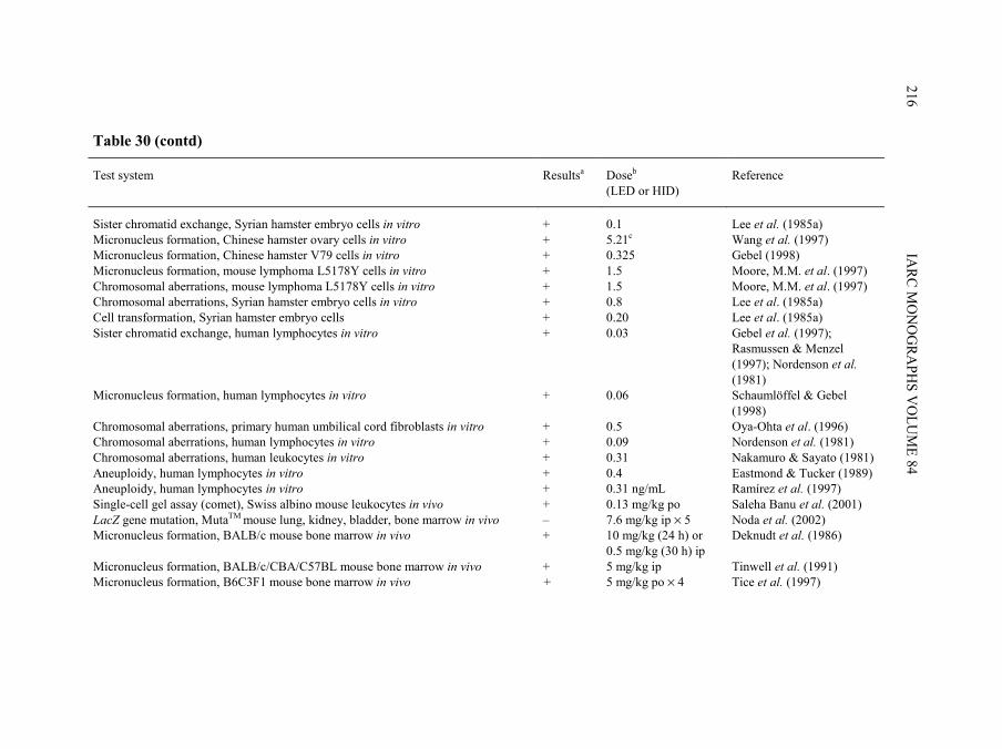
IARC M
ON
OG
RAPH
S VO
LUM
E 84216
Table 30 (contd)
Test system Resultsa Doseb (LED or HID)
Reference
Sister chromatid exchange, Syrian hamster embryo cells in vitro + 0.1 Lee et al. (1985a) Micronucleus formation, Chinese hamster ovary cells in vitro + 5.21c Wang et al. (1997) Micronucleus formation, Chinese hamster V79 cells in vitro + 0.325 Gebel (1998) Micronucleus formation, mouse lymphoma L5178Y cells in vitro + 1.5 Moore, M.M. et al. (1997) Chromosomal aberrations, mouse lymphoma L5178Y cells in vitro + 1.5 Moore, M.M. et al. (1997) Chromosomal aberrations, Syrian hamster embryo cells in vitro + 0.8 Lee et al. (1985a) Cell transformation, Syrian hamster embryo cells + 0.20 Lee et al. (1985a) Sister chromatid exchange, human lymphocytes in vitro + 0.03 Gebel et al. (1997);
Rasmussen & Menzel (1997); Nordenson et al. (1981)
Micronucleus formation, human lymphocytes in vitro + 0.06 Schaumlöffel & Gebel (1998)
Chromosomal aberrations, primary human umbilical cord fibroblasts in vitro + 0.5 Oya-Ohta et al. (1996) Chromosomal aberrations, human lymphocytes in vitro + 0.09 Nordenson et al. (1981) Chromosomal aberrations, human leukocytes in vitro + 0.31 Nakamuro & Sayato (1981) Aneuploidy, human lymphocytes in vitro + 0.4 Eastmond & Tucker (1989) Aneuploidy, human lymphocytes in vitro + 0.31 ng/mL Ramírez et al. (1997) Single-cell gel assay (comet), Swiss albino mouse leukocytes in vivo + 0.13 mg/kg po Saleha Banu et al. (2001) LacZ gene mutation, MutaTM mouse lung, kidney, bladder, bone marrow in vivo – 7.6 mg/kg ip × 5 Noda et al. (2002) Micronucleus formation, BALB/c mouse bone marrow in vivo + 10 mg/kg (24 h) or
0.5 mg/kg (30 h) ip Deknudt et al. (1986)
Micronucleus formation, BALB/c/CBA/C57BL mouse bone marrow in vivo + 5 mg/kg ip Tinwell et al. (1991) Micronucleus formation, B6C3F1 mouse bone marrow in vivo + 5 mg/kg po × 4 Tice et al. (1997)
pp167-228.qxd 11/10/2004 10:53 Page 216

ARSEN
IC IN D
RINK
ING
-WATER
217
Table 30 (contd)
Test system Resultsa Doseb (LED or HID)
Reference
Micronucleus formation, MutaTMmouse peripheral blood reticulocytes in vivo + 7.6 mg/kg ip × 5 Noda et al. (2002) Chromosomal aberrations, Swiss mouse bone marrow in vivo + 0.1 mg/kg sc × 4 Roy Choudhury et al. (1996) Chromosomal aberrations, Swiss mouse bone marrow in vivo + 2.5 mg/kg po Biswas et al. (1999) Dominant lethal mutation, Balb/c mouse in vivo – 5 mg/kg ip Deknudt et al. (1986)
Monomethylarsonic acid (MMAV) Gene mutation, mouse lymphoma L5178Y cells, Tk locus, in vitro + 2500 Moore, M.M. et al. (1997) Micronucleus formation, mouse lymphoma L5178Y cells in vitro + 4000 Moore, M.M. et al. (1997) Chromosomal aberrations, mouse lymphoma L5178Y cells in vitro + 4000 Moore, M.M. et al. (1997) Chromosomal aberrations, primary human umbilical cord fibroblasts in vitro + 196 Oya-Ohta et al. (1996)
Dimethylarsinic acid (DMAV) Gene mutation, mouse lymphoma L5178Y cells, Tk locus, in vitro + 5000 Moore, M.M. et al. (1997) Micronucleus formation, mouse lymphoma L5178Y cells in vitro – 10 000 Moore, M.M. et al. (1997) Chromosomal aberrations, mouse lymphoma L5178Y cells in vitro + 8000 Moore, M.M. et al. (1997) Chromosomal aberrations, primary human umbilical cord fibroblasts in vitro + 96.6 Oya-Ohta et al. (1996) DNA strand break, ICR CD-1 mouse lung in vivo + 1500 mg/kg Yamanaka et al. (1989);
Yamanaka & Okada (1994) DNA strand break, ICR CD-1 mouse liver, kidney and spleen in vivo – 1500 mg/kg Yamanaka et al. (1989);
Yamanaka & Okada (1994) LacZ gene mutation, MutaTM mouse lung, kidney, bladder, bone marrow in vivo – 10.6 mg/kg ip × 5 Noda et al. (2002) Micronucleus formation, MutaTM mouse peripheral blood reticulocytes – 10.6 mg/kg ip × 5 Noda et al. (2002) Aneuploidy, CD-1 mouse bone marrow in vivo + 300 mg/kg ip Kashiwada et al. (1998)
pp167-228.qxd 11/10/2004 10:53 Page 217

IARC M
ON
OG
RAPH
S VO
LUM
E 84218
Table 30 (contd)
Test system Resultsa Doseb (LED or HID)
Reference
Trimethylarsine oxide (TMAO) Chromosomal aberrations, primary human umbilical cord fibroblasts in vitro + 503 Oya-Ohta et al. (1996)
Arsenocholine Chromosomal aberrations, primary human umbilical cord fibroblasts in vitro + 4950 Oya-Ohta et al. (1996)
Arsenobetaine Cell transformation, mouse BALB/3T3 cells – 89 Sabbioni et al. (1991) Chromosomal aberrations, primary human umbilical cord fibroblasts in vitro + 1958 Oya-Ohta et al. (1996)
Tetramethylarsonium iodide Chromosomal aberrations, primary human umbilical cord fibroblasts in vitro + 4978 Oya-Ohta et al. (1996)
Arsenosugar (2′′,3′′-Dihydroxypropyl-5-deoxy-5-dimethylarsinoyl-ββ-D-riboside) Chromosomal aberrations, primary human umbilical cord fibroblasts in vitro + 4860 Oya-Ohta et al. (1996)
Methylarsonous acid (MAsIII) Single-cell gel (comet) assay, human lymphocytes in vitro + 2.12c Mass et al. (2001)
Dimethylarsinous acid (DMAsIII) Single-cell gel (comet) assay, human lymphocytes in vitro + 1.22c Mass et al. (2001)
a +, positive; –, negative; without exogenous metabolic system b LED, lowest effective dose; HID, highest ineffective dose unless otherwise stated; in-vitro tests, µg/mL; in-vivo tests, mg/kg bw/day; po, oral; ip, intraperitoneal c Estimated from graph in paper
pp167-228.qxd 11/10/2004 10:53 Page 218

Eguchi et al. (1997) had shown that pure DMAV but not MMAV had induced tetraploidsin Chinese hamster V79 cells.
MMAIII and DMAIII were investigated using human lymphocytes in the single-cell gelassay. At low micromolar doses, these methylated trivalent arsenicals showed a comet-liketail corresponding to DNA damage. In this study, neither AsIII, AsV, nor the methylatedpentavalent arsenicals produced significant nicking, strand breaks or alkali labile lesions inDNA compared with the methylated trivalent arsenicals.
Both hypomethylation and hypermethylation of DNA were associated with exposureto arsenic in cultures of human lung A549 cells and in human kidney UOK cells (Zhong& Mass, 2001). This could be consistent with the proposal that changes in DNAmethylation can activate some genes and repress others in response to exposure to arsenite.
(b) In-vivo studiesSwiss Albino mice administered arsenic trioxide (also called arsenite, AsIII) orally
showed a significantly increased DNA tail-length in leukocytes in the single-cell gel(comet) assay at the lowest dose tested.
Induction of DNA single-strand breaks was detected in the lung, but not in liver,kidney or spleen of ICR (CD-1) mice 12 h after oral administration of DMAV. The DNAdamage was completely repaired after a further 12-h interval.
No significant mutagenesis of the lacZ gene was observed in male transgenic MutaTM
mouse lung, kidney, bladder or bone marrow after five daily intraperitoneal injections ofarsenite (AsIII) or DMAV. However, arsenite significantly increased the frequencies ofmicronucleated reticulocytes in peripheral blood, whereas DMAV had no effect. [TheWorking Group noted that, in comparison with other studies using DMAV, the dose testedwas more than one order of magnitude lower.]
Sodium arsenite dissolved in water and administered intraperitoneally to CBA, BALB/cand C57BL mice resulted in a significant induction of micronuclei in the polychromaticerythrocytes, as did oral administration to B6C3F1 mice. Potassium arsenite tested only inC57BL mice was also positive in the micronucleus test in polychromatic erythrocytes.Arsenic sulfide (called orpiment) did not induce micronuclei to any quantifiable extent,presumably because of its low solubility and bioavailability, a reflection of elevated bloodlevels of arsenic in orpiment-treated animals. After oral or subcutaneous administration ofsodium arsenite for either 1, 6 or 30 consecutive days, elevated frequencies of chromosomalaberrations were found in the bone-marrow cells of Swiss albino mice.
Significantly elevated numbers of aneuploid cells were detected in bone-marrow cellsof ICR (CD-1) mice treated intraperitoneally with DMAV.
In an assay to detect point mutations caused by arsenic, virgin C57BL/6J mice andfemale metallothionein knock-out null mice (MT–/–) were exposed to drinking-watercontaining 500 µg/L arsenic for up to 26 months (Ng et al., 2001). Nine of 12 (75%) virginC57BL/6J and 8/11 (72.72%) MT–/– mice developed one or multiple mutations in exon 5of the p53 gene. The most prominent mutation (mutation hot spot) appeared in codon 163
ARSENIC IN DRINKING-WATER 219
pp167-228.qxd 11/10/2004 10:53 Page 219

of exon 5, in 9/12 (75%) and 10/14 (71.4%) of the tissues tested in C57BL/6J and MT–/–
mice, respectively.C57BL/6J mice fed methyl-deficient diets were administered arsenite in the drinking-
water at doses of 0, 2.6, 4.3, 9.5 or 14.6 mg/kg bw per day for 130 days. Arsenite treatmentincreased genomic hypomethylation in a dose-dependent manner and reduced thefrequency of methylation at several cytosine sites within the promoter region of the Ha-rasgene (Okoji et al., 2002).
Co-mutagenicity/co-genotoxicity of arsenic Trivalent arsenic was demonstrated to act as a synergistic co-mutagen in combination
with many genotoxic agents including ultraviolet (UV) light. For instance, when Chinese hamster ovary cells were treated simultaneously with UV
light and sodium arsenite, chromatid and chromosomal aberrations as well as Hprt muta-tions were increased synergistically. An additive effect in the induction of sister chromatidexchange was observed with a combined treatment of low doses of UV and AsIII but not witha combined treatment of higher doses of UV and arsenic (Lee et al., 1985b). Treatment ofChinese hamster ovary cells with sodium arsenite after incubation with the DNA-alkylatingagent methyl methanesulfonate also enhanced clastogenicity and Hprt mutagenicitysynergistically (Lee et al., 1986a). However, pretreatment with sodium arsenite resulted ina reduction in the mutagenicity of methyl methanesulfonate. Furthermore, post-treatment ofChinese hamster ovary cells with sodium arsenite was shown to increase UV- and alkylatingagent-induced chromosomal aberrations (Huang et al., 1986) and the clastogenicity ofDNA-cross-linking agents (Lee et al., 1986b). In the presence of sodium arsenite, γ-ray-induced chromosomal aberration frequency was potentiated in human peripherallymphocytes (Jha et al., 1992). In human UV-irradiated VH16 fibroblasts, micronuclei (butnot sister chromatid exchange) were induced synergistically by post-treatment with sodiumarsenite (Jha et al., 1992). According to the authors, the lack of synergistic effect on UV-induced sister chromatid exchange in this study may be because sodium arsenite waswashed off before the cells were seeded for division.
4.5 Mechanistic considerations
Several different mechanisms of arsenic-induced carcinogenicity have been proposed,and the trivalent species are implicated in most of these mechanisms (National ResearchCouncil, 1999, 2001; Simeonova & Luster, 2000; Kitchin, 2001; Hughes, 2002). It shouldbe noted, however, that the trivalent species are formed in vivo after exposure to penta-valent arsenic. Methylated trivalent arsenic is more toxic, and genotoxic, than trivalentinorganic arsenic; in contrast, methylated pentavalent arsenic is less toxic, and genotoxic,than pentavalent inorganic arsenic.
IARC MONOGRAPHS VOLUME 84220
pp167-228.qxd 11/10/2004 10:53 Page 220
4.5.1 Genotoxicity
Arsenic induces chromosomal aberrations, micronuclei, aneuploidy, endoreduplicationand gene amplification. These may play a role in the genomic instability that can resultfrom treatment with arsenic. Arsenic appears to have little if any ability to induce pointmutations (National Research Council, 1999, 2001). The methylated trivalent molecules ofarsenic are potent forms for the induction of DNA damage in cells in vitro, and they are theonly forms of arsenic that cause DNA breakage in vitro, a reaction that is mediated byreactive oxygen species (Yamanaka & Okada, 1994; Nesnow et al., 2002; Kitchin &Ahmad, 2003).
4.5.2 Altered DNA repair
Trivalent arsenic (AsIII) inhibits nucleotide-excision repair of UVC-induced DNAdamage in human fibroblasts by interacting with distinct steps of the repair process. Itimpaired the incision step at low concentrations and the ligation step at higher concen-trations (Hartwig et al., 1997).
AsIII inhibits several DNA-repair enzymes including DNA ligases I and II (Li &Rossman, 1989; Lee-Chen et al., 1992), and zinc-finger proteins bearing covalent disulfidelinkages seem to be potential targets of this metal. The activity of PARP, one of the zinc-finger DNA-repair enzymes, is inhibited in a human T-cell lymphoma-derived Molt-3 cellline and HeLa cells by low concentrations of arsenic (5 µM and 10 nM, respectively)(Yager & Wiencke, 1997; Hartwig et al., 2003). However, other zinc-finger DNA-repairenzymes such as mammalian xeroderma pigmentosum group A protein and bacterialformamido-pyrimidine-DNA glycosylase are not inhibited by AsIII (Asmuss et al., 2000).
4.5.3 Induction of oxidative stress
Exposure to arsenic results in the generation of reactive oxygen species both in vitroand in vivo. There is evidence that these may be involved in the DNA-damaging activitiesof AsIII, MMAIII and DMAIII. Arsenic species, particularly DMAIII, release iron fromferritin (Ahmad et al., 2000); this free iron can produce reactive oxygen species viaFenton and/or Haber-Weiss type reactions. Reactive oxygen species are detected inhuman–hamster hybrid cells exposed to arsenite (AsIII) (Liu, S.X. et al., 2001) and inφX174 DNA incubated in vitro with MMAIII or DMAIII (Nesnow et al., 2002). They arealso involved in stress responses that may alter DNA and gene expression. For example,8-OHdG formation and cyclooxygenase Cox-2 expression, most commonly used as amarker for the evaluation of oxidative DNA damage, are increased in the urinary bladdercancers of rats treated with dimethyl arsenite (Wei et al., 2002). The DMAIII producedin vivo in the urine of rats treated with DMAV (Cohen et al., 2002) and subsequent gene-ration of reactive oxygen species may be important factors in the arsenic-induced bladdercancer observed in these animals (Wei et al., 2002).
ARSENIC IN DRINKING-WATER 221
pp167-228.qxd 11/10/2004 10:53 Page 221

4.5.4 Altered DNA methylation
The alteration of DNA methylation by arsenic may also play a role in the developmentof cancer. In-vitro and in-vivo studies indicate that the carcinogenicity of arsenic may bemediated by alterations in the methylation status of DNA either by hypermethylation orhypomethylation (Mass & Wang, 1997; Zhao et al., 1997; Okoji et al., 2002).
4.5.5 Cell transformation
Arsenic induces cell transformation in Syrian hamster embryo cells, BALB/3T3 cellsand in the rat liver cell line TRL1215. Inoculation of the latter cells into nude mice gaverise to malignant tumours (fibrosarcoma and metastases to the lung) (Lee et al., 1985a;Bertolero et al., 1987; Zhao et al., 1997).
4.5.6 Altered cell proliferation
Increased cell proliferation has been demonstrated directly or indirectly in variousexperimental systems after exposure to arsenic (Germolec et al., 1997; Kitchin, 2001;Hughes, 2002).
Increases in ODC activity, a biomarker of cell proliferation, have been observed in thekidney or liver of rats treated with arsenic (Yamamoto et al., 1995; Brown & Kitchin,1996). Stimulation of cell proliferation had been shown in normal human epidermal kera-tinocytes treated in vitro by arsenic (Germolec et al., 1997).
Hyperplasia has been observed in the bladder of rats treated with DMAV (Cohen et al.,2002).
4.5.7 Altered cell signalling
Arsenic stimulates the activity of Jun kinases, which belong to the mitogen-activatedprotein kinase family, and increases the DNA binding of transcriptional factor AP-1. Arsenicalso induces the expression of proto-oncogenes such as C-JUN, C-FOS, C-MYC and tumourgrowth factor-α (Cavigelli et al., 1996; Germolec et al., 1998; Simeonova et al., 2000; Chenet al., 2001). A reduction in p53 protein levels concomitant with an increase in mdm2 proteinlevels were also observed in a keratinocyte (HaCaT) cell line treated with arsenic. Thedisruption of P53-MDM2 loop-regulating cell-cycle arrest as a model for arsenic-related skincarcinogenesis has been proposed (Hamadeh et al., 1999).
4.5.8 Altered steroid receptor binding and gene expression
Arsenic inhibited steroid binding to glycocorticoid receptors but had no effect on thebinding of ligands to androgen, estrogen, mineral corticoid or progesterone receptors.This specific inhibition may provide a method of using arsenic to block glucocorticoidreceptors selectively in assays of the progesterone receptor content of breast cancer tissues
IARC MONOGRAPHS VOLUME 84222
pp167-228.qxd 11/10/2004 10:53 Page 222
(Lopez et al., 1990). In MCF-7 cells, arsenite blocked the binding of estradiol tooestrogen receptor-α (ER-α) (Stoica et al., 2000).
Moreover, arsenic inhibited expression of ER-α but had no effect on expression ofER-β in breast cancer cell lines (Chen et al., 2002). Thus, the authors concluded that therole of arsenic in the expression of ER-α provides a novel therapeutic approach for ER-α-positive breast cancer (Chen et al., 2002).
4.5.9 Gene amplification
Arsenic enhanced the amplification of the dihydrofolate reductase (DHFR) gene inmouse 3T6 cells and gene amplification has been suggested as a possible mechanism ofthe carcinogenicity of arsenic (Lee et al., 1988).
5. Summary of Data Reported and Evaluation
5.1 Exposure data
Exposure of high levels of arsenic in drinking-water has been recognized for manydecades in some regions of the world, notably in China, Taiwan (China) and somecountries in Central and South America. More recently, it has been discovered that anumber of other regions have drinking-water that is highly contaminated with arsenic. Inmost of these regions, the drinking-water source is groundwater, naturally contaminatedfrom arsenic-rich geological formations. The primary regions where high concentrations ofarsenic have been measured in drinking-water include large areas of Bangladesh, Chinaand West Bengal (India) and smaller areas of Argentina, Australia, Chile, Mexico, Taiwan(China), the USA and Viet Nam. In some areas of Japan, Mexico, Thailand and othercountries, mining, smelting and other industrial activities have contributed to elevatedconcentrations of arsenic in local water sources.
Levels of arsenic in affected areas may range from tens to hundreds or even thousandsof micrograms per litre, whereas in unaffected areas levels are typically only a fewmicrograms per litre. The WHO guideline recommends that levels of arsenic in drinking-water should not exceed 10 µg/L. Arsenic occurs in drinking-water primarily as arsenate(AsV), although in reducing environments significant concentrations of arsenite (AsIII)have also been reported. Trace amounts of methylated arsenic species are typically foundin drinking-water, and higher levels are found in biological systems. Inorganic arsenic(arsenate plus arsenite) is the predominant form of arsenic in drinking-water.
In many areas where contamination of drinking-water by arsenic has been reported,current exposures have been reduced by various interventions.
ARSENIC IN DRINKING-WATER 223
pp167-228.qxd 11/10/2004 10:53 Page 223
5.2 Human carcinogenicity data
In previous monographs, the evidence of carcinogenicity to humans of exposure toarsenic and arsenic compounds, such as medical treatment with Fowler’s solution andinhalation exposure of mining and smelting workers, was evaluated as sufficient.
Informative epidemiological studies of cancer in relation to arsenic in drinking-waterinclude ecological studies and fewer case–control and cohort studies. For most other knownhuman carcinogens, the major source of causal evidence derives from case–control andcohort studies, with little evidence from ecological studies. In contrast, for arsenic indrinking-water, ecological studies provide important information for causal inference. Thereasons include large exposure contrasts and limited population migration. As a conse-quence of widespread exposure to local or regional water sources, ecological measurementsprovide a strong indication of individual exposure. Moreover, in the case of arsenic, the eco-logical estimates of relative risk are often so high that potential confounding with knowncausal factors cannot explain the results. Hence, in the reviews that follow, ecologicalstudies are presented in detail.
Urinary bladder cancerThe Working Group evaluated ecological studies in Taiwan (China), Chile, Argentina
and Australia, cohort studies from Taiwan, Japan and the USA and case–control studies inTaiwan, the USA and Finland.
There is extensive evidence of increased risks for urinary bladder cancer associatedwith arsenic in drinking-water. All studies that involved populations with high long-termexposures found substantial increases in the risk for bladder cancer. Key evidence derivesfrom ecological studies in Taiwan and Chile. In Taiwan, the evidence is supported bycase–control studies and cohort studies within the exposed communities that demonstrateevidence of dose–response relationships with levels of arsenic in drinking-water. The evi-dence of increased mortality from bladder cancer in Chile comes from a large populationwith exposure to arsenic in all major cities and towns of the contaminated region.
There is also evidence of increased risks for bladder cancer from a small cohort studyin Japan of persons drinking from wells that had been highly contaminated with arsenicwastes from a factory and an ecological study from Argentina with moderate exposure toarsenic in well-water. Two case–control studies that investigate low exposure to arsenicfound increased risks with increasing exposure in one or more subgroups.
Considered overall, the findings cannot be attributed to chance or confounding, andthey are consistent, with strong associations found in populations with high exposure.There is evidence of dose–response relationships within exposed populations.
Lung cancerThe Working Group evaluated ecological studies using mortality data in Taiwan
(China), Chile, Argentina and Australia, cohort studies in Taiwan, Japan and the USA andcase–control studies in Taiwan and Chile.
IARC MONOGRAPHS VOLUME 84224
pp167-228.qxd 11/10/2004 10:53 Page 224
Increased risk for lung cancer was consistently observed in ecological, case–controland cohort studies in Taiwan, Japan, Chile and Argentina. Evidence for a dose–responserelationship between arsenic in drinking-water and risk for lung cancer was also observedin ecological studies in Taiwan and Argentina, in cohort studies in south-western andnorth-eastern Taiwan and Japan and in case–control studies in south-western Taiwan andChile. The potential confounding effect of cigarette smoking was ruled out by direct andindirect evidence in studies from Taiwan and Chile.
Considered overall, the findings cannot be attributed to chance or confounding, areconsistent and demonstrate strong associations in populations with high exposure. Thereis evidence of a dose–response relationship.
Skin cancerThe Working Group evaluated ecological studies from Taiwan (China), Mexico, Chile
and the USA, cohort studies from Taiwan and a case–control study from the USA. The recognition that arsenic was potentially carcinogenic arose from occurrences of
skin cancer after ingestion of medicinal arsenic, arsenical pesticide residues and arsenic-contaminated drinking-water. Skin cancer is a commonly observed malignancy related tocontamination of drinking-water with arsenic. The characteristic arsenic-associated skintumours include keratinocytic malignancies (non-melanoma skin cancers), in particularsquamous-cell carcinomas, including Bowen disease, and multiple basal-cell carcinomas.
Ecological studies, largely from the south-west of Taiwan, indicate substantially ele-vated incidence of, prevalence of and mortality rates for skin cancer associated withdrinking-water highly contaminated with arsenic, with evidence of a dose–responserelationship. Findings in ecological studies were substantiated in two cohort studies in theregion of Taiwan that is endemic for arsenic. Increased mortality from skin cancer wasfound in Chile. A high prevalence of skin lesions, including skin cancers, was found inrural regions of Mexico. An excess risk for skin cancer was observed in a case–controlstudy in the USA conducted in an area with lower concentrations of arsenic in drinking-water. A cohort study from the south-west of Taiwan reported that differences in the levelsof serum β-carotene and urinary arsenic metabolites may modify the risk for arsenic-induced skin cancers.
Liver cancerThe Working Group evaluated ecological studies using mortality data in Taiwan
(China), Chile, Argentina and Australia, cohort studies in Taiwan, Japan and the USA anda case–control study in Taiwan of liver cancer cases identified from death certificates.
Increased mortality from liver cancer was observed in the ecological studies involvinga large population with high exposure to arsenic in Taiwan. Evidence for a dose–responserelationship between arsenic in drinking-water and liver cancer mortality was observed inboth ecological and case–control studies in Taiwan. Increased risks were also found insmall cohort studies in Taiwan and Japan. Findings on mortality from liver cancer observedin ecological studies in Chile are inconsistent.
ARSENIC IN DRINKING-WATER 225
pp167-228.qxd 11/10/2004 10:53 Page 225

The interpretation of these findings is limited by the small number of liver cancer cases,questionable accuracy of the diagnosis of liver cancer on death certificates and potentialconfounding or modifying effects of chronic hepatitis virus infection or other factors.
Kidney cancerThe Working Group evaluated ecological studies in Taiwan (China), Chile, Argentina
and Australia, and cohort studies from Taiwan and the USA.All studies that involved populations with high long-term exposures to arsenic found
increased risks for kidney cancer. Key evidence comes from ecological studies in Taiwanand Chile. In Taiwan, the evidence is supplemented by a small cohort study of patients withBlackfoot disease. The evidence of increased mortality from kidney cancer in Chile comesfrom a large population with exposure to arsenic in all major cities and towns of the region.There is also evidence of increased risk for kidney cancer in populations in Argentina withmoderate exposure to arsenic in well-water.
Relative risk estimates for kidney cancer were generally lower than those for urinarybladder cancer, and no studies have reported dose–response relationships on the basis ofindividual exposure assessment.
Other cancersThe Working Group evaluated ecological studies from Taiwan (China), Chile and the
USA, cohort studies from Japan and the USA and one case–control study each from Canadaand the USA.
Excess mortality from prostate cancer was found in south-west Taiwan. Inconsistentfindings were reported for other cancer sites.
5.3 Animal carcinogenicity data
Dimethylarsinic acid was tested for carcinogenicity by administration in drinking-water in mice and rats. It was also tested in two-stage initiation–promotion studies in miceand rats. Complete carcinogenicity was observed in the urinary bladder of rats and lungsof mice. Dimethylarsinic acid exerted its carcinogenic effect on spontaneous developmentof tumours in p53+/– and p53+/+ mice. Dimethylarsinic acid is a tumour promoter in theskin and lung of mice, and in the liver, urinary bladder, kidney and thyroid gland of rats.
After perinatal treatment, arsenic trioxide induced lung adenomas in mice and, afterintratracheal instillation to hamsters, it induced lung adenomas in two of three studies.Calcium arsenate administered to hamsters by intratracheal instillation induced lungadenomas. Sodium arsenate induced tumours at various organ sites in metallothioneinknockout mice. Transplacental exposure of mice to sodium arsenite induced liver and lungcarcinomas, ovarian tumours (benign and malignant) and adrenal cortical adenomas.Sodium arsenite promoted skin carcinogenesis in mice. Arsenic trisulfide was negative forcarcinogenicity when tested in hamsters by intratracheal instillation.
IARC MONOGRAPHS VOLUME 84226
pp167-228.qxd 11/10/2004 10:53 Page 226

5.4 Other relevant data
Arsenic in drinking-water is well absorbed in the gastrointestinal tract. The trivalentspecies of arsenic are formed in vivo after exposure to pentavalent arsenic. Arsenic is meta-bolized by a series of reductions and oxidations and by methylation reactions. Methylatedtrivalent arsenic is more toxic and less genotoxi than trivalent inorganic arsenic; in contrast,methylated pentavalent arsenic is less toxic and less genotoxic than pentavalent inorganicarsenic. There is a large variation in metabolism between animal species, population groupsand individuals. Both inorganic arsenic and its methylated metabolites are excreted in urine.
Acute effects due to ingestion of arsenic are characterized by severe vomiting anddiarrhoea with features of shock, muscle cramps and cardiac abnormalities. Subacute expo-sures affect primarily the respiratory, gastrointestinal, cardiovascular, nervous and haemato-poietic systems.
Most reports of chronic arsenic toxicity focus on skin manifestations such as pigmenta-tion, with depigmentation affecting trunks and limbs and keratosis affecting hands and feet.Chronic lung disease, peripheral neuropathy, hepatomegaly and peripheral vascular diseasehave frequently been reported in cases of chronic exposure to arsenic. Exposure to arsenichas been associated with an increased risk for diabetes mellitus. Other systemic manifesta-tions include cardiovascular effects, abdominal pain, anorexia, nausea, diarrhoea, cerebro-vascular disease, non-pitting oedema of hands, feet or legs, anaemia and generalized weak-ness. In a study in Taiwan (China), significantly higher mortality from cardiovascular andperipheral vascular disease was reported among patients with Blackfoot disease comparedwith the general population of Taiwan or unaffected residents in endemic areas of Blackfootdisease.
The acute toxicity of trivalent arsenic is greater than that of the pentavalent form. The50% lethal dose of arsenic trioxide in mice by the oral route varies from 15 to 48 mg/kg bw,whereas the acute lethal dose in humans varies from 1 to 3 mg/kg bw. In chronic toxicitystudies, arsenic inhibits mitochondrial respiration and induces apoptosis accompanied by aloss of the mitochondrial transmembrane potential. Metallothionein is thought to have aprotective effect against the toxicity of arsenic.
Arsenic can modify the urinary excretion of porphyrins in animals and humans. It alsointerferes with the activities of several enzymes of the haeme biosynthetic pathway. Themajor abnormalities in urinary porphyrin excretion in chronically exposed humans are(a) significant reductions in coproporphyrin III excretion, resulting in a decrease in theratio of coproporphyrinogen oxidase III to coproporphyrinogen oxidase I and (b) signifi-cant increases in uroporphyrin excretion.
Exposure to arsenite or arsenate results in generation of reduced oxygen species inlaboratory animals and human cells. Exposure to arsenicals either in vivo or in vitro in avariety of model systems causes induction of a number of major stress-protein familiessuch as heat-shock proteins. Recent studies in animals demonstrated altered geneexpression following acute treatment with arsenic that included DNA repair genes, acti-
ARSENIC IN DRINKING-WATER 227
pp167-228.qxd 11/10/2004 10:53 Page 227

vation of transcription factors, such as activator protein 1, and an increase in pro-inflam-matory cytokines. All of these events could play a role in the toxicity of arsenic.
Few studies have been conducted on the immunotoxicity of arsenic. All arsenic com-pounds evaluated in mouse spleen cells suppressed plaque-forming cell responses tosheep erythrocytes and proliferative response to mitogens. Furthermore, arsenic impairedstimulation and proliferation of human lymphocytes in vitro. Recent studies suggest thatapoptosis may be an important mechanism for arsenic-induced immunosuppression.
Experimental animal studies have demonstrated the developmental toxicity of trivalentand pentavalent arsenic, monomethylarsonic acid and dimethylarsinic acid. Limited humandata suggest that exposure to high concentrations of arsenic in drinking-water duringpregnancy may increase fetal and neonatal mortality.
The genotoxicity of arsenic is due largely to the trivalent arsenicals. In humans, arsenicis a chromosomal mutagen (an agent that induces mutations involving more than one gene,typically large deletions or rearrangements). Arsenic appears to have limited ability toinduce point mutations. Elevated frequencies of micronuclei, chromosomal aberrations andaneuploidy were detected in the peripheral lymphocytes or urothelial cells, or both, ofpeople exposed to elevated levels of arsenic. In vitro, arsenic was not a point mutagen inbacteria. In mammalian cells, arsenic caused various types of chromosomal mutations andaneuploidy. In combination with many genotoxic agents, including ultraviolet light, arsenicwas a synergistic co-mutagen. In vitro, arsenite was genotoxic at micromolar concentrations.Arsenate was approximately one order of magnitude less genotoxic than arsenite, dimethyl-arsinic acid and monomethylarsonic acid induced genotoxicity at millimolar concentrations.
Methylarsenous acid and dimethylarsinous acid are intermediary metabolites in themethylation of arsenic. Their genotoxicity has not been fully established, but recentresults implicate a major role for these metabolites and reduced (reactive) oxygen speciesin the induction of urinary bladder cancer in rats.
5.5 Evaluation
There is sufficient evidence in humans that arsenic in drinking-water causes cancers ofthe urinary bladder, lung and skin.
There is sufficient evidence in experimental animals for the carcinogenicity of dimethyl-arsinic acid.
There is limited evidence in experimental animals for the carcinogenicity of sodiumarsenite, calcium arsenate and arsenic trioxide.
There is inadequate evidence in experimental animals for the carcinogenicity of sodiumarsenate and arsenic trisulfide.
Taken together, the studies on inorganic arsenic provide limited evidence for carcino-genicity in experimental animals.
Overall evaluationArsenic in drinking-water is carcinogenic to humans (Group 1).
IARC MONOGRAPHS VOLUME 84228
pp167-228.qxd 11/10/2004 10:53 Page 228

6. References
Acharyya, S.K. (2002) Arsenic contamination in groundwater affecting major parts of southernWest Bengal and parts of western Chhattisgarh: Source and mobilization process. Current Sci.,82, 740–744
Agahian, B., Lee, J.S., Nelson, J.H. & Johns, R.E. (1990) Arsenic levels in fingernails as a biolo-gical indicator of exposure to arsenic. Am. ind. Hyg. Assoc. J., 51, 646–651
Ahmad, S.A., Bandaranayake, D., Khan, A.W., Hadi, S.K., Uddin, G. & Halim, M.A. (1997) Arseniccontamination in ground water and arsenicosis in Bangladesh. Int. J. environ. Health Res., 7,271–276
Ahmad, S., Anderson, W.L. & Kitchin, K.T. (1999) Dimethylarsinic acid effects on DNA damage andoxidative stress related biochemical parameters in B6C3F1 mice. Cancer Lett., 139, 129–135
Ahmad, S.A., Sayed, M.H.S.U., Hadi, S.A., Faruquee, M.H., Khan, M.H., Jalil, M.A., Ahmed, R.& Khan, A.W. (1999) Arsenicosis in a village in Bangladesh. Int. J. environ. Health Res., 9,187–195
Ahmad, S., Kitchin, K.T. & Cullen, W.R. (2000) Arsenic species that cause release of iron fromferritin and generation of activated oxygen. Arch. Biochem. Biophys., 382, 195–202
Ahmad, S.A., Sayed, M.H.S.U., Barua, S., Khan, M.H., Faruquee, M.H., Jalil, A., Hadi, S.A. &Talukder, H.K. (2001) Arsenic in drinking water and pregnancy outcomes. Environ. HealthPerspect., 109, 629–631
Alam, M.G.M., Allinson, G., Stagnitti, F., Tanaka, A. & Westbrooke, M. (2002) Arsenic contami-nation in Bangladesh groundwater: A major environmental and social disaster. Int. J. environ.Health Res., 12, 236–253
Alam, M.G.M., Snow, E.T. & Tanaka, A. (2003) Arsenic and heavy metal contamination of vege-tables grown in Samta village, Bangladesh. Sci. total Environ., 308, 83–96
Alonso, H. (1992) Arsenic enrichment in superficial waters. II Region northern Chile. In: Procee-dings of an International Seminar on Arsenic in the Environment and its Incidence on Health,Santiago, Universidad de Chile, pp. 101–108
Aposhian, H.V., Gurzau, E.S., Le, X.C., Gurzau, A., Healy, S.M., Lu, X., Ma, M., Yip, L.,Zakharyan, R.A., Maiorino, R.M., Dart, R.C., Tircus, M.G., Gonzalez-Ramirez, D., Morgan,D.L., Avram, D. & Aposhian, M.M. (2000a) Occurrence of monomethylarsonous acid in urineof humans exposed to inorganic arsenic. Chem. Res. Toxicol., 13, 693–697
Aposhian, H.V., Zheng, B., Aposhian, M.M., Le, X.C., Cebrian, M.E., Cullen, W., Zakharyan,R.A., Ma, M., Dart, R.C., Cheng, Z., Andrewes, P., Yip, L., O’Malley, G.F., Maiorino, R.M.,Van Voorhies, W., Healy, S.M. & Titcomb, A. (2000b) DMPS–arsenic challenge test. II.Modulation of arsenic species, including monomethylarsonous acid (MMAIII), excreted inhuman urine. Toxicol. appl. Pharmacol., 165, 74–83
Aragones Sanz, N., Palacios Diez, M., Avello de Miguel, A., Gomez Rodriguez, P., MartinezCortes, M. & Rodriguez Bernabeu, M.J. (2001) [Nivel de arsenico en abastecimientos de aguade consumo de origen subterraneo en la comunidad de Madrid.] Rev. esp. Salud. publica, 75,421–432 (in Spanish)
ARSENIC IN DRINKING-WATER 229
pp229-270.qxd 11/10/2004 10:56 Page 229

Arguello, R.A., Cenget, D.D. & Tello, E.E. (1938) [Cancer and arsenicism in the endemic regionof Córdoba.] Rev. Argentina Dermatofisiol., 22, 461–470 (in Spanish)
Armienta, M.A., Rodriguez, R. & Cruz, O. (1997) Arsenic content in hair of people exposed tonatural arsenic polluted groundwater at Zimapán, México. Bull. environ. Contam. Toxicol., 59,583–589
Aschengrau, A., Zierler, S. & Cohen, A. (1989) Quality of community drinking water and theoccurrence of spontaneous abortion. Arch. environ. Health, 44, 283–290
Asmuss, M., Mullenders, L.H.F., Eker, A. & Hartwig, A. (2000) Differential effects of toxic metalcompounds on the activities of Fpg and XPA, two zinc finger proteins involved in DNA repair.Carcinogenesis, 21, 2097–2104
Ballantyne, J.M. & Moore, J.N. (1988) Arsenic geochemistry in geothermal systems. Geochim.cosmochim. Acta, 52, 475–483
Barchowsky, A., Dudek, E.J., Treadwell, M.D. & Wetterhahn, K.E. (1996) Arsenic induces oxidantstress and NF-κB activation in cultured aortic endothelial cells. Free rad. Biol. Med., 21,783–790
Basu, D., Dasgupta, J., Mukherjee, A. & Guha Mazumder, D.N. (1996) Chronic neuropathy due toarsenic intoxication from geo-chemical source — A five-year follow-up study. JANEI, 1, 45–48
Basu, A., Mahata, J., Gupta, S. & Giri, A.K. (2001) Genetic toxicology of a paradoxical humancarcinogen, arsenic: A review. Mutat. Res., 488, 171–194
Basu, A., Mahata, J., Roy, A.K., Sarkar, J.N., Poddar, G., Nandy, A.K., Sarkar, P.K., Dutta, P.K.,Banerjee, A., Das, M., Ray, K., Roychaudhury, S., Natarajan, A.T., Nilsson, R. & Giri, A.K.(2002) Enhanced frequency of micronuclei in individuals exposed to arsenic through drinkingwater in West Bengal, India. Mutat. Res., 516, 29–40
Bates, M.N., Smith, A.H. & Cantor, K.P. (1995) Case–control study of bladder cancer and arsenicin drinking water. Am. J. Epidemiol., 141, 523–530
Bauman, J.W., Liu, J. & Klaassen, C.D. (1993) Production of metallothionein and heat-shockproteins in response to metals. Fundam. appl. Toxicol., 21, 15–22
Baxley, M.N., Hood, R.D., Vedel, G.C., Harrison, W.P. & Szczech, G.M. (1981) Prenatal toxicityof orally administered sodium arsenite in mice. Bull. environ. Contam. Toxicol., 26, 749–756
Beckman, G., Beckman, L. & Nordenson, I. (1977) Chromosome aberrations in workers exposedto arsenic. Environ. Health Perspect., 19, 145–146
Benramdane, L., Accominotti, M., Fanton, L., Malicier, D. & Vallon, J.-J. (1999) Arsenic speciationin human organs following fatal arsenic trioxide poisoning — A case report. Clin. Chem., 45,301–306
Berg, J.W. & Burbank, F. (1972) Correlations between carcinogenic trace metals in water suppliesand cancer mortality. Ann. N.Y. Acad. Sci., 199, 249–264
Berg, M., Tran, H.C., Nguyen, T.C., Pham, H.V., Schertenleib, R. & Giger, W. (2001) Arseniccontamination of groundwater and drinking water in Vietnam. A human health threat. Environ.Sci. Technol., 35, 2621–2626
Bergoglio, R.M. (1964) [Mortality in zones with arsenical woter in the Province of Córdoba,Argentine Republic.] Pren. méd. Argent., 51, 994–998 (in Spanish)
Bernstam, L. & Nriagu, J. (2000) Molecular aspects of arsenic stress. J. Toxicol. environ. Health,B3, 293–322
Bernstam, L., Lan, C.-H., Lee, J. & Nriagu, J.O. (2002) Effects of arsenic on human keratinocytes:Morphological, physiological, and precursor incorporation studies. Environ. Res., A89, 220–235
IARC MONOGRAPHS VOLUME 84230
pp229-270.qxd 11/10/2004 10:56 Page 230

Bertolero, F., Pozzi, G., Sabbioni, E. & Saffiotti, U. (1987) Cellular uptake and metabolic reductionof pentavalent to trivalent arsenic as determinants of cytotoxicity and morphological transfor-mation. Carcinogenesis, 8, 803–808
Bhattacharya, P., Chatterjee, D. & Jacks, G. (1997) Occurrence of arsenic-contaminated ground-water in alluvial aquifers from delta plains, eastern India: Options for safe drinking watersupply. Int. J. Water Resour. Dev., 13, 79–92
Bhattacharya, P., Larsson, M., Leiss, A., Jacks, G., Sracek, A. & Chatterjee, D. (1998) Genesis ofarseniferous groundwater in the alluvial aquifers of Bengal delta plains and strategies forlow-cost remediation. In: International Conference on Arsenic Pollution of Ground Water inBangladesh: Causes, Effects and Remedies, Dhaka, Dhaka Community Hospital Trust,pp. 120–123
Biagini, R.E. (1972) [Chronic hydroarsenicism and mortality from malignant cancers.] Semanamed., 141, 812–816 (in Spanish)
Biggs, M.L., Kalman, D.A., Moore, L.E., Hopenhayn-Rich, C., Smith, M.T. & Smith, A.H. (1997)Relationship of urinary arsenic to intake estimates and a biomarker of effect, bladder cellmicronuclei. Mutat. Res., 386, 185–195
Biswas, B.K., Dhar, R.K., Samanta, G., Mandal, B.K., Chakraborti, D., Faruk, I., Islam, K.S.,Chowdhury, M.M., Islam, A. & Roy, S. (1998) Detailed study report of Samta, one of thearsenic-affected villages of Jessore District, Bangladesh. Current Sci., 74, 134–145
Biswas, S., Talukder, G. & Sharma, A. (1999) Prevention of cytotoxic effects of arsenic by short-term dietary supplementation with selenium in mice in vivo. Mutat. Res., 441, 155–160
Black, M.M. (1967) Prolonged ingestion of arsenic. Pharma J., 9 December, 593–597Blackwell, R.Q., Yang, T.H. & Ai, I. (1961) Preliminary report on arsenic level in water and food
from the endemic Blackfoot area. J. formos. med. Assoc., 60, 1139–1140Blakley, B.R. (1987) The effect of arsenic on urethan-induced adenoma formation in Swiss mice.
Can. J. vet. Res., 51, 240–243Borgoño, J.M. & Greiber, R. (1971) [Epidemiological study of arsenic poisoning in the city of
Antofagasta.] Rev. méd. Chile, 99, 702–707 (in Spanish)Borgoño, J.M., Vicent, P., Venturino, H. & Infante, A. (1977) Arsenic in the drinking water of the
city of Antofagasta: Epidemiological and clinical study before and after the installation of atreatment plant. Environ. Health Perspect., 19, 103–105
Borgoño, J.M., Venturino, H. & Vicent, P. (1980) [Clinical and epidemiologic study of arsenicismin northern Chile.] Rev. méd. Chile, 108, 1039–1048 (in Spanish)
Borzsonyi, M., Bereczky, A., Rudnai, P., Csanady, M. & Horvath, A. (1992) Epidemiologicalstudies on human subjects exposed to arsenic in drinking water in Southeast Hungary. Arch.Toxicol., 66, 77–78
Boyle, R.W. & Jonasson, I.R. (1973) The geochemistry of arsenic and its use as an indicator ele-ment in geochemical prospecting. J. geochem. Explor., 2, 251–296
Braman, R.S. & Foreback, C.C. (1973) Methylated forms of arsenic in the environment. Science,182, 1247–1249
Bronaugh, R.L., Stewart, R.F. & Congdon, E.R. (1982) Methods for in vitro percutaneous absorp-tion studies. II. Animal models for human skin. Toxicol. appl. Pharmacol., 62, 481–488
Brown, J.L. & Kitchin, K.T. (1996) Arsenite, but not cadmium, induces ornithine decarboxylase andheme oxygenase activity in rat liver: Relevance to arsenic carcinogenesis. Cancer Lett., 98,227–231
ARSENIC IN DRINKING-WATER 231
pp229-270.qxd 11/10/2004 10:56 Page 231

Brown, M.M., Rhyne, B.C. & Goyer, R.A. (1976) Intracellular effects of chronic arsenic adminis-tration on renal proximal tubule cells. J. Toxicol. environ. Health, 1, 505–514
Buchet, J.P. & Lauwerys, R. (1985) Study of inorganic arsenic methylation by rat liver in vitro:Relevance for the interpretation of observations in man. Arch. Toxicol., 57, 125–129
Buchet, J.P. & Lauwerys, R. (1988) Role of thiols in the in vitro methylation of inorganic arsenicby rat liver cytosol. Biochem. Pharmacol., 37, 3149–3153
Buchet, J.P., Lauwerys, R. & Roels, H. (1981) Comparison of the urinary excretion of arsenicmetabolites after a single dose of sodium arsenite, monomethylarsonate or dimethylarsinate inman. Int. Arch. occup. environ. Health, 48, 71–79
Burk, D. & Beaudoin, A.R. (1977) Arsenate-induced renal agenesis in rats. Teratology, 16, 247–259Caceres, L., Gruttner, E. & Contreras, R. (1992) Water recycling in arid regions — Chilean case.
Ambio, 21, 138–144Calderon, R.L., Hudgens, E., Le, X.C., Schreinemachers, D. & Thomas, D.J. (1999) Excretion of
arsenic in urine as a function of exposure to arsenic in drinking water. Environ. HealthPerspect., 107, 663–667
Calderón, J., Navarro, M.E., Jimenez-Capdeville, M.E., Santos-Diaz, M.A., Golden, A.,Rodriguez-Leyva, I., Borja-Aburto, V. & Diaz-Barriga, F. (2001) Exposure to arsenic and leadand neuropsychological development in Mexican children. Environ. Res., A85, 69–76
Caltabiano, M.M., Koestler, T.P., Poste, G. & Greig, R.G. (1986) Induction of 32- and 34-kDa stressproteins by sodium arsenite, heavy metals, and thiol-reactive agents. J. biol. Chem., 261,13381–13386
Cantor, K.P., Hoover, R., Hartge, P., Mason, T.J., Silverman, D.T., Altman, R., Austin, D.F., Child,M.A., Key, C.R., Marrett, L.D., Myers, M.H., Narayana, A.S., Levin, L.I., Sullivan, J.W.,Swanson, G.M., Thomas, D.B. & West, D.W. (1987) Bladder cancer, drinking water source,and tap water consumption: A case–control study. J. natl Cancer Inst., 79, 1269–1279
Cao, S. (1996) [Current status of inorganic arsenic contamination in mainland China.] Chinese J.public Health, 15 (Suppl. 3), S6–S10 (in Chinese)
Carpenter, S.J. (1987) Developmental analysis of cephalic axial dysraphic disorders. Anat.Embryol., 176, 345–365
Cavigelli, M., Li, W.W., Lin, A., Su, B., Yoshioka, K. & Karin, M. (1996) The tumor promoterarsenite stimulates AP-1 activity by inhibiting a JNK phosphatase. EMBO J., 15, 6269–6279
Cebrián, M.E., Albores, A., Aguilar, M. & Blakely, E. (1983) Chronic arsenic poisoning in thenorth of Mexico. Hum. Toxicol., 2, 121–133
Cebrián, M.E., Albores, A., Connelly, J.C. & Bridges, J.W. (1988) Assessment of arsenic effects oncytosolic heme status using tryptophan pyrrolase as an index. J. biochem. Toxicol., 3, 77–86
Cebrián, M.E., Albores, A., García-Vargas, G., Del Razo, L.M. & Ostrosky-Wegman, P. (1994)Chronic arsenic poisoning in humans: The case of Mexico. In: O’Nriagu, J., ed., Arsenic in theEnvironment, Part II, Human Health and Ecosystem Effects, New York, John Wiley & Sons,pp. 93–107
Chaineau, E., Binet, S., Pol, D., Chatellier, G. & Meininger, V. (1990) Embryotoxic effects ofsodium arsenite and sodium arsenate on mouse embryos in culture. Teratology, 41, 105–112
Chakraborti, D., Samanta, G., Mandal, B.K., Roy Chowdhury, T., Chanda, C.R., Biswas, B.K.,Dhar, R.K., Basu, G.K. & Saha, K.C. (1998) Calcutta’s industrial pollution: Groundwaterarsenic contamination in a residential area and sufferings of people due to industrial effluentdischarge — An eight-year study report. Current Sci., 74, 346–355
IARC MONOGRAPHS VOLUME 84232
pp229-270.qxd 11/10/2004 10:56 Page 232

Chakraborti, D., Biswas, B.K., Basu, G.K., Chowdhury, U.K., Roy Chowdhury, T., Lodh, D.,Chanda, C.R., Mandal, B.K., Samanta, G., Chakraborti, K., Rahman, M.M., Paul, K., Roy, S.,Kabir, S., Ahmed, B., Das, R., Salim, M. & Quamruzzaman, Q. (1999a) Possible arsenic conta-mination of free groundwater source in Bangladesh. J. surface Sci. Technol., 15, 180–188
Chakraborti, D., Biswas, B.K., Roy Chowdhury, T., Basu, G.K., Mandal, B.K., Chowdhury, U.K.,Mukherjee, S.C., Gupta, J.P., Chowdhury, S.R. & Rathore, K.C. (1999b) Arsenic groundwatercontamination and sufferings of people in Rajnandgaon district, Madhya Pradesh, India.Current Sci., 77, 502–504
Chakraborti, D., Rahman, M.M., Paul, K., Chowdhury, U.K., Sengupta, M.K., Lodh, D., Chanda,C.R., Saha, K.C. & Mukherjee, S.C. (2002) Arsenic calamity in the Indian subcontinent —What lessons have been learned? Talanta, 58, 3–22
Chakraborti, D., Mukherjee, S.C., Pati, S., Sengupta, M.K., Rahman, M.M., Chowdhury, U.K.,Lodh, D., Chanda, C.R., Chakraborti, A.K. & Basu, G.K. (2003a) Arsenic groundwater conta-mination in Middle Ganga Plain, Bihar, India: A future danger. Environ. Health Perspect., 111,1194–1201
Chakraborti, D., Rahman, M.M., Paul, K., Chowdhury, U.K. & Quamruzzaman, Q. (2003b)Groundwater arsenic contamination. In: The Encyclopedia of Water Science, New York, MarcelDekker, pp. 324–329
Chakraborty, A.K. & Saha K.C. (1987) Arsenical dermatosis from tubewell water in West Bengal.Indian J. med. Res., 85, 326–334
Chatterjee, A., Das, D. & Chakraborti, D. (1993) A study of ground water contamination by arsenicin the residential area of Behala, Calcutta due to industrial pollution. Environ. Pollut., 80,57–65
Chatterjee, A., Das, D., Mandal, B.K., Roy Chowdhury, T., Samanta, G. & Chakraborti, D. (1995)Arsenic in ground water in six districts of West Bengal, India: The biggest arsenic calamity inthe world, Part I. Arsenic species in drinking water and urine of the affected people. Analyst,120, 645–650
Chatterjee, A., Shibata, Y., Yoshinaga, J. & Morita, M. (2000) Determination of arsenic compoundsby high-performance liquid chromatography-ultrasonic nebulizer-high power nitrogen-micro-wave-induced plasma mass spectrometry: An accepted coupling. Anal. Chem., 72, 4402–4412
Chávez, A., Pérez Hidalgo, C., Tovar, E. & Garmilla, M. (1964) [Studies in a community withchronic endemic arsenicism.] Salud públic Méx., 6, 435–441 (in Spanish)
Chen, C.-J. & Chiou, H.-Y. (2001) Chen and Chiou Respond to ‘Arsenic and cancer of the urinarytract’ by Cantor. Am. J. Epidemiol., 153, 422–423
Chen, H.-W. & Edwards, M. (1997) Arsenic occurrence and speciation in United States drinkingwaters: Implications for water utilities. In: Proceedings of the Water Quality Technology Con-ference, Denver, CO, American Water Works Association, pp. 468–484
Chen, C.-J. & Wang, C.-J. (1990) Ecological correlation between arsenic level in well water andage-adjusted mortality from malignant neoplasms. Cancer Res., 50, 5470–5474
Chen, K.P., Wu, H.Y. & Wu, T.C. (1962) Epidemiologic studies on blackfoot disease in Taiwan. 3.Physiochemical characteristics of drinking water in endemic blackfoot disease areas. Mem.Coll. Med. natl Taiwan Univ., 8, 115–129
Chen, C.-J., Chuang, Y.-C., Lin, T.-M. & Wu, H.-Y. (1985) Malignant neoplasms among residentsof a blackfoot disease-endemic area in Taiwan: High-arsenic artesian well water and cancers.Cancer Res., 45, 5895–5899
ARSENIC IN DRINKING-WATER 233
pp229-270.qxd 11/10/2004 10:56 Page 233

Chen, B., Burt, C.T., Goering, P.L., Fowler, B.A. & London, R.E. (1986) In vivo 31P nuclearmagnetic resonance studies of arsenite induced changes in hepatic phosphate levels. Biochem.biophys. Res. Commun., 139, 228–234
Chen, C.-J., Chuang, Y.-C., You, S.-L., Lin, T.-M. & Wu, H.-Y. (1986) A retrospective study onmalignant neoplasms of bladder, lung and liver in blackfoot disease endemic area in Taiwan.Br. J. Cancer, 53, 399–405
Chen, C.-J., Kuo, T.-L. & Wu, M.-M. (1988a) Arsenic and cancers. Lancet, i, 414–415Chen, C.-J., Wu, M.-M., Lee, S.-S., Wang, J.-D., Cheng, S.-H. & Wu, H.-Y. (1988b) Atherogenicity
and carcinogenicity of high-arsenic artesian well water. Multiple risk factors and relatedmalignant neoplasms of blackfoot disease. Arteriosclerosis, 8, 452–460
Chen, C.-J., Lin, L.-J., Hsueh, Y.-M., Chiou, H.-Y., Liaw, K.-F., Horng, S.-F., Chiang, M.-H.,Tseng, C.-H. & Tai, T.-Y. (1994) Ischemic heart disease induced by ingested inorganic arsenic.In: Chappell, W.R., Abernathy, C.O. & Cothern, C.R., eds, Arsenic Exposure and Health,Northwood, IL, Science and Technology Letters, pp. 83–90
Chen, C.-J., Hsueh, Y.-M., Lai, M.-S., Shyu, M.-P., Chen, S.-Y., Wu, M.-M., Kuo, T.-L. & Tai, T.-Y. (1995) Increased prevalence of hypertension and long-term arsenic exposure. Hypertension,25, 53–60
Chen, C.-J., Chiou, H.-Y., Chiang, M.-H., Lin, L.-J. & Tai, T.-Y. (1996) Dose–response relationshipbetween ischemic heart disease mortality and long-term arsenic exposure. Arterioscler.Thromb. vasc. Biol., 16, 504–510
Chen, C.-J., Chiou, H.-Y., Huang, W.-I., Chen, S.-Y., Hsueh, Y.-M., Tseng, C.-H., Lin, L.-J., Shyu,M.-P. & Lai, M.-S. (1997) Systemic non-carcinogenic effects and developmental toxicity ofinorganic arsenic. In: Abernathy, C.O., Calderon, R.L. & Chappell, W.R., eds, Arsenic Expo-sure and Health Effects, London, Chapman & Hall, pp. 124–134
Chen, Y.-C., Lin-Shiau, S.-Y. & Lin, J.-K. (1998) Involvement of reactive oxygen species andcaspase 3 activation in arsenite-induced apoptosis. J. cell. Physiol., 177, 324–333
Chen, K.-L.B., Amarasiriwardena, C.J. & Christiani, D.C. (1999) Determination of total arsenicconcentrations in nails by inductively coupled plasma mass spectrometry. Biol. trace ElementRes., 67, 109–125
Chen, Y., Megosh, L.C., Gilmour, S.K., Sawicki, J.A. & O’Brien, T.G. (2000) K6/ODC transgenicmice as a sensitive model for carcinogen identification. Toxicol. Lett., 116, 27–35
Chen, H., Liu, J., Zhao, C.Q., Diwan, B.A., Merrick, B.A. & Waalkes, M.P. (2001) Association ofc-myc overexpression and hyperproliferation with arsenite-induced malignant transformation.Toxicol. appl. Pharmacol., 175, 260–268
Chen, G.-C., Guan, L.-S., Hu, W.-L. & Wang, Z.-Y. (2002) Functional repression of estrogenreceptor α by arsenic trioxide in human breast cancer cells. Anticancer Res., 22, 633–638
Chen, Y.-C., Amarasiriwardena, C.J., Hsueh, Y.-M. & Christiani, D.C. (2002) Stability of arsenicspecies and insoluble arsenic in human urine. Cancer Epidemiol. Biomarkers Prev., 11,1427–1433
Chiang, J., Hermosilla, A.P., Rojas, H. & Henríquez, C. (1990) [Determination of arsenic in indi-viduals exposed to high pollution levels.] Rev. Chil. Nutr., 18, 39–50 (in Spanish)
Chiang, H.S., Guo, H.R., Hong, C.L., Lin, S.M. & Lee, E.F. (1993) The incidence of bladder cancerin the black foot disease endemic area in Taiwan. Br. J. Urol., 71, 274–278
Chilean Institute of National Standards (1984) [Standards for Potable Water], Santiago (in Spanish)
IARC MONOGRAPHS VOLUME 84234
pp229-270.qxd 11/10/2004 10:56 Page 234

Chiou, H.-Y., Hsueh, Y.-M., Liaw, K.-F., Horng, S.-F., Chiang, M.-H., Pu, Y.-S., Lin, J.S.-N.,Huang, C.-H. & Chen, C.-J. (1995) Incidence of internal cancers and ingested inorganicarsenic: A seven-year follow-up study in Taiwan. Cancer Res., 55, 1296–1300
Chiou, H.-Y., Hsueh, Y.-M., Hsieh, L.-L., Hsu, L.-I., Hsu, Y.-H., Hsieh, F.-I., Wei, M.-L., Chen,H.-C., Yang, H.-T., Leu, L.-C., Chu, T.-H., Chen-Wu, C., Yang, M.-H. & Chen, C.-J. (1997b)Arsenic methylation capacity, body retention, and null genotypes of glutathione S-transferaseM1 and T1 among current arsenic-exposed residents in Taiwan. Mutat. Res., 386, 197–207
Chiou, H.-Y., Huang, W.-I., Su, C.-L., Chang, S.-F., Hsu, Y.-H. & Chen, C.-J. (1997a) Dose–responserelationship between prevalence of cerebrovascular disease and ingested inorganic arsenic.Stroke, 28, 1717–1723
Chiou, H.Y., Chiou, S.T., Hsu, Y.H., Chou, Y.L., Tseng, C.H., Wei, M.L. & Chen, C.J. (2001) Inci-dence of transitional cell carcinoma and arsenic in drinking water: A follow-up study of 8102residents in an arseniasis-endemic area in northeastern Taiwan. Am. J. Epidemiol., 153,411–418
Choprapawon, C. & Porapakkham, Y. (2001) Occurrence of cancer in arsenic contaminated area,Ronpibool District, Nakorn Srithmmarat Province, Thailand. In: Chappell, W.R., Abernathy,C.O. & Calderon, R.L., eds, Arsenic Exposure and Health Effects IV, New York, ElsevierScience, pp. 201–206
Choprapawon, C. & Rodcline, A. (1997) Chronic arsenic poisoning in Ronpibool Nakhon SriThammarat, the Southern Province of Thailand. In: Abernathy, C.O., Calderon, R.L. & Chappell,W.R., eds, Arsenic Exposure and Health Effects, London, Chapman & Hall, pp. 69–77
Chouchane, S. & Snow, E.T. (2001) In vitro effect of arsenical compounds on glutathione-relatedenzymes. Chem. Res. Toxicol., 14 , 517–522
Chowdhury, T.R., Mandal, B.K., Samanta, G., Basu, G.K., Chowdhury, P.P., Chanda, C.R., Karan,N.K., Lodh, D., Dhar, R.K., Das, D., Saha, K.C. & Chakraborti, D. (1997) Arsenic in ground-water in six districts of West Bengal, India: The biggest arsenic calamity in the world: Thestatus report up to August, 1995. In: Abernathy, C.O., Calderon, R.L. & Chappell, W.R., eds,Arsenic Exposure and Health Effects, London, Chapman & Hall, pp. 93–111
Chowdhury, U.K., Biswas, B.K., Roy Chowdhury, T., Samanta, G., Mandal, B.K., Basu, G.C.,Chanda, C.R., Lodh, D., Saha, K.C., Mukherjee, S.K., Roy, S., Kabir, S., Quamruzzaman, Q.& Chakraborti, D. (2000a) Groundwater arsenic contamination in Bangladesh and WestBengal, India. Environ. Health Perspect., 108, 393–397
Chowdhury, U.K., Biswas, B.K., Roy Chowdhury, T., Mandal, B.K., Samanta, G., Basu, G.K.,Chanda, C.R., Lodh, D., Saha, K.C., Chakraborti, D., Mukherjee, S.C., Roy, S., Kabir, S. &Quamruzzaman, Q. (2000b) Arsenic groundwater contamination and sufferings of people inWest Bengal-India and Bangladesh. In: Roussel, A.M., Anderson, R.A. & Favrier, A.E., eds,Trace Elements in Man and Animals 10, New York, Kluwer Academic/Plenum, pp. 645–650
Chowdhury, U.K., Rahman, M.M., Mondal, B.K., Paul, K., Lodh, D., Biswas, B.K., Basu, G.K.,Chanda, C.R., Saha, K.C., Mukherjee, S.C., Roy, S., Das, R., Kaies, I., Barua, A.K., Palit,S.K., Quamruzzaman, Q. & Chakraborti, D. (2001) Groundwater arsenic contamination andhuman suffering in West Bengal, India and Bangladesh. Environ. Sci., 8, 393–415
Chowdhury, U.K., Rahman, M.M., Sengupta, M.K., Lodh, D., Chanda, R.C., Roy, S.,Quamruzzaman, Q., Tokunaga, H., Ando, M. & Chakraborti, D. (2003) Pattern of excretion ofarsenic compounds [arsenite, arsenate, MMA (V), DMA(V)] in urine of children compared toadults from an arsenic exposed area in Bangladesh. Environ. Sci. Health, A38, 87–113
ARSENIC IN DRINKING-WATER 235
pp229-270.qxd 11/10/2004 10:56 Page 235

Chung, J.S., Kalman, D.A., Moore, L.E., Kosnett, M.J., Arroyo, A.P., Beeris, M., Guha Mazumder,D.N., Hernandez, A.L. & Smith, A.H. (2002) Family correlations of arsenic methylationpatterns in children and parents exposed to high concentrations of arsenic in drinking water.Environ. Health Perspect., 110, 729–733
Cohen, S.M., Arnold, L.L., Uzvolgyi, E., Cano, M., St John, M., Yamamoto, S., Lu, X. & Le, X.C.(2002) Possible role of dimethylarsinous acid in dimethylarsinic acid-induced urothelial toxi-city and regeneration in the rat. Chem. Res. Toxicol., 15, 1150–1157
Colbourn, P., Alloway, B.J. & Thornton, I. (1975) Arsenic and heavy metals in soils associated withregional geochemical anomalies in South-West England. Sci. total Environ., 4, 359–363
Committee Constituted by Government of West Bengal (1994) Report on Arsenic Pollution inGroundwater in West Bengal (PHE-1/716/3D-1/88), Bengal, PHE Department
CONAMA (2000) [Technical Information Sheet: Analysis of Human Exposure to Arsenic in LargeCities] (Study No. 21–0022–002), Santiago, Ed. P. Pino Z. (in Spanish)
Concha, G., Vogler, G., Nermell, B. & Vahter, M. (1998a) Low-level arsenic excretion in breastmilk of native Andean women exposed to high levels of arsenic in the drinking water. Int.Arch. occup. environ. Health, 71, 42–46
Concha, G., Nermell, B. & Vahter, M.V. (1998b) Metabolism of inorganic arsenic in children withchronic high arsenic exposure in northern Argentina. Environ. Health Perspect., 106, 355–359
Concha, G., Vogler, G., Lezcano, D., Nermell, B. & Vahter, M. (1998c) Exposure to inorganicarsenic metobolites during early human development. Toxicol. Sci., 44, 185–190
Concha, G., Vogler, G., Nermell, B. & Vahter, M. (2002) Intra-individual variation in the meta-bolism of inorganic arsenic. Int. Arch. occup. environ. Health, 75, 576–580
COSUDE (2000) [Determination of National Baseline Content of Arsenic in Respirable Particu-late Material] (Proyecto CONAMA No. 22-0023-09), Santiago (in Spanish)
Cowlishaw, J.L., Pollard, E.J., Cowen, A.E. & Powell, L.W. (1979) Liver disease associated withchronic arsenic ingestion. Aust. N.Z. J. Med., 9, 310–313
Crecelius, E.A. (1977) Changes in the chemical speciation of arsenic following ingestion by man.Environ. Health Perspect., 19, 147–150
Crecelius, E.A. (1978) Modification of the arsenic speciation technique using hydride generation.Anal. Chem., 50, 826–827
Cross, J.D., Dale, I.M., Leslie, A.C.D. & Smith, H. (1979) Industrial exposure to arsenic. J. radio-anal. Chem., 48, 197–208
Cruz, A.C., Fomsgaard, I.S. & Lacayo, J. (1994) Lead, arsenic, cadmium and copper in LakeAsososca, Nicaragua. Sci. total Environ., 155, 229–236
Csanaky, I. & Gregus, Z. (2001) Effect of phosphate transporter and methylation inhibitor drugson the disposition of arsenate and arsenite in rats. Toxicol. Sci., 63, 29–36
Csanaky, I. & Gregus, Z. (2002) Species variations in the biliary and urinary excretion of arsenateand arsenite and their metabolites. Comp. Biochem. Physiol., C131, 355–365
Cullen, W.R. & Reimer, K.J. (1989) Arsenic speciation in the environment. Chem. Rev., 89, 713–764Cuzick, J., Evans, S., Gillman, M. & Price Evans, D.A. (1982) Medicinal arsenic and internal
malignancies. Br. J. Cancer, 45, 904–911Cuzick, J., Sasieni, P. & Evans, S. (1992) Ingested arsenic, keratoses, and bladder cancer. Am. J.
Epidemiol., 136, 417–421Dang, H.S., Jaiswal, D.D. & Somasundaram, S. (1983) Distribution of arsenic in human tissues and
milk. Sci. total Environ., 29, 171–175
IARC MONOGRAPHS VOLUME 84236
pp229-270.qxd 11/10/2004 10:56 Page 236

Das, D., Chatterjee, A., Samanta, G., Mandal, B., Roy Chowdhury, T., Samanta, G., Chowdhury,P.P., Chanda, C., Basu, G., Lodh, D., Nandi, S., Chakraborty, T., Mandal, S., Bhattacharya,S.M. & Chakraborti, D. (1994) Arsenic contamination in groundwater in six districts of WestBengal, India: The biggest arsenic calamity in the world. Analyst, 119, 168N–170N
Das, D., Chatterjee, A., Mandal, B.K., Samanta, G., Chakraborti, D. & Chanda, B. (1995) Arsenicin ground water in six districts of West Bengal, India: The biggest arsenic calamity in theworld. Part 2. Arsenic concentration in drinking water, hair, nails, urine, skin-scale and livertissue (biopsy) of the affected people. Analyst, 120, 917–924
Das, D., Samanta, G., Mandal, B.K., Roy Chowdhury, T., Chanda, C.R., Chowdhury, P.P., Basu,G.K. & Chakraborti, D. (1996) Arsenic in groundwater in six districts of West Bengal, India.Environ. Geochem. Health, 18, 5–15
Datta, D.V. (1976) Arsenic and non-cirrhotic portal hypertension (Letter to the Editor). Lancet, i,433
Datta, D.V. & Kaul, M.K. (1976) Arsenic content of tubewell water in villages in northern India.A concept of arsenicosis. J. Assoc. Phys. India, 24, 599–604
Datta, D.V., Mitra, S.K., Chhuttani, P.N. & Chakravarti, R.N. (1979) Chronic oral arsenic intoxi-cation as a possible aetiological factor in idiopathic portal hypertension (non-cirrhotic portalfibrosis) in India. Gut, 20, 378–384
Davis, M.K., Reich, K.D. & Tikkanen, M.W. (1994) Nationwide and California arsenic occurrencestudies. In: Chappell, W.R., Abernathy, C.O. & Cothern, C.R., eds., Arsenic Exposure andHealth Effects, Northwood, IL, Science and Technology Letters, pp. 31–40
De Kimpe, J., Cornelis, R., Mees, L. & Vanholder, R. (1996) Basal metabolism of intraperitoneallyinjected carrier-free 74As-labeled arsenate in rabbits. Fundam. appl. Toxicol., 34, 240–248
De Kimpe J., Cornelis R. & Vanholder, R. (1999) In vitro methylation of arsenite by rabbit livercytosol: Effect of metal ions, metal chelating agents, methyltransferase inhibitors and uremictoxins. Drug chem. Toxicol., 22, 613–628
Deknudt, G., Léonard, A., Arany, J., Jenar-Du Buisson, G. & Delavignette, E. (1986) In vivo studiesin male mice on the mutagenic effects of inorganic arsenic. Mutagenesis, 1, 33–34
Del Razo, L.M., Arellano, M.A. & Cebrián, M.E. (1990) The oxidation states of arsenic in well-water from a chronic arsenicism area of northern Mexico. Environ. Pollut., 64, 143–153
Del Razo, L.M., Garcia-Vargas, G.G., Vargas, H., Albores, A., Gonsebatt, M.E., Montero, R.,Ostrosky-Wegman, P., Kelsh, M. & Cebrián, M.E. (1997) Altered profile of urinary arsenicmetabolites in adults with chronic arsenicism. A pilot study. Arch. Toxicol., 71, 211–217
Del Razo, L.M., Aguilar, C., Sierra-Santoyo, A. & Cebrián, M.E. (1999) Interference in the quanti-tation of methylated arsenic species in human urine. J. anal. Toxicol., 23, 103–107
Del Razo, L.M., Styblo, M., Cullen, W.R. & Thomas, D.J. (2001a) Determination of trivalentmethylated arsenicals in biological matrices. Toxicol. appl. Pharmacol., 174, 282–293
Del Razo, L.M., Quintanilla-Vega, B., Brambila-Colombres, E., Calderón-Aranda, E.S., Manno,M. & Albores, A. (2001b) Stress proteins induced by arsenic. Toxicol. appl. Pharmacol., 177,132–148
Delnomdedieu, M., Basti, M.M., Otvos, J.D. & Thomas, D.J. (1993) Transfer of arsenite fromglutathione to dithiols: A model of interaction. Chem. Res. Toxicol., 6, 598–602
Delnomdedieu, M., Basti, M.M., Styblo, M., Otvos, J.D. & Thomas, D.J. (1994a) Complexationof arsenic species in rabbit erythrocytes. Chem. Res. Toxicol., 7, 621–627
ARSENIC IN DRINKING-WATER 237
pp229-270.qxd 11/10/2004 10:56 Page 237

Delnomdedieu, M., Basti, M.M., Otvos, J.D. & Thomas, D.J. (1994b) Reduction and binding ofarsenate and dimethylarsinate by glutathione: A magnetic resonance study. Chem.-biol. Inter-act., 90, 139–155
Devasagayam, T.P.A., Steenken, S., Obendorf, M.S.W., Schulz, W.A. & Sies, H. (1991) Formationof 8-hydroxy(deoxy)guanosine and generation of strand breaks at guanine residues in DNA bysinglet oxygen. Biochemistry, 30, 6283–6289
Dhaka Community Hospital Trust and School of Environmental Studies (1998) International Con-ference on Arsenic Pollution of Groundwater in Bangladesh: Causes, Effects and Remedies,Dhaka
Dhar, R.K., Biswas, B.K., Samanta, G., Mandal, B.K., Chakraborti, D., Roy, S., Jafar, A., Islam,A., Ara, G., Kabir, S., Khan, A.W., Ahmed, S.A. & Hadi, S.A. (1997) Groundwater arseniccalamity in Bangladesh. Current Sci., 73, 48–59
Diáz-Barriga, F., Santos, M.A., Mejía, J.J., Batres, L., Yáñez, L., Carrizales, L., Vera, E., Del Razo,L.M. & Cebrián, M.E. (1993) Arsenic and cadmium exposure in children living near a smeltercomplex in San Luis Potosí, Mexico. Environ. Res., 62, 242–250
Domingo, J.L., Bosque, M.A. & Piera, V. (1991) meso-2,3-Dimercaptosuccinic acid and preven-tion of arsenite embryotoxocity and teratogenicity in the mouse. Fundam. appl. Toxicol., 17,314–320
Dulout, F.N., Grillo, C.A., Seoane, A.I., Maderna, C.R., Nilsson, R., Vahter, M., Darroudi, F. &Natarajan, A.T. (1996) Chromosomal aberrations in peripheral blood lymphocytes from nativeAndean women and children from northwestern Argentina exposed to arsenic in drinkingwater. Mutat. Res., 370, 151–158
Durham, N.F. & Kosmus, W. (2003) Analytical considerations of arsenic contamination in water.Asian environ. Technol., 7(2), April/May
Eastmond, D.A. & Tucker, J.D. (1989) Identification of aneuploidy-inducing agents using cyto-kinesis-blocked human lymphocytes and an antikinetochore antibody. Environ. mol. Mutag.,13, 34–43
Eguchi, N., Kuroda, K. & Endo, G. (1997) Metabolites of arsenic induced tetraploids and mitoticarrest in cultured cells. Arch. environ. Contam. Toxicol., 32, 141–145
Engel, R.R. & Smith, A.H. (1994) Arsenic in drinking water and mortality from vascular disease:An ecologic analysis in 30 counties in the United States. Arch. environ. Health, 49, 418–427
Engel, R.R., Hopenhayn-Rich, C., Receveur, O. & Smith, A.H. (1994) Vascular effects of chronicarsenic exposure: A review. Epidemiol. Rev., 16, 184–209
Ercal, N., Gurer-Orhan, H. & Aykin-Burns, N. (2001) Toxic metals and oxidative stress. Part I:Mechanisms involved in metal-induced oxidative damage. Current Top. med. Chem., 1,529–539
Erickson, B.E. (2003) Field kits fail to provide accurate measure of arsenic in groundwater. Environ.Sci. Technol., 37, 35A–38A
Estrov, Z., Manna, S.K., Harris, D., Van, Q., Estey, E.H., Kantarjian, H.M., Talpaz, M. & Aggarwal,B.B. (1999) Phenylarsine oxide blocks interleukin-1β-induced activation of the nuclear trans-cription factor NF-κB, inhibits proliferation, and induces apoptosis of acute myelogenousleukemia cells. Blood, 94, 2844–2853
European Commission (1998) European Union Directive on the Quality of Water Intended forHuman Consumption (Council Directive 98/83/EC), Brussels
IARC MONOGRAPHS VOLUME 84238
pp229-270.qxd 11/10/2004 10:56 Page 238
Farago, M.E., Thornton, I., Kavanagh, P., Elliott, P. & Leonardi, G.S. (1997) Health aspects ofhuman exposure to high arsenic concentrations in soil in south-west England. In: Abernathy,C.O., Calderon, R.L. & Chappell, W.R., eds, Arsenic Exposure and Health Effects, New York,Chapman & Hall, pp. 210–226
Feldmann, J., Lai, V.W.-M., Cullen, W.R., Ma, M., Lu, X. & Le, X.C. (1999) Sample preparationand storage can change arsenic speciation in human urine. Clin. Chem., 45, 1988–1997
Ferreccio, C., González, C., Solari, J. & Noder, C. (1996) [Lung cancer in workers exposed toarsenic: A case–control study.] Rev. méd. Chile, 124, 119–123 (in Spanish)
Ferreccio, C., González, C., Milosavjlevic, V., Marshall, G., Sancha, A.M. & Smith, A.H. (2000)Lung cancer and arsenic concentrations in drinking water in Chile. Epidemiology, 11, 673–679
Fisher, D.L. (1982) Cultured rat embryo accumulation of DNA, RNA, and protein followingmaternal administration of sodium arsenate. Environ. Res., 28, 1–9
Flynn, H.C., McMahon, V., Diaz, G.C., Demergasso, C.S., Corbisier, P., Meharg, A.A. & Paton,G.I. (2002) Assessment of bioavailable arsenic and copper in soils and sediments from theAntofagasta region of northern Chile. Sci. total Environ., 286, 51–59
Foà, V., Colombi, A., Maroni, M., Buratti, M. & Calzaferri, G. (1984) The speciation of thechemical forms of arsenic in the biological monitoring of exposure to inorganic arsenic, Sci.total Environ., 34, 241–259
Focazio, M.J., Welch, A.H., Watkins, S.A., Helsel, D.R. & Horn, M.A. (2000) A RestrospectiveAnalysis on the Occurrence of Arsenic in Ground-water Resources of the United States andLimitations in Drinking-water-supply Characterizations (Water-Resources InvestigationsReport 99–4279), Reston, VA, Office of Water Quality, US Geological Survey
Foust, R.D., Jr, Mohapatra, P., Compton, A.-M. & Reifel, J. (2000) Groundwater arsenic in theVerde Valley in Central Arizona. In: Bhattacharya, P. & Welch, A.H., eds., Arsenic in Ground-water of Sedimentary Aquifers, 31st International Geological Congress, Rio de Janeiro, Brazil,pp. 43–46
Fowler, B.A., Woods, J.S. & Schiller, C.M. (1977) Ultrastructural and biochemical effects of pro-longed oral arsenic exposure on liver mitochondria of rats. Environ. Health Perspect., 19,197–204
Fowler, B.A., Woods, J.S. & Schiller, C.M. (1979) Studies of hepatic mitochondrial structure andfunction. Morphometric and biochemical evaluation of in vivo perturbation by arsenate. Lab.Invest., 41, 313–320
Francesconi, K.A. & Kuehnelt, D. (2002) Arsenic compounds in the environment. In:Frankenberger, W.T., Jr, ed., Environmental Chemistry of Arsenic, New York, Marcel Dekker,pp. 51–94
Franklin, M., Bean, W.B. & Hardin, R.C. (1950) Fowler’s solution as an etiologic agent incirrhosis. Am. J. med. Sci., 219, 589–596
Freeman, G.B., Schoof, R.A., Ruby, M.V., Davis, A.O., Dill, J.A., Liao, S.C., Lapin, C.A. &Bergstrom, P.D. (1995) Bioavailability of arsenic in soil and house dust impacted by smelteractivities following oral administration in cynomologus monkeys. Fundam. appl. Toxicol., 28,215–222
Frey, M.M. & Edwards, M.A. (1997) Surveying arsenic occurrence. J. Am. Water Works Assoc.,89, 105–117
ARSENIC IN DRINKING-WATER 239
pp229-270.qxd 11/10/2004 10:56 Page 239
Gallagher, P.A., Schwegel, C.A., Wei, X. & Creed, J.T. (2001) Speciation and preservation ofinorganic arsenic in drinking water sources using EDTA with IC separation and ICP–MSdetection. J. environ. Monit., 3, 371–376
Garai, R., Chakraborty, A.K., Dey, S.B. & Saha, K.C. (1984) Chronic arsenic poisoning from tube-well water. J. Indian med. Assoc., 82, 34–35
García-Vargas, G.G. & Hernández-Zavala, A. (1996) Urinary porphyrins and heme biosyntheticenzyme activities measured by HPLC in arsenic toxicity. Biomed. Chromatogr., 10, 278–284
García-Vargas, G.G., Del Razo, L.M., Cebrián, M.E., Albores, A., Ostrosky-Wegman, P., Montero,R., Gonsebatt, M.E., Lim, C.K. & De Matteis, F. (1994) Altered urinary porphyrin excretion ina human population chronically exposed to arsenic in Mexico. Hum. exp. Toxicol., 13, 839–847
Garland, M., Morris, J.S., Rosner, B.A., Stampfer, M.J., Spate, V.L., Baskett, C.J., Willett, W.C. &Hunter, D.J. (1993) Toenail trace element levels as biomarkers: Reproducibility over a 6-yearperiod. Cancer Epidemiol. Biomarkers Prev., 2, 493–497. Erratum in: Cancer Epidemiol. Bio-markers Prev., 3, 523 (1994)
Garland, M., Morris, J.S., Colditz, G.A., Stampfer, M.J., Spate, V.L., Baskett, C.K., Rosner, B.,Speizer, F.E., Willett, W.C. & Hunter, D.J. (1996) Toenail trace element levels and breastcancer: A prospective study. Am. J. Epidemiol., 144, 653–660
Gebel, T. (1998) Suppression of arsenic-induced chromosome mutagenicity by antimony. Mutat.Res., 412, 213–218
Gebel, T.W. (2001) Genotoxicity of arsenical compounds. Int. J. Hyg. environ Health, 203, 249–262Gebel, T., Christensen, S. & Dunkelberg, H. (1997) Comparative and environmental genotoxicity
of antimony and arsenic. Anticancer Res., 17, 2603–2607Geiszinger, A., Goessler, W. & Kosmus, W. (2002) Organoarsenic compounds in plants and soil on
top of an ore vein. Appl. organometal. Chem., 16, 245–249George, G.M., Frahm, L.J. & McDonnell, J.P. (1973) Dry ashing method for determination of total
arsenic in animal tissues: Collaborative study. J. Am. off. anal. Chem., 56, 793–797German, J. (1984) Embryonic stress hypothesis of teratogenesis. Am. J. Med., 76, 293–301German, J., Louie, E. & Banerjee, D. (1986) The heat-shock response in vivo: Experimental induc-
tion during mammalian organogenesis. Teratog. Carcinog. Mutag., 6, 555–562Germolec, D.R., Yoshida, T., Gaido, K., Wilmer, J.L., Simeonova, P.P., Kayama, F., Burleson, F.,
Dong, W., Lange, R.W. & Luster, M.I. (1996) Arsenic induces overexpression of growthfactors in human keratinocytes. Toxicol. appl. Pharmacol., 141, 308–318
Germolec, D.R., Spalding, J., Boorman, G.A., Wilmer, J.L., Yoshida, T., Simeonova, P.P.,Bruccoleri, A., Kayama, F., Gaido, K., Tennant, R., Burleson, F., Dong, W., Lang, R.W. &Luster, M.I. (1997) Arsenic can mediate skin neoplasia by chronic stimulation of keratinocyte-derived growth factors. Mutat. Res., 386, 209–218
Germolec, D.R., Spalding, J., Yu, H.-S., Chen, G.S., Simeonova, P.P., Humble, M.C., Bruccoleri,A., Boorman, G.A., Foley, J.F., Yoshida, T. & Luster, M.I. (1998) Arsenic enhancement of skinneoplasia by chronic stimulation of growth factors. Am. J. Pathol., 153, 1775–1785
Geubel, A.P., Mairlot, M.C., Buchet, J.P., Dive, C. & Lauwerys, R. (1988) Abnormal methylationcapacity in human liver cirrhosis. Int. J. clin. pharmacol. Res., 8, 117–122
Goessler, W. & Kuehnelt, D. (2002) Analytical methods for the determination of arsenic and arseniccompounds in the environment. In: Frankenberger, W.T., Jr, ed., Environmental Chemistry ofArsenic, New York, Marcel Dekker, pp. 27–50
IARC MONOGRAPHS VOLUME 84240
pp229-270.qxd 11/10/2004 10:56 Page 240

Gómez-Arroyo, S., Armienta, M.A., Cortés-Eslava, J. & Villalobos-Pietrini, R. (1997) Sister chro-matid exchanges in Vicia faba induced by arsenic-contaminated drinking water from Zimapan,Hidalgo, Mexico. Mutat. Res., 394, 1–7
Gong, Z., Lu, X., Cullen, W.R. & Le, X.C. (2001) Unstable trivalent arsenic metabolites, mono-methylarsonous acid and dimethylarsinous acid. J. anal. at. Spectrom., 16, 1409–1413
Gonsebatt, M.E., Vega, L., Herrera, L.A., Montero, R., Rojas, E., Cebrián, M.E. & Ostrosky-Wegman, P. (1992) Inorganic arsenic effects on human lymphocyte stimulation and prolife-ration. Mutat. Res., 283, 91–95
Gonsebatt, M.E., Vega, L., Montero, R., Garcia-Vargas, G., Del Razo, L.M., Albores, A., Cebrian,M.E. & Ostrosky-Wegman, P. (1994) Lymphocyte replicating ability in individuals exposed toarsenic via drinking water. Mutat. Res., 313, 293–299
Gonsebatt, M.E., Vega, L., Salazar, A.M., Montero, R., Guzmán, P., Blas, J., Del Razo, L.M.,García-Vargas, G., Albores, A., Cebrián, M.E., Kelsh, M. & Ostrosky-Wegman, P. (1997)Cytogenetic effects in human exposure to arsenic. Mutat. Res., 386, 219–228
Gonzalez Amarales, E. (1970) [Arsenicism in Antofagasta.] Bol. Inst. bacteriol. Chile, 30–38 (inSpanish)
Graham, J.H. & Helwig, E.B. (1959) Bowen’s disease and its relationship to systemic cancer. Am.med. Assoc. Arch. Dermatol., 80, 133–159
Gregus, Z., Gyurasics, A. & Csanaky, I. (2000) Biliary and urinary excretion of inorganic arsenic:Monomethylarsonous acid as a major biliary metabolite in rats. Toxicol. Sci., 56, 18–25
Grobe, J.W. (1977) Expert-testimony and therapeutic findings and observations in wine-dressersof the Mosel-region with late sequelae of arsenic intoxication. Berufsdermatosen, 25, 124–130
Guha Mazumder, D.N. (2001a) Arsenic and liver disease. J. Indian med. Assoc., 99, 311–320Guha Mazumder, D.N. (2001b) Clinical aspects of chronic arsenic toxicity. J. Assoc. Physicians
India, 49, 650–655Guha Mazumder, D.N., Chakraborty, A.K., Ghose, A., Gupta, J.D., Chakraborty, D.P., Dey, S.B. &
Chattopadhyay, N. (1988) Chronic arsenic toxicity from drinking tubewell water in rural WestBengal. Bull. World Health Org., 66, 499–506
Guha Mazumder, D.N., Das Gupta, J., Chakraborty, A.K., Chatterjee, A., Das, D. & Chakraborti,D. (1992) Environmental pollution and chronic arsenicosis in south Calcutta. Bull. WorldHealth Org., 70, 481–485
Guha Mazumder, D.N., Das Gupta, J., Santra, A., Pal, A., Ghose, A., Sarkar, S., Chattopadhaya,N. & Chakraborti, D. (1997) Non-cancer effects of chronic arsenicosis with special referenceto liver damage. In: Abernathy, C.O., Calderon, R.L. & Chappell, W.R., eds, Arsenic Exposureand Health Effects, London, Chapman & Hall, pp. 112–123
Guha Mazumder, D.N., De, B.K., Santra, A., Das Gupta, J., Ghosh, A., Pal, A., Roy, B., Pal, S. &Saha, J. (1998a) Clinical manifestations of chronic arsenic toxicity: Its natural history andtherapy: Experience of study in West Bengal, India. In: International Conference on ArsenicPollution of Ground Water in Bangladesh: Causes, Effects and Remedies, Dhaka, DhakaCommunity Hospital Trust, pp. 91–93
Guha Mazumder, D.N., Das Gupta, J., Santra, A., Pal, A., Ghose, A. & Sarkar, S. (1998b) Chronicarsenic toxicity in West Bengal — The worst calamity in the world. J. Indian med. Assoc., 96,4–7
ARSENIC IN DRINKING-WATER 241
pp229-270.qxd 11/10/2004 10:56 Page 241

Guha Mazumder, D.N., Haque, R., Ghosh, N., De, B.K., Santra, A., Chakraborty, D. & Smith, A.H.(1998c) Arsenic levels in drinking water and the prevalence of skin lesions in West Bengal,India. Int. J. Epidemiol., 27, 871–877
Guha Mazumder, D.N., De, B.K., Santra, A., Dasgupta, J., Ghosh, N., Roy, B.K., Ghoshal, U.C.,Saha, J., Chatterjee, A., Dutta, S, Haque, R., Smith, A.H., Chakraborty, D., Angle, C.R. &Centeno, J.A. (1999) Chronic arsenic toxicity: Epidemiology, natural history and treatment. In:Chappell, W.R., Abernathy, C.O. & Calderon, R.L., eds, Arsenic Exposure and Health Effects,Amsterdam, Elsevier Science, pp. 335–347
Guha Mazumder, D.N., Haque, R., Ghosh, N., De, B.K., Santra, A., Chakraborti, D. & Smith, A.H.(2000) Arsenic in drinking water and the prevalence of respiratory effects in West Bengal,India. Int. J. Epidemiol., 29, 1047–1052
Guha Mazumder, D.N., De, B.K., Santra, A., Ghosh, N., Das, S., Lahiri, S. & Das, T. (2001a) Ran-domized placebo-controlled trial of 2,3-dimercapto-1-propanesulfonate (DMPS) in therapy ofchronic arsenicosis due to drinking arsenic-contaminated water. Clin. Toxicol., 39, 665–674
Guha Mazumder, D.N., Ghosh, N., De, B.K., Santra, A., Das, S., Lahiri, S., Haque, R., Smith, A.H.& Chakraborti, D. (2001b) Epidemiological study on various non-carcinomatous manifestationsof chronic arsenic toxicity in a district of West Bengal. In: Chappell, W.R., Abernathy, C.O. &Calderon, R.L., eds, Arsenic Exposure and Health Effects IV, Amsterdam, Elsevier, pp. 153–164
Guo, H.-R., Chiang, H.-S., Hu, H., Lipsitz, S.R. & Monson, R.R. (1997) Arsenic in drinking waterand incidence of urinary cancers. Epidemiology, 8, 545–550
Guo, H.-R., Lipsitz, S.R., Hu, H. & Monson, R.R. (1998) Using ecological data to estimate aregression model for individual data: The association between arsenic in drinking water andincidence of skin cancer. Environ. Res., A79, 82–93
Guo, H.-R., Yu, H.-S., Hu, H. & Monson, R.R. (2001) Arsenic in drinking water and skin cancers:Cell-type specificity (Taiwan, R.O.C). Cancer Causes Control, 12, 909–916
Guo, X., Fujino, Y., Kaneko, S., Wu, K., Xia, Y. & Yoshimura, T. (2001) Arsenic contamination ofgroundwater and prevalence of arsenical dermatosis in the Hetao plain area, Inner Mongolia,China. Mol. cell. Biochem., 222, 137–140
Gurzau, E.S. & Gurzau, A.E. (2001) Arsenic in drinking water from groundwater in Transylvania,Romania: An overview. In: Chappell, W.R., Abernathy, C.O. & Calderon, R.L., eds, ArsenicExposure and Health Effects IV, New York, Elsevier Science, pp. 181–184
Hale, M. (1981) Pathfinder applications of arsenic, antimony and bismuth in geochemical explo-ration. J. geochem. Explor., 15, 307–323
Hamadeh, H.K., Vargas, M., Lee, E. & Menzel, D.B. (1999) Arsenic disrupts cellular levels of p53and mdm2: A potential mechanism of carcinogenesis. Biochem. biophys. Res. Commun., 263,446–449
Hantson, P., Verellen-Dumoulin, C., Libouton, J.M., Leonard, A., Leonard, E.D. & Mahieu, P.(1996) Sister chromatid exchanges in human peripheral blood lymphocytes after ingestion ofhigh doses of arsenicals. Int. Arch. occup. environ. Health, 68, 342–344
Haque, R., Guha Mazumder, D.N., Samanta, S., Ghosh, N., Kalman, D., Smith, M.M., Mitra, S.,Santra, A., Lahiri, S., Das, S., De, B.K. & Smith, A.H. (2003) Arsenic in drinking water andskin lesions: Dose–response data from West Bengal, India. Epidemiology, 14, 174–182
Harrington, J.M., Middaugh, J.P., Morse, D.L. & Housworth, J. (1978) A survey of a populationexposed to high concentrations of arsenic in well water in Fairbanks, Alaska. Am. J. Epidemiol.,108, 377–385
IARC MONOGRAPHS VOLUME 84242
pp229-270.qxd 11/10/2004 10:56 Page 242

Harrington-Brock, K., Cabrera, M., Collard, D.D., Doerr, C.L., McConnell, R., Moore, M.M.,Sandoval, H. & Fuscoe, J.C. (1999) Effects of arsenic exposure on the frequency of HPRT-mutant lymphocytes in a population of copper roasters in Antofagasta, Chile: A pilot study.Mutat. Res., 431, 247–257
Harrison, M.T. & McCoy, K.L. (2001) Immunosuppression by arsenic: A comparison of cathepsin Linhibition and apoptosis. Int. Immunopharmacol., 1, 647–656
Hartwig, A., Gröblinghoff, U.D., Beyersmann, D., Natarajan, A.T., Filon, R. & Mullenders, L.H.F.(1997) Interaction of arsenic(III) with nucleotide excision repair in UV-irradiated human fibro-blasts. Carcinogenesis, 18, 399–405
Hartwig, A., Pelzer, A., Asmuss, M. & Bürkle, A. (2003) Very low concentrations of arsenitesuppress poly(ADP-ribosyl)ation in mammalian cells. Int. J. Cancer, 104, 1–6
Hasegawa, H., Sohrin,Y., Matsui, M., Hojo, M. & Kawashima, M. (1994) Speciation of arsenic innatural waters by solvent extraction and hydride generation atomic absorption spectrometry.Anal. Chem., 66, 3247–3252
Hasegawa, H., Matsui, M., Okamura, S., Hojo, M., Iwasaki, N. & Sohrin, Y. (1999) Arsenicspeciation including ‘hidden’ arsenic in natural waters. Appl. organometal. Chem., 13, 113–119
Hayashi, H., Kanisawa, M., Yamanaka, K., Ito, T., Udaka, N., Ohji, H., Okudela, K., Okada, S. &Kitamura, H. (1998) Dimethylarsinic acid, a main metabolite of inorganic arsenics, has tumori-genicity and progression effects in the pulmonary tumors of A/J mice. Cancer Lett., 125, 83–88
Health Canada (1992) Guidelines for Canadian Drinking Water Quality — Supporting Documenta-tion for Arsenic, Ottawa
Health Canada (2003) Summary of Guidelines for Canadian Drinking Water Quality, Ottawa Healy, S.M., Casarez, E.A., Ayala-Fierro, F. & Aposhian, H.V. (1998) Enzymatic methylation of
arsenic compounds. V. Arsenite methyltransferase activity in tissues of mice. Toxicol. appl.Pharmacol., 148, 65–70
Healy, S.M., Wildfang, E., Zakharyan, R.A. & Aposhian, H.V. (1999) Diversity of inorganic arse-nite biotransformation. Biol. trace Elem. Res., 68, 249–266
Hei, T.K., Liu, S.X. & Waldren, C. (1998) Mutagenicity of arsenic in mammalian cells: Role ofreactive oxygen species. Proc. natl Acad. Sci. USA, 95, 8103–8107
Hernández-Zavala, A., Del Razo, L.M., García-Vargas, G.G., Aguilar, C., Borja, V.H., Albores, A.& Cebrián, M.E. (1999) Altered activity of heme biosynthesis pathway enzymes in individualschronically exposed to arsenic in Mexico. Arch. Toxicol., 73, 90–95
Hickey, C.W., Roper, D.S. & Buckland, S.J. (1995) Metal concentrations of resident and trans-planted freshwater mussels Hyridella menziesi (Unionacea: Hyriidae) and sediments in theWaikato River, New Zealand. Sci. total Environ., 175, 163–177
Hindmarsh, J.T. (2002) Caveats in hair analysis in chronic arsenic poisoning. Clin. Biochem., 35,1–11
Hindmarsh, J.T., McLetchie, O.R., Heffernan, L.P.M., Hayne, O.A., Ellenberger, H.A., McCurdy,R.F. & Thiebaux, H.J. (1977) Electromyographic abnormalities in chronic environmental arse-nicalism. J. anal. Toxicol., 1, 270–276
Hinwood, A., Bannister, R., Shugg, A. & Sim, M. (1998) Environmental arsenic in rural Victoria:An update. Water, July/August, 34–36
Hinwood, A.L., Jolley, D.J. & Sim, M.R. (1999) Cancer incidence and high environmental arsenicconcentrations in rural populations: Results of an ecological study. Int. J. environ. Health Res.,9, 131–141
ARSENIC IN DRINKING-WATER 243
pp229-270.qxd 11/10/2004 10:56 Page 243

Hirata, M., Tanaka, A., Hisanaga, A. & Ishinishi, N. (1990) Effects of glutathione depletion on theacute nephrotoxic potential of arsenite and on arsenic metabolism in hamsters. Toxicol. appl.Pharmacol., 106, 469–481
Honda, K.-I., Hatayama, T., Takahashi, K.-I. & Yukioka, M. (1992) Heat shock proteins in humanand mouse embryonic cells after exposure to heat shock or teratogenic agents. Teratog.Carcinog. Mutag., 11, 235–244
Hood, R.D. & Bishop, S.L. (1972) Teratogenic effects of sodium arsenate in mice. Arch. environ.Health, 24, 62–65
Hood, R.D. & Harrison, W.P. (1982) Effects of prenatal arsenite exposure in the hamster. Bull.environ. Contam. Toxicol., 29, 671–678
Hood, R.D. & Vedel-Macrander, G.C. (1984) Evaluation of the effect of BAL (2,3-dimercaptopro-panol) on arsenite-induced teratogenesis in mice. Toxicol. appl. Pharmacol., 73, 1–7
Hood, R.D., Harrison, W.P. & Vedel, G.C. (1982) Evaluation of arsenic metabolites for prenataleffects in the hamster. Bull. environ. Contam. Toxicol., 29, 679–687
Hopenhayn-Rich, C., Smith, A.H. & Goeden, H.M. (1993) Human studies do not support the methyl-ation threshold hypothesis for the toxicity of inorganic arsenic. Environ. Res., 60, 161–177
Hopenhayn-Rich, C., Biggs, M.L., Fuchs, A., Bergoglio, R., Tello, E.E., Nicolli, H. & Smith, A.H.(1996a) Bladder cancer mortality associated with arsenic in drinking water in Argentina. Epi-demiology, 7, 117–124
Hopenhayn-Rich, C., Biggs, M.L., Kalman, D.A., Moore, L.E. & Smith, A.H. (1996b) Arsenicmethylation patterns before and after changing from high to lower concentrations of arsenic indrinking water. Environ. Health Perspect., 104, 1200–1207
Hopenhayn-Rich, C., Biggs, M.L., Smith, A.H., Kalman, D.A. & Moore, L.E. (1996c) Methylationstudy of a population environmentally exposed to arsenic in drinking water. Environ. HealthPerspect., 104, 620–628
Hopenhayn-Rich, C., Biggs, M.L. & Smith, A.H. (1998) Lung and kidney cancer mortality asso-ciated with arsenic in drinking water in Córdoba, Argentina. Int. J. Epidemiol., 27, 561–569
Hopenhayn-Rich, C., Browning, S., Hertz-Picciotto, I., Ferreccio, C., Peralta, C. & Gibb, H. (2000)Chronic arsenic exposure and risk of infant mortality in two areas in Chile. Environ. HealthPerspect., 108, 667–673
Hotta, N. (1989) [Clinical aspects of chronic arsenic poisoning due to environmental and occupa-tional pollution in and around a small refining spot.] Nippon Taishitsugaku Zasshi [Jpn. J.Const. Med.], 53, 49–70 (in Japanese)
Hsueh, Y.-M., Cheng, G.-S., Wu, M.-M., Yu, H.-S., Kuo, T.-L. & Chen, C.-J. (1995) Multiple riskfactors associated with arsenic-induced skin cancer: Effects of chronic liver disease and mal-nutritional status. Br. J. Cancer, 71, 109–114
Hsueh, Y.-M., Chiou, H.-Y., Huang, Y.-L., Wu, W.-L., Huang, C.-C., Yang, M.-H., Lue, L.-C.,Chen, G.-S. & Chen, C.-J. (1997) Serum β-carotene level, arsenic methylation capability, andincidence of skin cancer. Cancer Epidemiol. Biomarkers Prev., 6, 589–596
Hsueh, Y.-M., Huang, Y.-L., Huang, C.-C., Wu, W.-L., Chen, H.-M., Yang, M.-H., Lue, L.-C. &Chen, C.-J. (1998) Urinary levels of inorganic and organic arsenic metabolites among resi-dents in an arseniasis-hyperendemic area in Taiwan. J. Toxicol. environ. Health, A54, 431–444
Huang, R.Y., Lee, T.C. & Jan, K.Y. (1986) Cycloheximide suppresses the enhancing effect ofsodium arsenite on the clastogenicity of ethyl methanesulphonate. Mutagenesis, 1, 467–470
Hughes, M.F. (2002) Arsenic toxicity and potential mechanisms of action. Toxicol. Lett., 133, 1–16
IARC MONOGRAPHS VOLUME 84244
pp229-270.qxd 11/10/2004 10:56 Page 244

Hughes, M.F. & Kenyon, E.M. (1998) Dose-dependent effects on the disposition of monomethyl-arsonic acid and dimethylarsinic acid in the mouse after intravenous administration. J. Toxicol.environ. Health, A53, 95–112
Hughes, M.F., Mitchell C.T., Edwards, B.C. & Rahman, M.S. (1995) In vitro percutaneous absorp-tion of dimethylarsinic acid in mice. J. Toxicol. environ. Health, 45, 279–290
Hughes, M.F., Del Razo, L.M. & Kenyon, E.M. (2000) Dose-dependent effects on tissue distri-bution and metabolism of dimethylarsinic acid in the mouse after intravenous administration.Toxicology, 143, 155–166
Hutchinson, J. (1888) On some examples of arsenic-keratosis of the skin and of arsenic-cancer.Trans. pathol. Soc. London, 39, 352–393
IARC (1980) IARC Monographs on the Evaluation of the Carcinogenic Risk of Chemicals toHumans, Vol. 23, Some Metals and Metallic Compounds, Lyon, IARCPress, pp. 39–143
IARC (1987) IARC Monographs on the Evaluation of Carcinogenic Risks to Humans, Suppl. 7,Overall Evaluations of Carcinogenicity: An Updating of IARC Monographs Volumes 1 to 42,Lyon, IARCPress, pp. 100–106
Infante-Rivard, C., Olson, E., Jacques, L. & Ayotte, P. (2001) Drinking water contaminants andchildhood leukemia. Epidemiology, 12, 13–19
Ishinishi, N., Tomita, M. & Hisanaga, A. (1980) Study on chronic toxicity of arsenic trioxide inrats with special reference to the liver damages. Fukuoka Acta med., 71, 27–40
Ishinishi, N., Yamamoto, A., Hisanga, A. & Imamasu, T. (1983) Tumorigenicity of arsenic trioxideto the lung in Syrian golden hamsters by intermittent instillations. Cancer Lett., 21, 141–147
Jaafar, R., Omar, I., Jidon, A.J., Wan-Khamizar, B.W.K., Siti-Aishah, B.M.A. & Sharifah-Noor-Akmal, S.H. (1993) Skin cancer caused by chronic arsenical poisoning — A report of threecases. Med. J. Malaysia, 48, 86–92
Jäättelä, M. (1999) Escaping cell death: Survival proteins in cancer. Exp. Cell Res., 248, 30–43Jackson, R. & Grainge, J.W. (1975) Arsenic and cancer. Can. med. Assoc. J., 113 , 396–399Jamett, A., Santander, M., Peña, L., Muñoz, L. & Gras, N. (1992) Arsenic levels in hair samples of
inhabitants of the secind region of Chile. In: Proceedings of an International Seminar on Arsenicin the Environment and its Incidence on Health, Santiago, Universidad de Chile, pp. 87–90
Jha, A.N., Noditi, M., Nilsson, R. & Natarajan, A.T. (1992) Genotoxic effects of sodium arseniteon human cells. Mutat. Res., 284, 215–221
Jiang, G., Lu, X., Gong, Z., Cullen, W.R. & Le, X.C. (2003) Trivalent arsenic species: Analysis,stability, and interaction with a protein. In: Chappell, W.R., Abernathy, C.O. & Calderon, R.L.,eds, Arsenic Exposure and Health Effects, Elsevier Science, 51–67
Johnston, D., Oppermann, H., Jackson, J. & Levinson, W. (1980) Induction of four proteins inchick embryo cells by sodium arsenite. J. biol. Chem., 255, 6975–6980
Kaltreider, R.C., Pesce, C.A., Ihnat, M.A., Lariviere, J.P. & Hamilton, J.W. (1999) Differentialeffects of arsenic(III) and chromium(VI) on nuclear transcription factor binding. Mol.Carcinog., 25, 219–229
Karagas, M.R., Morris, J.S., Weiss, J.E., Spate, V., Baskett, C. & Greenberg, E.R. (1996) Toenailsamples as an indicator of drinking water arsenic exposure. Cancer Epidemiol. BiomarkersPrev., 5, 849–852
Karagas, M.R., Tosteson, T.D., Blum, J., Morris, J.S., Baron, J.A. & Klaue, B. (1998) Design ofan epidemiologic study of drinking water arsenic exposure and skin and bladder cancer risk ina U.S. population. Environ. Health Perspect., 106 (Suppl. 4), 1047–1050
ARSENIC IN DRINKING-WATER 245
pp229-270.qxd 11/10/2004 10:56 Page 245

Karagas, M.R., Tosteson, T.D., Blum, J., Klaue, B., Weiss, J.E., Stannard, V., Spate, V. & Morris,J.S. (2000) Measurement of low levels of arsenic exposure: A comparison of water and toenailconcentrations. Am. J. Epidemiol., 152, 84–90
Karagas, M.R., Le, C.X., Morris, S., Blum, J., Lu, X., Spate, V., Carey, M., Stannard, V., Klaue, B.& Tosteson, T.D. (2001a) Markers of low level arsenic exposure for evaluating human cancerrisks in a US population. Int. J. occup. Med. Environ. Health, 14, 171–175
Karagas, M.R., Stukel, T.A., Morris, J.S., Tosteson, T.D., Weiss, J.E., Spencer, S.K. & Greenberg,E.R. (2001b) Skin cancer risk in relation to toenail arsenic concentrations in a US population-based case-control study. Am. J. Epidemiol., 153, 559–565
Karagas, M.R., Stukel, T.A. & Tosteson, T.D. (2002) Assessment of cancer risk and environmentallevels of arsenic in New Hampshire. Int. J. Hyg. environ. Health, 205, 85–94
Kasai, H. & Nishimura, S. (1984) Hydroxylation of deoxyguanosine at the C-8 position by ascorbicacid and other reducing agents. Nucleic Acids Res., 12, 2137–2145
Kasai, H., Yamaizumi, Z., Berger, M. & Cadet, J. (1992) Photosensitized formation of 7,8-dihydro-8-oxo-2′-deoxyguanosine (8-hydroxy-2′-deoxyguanosine) in DNA by riboflavin: A non singletoxygen mediated reaction. J. Am. chem. Soc., 114, 9692–9694
Kashiwada, E., Kuroda, K. & Endo, G. (1998) Aneuploidy induced by dimethylarsinic acid inmouse bone marrow cells. Mutat. Res., 413, 33–38
Kato, K., Yamanaka, K., Nakano, M., Hasegawa, A., Oku, N. & Okada, S. (1997) Cell-nuclearaccumulation of 72-kDa stress protein induced by dimethylated arsenics. Biol. pharm. Bull.,20, 364–369
Kato, K., Yamanaka, K., Hasegawa, A. & Okada, S. (1999) Dimethylarsinic acid exposure causesaccumulation of Hsp72 in cell nuclei and suppresses apoptosis in human alveolar cultured(L-132) cells. Biol. pharm. Bull., 22, 1185–1188
Kato, K., Yamanaka, K., Nakano, M., Hasegawa, A. & Okada, S. (2000) 72-kDA stress protein(Hsp72) induced by administration of dimethylarsinic acid to mice accumulates in alveolar flatcells of lung, a target organ for arsenic carcinogenesis. Biol. pharm. Bull., 23, 1212–1215
Katsnelson, B.A., Neizvestinova, Y.M. & Blokhin, V.A. (1986) [Stomach carcinogenesis inductionby chronic treatment with arsenic.] Vopr. Onkol., 32, 68–73 (in Russian)
Kenyon, E.M. & Hughes, M.F. (2001) A concise review of the toxicity and carcinogenicity ofdimethylarsinic acid. Toxicology, 160, 227–236
Keyse, S.M. & Tyrrell, R.M. (1989) Heme oxygenase is the major 32-kDa stress protein inducedin human skin fibroblasts by UVA radiation, hydrogen peroxide, and sodium arsenite. Proc.natl Acad. Sci. USA, 86, 99–103
Kilburn, K.H. (1997) Neurobehavioral impairment from long-term residential arsenic exposure. In:Abernathy, C.O., Calderon, R.L. & Chappell, W.R., eds, Arsenic Exposure and Health Effects,London, Chapman & Hall, pp. 159–175
Kingsley, G.R. & Schaffert, R.R. (1951) Microdetermination of arsenic and its application to bio-logical material. Anal. Chem., 23, 914–919
Kinniburgh, D.G. & Smedley, P.L., eds (2001) Arsenic Contamination of Groundwater inBangladesh (British Geological Survey Technical Report WC/00/19), Keyworth, British Geo-logical Survey
Kitchin, K.T. (2001) Recent advances in arsenic carcinogenesis: Modes of action, animal modelsystems, and methylated arsenic metabolites. Toxicol. appl. Pharmacol., 172, 249–261
IARC MONOGRAPHS VOLUME 84246
pp229-270.qxd 11/10/2004 10:56 Page 246

Kitchin, K.T. & Ahmad, S. (2003) Oxidative stress as a possible mode of action for arsenic carci-nogenesis. Toxicol. Lett., 137, 3–13
Klaassen, C.D. (1974) Biliary excretion of arsenic in rats, rabbits, and dogs. Toxicol. appl. Phar-macol., 29, 447–457
Klaassen, C.D. & Cagen, S.Z. (1981) Metallothionein as a trap for reactive organic intermediates.Adv. exp. Med. Biol., 136, 633–646
Koch, I., Feldmann, J., Wang, L., Andrewes, P., Reimer, K.J. & Cullen, W.R. (1999) Arsenic in theMeager Creek hot springs environment, British Columbia, Canada. Sci. total Environ., 236,101–117
Koljonen, T. (1992) Geochemical Atlas of Finland, Espoo, Geological Survey of FinlandKondo, H., Ishiguro, Y., Ohno, K., Nagase, M., Toba, M. & Takagi, M. (1999) Naturally occurring
arsenic in the groundwaters in the southern region of Fukuoka Prefecture, Japan. Water Res.,33, 1967–1972
Korte, N.E. & Fernando, Q. (1991) A review of arsenic (III) in groundwater. Crit. Rev. environ.Control, 21, 1–39
Kreppel, H., Bauman, J.W., Liu, J., McKim, J.M., Jr & Klaassen, C.D. (1993) Induction ofmetallothionein by arsenicals in mice. Fundam. appl. Toxicol., 20, 184–189
Kuo, T.L. (1968) Arsenic content of artesian well water in endemic area of chronic arsenicpoisoning. Rep. Inst. Pathol. natl Taiwan Univ., 20, 7–13
Kurokawa, M., Ogata, K., Idemori, M., Tsumori, S., Miyaguni, H., Inoue, S. & Hotta, N. (2001)Investigation of skin manifestations of arsenicism due to intake of arsenic-contaminatedgroundwater in residents of Samta, Jessore, Bangladesh. Arch. Dermatol., 137, 102–103
Kurttio, P., Komulainen, H., Hakala, E., Kahelin, H. & Pekkanen, J. (1998) Urinary excretion ofarsenic species after exposure to arsenic present in drinking water. Arch. environ. Contam.Toxicol., 34, 297–305
Kurttio, P., Pukkala, E., Kahelin, H., Auvinen, A. & Pekkanen, J. (1999) Arsenic concentrations inwell water and risk of bladder and kidney cancer in Finland. Environ. Health Perspect., 107,705–710
Lacayo, M.L., Cruz, A., Calero, S., Lacayo, J. & Fomsgaard, I. (1992) Total arsenic in water, fishand sediments from Lake Xolotlán, Managua, Nicaragua. Bull. environ. Contam. Toxicol., 49,463–470
Lahermo, P., Alfthan, G. & Wang, D. (1998) Selenium and arsenic in the environment in Finland.J. environ. Pathol. Toxicol. Oncol., 17, 205–216
Lai, M.-S., Hsueh, Y.-M., Chen, C.-J., Shyu, M.-P., Chen, S.-Y., Kuo, T.-L., Wu, M.-M. & Tai, T.-Y. (1994) Ingested inorganic arsenic and prevalence of diabetes mellitus. Am. J. Epidemiol.,139, 484–492
Lalor, G., Rattray, R., Simpson, P. & Vutchkov, M.K. (1999) Geochemistry of an arsenic anomalyin St. Elizabeth, Jamaica. Environ. Geochem. Health, 21, 3–11
Lamble, K.J. & Hill, S.J. (1996) Arsenic speciation in biological samples by on-line high-perfor-mance liquid chromatography–microwave digestion–hydride generation–atomic absorptionspectrometry. Anal. chim. Acta, 344, 261–270
Langner, H.W., Jackson, C.R., McDermott, T.R. & Inskeep, W.P. (2001) Rapid oxidation ofarsenite in a hot spring ecosystem, Yellowstone National Park. Environ. Sci. Technol., 35,3302–3309
ARSENIC IN DRINKING-WATER 247
pp229-270.qxd 11/10/2004 10:56 Page 247

Lantzsch, H. & Gebel, T. (1997) Genotoxicity of selected metal compounds in the SOS chromotest.Mutat. Res., 389, 191–197
Larochette, N., Decaudin, D., Jacotot, E., Brenner, C., Marzo, I., Susin, S.A., Zamzami, N. , Xie,Z., Reed, J. & Kroemer, G. (1999) Arsenite induces apoptosis via a direct effect on the mito-chondrial permeability transition pore. Exp. Cell Res., 249, 413–421
Larsen, N.A., Nielsen, B., Pakkenberg, H., Christoffersen, P., Damogarrd, E. & Heydorn, K. (1974)The concentrations of arsenic, manganese and selenium in organs from normal and uremicpersons determined by activation analysis. Ugeskr. Laeger, 136, 2586–2590
Le, X.C. (2002) Arsenic speciation in the environment and humans. In: Frankenberger, W.T., Jr,ed., Environmental Chemistry of Arsenic, New York, Marcel Dekker, pp. 95–116
Le, X.C. & Ma, M. (1997) Speciation of arsenic compounds by using ion-pair chromatographywith atomic spectrometry and mass spectrometry detection. J. Chromatogr., A764, 55–64
Lee, T.-C., Oshimura, M. & Barrett, J.C. (1985a) Comparison of arsenic-induced cell transforma-tion, cytotoxicity, mutation and cytogenetic effects in Syrian hamster embryo cells in culture.Carcinogenesis, 6, 1421–1426
Lee, T.-C., Huang, R.-Y. & Jan, K.-Y. (1985b) Sodium arsenite enhances the cytotoxicity, clasto-genicity, and 6-thioguanine-resistant mutagenicity of ultraviolet light in Chinese hamsterovary cells. Mutat. Res., 148, 83–89
Lee, T.-C., Wang-Wuu, S., Huang, R.Y., Lee, K.C.C. & Jan, K.Y. (1986a) Differential effects ofpre- and posttreatment of sodium arsenite on the genotoxicity of methyl methanesulfonate inChinese hamster ovary cells. Cancer Res., 46, 1854–1857
Lee, T.-C., Lee, K.C., Tzeng, Y.J., Huang, R.Y. & Jan, K.Y. (1986b) Sodium arsenite potentiatesthe clastogenicity and mutagenicity of DNA crosslinking agents. Environ. Mutag., 8, 119–128
Lee, T.-C., Tanaka, N., Lamb, P.W., Gilmer, T.M. & Barrett, J.C. (1988) Induction of gene ampli-fication by arsenic. Science, 241, 79–81
Le, X.-C., Cullen, W.R. & Reimer, K.J. (1994a) Effect of cysteine on the speciation of arsenic byusing hydride generation atomic absorption spectrometry. Anal. chim. Acta, 285, 277–285
Le, X.-C., Cullen, W.R. & Reimer, K.J. (1994b) Human urinary arsenic excretion after one-timeingestion of seaweed, crab, and shrimp. Clin. Chem., 40, 617–624
Le, X.C., Cullen, W.R. & Reimer, K.J. (1994c) Speciation of arsenic compounds in some marineorganisms. Environ. Sci. Technol., 28, 1598–1604
Le, X.C., Li, X.-F., Lai, V., Ma, M., Yalcin, S. & Feldmann, J. (1998) Simultaneous speciation ofselenium and arsenic using elevated temperature liquid chromatography separation withinductively coupled plasma mass spectrometry detection. Spectrochim. Acta, B53, 899–909
Le, X.C., Yalcin, S. & Ma, M. (2000a) Speciation of submicrogram per liter levels of arsenic inwater: On-site species separation integrated with sample collection. Environ. Sci. Technol., 34,2342–2347
Le, X.C., Ma, M., Cullen, W.R., Aposhian, H.V., Lu, X. & Zheng, B. (2000b) Determination ofmonomethylarsonous acid, key arsenic methylation intermediate, in human urine. Environ.Health Perspect., 108, 1015–1018
Lee-Chen, S.F., Yu, C.T. & Jan, K.Y. (1992) Effect of arsenite on the DNA repair of UV-irradiatedChinese hamster ovary cells. Mutagenesis, 7, 51–55
Lerda, D. (1994) Sister-chromatid exchange (SCE) among individuals chronically exposed toarsenic in drinking water. Mutat. Res., 312, 111–120
IARC MONOGRAPHS VOLUME 84248
pp229-270.qxd 11/10/2004 10:56 Page 248

Lerda, D.E. & Prosperi, C.H. (1996) Water mutagenicity and toxicology in Rio Tercero (Cordoba,Argentina). Water Res., 30, 819–824
Lewis, D.R., Southwick, J.W., Ouellet-Hellstrom, R., Rench, J. & Calderon, R.L. (1999) Drinkingwater arsenic in Utah: A cohort mortality study. Environ. Health Perspect., 107, 359–365
Li, J.-H. & Rossman, T.G. (1989) Inhibition of DNA ligase activity by arsenite: A possiblemechanism of its comutagenesis. Mol. Toxicol., 2, 1–9
Li, W., Wanibuchi, H., Salim, E.I., Yamamoto, S., Yoshida, K., Endo, G. & Fukushima, S. (1998)Promotion of NCI-Black-Reiter male rat bladder carcinogenesis by dimethylarsinic acid anorganic arsenic compound. Cancer Lett., 134, 29–36
Liebscher, K. & Smith, H. (1968) Essential and nonessential trace elements. A method of deter-mining whether an element is essential or nonessential in human tissue. Arch. environ.Health, 17, 881–890
Lin, T.-H., Huang, Y.-L. & Wang, M.-Y. (1998) Arsenic species in drinking water, hair, fingernails,and urine of patients with blackfoot disease. J. Toxicol. environ. Health, A53, 85–93
Lin S., Shi, Q., Nix, F.B., Styblo, M., Beck, M.A., Herbin-Davis, K.M., Hall, L.L., Simeonsson,J.B. & Thomas, D.J. (2002) A novel S-adenosyl-L-methionine:arsenic(III) methyltransferasefrom rat liver cytosol. J. biol. Chem., 277, 10795–10803
Lindgren, A., Vahter, M. & Dencker, L. (1982) Autoradiographic studies on the distribution ofarsenic in mice and hamster administered 74As-arsenite or -arsenate. Acta pharmacol. toxicol.,51, 253–265
Lindgren, A., Danielsson, B.R.G., Dencker, L. & Vahter, M. (1984) Embryotoxicity of arsenite andarsenate: Distribution in pregnant mice and monkeys and effects on embryonic cells in vitro.Acta pharmacol. toxicol., 54, 311–320
Liou, S.-H., Lung, J.-C., Chen, Y.-H., Yang, T., Hsieh, L.-L., Chen, C.-J. & Wu, T.-N. (1999)Increased chromosome-type chromosome aberration frequencies as biomarkers of cancer riskin a blackfoot endemic area. Cancer Res., 59, 1481–1484
Liu, J., Liu, Y., Goyer, R.A., Achanzar, W. & Waalkes, M.P. (2000) Metallothionein-I/II null miceare more sensitive than wild-type mice to the hepatotoxic and nephrotoxic effects of chronicoral or injected inorganic arsenicals. Toxicol. Sci., 55, 460–467
Liu, J., Kadiiska, M.B., Liu, Y., Lu, T., Qu, W. & Waalkes, M.P. (2001) Stress-related geneexpression in mice treated with inorganic arsenicals. Toxicol. Sci., 61, 314–320
Liu, S.X., Athar, M., Lippai, I., Waldren, C. & Hei, T.K. (2001) Induction of oxyradicals byarsenic: Implication for mechanism of genotoxicity. Proc. natl Acad. Sci. USA, 98, 1643–1648
Lo, M.C. (1975) Report on the Investigation of Arsenic Content of Well Water in the Province ofTaiwan, Nan-Tour, Taiwan Provincial Institute of Sanitary Department
Londesborough, S., Mattusch, J. & Wennrich, R. (1999) Separation of organic and inorganicarsenic species by HPLC–ICP–MS. Fresenius J. anal. Chem., 363, 577–581
Lopez, S., Miyashita, Y. & Simons, S.S., Jr (1990) Structurally based, selective interaction of arse-nite with steroid receptors. J. biol. Chem., 265, 16039–16042
Lu, F.J., Shih, S.R., Liu, T.M. & Shown, S.H. (1990) The effect of fluorescent humic substancesexisting in the well water of blackfoot disease endemic areas in Taiwan on prothrombin timeand activated partial thromboplastin time in vitro. Thromb. Res., 57, 747–753
Luo, Z.D., Zhang, Y.M., Ma, L., Zhang, G.Y., He, X., Wilson, R., Byrd, D.M., Griffiths, J.G., Lai, S.,He, L., Grumski, K. & Lamm, S.H. (1997) Chronic arsenicism and cancer in Inner Mongolia —Consequences of well-water arsenic levels greater than 50 µg/L. In: Abernathy, C.O., Calderon,
ARSENIC IN DRINKING-WATER 249
pp229-270.qxd 11/10/2004 10:56 Page 249

R.L. & Chappell, W.R., eds, Arsenic Exposure and Health Effects, London, Chapman & Hall,pp. 55–68
Lynn, S., Gurr, J.-R., Lai, H.-T. & Jan, K.-Y. (2000) NADH oxidase activation is involved inarsenite-induced oxidative DNA damage in human vascular smooth muscle cells. Circ. Res.,86, 514–519
Ma, L., Luo, Z., Zhang, Y., Zhang, G., Tai, S., Liang, S., Ren, S. & Zhang, M. (1996) [Currentstatus of endemic arsenic poisoning in Inner Mongolia.] Chinese J. public Health, 15(Suppl. 3), 15–39 (in Chinese)
Ma, H.Z., Xia, Y.J., Wu, K.G., Sun, T.Z. & Mumford, J.L. (1999) Human exposure to arsenic andhealth effects in Bayingnormen, Inner Mongolia. In: Chappell, W.R., Abernathy, C.O. &Calderon, R.L., eds, Arsenic Exposure and Health Effects, Amsterdam, Elsevier Science,pp. 127–131
Ma, L.Q., Komar, K.M., Tu, C., Zhang, W., Cai, Y. & Kennelley, E.D. (2001) A fern that hyper-accumulates arsenic. Nature, 409, 579
Mahieu, P., Buchet, J.P., Roels, H.A. & Lauwerys, R. (1981) The metabolism of arsenic in humansacutely intoxicated by As2O3. Its significance for the duration of BAL therapy. Clin. Toxicol.,18, 1067–1075
Maitani, T., Saito, N., Abe, M., Uchiyama, S. & Saito, Y. (1987) Chemical form-dependentinduction of hepatic zinc-thionein by arsenic administration and effect of co-administeredselenium in mice. Toxicol. Lett., 39, 63–70
Maiti, S. & Chatterjee, A.K. (2001) Effects on levels of glutathione and some related enzymes intissues after an acute arsenic exposure in rats and their relationship to dietary protein defi-ciency. Arch. Toxicol., 75, 531–537
Mäki-Paakkanen, J., Kurttio, P., Paldy, A. & Pekkanen, J. (1998) Association between the clasto-genic effect in peripheral lymphocytes and human exposure to arsenic through drinking water.Environ. mol. Mutag., 32, 301–313
Mallick, S. & Rajagopal, N.R. (1995) The mischief of oxygen on groundwater. In: Post ConferenceReport: Experts’ Opinions, Recommendations and Future Planning for Groundwater Problemof West Bengal, Calcutta, School of Environmental Studies, Jadavpur University, pp. 71–73
Mandal, B.K. & Suzuki, K.T. (2002) Arsenic round the world: A review. Talanta, 58, 201–235Mandal, B.K., Roy Chowdhury, T., Samanta, G., Basu, G.K., Chowdhury, P.P., Chanda, C.R., Lodh,
D., Karan, N.K., Dhar, R.K., Tamili, D.K., Das, D., Saha, K.C. & Chakraborti, D. (1996)Arsenic in groundwater in seven districts of West Bengal, India — The biggest arseniccalamity in the world. Current Sci., 70, 976–986
Mandal, B.K., Roy Chowdhury, T., Samanta, G., Basu, G.K., Chowdhury, P.P., Chanda, C.R., Lodh,D., Karan, N.K., Dhar, R.K., Tamili, D.K., Das, D., Saha, K.C. & Chakraborti, D. (1997)Chronic arsenic toxicity in West Bengal (Letter). Current Sci., 72, 114–117
Mandal, B.K., Chowdhury, T.R., Samanta, G., Mukherjee, D.P., Chanda, C.R., Saha, K.C. &Chakraborti, D. (1998) Impact of safe water for drinking and cooking on five arsenic-affectedfamilies for 2 years in West Bengal, India. Sci. total Environ., 218, 185–201
Mandal, B.K., Biswas, B.K., Dhar, R.K., Roy Chowdhury, T., Samanta, G., Basu, G.K., Chanda,C.R., Saha, K.C., Chakraborti, D., Kabir, S. & Roy, S. (1999) Groundwater arsenic contami-nation and sufferings of people in West Bengal, India and Bangladesh. Status report up toMarch, 1998. In: Sarkar, B., eds, Metals and Genetics, New York, Kluwer Academic/PlenumPublishers, pp. 41–65
IARC MONOGRAPHS VOLUME 84250
pp229-270.qxd 11/10/2004 10:56 Page 250

Mandal, B.K., Ogra, Y. & Suzuki, K.T. (2001) Identification of dimethylarsinous and monomethyl-arsonous acids in human urine of the arsenic-affected areas in West Bengal, India. Chem. Res.Toxicol., 14, 371–378
Mandal, B.K., Ogra, Y. & Suzuki, K.T. (2003) Speciation of arsenic in human nail and hair fromarsenic-affected area by HPLC-inductively coupled argon plasma mass spectrometry. Toxicol.appl. Pharmacol., 189, 73–83
Marafante, E., Bertolero, F., Edel, J., Pietra, R. & Sabbioni, E. (1982) Intracellular interaction andbiotransformation of arsenite in rats and rabbits. Sci. total Environ., 24, 27–39
Marafante, E., Vahter, M. & Envall, J. (1985) The role of the methylation in the detoxication ofarsenate in the rabbit. Chem.-biol. Interact., 56, 225–238
Marafante, E., Vahter, M., Norin, H., Envall, J., Sandström, M., Christakopoulos, A. & Ryhage, R.(1987) Biotransformation of dimethylarsinic acid in mouse, hamster and man. J. appl. Toxicol.,7, 111–117
Mass, M.J. & Wang, L. (1997) Arsenic alters cytosine methylation patterns of the promoter of thetumor suppressor gene p53 in human lung cells: A model for a mechanism of carcinogenesis.Mutat. Res., 386, 263–277
Mass, M.J., Tennant, A., Roop, B.C., Cullen, W.R., Styblo, M., Thomas, D.J. & Kligerman, A.D.(2001) Methylated trivalent arsenic species are genotoxic. Chem. Res. Toxicol., 14, 355–361
Matisoff, G., Khourey, C.J., Hall, J.F., Varnes, A.W. & Strain, W.H. (1982) The nature and sourceof arsenic in Northeastern Ohio ground water. Groundwater, 20, 446–456
Matschullat, J. (2000) Arsenic in the geosphere — A review. Sci. total Environ., 249, 297–312Matschullat, J., Borba, R.P., Deschamps, E., Figueireo, B.R., Gabrio, T. & Schwenk, M. (2000)
Human and environmental contamination in the Iron Quadrangle, Brazil. Appl. Geochem., 15,193–202
Matsui, M., Nishigori, C., Toyokuni, S., Takada, J., Akaboshi, M., Ishikawa, M., Imamura, S. &Miyachi, Y. (1999) The role of oxidative DNA damage in human arsenic carcinogenesis: Detec-tion of 8-hydroxy-2′-deoxyguanosine in arsenic-related Bowen’s disease. J. invest. Dermatol.,113, 26–31
McKenzie, E.J., Brown, K.L., Cady, S.L. & Campbell, K.A. (2001) Trace metal chemistry and sili-cification of microorganisms in geothermal sinter, Taupo Volcanic Zone, New Zealand. Geo-thermics, 30, 483–502
McLaren, S.J. & Kim, N.D. (1995) Evidence for a seasonal fluctuation of arsenic in New Zealand’slongest river and the effect of treatment on concentrations in drinking water. Environ. Pollut.,90, 67–73
Mealey J., Brownell, G.L. & Sweet, W.H. (1959) Radioarsenic in plasma, urine, normal tissue, andintracranial neoplasms. Arch. Neurol. Psychiatr., 81, 310–320
Meíja, J., Carrizales, L., Rodriguez, V.M., Jiménez-Capdeville, M.E. & Díaz-Barriga, F. (1999) [Amethod for health risk assessment of mining sites.] Salud publica Méx., 41 (Suppl. 2),S132–S140
Meranger, J.C., Subramanian, K.S. & McCurdy, R.F. (1984) Arsenic in Nova Scotian groundwater.Sci. total Environ., 39, 49–55
Milton, A.H. & Rahman, M. (1999) Environmental pollution and skin involvement pattern ofchronic arsenicosis in Bangladesh. J. occup. Health, 41, 207–208
Milton, A.H., Hasan, Z., Rahman, A. & Rahman, M. (2001) Chronic arsenic poisoning and respi-ratory effects in Bangladesh. J. occup. Health, 43, 136–140
ARSENIC IN DRINKING-WATER 251
pp229-270.qxd 11/10/2004 10:56 Page 251

Ministerio de Salud de la República de Chile (1986) Primera Jornada Sobre Arsenicismo Laboraly Ambiental II Region (Departamento de Programas del Ambiente Informe Técnico), Santiago,Servicio de Salud Antofagasta
Mirkes, P.E. & Cornel, L. (1992) A comparison of sodium arsenite-hyperthermia-induced stressresponses and abnormal development in cultured postimplantation rat embryos. Teratology,46, 251–259
Mirkes, P.E., Doggett, B. & Cornel, L. (1994) Induction of a heat shock response (HSP 72) in ratembryos exposed to selected chemical teratogens. Teratology, 49, 135–142
Moore, L.E., Warner, M.L., Smith, A.H., Kalman, D. & Smith, M.T. (1996) Use of the fluorescentmicronucleus assay to detect the genotoxic effects of radiation and arsenic exposure in exfoliatedhuman epithelial cells. Environ. mol. Mutag., 27, 176–184
Moore, L.E., Smith, A.H., Hopenhayn-Rich, C., Biggs, M.L., Kalman, D.A. & Smith, M.T. (1997a)Micronuclei in exfoliate bladder cells among individuals chronically exposed to arsenic indrinking water. Cancer Epidemiol. Biomarkers Prev., 6, 31–36
Moore, L.E., Smith, A.H., Hopenhayn-Rich, C., Biggs, M.L., Kalman, D.A. & Smith, M.T.(1997b) Decrease in bladder cell micronucleus prevalence after intervention to lower the con-centration of arsenic in drinking water. Cancer Epidemiol. Biomarkers Prev., 6, 1051–1056
Moore, M.M., Harrington-Brock, K. & Doerr, C.L. (1997) Relative genotoxic potency of arsenicand its methylated metabolites. Mutat. Res., 386, 279–290
Morikawa, T., Wanibuchi, H., Morimura, K., Ogawa, M. & Fukushima, S. (2000) Promotion ofskin carcinogenesis by dimethylarsinic acid in keratin (K6)/ODC transgenic mice. Jpn. J.Cancer Res., 91, 579–581
Morris, J.S., Schmid, M., Newman, S., Scheuer, P.J. & Sherlock, S. (1974) Arsenic and noncirrhoticportal hypertension. Gastroenterology, 66 , 86–94
Morrissey, R.E. & Mottet, N.K. (1983) Arsenic-induced exencephaly in the mouse and associatedlesions occurring during neurulation. Teratology, 28, 399–411
Morton, W., Starr, G., Pohl, D., Stoner, J., Wagner, S. & Weswig, P. (1976) Skin cancer and waterarsenic in Lane County, Oregon. Cancer, 37, 2523–2532
Mukherjee, S.C., Rahman, M.M., Chowdhury, U.K., Sengupta, M.K., Lodh, D., Chanda, C.R.,Saha, K.C. & Chakraborti, D. (2003) Neuropathy in arsenic toxicity from groundwater arseniccontamination in West Bengal, India. J. environ. Sci. Health, A38, 165–183
Muller, W.U., Streffer, C. & Fisher-Lahdo, C. (1986) Toxicity of sodium arsenite in mouse embryosin vitro and its influence on radiation risk. Arch. Toxicol., 59, 172–175
Nakadaira, H., Endoh, K., Katagiri, M. & Yamamoto, M. (2002) Elevated mortality from lung cancerassociated with arsenic exposure for a limited duration. J. occup. environ. Med., 44, 291–299
Nakamuro, K. & Sayato, Y. (1981) Comparative studies of chromosomal aberration induced bytrivalent and pentavalent arsenic. Mutat. Res., 88, 73–80
Nakano, M., Yamanaka, K., Hasegawa, A., Sawamura, R. & Okada, S. (1992) Preferential increaseof heterochromatin in venular endothelium of lung in mice after administration of dimethyl-arsinic acid, a major metabolite of inorganic arsenics. Carcinogenesis, 13, 391–393
National Health and Medical Research Council and Agriculture and Resource Management Councilof Australia and New Zealand (1996) Australian Drinking Water Guidelines Summary, Canberra
National Research Council (1999) Arsenic in Drinking Water, Washington DC, National AcademyPress, pp. 27–82
IARC MONOGRAPHS VOLUME 84252
pp229-270.qxd 11/10/2004 10:56 Page 252

National Research Council (2001) Arsenic in Drinking Water. 2001 Update, Washington DC,National Academy of Sciences, National Academy Press
Nemec, M.D., Holson, J.F., Farr, C.H. & Hood, R.D. (1998) Developmental toxicity assessment ofarsenic acid in mice and rabbits. Reprod. Toxicol., 12, 647–658
Nesnow, S., Roop, B.C., Lambert, G., Kadiiska, M., Mason, R.P., Cullen, W.R. & Mass, M.J.(2002) DNA damage induced by methylated trivalent arsenicals is mediated by reactiveoxygen species. Chem. Res. Toxicol., 15, 1627–1634
Neubauer, O. (1947) Arsenical cancer: A review. Br. J. Cancer, i, 192–251Neumann, E. & Schwank, R. (1960) Multiple malignant and benign epidermal and dermal tumours
following arsenic. Acta derm.-venereol., 40, 400–409Nevens, F., Fevery, J., Van Steenbergen, W., Sciot, R., Desmet, V. & De Groote, J. (1990) Arsenic
and non-cirrhotic portal hypertension. A report of eight cases. J. Hepatol., 11, 80–85Ng, J.C., Johnson, D., Imray, P., Chiswell, B. & Moore, M.R. (1998) Speciation of arsenic meta-
bolites in the urine of occupational workers and experimental rats using an optimised hydridecold- trapping method. Analyst, 123, 929–933
Ng, J.C., Seawright, A.A., Qi, L., Garnett, C.M., Chiswell, B. & Moore, M.R. (1999) Tumours inmice induced by exposure to sodium arsenate in drinking water. In: Chappell, W.R., Abernathy,C.O. & Calderon, R.L., eds, Arsenic Exposure and Health Effects, Amsterdam, Elsevier Science,pp. 217–223
Ng, J.C., Qi, L., Wang, J.-P., Xiao, X., Shahin, M., Moore, M.R. & Prakash, A.S. (2001) Mutationsin C57Bl/6J and metallothionein knock-out mice induced by chronic exposure to sodium arse-nate in drinking-water. In: Chappell, W.R., Abernathy, C.O. & Calderon, R.L., eds, ArsenicExposure and Health Effects IV, Amsterdam, Elsevier Science, pp. 231–242
Nichols, T.A., Morris, J.S., Mason, M.M., Spate, V.L., Baskett, C.K., Cheng, T.P., Tharp, C.J.,Scott, J.A., Horsman, T.L., Colbert, J.W., Rawson, A.E., Karagas, M.R. & Stannard, V. (1998)The study of human nails as an intake monitor for arsenic using neutron activation analysis.J. radioanal. nucl. Chem., 236, 51–56
Nickson, R., McArthur, J., Burgess, W., Ahmed, K.M., Ravenscroft, P. & Rahman, M. (1998)Arsenic poisoning of Bangladesh groundwater. Nature, 395, 338
Nickson, R.T., McArthur, J.M., Ravenscroft, P., Burgess, W.G. & Ahmed, K.M. (2000) Mechanismof arsenic release to groundwater, Bangladesh and West Bengal. Appl. Geochem., 15, 403–413
Nimick, D.A., Moore, J.N., Dalby, C.E. & Savka, M.W. (1998) The fate of geothermal arsenic inthe Madison and Missouri Rivers, Montana and Wyoming. Water Resource Res., 34,3051–3067
Nishikawa, T., Wanibuchi, H., Ogawa, M., Kinoshita, A., Morimura, K., Hiroi, T., Funae, Y.,Kishida, H., Nakae, D. & Fukushima, S. (2002) Promoting effects of monomethylarsonic acid,dimethylarsinic acid and trimethylarsine oxide on induction of rat liver preneoplastic gluta-thione S-transferase placental form positive foci: A possible reactive oxygen species mecha-nism. Int. J. Cancer, 100, 136–139
Noda, Y., Suzuki, T., Kohara, A., Hasegawa, A., Yotsuyanagi, T., Hayashi, M., Sofuni, T., Yamanaka,K. & Okada, S. (2002) In vivo genotoxicity evaluation of dimethylarsinic acid in MutaTM Mouse.Mutat. Res., 513, 205–212
Nordenson, I. & Beckman, L. (1982) Occupational and environmental risks in and around asmelter in northern Sweden. VII. Reanalysis and follow-up of chromosomal aberrations inworkers exposed to arsenic. Hereditas, 96, 175–181
ARSENIC IN DRINKING-WATER 253
pp229-270.qxd 11/10/2004 10:56 Page 253

Nordenson, I., Sweins, A. & Beckman, L. (1981) Chromosome aberrations in cultured human lym-phocytes exposed to trivalent and pentavalent arsenic. Scand. J. Work Environ. Health, 7,277–281
Nordstrom, D.K. (2002) Worldwide occurences of arsenic in ground water. Science, 296, 2143–2145O’Neil, M.J., ed. (2001) The Merck Index, 13th Ed., Whitehouse Station, NJ, Merck & Co.,
pp. 135–138Okoji, R.S., Yu, R.C., Maronpot, R.R. & Froines, J.R. (2002) Sodium arsenite administration via
drinking water increases genome-wide and Ha-ras DNA hypomethylation in methyl-deficientC57BL/6J mice. Carcinogenesis, 23, 777–785
Ostrosky-Wegman, P., Gonsebatt, M.E., Montero, R., Vega, L., Barba, H., Espinosa, J., Palao, A.,Cortinas, C., García-Vargas, G., Del Razo, L.M. & Cebrián, M. (1991) Lymphocyte prolife-ration kinetics and genotoxic findings in a pilot study on individuals chronically exposed toarsenic in Mexico. Mutat. Res., 250, 477–482
Oya-Ohta, Y., Kaise, T. & Ochi, T. (1996) Induction of chromosomal aberrations in cultured humanfibroblasts by inorganic and organic arsenic compounds and the different roles of glutathionein such induction. Mutat. Res., 357, 123–129
Page, G.W. (1981) Comparison of groundwater and surface water for patterns and levels of conta-mination by toxic substances. Environ. Sci. Technol., 15, 1475–1481
Pan, T.C., Lin, T.H., Tseng, C.L., Yang, M.H. & Huang, C.W. (1993) Trace elements in hair ofBlackfoot disease. Biol. trace Elem. Res., 39, 117–128
Pande, S.P., Deshpande, L.S. & Kaul, S.N. (2001) Laboratory and field assessment of arsenictesting field kits in Bangladesh and West Bengal, India. Environ. Monit. Assess., 68, 1–18
Pandey, P.K., Khare, R.N., Sharma, R., Sar, S.K., Pandey, M. & Binayake, P. (1999) Arsenicosisand deteriorating groundwater quality: Unfolding crisis in central-east Indian region. CurrentSci., 77, 686–693
Pandey, P.K., Yadav, S., Nair, S. & Bhui, A. (2002) Arsenic contamination of the environment. Anew perspective from central-east India. Environ. int., 28, 235–245
Pazirandeh, A., Brati, A.H. & Marageh, M.G. (1998) Determination of arsenic in hair using neutronactivation. Appl. Radiat. Isot., 49, 753–759
Peña, L., Jamett, A., Santander, M., Gras, N. & Muñoz, L. (1992) [Concentration of trace elementsin children in northern Chile.] Rev. med. Chile, 120, 20–24 (in Spanish)
Penedo, M. & Zigarán, A. (2002) Hidroarsenicismo en la Provincia de Córdoba. Actualización delMapa de Riesgo e Incidencia, Córdoba, CEPROCOR y Ministerio de Salud y SeguridadSocial de la Provincia de Córdoba
Peng, B., Sharma, R., Mass, M.J. & Kligerman, A.D. (2002) Induction of genotoxic damage is notcorrelated with the ability to methylate arsenite in vitro in the leukocytes of four mammalianspecies. Environ. mol. Mutag., 39, 323–332
Pershagen, G. & Björklund, N.E. (1985) On the pulmonary tumorigenicity of arsenic trisulfide andcalcium arsenate in hamsters. Cancer Lett., 27, 99–104
Pershagen, G., Nordberg, G. & Björklund, N.E. (1984) Carcinomas of the respiratory tract in hamstersgiven arsenic trioxide and/or benzo[a]pyrene by pulmonary route. Environ. Res., 34, 227–241
Peters, S.C., Blum, J.D., Klaue, B. & Karagas, M.R. (1999) Arsenic occurrence in New Hampshiredrinking water. Environ. Sci. Technol., 33, 1328–1333
IARC MONOGRAPHS VOLUME 84254
pp229-270.qxd 11/10/2004 10:56 Page 254

Petrick, J.S., Jagadish, B., Mash, E.A. & Aposhian, H.V. (2001) Monomethylarsonous acid(MMAIII) and arsenite: LD50 in hamsters and in vitro inhibition of pyruvate dehydrogenase.Chem. Res. Toxicol., 14, 651–656
Pfeifer, H.-R. & Zobrist, J. (2002) Arsenic in drinking water — Also a problem in Switzerland?EAWAG News, 53, 15–17
Pillai, A., Sunita, G. & Gupta, V.K. (2000) A new system for the spectrophotometric determinationof arsenic in environmental and biological samples. Anal. chim. Acta, 408, 111–115
Pomroy, C., Charbonneau, S.M., McCullough, R.S. & Tam, G.K.H. (1980) Human retentionstudies with 74As. Toxicol. appl. Pharmacol., 53, 550–556
Pongratz, R. (1998) Arsenic speciation in environmental samples of contaminated soils. Sci. totalEnviron., 224, 133–141
Quamruzzaman, Q., Roy, S., Rahman, M., Mia, S. & Arif, A.I. (1999) Rapid action programme:Emergency arsenic mitigation programme in two hundred villages in Bangladesh. In: Chappell,W., Abernathy, C.O. & Calderon, R.L., eds, Arsenic Exposure and Health Effects, Oxford,Elsevier Science, pp. 363–366
Queirolo, F. Stegen, S., Mondaca, J., Cortés, R., Rojas, R., Contreras, C., Munoz, L., Schwuger, M.J.& Ostapczuk, P. (2000a) Total arsenic, lead, cadmium, copper, and zinc in some salt rivers in thenorthern Andes of Antofagasta, Chile. Sci. total Environ., 255, 85–95
Queirolo, F., Stegen, S., Restovic, M., Paz, M., Ostapczuk, P., Schwuger, M.J. & Muñoz, L.(2000b) Total arsenic, lead and cadmium levels in vegetables cultivated at the Andean villagesof northern Chile. Sci. total Environ., 255, 75–84
Radabaugh, T.R. & Aposhian, H.V. (2000) Enzymatic reduction of arsenic compounds in mamma-lian systems: Reduction of arsenate to arsenite by human liver arsenate reductase. Chem. Res.Toxicol., 13, 26–30
Rahman, M. (2002) Arsenic and hypertension in Bangladesh. Bull. World Health Org., 80, 173Rahman, M. & Axelson, O. (2001) Arsenic ingestion and health effects in Bangladesh: Epidemio-
logical observations. In: Chappell, W.R., Abernathy, C.O. & Calderon, R.L., eds, ArsenicExposure and Health Effects IV, Amsterdam, Elsevier, pp. 193–199
Rahman, M.S., Hall, L.L. & Hughes, M.F. (1994) In vitro percutaneous absorption of sodium arse-nate in B6C3F1 mice. Toxicol. in Vitro, 8, 441–448
Rahman, M., Tondel, M., Ahmad, S.K. & Axelson, O. (1998) Diabetes mellitus associated witharsenic exposure in Bangladesh. Am. J. Epidemiol., 148, 198–203
Rahman, M., Tondel, M., Chowdhury, I.A. & Axelson, O. (1999a) Relations between exposure toarsenic, skin lesions, and glucosuria. Occup. environ. Med., 56, 277–281
Rahman, M., Tondel, M., Ahmad, S.A., Chowdhury, I.A., Faruquee, M.H. & Axelson, O. (1999b)Hypertension and arsenic exposure in Bangladesh. Hypertension, 33, 74–78
Rahman, M.M., Chowdhury, U.K., Mukherjee, S.C., Mondal, B.K., Paul, K., Lodh, D., Biswas,B.K., Chanda, C.R., Basu, G.K., Saha, K.C., Roy, S., Das, R., Palit, S.K., Quamruzzaman, Q.& Chakraborti, D. (2001) Chronic arsenic toxicity in Bangladesh and West Bengal, India —A review and commentary. Clin. Toxicol., 39, 683–700
Rahman, M.M., Mukherjee, D., Sengupta, M.K., Chowdhury, U.K., Lodh, D., Chanda, C.R., Roy, S.,Selim, M., Quamruzzaman, Q., Milton, A.H., Shahidullah, S.M., Rahman, M.T. & Chakraborti,D. (2002) Effectiveness and reliability of arsenic field kits: Are the million dollar screeningprojects effective or not? Environ. Sci. Technol., 36, 5385–5394
ARSENIC IN DRINKING-WATER 255
pp229-270.qxd 11/10/2004 10:56 Page 255

Rahman, M.M., Mandal, B.K., Roy Chowdhury, T., Sengupta, M.K., Chowdhury, U.K., Lodh, D.,Chanda, C.R., Basu, G.K., Mukherjee, S.C., Saha, K.C. & Chakraborti, D. (2003) Arsenicgroundwater contamination and sufferings of people in North 24-Parganas, one of the ninearsenic affected districts of West Bengal, India: The seven years study report. J. environ. Sci.Health, A38, 25–59
Ramírez, P., Eastmond, D.A., Laclette, J.P. & Ostrosky-Wegman, P. (1997) Disruption of micro-tubule assembly and spindle formation as a mechanism for the induction of aneuploid cells bysodium arsenite and vanadium pentoxide. Mutat. Res., 386, 291–298
Rasmussen, R.E. & Menzel, D.B. (1997) Variation in arsenic-induced sister chromatid exchange inhuman lymphocytes and lymphoblastoid cell lines. Mutat. Res., 386, 299–306
Rice, C.M., Ahcroft, W.A., Batten, D.J., Boyce, A.J., Caulfield, J.B.D., Fallick, A.E., Hole, M.J.,Jones, E., Pearson, M.J., Rogers, G., Saxton, J.M., Stuart, F.M., Trewin, N.H. & Turner, G.(1995) A Devonian auriferous hot-spring, system, Rhynie, Scotland. J. geol. Soc., 152, 229–250
Rivara, M.I., Cebrián, M., Corey, G., Hernández, M. & Romieu, I. (1997) Cancer risk in an arsenic-contaminated area of Chile. Toxicol. ind. Health, 13, 321–338
Rogers, E.H., Chernoff, N. & Kavlock, R.J. (1981) The teratogenic potential of cacodylic acid inthe rat and mouse. Drug chem. Toxicol., 4, 49–61
Romo-Kröger, C.M. & Llona, F. (1993) A case of atmospheric contamination at the slopes of theLos Andes mountain range. Atmos. Environ., 27A, 401–404
Romo-Kröger, C.M., Morales, J.R., Dinator, M.I. & Llona, F. (1994) Heavy metals in the atmospherecoming from a copper smelter in Chile. Atmos. Environ., 28, 705–711
Rosenberg, H.G. (1974) Systemic arterial disease and chronic arsenicism in infants. Arch. Pathol.,97, 360–365
Rossman, T.G., Stone, D., Molina, M. & Troll, W. (1980) Absence of arsenite mutagenicity in E. coliand Chinese hamster cells. Environ. Mutag., 2, 371–379
Rossman, T.G., Uddin, A.N., Burns, F.J. & Bosland, M.C. (2001) Arsenite is a cocarcinogen withsolar ultraviolet radiation for mouse skin: An animal model for arsenic carcinogenesis. Toxicol.appl. Pharmacol., 176, 64–71
Roth, F. (1957) After-effects of chronic arsenism in Moselle wine-makers. Dtsch. med. Wochenschr.,82, 211–217
Roussel, R.R. & Barchowsky, A. (2000) Arsenic inhibits NF-κB-mediated gene transcription byblocking IκB kinase activity and IκBα phosphorylation and degradation. Arch. Biochem.Biophys., 377, 204–212
RoyChoudhury, A., Das, T., Sharma, A. & Talukder, G. (1996) Dietary garlic extract in modifyingclastogenic effects of inorganic arsenic in mice: Two-generation studies. Mutat. Res., 359,165–170
Roychowdhury, T., Uchino, T., Tokunaga, H. & Ando, M. (2002) Survey of arsenic in food compo-sites from an arsenic-affected area of West Bengal, India. Food chem. Toxicol., 40, 1611–1621
Roychowdhury, T., Tokunaga, H. & Ando, M. (2003) Survey of arsenic and other heavy metals infood composites and drinking water and estimation of dietary intake by the villagers from anarsenic-affected area of West Bengal, India. Sci. total Environ., 308, 15–35
Rudnay, P. & Börzsönyi, M. (1981) [The tumorigenic effect of treatment with arsenic trioxide.]Magyar Onkol., 25, 73–77 (in Hungarian)
Rutishauser, U., Acheson, A., Hall, A.K., Mann, D.M. & Sunshine, J. (1988) The neural cell adhesionmolecule (NCAM) as a regulator of cell–cell interactions. Science, 240, 53–57
IARC MONOGRAPHS VOLUME 84256
pp229-270.qxd 11/10/2004 10:56 Page 256

Ryabukhin, Y.S. (1978) Activation Analysis of Hair as an Indicator of Contamination of Man byEnvironmental Trace Element Pollutants (IAEA Report 50), Vienna, International AtomicEnergy Agency
Saad, A. & Hassanien, M.A. (2001) Assessment of arsenic level in the hair of the nonoccupationalEgyptian population: Pilot study. Environ. int., 27, 471–478
Sabbioni, E., Fischbach, M., Pozzi, G., Pietra, R., Gallorini, M. & Piette, J.L. (1991) Cellular reten-tion, toxicity and carcinogenic potential of seafood arsenic. I. Lack of cytotoxicity and trans-forming activity of arsenobetaine in the BALB/3T3 cell line. Carcinogenesis, 12, 1287–1291
Saha, K.C. (1984) Melanokeratosis from arsenic contaminated tubewell water. Indian J. Dermatol.,29, 37–46
Saha, K.C. (1995) Chronic arsenical dermatoses from tube-well water in West Bengal during1983–87. Indian J. Dermatol., 40, 1–12
Saha, K.C. (2001) Cutaneous malignancy in arsenicosis. Br. J. Dermatol., 145, 185Saha, K.C. & Chakraborti, D. (2001) Seventeen years experience of arsenicosis in West Bengal,
India. In: Chappell, W.R., Abernathy, C.O. & Calderon, R.L., eds, Arsenic Exposure and HealthEffects IV, New York, Elsevier, pp. 387–395
Saha, K.C. & Poddar, D. (1986) Further studies on chronic arsenical dermatosis. Indian J. Dermatol.,31, 29–33
Saleha Banu, B., Danadevi, K., Jamil, K., Ahuja, Y.R., Visweswara, R.K. & Ishaq, M. (2001) In vivogenotoxic effect of arsenic trioxide in mice using comet assay. Toxicology, 162, 171–177
Salim, E.I., Wanibuchi, H., Morimura, K., Wei, M., Mitsuhashi, M., Yoshida, K., Endo, G. &Fukushima, S. (2003) Carcinogenicity of dimethylarsinic acid in p53+/- heterozygous knockoutand wild type C57BL/6J mice. Carcinogenesis, 24, 335–342
Sampayo-Reyes, A., Zakharyan, R.A., Healy, S.M. & Aposhian, H.V. (2000) Monomethylarsonicacid reductase and monomethylarsonous acid in hamster tissue. Chem. Res. Toxicol., 13,1181–1186
Sancha, A.M. (1997) Determinación de Línea Base de Arsénico ambiental (Proyecto FONDEF 2 –24), Santiago, Facultad de Ciencias Físicas y Matemáticas de la Universidad de Chile
Sandoval, H. & Venturino, H. (1987) [Environmental contamination with arsenic in Chile.] Cuad.méd. Soc., 28, 30–37 (in Spanish)
Santolaya, B.R., Slazar, L., Santolaya, C.R., Sandoval, M. & Alfaro, R. (1995) Arsénico ImpactoSobre el Hombre y su Entorno. II Region de Chile (Antofagasta) (Programa Ambiente 02(1992–1993)), Chuquicamata, Centro de Investigaciones Ecobiológicas y Médicas de Altura,División Chuqicamata Codelco Chile
Santra, A., Das Gupta, J., De, B.K., Roy, B. & Guha Mazumder, D.N. (1999) Hepatic manifesta-tions in chronic arsenic toxicity. Indian J. Gastroenterol., 18, 152–155
Santra, A., Maiti, A., Das, S., Lahiri, S., Charkaborty, S.K. & Guha Mazumder, D.N. (2000) Hepaticdamage caused by chronic arsenic toxicity in experimental animals. Clin. Toxicol., 38, 395–405
Sarin, S.K., Sharma, G., Banerjee, S., Kathayat, R. & Malhotra, V. (1999) Hepatic fibrogenesisusing chronic arsenic ingestion: Studies in a murine model. Indian J. exp. Biol., 37, 147–151
de Sastre, M.S.R., Varillas, A. & Kirschbaum, P. (1992) Arsenic content in water in the northwestarea of Argentina. In: International Seminar Proceedings (91–99) on Arsenic in the Environ-ment and its Incidence on Health, Santiago, Universidad de Chile, pp. 123–130
Sato, M. & Bremner, I. (1993) Oxygen free radicals and metallothionein. Free Radic. Biol. Med.,14, 325–337
ARSENIC IN DRINKING-WATER 257
pp229-270.qxd 11/10/2004 10:56 Page 257
Schaumlöffel, N. & Gebel, T. (1998) Heterogeneity of the DNA damage provoked by antimony andarsenic. Mutagenesis, 13, 281–286
Schiller, C.M., Fowler, B.A. & Woods, J.S. (1977) Effects of arsenic on pyruvate dehydrogenaseactivation. Environ. Health Perspect., 19, 205–207
Scott, N., Hatlelid, K.M., MacKenzie, N.E. & Carter, D.E. (1993) Reactions of arsenic(III) andarsenic(V) species with glutathione. Chem. Res. Toxicol., 6, 102–106
Sekhar, K.C., Chary, N.S., Kamala, C.T., Rao, V.R., Balaram, V. & Anjaneyulu, Y. (2003) Riskassessment and pathway study of arsenic in industrially contaminated sites of Hyderabad: Acase study. Environ. int., 29, 601–611
Shibata, Y. & Morita, M. (1989) Speciation of arsenic by reversed-phase high performance liquidchromatography–inductively coupled plasma mass spectrometry. Anal. Sci., 5, 107–109
Shibutani, S., Takeshita, M. & Grollman, A.P. (1991) Insertion of specific bases during DNAsynthesis past the oxidation-damaged base 8-oxodG. Nature, 349, 431–434
Shiobara, Y., Ogra, Y. & Suzuki, K.T. (2001) Animal species differences in the uptake of dimethyl-arsinous acid (DMAIII) by rat blood cells. Chem. Res. Toxicol., 14, 1446–1452
Shirachi, D.Y., Johansen, M.G., McGowan, J.P. & Tu, S.-H. (1983) Tumorigenic effect of sodiumarsenite in rat kidney. Proc. West. pharmacol. Soc., 26, 413–415
Shotyk, W., Cheburkin, A.K., Appleby, P.G., Fankhauser, A. & Kramers, J.D. (1996) Two thousandyears of atmospheric arsenic, antimony, and lead deposition recorded in an ombrotrophic peatbog profile, Jura Mountains, Switzerland. Earth planet. Sci. Lett., 145, E1–E7
Shraim, A., Chiswell, B. & Olszowy, H. (1999) Speciation of arsenic by hydride generation–atomicabsorption spectrometry (HG–AAS) in hydrochloric reaction medium. Talanta, 50, 1109–1127
Shraim, A., Chiswell, B. & Olszowy, H. (2000) Use of perchloric acid as a reaction medium forspeciation of arsenic by hydride generation–atomic absorption spectrometry. Analyst, 125,949–953
Shraim, A., Hirano, S. & Yamauchi, H. (2001) Extraction and speciation of arsenic in hair usingHPLC-ICPMS. Anal. Sci., 17 (Suppl.), i1729–i1732
Shrestha, R.R., Maskey, A. & Khadka, P.K. (2002) Groundwater arsenic contamination in Nepal:A new challenge for water supply sector. In: Fifth International Conference on Arsenic Expo-sure and Health — Speakers’ Abstracts, Denver, CO, University of Colorado
Silva, R.A., Muñoz, S.E., Perez, C.A. & Eynard, A.R. (2000) Effects of dietary fat on benz-a-pyrene-induced forestomach tumorigenesis in mice chronically exposed to arsenic. Exp. Toxicol.Pathol., 52, 11–16
Simeonova, P.P. & Luster, M.I. (2000) Mechanisms of arsenic carcinogenicity: Genetic or epi-genetic mechanisms? J. environ. Pathol. Toxicol. Oncol., 19, 281–286
Simeonova, P.P., Wang, S., Toriuma, W., Kommineni, V., Matheson, J., Unimye, N., Kayama, F.,Harki, D., Vallyathan, V. & Luster, M.I. (2000) Arsenic mediates cell proliferation and geneexpression in the bladder epithelium: Association with activating protein-1 transactivation.Cancer Res., 60, 3445–3453
Siripitayakunkit, U., Visudhiphan, P., Pradipasen, M. & Vorapongsathron, T. (1999) Associationbetween chronic arsenic exposure and children’s intelligence in Thailand. In: Chappell, W.R.,Abernathy, C.O. & Calderon, R.L., eds, Arsenic Exposure and Health Effects, Amsterdam,Elsevier Science, pp. 141–149
SM 3113 (1999) Standard Methods for the Examination of Water and Wastewater. Metals byElectrothermal Atomic Absorption Spectrometry, American Water Works Association
IARC MONOGRAPHS VOLUME 84258
pp229-270.qxd 11/10/2004 10:56 Page 258
SM 3114 (1999) Standard Methods for the Examination of Water and Wastewater. Arsenic andSelenium Hydride by Hydride Generation/Atomic Absorption Spectrometry, American WaterWorks Association
SM 3120 (1999) Standard Methods for the Examination of Water and Wastewater. Metals byPlasma Emission Spectrometry, American Water Works Association
Smedley, P.L. (1996) Arsenic in rural groundwater in Ghana. J. Afr. Earth Sci., 22, 459–470Smedley, P.L. & Kinniburgh, D.G. (2002) A review of the source, behaviour and distribution of
arsenic in natural waters. Appl. Geochem., 17, 517–568Smedley, P.L., Nicolli, H.B., Macdonald, D.M.J., Barros, A.J. & Tullio, J.O. (2002) Hydrochemistry
of arsenic and other inorganic constituents in groundwaters from La Pampa, Argentina. Appl.Geochem., 17, 259–284
Smith, A.H., Hopenhayn-Rich, C., Bates, M.N., Goeden, H.M., Hertz-Picciotto, I., Duggan, H.M.,Wood, R., Kosnett, M.J. & Smith, M.T. (1992) Cancer risks from arsenic in drinking water.Environ. Health Perspect., 97, 259–267
Smith, A.H., Goycolea, M., Haque, R. & Biggs, M.L. (1998) Marked increase in bladder and lungcancer mortality in a region of northern Chile due to arsenic in drinking water. Am. J. Epi-demiol., 147, 660–669
Smith, A.H., Lingas, E.O. & Rahman, M. (2000a) Contamination of drinking water by arsenic inBangladesh: A public health emergency. Bull. World Health Org., 78, 1093–1103
Smith, A.H., Arroyo, A.P., Guha Mazumder, D.N., Kosnett, M.J., Hernandez, A.L., Beeris, M.,Smith, M.M. & Moore, L.E. (2000b) Arsenic-induced skin lesions among the Atacameñopeople in northern Chile despite good nutrition and centuries of exposure. Environ. HealthPerspect., 108, 617–620
Smith, A.H., Lopipero, P.A., Bates, M.N. & Steinmaus, C.M. (2002) Arsenic epidemiology anddrinking water standards. Science, 296, 2145–2146
Sommers, S.G. & McManus, R.G. (1953) Multiple arsenical cancers of skin and internal organs.Cancer, 6, 347–359
Southwick, J.W., Western, A.E. & Beck, M.M. (1983) An epidemiological study of arsenic indrinking water in Millard County, Utah. In: Lederer, W. & Fensterheim, R., eds, Arsenic:Industrial, Biomedical, Environmental Perspectives, New York, Van Nostrand Reinhold,pp. 210–225
Sriwana, T., van Bergen, M.J., Sumarti, S., de Hoog, J.C.M., van Os, B.J.H., Wahyuningsih, R. &Dam, M.A.C. (1998) Volcanogenic pollution by acid water discharges along Ciwidey River,West Java (Indonesia). J. geochem. Explor., 62, 161–182
Steering Committee Arsenic Investigation Project (1991) National Drinking Water Mission Sub-mission Project on Arsenic Pollution in Groundwater in West Bengal (Final Report), Kalkota,Public Health and Environment Department, Government of West Bengal
Sternowsky, H.-J., Moser, B. & Szadkowsky, D. (2002) Arsenic in breast milk during the first3 months of lactation. Int. J. Hyg. environ. Health, 205, 405–409
Stocker, J., Balluch, D., Gsell, M., Harms, H., Feliciano, J., Daunert, S., Malik, K.A. & van derMeer, J.R. (2003) Development of a set of simple bacterial biosensors for quantitative and rapidmeasurements of arsenite and arsenate in potable water. Environ. Sci. Technol., 37, 4743–4750
Stoica, A., Pentecost, E. & Martin, M.B. (2000) Effects of arsenite on estrogen receptor-αexpression and activity in MCF-7 breast cancer cells. Endocrinology, 141, 3595–3602
ARSENIC IN DRINKING-WATER 259
pp229-270.qxd 11/10/2004 10:56 Page 259

Styblo, M., Yamauchi, H. & Thomas, D.J. (1995) Comparative in vitro methylation of trivalent andpentavalent arsenicals. Toxicol. appl. Pharmacol., 135, 172–178
Styblo, M., Delnomdedieu, M. & Thomas, D.J. (1996) Mono- and dimethylation of arsenic in ratliver cytosol in vitro. Chem.-biol. Interact., 99, 147–164
Styblo, M., Del Razo, L.M., LeCluyse, E.L., Hamilton, G.A., Wang, C., Cullen, W.R. & Thomas,D.J. (1999) Metabolism of arsenic in primary cultures of human and rat hepatocytes. Chem.Res. Toxicol., 12, 560–565
Styblo, M., Del Razo, L.M., Vega, L., Germolec, D.R., LeCluyse, E.L., Hamilton, G.A., Reed, W.,Wang, C., Cullen, W.R. & Thomas, D.J. (2000) Comparative toxicity of trivalent and penta-valent inorganic and methylated arsenicals in rat and human cells. Arch. Toxicol., 74, 289–299
Sun, G., Pi, J., Li, B., Guo, X., Yamauchi, H. & Yoshida, T. (2001) Progresses on researches ofendemic arsenism in China: Population at risk, intervention actions, and related scientificissues. In: Chappell, W.R., Abernathy, C.O. & Calderon, R.L., eds, Arsenic Exposure andHealth Effects IV, Amsterdam, Elsevier, pp. 79–85
Swart, D.J. & Simeonsson, J.B. (1999) Development of an electrothermal atomization laser-excitedatomic fluorescence spectrometry procedure for direct measurements of arsenic in diluted serum.Anal. Chem., 71, 4951–4955
Szuler, I.M., Williams, C.N., Hindmarsh, J.T. & Park-Dincsoy, H. (1979) Massive varicealhemorrhage secondary to presinusoidal portal hypertension due to arsenic poisoning. Can.med. Assoc. J., 120, 168–171
Tabacova, S., Hunter E.S., III & Gladen, B.C. (1996) Developmental toxicity of inorganic arsenicin whole embryo culture: Oxidation state, dose, time, and gestational age dependence. Toxicol.appl. Pharmacol., 138, 298–307
Taiwan Environmental Protection Agency (2000) [Standards for Drinking Water Quality], Taipei(in Chinese)
Takatsu, A. & Uchiumi, A. (1998) Abnormal arsenic accumulation by fish living in a naturally aci-dified lake. Analyst, 123, 73–75
Takeuchi, X. (1979) Embryotoxicity of arsenic acid: Light and electron microscopy of its effect onneurolation-stage rat embryo. J. toxicol. Sci., 4, 405–416
Tam, G.K.H., Charbonneau, S.M., Bryce, F., Pomroy, C. & Sandi, E. (1979) Metabolism of inorganicarsenic (74As) in humans following oral ingestion. Toxicol. appl. Pharmacol., 50, 319–322
Tamaki, S. & Frankenberger, W.T. (1992) Environmental biochemistry of arsenic. Rev. environ.Contam. Toxicol., 124, 79–110
Tandukar, N., Bhattacharya, P. & Mukherjee, A.B. (2001) Preliminary assessment of arsenic conta-mination in groundwater in Nepal. In: Proceedings of the International Conference on Arsenicin the Asia-Pacific Region: Managing Arsenic for our Future, Adelaide, Australia, November2001, Collingwood, CSIRO-Land and Water, p. 103
Tay, C.H. (1974) Cutaneous manifestations of arsenic poisoning due to certain Chinese herbalmedicine. Austr. J. Dermatol., 15, 121–131
Thomas, D.J., Styblo, M. & Lin, S. (2001) The cellular metabolism and systemic toxicity ofarsenic. Toxicol. appl. Pharmacol., 176, 127–144
Thompson, T.S., Le, M.D., Kasick, A.R. & Macaulay, T.J. (1999) Arsenic in well water supplies inSaskatchewan. Bull. environ. Contam. Toxicol., 63, 478–483
IARC MONOGRAPHS VOLUME 84260
pp229-270.qxd 11/10/2004 10:56 Page 260
Thornalley, P.J. & Vašák, M. (1985) Possible role for metallothionein in protection againstradiation-induced oxidative stress. Kinetics and mechanism of its reaction with superoxideand hydroxyl radicals. Biochim. biophys. Acta, 827, 36–44
Tian, D., Ma, H., Feng, Z., Xia, Y., Le, X.C., Ni, Z., Allen, J., Collins, B., Schreinemachers, D. &Mumford, J.L. (2001) Analyses of micronuclei in exfoliated epithelial cells from individualschronically exposed to arsenic via drinking water in Inner Mongolia, China. J. Toxicol.environ. Health, A64, 473–484
Tice, R.R., Yager, J.W., Andrews, P. & Crecelius, E. (1997) Effect of hepatic methyl donor statuson urinary excretion and DNA damage in B6C3F1 mice treated with sodium arsenite. Mutat.Res., 386, 315–334
Tinwell, H., Stephens, S.C. & Ashby, J. (1991) Arsenite as the probable active species in the humancarcinogenicity of arsenic: Mouse micronucleus assays on Na and K arsenite, orpiment, andFowler’s solution. Environ. Health Perspect., 95, 205–210
Tondel, M., Rahman, M., Magnuson, A., Chowdhury, I.A., Faruquee, M.H. & Ahmad, S.A. (1999)The relationship of arsenic levels in drinking water and the prevalence rate of skin lesions inBangladesh. Environ. Health Perspect., 107, 727–729
Tsai, S.-M., Wang, T.-N. & Ko, Y.-C. (1999) Mortality for certain diseases in areas with high levelsof arsenic in drinking water. Arch. environ. Health, 54, 186–193
Tseng, W.-P. (1977) Effects and dose–response relationships of skin cancer and blackfoot diseasewith arsenic. Environ. Health Perspect., 19, 109–119
Tseng, W.P., Chu, H.M., How, S.W., Fong, J.M., Lin, C.S. & Yeh, S. (1968) Prevalence of skincancer in an endemic area of chronic arsenicism in Taiwan. J. natl Cancer Inst., 40, 453–463
Tseng, C.-H., Chong, C.-K., Chen, C.-J. & Tai, T.-Y. (1996) Dose–response relationship betweenperipheral vascular disease and ingested inorganic arsenic among residents in blackfootdisease endemic villages in Taiwan. Atherosclerosis, 120, 125–133
Tseng, C.-H., Tai, T.-Y., Chong, C.-K., Tseng, C.-P., Lai, M.-S., Lin, B.J,. Chiou, H.-Y., Hsueh, Y.-M., Hsu, K.-H. & Chen, C.-J. (2000) Long-term arsenic exposure and incidence of non-insulin-dependent diabetes mellitus: A cohort study in arseniasis-hyperendemic villages inTaiwan. Environ. Health Perspect., 108, 847–851
Tsuda, T., Nagira, T., Yamamoto, M., Kurumatani, N., Hotta, N., Harada, M. & Aoyama, H. (1989)Malignant neoplasms among residents who drank well water contaminated by arsenic from aKing’s Yellow factory. Sangyo Ika Daigaku Zasshi, 11 (Suppl.), 289–301
Tsuda, T., Babazono, A., Yamamoto, E., Kurumatani, N., Mino, Y., Ogawa, T., Kishi, Y. &Aoyama, H. (1995) Ingested arsenic and internal cancer: A historical cohort study followed for33 years. Am. J. Epidemiol., 141, 198–209
Turpeinen, R., Pantsar-Kallio, M., Häggblom, M. & Kairesalo, T. (1999) Influence of microbes onthe mobilization, toxicity and biomethylation of arsenic in soil. Sci. total Environ., 236, 173–180
Turpeinen, R., Pantsar-Kallio, M. & Kairesalo, T. (2002) Role of microbes in controlling thespeciation of arsenic and production of arsines in contaminated soils. Sci. total Environ., 285,133–145
UNESCAP-UNICEF-WHO (2001) Meeting on Geology and Health: Solving the Arsenic Crisis inthe Asia-Pacific Region, 2–4 May 2001, Bangkok
UNIDO (2001) Study Report under the Project Concerted Action on Elimination/Reduction ofArsenic in Groundwater, West Bengal, India (Project NC/IND/99/967), Vols I and II, NewDelhi, United Nations Industrial Development Organization
ARSENIC IN DRINKING-WATER 261
pp229-270.qxd 11/10/2004 10:56 Page 261

US Environmental Protection Agency (1994a) Method 200.7. Methods for the Determination ofMetals in Environmental Samples, Washington DC
US Environmental Protection Agency (1994b) Method 200.8. Methods for the Determination ofMetals in Environmental Samples, Washington DC
US Environmental Protection Agency (1996a) Method 1638. Determination of Trace Elements inAmbient Waters by Inductively Coupled Plasma–Mass Spectrometry, Washington DC
US Environmental Protection Agency (1996b) Method 7063. Arsenic in Aqueous Samples andExtracts by Anodic Stripping Boltammetry (AVS), Washington DC
US Environmental Protection Agency (1996c) Method 1632. Inorganic Arsenic in Water by HydrideGeneration Quartz Furnace Atomic Absorption, Washington DC
US Environmental Protection Agency (2001) National primary drinking water regulations; arsenicand clarifications to compliance and new source contaminants monitoring; final rule. Fed.Regist., 66, 6976–7066
US Environmental Protection Agency (2002) Drinking water regulations for arsenic and classifi-cations to compliance and new source contaminants monitoring. In: Implementation Guidancefor Arsenic Rules, Washington DC
US Environmental Protection Agency-Battelle (2002a) The Environmental Technology VerificationProgram, EPA/ETV Joint Verification Statement, Arsenic Test Kit, QuickTM, Washington DC
US Environmental Protection Agency-Battelle (2002b) The Environmental Technology VerificationProgram, EPA/ETV Joint Verification Statement, Arsenic Test Kit, AS 75, Washington DC
Vahter, M. (1999a) Variation in human metabolism of arsenic. In: Chappell, W.R., Abernathy, C.O.& Calderon, R.L., eds, Arsenic Exposure and Health Effects, Oxford, Elsevier Science,pp. 267–279
Vahter, M. (1999b) Methylation of inorganic arsenic in different mammalian species and popula-tion groups. Sci. Prog., 82, 69–88
Vahter, M. (2002) Mechanism of arsenic biotransformation. Toxicology, 181–182, 211–217Vahter, M. & Concha, G. (2001) Role of metabolism in arsenic toxicity. Pharmacol. Toxicol., 89, 1–5Vahter, M. & Marafante, E. (1987) Effects of low dietary intake of methionine, choline or proteins
on the biotransformation of arsenite in the rabbit. Toxicol. Lett., 37, 41–46Vahter, M. & Marafante, E. (1989) Intracellular distribution and chemical forms of arsenic in
rabbits exposed to arsenate. Biol. trace Elem. Res., 21, 233–239Vahter, M. & Norin, H. (1980) Metabolism of 74As-labeled trivalent and pentavalent inorganic
arsenic in mice. Environ. Res., 21, 446–457Vahter, M., Marafante, E., Lindgren, A. & Dencker, L. (1982) Tissue distribution and subcellular
binding of arsenic in marmoset monkeys after injection of 74As-arsenite. Arch. Toxicol., 51,65–77
Vahter, M., Marafante, E. & Dencker, L. (1984) Tissue distribution and retention of 74As-dimethyl-arsinic acid in mice and rats. Arch. environ. Contam. Toxicol., 13, 259–264
Vahter, M., Couch, R., Nermell, B. & Nilsson, R. (1995a) Lack of methylation of inorganic arse-nic in the chimpanzee. Toxicol. appl. Pharmacol., 133, 262–268
Vahter, M., Concha, G., Nermell, B., Nilsson, R., Dulout, F. & Natarajan, A.T. (1995b) A uniquemetabolism of inorganic arsenic in native Andean women. Eur. J. Pharmacol., 293, 455–462
Van Duuren, B.L., Melchionne, S., Seidman, I. & Pereira, M.A. (1986) Chronic bioassays ofchlorinated humic acids in B6C3F1 mice. Environ. Health Perspect., 69, 109–117
IARC MONOGRAPHS VOLUME 84262
pp229-270.qxd 11/10/2004 10:56 Page 262

Venturino, H. (1987) [Determination of urinary arsenic concentration in different regions of Chile.Epidemiological study] Cuad. méd. Soc., 28, 38–40 (in Spanish)
Vig, B.K., Figueroa, M.L., Cornforth, M.N. & Jenkins, S.H. (1984) Chromosome studies in humansubjects chronically exposed to arsenic in drinking water. Am. J. ind. Med., 6, 325–338
Vogel, A.E. (1954) Special tests for small amounts of arsenic. In: Vogel, A.E., ed., A Text-book ofMacro and Semimicro Qualitative Inorganic Analysis, London, Longmans, pp. 242–247
Waalkes, M.P., Ward, J.M., Liu, J. & Diwan, B.A. (2003) Transplacental carcinogenicity of inorganicarsenic in the drinking water: Induction of hepatic, ovarian, pulmonary and adrenal tumors inmice. Toxicol. appl. Pharmacol., 186, 7–17
Wagner, S.L., Maliner, J.S., Morton, W.E. & Braman, R.S. (1979) Skin cancer and arsenical intoxi-cation from well water. Arch. Dermatol., 115, 1205–1207
Wang, L. (1996) Current status of arsenic-related health hazards in Xinjiang. Chinese J. publicHealth, 15 (Suppl. 3), 53–58
Wang, C. & Lazarides, E. (1984) Arsenite-induced changes in methylation of the 70,000 Daltonheat shock proteins in chicken embryo fibroblasts. Biochem. biophys. Res. Commun., 119,735–743
Wang, T.-S., Kuo, C.-F., Jan, K.-Y. & Huang, H. (1996) Arsenite induces apoptosis in Chinesehamster ovary cells by generation of reactive oxygen species. J. cell. Physiol., 169, 256–268
Wang, T.S., Shu, Y.F., Liu, Y.C., Jan, K.Y. & Huang, H. (1997) Glutathione peroxidase and catalasemodulate the genotoxicity of arsenite. Toxicology, 121, 229–237
Wang, C.-H., Jeng, J.-S., Yip, P.-K., Chen, C.-L., Hsu, L.-I., Hsueh, Y.-M., Chiou, H.-Y., Wu, M.-M. & Chen, C.-J. (2002) Biological gradient between long-term arsenic exposure and carotidatherosclerosis. Circulation, 105, 1804–1809
Wanibuchi, H., Yamamoto, S., Chen, H., Yoshida, K., Endo, G., Hori, T. & Fukushima, S. (1996)Promoting effects of dimethylarsinic acid on N-butyl-N-(4-hydroxybutyl)nitrosamine-inducedurinary bladder carcinogenesis in rats. Carcinogenesis, 17, 2435–2439
Wanibuchi, H., Hori, T., Meenakshi, V., Ichihara, T., Yamamoto, S., Yano, Y., Otani, S., Nakae, D.,Konishi, Y. & Fukushima, S. (1997) Promotion of rat hepatocarcinogenesis by dimethylarsinicacid: Association with elevated ornithine decarboxylase activity and formation of 8-hydroxy-deoxyguanosine in the liver. Jpn. J. Cancer Res., 88, 1149–1154
Warner, K.L. (2001) Arsenic in shallow and deep glacial drift aquifers — Illinois. In: Proceedingsof the 2001 USGS Workshop on Arsenic in the Environment, 21–22 February 2001, Denver,Colorado, Denver, CO, USGS Arsenic Studies Group, US Geological Survey
Warner, M.L., Moore, L.E., Smith, M.T., Kalman, D.A., Fanning, E. & Smith, A.H. (1994)Increased micronuclei in exfoliated bladder cells of individuals who chronically ingestarsenic-contaminated water in Nevada. Cancer Epidemiol. Biomarkers Prev., 3, 583–590
Wei, M., Wanibuchi, H., Yamamoto, S., Li, W. & Fukushima, S. (1999) Urinary bladder carcino-genicity of dimethylarsinic acid in male F344 rats. Carcinogenesis, 20, 1873–1876
Wei, M., Wanibuchi, H., Morimura, K., Iwai, S., Yoshida, K., Endo, G., Nakae, D. & Fukushima,S. (2002) Carcinogenicity of dimethylarsinic acid in male F344 rats and genetic alterations ininduced urinary bladder tumors. Carcinogenesis, 23, 1387–1397
Welch, A.H., Lico, M.S. & Hughes, J.L. (1988) Arsenic in ground water of the western UnitedStates. Ground Water, 26, 333–347
ARSENIC IN DRINKING-WATER 263
pp229-270.qxd 11/10/2004 10:56 Page 263

Welch, A.H., Helsel, D.R., Focazio, M.J. & Watkins, S.A. (1999) Arsenic in ground water suppliesof the United States. In: Chappell, W.R., Abernathy, C.O. & Calderon, R.L., eds., ArsenicExposure and Health Effects, Oxford, Elsevier Science, pp. 9–17
Welch, A.H., Westjohn, D.B., Helsel, D.R. & Wanty, R.B. (2000) Arsenic in ground water of theUnited States: Occurrence and geochemistry. Ground Water, 38, 589–604
Wester, R.C., Maibach, H.I., Sedik, L., Melendres, J. & Wade, M. (1993) In vivo and in vitro percu-taneous absorption and skin decontamination of arsenic from water and soil. Fundam. appl.Toxicol., 20, 336–340
WHO (1981) Arsenic (Environmental Health Criteria 18), Geneva, International Programme onChemical Safety
WHO (1998) Guidelines for Drinking-water Quality, 2nd Ed., Vol. 2, Health Criteria and OtherSupporting Information, Geneva
WHO (2000) Guidelines for Drinking-water Quality Training Pack [http://www.who.int/water_sanitation_sanitation_health/Training_mat/GDWQtraining.htm]
WHO (2001) Arsenic and Arsenic Compounds (Environmental Health Criteria 224), 2nd Ed.,Geneva, International Programme on Chemical Safety
Wijeweera, J.B., Gandolfi, A.J., Parrish, A. & Lantz, R.C. (2001) Sodium arsenite enhances AP-1and NFκB DNA binding and induces stress protein expression in precision-cut rat lung slices.Toxicol. Sci., 61, 283–294
van Wijk, R., Welters, M., Souren, J.E.M., Ovelgonne, H. & Wiegant, F.A.C. (1993) Serum-stimu-lated cell cycle progression and stress protein synthesis in C3H10T1/2 fibroblasts treated withsodium arsenite. J. cell. Physiol., 155, 265–272
Wildfang, E., Zakharyan, R.A. & Aposhian, H.V. (1998) Enzymatic methylation of arsenic com-pounds. VI. Characterization of hamster liver arsenite and methylarsonic acid methyltrans-ferase activities in vitro. Toxicol. appl. Pharmacol., 152, 366–375
Wildfang, E., Radabaugh, T.R. & Aposhian, H.V. (2001) Enzymatic methylation of arsenic com-pounds. IX. Liver arsenite methyltransferase and arsenate reductase activities in primates.Toxicology, 168, 213–221
Willhite, C.C. (1981) Arsenic-induced axial skeletal (dysraphic) disorders. Exp. mol. Pathol., 34,145–158
Williams, M., Fordyce, F., Paijitprapapon, A. & Charoenchaisri, P. (1996) Arsenic contaminationin surface drainage and groundwater in part of the southeast Asian tin belt, Nakhon SiThammarat Province, southern Thailand. Environ. Geol., 27, 16–33
Winski, S.L. & Carter, D.E. (1995) Interactions of rat red blood cell sulfhydryls with arsenate andarsenite. J. Toxicol. environ. Health., 46, 379–397
Winski, S.L. & Carter, D.E. (1998) Arsenate toxicity in human erythrocytes: Characterization ofmorphologic changes and determination of the mechanism of damage. J. Toxicol. environ.Health, A53, 345–355
Wlodarczyk, B., Bennett, G.D., Calvin, J.A., Craig, J.C. & Finnell, R.H. (1996) Arsenic-inducedalterations in embryonic transcription factor gene expression: Implications for abnormalneural development. Develop. Genet., 18, 306–315
Wong, O., Whorton, M.D., Foliart, D.E. & Lowengart, R. (1992) An ecologic study of skin cancerand environmental arsenic exposure. Int. Arch. occup. environ. Health, 64, 235–241
IARC MONOGRAPHS VOLUME 84264
pp229-270.qxd 11/10/2004 10:56 Page 264

Woods, J.S. & Fowler, B.A. (1978) Altered regulation of mammalian hepatic heme biosynthesisand urinary porphyrin excretion during prolonged exposure to sodium arsenate. Toxicol. appl.Pharmacol., 43, 361–371
Woolson, E.A. (1977) Generation of alkylarsines from soil. Weed Sci., 25, 412–416Wu, W. & Welsh, M.J. (1996) Expression of the 25-kDa heat-shock protein (HSP27) correlates
with resistance to the toxicity of cadmium chloride, mercuric chloride, cis-platinum(II)-diam-mine dichloride, or sodium arsenite in mouse embryonic stem cells transfected with sense orantisense HSP27 cDNA. Toxicol. appl. Pharmacol., 141, 330–339
Wu, M.-M., Kuo, T.-L., Hwang, Y.-H. & Chen, C.-J. (1989) Dose–response relation betweenarsenic concentration in well water and mortality from cancers and vascular diseases. Am. J.Epidemiol., 130, 1123–1132
Wu, M.-M., Chiou, H.-Y., Wang, T.-W., Hsueh, Y.-M., Wang, I.-H., Chen, C.-J. & Lee, T.-C.(2001) Association of blood arsenic levels with increased reactive oxidants and decreased anti-oxidant capacity in human population of northeastern Taiwan. Environ. Health Perspect., 109,1011–1017
Wyatt, C.J., Lopez Quiroga, V., Olivas Acosta, R.T. & Méndez, R.O. (1998a) Excretion of arsenic(As) in urine of children, 7–11 years, exposed to elevated levels of As in the city water supplyin Hermosillo, Sonora, Mexico. Environ. Res., A78, 19–24
Wyatt, C.J., Fimbres, C., Romo, L., Méndez, R.O. & Grijalva, M. (1998b) Incidence of heavymetal contamination in water supplies in northern Mexico. Environ. Res., 76, 114–119
Yager, J.W. & Wiencke, J.K. (1997) Inhibition of poly(ADP-ribose) polymerase by arsenite. Mutat.Res., 386, 345–351
Yalçin, S. & Le, X.C. (2001) Speciation of arsenic using solid phase extraction cartridges. J.environ. Monit., 3, 81–85
Yamamoto, A., Hisanaga, A. & Ishinishi, L.N. (1987) Tumorigenicity of inorganic arsenic com-pounds following intratracheal instillations to the lungs of hamsters. Int. J. Cancer, 40, 220–223
Yamamoto, S., Konishi, Y., Matsuda, T., Murai, T., Shibata, M.-A., Matsui-Yuasa, I., Otani, S.,Kuroda, K., Endo, G. & Fukushima, S. (1995) Cancer induction by an organic arsenic com-pound, dimethylarsinic acid (cacodylic acid), in F344/DuCrj rats after pretreatment with fivecarcinogens. Cancer Res., 55, 1271–1276
Yamanaka, K. & Okada, S. (1994) Induction of lung-specific DNA damage by metabolicallymethylated arsenics via the production of free radicals. Environ. Health Perspect., 102(Suppl. 3), 37–40
Yamanaka, K., Hasegawa, A., Sawamura, R. & Okada, S. (1989) Dimethylated arsenics induceDNA strand breaks in lung via the production of active oxygen in mice. Biochem. biophys.Res. Commun., 165, 43–50
Yamanaka, K., Hoshino, M., Okamoto, M., Sawamura, R., Hasegawa, A. & Okada, S. (1990)Induction of DNA damage by dimethylarsine, a metabolite of inorganic arsenics, is for themajor part likely due to its peroxyl radical. Biochem. biophys. Res. Commun., 168, 58–64
Yamanaka, K., Hasegawa, A., Sawamura, R. & Okada, S. (1991) Cellular response to oxidativedamage in lung induced by the administration of dimethylarsinic acid, a major metabolite ofinorganic arsenics, in mice. Toxicol. appl. Pharmacol., 108, 205–213
Yamanaka, K., Ohtsubo, K., Hasegawa, A., Hayashi, H., Ohji, H., Kanisawa, M. & Okada, S.(1996) Exposure to dimethylarsinic acid, a main metabolite of inorganic arsenics, strongly
ARSENIC IN DRINKING-WATER 265
pp229-270.qxd 11/10/2004 10:56 Page 265
promotes tumorigenesis initiated by 4-nitroquinoline 1-oxide in the lungs of mice. Carcino-genesis, 17, 767–770
Yamanaka, K., Katsumata, K., Ikuma, K., Hasegawa, A., Nakano, M. & Okada, S. (2000) The roleof orally administered dimethylarsinic acid, a main metabolite of inorganic arsenics, in the pro-motion and progression of UVB-induced skin tumorigenesis in hairless mice. Cancer Lett., 152,79–85
Yamanaka, K., Mizoi, M., Kato, K., Hasegawa, A., Nakano, M. & Okada, S. (2001a) Oral admi-nistration of dimethylarsinic acid, a main metabolite of inorganic arsenic, in mice promotesskin tumorigenesis initiated by dimethylbenz(a)anthracene with or without ultraviolet B as apromoter. Biol. pharm. Bull., 24, 510–514
Yamanaka, K., Takabayashi, F., Mizoi, M., An, Y., Hasegawa, A. & Okada, S. (2001b) Oral expo-sure of dimethylarsinic acid, a main metabolite of inorganic arsenics, in mice leads to anincrease in 8-oxo-2′-deoxyguanosine level, specifically in the target organs for arsenic carci-nogenesis. Biochem. biophys. Res. Commun., 287, 66–70
Yamauchi, H. & Yamamura, Y. (1979) Urinary inorganic arsenic and methylarsenic excretionfollowing arsenate-rich seaweed ingestion (author’s transl). Sangyo Igaku, 21, 47–54 (inJapanese)
Yamauchi, H. & Yamamura, Y. (1984) Metabolism and excretion of orally administered dimethyl-arsinic acid in the hamster. Toxicol. appl. Pharmacol., 74, 134–140
Yamauchi, H. & Yamamura, Y. (1985) Metabolism and excretion of orally administrated arsenictrioxide in the hamster. Toxicology, 34, 113–121
Yamauchi, H., Yamato, N. & Yamamura, Y. (1988) Metabolism and excretion of orally and intra-peritoneally administered methylarsonic acid in the hamster. Bull. environ. Contam. Toxicol.,40, 280–286
Yeh, S. (1963) Studies on endemic chronic arsenism in southwest coast of Taiwan. Rep. Inst.Pathol. natl Taiwan Univ., 14, 1–23 (in Chinese)
Yeh, S. (1973) Skin cancer in chronic arsenicism. Hum. Pathol., 4, 469–485Yeh, S., How, S.W. & Lin, C.S. (1968) Arsenical cancer of skin. Histologic study with special refe-
rence to Bowen’s disease. Cancer, 21, 312–339Yoshida, T., Shimamura, T. & Shigeta, S. (1986) Immunological effects of arsenic compounds on
mouse spleen cells in vitro. Tokai J. exp. clin. Med., 11, 353–359Yoshida, T., Shimamura, T. & Shigeta, S. (1987) Enhancement of the immune response in vitro by
arsenic. Int. J. Immunopharmacol., 9, 411–415Yoshida, K., Chen, H., Inoue, Y., Wanibuchi, H., Fukushima, S., Kuroda, K. & Endo, G. (1997) The
urinary excretion of arsenic metabolites after a single oral administration of dimethylarsinicacid to rats. Arch. environ. Contam. Toxicol., 32, 416–421
Yu, R.C., Hsu, K.-H., Chen, C.-J. & Froines, J.R. (2000) Arsenic methylation capacity and skincancer. Cancer Epidemiol. Biomarkers Prev., 9, 1259–1262
Zakharova, T., Tatano, F. & Menshikov, V. (2002) Health cancer risk assessment for arsenic expo-sure in potentially contaminated areas by fertilizer plants: A possible regulatory approachapplied to a case study in Moscow Region, Russia. Regul. Toxicol. Pharmacol., 36, 22–33
Zakharyan, R.A. & Aposhian, H.V. (1999) Enzymatic reduction of arsenic compounds in mamma-lian systems: The rate-limiting enzyme of rabbit liver arsenic biotransformation is MMAv
reductase. Chem. Res. Toxicol., 12, 1278–1283
IARC MONOGRAPHS VOLUME 84266
pp229-270.qxd 11/10/2004 10:56 Page 266

Zakharyan, R., Wu, Y., Bogdan, G.M. & Aposhian, H.V. (1995) Enzymatic methylation of arseniccompounds: Assay, partial purification, and properties of arsenite methyltransferase andmonomethylarsonic acid methyltransferase of rabbit liver. Chem. Res. Toxicol., 8, 1029–1038
Zakharyan, R.A., Wildfang, E. & Aposhian, H.V. (1996) Enzymatic methylation of arsenic com-pounds. III. The marmoset and tamarin, but not the rhesus, monkeys are deficient in methyl-transferases that methylate inorganic arsenic. Toxicol. appl. Pharmacol., 140, 77–84
Zakharyan, R.A., Sampayo-Reyes, A., Healy, S.M., Tsaprailis, G., Board, P.G., Liebler, D.C. &Aposhian, H.V. (2001) Human monomethylarsonic acid (MMAV) reductase is a member ofthe glutathione-S-transferase superfamily. Chem. Res. Toxicol., 14, 1051–1057
Zaldívar, R. (1974) Arsenic contamination of drinking water and foodstuffs causing endemicchronic poisoning. Beitr. Pathol., 151, 384–400
Zaldívar, R. & Guillier, A. (1977) Environmental and clinical investigations on endemic chronicarsenic poisoning in infants and children. Zbl. Bakt. Hyg. I. Abt. Orig. B, 165, 226–234
Zaldívar, R., Villar, I., Robinson, H., Wetterstrand, W.H., Ghai, G.L. & Brain, C. (1978) [Chronicarsenic poisoning. Epidemiological, clinical, pathological, toxicological and nutritional data.]Rev. méd. Chile, 106, 1027–1030 (in Spanish)
Zaldívar, R., Prunés, L. & Ghai, G.L. (1981) Arsenic dose in patients with cutaneous carcinomataand hepatic haemangio-endothelioma after environmental and occupational exposure. Arch.Toxicol., 47, 145–154
Zhang, X., Cornelis, R., De Kimpe, J., Mees, L. & Lameire, N. (1997) Speciation of arsenic inserum, urine, and dialysate of patients on continuous ambulatory peritoneal dialysis. Clin.Chem., 43, 406–408
Zhang, X., Cornelis, R., De Kimpe, J., Mees, L. & Lameire, N. (1998a) Study of arsenic-proteinbinding in serum of patients of continuous ambulatory peritoneal dialysis. Clin. Chem., 44,141–147
Zhang, X., Cornelis, R., Mees, L. Vanholder, R. & Lameire, N. (1998b) Chemical speciation ofarsenic in serum of uraemic patients. Analyst, 123, 13–17
Zhao, C.Q., Young, M.R., Diwan, B.A., Coogan, T.P. & Waalkes, M.P. (1997) Association ofarsenic-induced malignant transformation with DNA hypomethylation and aberrant geneexpression. Proc. natl Acad. Sci. USA, 94. 10907–10912
Zhong, C.X. & Mass, M.J. (2001) Both hypomethylation and hypermethylation of DNA associatedwith arsenite exposure in cultures of human cells identified by methylation-sensitive arbi-trarily-primed PCR. Toxicol. Lett., 122, 223–234
Zierler, S., Theodore, M., Cohen, A. & Rothman, K.J. (1988) Chemical quality of maternal drinkingwater and congenital heart disease. Int. J. Epidemiol., 17, 589–594
ARSENIC IN DRINKING-WATER 267
pp229-270.qxd 11/10/2004 10:56 Page 267