Embed Size (px)
Citation preview
Archaeal virus with exceptional virion architectureand the largest single-stranded DNA genomeTomohiro Mochizukia, Mart Krupovica, Gérard Pehau-Arnaudetb, Yoshihiko Sakoc, Patrick Forterrea,and David Prangishvilia,1
aUnité de Biologie Moléculaire du Gène chez les Extrêmophiles, Département de Microbiologie, and bPlate-Forme de Microscopie Ultrastructurale, InstitutPasteur, 75015 Paris, France; and cLaboratory of Marine Microbiology, Graduate School of Agriculture, Kyoto University, Kyoto 606-8502, Japan
Edited* by Michael G. Rossmann, Purdue University, West Lafayette, IN, and approved June 27, 2012 (received for review March 2, 2012)
Known viruses build their particles using a restricted number ofredundant structural solutions. Here, we describe the Aeropyrumcoil-shaped virus (ACV), of the hyperthermophilic archaeon Aero-pyrum pernix, with a virion architecture not previously observedin the viral world. The nonenveloped, hollow, cylindrical virion isformed from a coiling fiber, which consists of two intertwininghalves of a single circular nucleoprotein. The virus ACV is alsoexceptional for its genomic properties. It is the only virus with asingle-stranded (ss) DNA genome among the known hyperthermo-philic archaeal viruses. Moreover, the size of its circular genome,24,893 nt, is double that of the largest known ssDNA genome,suggesting an efficient solution for keeping ssDNA intact at 90–95 °C, the optimal temperature range of A. pernix growth. Thegenome content of ACV is in line with its unique morphologyand confirms that ACV is not closely related to any known virus.
Archaea | hyperthermophile | virion structure
Cells from each of the three domains of life, Archaea, Bac-teria, and Eukarya, are infected by a plethora of diverse
viruses (1, 2). These viruses are now increasingly recognized tohave played a major role in the history of life on our planet,introducing new functions in cellular genomes, promoting genetransfer, and controlling microbial populations (3–5). However,our knowledge of viral biodiversity is still in its infancy. Most ofthe currently studied viruses infect a limited number of modelorganisms—certain groups of animals, plants, and bacteria.Furthermore, whereas viruses infecting Eukarya and Bacteriahave been studied for a century, the study of viruses infectingArchaea was initiated only 30 y ago and remains limited to asmall number of laboratories (2, 6). As a consequence, the numberof described species of archeoviruses represents less than 1% ofthe known viruses from the other two domains of life (1). De-spite this limited number, the morphological diversity of archaealviruses is astounding. Archaeal viruses include all morphotypesof bacterial double-stranded (ds) DNA viruses, with head-tailed,icosahedral, and pleomorphic virions and, additionally, diverseArchaea-specific morphotypes never observed among virusesinfecting the Bacteria or Eukarya domains (6, 7). It is noteworthythat exceptional viral morphotypes are associated mainly with thehyperthermophilic Archaea and include droplet-shaped, bottle-shaped, fusiform, and other unexpected morphologies. Virusessimilar to the bacterial head-tailed viruses (order Caudovirales)have never been isolated from hyperthermophiles; viruses ofthis type infect mesophilic Archaea—the extreme halophilesand methanogens (8, 9). Not only the morphotypes but also thegenomes of hyperthermophilic archaeal viruses are exceptional:more than 90% of their putative genes have no recognizablefunctions or detectable homologs in other viruses or cellular lifeforms (6, 10, 11). Moreover, the ways in which some of theseviruses interact with their hosts are also particular, as recentlydemonstrated by the description of a unique virion releasemechanism (12–14).The distinctive morphological and genomic characteristics of
hyperthermophilic archaeal viruses could reflect the features of
the ancient virosphere, and their description may contribute tounderstanding the evolutionary history of viruses and their inter-actions with hosts (2, 6, 15). It is noteworthy that known hyper-thermophilic archaeal viruses carry dsDNA genomes (6). Althoughisolation procedures are not biased toward dsDNA viruses anda haloarchaeal virus with a single-stranded (ss) DNA genome hasbeen described (16), no virus with an ssDNA or an RNA genomehas been isolated from a hyperthermic environment (17). As such,the very possibility of their existence in the harsh conditions ofhyperthermic habitats was questionable.Here, we demonstrate that ssDNA viruses do exist in extreme
geothermal environments and describe the unique virion structureand genomic properties of an Aeropyrum coil-shaped virus (ACV),a hyperthermophilic archaeal virus with an ssDNA genome.
ResultsVirus Isolation. The virus ACV was discovered and isolated froma culture of Aeropyrum pernix established from an environmentalsample collected at the coastal Yamagawa Hot Spring in Japan,where the temperature reaches 104 °C (18), as described in SIMaterials and Methods. The virus could not be replicated in anyof the 60 available strains of A. pernix, nor in Aeropyrum camini(SI Materials and Methods). Consequently, ACV was propa-gated in the original A. pernix culture, precluding formaldemonstration of infectivity.
Virion Structure. Negatively stained virions of ACV analyzed bytransmission electron microscopy (TEM) appeared as rigid cy-lindrical particles of 220 ± 10 × 28 ± 2 nm with appendages of20 ± 2 nm protruding from both termini at 45° angles to the axisof the cylinder (Fig. 1A). The appendages in ∼80% of the virionsprotruded from the same face of the virion, whereas ∼20% didso from the opposite faces (Fig. S1). The virion surface dem-onstrated a clear periodic pattern, which could represent eitherstacked discs or a helix with a shallow rise. Approximately 40discs or 40 turns of the putative helix were readily distinguishable(Fig. 1B).ACV virions embedded in vitreous ice and observed with cryo-
EM appeared to be more flexible than the negatively stainedvirions (Fig. 1 C and D). The ice-embedded virions measured230 ± 10 × 19 ± 1 nm. The closer examination of cryo-EMimages revealed periodic, higher density regions along the sidesof the cylindrical particles appearing as darker dots in Fig. 1D.
Author contributions: T.M. and D.P. designed research; T.M. performed research; Y.S.contributed new reagents/analytic tools; T.M., M.K., G.P.-A., P.F., and D.P. analyzed data;and T.M., M.K., P.F., and D.P. wrote the paper.
The authors declare no conflict of interest.
*This Direct Submission article had a prearranged editor.
Data deposition: The data reported in this paper have been deposited in the EuropeanMolecular Biology Laboratory (EMBL) database, http://www.ebi.ac.uk/embl (accession no.HE681887).1To whom correspondence should be addressed. E-mail: [email protected].
This article contains supporting information online at www.pnas.org/lookup/suppl/doi:10.1073/pnas.1203668109/-/DCSupplemental.
13386–13391 | PNAS | August 14, 2012 | vol. 109 | no. 33 www.pnas.org/cgi/doi/10.1073/pnas.1203668109

The staggered position of these dots on the opposite sides of theparticle (Fig. S2) was more consistent with the helical organi-zation of the virion rather than the alternative possibility, i.e.,stacking of discs. The helical organization of the ACV virion waseven more evident from the analysis of partially disassembledparticles (see below). From the examination of TEM and cryo-EMimages, we also conclude that the virion of ACV is nonenveloped.We attempted to measure the pitch of the virion helix using
the Fourier transformation of both TEM and cryo-EM images.However, only images of negatively stained ACV virions, but notthose analyzed by cryo-EM, could be used for such measure-ments due to the rigid cylindrical shape of the former. Forcomparison, virions of other helical viruses, the tobacco mosaicvirus (TMV) and the archaeal rudivirus SIRV2 (19), were addedto the analyzed sample (Fig. 2 and Fig. S3). The helix pitch ofTMV and SIRV2 was estimated to be ∼2.3 nm, whereas the pitchof the ACV helix was nearly twice as wide at ∼4.8 nm. These resultssuggest an unusual nucleoprotein arrangement in the ACV virion.
Considering the length of the virion and the measured pitchof the helix, we were able to calculate the number of helix turnsas 220/4.8 = 45, which is close to the number 40 estimatedby manually counting the observable helix turns in negativelystained virions (Fig. 1B). Using these parameters and assumingthe diameter to be equal to 28 nm, the helix-forming filamentwould be ∼4,000 nm in length (http://deepfriedneon.com/tesla_f_calchelix.html).Due to the inherent fragility of the virions, their purification
resulted in a fraction of partially or completely disassembledviral particles. The broken particles could be observed in topview, down the long axis of the cylindrical virion; the images wereconsistent with the absence of an envelope and revealed theexistence of a central cavity that apparently extended throughoutthe full length of the virion (Fig. S4). The helix of some damagedvirions was partially unwound, revealing the constituent filamentand suggesting that the helix may be right-handed (Fig. 3A). Theobservation of completely unwound virions (Fig. 3B) allowed fora rough estimation of the length of the helix-forming nucleo-protein filament in the range of 4,000 nm, as is also suggested bythe above-described calculations. Moreover, an image analysisrevealed that this filament represents a rope-like helical fibercomposed of two intertwined strands, which are clearly visible inthe Insets in Fig. 3B.The virions purified by isopycnic centrifugation in caesium
chloride (CsCl) gradient yielded in SDS/PAGE two major bandsof approximately equal intensity, corresponding to proteins withmolecular mases of ∼23 and 18.5 kDa and a few minor bandsof proteins with molecular masses in the range of ∼5–13 kDa(Fig. S5). The identity of the major capsid proteins could not bedetermined by MALDI-TOF mass spectrometry analysis, possi-bly due to extensive posttranslational modification of theseproteins (see below).
Virus Genome. The nucleic acid extracted from the ACV virionswas completely degraded by DNase I but not by RNase and thusrepresented DNA. As the first step in the determination of itsnucleotide sequence, segments of the viral genome were PCR-amplified using a sequence-independent, single-primer ampli-fication (SISPA) method (20), as described in Materials andMethods. The nucleotide sequence of the largest produced DNAfragment (1.5 kb) was used to design primers for the amplifica-tion of the remainder of the presumably circular viral genome(Materials and Methods). The formation of a large PCR product,∼20 kb in size, confirmed the circular nature of the viral genome.Its nucleotide sequence was determined and assembled using thesequence of the 1.5-kb DNA fragment to produce the completesequence of the viral genome. The sequences of the smaller PCR
Fig. 1. Electron micrographs of ACV virions. (A and B) Negatively stained with 2% (wt/vol) uranyl acetate. (C and D) Sample embedded in vitreous ice. (Scalebars, 100 nm.)
Fig. 2. Comparison of ACV to other helical viruses. Portions of negativelystained virions of ACV, tobacco mosaic virus (TMV), and Sulfolobus islandicusrod-shaped virus 2 (SIRV2) are in the left column, and their cryo-EM imagesare in the right column. Original micrographs are shown in Fig. S3. The pitchof the virion helix, determined by Fourier transformation of the negativelystained images, is indicated.
Mochizuki et al. PNAS | August 14, 2012 | vol. 109 | no. 33 | 13387
MICRO
BIOLO
GY

products amplified from DNA extracted from the ACV virionsalso matched the assembled viral genome.Although ACV DNA was sensitive to DNase I, it was not
sensitive to type II restriction endonucleases, which had recog-nition sequences in the viral genome and were insensitive toDNA methylation (e.g., NdeI). To determine whether the pack-aged ACV genome consisted of ssRNA or dsDNA, we conductedprimer extension experiments. Using the Klenow enzyme anda mixture of random hexamers as primers, it was possible to copyACV DNA without a template denaturation step (Fig. S6). Theresults strongly suggested that the ACV DNA was single-stranded.To determine the orientation of the viral ssDNA, we applied
the modified SISPA method that we previously developed andverified with the ssDNA genomes of the M13 and phiX174viruses (21). Briefly, the experiment proceeded as follows: DNAcomplementary to the segments of ACV DNA was produced byelongation of the semirandom primer FR26RV-N (20). Aftercomplete removal of the excessive primer FR26RV-N, this stepwas followed by PCR amplification of the synthesized molecules
using FR20RV (20) and an ACV-specific oligonucleotide as pri-mers (Materials and Methods). Amplification occurred only if thedirection of the ACV-specific primer and the ACV genome co-incided. The results of the experiment shown in Fig. 4 clearlydemonstrated the formation of PCR products with three ACV-specific “forward” primers and the absence of synthesis withthree ACV-specific “reverse” primers. The orientation of thefragments was further verified by their sequence. This analysisnot only confirmed the single-stranded nature of the genome butalso allowed us to determine that ACV is a positive-sense ssDNAvirus (see below).
Genome Sequence. The circular ssDNA genome of ACV consistsof 24,893 nt and has a G+C content of 46.7%. The latter value issignificantly lower than that of A. pernix K1 (56.3%) (22) and thethree A. pernix viruses previously isolated (52.7–56.5%) (18, 23).The genome contains 57 predicted ORFs larger than the 40codons (Fig. 5) that occupy 93.5% of the genome. All but one ofthese ORFs are present on the DNA strand that is packaged intothe ACV virions, indicating that the genome is positive sense.Homology searches using the BLASTP program (24) revealed
that only four (7%) ACV gene products share significant ho-mology (cutoff of E = 1e-05) with sequences in the nonredundantprotein database (Table S1). Three of these proteins were pre-dicted to be involved in carbohydrate metabolism (ORFs 19, 38,and 39), whereas the fourth (ORF25) was significantly similar(E = 3e-39) to trypsin-like serine proteases. To obtain furtherinformation on the functions encoded by ACV, we carried outa more sensitive, hidden Markov model-based HHpred analysis(25), which allowed us to identify a set of more remote homo-logs. As a result, we collectively assigned functions to 14 ACVgene products (25%; Table S1).Six of the ACV ORFs were predicted to encode DNA-binding
proteins containing ribbon-helix-helix (RHH) (ORFs 29, 35, and51) or winged helix-turn-helix (wHTH) (ORFs 43, 52 and 55)motifs that may be responsible for controlling the expression ofviral and/or cellular genes. In addition, ORF33 encodes a proteinwith an N-terminal DNA-binding HTH motif (Fig. S7A) followedby a domain related to the catalytic domains of diverse tyrosinerecombinases, such as siphovirus λ integrase, P1 Cre recombi-nase, yeast Flp recombinase, bacterial XerD/C recombinases, and
Fig. 3. Transmission electron micrographs of disrupted ACV virions. (A)Fragments of partially disassembled virions connected by a twisted filament.(B) A completely unwound helix-forming filament; the regions where twoconstituent strands of the helix-forming filament are clearly distinguishableare shown in Insets and are indicated by arrowheads. [Scale bars: (A) 200 nm;(B) 500 nm; (Insets) 100 nm.]
Fig. 4. Analysis of single-stranded DNA orientation. Agarose gel electro-phoresis of PCR products after step 2 of the Strand Orientation–SISPAmethod. Alongwith FR20RV, the following primers were used: lane 1, 1F; lane2, 2F; lane 3, 3F; lane 4, 4R; lane 5, 5R; lane 6, 6R; lane 7, negative control; andlane M, DNA markers.
13388 | www.pnas.org/cgi/doi/10.1073/pnas.1203668109 Mochizuki et al.

eukaryotic type IB topoisomerase. A search against the conserveddomain database (26) revealed that all active-site residues typicalfor this family of integrases/recombinases were conserved inACV ORF33 (Fig. S7B). Notably, as in the case of the Cre andFlp recombinases, ACV ORF33 possesses an active-site trypto-phan instead of the histidine residue found in the active sites ofthe vast majority of tyrosine recombinases (27, 28). The closesthomolog of ORF33 (with the same domain organization) isencoded in the genome of the ammonia-oxidizing soil thau-marchaeon Candidatus Nitrosopumilus koreensis, whereas weakhits to proteins from other organisms are confined to the cata-lytic domain. Consequently, the product of ORF33 is a highlydivergent member of the tyrosine recombinase family, but itsexact function during the viral replication cycle is unknown.ORFs 12 and 13 encode thioredoxin-like proteins with con-
served CXXC motifs (cysteines separated by two amino acids).Whereas ORF13 appears to encode a soluble protein, theproduct of ORF12 possesses a predicted N-terminal trans-membrane domain (Table S1) and is therefore likely to betethered to the cellular membrane.Finally, ORF16 encodes a putative zinc ribbon protein with
a characteristic CXXC...CXXC motif (Fig. S8). Searches againstthe Protein Data Bank (PDB) database using HHpred revealeda number of potential homologs of ORF16 (prediction proba-bility greater than 95%), including the RNA polymerase subunitP, RpoP (PDB ID: 2WAQ), and the N-terminal domain of thetranscription factor II B (PDB ID: 1PFT). Notably, the size ofthe ORF16 product (72 aa) falls in the typical range of archaealRpoP proteins (43–80 aa; SI Text).An all-against-all comparison of the ACV gene products
revealed the expansion of three different genes (ORFs 5, 14,40), all coding for hypothetical proteins (Table S1). ThreeORF14-like paralogs exist in the ACV genome, whereasORF5- and ORF40-like genes are present in two copies each.ORF5 encodes a protein with an N-terminal transmembranedomain. Peculiarly, this domain is not present in the
paralogous ORF6, suggesting that gene duplication was fol-lowed by functional diversification.
DiscussionThe structural and genomic features of the virus ACV are ex-ceptional and clearly distinguish it from all other viruses describedto date. The ACV virions differed in their appearance dependingon the electron microscopy technique used for their observation.Negatively stained virions observed with TEM appeared as rigidcircular cylinders with short appendages at both ends (Fig. 1).The analysis of partially disassembled virions (Fig. 3A) allowedus to suggest that the virion cylinder is hollow and formed from acoiling nucleoprotein filament. The helical structure of the ACVvirion was even more apparent in the cryo-EM analysis (Fig. 1 Cand D). Notably, virions embedded in vitreous ice appearedmore flexible than the negatively stained virions (Fig. 1 A and B).Because cryo-EM allows for the observation of samples in theirnatural conformation, avoiding possible artifacts caused by de-hydration due to negative staining, we consider the images ofparticles embedded in ice to be closer to the authentic structureof the ACV virion. The flexible coil of the native virion couldindeed be prone to contraction and stiffening upon dehydrationdue to uranyl acetate staining. This process would explain theobserved differences in virion appearance under the two con-ditions. The short appendages observed at the virion termini couldresult from a modification of the ends of the coil-forming filament.The TEM observations of disassembled virions (see, e.g., Fig.
3) highlighted that at least two levels of helical organization mustexist in the virion and that the filament, which forms the virionhelix, can itself have a helical structure, being composed of twointertwining strands. Each of these strands is most likely a nu-cleoprotein containing a strand of ssDNA. Because the viralssDNA is circular, the two strands likely represent the two halvesof the same circular nucleoprotein. On the basis of these consid-erations, a tentative model for the structural organization of theACV virion can be proposed (Fig. 6).To our knowledge, the virion architecture suggested for ACV
has not been reported for any known virus with either a DNAor an RNA genome. The only known filamentous viruses withssDNA genomes, members of the Inoviridae family, have virionsthat differ significantly in structure from the ACV virion. Theformer have cylindrical protein shells encasing two antiparallelstrands of circular ssDNA that are partly base-paired and adopta conformation similar to classical A- and B-form dsDNA (29).By contrast, the protein shell of the ACV virion apparently
Fig. 5. Circular genome map of ACV. The ORFs are marked with arrowsindicating the direction of transcription. The ORFs encoding putative pro-teins for which functions could be predicted are color-coded as follows:thioredoxins, yellow; carbohydrate metabolism, green; DNA binding, blue;serine protease, gray; and recombinase, red.
Fig. 6. Schematic representation of different levels of organization of thecircular ACV nucleoprotein. As described in the text, the two halves of thecircular nucleoprotein (A) intertwine with each other and form a nucleo-protein filament (B), which is condensed into the helical coil of the virion (C).
Mochizuki et al. PNAS | August 14, 2012 | vol. 109 | no. 33 | 13389
MICRO
BIOLO
GY
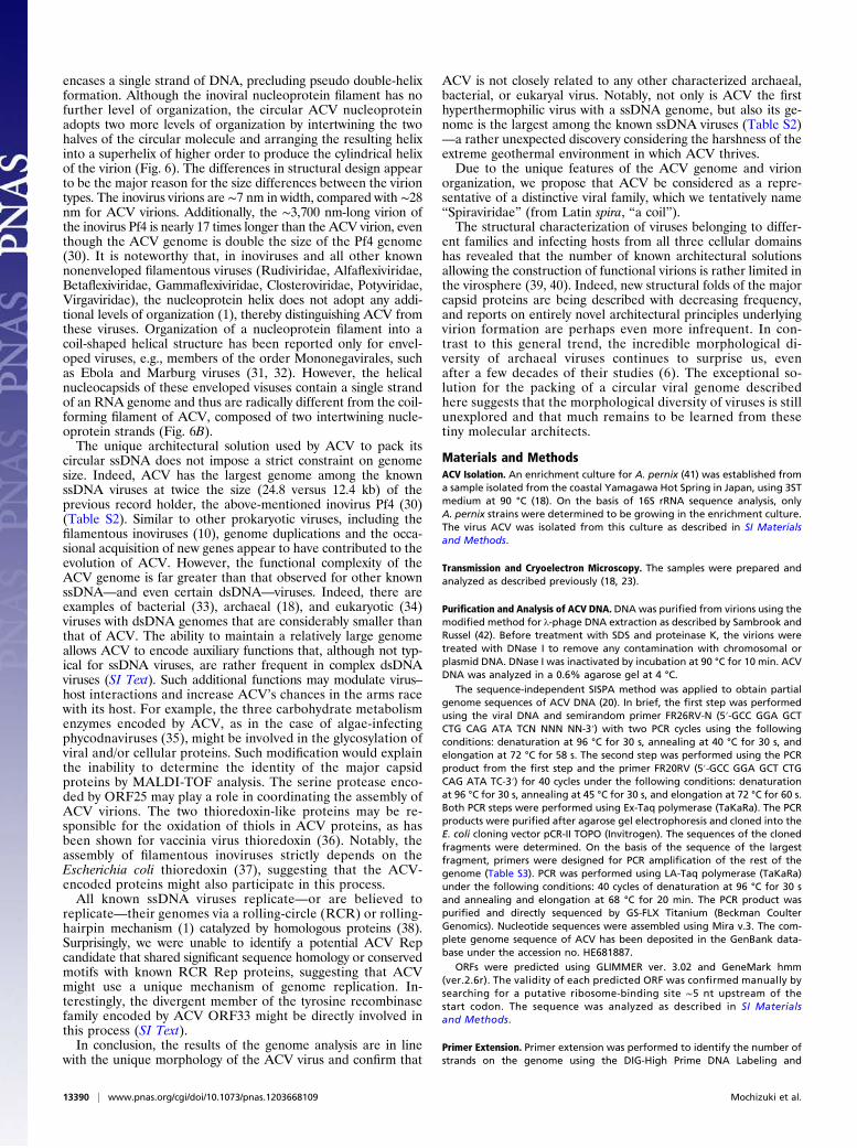
encases a single strand of DNA, precluding pseudo double-helixformation. Although the inoviral nucleoprotein filament has nofurther level of organization, the circular ACV nucleoproteinadopts two more levels of organization by intertwining the twohalves of the circular molecule and arranging the resulting helixinto a superhelix of higher order to produce the cylindrical helixof the virion (Fig. 6). The differences in structural design appearto be the major reason for the size differences between the viriontypes. The inovirus virions are ∼7 nm in width, compared with ∼28nm for ACV virions. Additionally, the ∼3,700 nm-long virion ofthe inovirus Pf4 is nearly 17 times longer than the ACV virion, eventhough the ACV genome is double the size of the Pf4 genome(30). It is noteworthy that, in inoviruses and all other knownnonenveloped filamentous viruses (Rudiviridae, Alfaflexiviridae,Betaflexiviridae, Gammaflexiviridae, Closteroviridae, Potyviridae,Virgaviridae), the nucleoprotein helix does not adopt any addi-tional levels of organization (1), thereby distinguishing ACV fromthese viruses. Organization of a nucleoprotein filament into acoil-shaped helical structure has been reported only for envel-oped viruses, e.g., members of the order Mononegavirales, suchas Ebola and Marburg viruses (31, 32). However, the helicalnucleocapsids of these enveloped visuses contain a single strandof an RNA genome and thus are radically different from the coil-forming filament of ACV, composed of two intertwining nucle-oprotein strands (Fig. 6B).The unique architectural solution used by ACV to pack its
circular ssDNA does not impose a strict constraint on genomesize. Indeed, ACV has the largest genome among the knownssDNA viruses at twice the size (24.8 versus 12.4 kb) of theprevious record holder, the above-mentioned inovirus Pf4 (30)(Table S2). Similar to other prokaryotic viruses, including thefilamentous inoviruses (10), genome duplications and the occa-sional acquisition of new genes appear to have contributed to theevolution of ACV. However, the functional complexity of theACV genome is far greater than that observed for other knownssDNA—and even certain dsDNA—viruses. Indeed, there areexamples of bacterial (33), archaeal (18), and eukaryotic (34)viruses with dsDNA genomes that are considerably smaller thanthat of ACV. The ability to maintain a relatively large genomeallows ACV to encode auxiliary functions that, although not typ-ical for ssDNA viruses, are rather frequent in complex dsDNAviruses (SI Text). Such additional functions may modulate virus–host interactions and increase ACV’s chances in the arms racewith its host. For example, the three carbohydrate metabolismenzymes encoded by ACV, as in the case of algae-infectingphycodnaviruses (35), might be involved in the glycosylation ofviral and/or cellular proteins. Such modification would explainthe inability to determine the identity of the major capsidproteins by MALDI-TOF analysis. The serine protease enco-ded by ORF25 may play a role in coordinating the assembly ofACV virions. The two thioredoxin-like proteins may be re-sponsible for the oxidation of thiols in ACV proteins, as hasbeen shown for vaccinia virus thioredoxin (36). Notably, theassembly of filamentous inoviruses strictly depends on theEscherichia coli thioredoxin (37), suggesting that the ACV-encoded proteins might also participate in this process.All known ssDNA viruses replicate—or are believed to
replicate—their genomes via a rolling-circle (RCR) or rolling-hairpin mechanism (1) catalyzed by homologous proteins (38).Surprisingly, we were unable to identify a potential ACV Repcandidate that shared significant sequence homology or conservedmotifs with known RCR Rep proteins, suggesting that ACVmight use a unique mechanism of genome replication. In-terestingly, the divergent member of the tyrosine recombinasefamily encoded by ACV ORF33 might be directly involved inthis process (SI Text).In conclusion, the results of the genome analysis are in line
with the unique morphology of the ACV virus and confirm that
ACV is not closely related to any other characterized archaeal,bacterial, or eukaryal virus. Notably, not only is ACV the firsthyperthermophilic virus with a ssDNA genome, but also its ge-nome is the largest among the known ssDNA viruses (Table S2)—a rather unexpected discovery considering the harshness of theextreme geothermal environment in which ACV thrives.Due to the unique features of the ACV genome and virion
organization, we propose that ACV be considered as a repre-sentative of a distinctive viral family, which we tentatively name“Spiraviridae” (from Latin spira, “a coil”).The structural characterization of viruses belonging to differ-
ent families and infecting hosts from all three cellular domainshas revealed that the number of known architectural solutionsallowing the construction of functional virions is rather limited inthe virosphere (39, 40). Indeed, new structural folds of the majorcapsid proteins are being described with decreasing frequency,and reports on entirely novel architectural principles underlyingvirion formation are perhaps even more infrequent. In con-trast to this general trend, the incredible morphological di-versity of archaeal viruses continues to surprise us, evenafter a few decades of their studies (6). The exceptional so-lution for the packing of a circular viral genome describedhere suggests that the morphological diversity of viruses is stillunexplored and that much remains to be learned from thesetiny molecular architects.
Materials and MethodsACV Isolation. An enrichment culture for A. pernix (41) was established froma sample isolated from the coastal Yamagawa Hot Spring in Japan, using 3STmedium at 90 °C (18). On the basis of 16S rRNA sequence analysis, onlyA. pernix strains were determined to be growing in the enrichment culture.The virus ACV was isolated from this culture as described in SI Materialsand Methods.
Transmission and Cryoelectron Microscopy. The samples were prepared andanalyzed as described previously (18, 23).
Purification and Analysis of ACV DNA. DNA was purified from virions using themodified method for λ-phage DNA extraction as described by Sambrook andRussel (42). Before treatment with SDS and proteinase K, the virions weretreated with DNase I to remove any contamination with chromosomal orplasmid DNA. DNase I was inactivated by incubation at 90 °C for 10 min. ACVDNA was analyzed in a 0.6% agarose gel at 4 °C.
The sequence-independent SISPA method was applied to obtain partialgenome sequences of ACV DNA (20). In brief, the first step was performedusing the viral DNA and semirandom primer FR26RV-N (5′-GCC GGA GCTCTG CAG ATA TCN NNN NN-3′) with two PCR cycles using the followingconditions: denaturation at 96 °C for 30 s, annealing at 40 °C for 30 s, andelongation at 72 °C for 58 s. The second step was performed using the PCRproduct from the first step and the primer FR20RV (5′-GCC GGA GCT CTGCAG ATA TC-3′) for 40 cycles under the following conditions: denaturationat 96 °C for 30 s, annealing at 45 °C for 30 s, and elongation at 72 °C for 60 s.Both PCR steps were performed using Ex-Taq polymerase (TaKaRa). The PCRproducts were purified after agarose gel electrophoresis and cloned into theE. coli cloning vector pCR-II TOPO (Invitrogen). The sequences of the clonedfragments were determined. On the basis of the sequence of the largestfragment, primers were designed for PCR amplification of the rest of thegenome (Table S3). PCR was performed using LA-Taq polymerase (TaKaRa)under the following conditions: 40 cycles of denaturation at 96 °C for 30 sand annealing and elongation at 68 °C for 20 min. The PCR product waspurified and directly sequenced by GS-FLX Titanium (Beckman CoulterGenomics). Nucleotide sequences were assembled using Mira v.3. The com-plete genome sequence of ACV has been deposited in the GenBank data-base under the accession no. HE681887.
ORFs were predicted using GLIMMER ver. 3.02 and GeneMark hmm(ver.2.6r). The validity of each predicted ORF was confirmed manually bysearching for a putative ribosome-binding site ∼5 nt upstream of thestart codon. The sequence was analyzed as described in SI Materialsand Methods.
Primer Extension. Primer extension was performed to identify the number ofstrands on the genome using the DIG-High Prime DNA Labeling and
13390 | www.pnas.org/cgi/doi/10.1073/pnas.1203668109 Mochizuki et al.

Detection Starter Kit I (Roche). Viral DNA (14 ng) was treated in a 20-μL assaycontaining 2.7 U of Klenow fragment, 0.02 mM dNTP, and 0.12 mM digox-igenin (DIG)-labeled dUTP for 6 h at 37 °C. The product was subjected toelectrophoresis in a 0.9% agarose gel and transferred onto a Hybond-N+
membrane (Amersham). The DIG-labeled nucleotides were detected as sug-gested by the manufacturer. In the same way, the following control sampleswere treated and analyzed: (i) 14 ng of single-stranded M13; (ii) 14 ng ofdouble-stranded M13; (iii) 25 ng of linearized double-stranded plasmidpBR328 (provided with the kit); and (iv) 25 ng of linearized double-strandedplasmid pBR328, heat-denatured before elongation.
Identification of Genome-Strand Orientation. Orientation of the single-stranded DNA was identified using the modified SISPA method. In brief,partial copies of the complementary strand were first produced using thesemirandom primer FR26RV. After removal of excess primers using a PCRpurification kit (Machery-Nagel), a second step was performed with 45 PCR
cycles using the purified product from the first step as a template. The usedprimers were FR20RV and one of the viral sequence-specific primers designedon the basis of the viral genome sequence to cover its different parts (Table S3).The resulting PCR product was visualized by electrophoresis in an agarosegel. The PCR product was formed and observed only if the sequence-specificprimer used in the second step coincided in its direction with the direction ofthe single-stranded template (viral genome).
ACKNOWLEDGMENTS. We thank Hiroshi Nishimura, Satoshi Kawaichi, andYasuko Yoneda for sample and strain preparations; Elina Roine for com-ments on the viral DNA analysis; and Soizick Lucas-Staat for assistance withelectron microscopy. This work was supported by the Programme Blanc ofthe Agence Nationale de la Recherche, France (Grant ANR 09-BLN-0288.01).T.M. was supported by Bourse du Gouvernement Français (Dossier 2008661)and Allocations Pasteur-Weizmann. M.K. was supported by the EuropeanMolecular Biology Organization (ALTF 347-2010).
1. King AMQ, Adams MJ, Carstens EB, Lefkowitz EJ (2011) Virus Taxonomy: Ninth Reportof the International Committee on Taxonomy of Viruses (Elsevier Academic Press,San Diego).
2. Prangishvili D, Forterre P, Garrett RA (2006) Viruses of the Archaea: A unifying view.Nat Rev Microbiol 4:837–848.
3. Suttle CA (2007) Marine viruses: Major players in the global ecosystem. Nat RevMicrobiol 5:801–812.
4. Rohwer F, Prangishvili D, Lindell D (2009) Roles of viruses in the environment. EnvironMicrobiol 11:2771–2774.
5. Forterre P, Prangishvili D (2009) The great billion-year war between ribosome- andcapsid-encoding organisms (cells and viruses) as the major source of evolutionarynovelties. Ann N Y Acad Sci 1178:65–77.
6. Pina M, Bize A, Forterre P, Prangishvili D (2011) The archeoviruses. FEMSMicrobiol Rev35:1035–1054.
7. Ackermann HW (2007) 5500 phages examined in the electron microscope. Arch Virol152:227–243.
8. Atanasova NS, Roine E, Oren A, Bamford DH, Oksanen HM (2012) Global network ofspecific virus-host interactions in hypersaline environments. Environ Microbiol 14:426–440.
9. Pfister P, Wasserfallen A, Stettler R, Leisinger T (1998) Molecular analysis of Meth-anobacterium phage psiM2. Mol Microbiol 30:233–244.
10. Krupovic M, Prangishvili D, Hendrix RW, Bamford DH (2011) Genomics of bacterialand archaeal viruses: Dynamics within the prokaryotic virosphere. Microbiol Mol BiolRev 75:610–635.
11. Prangishvili D, Garrett RA, Koonin EV (2006) Evolutionary genomics of archaeal vi-ruses: Unique viral genomes in the third domain of life. Virus Res 117:52–67.
12. Bize A, et al. (2009) A unique virus release mechanism in the Archaea. Proc Natl AcadSci USA 106:11306–11311.
13. Brumfield SK, et al. (2009) Particle assembly and ultrastructural features associatedwith replication of the lytic archaeal virus sulfolobus turreted icosahedral virus. J Virol83:5964–5970.
14. Quax TE, et al. (2011) Simple and elegant design of a virion egress structure inArchaea. Proc Natl Acad Sci USA 108:3354–3359.
15. Prangishvili D (2003) Evolutionary insights from studies on viruses of hyper-thermophilic archaea. Res Microbiol 154:289–294.
16. Pietilä MK, Roine E, Paulin L, Kalkkinen N, Bamford DH (2009) An ssDNA virus in-fecting Archaea: A new lineage of viruses with a membrane envelope. Mol Microbiol72:307–319.
17. Prangishvili D (2006) Hyperthermophilic virus-host systems: Detection and isolation.Methods in Microbiology: Extremophiles, eds Rainey FA, Oren A (Elsevier AcademicPress, London), Vol 35, pp 331–347.
18. Mochizuki T, et al. (2010) Diversity of viruses of the hyperthermophilic archaeal genusAeropyrum, and isolation of the Aeropyrum pernix bacilliform virus 1, APBV1, thefirst representative of the family Clavaviridae. Virology 402:347–354.
19. Prangishvili D, et al. (1999) A novel virus family, the Rudiviridae: Structure, virus-hostinteractions and genome variability of the sulfolobus viruses SIRV1 and SIRV2.Genetics 152:1387–1396.
20. Reyes GR, Kim JP (1991) Sequence-independent, single-primer amplification (SISPA) ofcomplex DNA populations. Mol Cell Probes 5:473–481.
21. Mochizuki T, Prangishvili D (2012) A simple and sensitive method for determiningthe strand orientation of single-stranded viral genomes. J Virol Methods, 10.1016/j.jviromet.2012.05.017.
22. Kawarabayasi Y, et al. (1999) Complete genome sequence of an aerobic hyper-ther-
mophilic crenarchaeon, Aeropyrum pernix K1. DNA Res 6:83–101, 145–152.23. Mochizuki T, Sako Y, Prangishvili D (2011) Provirus induction in hyperthermophilic
Archaea: Characterization of Aeropyrum pernix spindle-shaped virus 1 and Aero-
pyrum pernix ovoid virus 1. J Bacteriol 193:5412–5419.24. Altschul SF, et al. (1997) Gapped BLAST and PSI-BLAST: A new generation of protein
database search programs. Nucleic Acids Res 25:3389–3402.25. Söding J (2005) Protein homology detection by HMM-HMM comparison. Bioinformatics
21:951–960.26. Marchler-Bauer A, Bryant SH (2004) CD-Search: Protein domain annotations on the
fly. Nucleic Acids Res 32(Web Server issue):W327–331.27. Gopaul DN, Guo F, Van Duyne GD (1998) Structure of the Holliday junction in-
termediate in Cre-loxP site-specific recombination. EMBO J 17:4175–4187.28. Nunes-Düby SE, Kwon HJ, Tirumalai RS, Ellenberger T, Landy A (1998) Similarities and
differences among 105 members of the Int family of site-specific recombinases. Nu-
cleic Acids Res 26:391–406.29. Day LA (2011) Inoviridae. Virus Taxonomy: Ninth Report of the International Com-
mittee on Taxonomy of Viruses, eds King AMQ, Adams MJ, Carstens EB, Lefkowitz EJ
(Elsevier Academic Press, San Diego), pp 375–383.30. Webb JS, Lau M, Kjelleberg S (2004) Bacteriophage and phenotypic variation in
Pseudomonas aeruginosa biofilm development. J Bacteriol 186:8066–8073.31. Bharat TA, et al. (2011) Cryo-electron tomography of Marburg virus particles and
their morphogenesis within infected cells. PLoS Biol 9:e1001196.32. Bharat TAM, et al. (2012) Structural dissection of Ebola virus and its assembly de-
terminants using cryo-electron tomography. Proc Natl Acad Sci USA 109:4275–4280.33. Krupovic M, et al. (2006) Genome characterization of lipid-containing marine bac-
teriophage PM2 by transposon insertion mutagenesis. J Virol 80:9270–9278.34. Fanning E, Zhao K (2009) SV40 DNA replication: from the A gene to a nanomachine.
Virology 384:352–359.35. Van Etten JL, Gurnon JR, Yanai-Balser GM, Dunigan DD, Graves MV (2010) Chlorella
viruses encode most, if not all, of the machinery to glycosylate their glycoproteins
independent of the endoplasmic reticulum and Golgi. Biochim Biophys Acta 1800:
152–159.36. Senkevich TG, White CL, Koonin EV, Moss B (2002) Complete pathway for protein
disulfide bond formation encoded by poxviruses. Proc Natl Acad Sci USA 99:
6667–6672.37. Russel M (1995) Moving through the membrane with filamentous phages. Trends
Microbiol 3:223–228.38. Ilyina TV, Koonin EV (1992) Conserved sequence motifs in the initiator proteins for
rolling circle DNA replication encoded by diverse replicons from Eubacteria, eucar-
yotes and Archaebacteria. Nucleic Acids Res 20:3279–3285.39. Krupovi�c M, Bamford DH (2010) Order to the viral universe. J Virol 84:12476–12479.40. Krupovic M, Bamford DH (2011) Double-stranded DNA viruses: 20 families and only
five different architectural principles for virion assembly. Curr Opin Virol 1:118–124.41. Sako Y, et al. (1996) Aeropyrum pernix gen. nov., sp. nov., a novel aerobic hyper-
thermophilic archaeon growing at temperatures up to 100 degrees C. Int J Syst
Bacteriol 46:1070–1077.42. Sambrook J, Russel DW (2001) Molecular Cloning: A Laboratory Manual (Cold Spring
Harbor Laboratory Press, Cold Spring Harbor, NY).
Mochizuki et al. PNAS | August 14, 2012 | vol. 109 | no. 33 | 13391
MICRO
BIOLO
GY