Embed Size (px)
Citation preview

University of Arkansas, Fayetteville University of Arkansas, Fayetteville
ScholarWorks@UARK ScholarWorks@UARK
Graduate Theses and Dissertations
12-2018
Aqueous Ozone Inactivation of Viruses and Bacteria on Biotic and Aqueous Ozone Inactivation of Viruses and Bacteria on Biotic and
Abiotic Surfaces Abiotic Surfaces
Cailin Dawley University of Arkansas, Fayetteville
Follow this and additional works at: https://scholarworks.uark.edu/etd
Part of the Food Microbiology Commons, Food Studies Commons, and the Pathogenic Microbiology
Commons
Citation Citation Dawley, C. (2018). Aqueous Ozone Inactivation of Viruses and Bacteria on Biotic and Abiotic Surfaces. Graduate Theses and Dissertations Retrieved from https://scholarworks.uark.edu/etd/3022
This Thesis is brought to you for free and open access by ScholarWorks@UARK. It has been accepted for inclusion in Graduate Theses and Dissertations by an authorized administrator of ScholarWorks@UARK. For more information, please contact [email protected].

1
Aqueous Ozone Inactivation of Viruses and Bacteria on Biotic and Abiotic Surfaces
A thesis submitted in partial fulfillment
of the requirements for the degree of
Master of Science in Food Science
by
Cailin R. Dawley
University of Arkansas
Bachelor of Science in Food Science, 2016
December 2018
University of Arkansas
This thesis is approved for recommendation to the Graduate Council.
________________________________________
Kristen Gibson, Ph.D.
Thesis Director
________________________________________
Ruben Morawicki, Ph.D.
Committee Member
________________________________________
Young Min Kwon, Ph.D.
Committee Member

2
Abstract:
Produce is susceptible to contamination by foodborne pathogens. Food service establishments
utilize sanitizing agents to reduce microbes on produce surfaces. The research objectives were to
evaluate the efficacy of aqueous ozone 1) on the inactivation of viruses and bacteria on produce;
2) on the inactivation of viruses on stainless steel; and 3) against viruses in association with
bacteria on produce surfaces. For objective 1, Boston bibb lettuce and cherry tomatoes were spot
inoculated with a cocktail of viruses (murine norovirus (MNV) and MS2 bacteriophage) or
bacteria (Enterobacter cloacae and Bacillus cereus) and washed for 40 min with samples taken
every 10 min. For objective 2, stainless steel (SS) coupons were spot inoculated with the same
cocktail of viruses and washed for 0.5, 3, and 10 min. For objective 3, Boston bibb was spot
inoculated with either MNV and E. cloacae or MNV and B. cereus and washed for 40 min with
samples taken every 10 min. Inocula were allowed to dry for > 90 min. A batch wash ozone
sanitation system (BWOSS) was prepared with ice (3-5°C) and 0.5 ppm ozone or no ozone.
Surfaces were treated with either an ozone or water wash with samples taken over time and
repeated in at least duplicate. Samples were processed to determine plaque forming units (PFU)
and colony forming units (CFU). In objective 1, there were no significant differences in
inactivation of MNV, MS2, or bacteria with ozone compared to water only. There was greater
variability in viral reduction while bacterial inactivation increased over time. In objective 2, there
was no significant difference in inactivation of MNV or MS2 on SS, but the variability was
reduced. The log reduction difference between ozone and water for MNV and MS2 after 10 min
was 0.25 and 0.51 PFU/ml, respectively. In objective 3, MNV-bacteria association on lettuce did
not impact ozone inactivation of MNV. The log reduction differences between ozone and water

3
for MNV with B. cereus and MNV with E. cloacae after 40 min were 0.95 and -0.36 PFU/ml,
respectively. Further research is needed on how food matrices effects viral inactivation by ozone.
4
©2018 by Cailin R. Dawley
All Rights Reserved
5
Acknowledgements:
I am incredibly grateful for everything that Dr. Kristen Gibson has done for me in my pursuit of
my Masters. She has been incredibly patient as I learned the ins and outs of research, especially
tissue culture. Her expanse knowledge in the field is awe inspiring and I am so blessed to have
spent the last two years learning from her. I also would like to thank Giselle Almeida, laboratory
assistant for Dr. Kristen Gibson, for her support in my laboratory training and patience as I asked
countless questions throughout my time here. I also am grateful for my committee members, Dr.
Ruben Morawicki and Dr. Young Min Kwon, their insight was helpful in the course of my
research. Kevin Thompson was also a crucial part of this research. He aided in statistical analysis
and was incredibly helpful with helping me understand my data. Additionally, my lab mates
were an amazing support system: Sarah Jones, Wenjun Deng, Kacy Wright, Gina Riggio, and
Thomas Yeargin. They helped solve problems, made media, and reminded me that I was so close
to being done. I would also like to thank my parents and roommates for listening to me talk
about my research for the last two years. A major thank you also goes to my campus ministry,
Razorbacks for Christ, a place where I could go bake cookies when I was stressed and be
welcomed by several friends that would make me laugh and forget about my massive to-do list. I
would not have made it through this program without their support. I also know I would not have
done this if God had not opened this opportunity, and I will be forever grateful that he did
because this experience has changed my life.

6
Table of Contents:
Chapter 1: Literature Review 1
1. Foodborne Illnesses 2
a. Outbreaks and Fresh Produce 2
2. Strategies to control pathogens in Retail Food Service 3
3. Ozone as a disinfectant 5
a. Ozone inactivation of bacteria 5
b. Ozone inactivation of viruses 7
4. Microbe-Microbe Interactions 8
a. Virus and Bacteria 8
5. Future Research and Objectives 9
References 11
Appendix 16
Chapter 2: Virus-Bacterial Interactions: Implications for Prevention and Control of
Human Enteric Viruses from Environment to Host 20
Abstract 21
1. Introduction 22
2. In Vivo Implications of Virus-Bacteria Interactions 23
a. Infectivity and Pathogenesis 24
b. Protection and Competitive Exclusion 25
c. Role in Recombination 29
3. In Situ Implications of Virus-Bacteria Interactions 31
a. Food Contact Surfaces 31
b. Water resources-Biofilms 33
c. Specialty Crops 35
4. In Vitro Implications of Virus-Bacteria Interactions 37
5. Conclusion 39
References 40
Chapter 3: Inactivation of microorganisms on Boston bibb lettuce and cherry tomatoes by
aqueous ozone 46
Abstract 47
1. Introduction 49
2. Material and Methods 51
a. Microbe Cultivation 51
b. Produce 54
c. Inoculation of Produce 54
d. Treatments 55
e. Microbial Analysis 55
f. Statistical Analysis 56
3. Results 57
4. Discussion 59
7
5. Conclusion 62
References 63
Appendix 66
Chapter 4: Aqueous ozone inactivation of viruses on stainless steel surfaces 70
Abstract 71
1. Introduction 72
2. Material and Methods 73
a. Microbe Cultivation 73
b. Stainless Steel 74
c. Treatments 75
d. Microbial Analysis 76
e. Statistical Analysis 76
3. Results 76
4. Discussion 77
5. Conclusion 78
References 80
Appendix 82
Chapter 5: Aqueous ozone inactivation of viruses in association with bacteria on Boston
bibb lettuce 83
Abstract 84
1. Introduction 85
2. Material and Methods 86
a. Microbe Cultivation 86
b. Microbe-Microbe Interactions 88
c. Produce 88
d. Inoculation of Produce 89
e. Treatments 89
f. Microbial Analysis 90
g. Statistical Analysis 90
3. Results 90
4. Discussion 91
5. Conclusion 92
References 93
Appendix 95
Chapter 6: Overall Conclusions 97
References 101
Appendix 102
IBC Approval Letter 102
IBC Registration for Research Project 103

8
List of Unpublished Papers
1. Chapter 2: Dawley, Cailin and Gibson, Kristen. (2018) Virus-Bacterial Interactions:
Implications for Prevention and Control of Human Enteric Viruses from Environment to Host.
Foodborne Pathogens and Disease. Submitted for Publication.

1
Chapter 1: Literature Review
2
1. Foodborne Illnesses
Of 31 major foodborne pathogens, it is estimated there are 9.4 million cases of foodborne
illness in the United States each year (Scallan et al., 2011). Foodborne pathogens can
contaminate food anywhere from the farm to the fork, due to the range of environments it incurs
before reaching the consumer. These environments are the production field, processing facilities,
transportation, handling by food service establishment employees, or in homes during
preparation. Foods that are especially susceptible to contamination are produce, dairy, poultry,
eggs, and beef (Painter et al., 2013). Major foodborne pathogens are introduced into these
environments through a variety of routes. In some cases, animals are hosts to pathogens such as
Salmonella, Listeria, Escherichia coli, and Campylobacter (Swartz, 2002). Sometimes humans
are hosts and can transfer the pathogen (e.g. human norovirus) during handling (Berger et al.,
2010). Of the 31 major pathogens, human noroviruses are the primary cause of foodborne
illnesses in the United States (Scallan et al., 2011).
a. Outbreaks and Fresh Produce
Fresh produce, specifically leafy greens, have been found to be a common source of
foodborne pathogens resulting in 46% of estimated foodborne illnesses (Painter et al., 2013).
Norovirus is often associated with leafy greens as well; from 2001-2008, 33% of norovirus
outbreaks were associated with leafy greens (Hall et al., 2012). Produce is susceptible to
contamination with pathogens for several reasons. It is consumed raw, unlike products that are
cooked or pasteurized before eating. Produce is also processed to be ready to eat which can cause
tears and breaks to the surface of the produce allowing some microbes to grow on the surface or
be internalized (Benson, 2010). Produce can become contaminated prior to retail or during
preparation at the food service establishment (FSE). Prior to harvest, contamination can come
3
from the soil, water, or wild animal excrements carrying pathogens that adhere to produce
(Benson, 2010). Then as produce is transferred from the field to the store there are more
opportunities for contamination: field workers, packaging, and transportation (Johnston et al.,
2005). At the FSE, the contamination more than likely comes from the employees. This could be
in the form of poor personal hygiene or from improper hand washing leading to the transmission
of viruses such as norovirus and Hepatitis A virus (Strohbehn et al., 2008).
Contamination of fresh produce has led to several outbreaks. For instance, E. coli O157:H7
has been associated with contaminated leafy greens resulting in various outbreaks. A multistate
outbreak of E. coli O157:H7 was linked to bagged spinach that was sold for at home use (Grant
et al., 2008). The authors hypothesized that the bacteria were either internalized by the spinach
plant in the field or attached to the cut surfaces of the prepackaged spinach. Another outbreak of
E. coli O157:H7 in Wisconsin was linked to bagged spinach where the pathogen was detected in
river water, cattle feces, and pig feces near the field the spinach was grown in (Wendel et al.,
2009). Non-typhoidal Salmonella has been associated with outbreaks due to contaminated
tomatoes. In a multistate outbreak across 26 states, tomatoes were contaminated by Salmonella
Newport which was found to be present in the irrigation water at the field the tomatoes were
grown in (Greene et al., 2008).
2. Strategies to control pathogens in retail food service
The U.S. Food and Drug Administration (FDA) has published recommendations on how to
prevent contamination of food products after receiving. It includes separating products, washing
hands, and cleaning equipment. There is a specific section on washing produce which is as
follows:

4
Washing Fruits and Vegetables A) Except as specified in (B) of this section and except
for whole, raw fruits and vegetables that are intended for washing by the consumer before
consumption, raw fruits and vegetables shall be thoroughly washed in water to remove
soil and other contaminants before being cut, combined with other ingredients, cooked,
served, or offered for human consumption in ready to eat form. (B) Fruits and vegetables
may be washed by using chemicals as specified under 21 CFR 7-204.12 (U.S.
Department of Health and Human Services, 2009).
Retail and institutional food service can apply several different strategies to control
pathogens and prevent contamination once produce reaches the retailer. Retailers can take steps
to prevent improper food handling by workers. These steps can include enforcing good hygiene,
hand washing, and sending ill workers home. Retailers can also make sure that suppliers are
following good manufacturing practices (GMPs) to prevent contamination before the products
reach them. The use of refrigeration when the produce is not being served can keep bacterial
pathogens from multiplying (Lynch et al., 2009). Retailers can also wash produce with a
sanitizing solution of chlorine, ozone, or lemon juice and vinegar to reduce microbes on the
surface of produce (Berger et al., 2010). A study by Allende et al. (2008) investigated
commercial sanitizing agents for the inactivation of epiphytic microbes on the surface of leafy
greens via submersion in these sanitizers. The sanitizers they used were chlorine (sodium
hypochlorite), Sanova (acidified sodium chlorite), Sanoxol 20 (hydrogen peroxide and
peroxyacetic acid), Tsunami 100 (peroxyacetic acid and hydrogen peroxide), Purac FCC 80
(lactic acid), Citrox 14W (organic acid and flavonoids) and Catallix (lactoperoxidase, hydrogen
peroxide and thiocyanate) under the manufacturer’s suggested concentrations and exposure time.
The researchers found that chlorine, Sanova, Purac FCC 80, Citrox 14W and Catallix all had a
significant impact on the microbial epiphytes present on the leafy greens compared with the
water only wash.
5
3. Ozone as a disinfectant
Ozone (O3) is a strong oxidant that can be effective against several different microorganisms
(Alexandre et al. 2011). Aqueous ozone is being used in the food industry during produce
washes, disinfection of processing equipment, and disinfection of the environment (Mahapatra et
al., 2005) Ozone is an effective disinfectant against E. coli O157:H7, Salmonella, Listeria
monocytogenes, Staphylococcus aureus, foodborne viruses, and protozoa (Achen et al., 2001;
Cursons et al., 1980; Lim et al., 2010; Restaino et al., 1995). When considering ozone as a
disinfectant, it has some positive characteristics that aid in its ability to disinfect and some
drawbacks that need to be considered. Ozone is soluble in water which creates a solution, but it
is not stable long term; it will decompose into oxygen over time. Due to the short half-life of
ozone, there is no residue left on the produce after being washed unlike chlorine that leaves
residues on produce and surfaces that can be toxic in high concentrations (Karaca and Velioglu,
2007).
Ozone is also sensitive to pH as it is more stable in acidic conditions than alkaline (Khadre et
al., 2001). Additionally, the half-life of ozone is increased at colder temperatures thus enhancing
its stability (Batakliev et al., 2014). Ozone effectiveness can be impacted by the presence of
organic matter by changing the pathway of ozone. Some organic matter converts the hydroxyl
radical into a superoxide radical which slows the effectiveness of ozone while other organic
compounds can stabilize ozone (Kim et al., 1999).
a. Ozone inactivation of bacteria
Ozone has been shown to be effective in inactivating a range of bacteria. Ozone oxidizes
lipids present in the cell membrane of the bacteria, intracellular enzymes, as well as the bacterial
genome (Guzel-Seydim et al., 2004). The effectiveness is in relation to the type of bacteria: gram
6
positive or gram negative. Gram negative bacteria are reported to be less resistant to ozone while
gram positive bacteria have greater resistance due to the cellular structure of the outer
membrane. Gram positive bacteria contains a higher concentration of peptidoglycan which is
more resistant to ozone than the lipoproteins present in gram negative bacteria (Khadre et al.,
2001). Ozone also has the potential to inactivate bacterial spores such as Bacillus cereus by
degrading the outer spore coat and exposing the core to disinfection (Khadre et al., 2001).
Ozone has been used with bacteria in suspension and bacteria inoculated on food products.
Table 1 highlights studies that have used ozone as a sanitizing agent for bacteria. Kim and
Yousef (2000) investigated the inactivation of bacteria in suspension when exposed to ozone.
The researchers used a low concentration of ozone (0.2-0.3ppm) and observed at least a one log
reduction after 30s for all of the microbes tested. Ozone is highly effective against bacteria in
suspension; however, adding in a food matrix requires higher concentrations of ozone and a
longer contact time to experience the same reduction. This can be seen in the study by Bialka and
Demirci (2007). Here, the authors inoculated blueberries with E. coli and Salmonella and
required over 2 minutes to observe at least a log reduction with a higher concentration of ozone
(1.7-8.9ppm) compared to the previously mentioned study by Kim and Yousef (2000). The main
limitation in comparing these studies is the difference between microbes in suspension and
microbes on the surface of produce which effects the results. Besides the presence of a biotic
surface, there are two different ways of generating aqueous ozone: ozonated water which was
utilized in the study by Kim and Yousef (2000) and bubbling ozone which was utilized by Bialka
and Demirci (2007). Ozonated water is water that ozone has been generated in to reach a certain
concentration and then the generator of ozone is removed whereas bubbling ozone has a
continuous supply of ozone added to the water (Achen and Yousef, 2001). Bubbling ozone

7
generates a greater log reduction than ozonated water due to the ozone always being replenished
(Achen and Yousef, 2001) (Table 1).
b. Ozone inactivation of viruses
Viruses are also reportedly susceptible to ozone. This is primarily due to the composition of a
virus particle. It is genetic material surrounded by a capsid made of proteins that can be
denatured by ozone thus releasing the genetic material which leads to inactivation (Khadre et al.,
2001). It could also be hypothesized that ozone could damage the viral capsid and lead to an
inability to bind to receptors which has been documented in thermal treatments (D. Li et al.,
2012). Ozone inactivation of viruses has been investigated with virus in suspension. Ozone has
been found to be more effective in deactivating viruses at lower pH’s and with lower
temperatures. Table 2 highlights studies that have documented the impact of ozone on the
inactivation of viruses. Lim et al. (2010) investigated the inactivation of murine norovirus
(MNV) at 5°C and 20°C and at pH 5.6 and 7. The authors observed a more rapid inactivation of
MNV at 5°C than at 20°C, but this was not significantly different. The pH did have a significant
impact on the inactivation of MNV, with a more rapid inactivation of the viral surrogate at pH
5.6 than at pH 7, regardless of temperature. Studies have also been conducted on biotic and
abiotic surfaces such as green onions and stainless-steel utensils inoculated with viruses. Green
onions were inoculated with adenovirus, MNV, and Hepatitis A virus, and log reductions of 4.3,
2.5, and 2.9 were achieved, respectively, in 6.25 ppm ozone after 10 minutes (Hirneisen and
Kniel, 2013). Gibson and Almeida (2016) reported dipping a stainless-steel utensil in a virus
solution containing PRD1 bacteriophage then placing the inoculated utensil in a circulating
ozone washer at 0.5ppm. The authors observed a 6.44 log reduction at 30 seconds (Almeida and
Gibson, 2016) (Table 2).
8
4. Microbe-Microbe Interactions
Microorganisms do not exist in monoculture but rather in a diverse community of microbes.
A community that is often discussed in the food industry are biofilms. Biofilms are
microorganisms that attach to and multiply on a surface while attracting nutrients (Kumar and
Anand, 1998). These communities may allow interactions to form which could create stronger
resistances to being inactivated. Research has begun to investigate these interactions but several
questions still remain.
a. Virus and Bacteria
As previously stated, microbes do not exist in isolation. Microbial communities such as
biofilms may harbor bacteria including pathogens, but can also protect viruses from desiccation
and other environmental stressors (Lacroix-Gueu et al., 2005). Similar studies that occurred in
vivo showed that in the presence of bacteria (gram positive and gram negative) or in the presence
of bacterial cell components viruses had increased infectivity (Kuss et al., 2011).
When it comes to foodborne pathogens, human noroviruses are a primary concern due to the
volume of illnesses. A majority of human noroviruses bind to histo-blood group antigens
(HBGA) (Huang et al., 2005). Some bacteria, such as enteric bacteria, have been known to
possess HBGA-like structures that viruses are able to bind to as well (Miura et al., 2013). Jones
et al. (2014) took this knowledge and showed that enteric bacteria can act as co-factors to aid in
viral attachment to B-cells. These studies show that viral interactions with bacteria have the
potential to exist and increase viability and virulence of the virus.
i. Disinfection
Understanding the impact of virus-bacteria interactions on the efficacy of a disinfection
method is an important research area. If these interactions occur, how do the microbes respond to
9
current disinfection processes? Heat treatment is the only disinfectant that has been applied to
these interactions thus far. Li et al. (2015) utilized several genera of norovirus allowing the virus
to interact with various bacteria. Three different treatment groups were evaluated: virus like
particles (VLPs) of human norovirus GI.1; VLPs of human norovirus GI.1 and bacteria with
HBGA-like substances; and VLPs of human norovirus GI.1 and bacteria with no HBGA-like
substances. Results indicated that when viruses interacted with bacteria with HBGA-like
structures, the viruses had a higher immunoreactivity after applying heat (Li et al., 2015). On the
other hand, Li et al. (2017) found that when Tulane virus (TV) was in association with bacteria
with HBGA-like structures the interaction did not protect the virus against heat inactivation. The
authors hypothesized that the difference in results was due to the different viruses used: VLPs of
human norovirus versus a viral surrogate (TV).
5. Future Research and Objectives
Due to heat stress being the only form of inactivation that has been applied to virus-bacteria
interactions, there is a range of possible research in this area of microbiology, such as
investigating common disinfectants including chlorine, bleach, peroxide, or ozone and their
impact on viruses in association with bacteria. The disinfectant of interest in this specific study is
ozone. This is due to ozone being effective against several microbes individually. This could
mean that ozone might be an effective disinfectant for microbe-microbe interactions that have
very little published research.
Ozone washers have been proposed for the retail setting especially for fresh produce.
Produce is a possible environment that microbe-microbe interactions could occur. Since leafy
greens have been linked to several outbreaks of foodborne illnesses especially norovirus
10
outbreaks, this commodity is a possible environment that could highlight microbe-microbe
interactions.
A batch wash ozone sanitation system (BWOSS) will be used to evaluate the efficacy of an
ozone washer to inactivate viruses with different attachment times. This research will further
elucidate the susceptibility of viruses when adhered to different surfaces: Boston bib lettuce,
cherry tomatoes, and stainless steel. The second area of research focus is to investigate the ability
of the BWOSS to inactivate viruses associated with bacteria when in combination on the fresh
produce surface. These interactions could possibly prevent current control strategies from
working at the optimal level which is important for food safety.
11
References:
Achen, M., & Yousef, A. E. (2001). Efficacy of Ozone Against Escherichia coli O157:H7 on
Apples. Journal of Food Science, 66(9), 1380–1384. https://doi.org/10.1111/j.1365-
2621.2001.tb15218.x
Alexandre, E. M. C., Santos-Pedro, D. M., Brandão, T. R. S., & Silva, C. L. M. (2011). Influence
of aqueous ozone, blanching and combined treatments on microbial load of red bell
peppers, strawberries and watercress. Journal of Food Engineering, 105(2), 277–282.
https://doi.org/10.1016/J.JFOODENG.2011.02.032
Allende, A., Selma, M. V., López-Gálvez, F., Villaescusa, R., & Gil, M. I. (2008). Role of
commercial sanitizers and washing systems on epiphytic microorganisms and sensory
quality of fresh-cut escarole and lettuce. Postharvest Biology and Technology, 49(1),
155–163. https://doi.org/10.1016/J.POSTHARVBIO.2007.12.010
Almeida, G., & Gibson, K. E. (2016). Evaluation of a Recirculating Dipper Well Combined with
Ozone Sanitizer for Control of Foodborne Pathogens in Food Service Operations. Journal
of Food Protection, 79(9), 1537–1548. https://doi.org/10.4315/0362-028X.JFP-16-055
Batakliev, T., Georgiev, V., Anachkov, M., Rakovsky, S., & Zaikov, G. E. (2014). Ozone
decomposition. Interdisciplinary Toxicology, 7(2), 47–59. https://doi.org/10.2478/intox-
2014-0008
Benson, S. M. (2010). Guidance for Improving the Federal Response to Foodborne Illness
Outbreaks Associated with Fresh Produce. Food & Drug Law Journal, 65(3), 503–524.
Berger, C. N., Sodha, S. V., Shaw, R. K., Griffin, P. M., Pink, D., Hand, P., & Frankel, G.
(2010). Fresh fruit and vegetables as vehicles for the transmission of human pathogens.
Environmental Microbiology, 12(9), 2385–2397. https://doi.org/10.1111/j.1462-
2920.2010.02297.x
Bialka, K. L., & Demirci, A. (2007). Decontamination of Escherichia coli O157:H7 and
Salmonella enterica on Blueberries Using Ozone and Pulsed UV-Light. Journal of Food
Science, 72(9), M391–M396. https://doi.org/10.1111/j.1750-3841.2007.00517.x
Cursons, R. T. M., Brown, T. J., & Keys, E. A. (1980). Effect of Disinfectants on Pathogenic
Free-Living Amoebae: in Axenic Conditions. APPLIED AND ENVIRONMENTAL
MICROBIOLOGY, 40(1), 62–66.
Grant, J., Wendelboe, A. M., Wendel, A., Jepson, B., Torres, P., Smelser, C., & Rolfs, R. T.
(2008). Spinach-associated Escherichia coli O157:H7 Outbreak, Utah and New Mexico,
2006. Emerging Infectious Diseases, 14(10), 1633–1636.
https://doi.org/10.3201/eid1410.071341
12
Greene, S. K., Daly, E. R., Talbot, E. A., Demma, L. J., Holzbauer, S., Patel, N. J., … Painter.
(2008). Recurrent multistate outbreak of Salmonella Newport associated with tomatoes
from contaminated fields, 2005. Epidemiology and Infection, 136(02), 157–65.
https://doi.org/10.1017/S095026880700859X
Guzel-Seydim, Z. B., Greene, A. K., & Seydim, A. C. (2004). Use of ozone in the food industry,
37, 453–460. https://doi.org/10.1016/j.lwt.2003.10.014
Hall, A. J., Eisenbart, V. G., Etingüe, A. L., Gould, L. H., Lopman, B. A., & Parashar, U. D.
(2012). Epidemiology of Foodborne Norovirus Outbreaks, United States, 2001–2008.
Emerging Infectious Diseases, 18(10), 1566–1573.
https://doi.org/10.3201/eid1810.120833
Hirneisen, K. A., & Kniel, K. E. (2013). Inactivation of internalized and surface contaminated
enteric viruses in green onions. International Journal of Food Microbiology, 166(2), 201–
206. https://doi.org/10.1016/j.ijfoodmicro.2013.07.013
Huang, P., Farkas, T., Zhong, W., Tan, M., Thornton, S., Morrow, A. L., & Jiang, X. (2005).
Norovirus and Histo-Blood Group Antigens: Demonstration of a Wide Spectrum of
Strain Specificities and Classification of Two Major Binding Groups among Multiple
Binding Patterns. Journal of Virology, 79(11), 6714–6722.
https://doi.org/10.1128/JVI.79.11.6714-6722.2005
Johnston, L. M., Jaykus, L.-A., Moll, D., Martinez, M. C., Anciso, J., Mora, B., & Moe, C. L.
(2005). A Field Study of the Microbiological Quality of Fresh Produce. Journal of Food
Protection, 68(9), 1840–1847. https://doi.org/10.4315/0362-028X-68.9.1840
Jones, M. K., Watanabe, M., Zhu, S., Graves, C. L., Keyes, L. R., Grau, K. R., … Karst, S. M.
(2014). Enteric bacteria promote human and mouse norovirus infection of B cells.
Science (New York, N.Y.), 346(6210), 755–9. https://doi.org/10.1126/science.1257147
Karaca, H., & Velioglu, Y. S. (2007). Ozone Applications in Fruit and Vegetable Processing.
Food Reviews International, 23(1), 91–106. https://doi.org/10.1080/87559120600998221
Khadre, M. A., & Yousef, A. E. (2001). Sporicidal action of ozone and hydrogen peroxide: a
comparative study. International Journal of Food Microbiology, 71, 131–138.
Khadre, M. A., Yousef, A. E., & Kim, J.-G. (2001). Microbiological Aspects of Ozone
Applications in Food: A Review. Journal of Food Science, 66(9), 1242–1252.
https://doi.org/10.1111/j.1365-2621.2001.tb15196.x
Kim, J.-G., & Yousef, A. E. (2000). Inactivation Kinetics of Foodborne Spoilage and Pathogenic
Bacteria by Ozone JFS: Food Microbiology and Safety. Journal of Food Science, 65(3).
Kim, J.-G., Yousef, A. E., & Chism, G. W. (1999). Use of Ozone to Inactivate Microorganisms
on Lettuce. Journal of Food Safety, 19(1), 17–34. https://doi.org/10.1111/j.1745-
4565.1999.tb00231.x

13
Kim, J.-G., Yousef, A. E., & Dave, S. (1999). Application of Ozone for Enhancing the
Microbiological Safety and Quality of Foods: A Review. Journal of Food Protection,
62(9), 1071–1087. https://doi.org/10.4315/0362-028X-62.9.1071
Koseki, S., & Isobe, S. (2006). Effect of Ozonated Water Treatment on Microbial Control and on
Browning of Iceberg Lettuce (Lactuca sativa L.). Journal of Food Protection, 69(1), 154–
160. https://doi.org/10.4315/0362-028X-69.1.154
Kumar, C. G., & Anand, S. . (1998). Significance of microbial biofilms in food industry: a
review. International Journal of Food Microbiology, 42(1–2), 9–27.
https://doi.org/10.1016/S0168-1605(98)00060-9
Kuss, S. K., Best, G. T., Etheredge, C. A., Pruijssers, A. J., Frierson, J. M., Hooper, L. V., …
Pfeiffer, J. K. (2011). Intestinal Microbiota Promote Enteric Virus Replication and
Systemic Pathogenesis. Science, 334(6053), 249–252.
https://doi.org/10.1126/science.1211057
Lacroix-Gueu, P., Briandet, R., Lévêque-Fort, S., Bellon-Fontaine, M.-N., & Fontaine-Aupart,
M.-P. (2005). In situ measurements of viral particles diffusion inside mucoid biofilms.
Comptes Rendus Biologies, 328, 1065–1072.
Li, D., Baert, L., Xia, M., Zhong, W., Van Coillie, E., Jiang, X., & Uyttendaele, M. (2012).
Evaluation of methods measuring the capsid integrity and/or functions of noroviruses by
heat inactivation. Journal of Virological Methods, 181(1), 1–5.
https://doi.org/10.1016/J.JVIROMET.2012.01.001
Li, D., Breiman, A., le Pendu, J., & Uyttendaele, M. (2015). Binding to histo-blood group
antigen-expressing bacteria protects human norovirus from acute heat stress. Frontiers in
Microbiology, 6, 659. https://doi.org/10.3389/fmicb.2015.00659
Li, Q., Wang, D., Yang, D., Shan, L., & Tian, P. (2017). Binding of Escherichia coli Does Not
Protect Tulane Virus from Heat-Inactivation Regardless the Expression of HBGA-Like
Molecules. Frontiers in Microbiology, 8. https://doi.org/10.3389/fmicb.2017.01746
Lim, M. Y., Kim, J.-M., Lee, J. E., & Ko, G. (2010). Characterization of Ozone Disinfection of
Murine Norovirus. Applied and Environmental Microbiology, 76(4), 1120–1124.
https://doi.org/10.1128/AEM.01955-09
Lynch, M. F., Tauxe, R. V., & Hedberg, C. W. (2009). The growing burden of foodborne
outbreaks due to contaminated fresh produce: risks and opportunities. Epidemiology and
Infection, 137(03), 307. https://doi.org/10.1017/S0950268808001969
Mahapatra, A. K., Muthukumarappan, K., & Julson, J. L. (2005). Applications of Ozone,
Bacteriocins and Irradiation in Food Processing: A Review. Critical Reviews in Food
Science and Nutrition, 45(6), 447–461. https://doi.org/10.1080/10408390591034454

14
Miura, T., Sano, D., Suenaga, A., Yoshimura, T., Fuzawa, M., Nakagomi, T., … Okabe, S.
(2013). Histo-blood group antigen-like substances of human enteric bacteria as specific
adsorbents for human noroviruses. Journal of Virology, 87(17), 9441–51.
https://doi.org/10.1128/JVI.01060-13
Painter, J. A., Hoekstra, R. M., Ayers, T., Tauxe, R. V., Braden, C. R., Angulo, F. J., & Griffin,
P. M. (2013). Attribution of Foodborne Illnesses, Hospitalizations, and Deaths to Food
Commodities by using Outbreak Data, United States, 1998–2008. Emerging Infectious
Diseases, 19(3), 407–415. https://doi.org/10.3201/eid1903.111866
Restaino, L., Frampton, E. W., Hemphill, J. B., & Palnikar, P. (1995). Efficacy of ozonated
water against various food-related microorganisms. Applied and Environmental
Microbiology, 61(9), 3471–5.
Rodgers, S. L., Cash, J. N., Siddiq, M., & Ryser, E. T. (2004). A Comparison of Different
Chemical Sanitizers for Inactivating Escherichia coli O157:H7 and Listeria
monocytogenes in Solution and on Apples, Lettuce, Strawberries, and Cantaloupe.
Journal of Food Protection, 67(4), 721–731.
Roy, D., Englebrecht, R. S., & Chian, E. S. K. (1982). Comparative inactivation of six
enterovirus by ozone. American Water Works Association, 74(12), 660–664.
Scallan, E., Hoekstra, R. M., Angulo, F. J., Tauxe, R. V., Widdowson, M.-A., Roy, S. L., …
Griffin, P. M. (2011). Foodborne Illness Acquired in the United States—Major
Pathogens. Emerging Infectious Diseases, 17(1), 7–15.
https://doi.org/10.3201/eid1701.P11101
Selma, M. V., Beltrán, D., Allende, A., Chacón-Vera, E., & Gil, M. I. (2007). Elimination by
ozone of Shigella sonnei in shredded lettuce and water. Food Microbiology, 24(5), 492–
499. https://doi.org/10.1016/j.fm.2006.09.005
Selma, M. V., Beltrán, D. N., Chacón-Vera, E., & Gil, M. I. (2006). Effect of Ozone on the
Inactivation of Yersinia enterocolitica and the Reduction of Natural Flora on Potatoes.
Journal of Food Protection, 69(10), 2357–2363.
Strohbehn, C., Sneed, J., Paez, P., & Meyer, J. (2008). Hand Washing Frequencies and
Procedures Used in Retail Food Services. Journal of Food Protection, 71(8), 1641–1650.
Swartz, M. N. (2002). Human Diseases Caused by Foodborne Pathogens of Animal Origin.
Clinical Infectious Diseases, 34(s3), S111–S122. https://doi.org/10.1086/340248
U.S. Department of Health and Human Services. (2009). 2009 Recommendations of the United
States Public Health Service Food and Drug Administration.
Vaughn, J. M., Chen, Y. S., Lindburg, K., & Morales, D. (1987). Inactivation of Human and
Simian Rotaviruses by Ozone. Applied and Environmental Microbiology, 53(9), 2218–
2221.

15
Wendel, A. M., Hoang Johnson, D., Sharapov, U., Grant, J., Archer, J. R., Monson, T., … Davis,
J. P. (2009). Multistate Outbreak of Escherichia coli O157:H7 Infection Associated with
Consumption of Packaged Spinach, August–September 2006: The Wisconsin
Investigation. Clinical Infectious Diseases, 48(8), 1079–1086.
https://doi.org/10.1086/597399

16
Appendix
Table 1. Previous studies on bacterial survival while using ozone
Bacteria Method of Ozone
(Aqueous)
Ozone
Concentration
Microbe
Concentration
Results Reference
P. fluorescens Lettuce inoculated and
washed in water with
bubbling ozone
3-10ppm 104 cfu/mL 1.5-1.9 log reduction in 3min
3.9-4.6 log reduction in 5min
(Kim et al.,
1999)
E. coli O157:H7
L. monocytogenes
L. mesenteroides
P. fluorescens
Cell suspension was
placed in ozonated water
and samples taken at 30s
Varied by batch EC:1-3x10⁹cfu/mL
LMo:1-2x10⁸cfu/mL
LMe: 107cfu/ml
PM: 1-3x10⁹cfu/mL
EC: 0.3ppm-1.3 log red.
LMo: 0.4ppm-4.6 log red
LMe: 0.3ppm-1.3 log red
PM: 0.2ppm-0.9 log red.
(Kim and
Yosef,
2000)
E. coli O157:H7 Inoculated apples dipped
in ozonated water and
immersed in water with
bubbling ozone
Ozonated Water:
22-24ppm
Bubbling Ozone:
25ppm
109 cfu/mL Maximum decreases:
OW: 3min immersion 2.6 log
red
BO: 3min washing 3.7 log red
(Achen and
Yousef,
2001)
E. coli O157:H7
Listeria
monocytogenes
Inoculated lettuce and
strawberries in an
aqueous ozone till there
is a one log reduction
3 ppm 106 cfu/g EC: 1 log reduction in shredded
lettuce 92s and strawberries in
20s
LMo: 1 log reduction in
shredded lettuce 96s in
strawberries 20s
(Rodgers et
al., 2004)
16
EC Escherichia coli O157:H7, LMo Listeria monocytogenes, LMe Leuconostoc mesenteroides, PM Pseudomonas
fluorescens, S Salmonella enterica, OW ozonated water, BO bubbling ozone

17
Table 1. (Continued) Previous studies on bacterial survival while using ozone
Bacteria Method of Ozone
(Aqueous)
Ozone
Concentration
Microbe
Concentration
Results Reference
E.
coli O157:H7
Lettuce inoculated
immersed in ozone
solution in a bag with
agitation for different
time
5.2ppm, 9.7ppm,
16.5ppm
8.10log10 cfu/g 5.2 ppm no significant
reduction
9.7 ppm
(10min):1.41log10
(15min):1.42log10 reduction
16.5ppm
(10min):1.68log10 reduction
(15min):1.8log10 reduction
(Koseki
and Isobe,
2006)
Yersinia
enterocolitica
Inoculated surfaces of
potatoes were immersed
in aqueous ozone for one
minute
5 ppm 109cfu/ml 1.6 log reduction in 1 min
(Selma et
al., 2006)
E. coli O157:H7
Salmonella
enterica
Inoculated blueberries
were immersed in
aqueous ozone solution
log reductions recorded
over time
Varied (1.7-
8.9ppm)
106cfu/g EC: 2min-1.7ppm-1.3 log red.
16 min-7.6ppm-2.5 log red.
64min-8.9ppm-4.9 log red.
S: 2min-1.7ppm-0.7log red.
16min-7.6ppm-3.5log red.
64min-8.9ppm-4.7log red.
(Bialka and
Demirci,
2007)
Shigella sonnei
Shredded lettuce was
inoculated then
submerged into different
ozonate water
concentrations for 5 min
1ppm
2ppm
5ppm
109cfu/ml 1ppm: 0.7 log red.
2ppm: 1.4 log red.
5ppm: 1.8 log red.
(Selma et
al., 2007)
EC Escherichia coli O157:H7, LMo Listeria monocytogenes, LMe Leuconostoc mesenteroides, PM Pseudomonas fluorescens, S
Salmonella enterica, OW ozonated water, BO bubbling ozone
17

18
Table 2. Previous studies on virus survival while using ozone
Virus Aqueous Ozone Method Ozone
Concentration
Virus
Concentration
Results Reference
Poliovirus 2
Echovirus 1
Poliovirus 1
Coxsackievirus B5
Echovirus 5
Coxsackievirus A9
Viral suspension was added
with aqueous ozone in a
beaker and samples were
collected at 2 min, 20°C, and
pH 7.2
0.15ppm 106 PFU/mL PV2: 1.4 log reduction
EV1: 2.6 log reduction
PV1: 2.8 log reduction
CXB5: 2.9 log reduction
E5: 3.4 log reduction
CXA9: 3.9 log reduction
(Roy et
al., 1982)
Simian Rotavirus
SA-11
Human Rotavirus
type 2
Virus in suspension was
added to ozonated water and
gently mixed with a stirrer
with samples taken over time
at pH 6 and 9 over time
Varied 107 PFU/mL SA-11 (pH6):
(0.10ppm) 5 log reduction in 30s
(0.25ppm) 5 log reduction in 10s
pH 9
(0.15ppm) 3.4 log reduction in
60s
(0.30ppm) 5 log reduction in 10s
HRV (pH 6):
(0.05ppm) 5 log reduction in 10s
pH 9
(0.10ppm) 5 log reduction in 10s
(Vaughn
et al.,
1987)
Murine Norovirus Virus was suspended in a
solution and ozonated water
was then added at two pH
and temps. Then measured
by plaque assay
1ppm 104 PFU/mL 20° (pH 7): 2 log reduction
20° (pH 5.6): 2.4 log reduction
5° (pH 7): 2.8 log reduction
5° (pH 5.6): 3 log reduction
(Lim et
al., 2010)
V2 Poliovirus 2, EV1 Echovirus 1, PV1 Poliovirus 1, CXB5 coxsackievirus B5, E5 Echovirus 5, CXA9 Coxsackievirus A9, SA-
11 Simian Rotavirus SA-11, HRV Human Rotavirus type 2, AD41 Adenovirus 41, MNV Human Murine Norovirus, HAV
Hepatitis A
18

19
Table 2. (Continued) Previous studies on virus survival while using ozone
Virus Aqueous Ozone
Method
Ozone
Concentration
Virus Concentration Results Reference
Adenovirus 41
Human Murine
Norovirus
Hepatitis A
Virus inoculated on the
surface of onions and
then placed in water with
bubbling ozone at 20°C
for 10min
6.25ppm AD41: 105PFU/mL
MNV: 106 PFU/mL
HAV: 106 PFU/mL
AD41: 4.3 log reduction
MNV: 2.5 log reduction
HAV: 2.9 log reduction
(Hirneisen
and Kniel,
2013)
PRD1
bacteriophage
Virus particles were
added to a solution then
a stainless-steel scoop
was dipped in it. Then
placed in a continuous
ozone washer
0.5ppm 105-106 PFU/mL 30s: 6.44 log reduction
180s: 6.44 log reduction
(Almeida
and
Gibson,
2016)
V2 Poliovirus 2, EV1 Echovirus 1, PV1 Poliovirus 1, CXB5 coxsackievirus B5, E5 Echovirus 5, CXA9 Coxsackievirus A9, SA-11
Simian Rotavirus SA-11, HRV Human Rotavirus type 2, AD41 Adenovirus 41, MNV Human Murine Norovirus, HAV Hepatitis A
19

20
Chapter 2: Virus-Bacterial Interactions: Implications for Prevention and Control of
Human Enteric Viruses from Environment to Host

21
Abstract
Human enteric viruses, specifically human noroviruses (hNoV), are the most common cause of
foodborne illness boasting a wide range of transmission routes. These include person-to-person,
contact with contaminated fomites as well as ingestion of contaminated water and food. Because
of this, the control and prevention of enteric viruses in food and other relevant environments has
been a research focus over the past few decades. Interestingly, viruses as well as many other
pathogens are often studied in isolation even though it is known that microorganisms do not
occur in isolation but rather as part of complex microbial communities—both external from the
host as well as within the host. Therefore, the overall goal of this review is to present the current
evidence on virus-microbe interactions as these relate to the infectivity as well as the control and
prevention of epidemiologically relevant foodborne viruses (such as hNoV) within our food
systems. Therefore, this review is divided into in vivo, in situ, and in vitro implications of virus-
microbe interactions through discussion of studies investigating the complex relationships
between human enteric viruses and microbial co-habitants, specifically hNoV and bacteria.

22
1. Introduction
The U.S. Centers for Disease Control and Prevention (CDC) estimates that there are 9.4
million foodborne illnesses caused by 31 major pathogens each year in the United States, of
which 5.5 million of those illnesses are associated with foodborne viruses (Scallan et al., 2011).
Due to the high prevalence of virus related foodborne illnesses, it is important to characterize
how viruses behave in food systems in order to implement prevention and control strategies. The
most common foodborne viruses are human norovirus (hNoV) and Hepatitis A virus (HAV)
(Hall, 2016). These viruses have various, well-documented modes of transmission including
from person-to-person, contact with contaminated fomites as well as ingestion of contaminated
water and food. One of the most common modes of transmission is via infected food handlers
with inadequate hygiene while in contact with food (Koopmans et al., 2002). In addition, food
may become contaminated prior to preparation such as during production which is often the case
with shellfish (e.g., bivalve mollusks) as well as for leafy greens and berries—the most common
commodities implicated in viral foodborne disease outbreaks (Marsh et al., 2018). With respect
to shellfish and fresh produce, these commodities are susceptible to contaminants from the
surrounding production environment, especially from water sources utilized during production
and processing (Greening and Cannon, 2016).
While oftentimes pathogens are studied in isolation, it is known that viruses—and
microorganisms in general—do not occur in isolation but are rather a part of complex microbial
communities. The most well-known example of a complex microbial community are biofilms
that form on various types of surfaces. Biofilms are composed of microorganisms that attach to
surfaces and multiply, and as the microbes multiply the colonies attract nutrients, organic
compounds, and other microbes thus creating a biofilm (Kumar and Anand, 1998). These
23
microbial communities have had an impact on the food industry for years. Pathogenic microbes
can be attracted to biofilms that develop on food contact surfaces. Due to this, there is an
increased chance of contamination in food processing when biofilms are present (Shi and Zhu,
2009). Biofilms also reside in the natural environment – aquatic and sediment – in which food
production occurs (Winkelströter et al., 2013). Besides microbial communities in the form of
biofilms, there are also communities on the surfaces of foods as well as within the human
gastrointestinal system. These communities are being investigated in order to understand how
they affect the infectivity and pathogenicity of viruses (Jones et al., 2014; Kuss et al., 2011;
Monedero et al., 2018).
The past ten years has seen an explosion of studies attempting to better understand complex
microbial communities including the interactions of viruses with other microbes (e.g., free-living
protozoa and bacteria) as well as specific bacterial cell components (Atanasova et al., 2018;
Hsueh and Gibson, 2015; Moore and Jaykus, 2018). The overall goal of this review is to present
the current evidence on virus-microbe interactions as it may relate to the infectivity as well as the
control and prevention of epidemiologically relevant foodborne viruses within our food systems.
Therefore, this review is divided into in vivo, in situ, and in vitro implications of virus-microbe
interactions through discussion of studies investigating the complex relationships between
human enteric viruses and microbial co-habitants, specifically hNoV and bacteria.
2. In Vivo Implications of Virus-Bacteria Interactions
It is established that viruses exist in diverse microbial communities, and it is important to
understand how these ecosystems impact infectivity and pathogenesis of the viruses within the
host. Several studies utilizing cell culture model systems over the past decade have investigated
these interactions, and these data are used here to consider the potential implications to the virus

24
host. Moreover, Berger and Mainou (2018) recently provided an in-depth review on the
interaction of enteric bacteria with eukaryotic viruses—such as those discussed herein—and the
impact on the viral infection process.
a. Infectivity and pathogenesis
One such study by Kuss et al. (2011) explored interactions of poliovirus (PV; serotype 1,
Mahoney) with bacteria and bacterial cell components. The authors found that when PV is
incubated in the presence of gram-negative and gram-positive bacteria, the virus had increased
viability as determined by plaque assays. The largest increase in viability was seen with the
gram-positive bacterium Bacillus cereus. Further investigation revealed that B. cereus increased
adherence of PV to HeLa cells thus aiding the infection process. In addition, Kuss et al. (2011)
reported an increased yield of plaque forming units (PFU) of polioviruses in the presence of
bacterial components, such as lipopolysaccharide (LPS) and peptidoglycan (PG). Further
research was conducted based on that of Kuss et al. (2011) findings. Robinson et al. (2014)
investigated the mechanisms leading to the increase in yield of poliovirus. More specifically,
following incubation of PV with LPS, the authors observed that LPS associated with PV binds
directly to the PV receptor. As a result, PV associated with bacterial cell components had an
increase in attachment to the host cells. It was also discovered that only a few sites on the viral
capsid—specifically, the lysine amino acid at position 99 located in the surface exposed BC loop
region of viral protein 1 (VP1)—had to bind with LPS to lead to an increase in attachment.
Another study examined murine norovirus (MNV)—a hNoV surrogate—strain types 1 and
3, and the ability of MNV to infect B cells in the presence of enteric bacteria (Jones et al., 2014).
To begin, the authors investigated whether MNV infects B-cells which then led the authors to
determine whether hNoV (GII.4 Sydney) also infected B-cells. Once it was established that

25
hNoV also infected B-cells, further investigation examined what occurs with the addition of the
enteric bacteria, Enterobacter cloacae. It has been shown that hNoV can bind to histo-blood
group antigen (HBGA) like structures (Harrington et al. 2004), and E. cloacae is documented to
possess the H-type HGBA that allows hNoV to bind (Miura et al., 2013). The results of the study
by Jones et al. (2014) revealed that enteric bacteria, such as E. cloacae, can act as co-factors to
aid in the virus’s attachment to and infection in B-cells. These two key studies demonstrate that
the interactions between viruses and specific bacterial strains have the potential to increase
infectivity during in vitro studies. However, do these observed interactions and enhanced
infectivity translate to the infection process in the actual host? The authors of the two cell culture
studies above did in fact use the mouse host to provide further evidence. Both treated one group
of mice with antibiotics to deplete the natural microbiota in the gut and then challenged the mice
with poliovirus or MNV. In both studies, the group of mice treated with antibiotics had a
reduction in viral replication (Jones et al., 2014; Kuss et al., 2011). These results further support
the idea that virus-bacteria interactions can potentially impact the infectivity of viruses in a host.
b. Protection and competitive exclusion
A further review of gut microbiota and viruses indicates that these principles can translate
into other hosts including humans. In a study investigating the effectiveness of a vaccine against
porcine rotavirus (pRV)—an enteric virus that infects swine—the researchers first inoculated
gnotobiotic pigs with either healthy or diseased children’s feces and then administered the pRV
vaccine (Twitchell et al., 2016). The diseased feces were from children in Nicaragua that
demonstrated a high enteropathy score (i.e., an indication of intestinal inflammation and poor
gastrointestinal health) and had previously received the human rotavirus vaccine. Next, the pigs
were challenged with infectious pRV particles, and the pigs inoculated with healthy feces

26
demonstrated a lower incidence of infection and a stronger adaptive immunity to the pRV
vaccine than compared to the pigs inoculated with diseased feces. These results indicate that the
intestinal microbiota affects the infection process of the virus for better (e.g., protective) or for
worse (e.g., increased susceptibility) (Twitchell et al., 2016). In an editorial by Iturriza-Gómara
and Cunliffe (2017), the authors discuss the link between the gut microbiome and the efficacy of
enteric virus vaccines used in areas with elevated morbidity due to infectious disease. Iturriza-
Gómara and Cunliffe highlight the findings of Harris et al. (2017) who reported significant
differences in the gut microbiota of infants who responded positively versus that of infants that
responded poorly to an administered RV vaccine. While the difference could be strictly due to a
decrease in the immune-modulating capacity of the LPS of the more abundant bacteria in the
infants with low response to the RV vaccine, another theory is possible. More specifically,
because the RV vaccine contains live attenuated virus, the gut bacteria in the positive response
group could be expressing HBGA or other relevant glycans that are necessary for RV cell entry
and replication; thus, these bacteria are helping to elicit an immune response and future
protection from infection with wild-type RV (Harris et al., 2017).
Investigations have also explored the role probiotics may play in host protection from and/or
during viral infection as observed in the Twitchell et al. (2016) study discussed previously. More
specifically, Rubio-del-Campo et al. (2014) explored the interaction of hNoV (GI.1 and GII.4) P-
particles—the protruding domain of the viral protein 1 [VP1] capsid protein—with lactic acid
bacteria including Lactococcus lactis and nine types of Lactobacillus sp. along with Escherichia
coli strain Nissle 1917. The authors observed varying degrees of ability to bind hNoV P-particles
among all eleven bacteria assayed with the best and worst binding observed for Lactobacillus
casei BL23 and the gram-negative E. coli Nissle 1917, respectively. Following confirmation of

27
bacterial cell binding, the investigators explored the effects of bacteria on the binding of hNoV
GI.1 P-particles to HT-29 enterocyte cultures. These studies revealed that total inhibition of P-
particle binding to HT-29 cells was achieved in the presence of high concentrations (OD550 >
0.5) of E. coli and less so with L. casei BL23. Of more interest, however, is the observation of
this inhibitory effect only via competitive exclusion (i.e., simultaneous inoculation of bacteria
and P-particles) and not when HT-29 cells were pre-treated with bacteria or when P-particles
were already attached to the cells. In the latter scenario, the addition of bacteria to the cells with
P-particles already attached actually enhanced P-particle retention on the enterocytes by up to 4-
fold depending on the bacteria type and density. Rubio-del-Campo and co-authors hypothesized
that during competitive exclusion hNoV GI.1 P-particle association with bacterial cells may limit
binding to HT-29 cells; however, this simple association may not fully explain the inhibitory
mechanism, especially in the case of E. coli Nissle 1917. It is plausible that this probiotic strain
of E. coli could prevent hNoV GI.1 P-particle binding to enterocytes via a non-microbicidal
substance as was previously shown for preventing invasion of intestinal cells by bacterial
pathogen (Altenhoefer et al., 2004).
Along with Rubio-del-Campo et al. (2014), additional studies on the role of probiotics in
both hNoV and its surrogates binding to host cells have also been published. Li et al. (2016)
investigated the effect of Bifidobacterium adolescentis against both MNV and hNoV virus-like
particles (VLP). The authors determined that B. adolescentis primarily decreased MNV
replication in the murine macrophage cell line (RAW 264.7 cells) as opposed to denaturing the
MNV protein capsid via lactic acid production or inhibition of host cell binding. With respect to
hNoV VLPs, the authors observed that B. adolescentis actually did impact the binding of VLPs
to the cells. More specifically, hNoV GI.1 VLP binding to Caco-2 cells was decreased

28
significantly whereas binding to HT-29 cells was marginally impacted. Interestingly, hNoV
GII.4 VLP binding to Caco-2 cells was not impacted by the presence of B. adolescentis.
Additional investigations by Shearer et al. (2014) and Aboubkar et al. (2014) also explored
probiotic interactions with hNoV surrogates—specifically Tulane virus and MNV as well as
feline calicivirus, respectively. However, these studies primarily consider cell-free spent media
from probiotic culture for the purpose of viral inactivation.
Based on this evidence, researchers have recently considered the role gut microbiota may
play in hNoV infection. Prior to 2016, the hNoV research community relied on surrogates and
limited human volunteer studies to understand the mechanisms behind hNoV infection
processes; however, Ettayebi et al. (2016) published the first evidence of reproducible hNoV
replication using an ex vivo human intestinal enteroid (HIE) model. Following the lead of Jones
et al. (2014) who reported MNV as well as hNoV infection of B cells in the presence of enteric
bacteria, Ettayebi and others claimed that hNoV did not require bacterial co-factors for infection
nor did LPS promote replication. Although, the investigators acknowledge that hNoV replication
within the HIEs varied greatly by strain type as well as HIE origin (i.e. FUT2 secretor status of
the patient from which the biopsy was taken). In the area of hNoV-bacteria interactions within
this new culture model, more work is needed to characterize the individual requirements for
infection of each hNoV genotype. This is especially apropos given the nearly parallel (in time)
publication on the role E. cloacae plays in the shedding of hNoV by gnotobiotic pigs inoculated
with the hNoV GII.4/200b variant (Lei et al., 2016). The authors support the conclusions of
Ettayebi et al. (2016) with respect to enterocytes specifically being the site of infection.
However, Lei et al. (2016) also reported that pigs colonized with E. cloacae inhibited hNoV
infection by reducing both the concentration of hNoV in the feces and the duration of shedding

29
compared to the control group. Similarly, Rodríguez-Díaz (2017) reported that individuals with a
greater abundance of certain bacterial families—for example, Ruminococcaceae bacteria—might
have lower susceptibility to infections with RV and hNoV. However, limitations linked to the
sample population and the interdependency of gut microbiota composition and secretor status are
not conducive to generalizability of results to the greater population.
c. Role in recombination
Besides directly impacting virus infectivity, virus-bacteria interactions in vivo may also
indirectly play a role in recombination events that viruses can undergo. Recombination happens
as viruses interact with other viruses during the replication process within the host, and this
allows the virus to acquire new genes (Worobey and Holmes, 1999). These newly acquired genes
can lead to viral evolution and a potential increase in virulence (Bull et al., 2007). These
recombination events can happen in a variety of ways and settings including during animal
production. For instance, Mattison et al. (2007) examined swine and cattle fecal samples and
retail meat (raw chicken, beef, and pork) for the presence of noroviruses—both animal and
human. It has been established that swine and bovine-specific NoV strains are present in these
animals and can infect their respective hosts (Scipioni et al., 2008). For this reason, the authors
were interested in whether hNoV strains could simultaneously be present in livestock, and thus
possibly cause indirect zoonotic transmission through fecal contamination of retail meat
products. The authors reported the detection of human-like GII.4 (genogroup II, cluster 4) NoV
in cattle and swine fecal samples alongside GIII (bovine) and GII.18 (swine). In addition, one
raw pork meat sample tested positive for a hNoV in the GII.4 cluster. Since hNoV strains were
found to be in the presence of NoV strains infectious to cattle and swine, Mattison et al. (2007)
suggested the opportunity for recombination of the virus along with its new virulence factors.

30
More recently, Sisay et al. (2016) confirmed the presence of hNoV GII.1 in collected swine fecal
samples—demonstrating both zoonotic as well as viral evolutionary potential.
Other studies have investigated the whole virome—a collection of viruses that make up a
viral community within a given ecosystem. A study by Shan et al. (2011) looked specifically at
the virome of food production animals. Here, the authors explored the virome associated with the
feces of healthy and diarrheic piglets on high density farms. The majority (68%) of classified
sequences in the piglets’ intestines were viruses with 99% of those being mammalian RNA
viruses from the families Picornaviridae (kobuviruses, enteroviruses), Astroviridae,
Coronaviridae, and Caliciviridae (sapoviruses). Shan and co-authors posit that the level of
presumed co-infection of diverse viruses observed in their study presents favorable conditions
for viral recombination and viral evolution. In the context of virus-bacteria interactions, what
role do these interactions play in co-infections and the possibility of allowing accelerated viral
evolution? Fortunately, an in vitro study by Erickson et al. (2018) took this step forward. The
authors investigated the bacterial strains that aid in co-infection of cells and found that, when co-
infection occurs, the bacteria 1) aids in recombination events and 2) prevents deleterious
mutations from occurring, ultimately causing an advantageous impact on the fitness of the virus
and viral population diversity. The authors established this using poliovirus (serotype 1
Mahoney) and forty-one bacterial isolates recovered from the feces of healthy mice. Through
experimental procedures, Erickson and co-authors were able to observe a 4.6-fold increase in
recombination in the presence of co-infection aiding bacteria over the control group that
contained no bacteria.
The research presented above provides evidence that virus-bacteria interactions can increase
viability and virulence by allowing co-infection and recombination of viruses. There are still

31
many questions on how these interactions affect human viruses since most research is completed
using virus surrogates. These surrogates represent the human enteric virus well, but do not
behave completely like the human strains.
3. In Situ Implications of Virus-Bacteria Interactions
Besides the interactions within the body, viruses can also form interactions with bacteria in
the environment, whether on surfaces, conduits, or food. Biofilms are an example where
microbial communities aid in persistence of microbes which in this case could include viruses.
Biofilms are composed of microbes that adhere to a surface and include bacteria that secrete
extracellular polymeric substances (EPS). The EPS act as a binding agent and allows the
microbes to stay attached and attract other microbes to the biofilm creating a community of
diverse microbes. Similar to bacteria, viruses can imbed themselves into bacterial biofilms to
gain protection from desiccation and other environmental stressors (Lacroix-Gueu et al., 2005).
While previous work is related to bacteriophage in biofilms, it gives merit to the hypothesis that
these microbial communities present in situ have an impact on foodborne viruses as a result of
the interactions between human enteric viruses and bacteria.
a. Food contact surfaces
Food products have natural microflora—some may be pathogenic, while others are naturally
occurring (Wang et al., 2017). As food is processed, the ingredients and products encounter non-
porous surfaces where microorganisms can be transferred resulting in adherence to the surface
and possibly biofilm formation. There have been several studies that look at the development of
biofilms with bacterial foodborne pathogens such as Staphylococcus aureus, Salmonella, Listeria
monocytogenes, and Escherichia coli O157:H7 (da Silva Meira et al., 2012; Di Bonaventura et
al., 2008; Dourou et al., 2011; Yang et al., 2009). Because viruses do not propagate outside of a

32
host, studies on viruses and fomite surfaces have been limited to investigating their persistence
under varying conditions. For instance, Escudero et al. (2012) examined viral persistence on
food contact surfaces (stainless steel, ceramic, and formica) and reported that hNoV GI.1
(Norwalk strain), GII.2 (Snow Mountain strain), and MNV (type 1) were able to survive on
surfaces for 42 days. These results have been substantiated by other researchers as reviewed by
Kotwal and Cannon (2014). Unfortunately, most published studies investigate viruses in
isolation as opposed to in complex microbial systems, such as biofilms, that are present in the
real-world. This paucity of published data related to interactions between viruses and bacteria on
surfaces was also previously noted by Vasickova et al. (2010).
Recently, Schumacher et al. (2016) investigated the spread of porcine epidemic diarrhea
virus (PEDV)—an animal coronavirus—within an animal food manufacturing facility. The
authors reported that one batch of feed contaminated with PEDV distributed the virus to both
animal and non-animal food contact surfaces throughout the facility. Moreover, the control
measures typically employed for the prevention of cross-contamination of bacterial contaminants
were not adequate for the control of PEDV. While the authors did not specifically look at the
interaction of PEDV with bacteria, research has shown that—once diffusion through the biofilm
occurs—viruses can utilize the protective aspects of the biofilm in order to avoid environmental
stressors (Bridier et al., 2015; Habimana et al., 2011). It can be speculated that specific
associations of viruses with bacteria may allow for easier entry of virus particles into the biofilm
resulting in a reservoir of viruses that are as difficult to remove and inactivate as their bacterial
counterparts (Belessi et al., 2011; Corcoran et al., 2014; Coughlan et al., 2016). However,
specific studies on virus-bacteria interactions on food contact surfaces are nearly nonexistent and
is an area that needs to be further explored.
33
b. Water resources – Biofilms
Biofilms in our water conveyance systems are not a novel occurrence and have been
investigated for years. A review by Skraber et al. (2005) examined how viruses in water
distribution systems can cause health concerns. Another review by Wingender and Flemming
(2011) discussed research on the ability of pathogenic bacteria to persist in drinking water
biofilms and act as reservoirs for a variety of pathogenic microorganisms. These reviews point to
similar references, such as Quignon et al. (1997). The authors of this seminal study demonstrated
that viruses can incorporate into biofilms within water distribution systems. The researchers
evaluated how poliovirus-1 (Sabin strain) behaved in a water distribution system and found that
the virus was always recovered at a higher percentage from the biofilms than from the water
alone. The main concern within the water industry is that sloughing off of the biofilm can occur
and result in pathogenic microorganisms reaching the consumer (Ashbolt, 2015). This
transmission of pathogens via water to the consumer could occur either directly or indirectly.
Directly from drinking the contaminated water while indirectly via consumption of contaminated
food products that have come in contact with the water through irrigation or processing (Lynch
et al., 2009).
There are several types of microbes that have been detected in irrigation waters, and
Uyttendaele et al. (2015) recently published a thorough review of irrigation water quality in the
fresh produce industry. A study conducted in Belgium monitored microbes not only on the
surface of the produce but also in the irrigation water of several farms (Holvoet et al., 2014). The
authors found that within the irrigation water E. coli were a regular occurrence with positive
detection in 75% of the samples and that Campylobacter spp. was occasionally detected with a
30.9% presence in samples. As indicated by Holvoet and co-authors, the prevalence of both
34
Campylobacter and E. coli was quite high and comparable to previous reports. Of the farms that
were sampled, six used open wells and two used bore hole water for irrigation, and the samples
were collected either from the water source or if able, from the outlet of irrigation. While this
study targeted only bacteria, it provides evidence of the susceptibility of irrigation water sources
to human pathogens including viruses as reported by Kokkinos et al. (2017).
Regarding viruses, Kokkinos et al. (2017) investigated the presence of enteric viruses in
irrigation waters within leafy green and berry production chains in multiple countries. The
researchers reported Hepatitis E virus and hNoV GII in 1 of 20 and 4 of 28 samples within leafy
green production, respectively. In berry production, norovirus GII was detected in 2 of 56
samples. Here, the water samples were collected from a variety of systems in which water was
most often pumped directly to the produce while some production water sources were stored in
open basins. In these instances, the contamination could be introduced via direct fecal
contamination or even association with and detachment from biofilms within the water pipes. For
instance, Pachepsky et al. (2012) focused on the effect of biofilms in aluminum irrigation pipes
and observed that the concentration of E. coli was always greater in the biofilm rather than the
water. Moreover, E. coli concentrations were higher in the sprinkler water, or irrigation output,
than the intake creek water—indicating the release of microbes from the biofilms. Given that
viruses can associate with E. coli along with other bacteria within biofilms, one can speculate
that viruses could enter the irrigation water just as easily as bacteria, especially if physically
associated with bacteria during biofilm detachment.
Another less obvious reservoir of human pathogens in water resources used in food
production are those found in fresh water sediments. Interestingly, sediments contain their own
biological compartments (i.e., biofilms) and if disturbed via heavy rains, increased flow, or

35
activities occurring within the waterbody, these sediments can significantly contribute to the
microbial population of the water column (Pachepsky and Shelton, 2011). A study by Yakirevich
et al. (2013) observed the prolonged release of E. coli after artificial high-water flow events even
when water levels returned to base flow indicating continued detachment from sediments.
Unfortunately, this study did not measure levels of pathogens. For some perspective on the
potential contributions of the sediments to microbial load, Pachepsky and Shelton (2011)
described sediment densities of E. coli ranging from 1 to 500,000 colony forming units (CFU) or
most probable number (MPN) per gram of dry weight sediment from an analysis of over 20
published studies. It is also well-known that viruses associate with particulates in the
environment including aquatic environments (Gerba, 1984). Although specific to coastal and
estuarine sediments, Hassard et al. (2016) reviewed the reported abundance of enteric viruses in
these sediments and listed levels ranging from non-detect to >6,000,000 viruses per 100 grams of
weight wet sediment. Research in this area has also revealed that protection from degradation is
conferred to viruses when associated with sediments (Hassard et al. 2016). Therefore, it is
conceivable that microbial settling and resuspension—including bacteria-associated viruses—are
essential processes driving microbial contamination of freshwater including water sources used
for irrigation purposes.
c. Specialty crops
As with other natural environments, specialty crops such as fresh produce have their own
unique microflora. Several studies have investigated the microbial diversity present on the
phyllosphere—the total above-ground portions of plants—of a variety of fresh produce. Leff et
al. (2013) observed that, while each produce type has a distinct microbial community, the
majority of the microorganisms belonged to the family Enterobacteriaceae in the case of sprouts,

36
spinach, lettuce, tomatoes, peppers, and strawberries. Meanwhile, Jackson et al. (2013) reported
that Pseudomonas spp. were ubiquitous in leafy greens by both culture-dependent and culture-
independent analyses. As reviewed by Deng and Gibson (2017), numerous types of
microorganisms inhabit leafy green phyllospheres including viruses, some of which may be
pathogenic to humans. Baert et al. (2011) investigated the prevalence of hNoV on a variety of
fresh produce: leafy greens, red fruits, cucumbers, and tomatoes. Out of 850 samples, 216
(25.4%) tested positive for hNoV (GI or GII) by real time, reverse transcription PCR (RT-PCR);
however, these presumptive positives could not be confirmed via sequencing. Similarly, Stals et
al. (2011) reported that 18 of 75 (24%) fruit samples tested positive for hNoV (GI and/or GII)
and also could not confirm their results.
Looking beyond hNoV, Aw et al. (2016) were the first to characterize the virome of lettuce.
The researchers collected samples of romaine and iceberg head lettuce from a produce
distribution center and then conducted viral metagenomic analysis. The authors observed that
human and animal viruses—rotavirus and picobirnavirus, respectively—were present on the
samples prior to retail distribution. Aw and co-authors also confirmed presents of numerous
viruses that require other hosts such as plants, bacteria, invertebrate, amoeba, fungi, and alga.
Along these lines, the interactions of viruses with fresh produce, specifically leafy greens, in the
presence of both biotic and abiotic (i.e. flooding, heat stress, mechanical stress) factors have been
investigated (Esseili et al., 2015; Gao et al. 2016). Deng and Gibson (2017) described various
interactions that may be occurring including specific binding, non-specific binding,
internalization, and microbial-assisted binding.
As discussed in the previous section “Water resources – biofilms”, irrigation water can
transport and harbor microorganisms and deliver them to crops; thus, irrigation waters also effect

37
the microbiome on the surface of fresh produce. Jongman et al. (2017) applied next generation
sequencing to characterize the bacterial composition of both irrigation waters and leafy greens in
South Africa by targeting the V1-V3 hypervariable region of the 16S ribosomal RNA (rRNA)
gene found in prokaryotes. The authors examined several variables and determined that the
bacterial microbiome of the fresh produce sample in their study was influenced by water quality,
similar to the findings of Kokkinos et al. (2012). With both bacteria and viruses being present on
fresh produce, this could result in interactions as previously speculated (Deng and Gibson, 2017).
4. In Vitro Implications of Virus-Bacteria Interactions
Interactions have been demonstrated in vivo and in situ, and the observations by Miura et al.
(2013) set the stage for investigating these interactions. As mentioned previously, Miura et al.
(2013) revealed that hNoV virus-like particles (VLPs) bound to the EPS of Enterobacter sp.
SENG-6 which contained HBGA-like structures similar to A type. The knowledge that bacteria
have HBGA-like structures that viruses are able to bind to has led to additional investigations on
how these interactions impact persistence and survival of enteric viruses relevant to human
health. Li et al. (2015) examined how thermal treatment would affect hNoV VLPs (GI.1 and
GII.4 strains) associated with bacteria. The authors revealed that, when VLPs were in association
with bacteria expressing HBGA-like structures, there was a higher antigen integrity versus
interactions with bacteria not expressing similar structures. This means that hNoV VLPs could
be detected via antibody-based assays such as direct ELISA and porcine gastric mucin-binding
assays at greater frequency with associated with an HBGA expressing E. coli than with non-
HBGA expressing E. coli. Conversely, Li et al. (2017) investigated viral infectivity of thermally-
treated Tulane virus (TV)—a hNoV surrogate—in association with the HBGA type B expressing
bacterium, E. coli O86:H2. Here, the authors revealed that the association of TV with E. coli
38
O86:H2 did not confer any heat resistance to TV compared to a TV+PBS control. Moreover, the
infectivity of TV due to heat denaturation was reduced in a dose-dependent manner. The
divergent results of these two similar studies could be due to the use of TV as opposed to hNoV
VLPs, different bacterial strains, and differences in methods of evaluation (plaque assay for
infectivity versus antibody-antigen based detection).
Yet another investigation explored the association of enteroviruses (coxsackievirus A21,
coxsackievirus B5, echovirus 30, poliovirus) with bacterial cell components (LPS and PG), and
the impact this association had on common drinking water disinfection strategies (chlorine,
ultraviolet radiation) as well as thermal treatment. Waldman et al. (2017) demonstrated that LPS
and PG of bacterial origin could stabilize the capsid of echovirus 30 resulting in enhanced
thermal protection at 50C for at least 1 h. Based on previous data by Robinson et al. (2014),
with regard to the specific capsid binding protein for LPS, the authors hypothesized that
differences would exist amongst the enterovirus serotypes selected for the study based on
polymorphisms in the BC loop protein sequence across the enterovirus genus within the
Picornaviridae family. As such there was variability in protection across enterovirus serotypes as
well as across the LPS and PG of different bacterial origins. Similar to heat treatment, protective
effects were demonstrated when echovirus 30 was exposed to 0.2 ppm free chlorine over a
period of 3 to 12 minutes. Meanwhile, these bacterial components did not exert any protective
effects on echovirus 30 during exposure to UV radiation. Based on these data, Waldman and co-
authors suggest that when inactivation steps target the viral genome (e.g. UV radiation), bacterial
cell components do not provide a protective effect and that these virus-bacteria interactions may
only be beneficial when the mechanism of action is for destabilization of viral capsid. Overall,

39
the limited published studies on the protective effect of virus-microbe interactions regarding
persistence and resistance to common control measure still leave many questions unanswered.
5. Conclusion
It has been established that virus-bacteria interactions can happen in a variety of settings: in
vivo, in situ, and in vitro. Therefore, a key question is how do these interactions impact virus
prevention and control? There are scientifically-validated strategies available to prevent enteric
virus transmission, but most of these validated approaches are based on lab-controlled studies
using viruses within single organism environments. Based on the evidence presented, whole cell
bacteria as well as their cell components can impact the efficacy of the strategies aimed at the
prevention and control of viruses. Moreover, virus selection in studies related to bacterial
interactions – or microbial interactions in general – will be critical as previous work has already
demonstrated variability between virus types within a given virus family as well as at the genus
level. These differences are effectively demonstrated by the differing observations of thermal
stability of viruses in the presence of bacteria reported in Li et al. (2015) and Li et al. (2017)
where one used hNoV VLPs and the other used Tulane virus—a virus within the same family as
hNoV—respectively. Another aspect to consider is the role virus-microbe interactions may play
in downstream virus detection. Can these interactions be capitalized on to aid in virus recovery
and detection, or do they provide another source of interference and complexity to the already
tedious methods utilized in foodborne virus research? In conclusion, while human enteric virus
research is moving into a new frontier, there is clearly more work to be done to further elucidate
the significance of microbe-microbe interactions within the host as well as the environment.

40
References:
Aboubakr HA, El-Banna AA, Youssef MM, Al-Sohaimy SAA, Goyal SM. Antiviral effects of
Lactococcus lactis on feline calicivirus, a human norovirus surrogate. Food Environ Virol
2014;6:282–289.
Altenhoefer A, Oswald S, Sonnenborn U, Enders C, Schulze J, Hacker J, Oelschlaeger TA. The
probiotic Escherichia coli strain Nissle 1917 interferes with invasion of human intestinal
epithelial cells by different enteroinvasive bacterial pathogens. FEMS Immunol Med
Microbiol 2004;40:223–229.
Ashbolt NJ. Microbial contamination of drinking water and human health from community water
systems. Curr Environ Health Rep 2015;2:95–106.
Atanasova ND, Dey R., Candis S, Li Q, Pang X.-L, Ashbolt NJ. Persistence of infectious
enterovirus within free-living amoebae - A novel waterborne risk pathway? Water Res 2018
(in press);144:204–214.
Aw TG, Wengert S, Rose JB. Metagenomic analysis of viruses associated with field-grown and
retail lettuce identifies human and animal viruses. Int J Food Microbiol 2016;223:50–56.
Baert L, Mattison K, Loisy-Hamon F, Harlow J, Martyres A, Lebeau B, Stals A, Van Coillie E,
Herman L, Uyttendaele M. Review: norovirus prevalence in Belgian, Canadian and French
fresh produce: a threat to human health? Int J Food Microbiol 2001;151:261–269.
Belessi C-E A, Gounadaki AS, Psomas AN, Skandamis PN. Efficiency of different sanitation
methods on Listeria monocytogenes biofilms formed under various environmental
conditions. Int J Food Microbiol 2011;145:S46–S52.
Berger AK, Mainou BA. Interactions between enteric bacteria and eukaryotic viruses impact the
outcome of infection. Viruses 2018; 10:pii: E19.
Bridier A, Sanchez-Vizuete P, Guilbaud M, Piard, J-C, Naïtali M, Briandet R. Biofilm-
associated persistence of food-borne pathogens. Food Microbiol 2015;45:167–178.
Bull RA, Tanaka MM, White PA. Norovirus recombination. J Gen Virol 2007;88:3347–3359.
Corcoran M, Morris D, De Lappe N, O’Connor J, Lalor P, Dockery P, Cormican M. Commonly
used disinfectants fail to eradicate Salmonella enterica biofilms from food contact surface
materials. Appl Environ Microbiol 2014;80:1507–1514.
Coughlan LM, Cotter PD, Hill C, Alvarez-Ordóñez A. New weapons to fight old enemies: Novel
strategies for the (bio)control of bacterial biofilms in the food industry. Front Microbiol
2016;7:1641.
da Silva Meira QG, de Medeiros Barbosa I, Alves Aguiar Athayde AJ, de Siqueira-Júnior JP, de
Souza EL. Influence of temperature and surface kind on biofilm formation by
Staphylococcus aureus from food-contact surfaces and sensitivity to sanitizers. Food
Control 2012;25:469–475.
Deng, W, Gibson KE. Interaction of microorganisms within leafy green phyllospheres: Where do
human noroviruses fit in? Int J Food Microbiol 2017;258:28–37.

41
Di Bonaventura G, Piccolomini R, Paludi D, D’Orio V, Vergara A, Conter M, Ianieri A.
Influence of temperature on biofilm formation by Listeria monocytogenes on various food-
contact surfaces: relationship with motility and cell surface hydrophobicity. J Appl
Microbiol 2008;104:1552–1561.
Dourou D, Beauchamp CS, Yoon Y, Geornaras I, Belk KE, Smith GC, Nychas G-JE, Sofos JN.
Attachment and biofilm formation by Escherichia coli O157:H7 at different temperatures,
on various food-contact surfaces encountered in beef processing. Int J Food Microbiol
2011;149:262–268.
Erickson AK, Jesudhasan PR, Mayer MJ, Narbad A, Winter, SE, Pfeiffer JK. (2018). Bacteria
facilitate enteric virus co-infection of mammalian cells and promote genetic recombination.
Cell Host Microbe 2018;23:77–88.
Escudero BI, Rawsthorne H, Gense G, Jaykus LA. Persistence and transferability of noroviruses
on and between common surfaces and foods. J Food Prot 2012;75:927–935.
Esseili MA, Gao X, Tegtmeier S, Saif LJ, Wang Q. Abiotic stress and phyllosphere bacteria
influence the survival of human norovirus and its surrogates on preharvest leafy greens.
Appl Environ Microbiol 2015;82:352-363.
Ettayebi K, Crawford SE, Murakami K, Broughman JR, Karandikar U, Tenge VR, Neill FH,
Blutt SE, Zeng, X, Qu L, Kou B, Opekun AR, Burrin D, Graham DY, Ramani S, Atmar RL,
Estes MK. Replication of human noroviruses in stem cell-derived human enteroids. Science
2016;353:1387–1393.
Gao X, Esseili MA, Lu Z, Saif LJ, Wang, Q. Recognition of histo-blood group antigen-like
carbohydrates in lettuce by human GII.4 norovirus. Appl Environ Microbiol 2016;82:2966-
2974.
Gerba CP. Applied and theoretical aspects of virus adsorption to surfaces. Adv Appl Microbiol
1984;30:133–168.
Greening G andCannon JL. Human and animal viruses in food (Including taxonomy of enteric
viruses). In: Viruses in Foods. Goyal SM and Cannon JL (eds.) Springer, Cham, 2016, pp.
5–57.
Habimana O, Steenkeste K, Fontaine-Aupart M-P, Bellon-Fontaine M-N, Kulakauskas S,
Briandet R. Diffusion of nanoparticles in biofilms is altered by bacterial cell wall
hydrophobicity. App Environ Microbiol, 2011;77:367–368.
Hall AJ. Epidemiology of food-borne viruses. In: Viruses in Foods. Goyal SM and Cannon JL
(eds.) Springer, Cham, 2016, pp. 131–145.
Harrington PR, Vinjé J, Moe CL, Baric, RS. Norovirus capture with histo-blood group antigens
reveals novel virus-ligand interactions. J Virol 2004;78:3035–3045.
Harris VC, Armah G, Fuentes S, Korpela KE, Parashar U, Victor JC, Tate J, de Weerth C,
Giaquinto C, Wiersinga WJ, Lewis K DC, de Vos WM. Significant correlation between the
infant gut microbiome and rotavirus vaccine response in rural Ghana. J Infect Dis
2017;215:34–41.
42
Hassard F, Gwyther CL, Farkas K, Andrews A, Jones V, Cox B, Brett H, Jones DL, McDonald
JE, Malham SK. Abundance and distribution of enteric bacteria and viruses in coastal and
estuarine sediments—a Review. Front Microbiol 2016;7:1692.
Holvoet K, Sampers I, Seynnaeve M, Uyttendaele M. Relationships among hygiene indicators
and enteric pathogens in irrigation water, soil and lettuce and the impact of climatic
conditions on contamination in the lettuce primary production. Int J Food Microbiol
2014;171:21–31.
Hsueh T-Y, Gibson KE. Interactions between human norovirus surrogates and Acanthamoeba
spp. Appl Environ Microbiol 2015;81:4005–4013.
Iturriza-Gómara M, Cunliffe NA. The gut microbiome as possible key to understanding and
improving rotavirus vaccine performance in high–disease burden settings. J Infect Dis
2017;215:8–10.
Jackson CR, Randolph KC, Osborn SL, Tyler HL. Culture dependent and independent analysis
of bacterial communities associated with commercial salad leaf vegetables. BMC Microbiol
2013;13(1):274.
Jones MK, Watanabe M, Zhu S, Graves CL, Keyes LR, Grau KR, Gonzalez-Hernanadez MB,
Iovine NM, Wobus CE, Vinjé J, Tibbetts SA, Wallet SM, Karst SM. Enteric bacteria
promote human and mouse norovirus infection of B cells. Science 2014;346:755–9.
Jongman M, Chidamba L, Korsten L. Bacterial biomes and potential human pathogens in
irrigation water and leafy greens from different production systems described using
pyrosequencing. J Appl Microbiol 2017;123:1043–1053.
Kokkinos P, Kozyra I, Lazic S, Bouwknegt M, Rutjes S, Willems K, Moloney R, de Roda
Husman AM, Kaupke A, Legaki E, D' Agostino M, Cook N, Rzeżutka A, Petrovic T,
Vantarakis A. Harmonised investigation of the occurrence of human enteric viruses in the
leafy green vegetable supply chain in three European countries. Food Environ Virol
2012;4:179–191.
Kokkinos P, Kozyra I, Lazic S, Söderberg K, Vasickova P, Bouwknegt, M, Rutjes S, Willems K,
Moloney R, de roda Husman AM, Kaupke A, Legaki E, D'Agostino M, Cook N, von
Bonsdorff C-H, Rzeżutka A, Petrovic T, Maunula L, Pavlik I, Vantarakis, A. Virological
quality of irrigation water in leafy green vegetables and berry fruits production chains. Food
Environ Virol 2017;9:72–78.
Koopmans M, von Bonsdorff CH, Vinjé J, de Medici D, Monroe S. Foodborne viruses. FEMS
Microbiol Rev 2002;26:187–205.
Kotwal G, Cannon JL. Environmental persistence and transfer of enteric viruses. Curr Opin Virol
2014;4:37–43.
Kumar CG, Anand S. Significance of microbial biofilms in food industry: a review. Int J Food
Microbiol 1998;42(1–2):9–27.
Kuss SK, Best GT, Etheredge CA, Pruijssers AJ, Frierson JM, Hooper LV, Dermody TS,
Pfeiffer JK. Intestinal microbiota promote enteric virus replication and systemic
pathogenesis. Science 2011;334:249–252.

43
Lacroix-Gueu P, Briandet R, Lévêque-Fort S, Bellon-Fontaine M-N, Fontaine-Aupart M-P. In
situ measurements of viral particles diffusion inside mucoid biofilms. C R Biol 2005;328:
1065–1072.
Leff JW, Fierer N. Bacterial communities associated with the surfaces of fresh fruits and
vegetables. PLoS ONE 2013;8:e59310.
Lei S, Samuel H, Twitchell E, Bui T, Ramesh A, Wen K, Weiss M, Li G, Yan X, Jiang X, Yuan
L. Enterobacter cloacae inhibits human norovirus infectivity in gnotobiotic pigs. Sci Rep
2016;6:25017.
Li D, Breiman A, le Pendu J, Uyttendaele M. Binding to histo-blood group antigen-expressing
bacteria protects human norovirus from acute heat stress. Front Microbiol 2015;6:659.
Li D, Breiman A, Le Pendu J, Uyttendaele M. Anti-viral effect of Bifidobacterium adolescentis
against noroviruses. Front Microbiol 2016;7:864.
Li Q, Wang D, Yang D, Shan L, Tian P. Binding of Escherichia coli does not protect Tulane
virus from heat-inactivation regardless the expression of HBGA-like molecules. Front
Microbiol 2017;8:1746.
Lynch MF, Tauxe RV, Hedberg CW. The growing burden of foodborne outbreaks due to
contaminated fresh produce: risks and opportunities. Epidemiol Infect 2009;137:307.
Marsh Z, Shah MP, Wikswo ME, Barclay L, Kisselburgh H, Kambhampati A, Cannon JL,
Parashar UD, Vinjé J, Hall AJ. Epidemiology of foodborne norovirus outbreaks–United
States, 2009–2015. Food Safety 2018;6:58–66.
Mattison K, Shukla A, Cook A, Pollari F, Friendship R, Kelton D, Bidawid S, Farber JM.
Human noroviruses in swine and cattle. Emerg Infect Dis 2007;13:1184–1188.
Miura T, Sano D, Suenaga A, Yoshimura T, Fuzawa M, Nakagomi T, Nakagomi O, Okabe, S.
Histo-blood group antigen-like substances of human enteric bacteria as specific adsorbents
for human noroviruses. J Virol 2013;87:9441–51.
Monedero V, Buesa J, Rodríguez-Díaz J. The interactions between host glycobiology, bacterial
microbiota, and viruses in the gut. Viruses 2018;10.
Moore MD, Jaykus LA. Virus-bacteria interactions: implications and potential for the applied
and agricultural sciences. Viruses 2018;10.
Pachepsky YA, Shelton DR. Escherichia coli and fecal coliforms in freshwater and estuarine
sediments. Crit Rev Environ Sci Technol 2011;41:1067–1110.
Pachepsky Y, Morrow J, Guber A, Shelton D, Rowland R, Davies G. Effect of biofilm in
irrigation pipes on microbial quality of irrigation water. Lett App Microbiol 2012;54:217–
224.
Quignon F, Kiene L, Levi Y, Sardin M, Schwartzbod L. Virus behavior within a distribution
system. Water Sci Technol 1997;35:311–318.

44
Robinson CM, Jesudhasan PR, Pfeiffer JK. Bacterial lipopolysaccharide binding enhances virion
stability and promotes environmental fitness of an enteric virus. Cell Host Microbe
2014;15:36–46.
Rodríguez-Díaz J, García-Mantrana I, Vila-Vicent S, Gozalbo-Rovira R, Buesa J, Monedero V,
Collado M. C. (2017). Relevance of secretor status genotype and microbiota composition in
susceptibility to rotavirus and norovirus infections in humans. Scientific Reports, 2017; 7:
45559.
Rubio-del-Campo A, Coll-Marqués JM, Yebra MJ, Buesa J, Pérez-Martínez G, Monedero V,
Rodríguez-Díaz J. Noroviral P-particles as an in vitro model to assess the interactions of
noroviruses with probiotics. PLoS ONE 2014;9:e89586.
Scallan E, Hoekstra RM, Angulo FJ, Tauxe RV, Widdowson, M-A, Roy SL, Jones JL, Griffin
PM. Foodborne illness acquired in the United States—major pathogens. Emerg Infect Dis
2011;17:16–22.
Schumacher LL, Cochrane RA, Evans CE, Kalivoda JR, Woodworth JC, Huss AR, Stark CR,
Jones CK, Chen Q, Main R, Zhang J, Gauger PC, Dritz SS, Tokach MD. Evaluating the
effect of manufacturing porcine epidemic diarrhea virus (PEDV)-contaminated feed on
subsequent feed mill environmental surface contamination. J Anim Sci 2016;94:77.
Scipioni A, Mauroy A, Vinjé J, Thiry E. Animal noroviruses. Vet J 2008;178:32–45.
Shan T, Li L, Simmonds P, Wang C, Moeser A, Delwart E. The fecal virome of pigs on a high-
density farm. J Virol 2011;85:11697–708.
Shearer AEH, Hoover DG, Kniel KE. Effect of bacterial cell-free supernatants on infectivity of
norovirus surrogates. J Food Prot 2014;77:145–149.
Shi X, Zhu X. Biofilm formation and food safety in food industries. Trends Food Sci Technol
2009;20:407–413.
Sisay Z, Djikeng A, Berhe N, Belay G, Abegaz WE, Wang QH, Saif LJ. First detection and
molecular characterization of sapoviruses and noroviruses with zoonotic potential in swine
in Ethiopia. Arch Virol 2016;61:2739–2747.
Skraber S, Schijven J, Gantzer C, de Roda Husman AM. Pathogenic viruses in drinking-water
biofilms: a public health risk? Biofilms 2005;2:105–117.
Stals A, Baert L, Jasson V, Van Coillie E, Uyttendaele M. Screening of fruit products for
norovirus and the difficulty of interpreting positive PCR results. J Food Prot 2011;74:425–
431.
Twitchell EL, Tin C, Wen K, Zhang H, Becker-Dreps S, Azcarate-Peril MA, Vilchez S, Li G,
Ramesh A, Weiss M, Lei S, Bui T, Yang X, Schultz-Cherry S, Yuan L. Modeling human
enteric dysbiosis and rotavirus immunity in gnotobiotic pigs. Gut Pathog 2016;8:51.
Uyttendaele M, Jaykus LA, Amoah P, Chiodini A, Cunliffe D, Jacxsens L, Holvoet K, Korsten
L, Lau M, McClure P, Medema G, Samper I, Rao Jasti P. Microbial hazards in irrigation
water: standards, norms, and testing to manage use of water in fresh produce primary
production. Comp Rev Food Sci Food Safety 2015;14:336–356.

45
Vasickova P, Pavlik I, Verani M, Carducci A. Issues concerning survival of viruses on surfaces.
Food Environ Virol 2010;2:24–34.
Waldman P, Meseguer A, Lucas F, Moulin L, Wurtzer S. Interaction of human enteric viruses
with microbial compounds: implication for virus persistence and disinfection treatments.
Environ Sci Technol 2017;51:13633–13640.
Wang Y, Zhang W, Fu L. Food Spoilage Microorganisms: Ecology and Control, 1st edition.
Boac Raton, FL: CRC Press, 2017.
Wingender J, Flemming HC. Biofilms in drinking water and their role as reservoir for pathogens.
Int J Hyg Environ Health 2011;214:417–423.
Winkelströter LK, Teixeira FBDR, Silva EP, Alves VF, De Martinis ECP. Unraveling microbial
biofilms of importance for food microbiology. Microb Ecol 2013;68:35–46.
Worobey M, Holmes EC. Evolutionary aspects of recombination in RNA viruses. J Gen Virol
1999;80:2535–2543.
Yakirevich A, Pachepsky YA, Guber AK, Gish TJ, Shelton DR, Cho KH. Modeling transport of
Escherichia coli in a creek during and after artificial high-flow events: Three-year study and
analysis. Water Res 2013;47:2676–2688.
Yang H, Kendall PA, Medeiros LC, Sofos JN. Efficacy of sanitizing agents against Listeria
monocytogenes biofilms on high-density polyethylene cutting board surfaces. J Food Prot
2009;72:990–998.

46
Chapter 3: Inactivation of microorganisms on Boston bibb lettuce and cherry tomatoes by
aqueous ozone

47
Abstract
Produce is susceptible to contamination throughout the supply chain. To further protect
consumers, retail foodservice establishments may treat produce with sanitizing agents, such as
aqueous ozone, to reduce microorganisms that may be present on the surface of produce. The
primary objective of this study was to investigate the efficacy of aqueous ozone against viruses
and bacteria on fresh produce. Boston bibb lettuce and cherry tomatoes were inoculated with
either viruses (murine norovirus [MNV-1] and MS2 bacteriophage) or bacteria (Enterobacter
cloacae and Bacillus cereus) and then treated with aqueous ozone or water only in a batch wash
ozone sanitation system (BWOSS). Virus experiments also compared 90 min and 24 h virus
attachment times before ozone treatment. Each wash consisted of 40 min with 25 g samples
taken every 10 min. Ozone concentrations ranged from 0.5 ppm to 1 ppm with water
temperatures of 3 to 5°C achieved by ice prior to wash. After treatment, samples were processed
for recovery of plaque forming units (PFU) or colony forming units (CFU), and log reductions in
PFU/ml or CFU/ml were calculated. The log reduction difference between ozone and water at
the 24 h attachment time for MS2 and MNV-1 on Boston bibb lettuce after a 40 min wash was
0.33 and -0.16 log10 PFU/ml, respectively. The difference between ozone and water only wash
for cherry tomatoes under the same conditions for MS2 and MNV-1 was -0.99 and 0.31 log10
PFU/ml, respectively. Ozone achieved a 0.55 and 0.93 log10 CFU/ml greater reduction over
water alone of E. cloacae and B. cereus, respectively, on Boston bibb after a 40 min wash. For
tomatoes, the difference between ozone treatment and water only for E. cloacae and B. cereus
was 0.12 and 0.92 log10 CFU/ml, respectively. Overall, there were no significant differences (p-
value > 0.05) between ozone and water only washes for any experimental combinations. Future
48
studies should focus on continued optimization of aqueous ozone treatment for enhanced
inactivation of microorganisms on fresh produce.
49
1. Introduction
It is estimated that there are over 9 million illnesses that are due to foodborne pathogens
every year (Painter et al., 2013). Of the 9 million illnesses, contaminated produce contributed to
46% of illnesses with leafy greens associated with 22% of the illnesses alone (Painter et al.,
2013). Produce is at risk for foodborne illness because it is consumed raw unlike other food
products that have a pathogen kill step in place: pasteurization, cooking, or sterilization (Benson,
2010). Pathogens that are often associated with produce include: Escherichia coli O157:H7,
Salmonella, and human norovirus (Grant et al., 2008; Greene et al., 2008; Hall et al., 2012).
Produce contamination can come from anywhere between farm and fork. Pathogens can be
transmitted to produce from water, soil, animal excretions, or during preparation at retail
(Benson, 2010). Retail settings present several opportunities for cross-contamination of fresh
produce to occur: from mishandling of the product, poor handwashing, or poor hygiene by
employees. Therefore, it is also important to prevent and control these pathogens at retail prior to
reaching the consumer. Wash treatments are the primary prevention method considered for fresh
produce; however, issues arise when wash treatments do not inactivate all of the microbes on the
produce allowing any remaining viable microorganisms to potentially cross-contaminate a
subsequent batch of fresh produce (Olaimat and Holley, 2012). Due to this, there is an
opportunity to implement new processes for inactivation of pathogens on fresh produce in retail
foodservice settings. One such mitigation strategy includes the addition of ozone—a strong
oxidant and sanitizer—to wash water used for fresh produce prior to preparation.
By implementing an ozone wash into the retail setting, this additional control strategy could
inactivate pathogens potentially present on the produce and ultimately reduce the number of
foodborne illnesses. When using ozone as a sanitizer, there are several variables that impact

50
efficacy. There are extrinsic factors, such as water temperature, free chlorine, and pH, that effect
the stability of the ozone and ultimately the efficacy. Besides extrinsic factors, there are intrinsic
factors that need to considered: concentration and form (gaseous or aqueous) (Jin-Gab Kim,
Yousef, & Khadre, 2003). When it comes to disinfection, ozone has been shown to reduce
viruses including murine norovirus, poliovirus, and human rotavirus while in suspension (Lim et
al., 2010; Roy et al., 1982; Vaughn et al., 1987). Unfortunately, there is very limited research on
the reduction of viruses in association with food matrices. On the other hand, aqueous ozone has
been shown to be effective for several different types of bacteria on produce. Examples include
E. coli on the surface of apples (3.7 log reduction in 3 min); 1 log reduction in 96 s of Listeria
monocytogenes on the surface of lettuce; and 3.5 log reduction in 16 min of a cocktail of
Salmonella enterica—including the serovars Agona, Baildon, Gaminara, Michigan, and
Montevideo—on the surface of blueberries (Achen and Yousef, 2001; Bialka and Demirci, 2007;
Rodgers et al., 2004).
Inactivation of various spoilage and opportunistic microbes by ozone has also been
previously reported. However, there are still some bacterial strains of interest that have not been
investigated for ozone inactivation Enterobacter cloacae and Bacillus cereus. B. cereus is a gram
positive bacterium that can cause illness when the toxins are ingested; it is found in the soil and
thus has also been found on produce (Stenfors Arnesen et al., 2008). E. cloacae is a gram
negative rod that is found in the soil and the intestinal tract of humans, and often related to
nosocomial infections (Harbarth et al., 1999). Similar to B. cereus, E. cloacae has been found on
the surface of produce (Al-Kharousi et al., 2016). Since these bacteria may naturally occur on the
surface, they could possibly interact with viruses (Moore and Jaykus, 2018). This interaction
could result in a change in inactivation of viruses due to the presence of the bacteria (Li et al.,

51
2015; Li et al., 2017). In chapter 5, these bacteria will be investigated in association with viruses,
but prior to investigating the association it was important to establish the impact of ozone on the
bacteria alone.
Viral surrogates such as murine norovirus (MNV-1) are often used in place of human
noroviruses because they are easier to cultivate and may behave similarly to the human strain
(Wobus et al., 2006). Therefore, the purpose of this research is 1) to evaluate the inactivation of
viruses and bacteria by aqueous ozone when they are associated with different produce types
(leafy greens and tomatoes) and 2) to investigate attachment time of viruses to fresh produce and
its impact on ozone inactivation.
2. Materials and Methods
a. Microbe cultivation
i. Viruses
1. MS2 Bacteriophage
MS2 stock was generated, as described previously by Gibson, Crandall, and Ricke (2012),
and aliquoted in small volumes. MS2 was kindly provided by Dr. Stephanie Friedman from the
United States Environmental Protection Agency (EPA) Gulf Ecology Division in Gulf Breeze,
FL. Stock was generated by scraping the soft agar layer containing bacteriophage lysed E. coli
C3000 (ATCC 15597) into centrifuge tubes. The cell lysate was then suspended with 23 ml of 1
× phosphate buffered saline (PBS), vortexed, and centrifuged at 185 × g for 25 min. The
supernatant was collected and filtered through a 0.22 µm filter (Millipore Corporation, Billerica,
MA) and then aliquoted and stored at -80°C. The MS2 stock concentration was determined based
on titration by double agar layer (DAL) method with bacterial host E. coli C3000. DAL assay
involves adding 100 μl of sample plus 100 μl of log phase bacterial host (E. coli C3000) to 5 ml

52
of soft agar (0.7% tryptic soy agar (TSA)) and then poured on the top of TSA plates. The soft
agar was allowed to solidify and then the plates were incubated for 24 h at 37°C. The plaques
were counted and PFU/ml was then calculated.
2. Murine norovirus (MNV-1)
Murine norovirus type 1 (MNV-1) was prepared as described previously by Bae and Schwab
(2008) with modifications. MNV-1 was kindly provided by Dr. Kellogg Schwab at Johns
Hopkins Bloomberg School of Public Health in Baltimore, MD. MNV-1 was propagated in
monolayers of RAW 264.7 (mouse leukameic monocyte macrophage, ATCC TIB-71) cells.
Cells were cultured in Dulbecco modified Eagle’s medium (DMEM) (Sigma-Aldrich, St. Louis,
MO) containing less than 10% low endotoxin, fetal bovine serum (FBS: GibcoLife Technology,
Gaithersburg, MD), 1% 100 × penicillin-streptomycin solution (GibcoLife Technology), 1%
HEPES (Sigma-Aldrich), 1% glutamine (Hyclone, Logan, UT), and 1% non-essential amino
acids (Corning, New York, NY). After reaching 90% confluence, cells were infected with MNV-
1 at a multiplicity of infection (MOI) of 0.05 for virus stock production. The virus was extracted
from cell lysate after complete cytopathic effect as described by Hsueh and Gibson (2015) with
modifications. Briefly, culture flasks containing infected cells were subjected to three freeze-
thaw cycles at -80°C and 37°C respectively, followed by centrifugation at 5000 × g for 20 min at
4°C then filtration with 0.1 μm filter and stored at -80°C. To determine infectious titer, the
plaque assay as reported by Hsueh and Gibson (2015) was used with modifications. Briefly, six-
well plates were seeded with 2x10⁶ RAW cells per well and grown to 90% confluence in 2 ml of
complete growth medium. Cell monolayers were inoculated with virus stocks for 1 h at 37°C
with rocking followed by removal of the inocula. Cells were covered with 2 ml of prepared
overlay medium containing: 25% of 6% low melting point agarose, 50% 2 × minimum essential

53
medium eagle (MEM) (Corning) (100 ml 10× MEM, 10 ml glutamine, 29.3 ml sodium
bicarbonate, and 360.7 ml sterile distilled water filtered through 0.22 µm filter (Millipore
Corporation, Billerica, MA)), 10% low endotoxin FBS, 1% 100 × penicillin-streptomycin
solution, 1% glutamine, 0.5% HEPES, and 12.5% sterile distilled water. The plates were
incubated for 72 h. Next, 2 ml of 0.01% neutral red (Sigma-Aldrich) prepared in 1 × phosphate
buffered saline (PBS) was added to each well to visualize plaques. Plaques were counted after 1h
to determine virus titer (PFU/ml).
ii. Bacteria
Enterobacter cloacae (ATCC 39979) with rifampicin resistance was streaked from a frozen
50% glycerol stock onto Luria Bertani (LB) agar (Alfa Aesar, Tewksbury, MA) with 100 µg/ml
of rifampicin (Alfa Aesar) using a sterile inoculation loop and incubated overnight at 37°C. A
single CFU was selected from the plate, placed in 5 ml of LB broth with 100 µg/ml rifampicin in
a 50 ml centrifuge tube, and incubated overnight at 37°C with shaking at 150 rpm. Bacillus
cereus (ATCC 14579) was also streaked from a frozen, 50% glycerol stock onto a nutrient agar
(NA) plate (Becton, Dickson, and Company, Franklin Lakes, NJ) using a sterile inoculation loop
and incubated overnight at 30°C. A single CFU was selected from the plate, placed in 5 ml of
nutrient broth (NB) (Becton, Dickson, and Company) in a 50 ml centrifuge tube, and incubated
overnight at 30°C with shaking at 150 rpm. Following overnight growth of both bacteria, the
culture tubes were centrifuged at 5,000 × g for 10 min to pellet the bacterial cells. The
supernatant was decanted, and the pellet was resuspended in 5 ml of 1 × PBS, vortexed, and
centrifuged again. This washing step was repeated two more times. After the final wash, the
bacterial pellet was resuspended in 5 ml of buffered phosphate water (BPW), and the
concentration of each bacterial culture was determined by spread plate enumeration of

54
microorganisms. LB agar with 100 µg/ml of rifampicin and B. cereus agar (Oxoid, Altrincham,
Cheshire, England) supplemented with selective supplement (Oxoid) and egg yolk emulsion
(Dalynn, Calgary, Canada) were used for E. cloacae and B. cereus, respectively.
b. Produce
Boston bibb loose leaf lettuce and cherry tomatoes were used. The Boston bibb was grown
hydroponically and procured from two separate places: 1) grown in a greenhouse at Ozark All
Seasons in Windsor, AR and purchased from a local grocer and 2) grown in a freight farm—a
hydroponic farming system that is built into a shipping box—on the University of Arkansas
Fayetteville campus and donated. In both instances, Boston bibb leaves were placed in a
sterilized plastic container with a lid and stored at 4°C until use. Cherry tomatoes were received
from a local produce distributor or from a local grocery store and transported and stored at 4°C
until use.
c. Inoculation of Produce
For virus inoculation, 25 g of Boston bibb leaves were measured out for each sampling time
point (n=5) for a total of 125 g for each experiment. Each 25 g was then spot inoculated with 100
µl each of 108 PFU/ml MS2 and 106 PFU/ml MNV-1. For cherry tomatoes, 2 tomatoes (20-25 g)
were measured out for each sampling point (n=5), and inoculated similarly. For inoculation with
bacteria, the same amount of produce and similar inoculation methods were used as described for
viruses. The Boston bibb was inoculated with 100 µl each of 5 × 10⁷ CFU/ml E. cloacae and 6 ×
106 CFU/ml B. cereus. Due to a low recovery of bacteria based on preliminary work, tomatoes
were inoculated with 100 µl each of 7 × 10⁸ CFU/ml E. cloacae and 3 × 10⁸ CFU/ml B. cereus.
Following inoculation, the produce was allowed to dry until the surface was visibly dry,
approximately 1 h. Following the initial 1 h drying period, for virus experiments, the produce

55
was stored overnight and then exposed to the ozone wash or to the water only wash. An
additional short attachment time of 90 min followed by an ozone wash was also conducted for
viruses. For bacteria experiments, the produce was stored overnight prior to the ozone wash or to
the water only wash.
d. Treatments
The batch wash ozone sanitation system (BWOSS) utilized in the present study was
developed by Recycled Hydro Solutions (Rogers, AR). The BWOSS contains a one
compartment sink measuring 43 cm2 with a depth of 30 cm and fabricated using 16-gauge
stainless steel. During operation, the sink is filled with water, and once filled, water from the sink
is passed through a Venturi injector, which creates aqueous ozone continuously. The ozonated
water is then passed back into the sink basin. The BWOSS has a dissolved ozone meter (Model
Q46, ATI, Collegeview, PA) which was corroborated by the indigo trisulfonate method (SM
4500-OS3 B) using a Hach Pocket Colorimeter II (Hach Company, Loveland, CO) and Ozone
AccuVac Ampules (Hach) (American Public Health Association, American Water Works
Association, and Water and Environment Federation, 2012).
The sink holds approximately 34.07 L (9 gallons) of water. Ice was added to the sink to aid in
stabilization of ozone until the water temperature reached 4°C. The produce was placed in the
BWOSS once ozone reached a concentration of 0.5 ppm—as indicated by the dissolved ozone
meter—and the water temperature in the sink was stable at 4°C. The produce samples were
submersed in the sink for a total of 40 min with subsamples taken every ten minutes.
e. Microbial analysis
Following treatment, produce samples were placed in Whirl-pak bags with 75 ml of BPW.
The bags containing lettuce were then placed in a stomacher (Seward Stomacher 400 Circulator,

56
West Sussex, United Kingdom) for one minute at 260 rpm. The eluate was then serially diluted.
Based on preliminary data, the cherry tomatoes were hand massaged for 1 min in order to
prevent the tomatoes from breaking. We speculate that the pH of the eluate dropped when the
tomatoes broke resulting in interference with the plaque assay leading to inconclusive results due
to cytopathic effects. The resulting eluate from the hand massaged tomatoes was then serially
diluted in BPW.
For MS2, 100 μl of each dilution was plated in duplicate using DAL method. Then the
plaque forming units (PFU) were counted and PFU/ml was calculated. For MNV-1, plaque assay
was performed as previously described (Section MNV-1) and PFU were counted in order to
calculate PFU/ml. Viral experiments were conducted at least in duplicate.
For bacteria, the eluate was ten-fold serially diluted, and 100 μl of each dilution was plated
on LB agar with 100 μg/ml of rifampicin and B. cereus agar for detection of E. cloacae and B.
cereus, respectively. The plates were incubated overnight at 37°C and 35°C, respectively.
Colonies were counted to determine CFU/ml. All bacterial experiments were conducted in
duplicate.
f. Statistical analysis
Statistical analyses were performed using JMP Pro 13 software (SAS institute, Inc., Cary,
NC). Significance was determined by a critical p-value 0.05 by one-way analysis of variance
(ANOVA) for all scenarios. Based on the results, a Tukey-Kramer honestly significant difference
(HSD) test was conducted to confirm differences between groups.

57
3. Results
a. Viral Inactivation
A baseline recovery was used to calculate the amount of virus that can be recovered from
each produce without any treatment. The average recovery was 107 PFU/ml for MS2 and 105
PFU/ml for MNV-1 for both Boston bibb and cherry tomatoes. The samples were then compared
to the baseline recovery to give the log reductions over time for the various treatments.
MS2 inactivation on Boston bibb did not reveal a significant difference between the ozone
wash and the water only wash or between the two attachment times (Table 1). There was a small
observed difference between attachment times where a 90 min attachment resulted in a lower
reduction in MS2 on Boston bibb compared with a 24 h attachment at all sampling points except
at 40 min. When comparing ozone washes to water only washes there was a greater reduction
based on PFU/ml values with ozone achieving a greater reduction of MS2 at both attachment
times; however, it was not a significant difference.
MNV-1 inactivation on Boston bibb also did not display a significant difference between the
ozone wash and the water only wash or between the two attachment times (Table 2). There was
a small observed difference between attachment times. However, differing from MS2, there was
a greater reduction in MNV-1 after a 90 min attachment compared to a 24 h attachment at all
sampling points. When comparing ozone washes to water only washes, there was a greater
reduction with ozone than water, except at time 40 min with 24 h attachment. Again, this
observed trend was not statistically significant.
In Tables 3 and 4, MS2 and MNV-1 inactivation on cherry tomatoes is reported, respectively.
Besides at time 10 min, a 90 min attachment time has a higher reduction than a 24 h attachment
time on cherry tomatoes, which was also seen with Boston Bibb. When comparing the results

58
between the treatments for cherry tomatoes, there is not a trend that can be observed given the
variation within the data. Again, there is not a significant difference between the treatments. For
MNV-1, unlike the Boston bibb results, the 24 h attachment on tomatoes had a greater reduction
than the 90 min attachment when treated with aqueous ozone. There was variability in the trend
in log reduction of MNV-1 over time when comparing ozone and water only treatments.
Specifically, water only had a greater log reduction than ozone at 10 and 20 min, but at 30 and
40 min, ozone washes had a greater log reduction than water.
b. Bacterial Inactivation
A baseline recovery was also used for bacteria in order to calculate the log reduction over
time. The average recovery from both produce types was 106 CFU/ml for E. cloacae and 105 B.
cereus. Bacterial inactivation on Boston bibb is shown in Table 5. When comparing ozone and
water only treatments of lettuce inoculated with both E. cloacae and B. cereus, initially water
had a greater reduction than ozone at 10 min. However, in general, ozone achieved greater
reduction than water for the remaining time points. Despite ozone having a greater reduction
than water, there were no statistically significant differences. With cherry tomatoes, the bacterial
inactivation is shown in Table 6. Ozone treatment had a higher observed log reduction than water
only for both bacteria. When comparing the two bacteria on Boston bibb, E. cloacae experienced
a greater log reduction of 3.33 log CFU/ml after a 40 min exposure compared to B. cereus which
had a 2.83 log CFU/ml. Although for tomatoes, B. cereus experienced a greater log reduction of
2.82 log CFU/ml over the 2.58 log CFU/ml for E. cloacae.
c. Ozone Concentration
The average ozone concentration for each treatment is recorded in Table 7. The ozone
concentration ranges from 0.48-0.99 ppm. The treatments all have an initial concentration of at

59
least 0.48 ppm of ozone. As time increases, the concentration of ozone also increases reaching at
least 0.90 ppm in all treatments except viruses on tomatoes with a 24 h attachment which
reached a concentration of 0.69 ppm.
4. Discussion
One of the major conclusions from this study was that ozone did not provide a significant
impact on virus reduction on Boston bibb lettuce or cherry tomatoes when compared to the water
only treatments. When examining the effects of aqueous ozone on viral inactivation, there are
very few studies that have investigated the inactivation on fresh produce. Hirneisen et al. (2011)
reported a more than three-log reduction of MNV-1 after 10 min on lettuce and on green onions.
Moreover, the authors observed a five-log reduction of MNV-1 inoculated in water after a 10
min exposure to ozone and concluded that the food matrices played a role in viral inactivation.
The authors reported that there was a greater log reduction of the viruses in suspension than on
onions or lettuce. However, Hirneisen and co-authors did not publish the results of a water wash
only, but rather referenced that a water wash was performed as a control. Through comparison of
ozone and water only treatments in the present study, it seems these results contradict those
reported by Hirneisen et al. (2011). More specifically, the present study showed that ozone did
not have a significant impact on log reduction of viruses. In fact, it may be hypothesized that
reduction in viruses on produce is primarily due to physical removal followed by actual
inactivation of viruses in the wash water by aqueous ozone. However, these discrepancies could
be due to the differing sample sizes, the volume of water the samples were treated with, or the
concentration of the ozone applied. More specifically, Hirneisen et al. (2011) exposed 5 g of
produce to an ozone wash in a beaker at a concentration of 6.25 ppm whereas, in the current
study, 25 g of produce was submerged in a sink of aqueous ozone at a concentration between 0.5

60
ppm and 1 ppm. Even though there are conflicting results, both studies indicate that food
matrices play a key role in protecting viruses from ozone inactivation.
It was also shown in this study that attachment time for viruses does not affect the efficacy of
ozone. To our knowledge, no other published studies that have investigated the attachment time
of viruses to fresh produce and the impact on sanitizer efficacy. The results reported here show
that the attachment time of viruses has no significant effect with respect to ozone inactivation.
This may indicate that no matter how long the virus has been present, whether that means
contamination in the retail setting or contamination in the field, aqueous ozone has a similar level
of efficacy.
Even though the conclusions are the same for both Boston bibb and cherry tomatoes, there
was a visible difference in the reductions of microorganisms between the two produce types.
This could be due to the surface topography of the produce. Lu et al. (2015) looked at the
influence of the epicuticular surfaces of lettuce and tomatoes as it related to the adsorption of
rotavirus. The authors found that these surfaces play a role in the effectiveness of sanitation
treatments and could be the cause of the differences in log reductions of viruses between produce
types. Interactions of the microbe with the produce may also play a role in the inactivation
difference between produce types. A majority of human noroviruses are known to bind to histo-
blood group antigens (HBGA) on cells lining the gastrointestinal tract which can lead to
infections in human. It has been previously observed that lettuce possess HBGA-like
carbohydrates and other carbohydrate moieties that norovirus particles were able to attach to and
thus could not be removed by simple washing (Gao et al., 2016). The viral surrogate used in the
present study, MNV-1, binds to sialic acid, another type of carbohydrate moiety (Taube et al.,
61
2009). These types of interactions could be an explanation as to why there was a lower reduction
of viruses on the lettuce than tomatoes.
When it comes to bacteria, Takeuchi et al. (2000) observed that species of bacteria (E. coli
O157:H7, L. monocytogenes, Salmonella Typhimurium, and Pseudomonas fluorescens) attach to
lettuce differently. The authors found that E. coli and L. monocytogenes attached to cut edges
while P. fluorescens preferred the surface of the lettuce; meanwhile, Salmonella did not have a
preference in attachment site. They further explained that these differences are due to the ability
of the bacteria to bind to the hydrophobic cuticle layer. This could be an explanation as to why
log reductions are different between the two bacteria for the produce analyzed. In general, the
present study demonstrates that ozone does not have a significant impact on the removal and/or
inactivation of E. cloacae and B. cereus on either produce type. Previous studies on ozone
inactivation of bacteria on fresh produce have reported findings that conflict with those reported
here. Kim et al. (1999) investigated the inactivation of P. fluorescens on the surface of shredded
lettuce. The authors found that bubbling ozone into the wash water was significantly different
from the water only wash for inactivation of P. fluorescens. However, their samples were placed
in a beaker with 500 ml of water and stirred while the ozone is generated which may have
impacted the result. The stirring could have increased the physical removal of the bacteria from
the surface. Similarly, Selma et al. (2007) concluded that ozone significantly reduced Shigella
sonnei when inoculated on shredded lettuce. This experiment took place in a 50-L tank and the
sample size was 30 g with the longest exposure time being 5 min at various ozone concentrations
(1, 2, and 5 ppm). In the present study, the sample size was 25g which is similar to Selma et al.
(2007) but the studies differ in contact times (5 min compared to 40 min in the current study) and
in the concentrations of ozone applied (1, 2, and 5 ppm compared to 0.5-1 ppm in the current

62
study). These differences could be a possible explanation for the discrepancies between the
present study and other published studies.
The bacteria investigated in the present study where chosen because both bacteria are present
in the phyllosphere of leafy greens and fresh produce in general (Al-Kharousi et al., 2016;
Stenfors Arnesen et al., 2008). In addition, these bacteria also represent both gram positive and
gram negative species. Gram negative bacteria are reportedly more sensitive to ozone due to the
composition of the cell membrane not containing as much peptidoglycan that is present in gram
positive bacteria (Kim et al., 1999). Although that was true for gram negative bacteria on Boston
bibb in the present study, tomatoes had a greater reduction of gram positive bacteria. This could
mean that produce type may be a key aspect in the inactivation of the microbes. It is also
important to note that unpublished data has shown that when the sink is drained, microbes were
left on the surface of the sink with water only washes whereas there were no microbes left on the
surface of the sink when ozone was used. This indicates that adding ozone to wash water could
prevent cross contamination during produce washing.
5. Conclusion
In conclusion, an ozone wash does not increase inactivation of viruses on fresh produce
compared to the use of water alone. Additionally, the two different attachment times had no
significant difference in viral inactivation by ozone. Ozone does, however, have the potential to
make an impact on bacterial inactivation when compared to water only. The surface of the
produce could play a key role in the extent of inactivation of microbes and needs to be
considered when evaluating sanitizers.
63
References:
Achen, M., & Yousef, A. E. (2001). Efficacy of Ozone Against Escherichia coli O157:H7 on
Apples. Journal of Food Science, 66(9), 1380–1384. https://doi.org/10.1111/j.1365-
2621.2001.tb15218.x
Al-Kharousi, Z. S., Guizani, N., Al-Sadi, A. M., Al-Bulushi, I. M., & Shaharoona, B. (2016).
Hiding in Fresh Fruits and Vegetables: Opportunistic Pathogens May Cross Geographical
Barriers. International Journal of Microbiology, 2016, 4292417.
https://doi.org/10.1155/2016/4292417
Anders, R., & Chrysikopoulos, C. V. (2006). Evaluation of the Factors Controlling the Time-
Dependent Inactivation Rate Coefficients of Bacteriophage MS2 and PRD1.
https://doi.org/10.1021/ES051604B
Bae, J., & Schwab, K. J. (2008). Evaluation of murine norovirus, feline calicivirus, poliovirus,
and MS2 as surrogates for human norovirus in a model of viral persistence in surface
water and groundwater. Applied and Environmental Microbiology, 74(2), 477–84.
https://doi.org/10.1128/AEM.02095-06
Benson, S. M. (2010). Guidance for Improving the Federal Response to Foodborne Illness
Outbreaks Associated with Fresh Produce. Food & Drug Law Journal, 65(3), 503–524.
Bialka, K. L., & Demirci, A. (2007). Decontamination of Escherichia coli O157:H7 and
Salmonella enterica on Blueberries Using Ozone and Pulsed UV-Light. Journal of Food
Science, 72(9), M391–M396. https://doi.org/10.1111/j.1750-3841.2007.00517.x
Esseili, M. A., Saif, L. J., Farkas, T., & Wang, Q. (2015). Feline Calicivirus, Murine Norovirus,
Porcine Sapovirus, and Tulane Virus Survival on Postharvest Lettuce. Applied and
Environmental Microbiology, 81(15), 5085–92. https://doi.org/10.1128/AEM.00558-15
Gao, X., Esseili, M. A., Lu, Z., Saif, L. J., & Wang, Q. (2016). Recognition of Histo-Blood
Group Antigen-Like Carbohydrates in Lettuce by Human GII.4 Norovirus. Applied and
Environmental Microbiology, 82(10), 2966–74. https://doi.org/10.1128/AEM.04096-15
Gibson, K. E., Crandall, P. G., & Ricke, S. C. (2012). Removal and Transfer of Viruses on Food
Contact Surfaces by Cleaning Cloths. Applied and Environmental Microbiology, 78(9),
3037–3044. https://doi.org/10.1128/AEM.00027-12
Grant, J., Wendelboe, A. M., Wendel, A., Jepson, B., Torres, P., Smelser, C., & Rolfs, R. T.
(2008). Spinach-associated Escherichia coli O157:H7 Outbreak, Utah and New Mexico,
2006. Emerging Infectious Diseases, 14(10), 1633–1636.
https://doi.org/10.3201/eid1410.071341
64
Greene, S. K., Daly, E. R., Talbot, E. A., Demma, L. J., Holzbauer, S., Patel, N. J., … Painter.
(2008). Recurrent multistate outbreak of Salmonella Newport associated with tomatoes
from contaminated fields, 2005. Epidemiology and Infection, 136(02), 157–65.
https://doi.org/10.1017/S095026880700859X
Hall, A. J., Eisenbart, V. G., Etingüe, A. L., Gould, L. H., Lopman, B. A., & Parashar, U. D.
(2012). Epidemiology of Foodborne Norovirus Outbreaks, United States, 2001–2008.
Emerging Infectious Diseases, 18(10), 1566–1573.
https://doi.org/10.3201/eid1810.120833
Harbarth, S., Sudre, P., Dharan, S., Cadenas, M., & Pittet, D. (1999). Outbreak of Enterobacter
cloacae Related to Understaffing, Overcrowding, and Poor Hygiene Practices. Infection
Control & Hospital Epidemiology, 20(09), 598–603. https://doi.org/10.1086/501677
Hirneisen, K. A., Markland, S. M., & Kniel, K. E. (2011). Ozone Inactivation of Norovirus
Surrogates on Fresh Produce. Journal of Food Protection, 74(5), 836–839.
https://doi.org/10.4315/0362-028X.JFP-10-438
Hsueh, T.-Y., & Gibson, K. E. (2015). Interactions between Human Norovirus Surrogates and
Acanthamoeba spp. Applied and Environmental Microbiology, 81(12), 4005–13.
https://doi.org/10.1128/AEM.00649-15
Kim, J.-G., Yousef, A. E., & Chism, G. W. (1999). Use of Ozone to Inactivate Microorganisms
on Lettuce. Journal of Food Safety, 19(1), 17–34. https://doi.org/10.1111/j.1745-
4565.1999.tb00231.x
Kim, J.-G., Yousef, A. E., & Dave, S. (1999). Application of Ozone for Enhancing the
Microbiological Safety and Quality of Foods: A Review. Journal of Food Protection,
62(9), 1071–1087.
Kim, J.-G., Yousef, A. E., & Khadre, M. A. (2003). Ozone and its current and future application
in the food industry. In Advances in Food and Nutrition Research (p. 45:167-218).
https://doi.org/10.1016/S1043-4526(03)45005-5
Li, D., Breiman, A., le Pendu, J., & Uyttendaele, M. (2015). Binding to histo-blood group
antigen-expressing bacteria protects human norovirus from acute heat stress. Frontiers in
Microbiology, 6, 659. https://doi.org/10.3389/fmicb.2015.00659
Li, Q., Wang, D., Yang, D., Shan, L., & Tian, P. (2017). Binding of Escherichia coli Does Not
Protect Tulane Virus from Heat-Inactivation Regardless the Expression of HBGA-Like
Molecules. Frontiers in Microbiology, 8. https://doi.org/10.3389/fmicb.2017.01746
Lu, L., Ku, K.-M., Palma-Salgado, S. P., Storm, A. P., Feng, H., Juvik, J. A., & Nguyen, T. H.
(2015). Influence of Epicuticular Physicochemical Properties on Porcine Rotavirus
Adsorption to 24 Leafy Green Vegetables and Tomatoes. PLOS ONE, 10(7), e0132841.
https://doi.org/10.1371/journal.pone.0132841
65
Moore, M. D., & Jaykus, L.-A. (2018). Virus-Bacteria Interactions: Implications and Potential
for the Applied and Agricultural Sciences. Viruses, 10(2).
https://doi.org/10.3390/v10020061
Olaimat, A. N., & Holley, R. A. (2012). Factors influencing the microbial safety of fresh
produce: A review. Food Microbiology, 32(1), 1–19.
https://doi.org/10.1016/j.fm.2012.04.016
Painter, J. A., Hoekstra, R. M., Ayers, T., Tauxe, R. V., Braden, C. R., Angulo, F. J., & Griffin,
P. M. (2013). Attribution of Foodborne Illnesses, Hospitalizations, and Deaths to Food
Commodities by using Outbreak Data, United States, 1998–2008. Emerging Infectious
Diseases, 19(3), 407–415. https://doi.org/10.3201/eid1903.111866
Rodgers, S. L., Cash, J. N., Siddiq, M., & Ryser, E. T. (2004). A Comparison of Different
Chemical Sanitizers for Inactivating Escherichia coli O157:H7 and Listeria
monocytogenes in Solution and on Apples, Lettuce, Strawberries, and Cantaloupe.
Journal of Food Protection, 67(4), 721–731.
Selma, M. V., Beltrán, D., Allende, A., Chacón-Vera, E., & Gil, M. I. (2007). Elimination by
ozone of Shigella sonnei in shredded lettuce and water. Food Microbiology, 24(5), 492–
499. https://doi.org/10.1016/j.fm.2006.09.005
Stenfors Arnesen, L. P., Fagerlund, A., & Granum, P. E. (2008). From soil to gut: Bacillus
cereus and its food poisoning toxins. FEMS Microbiology Reviews, 32(4), 579–606.
https://doi.org/10.1111/j.1574-6976.2008.00112.x
Takeuchi, K., Matute, C. M., Hassan, A. N., & Frank, J. F. (2000). Comparison of the
Attachment of Escherichia coli O157:H7, Listeria monocytogenes, Salmonella
Typhimurium, and Pseudomonas fluorescens to Lettuce Leaves. Journal of Food
Protection, 63(10), 1433–1437. https://doi.org/10.4315/0362-028X-63.10.1433
Taube, S., Perry, J. W., Yetming, K., Patel, S. P., Auble, H., Shu, L., … Wobus, C. E. (2009).
Ganglioside-Linked Terminal Sialic Acid Moieties on Murine Macrophages Function as
Attachment Receptors for Murine Noroviruses. Journal of Virology, 83(9), 4092–4101.
https://doi.org/10.1128/JVI.02245-08
Wobus, C. E., Thackray, L. B., Virgin, H. W., & IV. (2006). Murine norovirus: a model system
to study norovirus biology and pathogenesis. Journal of Virology, 80(11), 5104–12.
https://doi.org/10.1128/JVI.02346-05

66
Appendix
Table 1. Log reduction of MS2 on Boston bibb lettuce after exposure to aqueous ozone or
water over time.
Time
(min)
Mean Log Reduction in PFU (SD) by Treatment1
Ozone (90 min) Ozone (24 h) Water (24 h)
10 2.14 (±0.30) 3.85* (±1.13) 2.02 (±0.42)
20 2.99 (±0.93) 3.49 (±0.27) 2.64 (±0.55)
30 2.78 (±0.70) 2.92 (±1.20) 2.14 (±0.09)
40 2.95 (±0.73) 2.46 (±0.51) 2.13 (±0.47)
SD = one standard deviation from the mean 1Ozone (90 min) = exposure to ozone after 90 min drying time for inoculant; Ozone (24 h) =
exposure to ozone after 24 h drying time for inoculant; Water (24 h) = no ozone exposure after
24 h drying time for inoculant
*A sample reached the limit of detection (10 PFU/ml in 75ml sample)
Table 2. Log reduction of MNV-1 on Boston bibb lettuce after exposure to aqueous ozone
or water over time.
Time
(min)
Mean Log Reduction in PFU (SD) by Treatment1
Ozone (90 min) Ozone (24 h) Water (24 h)
10 2.54* (±1.16) 1.12* (±1.08) 0.91 (±1.33)
20 2.51* (±1.21) 1.50* (±0.90) 1.47 (±0.93)
30 1.69 (±0.53) 1.53 (±0.52) 1.22 (±0.51)
40 1.63 (±0.17) 1.47 (±0.31) 1.63 (±0.70)
SD = one standard deviation from the mean 1Ozone (90 min) = exposure to ozone after 90 min drying time for inoculant; Ozone (24 h) =
exposure to ozone after 24 h drying time for inoculant; Water (24 h) = no ozone exposure after
24 h drying time for inoculant
*A sample reached the limit of detection (10 PFU/ml in 75ml sample)
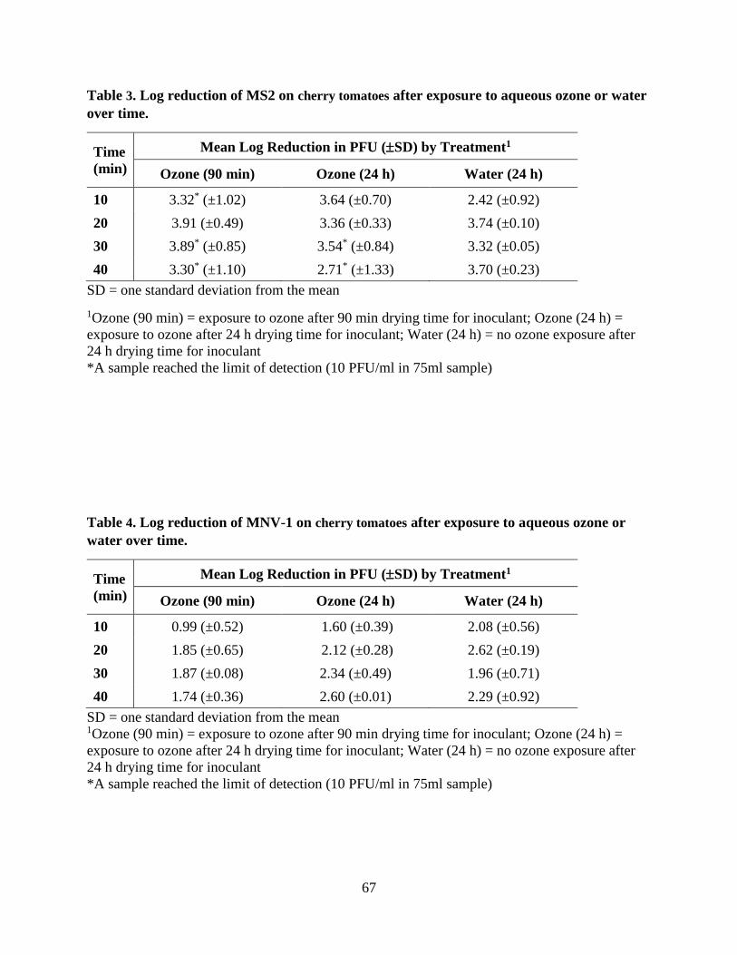
67
Table 3. Log reduction of MS2 on cherry tomatoes after exposure to aqueous ozone or water
over time.
Time
(min)
Mean Log Reduction in PFU (SD) by Treatment1
Ozone (90 min) Ozone (24 h) Water (24 h)
10 3.32* (±1.02) 3.64 (±0.70) 2.42 (±0.92)
20 3.91 (±0.49) 3.36 (±0.33) 3.74 (±0.10)
30 3.89* (±0.85) 3.54* (±0.84) 3.32 (±0.05)
40 3.30* (±1.10) 2.71* (±1.33) 3.70 (±0.23)
SD = one standard deviation from the mean
1Ozone (90 min) = exposure to ozone after 90 min drying time for inoculant; Ozone (24 h) =
exposure to ozone after 24 h drying time for inoculant; Water (24 h) = no ozone exposure after
24 h drying time for inoculant
*A sample reached the limit of detection (10 PFU/ml in 75ml sample)
Table 4. Log reduction of MNV-1 on cherry tomatoes after exposure to aqueous ozone or
water over time.
Time
(min)
Mean Log Reduction in PFU (SD) by Treatment1
Ozone (90 min) Ozone (24 h) Water (24 h)
10 0.99 (±0.52) 1.60 (±0.39) 2.08 (±0.56)
20 1.85 (±0.65) 2.12 (±0.28) 2.62 (±0.19)
30 1.87 (±0.08) 2.34 (±0.49) 1.96 (±0.71)
40 1.74 (±0.36) 2.60 (±0.01) 2.29 (±0.92)
SD = one standard deviation from the mean 1Ozone (90 min) = exposure to ozone after 90 min drying time for inoculant; Ozone (24 h) =
exposure to ozone after 24 h drying time for inoculant; Water (24 h) = no ozone exposure after
24 h drying time for inoculant
*A sample reached the limit of detection (10 PFU/ml in 75ml sample)

68
Table 5. Log reduction of Enterobacter cloacae and Bacillus cereus on Boston bibb lettuce after
exposure to aqueous ozone or water over time.
Time
(min)
Mean Log Reduction in CFU (SD) by Treatment
E. cloacae B. cereus
Ozone Water Ozone Water
10 1.81 (±0.05) 2.80 (±0.35) 1.61 (±0.32) 2.13 (±0.42)
20 2.13 (±1.57) 1.67 (±0.02) 2.45 (±0.06) 2.11 (±0.01)
30 2.41 (±0.60) 1.48 (±0.09) 2.13 (±0.32) 2.15 (±0.28)
40 3.33* (±0.95) 2.78* (±1.73) 2.83* (±1.08) 1.90 (±0.44)
SD = one standard deviation from the mean *= A sample reached the limit of detection (10 CFU/ml in 75ml sample)
Table 6. Log reduction of Enterobacter cloacae and Bacillus cereus on cherry tomatoes after
exposure to aqueous ozone or water over time.
Time
(min)
Mean Log Reduction in CFU (SD) by Treatment
E. cloacae B. cereus
Ozone Water Ozone Water
10 1.58 (±0.33) 1.39 (±0.53) 1.62 (±0.47) 0.36 (±0.23)
20 1.44 (±0.66) 1.35 (±0.14) 2.06 (±0.22) 1.51 (±0.10)
30 2.46 (±0.14) 2.01 (±0.76) 2.71 (±1.07) 1.59 (±0.86)
40 2.58 (±1.11) 2.46 (±0.21) 2.82* (±1.38) 1.90 (±0.21)
SD = one standard deviation from the mean *A sample reached the limit of detection (10 CFU/ml in 75ml sample)

69
Table 7. Mean ozone concentration (ppm) for ozone treatments over time.
Time
(min)
Mean Ozone Concentration in ppm (SD) by Treatment1
Boston Bibb-Viruses Cherry Tomatoes-Viruses Boston
Bibb-
Bacteria
Cherry
Tomatoes-
Bacteria 90 min 24 h 90 min 24 h
0 0.51 (±0.02) 0.65 (±0.15) 0.51 (±0.04) 0.48 (±0.05) 0.54 (±0.06) 0.51 (±0.01)
10 0.78 (±0.01) 0.69 (±0.05) 0.78 (±0.06) 0.76 (±0.03) 0.73 (±0.01) 0.72 (±0.02)
20 0.91 (±0.02) 0.74 (±0.01) 0.90 (±0.08) 0.89 (±0.03) 0.84 (±0.01) 0.85 (±0.00)
30 0.94 (±0.04) 0.74 (±0.5) 0.96 (±0.11) 0.96 (±0.06) 0.92 (±0.01) 0.93 (±0.03)
40 0.96 (±0.02) 0.69 (±0.12) 0.98 (±0.10) 0.99 (±0.07) 0.94 (±0.00) 0.97 (±0.28)
SD = one standard deviation from the mean

70
Chapter 4: Aqueous ozone inactivation of viruses on stainless steel surfaces

71
Abstract
Norovirus is a common foodborne illness, most often related to an infected food handler
transferring the virus directly to food or indirectly to food contact surfaces. Implementing risk
management strategies, such as aqueous ozone, to reduce viral contamination is important for the
retail industry. Stainless steel coupons (10 cm2) were spot inoculated with 100 µl of both MNV-1
and MS2 at 106 and 108 PFU/ml, respectively. The viruses were allowed to dry and attach to the
coupons for 90 min and then the coupons were placed in a batch wash ozone sanitation system
(BWOSS) with either an ozone wash or a water only wash. The water was iced to achieve a
temperature between 3-5°C with an ozone concentration of 0.5 ppm to 1 ppm. The coupons were
exposed to either treatment for times of 0.5, 3, and 10 min. The coupons were removed from the
sink and flooded with buffered phosphate water in a sterile petri dish in order to recover the
viruses. The eluate was serially diluted and processed by double agar layer method and plaque
assay for MS2 and MNV-1, respectively, to determine PFU/ml. The log reduction difference
between ozone and water for MNV-1 and MS2 after 10 min was 0.25 and 0.51 PFU/ml. Overall,
there was no significant difference between an ozone wash and a water only wash for the
inactivation of MNV-1and MS2 on stainless steel surfaces.

72
1. Introduction
In Chapter 3, ozone was investigated for its inactivation of viruses and bacteria on fresh
produce, and ozone was not found to significantly contribute to the inactivation of viruses. Based
on those results, it was hypothesized that the food matrix may contain structures and other
compounds that aid in viral attachment and prevent ozone inactivation. For instance, Gao et al.
(2016) showed that virus like particles (VLPs) of human norovirus GII.4 were able to bind to
surface carbohydrates in the cell wall of lettuce.
Viral inactivation on food matrices has been shown to be variable. Hirneisen et al., (2010)
indicates that the efficacy of ozone on food matrices is greatly affected by variables such as the
surface topography of the food, the type of microorganism contaminating the food, and the
strength of association of the microbe to the produce. There are only a few studies that have
researched viral inactivation on produce by ozone. Hirneisen et al. (2011), researched the
inactivation of murine norovirus (MNV) and feline calcivirus (FCV) on green onions and lettuce
by ozone. The researchers did not always have viral inactivation over time there were a few
cases of variability recorded. However, the researchers recorded at least a 2 log reduction of
FCV on both produce and over a 3 log reduction of MNV. They concluded that the main
contributor to variability was due to produce type. The authors believe that the difference in
organic composition of the produce could be the reason for the variability. Similarly, Chapter 3
had variability in inactivation of MNV and MS2 bacteriophage on both Boston bibb lettuce and
cherry tomatoes.
Due to the variability in virus inactivation observed in Chapter 3, the use of an abiotic
surface such as stainless steel for the evaluation of ozone efficacy against viruses was considered
here in order to eliminate the impact of the specific interactions occurring on biotic surfaces.

73
2. Materials and Methods
a. Microbe cultivation
i. MS2 Bacteriophage
MS2 stock was generated, as described previously by Gibson, Crandall, and Ricke (2012),
and aliquoted in small volumes. MS2 was kindly provided by Dr. Stephanie Friedman from the
United States Environmental Protection Agency (EPA) Gulf Ecology Division in Gulf Breeze,
FL. Stock was generated by scraping the soft agar layer containing bacteriophage lysed E. coli
C3000 (ATCC 15597) into centrifuge tubes. The cell lysate was then suspended with 23 ml of 1
× phosphate buffered saline (PBS), vortexed, and centrifuged at 185 × g for 25 min. The
supernatant was collected and filtered through a 0.22 µm filter (Millipore Corporation, Billerica,
MA) and then aliquoted and stored at -80°C. The MS2 stock concentration was determined based
on titration by double agar layer (DAL) method with bacterial host E. coli C3000. DAL assay
involves adding 100 μl of sample plus 100 μl of log phase bacterial host (E. coli C3000) to 5 ml
of soft agar (0.7% tryptic soy agar (TSA)) and then poured on the top of TSA plates. The soft
agar was allowed to solidify and then the plates were incubated for 24 h at 37°C. The plaques
were counted and PFU/ml was then calculated.
ii. Murine Norovirus (MNV-1)
Murine norovirus type 1 (MNV-1) was prepared as described previously by Bae and Schwab
(2008) with modifications. MNV-1 was kindly provided by Dr. Kellogg Schwab at Johns
Hopkins Bloomberg School of Public Health in Baltimore, MD. MNV-1 was propagated in
monolayers of RAW 264.7 (mouse leukameic monocyte macrophage, ATCC TIB-71) cells.
Cells were cultured in Dulbecco modified Eagle’s medium (DMEM) (Sigma-Aldrich, St. Louis,
MO) containing less than 10% low endotoxin, fetal bovine serum (FBS: GibcoLife Technology,

74
Gaithersburg, MD), 1% 100 × penicillin-streptomycin solution (GibcoLife Technology), 1%
HEPES (Sigma-Aldrich), 1% glutamine (Hyclone, Logan, UT), and 1% non-essential amino
acids (Corning, New York, NY). After reaching 90% confluence, cells were infected with MNV-
1 at a multiplicity of infection (MOI) of 0.05 for virus stock production. The virus was extracted
from cell lysate after complete cytopathic effect as described by Hsueh and Gibson (2015) with
modifications. Briefly, culture flasks containing infected cells were subjected to three freeze-
thaw cycles at -80°C and 37°C respectively, followed by centrifugation at 5000 × g for 20 min at
4°C then filtration with 0.1 μm filter and stored at -80°C. To determine infectious titer, the
plaque assay as reported by Hsueh and Gibson (2015) was used with modifications. Briefly, six-
well plates were seeded with 2x10⁶ RAW cells per well and grown to 90% confluence in 2 ml of
complete growth medium. Cell monolayers were inoculated with virus stocks for 1 h at 37°C
with rocking followed by removal of the inocula. Cells were covered with 2 ml of prepared
overlay medium containing: 25% of 6% low melting point agarose, 50% 2 × minimum essential
medium eagle (MEM) (Corning) (100 ml 10× MEM, 10 ml glutamine, 29.3 ml sodium
bicarbonate, and 360.7 ml sterile distilled water filtered through 0.22 µm filter (Millipore
Corporation, Billerica, MA)), 10% low endotoxin FBS, 1% 100 × penicillin-streptomycin
solution, 1% glutamine, 0.5% HEPES, and 12.5% sterile distilled water. The plates were
incubated for 72 h. Next, 2 ml of 0.01% neutral red (Sigma-Aldrich) prepared in 1 × phosphate
buffered saline (PBS) was added to each well to visualize plaques. Plaques were counted after 1h
to determine virus titer (PFU/ml).
b. Stainless Steel
Stainless steel sheets were (type 304/16 gauge, unpolished; Rose Metal Products, Springfield,
Missouri, USA) cut into 10 cm2 coupons. The stainless steel was first autoclaved then 100µl of

75
MS2 at 108 PFU/ml and 100µl of MNV at 106 PFU/ml were spotted on the surface of each
coupon in 10 µl drops and allowed to dry for 90 min in a biological safety cabinet.
c. Treatments
The stainless steel coupons were placed in a batch wash ozone sanitation system (BWOSS).
The BWOSS utilized in the present study was developed by Recycled Hydro Solutions (Rogers,
AR). The BWOSS contains a one compartment sink measuring 43 cm2 with a depth of 30 cm
and fabricated using 16-gauge stainless steel. During operation, the sink is filled with water, and
once filled, water from the sink is passed through a Venturi injector, which creates aqueous
ozone continuously. The ozonated water is then passed back into the sink basin. The BWOSS
has a dissolved ozone meter (Model Q46, ATI, Collegeview, PA) which was corroborated by the
indigo trisulfonate method (SM 4500-OS3 B) using a Hach Pocket Colorimeter II (Hach
Company, Loveland, CO) and Ozone AccuVac Ampules (Hach) (American Public Health
Association, American Water Works Association, and Water and Environment Federation,
2012).
The sink holds approximately 34.07 L (9 gallons) of water. Ice was added to the sink to aid in
stabilization of ozone until the water temperature reached 4°C. The coupons were placed in the
BWOSS once it reached 0.5 ppm as indicated by the dissolved ozone meter and the water
temperature in the sink was stable at 4°C. The coupons were in the sink for a total of 10 minutes
with two samples (n=6) taken at 30 s, 3 min, and 10 min. A water only wash was also conducted
in the same sink as a control. Experiments were conducted in duplicate.

76
d. Microbial analysis
The coupons were placed in sterile Petri dishes and flooded with 2 ml of buffered phosphate
water (BPW), and the coupons were then scraped with a cell scraper. The eluate was serially
diluted and analyzed.
For MS2, 100 μl of each dilution was plated in duplicate using DAL method. Then the
plaque forming units (PFU) were counted, and PFU/ml was calculated. For MNV-1, plaque
assay was performed as previously described (Section MNV-1), and PFU were counted in order
to calculate PFU/ml.
e. Statistical analysis
Statistical analyses were performed using JMP Pro 13 software (SAS institute, Inc., Cary,
NC). Significance was determined by a critical p-value 0.05 by one-way analysis of variance
(ANOVA) for all scenarios. Based on the results, a Tukey-Kramer honestly significant difference
(HSD) test was conducted to confirm differences between groups.
4. Results
A baseline recovery was used to calculate the amount of the viruses that can be recovered
from stainless steel without any treatment. The average baseline recovery for MNV-1 was
6.4×10⁴ PFU/ml and for MS2 1.2×10⁷ PFU/ml. The treatment results are the difference between
the baseline values and the experimental sample values. The average ozone concentration for 0,
0.5, 3, and 10 min are as follows: 0.52, 0.56, 0.66, 0.80 ppm.
Both treatments applied to stainless steel coupons resulted in log reductions over time (Table
1). An observed increase in log reduction of 0.83 to 3.53 PFU/ml over time with ozone was seen
for MNV-1. Similarly, with a water only wash, the log reduction was 0.57 to 3.28 PFU/ml over
time for MNV-1. There was a visible difference between the ozone wash and water only wash

77
for MNV-1, though there was not a significant difference. An increase in log reduction from 0.56
to 4.56 PFU/ml over time with ozone was also observed for MS2. Again, the water wash also
showed an increase in log reduction over time of 0.34 to 4.05 PFU/ml for MS2. Similar to MNV-
1, MS2 did not have a significant difference between the treatments.
5. Discussion
Even though there is not a significant difference between ozone and water wash, it was
hypothesized that log reductions would increase over time as viruses are inactivated and/or
physically removed. This was seen with the stainless steel results and not in the produce results
(See Chapter 3). This is related to the limitations in recovery. Produce was washed in a larger
volume (75 ml) than the stainless steel (2 ml) which dictates the limit of detection. Recovery
from produce is also more difficult because of the surface topography hindering the removal of
the virus in comparison to the smooth surface of the stainless steel (K A Hirneisen et al., 2011).
Viruses have also been known to form interactions with the surface of lettuce due to surface
glycans (Gao et al., 2016). However, there are no known studies that have looked at the
inactivation of viruses by ozone and compared biotic and abiotic surfaces.
Previous research has reported similar log reductions on abiotic surfaces as are reported here.
A study showed that a 6-log reduction in the bacteriophage, PRD1, was achieved on the surface
of a stainless steel ice cream dipper in an aqueous ozone dipper well (Almeida and Gibson,
2016). The results in the current study are not near a 6-log reduction, but there is still an overall
log reduction of 3.5 and 4.5 for MNV-1 and MS2, respectively, after a 10 min exposure. There
are differences between the current study and the study by Almeida and Gibson (2016). The first
notable difference is the use of a different bacteriophage. PRD1 is a double stranded DNA
bacteriophage while MS2 is a single stranded RNA bacteriophage (Anders and Chrysikopoulos,

78
2006). DNA and RNA phage are reported to behave differently. When exposed to sanitizing
practices such as peracetic acid and UV irradiation, DNA phage are more sensitive to
inactivation than RNA phage (Rajala-Mustonen et al., 1997). Peracetic acid and ozone are both
strong oxidizers which could indicate that similar tendencies for phages could be seen with
ozone; conversely, UV irradiation damages the genetic material which could have been the
reason Rajala-Mustonen et al. (1997) saw that DNA phage were more sensitive than RNA phage.
Besides the bacteriophage used, the method of inoculation between the two studies was different.
Almeida and Gibson (2016) dipped the ice cream dipper in the inoculum whereas the current
study spot inoculated. Moreover, the ice cream dipper was then immediately submerged in the
ozone wash as opposed to allowing for a drying period. The dip inoculation method is limited
due to the application of an unknown microbial concentration thus leading to consistency issues
between inoculation events; meanwhile, a spot inoculation of a known concentration eliminates
this issue (Beuchat et al., 2001). Last, the ice cream dipper was then swabbed for the presence of
microorganisms whereas the stainless steel coupons used in the present study were flooded in a
buffer solution. This difference in recovery methods affects the limit of detection since the
eluate volumes also differe. The differences discussed above could certainly account for the
opposing results.
6. Conclusion
By exploring the effect of aqueous ozone on viruses on an abiotic surface, we were able to
show ozone efficacy without confounding variables that are present on biotic surfaces such as on
produce. By eliminating confounding variables, the variation in the data was reduced in
comparison to the produce results in Chapter 3. Surface results show an increase in log reduction
over time which contrasts what was shown on produce in Chapter 3. Even though there was not a

79
significant difference between the ozone and water washes on the surface of the stainless steel,
there was a visible difference which indicates that the food matrix plays a critical role in a virus’s
ability to evade inactivation via ozone.

80
References:
Almeida, G., & Gibson, K. E. (2016). Evaluation of a Recirculating Dipper Well Combined with
Ozone Sanitizer for Control of Foodborne Pathogens in Food Service Operations. Journal
of Food Protection, 79(9), 1537–1548. https://doi.org/10.4315/0362-028X.JFP-16-055
American Public Health Association, American Water Works Association, & Water and
Environment Federation. (2012). Standard methods for the examination of water and
wastewater.
Bae, J., & Schwab, K. J. (2008). Evaluation of murine norovirus, feline calicivirus, poliovirus,
and MS2 as surrogates for human norovirus in a model of viral persistence in surface
water and groundwater. Applied and Environmental Microbiology, 74(2), 477–84.
https://doi.org/10.1128/AEM.02095-06
Bae, Y.-M., Baek, S.-Y., & Lee, S.-Y. (2012). Resistance of pathogenic bacteria on the surface
of stainless steel depending on attachment form and efficacy of chemical sanitizers.
International Journal of Food Microbiology, 153(3), 465–473
Beuchat, L. R., Farber, J. M., Garrett, E. H., Harris, L. J., Parish, M. E., Suslow, T. V., & Busta,
F. F. (2001). Standardization of a Method To Determine the Efficacy of Sanitizers in
Inactivating Human Pathogenic Microorganisms on Raw Fruits and Vegetables. Journal
of Food Protection, 64(7), 1079–1084. https://doi.org/10.4315/0362-028X-64.7.1079
Fallahi, S., & Mattison, K. (2011). Evaluation of Murine Norovirus Persistence in Environments
Relevant to Food Production and Processing. Journal of Food Protection, 74(11), 1847–
1851. https://doi.org/10.4315/0362-028X.JFP-11-081
Gao, X., Esseili, M. A., Lu, Z., Saif, L. J., & Wang, Q. (2016). Recognition of Histo-Blood
Group Antigen-Like Carbohydrates in Lettuce by Human GII.4 Norovirus. Applied and
Environmental Microbiology, 82(10), 2966–2974. https://doi.org/10.1128/AEM.04096-
15
Gibson, K. E., Crandall, P. G., & Ricke, S. C. (2012). Removal and Transfer of Viruses on Food
Contact Surfaces by Cleaning Cloths. Applied and Environmental Microbiology, 78(9),
3037–3044. https://doi.org/10.1128/AEM.00027-12
Girard, M., Ngazoa, S., Mattison, K., & Jean, J. (2010). Attachment of Noroviruses to Stainless
Steel and Their Inactivation, Using Household Disinfectants. Journal of Food Protection,
73(2), 400–404. https://doi.org/10.4315/0362-028X-73.2.400
Hirneisen, K. A., Black, E. P., Cascarino, J. L., Fino, V. R., Hoover, D. G., & Kniel, K. E.
(2010). Viral Inactivation in Foods: A Review of Traditional and Novel Food-Processing
Technologies. Comprehensive Reviews in Food Science and Food Safety, 9(1), 3–20.
https://doi.org/10.1111/j.1541-4337.2009.00092.x
81
Hirneisen, K. A., Markland, S. M., & Kniel, K. E. (2011). Ozone Inactivation of Norovirus
Surrogates on Fresh Produce. Journal of Food Protection, 74(5), 836–839.
https://doi.org/10.4315/0362-028X.JFP-10-438
Hsueh, T.-Y., & Gibson, K. E. (2015). Interactions between Human Norovirus Surrogates and
Acanthamoeba spp. Applied and Environmental Microbiology, 81(12), 4005–13.
https://doi.org/10.1128/AEM.00649-15
Kusumaningrum, H., Riboldi, G., Hazeleger, W., & Beumer, R. (2003). Survival of foodborne
pathogens on stainless steel surfaces and cross-contamination to foods. International
Journal of Food Microbiology, 85(3), 227–236. https://doi.org/10.1016/S0168-
1605(02)00540-8
Mattison, K., Karthikeyan, K., Abebe, M., Malik, N., Sattar, S. A., Farber, J. M., & Bidawid, S.
(2007). Survival of Calicivirus in Foods and on Surfaces: Experiments with Feline
Calicivirus as a Surrogate for Norovirus. Journal of Food Protection, 70(2), 500–503.
https://doi.org/10.4315/0362-028X-70.2.500
Moe, C. L. (2009). Preventing Norovirus Transmission: How Should We Handle Food Handlers?
Clinical Infectious Diseases, 48(1), 38–40. https://doi.org/10.1086/594119
Rajala-Mustonen, R. L., Toivola, P. S., & Heinonen-Tanski, H. (1997). Effects of peracetic acid
and UV irradiation on the inactivation of coliphages in wastewater. Water Science and
Technology, 35(11–12), 237–241. https://doi.org/10.1016/S0273-1223(97)00265-5
Roy, D., Englebrecht, R. S., & Chian, E. S. K. (1982). Comparative inactivation of six
enterovirus by ozone. American Water Works Association, 74(12), 660–664.

82
Appendix
Table 1. Log reduction of MNV-1 and MS2 on stainless steel after exposure to aqueous
ozone or water over time.
Time
(min)
Mean Log Reduction in CFU (SD) by Treatment
MNV-1 MS2
Ozone Water Ozone Water
0.5 0.83 (±0.09) 0.57 (±0.01) 0.56(±0.14) 0.34 (±0.34)
3 2.39 (±0.50) 1.89 (±0.15) 1.82 (±1.40) 0.72 (±0.29)
10 3.53* (±0.03) 3.28* (±0.64) 4.56* (±0.99) 4.05 (±0.50)
SD = one standard deviation from the mean
*= A sample reached the limit of detection (10 CFU/ml in a 2ml sample)

83
Chapter 5: Aqueous ozone inactivation of viruses in association with bacteria on Boston
bibb lettuce

84
Abstract
Viruses do not exist in monocultures in the environment; they instead exist in microbial
communities. The extent to which these communities effect virus inactivation is not well known.
These communities may exist on the surface of fresh produce, and the interaction of human
enteric viruses with their microbial co-habitants is an under explored area of research. A key
question is how do these virus-bacteria interactions impact the efficacy of control strategies?
Therefore, aqueous ozone was applied as a sanitizing agent on Boston bibb lettuce to investigate
the impact of virus-bacteria interactions on the inactivation of viruses on fresh produce. Samples
of Boston bibb (25 g) were spot inoculated with 200 µl of a mixture of 100 µl of murine
norovirus (MNV-1; 106 PFU/ml) and 100 µl of either Bacillus cereus or Enterobacter cloacae
(106 CFU/ml) that had been allowed to incubate for 1 h at room temperature prior to inoculation.
The mixture was allowed to dry and attach to the lettuce (90 min) and then the lettuce was placed
in the batch wash ozone sanitation system (BWOSS) with either an ozone wash or a water only
wash. The water was iced to achieve a temperature between 3-5°C and the ozone concentration
was 0.5 ppm to 1 ppm. The lettuce was exposed to either treatment for 10, 20, 30, and 40 min.
The lettuce was removed from the sink and washed with an antibiotic solution. The eluate was
serially diluted and processed by plaque assay to determine the concentration of MNV-1 in
PFU/ml. The log reduction differences between ozone and water for MNV-1 only, MNV-1 with
B. cereus, and MNV-1 with E. cloacae after 40 min were 0, 0.95, and -0.36 PFU/ml,
respectively. Overall, there was no significant difference between virus alone and virus
associated with either bacterium. However, the results from MNV-1 with B. cereus indicate that
association with bacteria of different gram types (i.e. gram positive versus gram negative) could
impact viral inactivation by aqueous ozone.
85
1. Introduction
Throughout the supply chain there are several opportunities for microorganisms to interact
with microbial co-habitants on the surface of produce. These interactions could possibly create
stronger resistance to current microbial control strategies thus placing the consumer at an
increased risk for foodborne illness. Most studies focus on microbes in isolation; however, that is
not the case as these microbes exist in microbial communities and interact with one another.
Currently, there is very little research on how existing control strategies respond to these
interactions, especially viral interactions with bacteria. Enteric viruses, for example human
norovirus, are often of concern when it comes to food safety. Norovirus and leafy greens have
the highest number of illnesses when comparing pathogen-commodity pairs (Gould et al., 2013).
Produce can be contaminated via various routes before reaching the consumer: soil, water,
animal excrements, and mishandling by workers (Benson, 2010; Strohbehn et al, 2008). Also, the
produce itself has naturally occurring microbes on its surface (Whipps et al., 2008). Each of
these could contribute to microbes possibly interacting on the surface of produce. A recent
review by Deng and Gibson (2017), discuss more specifically the interactions that may occur
between human noroviruses and the microbial community of leafy greens. Interestingly, Almand
et al. (2017) reported on the specificity of interactions between noroviruses and bacteria stated
that these were specific to norovirus strains since other viruses tested did not readily interact with
bacteria. It has also been reported that noroviruses are able to bind to carbohydrate moieties
present on the surface of romaine lettuce (Esseili et al., 2015; Gao et al., 2016).
Of the limited research, it has been shown that when viruses interact with bacteria expressing
specific surface antigens, histo-blood group antigens (HBGA’s), viruses are more resistant to
disinfectant processes. For example, when heat is applied, human norovirus particles in

86
association with bacteria had a higher immunoreactivity than human norovirus particles alone (Li
et al. 2015). Conversely, Li et al. (2017) published results indicating that Tulane virus—a
cultivable human norovirus surrogate—was not protected from heat while interacting with
bacteria expressing HBGA-like molecules. These conflicting results raise several questions when
it comes to the interactions of viruses and bacteria and these may impact virus inactivation under
specific sanitation practices.
Murine norovirus and poliovirus (serotype 1, Mahoney) have been shown to interact with
gram positive and gram negative bacteria including Bacillus cereus, Enterococcus faecalis,
Escherichia coli, and Enterobacter cloacae (Jones et al., 2014; Kuss et al., 2011). Therefore, this
study aimed to evaluate the efficacy of aqueous ozone on the inactivation of viruses while in
association with bacteria often found on the phyllosphere of lettuce.
2. Materials and Methods
a. Microbe Cultivation
i. Virus
Murine norovirus type 1 (MNV-1) was prepared as described previously by Bae and Schwab
(2008) with modifications. MNV-1 was kindly provided by Dr. Kellogg Schwab at Johns
Hopkins Bloomberg School of Public Health in Baltimore, MD. MNV-1 was propagated in
monolayers of RAW 264.7 (mouse leukameic monocyte macrophage, ATCC TIB-71) cells.
Cells were cultured in Dulbecco modified Eagle’s medium (DMEM) (Sigma-Aldrich, St. Louis,
MO) containing less than 10% low endotoxin, fetal bovine serum (FBS: GibcoLife Technology,
Gaithersburg, MD), 1% 100 × penicillin-streptomycin solution (GibcoLife Technology), 1%
HEPES (Sigma-Aldrich), 1% glutamine (Hyclone, Logan, UT), and 1% non-essential amino
acids (Corning, New York, NY). After reaching 90% confluence, cells were infected with MNV-
87
1 at a multiplicity of infection (MOI) of 0.05 for virus stock production. The virus was extracted
from cell lysate after complete cytopathic effect as described by Hsueh and Gibson (2015) with
modifications. Briefly, culture flasks containing infected cells were subjected to three freeze-
thaw cycles at -80°C and 37°C respectively, followed by centrifugation at 5000 × g for 20 min at
4°C then filtration with 0.1 μm filter and stored at -80°C. To determine infectious titer, the
plaque assay as reported by Hsueh and Gibson (2015) was used with modifications. Briefly, six-
well plates were seeded with 2x10⁶ RAW cells per well and grown to 90% confluence in 2 ml of
complete growth medium. Cell monolayers were inoculated with virus stocks for 1 h at 37°C
with rocking followed by removal of the inocula. Cells were covered with 2 ml of prepared
overlay medium containing: 25% of 6% low melting point agarose, 50% 2 × minimum essential
medium eagle (MEM) (Corning) (100 ml 10× MEM, 10 ml glutamine, 29.3 ml sodium
bicarbonate, and 360.7 ml sterile distilled water filtered through 0.22 µm filter (Millipore
Corporation, Billerica, MA)), 10% low endotoxin FBS, 1% 100 × penicillin-streptomycin
solution, 1% glutamine, 0.5% HEPES, and 12.5% sterile distilled water. The plates were
incubated for 72 h. Next, 2 ml of 0.01% neutral red (Sigma-Aldrich) prepared in 1 × phosphate
buffered saline (PBS) was added to each well to visualize plaques. Plaques were counted after 1h
to determine virus titer (PFU/ml).
ii. Bacteria
Enterobacter cloacae (ATCC 39979) with rifampicin resistance was streaked from a frozen
50% glycerol stock onto Luria Bertani (LB) agar (Alfa Aesar, Tewksbury, MA) with 100 µg/ml
of rifampicin (Alfa Aesar) using a sterile inoculation loop and incubated overnight at 37°C. A
single CFU was selected from the plate, placed in 5 ml of LB broth with 100 µg/ml rifampicin in
a 50 ml centrifuge tube, and incubated overnight at 37°C with shaking at 150 rpm. Bacillus
88
cereus (ATCC 14579) was also streaked from a frozen, 50% glycerol stock onto a nutrient agar
(NA) plate (Becton, Dickson, and Company, Franklin Lakes, NJ) using a sterile inoculation loop
and incubated overnight at 30°C. A single CFU was selected from the plate, placed in 5 ml of
nutrient broth (NB) (Becton, Dickson, and Company) in a 50 ml centrifuge tube, and incubated
overnight at 30°C with shaking at 150 rpm. Following overnight growth of both bacteria, the
culture tubes were centrifuged at 5,000 × g for 10 min to pellet the bacterial cells. The
supernatant was decanted, and the pellet was resuspended in 5 ml of 1 × PBS, vortexed, and
centrifuged again. The bacterial pellet was resuspended in 5 ml of 1 × PBS and the concentration
of each bacterial culture was determined by spread plate enumeration of microorganisms. LB
agar with 100 µg/ml of rifampicin and B. cereus agar (Oxoid, Altrincham, Cheshire, England)
supplemented with selective supplement (Oxoid) and egg yolk emulsion (Dalynn, Calgary,
Canada) were used for E. cloacae and B. cereus, respectively.
b. Microbe-Microbe Interaction
Enterobacter cloacae and B. cereus were serially diluted to 106 CFU/ml in 1×PBS. For
interactions, 100 µl of bacteria was mixed with 100 µl of MNV (106 PFU/ml) at room
temperature in an Eppendorf tube and allowed to associate for 1 h. An abstract presented at the
International Association of Food Protection (IAFP) 2017 Annual Meeting in Tampa, FL,
confirms association of MNV and E. cloacae or B. cereus in the time specified (Almeida and
Gibson, 2017).
c. Produce
Boston bibb loose leaf lettuce was used. The Boston bibb was grown hydroponically by
Ozark All Seasons in Windsor, AR and purchased from a local grocer. The Boston bibb leaves
were removed and placed in a sterilized plastic container with a lid and stored at 4°C until use.

89
d. Inoculation of Produce
For inoculation, 25 g of Boston bibb leaves were measured out for each sampling time point
(n = 5) for a total of 125 g for each experiment. Each 25 g was then spot inoculated with the 200
µl of the mixture containing viruses and bacteria. Following inoculation, the produce was
allowed to dry for 90 min before being treated.
e. Treatments
The batch wash ozone sanitation system (BWOSS) utilized in the present study was
developed by Recycled Hydro Solutions (Rogers, AR). The BWOSS contains a one
compartment sink measuring 43 cm2 with a depth of 30 cm and fabricated using 16-gauge
stainless steel. During operation, the sink is filled with water, and once filled, water from the sink
is passed through a Venturi injector, which creates aqueous ozone continuously. The ozonated
water is then passed back into the sink basin. The BWOSS has a dissolved ozone meter (Model
Q46, ATI, Collegeview, PA) which was corroborated by the indigo trisulfonate method (SM
4500-OS3 B) using a Hach Pocket Colorimeter II (Hach Company, Loveland, CO) and Ozone
AccuVac Ampules (Hach) (American Public Health Association, American Water Works
Association, and Water and Environment Federation, 2012).
The sink holds approximately 34.07 L (9 gallons) of water. Ice was added to the sink to aid
in stabilization of ozone until the water temperature reached 4°C. The produce was placed in the
BWOSS once ozone reached a concentration of 0.5 ppm—as indicated by the dissolved ozone
meter—and the water temperature in the sink was stable at 4°C. The produce samples were
submersed in the sink for a total of 40 min with subsamples taken every ten minutes.

90
f. Microbial Analysis
Produce samples were placed in Whirl-pak bags with 75 ml of buffered phosphate water
(BPW) containing 40 µg/ml of penicillin/streptomycin. The bags were then placed in the
stomacher (Seward Stomacher 400 Circulator, West Sussex, United Kingdom) for 1 min at 260
rpm. A plaque assay was performed as previously described (Virus Section). PFU were counted
and then PFU/ml was calculated.
g. Statistical Analysis
Statistical analyses were performed using JMP Pro 13 software (SAS institute, Inc., Cary,
NC). Significance was determined by a critical p-value 0.05 by one-way analysis of variance
(ANOVA) for all scenarios. Based on the results, a Tukey-Kramer honestly significant difference
(HSD) test was conducted to confirm differences between groups.
3. Results
A baseline recovery was used to calculate the amount of virus that can be recovered from the
surface of Boston bibb. This baseline recovery was then used to calculate the log reduction for
all time points. In Table 1, the average ozone concentrations are recorded for the treatments. The
initial concentration of ozone is at least 0.5 ppm and reached a final concentration of at least 0.85
ppm for all treatments.
In Table 2, the results from the water wash treatments are displayed. Results from Chapter 3
are included for comparison. There was no significant difference between MNV only and any of
the virus-bacteria interactions. If log reduction at 40 min is examined separately, there is no
visible difference between MNV-1 only (1.63 PFU/ml) and MNV-1 with B. cereus (1.65
PFU/ml); however, MNV-1 with E. cloacae had a higher log reduction at this time point (2.20
PFU/ml).

91
The results from the ozone treatment are displayed in Table 3. There was no significant
difference between MNV-1 only and any of the virus-bacteria interactions with the ozone wash;
however, there is a visible difference. After 40 min, the log reduction with an ozone wash for
MNV-1, MNV-1 with B. cereus, and MNV-1 with E. cloacae was 1.63, 2.60, 1.83 PFU/ml,
respectively. The interaction of MNV-1 and B. cereus had consistently greater log reductions
over time (30 and 40 min) in comparison to the MNV-1 only log reductions.
In comparing the results from the interactions separately, MNV-1 with B. cereus, there was a
significant difference between the ozone and water treatments. This, however, was not the case
for MNV-1 with E. cloacae which had no significant difference between the treatments.
4. Discussion
This is the first study that has investigated the effects of aqueous ozone on the inactivation of
a norovirus surrogate while in combination with bacteria. As indicated previously, there have
been studies that have investigated how these interactions impact norovirus and norovirus
surrogate response to heat stress. Li et al. (2015) published that when human norovirus virus like
particles (VLP) were in association with HBGA-like expressing bacteria, the VLP’s had a higher
antigen integrity than in association with bacteria without the antigen. The researchers indicate
that this interaction could protect the virus from heat stress. Then Li et al. (2017) conducted a
similar study and found that when Tulane virus (TV) was bound to HBGA-like expressing
bacteria that this interaction did not protect it from heat stress. The conflicting results in these
two studies can be contributed to the different viruses used, human norovirus VLP’s versus TV.
The study that utilized VLP’s had the limitation of a binding assay which does not indicate
infectivity whereas the TV study could measure the virus infectivity to demonstrate if the virus

92
was denatured or not. The discrepancy between the studies testifies to how complex these
interactions may be.
The results in this study indicate there is not a significant difference in viral inactivation by
aqueous ozone while interacting with bacteria; however, there were visible differences noted.
When MNV-1 was associated with B. cereus and then exposed to the ozone wash, MNV-1
experienced a greater log reduction than when associated with E. cloacae. This could indicate
that, when viruses associate with gram positive bacteria, the virus is more susceptible to
inactivation due to ozone, or that when in association with gram negative bacteria, it neither aids
or enhances the inactivation. This could be due to the difference in the composition of the cell
membrane between gram positive and gram negative. Another possibility could be that the
interaction between MNV-1 and B. cereus has a weaker association than MNV-1 and E. cloacae
which could lead to the greater reduction that was recorded. Further research is needed in order
to further understand these interactions.
5. Conclusion
Viruses do not exist in a monoculture but rather in microbial communities. The interaction
between viruses and bacteria had no significant impact on viral inactivation when compared to
the virus only results. There is a possibility that viruses in association with gram positive bacteria
may be more susceptible to inactivation by ozone, but this is only speculation and further
investigation is needed.

93
References:
Almand, E. A., Moore, M. D., Outlaw, J., Jaykus, L.-A., Hargitt, E., Alvarez, X., & Cameron, D.
(2017). Human norovirus binding to select bacteria representative of the human gut
microbiota. PLOS ONE, 12(3), e0173124. https://doi.org/10.1371/journal.pone.0173124
Almeida, G., & Gibson, K. (2017). Rapid Association of Enteric Viruses with Whole Cell
Bacteria in Suspension. International Association of Food Protection. Poster presentation
at the Annual Meeting for International Association of Food Protection. Tampa, FL.
Bae, J., & Schwab, K. J. (2008). Evaluation of murine norovirus, feline calicivirus, poliovirus,
and MS2 as surrogates for human norovirus in a model of viral persistence in surface
water and groundwater. Applied and Environmental Microbiology, 74(2), 477–84.
https://doi.org/10.1128/AEM.02095-06
Benson, S. M. (2010). Guidance for Improving the Federal Response to Foodborne Illness
Outbreaks Associated with Fresh Produce. Food & Drug Law Journal, 65(3), 503–524.
Esseili, M. A., Saif, L. J., Farkas, T., & Wang, Q. (2015). Feline Calicivirus, Murine Norovirus,
Porcine Sapovirus, and Tulane Virus Survival on Postharvest Lettuce. Applied and
Environmental Microbiology, 81(15), 5085–92. https://doi.org/10.1128/AEM.00558-15
Gao, X., Esseili, M. A., Lu, Z., Saif, L. J., & Wang, Q. (2016). Recognition of Histo-Blood
Group Antigen-Like Carbohydrates in Lettuce by Human GII.4 Norovirus. Applied and
Environmental Microbiology, 82(10), 2966–74. https://doi.org/10.1128/AEM.04096-15
Gould, H. L., Walsh, K. A., Vieira, A. R., Williams, I. T., Hall, A. J., & Cole, D. (2013).
Surveillance for Foodborne Disease Outbreaks — United States, 1998–2008.
Hsueh, T.-Y., & Gibson, K. E. (2015). Interactions between Human Norovirus Surrogates and
Acanthamoeba spp. Applied and Environmental Microbiology, 81(12), 4005–13.
https://doi.org/10.1128/AEM.00649-15
Jones, M. K., Watanabe, M., Zhu, S., Graves, C. L., Keyes, L. R., Grau, K. R., … Karst, S. M.
(2014). Enteric bacteria promote human and mouse norovirus infection of B cells.
Science (New York, N.Y.), 346(6210), 755–9. https://doi.org/10.1126/science.1257147
Kuss, S. K., Best, G. T., Etheredge, C. A., Pruijssers, A. J., Frierson, J. M., Hooper, L. V., …
Pfeiffer, J. K. (2011). Intestinal Microbiota Promote Enteric Virus Replication and
Systemic Pathogenesis. Science, 334(6053), 249–252.
https://doi.org/10.1126/science.1211057
Li, D., Breiman, A., le Pendu, J., & Uyttendaele, M. (2015). Binding to histo-blood group
antigen-expressing bacteria protects human norovirus from acute heat stress. Frontiers in
Microbiology, 6, 659. https://doi.org/10.3389/fmicb.2015.00659

94
Li, Q., Wang, D., Yang, D., Shan, L., & Tian, P. (2017). Binding of Escherichia coli Does Not
Protect Tulane Virus from Heat-Inactivation Regardless the Expression of HBGA-Like
Molecules. Frontiers in Microbiology, 8. https://doi.org/10.3389/fmicb.2017.01746
Strohbehn, C., Sneed, J., Paez, P., & Meyer, J. (2008). Hand Washing Frequencies and
Procedures Used in Retail Food Services. Journal of Food Protection, 71(8), 1641–1650.
Whipps, J. M., Hand, P., Pink, D., & Bending, G. D. (2008). Phyllosphere microbiology with
special reference to diversity and plant genotype. Journal of Applied Microbiology,
105(6), 1744–1755. https://doi.org/10.1111/j.1365-2672.2008.03906.x
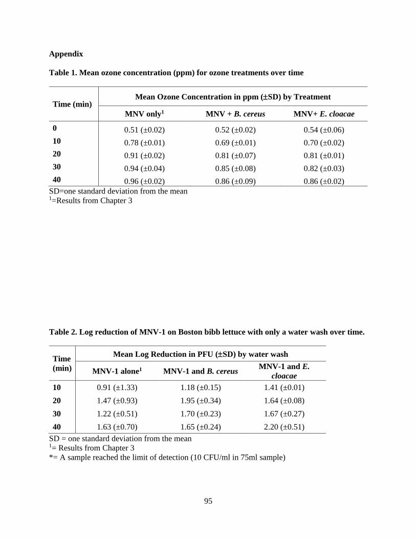
95
Appendix
Table 1. Mean ozone concentration (ppm) for ozone treatments over time
Time (min) Mean Ozone Concentration in ppm (SD) by Treatment
MNV only1 MNV + B. cereus MNV+ E. cloacae
0 0.51 (±0.02) 0.52 (±0.02) 0.54 (±0.06)
10 0.78 (±0.01) 0.69 (±0.01) 0.70 (±0.02)
20 0.91 (±0.02) 0.81 (±0.07) 0.81 (±0.01)
30 0.94 (±0.04) 0.85 (±0.08) 0.82 (±0.03)
40 0.96 (±0.02) 0.86 (±0.09) 0.86 (±0.02)
SD=one standard deviation from the mean 1=Results from Chapter 3
Table 2. Log reduction of MNV-1 on Boston bibb lettuce with only a water wash over time.
Time
(min)
Mean Log Reduction in PFU (SD) by water wash
MNV-1 alone1 MNV-1 and B. cereus MNV-1 and E.
cloacae
10 0.91 (±1.33) 1.18 (±0.15) 1.41 (±0.01)
20 1.47 (±0.93) 1.95 (±0.34) 1.64 (±0.08)
30 1.22 (±0.51) 1.70 (±0.23) 1.67 (±0.27)
40 1.63 (±0.70) 1.65 (±0.24) 2.20 (±0.51)
SD = one standard deviation from the mean 1= Results from Chapter 3
*= A sample reached the limit of detection (10 CFU/ml in 75ml sample)

96
Table 3. Log reduction of MNV-1 on Boston bibb lettuce by an aqueous ozone wash over
time.
Time
(min)
Mean Log Reduction in PFU (SD) by ozone
MNV-1 alone1 MNV-1 and B. cereus MNV-1 and E.
cloacae
10 2.54* (±1.16) 2.04 (±0.07) 1.42 (±0.16)
20 2.51* (±1.21) 2.38 (±0.07) 2.04 (±0.75)
30 1.69 (±0.53) 2.77* (±0.04) 2.21 (±0.51)
40 1.63 (±0.17) 2.60* (±0.13) 1.84 (±0.25)
SD = one standard deviation from the mean 1= Results from Chapter 3
*= A sample reached the limit of detection (10 CFU/ml in 75ml sample)

97
Chapter 6: Overall Conclusion

98
Contaminated produce is responsible for a large portion of foodborne illnesses (Painter et al.,
2013). Produce is susceptible to contamination because it is a ready to eat product that is
consumed raw, which eliminates common sanitization practices like pasteurization and cooking
(Benson, 2010). The U.S. Food and Drug Administration (FDA) recommends several practices
such as separation of products (i.e. meat and produce), washing of hands and equipment, and
washing of produce to prevent further contamination in the retail setting (U.S. Department of
Health and Human Services, 2009).
Produce washes often contain sanitizing agents such as chlorine, ozone, and organic acids
(Berger et al., 2010). The one of interest for this research is ozone, specifically aqueous ozone.
Ozone has been demonstrated to be effective against both bacteria and viruses in suspension
(Khadre and Yousef, 2001; Roy et al., 1982). There is research that shows the efficacy of ozone
on bacteria on a food matrix (Bialka and Demirci, 2007; Koseki and Isobe, 2006; Selma et al.,
2007). There is very little research on the efficacy of ozone against viruses on a food matrix with
the only published article being Hirneisen and Kniel (2013) which investigated the inactivation
of viruses by bubbling ozone on onions. There is no known published research on ozone
inactivation of viruses in complex scenarios including microbe-microbe interactions.
The primary objectives of my research were to evaluate the efficacy of aqueous ozone 1) on
the inactivation of viruses and bacteria on produce; 2) on the inactivation of viruses on stainless
steel; and 3) against viruses in association with bacteria on produce surfaces.
To begin with, two types of produce, Boston bibb lettuce and cherry tomatoes, were
inoculated with either a cocktail of viruses (murine norovirus (MNV) or MS2 bacteriophage)
with two attachment times (24 h and 90 min) or bacteria (Enterobacter cloacae and Bacillus
cereus). The produce was then washed in the batch wash ozone sanitation system (BWOSS) with
99
an ozone wash or a water only wash for 40 min with samples taken every 10 min. Log reductions
were then calculated for each sample. It was determined there was not a significant difference in
ozone efficacy between the two different attachment times for viruses. Ozone also did not have a
significant impact on the reduction of viruses or bacteria in comparison to ozone. It is important
to note there was a lot of variability among the viral samples which was not seen in the bacterial
samples.
Due to the variability in viral data, virus inactivation on stainless steel was conducted to
eliminate any added variability due to the food matrix. Stainless steel coupons were spot
inoculated with a viral cocktail of MNV and MS2 bacteriophage and allowed to dry for 90 min
prior to being placed into the BWOSS for either an ozone wash or a water only wash for a total
of 10 min with samples taken at 0.5, 3, and 10 min. Log reductions were then calculated for each
sample. The variability was reduced and log reductions increased over time; however, there was
not a significant difference between the ozone and water washes. Due to these results it is
hypothesized that there is a possible interaction that viruses have with the surface of produce that
aids the virus from deactivation by ozone.
To evaluate inactivation of viruses in association with bacteria, Boston bibb was spot
inoculated with a combination of MNV and E. cloacae or MNV and B. cereus. The virus and
bacteria were allowed to associate for an hour prior to inoculating the produce. Again, the
produce was placed in the BWOSS with either an ozone wash or a water only wash for 40 min
with samples taken every 10 min. Viral log reductions were then calculated for each sample.
There was no significant difference between the ozone and water wash for MNV with E.
cloacae; however, there was a significant difference for MNV with B. cereus. This could
indicate that if MNV is in association with B. cereus that the virus is more susceptible to
100
inactivation. When comparing both associations with the viral only wash results, there was no
significant difference between ozone and water washes with any of the variables.
Overall, it can be hypothesized that there are interactions that occur between viruses and the
surface of produce that are preventing complete viral inactivation. Further research is needed to
fully understand the impact of a food matrix on the inactivation of viruses alone and in
association with bacteria.
101
References:
Benson, S. M. (2010). Guidance for Improving the Federal Response to Foodborne Illness
Outbreaks Associated with Fresh Produce. Food & Drug Law Journal, 65(3), 503–524.
Berger, C. N., Sodha, S. V., Shaw, R. K., Griffin, P. M., Pink, D., Hand, P., & Frankel, G.
(2010). Fresh fruit and vegetables as vehicles for the transmission of human pathogens.
Environmental Microbiology, 12(9), 2385–2397. https://doi.org/10.1111/j.1462-
2920.2010.02297.x
Bialka, K. L., & Demirci, A. (2007). Decontamination of Escherichia coli O157:H7 and
Salmonella enterica on Blueberries Using Ozone and Pulsed UV-Light. Journal of Food
Science, 72(9), M391–M396. https://doi.org/10.1111/j.1750-3841.2007.00517.x
Hirneisen, K. A., & Kniel, K. E. (2013). Inactivation of internalized and surface contaminated
enteric viruses in green onions. International Journal of Food Microbiology, 166(2), 201–
206. https://doi.org/10.1016/j.ijfoodmicro.2013.07.013
Khadre, M. A., & Yousef, A. E. (2001). Sporicidal action of ozone and hydrogen peroxide: a
comparative study. International Journal of Food Microbiology, 71, 131–138. Retrieved
from www.elsevier.comrlocaterijfoodmicro
Koseki, S., & Isobe, S. (2006). Effect of Ozonated Water Treatment on Microbial Control and on
Browning of Iceberg Lettuce (Lactuca sativa L.). Journal of Food Protection, 69(1), 154–
160. https://doi.org/10.4315/0362-028X-69.1.154
Painter, J. A., Hoekstra, R. M., Ayers, T., Tauxe, R. V., Braden, C. R., Angulo, F. J., & Griffin,
P. M. (2013). Attribution of Foodborne Illnesses, Hospitalizations, and Deaths to Food
Commodities by using Outbreak Data, United States, 1998–2008. Emerging Infectious
Diseases, 19(3), 407–415. https://doi.org/10.3201/eid1903.111866
Roy, D., Englebrecht, R. S., & Chian, E. S. K. (1982). Comparative inactivation of six
enterovirus by ozone. American Water Works Association, 74(12), 660–664.
Selma, M. V., Beltrán, D., Allende, A., Chacón-Vera, E., & Gil, M. I. (2007). Elimination by
ozone of Shigella sonnei in shredded lettuce and water. Food Microbiology, 24(5), 492–
499. https://doi.org/10.1016/j.fm.2006.09.005
U.S. Department of Health and Human Services. (2009). 2009 Recommendations of the United
States Public Health Service Food and Drug Administration.

102
Appendix
IBC Approval Letter

103
IBC Registration for Research Project

104

105

106

107

108

109

110

111

112

113

114