Embed Size (px)
Citation preview

1
Aqueous and Lens Physiology
Lisa Ostrin, OD, PhD, FAAORoom 2153
Aqueous and Lens Physiology
• Aqueous humor – secretion
– outflow
• Crystalline lens– Embryology
– Structure
– Function

2
Gross Anatomy
• Anterior chamber
• Posterior chamber
vs
• Anterior segment
• Posterior segment
Anterior Segment Anatomy
Anterior and posterior chambers
3
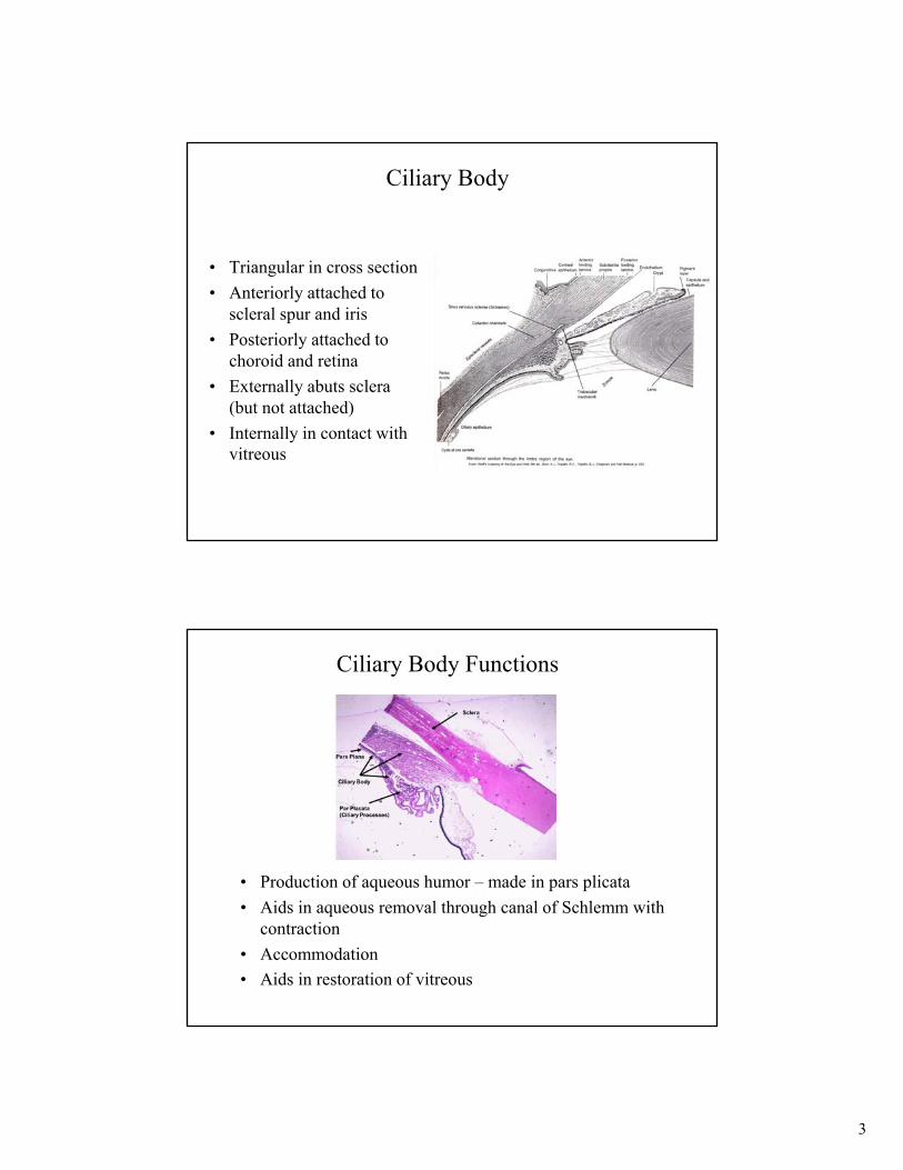
Ciliary Body
• Triangular in cross section
• Anteriorly attached to scleral spur and iris
• Posteriorly attached to choroid and retina
• Externally abuts sclera (but not attached)
• Internally in contact with vitreous
Ciliary Body Functions
• Production of aqueous humor – made in pars plicata
• Aids in aqueous removal through canal of Schlemm with contraction
• Accommodation
• Aids in restoration of vitreous

4
Ciliary Body Regions
• Pars plicata – aka corona ciliaris– Anterior portion
– About 70 meridional ridges known as ciliary processes
– Connected to base of iris, extend into posterior chamber
– Contains zonules that connect to lens capsule
View from posterior chamber
Ciliary Body Ridges

5
Zonular Fibers
Hogan’s figure 7-6, pg. 272
Ciliary Body Regions
• Pars plana – aka orbicularis ciliaris aka vitreous base– Posterior flattened portion
– Continuous choroid and retina posteriorly
– Ora serrata is where pars plana and retina meet• Scalloped appearance
• Also called dentate processes

6
Ora Serrata
Pars plana
Ora serattaRetina
Ciliary Body Layers
1. Suprachoroidal space
2. Ciliary muscle
3. Layer of vessels and ciliary processes
4. Basal Lamina
5. Epithelium
6. Internal limiting membrane

7
1. Suprachordal Lamina
• Suprachoroidal space
• Resembles the choroid posteriorly, but more serous anteriorly
2. Ciliary Muscle
• Multi-unit smooth muscle
• Innervated by CN III, parasympathetic fibers
• 3 parts– Longitudinal – aka Brucke’s muscle, outer layer, runs from anterior
to posterior along length of ciliary body
– Circular – aka Muller’s muscle, innermost bundles, run circumferentially, major arterial circle is just anterior
– Radial – few in number, between longitudinal and circular

8
3. Layer of Vessels
• Ciliary processes are essentially blood vessels– Fold of connective tissue with vascular core covered by double
layer of ciliary epithelium
• Most vascular part of eye
• Direct communication with choroid
4. Basal Lamina
• Continuation forward of Bruch’s Membrane of choroid
• Internal surface has thickened ridges – Forms sockets
– Provides firm anchorage
– Helps withstand traction of zonular fibers

9
5. EpitheliumAka pars ciliaris retinae
• Pigmented ciliary epithelium – between basal lamina and unpigmented ciliary epithelium
– continuous with RPE posteriorly
– Continuous with anterior epithelium/dilator of iris anteriorly
– Desmosomes and tight junctions with unpigmented ciliary epithelium
5. EpitheliumAka pars ciliaris retinae
• Unpigmented ciliary epithelium – Continuation of neural retina
– Single cell layer
– Continuous with posterior/pigmented epithelium of iris anteriorly
– Firmly attached to pigmented epithelium

10
Ciliary Body Epithelium
ILM
6. Internal Limiting Membrane
Vitreous attachments
Vitreous attachment
• Continuation forward of internal limiting membrane of retina
• More tightly adherent to vitreous than most of retina

11
IRIS CILIARY BODY POSTERIOR GLOBE
Anterior epithelium Pigmented epithelium
Posterior pigmented epithelium
Unpigmented epithelium
Layer of vessels Choroid vessels
Basal Lamina Bruch’s membrane
RPE
Neural retina
ILL ILL
Ciliary Body Blood Supply
• Primary supply is from long posterior and anterior ciliary arteries from the ophthalmic artery
• Major arterial circle (of iris) gives off branches to ciliary body

12
Ciliary Body Blood Supply
Copyright 2013 wlmiller
Venous Drainage and Lymphatics
• Venous system– all drain to episcleral veins → into the ophthalmic vein
• Lymphatics– medial trunk to submaxillary nodes
– lateral trunk to pre-auricular nodes

13
Ciliary Body and Iris Innervation
• Motor– Long and short ciliary nerves (same as iris)
• SHORT: Parasympathetic from EW nucleus via inferior division of CN III, enter eye via short ciliary nerves – increases aqueous outflow, no effect on aq humor production
• Parasympathetic from VII through pterygopalatine ganglion, increases IOP likely due to increased episcleral venous pressure
• LONG: Sympathetic from cervical sympathetic trunk, synapse in superior cervical ganglia, enter eye via long ciliary nerves – can affect inflow and outflow
Ciliary Body and Iris Innervation
• Sensory (LONG CILIARY NERVES)– Nasociliary branch of ophthalmic division of CN V
– run in long ciliary nerves
– Enter ciliary body, terminate in iris, cornea and ciliary muscle

14
EW nucleus
CN V, ophthalmic division, nasociliary branch,
Long ciliary nerves
Superior Ciliary ganglion
Short ciliary nerves
cervical sympathetic trunkSuperior Cervical ganglion
Iris, cornea, cb
Ciliary Body/Sphincter/BVIII
Long ciliary nerves Dilator/BV
Motor parasympathetic
Motor sympathetic
Sensory
Ciliary Body and Iris Innervation
pterygopalatineganglion
Lacrimal gland?VII
Motor nucleus
Signal Transduction in Ciliary Epithelium
• Reducing inflow• alpha agonists
decrease cAMP• beta blockers
decrease of cAMP

15
Adrenergic Receptors (Sympathetic)
• Alpha 1 receptors– mydriasis, vasoconstriction– decreases facility of outflow
• Alpha 2 receptors– vasoconstriction– agonists
• involved in glaucoma therapy• increases outflow facility; useful in inflammation• examples: clonidine, apraclonidine
– Reduces production by decreasing cAMP
Adrenergic Receptors(Sympathetic)
• Beta 2 receptors– (1 mostly in the heart)– 2 (most of receptors in trabecular meshwork and CB)
• Adrenergic activation of receptor• Stimulations intramembrane G-protein • G-proteins activates adenylate cyclase• This increases cAMP (2nd messenger) and leads to
phosphorylation of a kinase• Increases aqueous production
16
• Positive internal pressure of eyeball (maintain IOP)
• Nourishment of avascular cornea and lens
• Removal of metabolic waste
• Transport neurotransmitters
• Can be used for drug delivery
Aqueous HumorFunctions
Aqueous Humorgeneral characteristics
• Hyperosmotic• Acidic• Excess ascorbate (slight excess Cl-,
lactic acid)• Deficit of protein (slight deficit
glucose, Na+, bicarbonate, CO2, )

17
• Aqueous flow – From the posterior chamber, through pupil to anterior chamber
– Temperature gradient creates flow pattern in anterior chamber
Gabelt, & Kaufman, 2011
Aqueous Humor Flow
Aqueous Humor Flow
• blood flow to ciliary processes is 125 µl/min
• 4% of plasma enters pars plicata
• Aqueous turnover – has diurnal variation– 3.0 μL/minute in morning
– 2.4 μL/minute in afternoon
– 1.5 μL/minute at night
• Posterior chamber volume 60μL
• Anterior chamber volume 150 μL
• Complete turnover every 30 (PC) and 120 (AC) minutes

18
Aqueous Humor Composition
• Electrolytes– Na– Chloride– Glucose
• Oxygen• Ascorbate
– very high concentration– active secretory mechanism– why needed in aqueous
• may be used as an antioxident• Diurnal animals have higher
levels possibly to protect against UV
Aqueous Humor Composition
• Proteins– IgG > IgM and IgA
• in low concentrations 3 mg/100ml• during disease .... and get Tyndall effect• little to no IgA, IgM, IgD
– lens and vitreous proteins in low concentration– Lactate dehydrogenase
• high in patients with retinoblastoma• Lipids
– low in aqueous
19
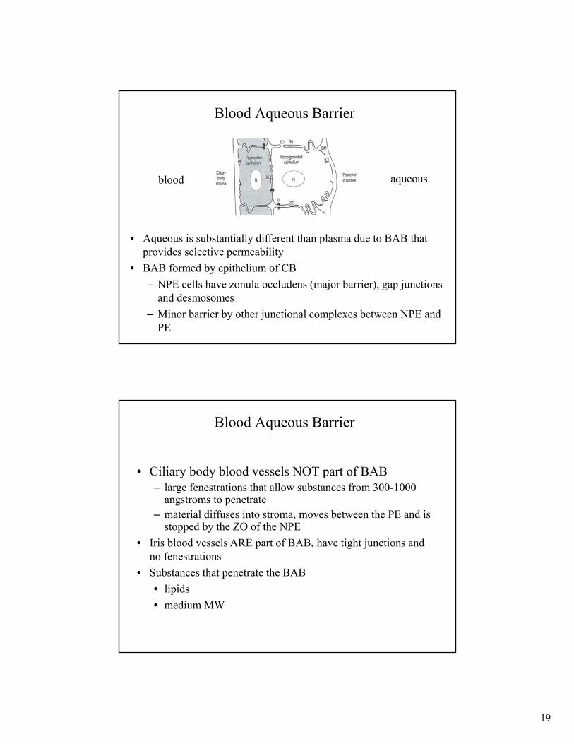
Blood Aqueous Barrier
• Aqueous is substantially different than plasma due to BAB that provides selective permeability
• BAB formed by epithelium of CB
– NPE cells have zonula occludens (major barrier), gap junctions and desmosomes
– Minor barrier by other junctional complexes between NPE and PE
blood aqueous
Blood Aqueous Barrier
• Ciliary body blood vessels NOT part of BAB– large fenestrations that allow substances from 300-1000
angstroms to penetrate– material diffuses into stroma, moves between the PE and is
stopped by the ZO of the NPE
• Iris blood vessels ARE part of BAB, have tight junctions and no fenestrations
• Substances that penetrate the BAB
• lipids
• medium MW

20
Blood Aqueous Barrier
• Use of hyperosmotics can make use of BAB– A rise in IOP..... patient ingests a hyperosmotic..…
• This sets up an osmolality difference between aqueous and blood.... remain in blood and are unable to ..
• Therefore they draw fluids out of the aqueous
• good for short-term treatment only
• Other theories: decrease vitreous volume and hypothalamus influence
Mechanisms of Aqueous Formation
• Diffusion (minor) – lipid soluble substances, passive, moves down concentration gradient
• Ultrafiltration (minor) – through fenestrations in ciliary capillaries, includes water, water soluble substances, minor, dialysis, passive, osmostic gradient
• Active secretion (major) – 80-90% of aqueous, active transport in NPE, generation of an osmotic gradient, two major enzymes:– Na-K ATPase in NPE
– Carbonic Anhydrase (CA) in PE & NPE

21
G-protein CoupledEnergy from ATP
& AQP4
Antiport
Symport
CO2 + H2O ↔ H2CO3 ↔ H+ + HCO3-
Ion Transport in Ciliary Epithelium
Copyright 2013 wlmiller
Symport

22
Aqueous Humor Formation
• Active secretion– 80-90 % of aqueous– Main site is NPE– Evidence
• concentrations of [ascorbate], [lactate], [amino acids] all higher than Gibbs-Donan equilibrium
• [Na] and [bicarb.] higher than G-D• effects of metabolic inhibitors or temperature decrease
will decrease secretion (IOP)• Membrane permeable to K+
• Net direction is secretion across the epithelium, but hydrostatic and oncotic forces favor resorbption
• Active transport produces an osmotic gradient across the ciliary epithelium, which promotes the movement of other plasma constituents by ultrafiltration and diffusion
Aqueous Humor Active Secretion
23
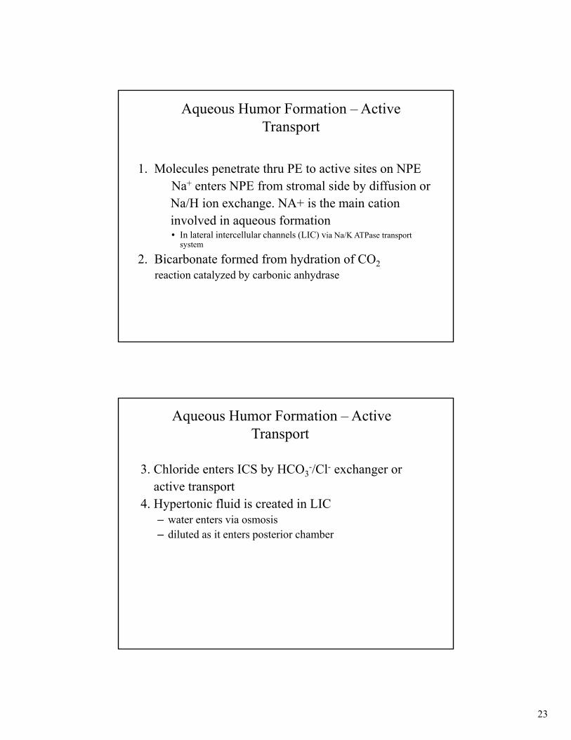
1. Molecules penetrate thru PE to active sites on NPENa+ enters NPE from stromal side by diffusion or Na/H ion exchange. NA+ is the main cationinvolved in aqueous formation• In lateral intercellular channels (LIC) via Na/K ATPase transport
system
2. Bicarbonate formed from hydration of CO2reaction catalyzed by carbonic anhydrase
Aqueous Humor Formation – Active Transport
Aqueous Humor Formation – Active Transport
3. Chloride enters ICS by HCO3-/Cl- exchanger or
active transport 4. Hypertonic fluid is created in LIC
– water enters via osmosis– diluted as it enters posterior chamber

24
Calcium-activated Potassium Channels
• Barros et. al. Curr. Eye Res. 10:731, 1991
• showed “super” K channels– large conductance, voltage
– activated by Calcium
– play a role in aqueous humor secretion
– did not show role to be related to other known roles in other epithelial types
Carbonic Anhydrase
• Responsible for formation of H+ and HCO3-
– enhances aqueous secretion
– CA inhibitors block formation of H+ and HCO3-
• H+ used in ion exchange with Na+
– Topical CA antagonist for glaucoma: dorzolamide (Trusopt)

25
Kiel et al 2011
Aqueous Humor Formation
Factors Affecting Aqueous Formation
• Innervation- generally…– sympathetics decrease– parasympathetics increase– Blood pressure increases then increase IOP– others decreasing formation: exercise, aging, anesthetics,
inflammation, acidosis, hypothermia
26
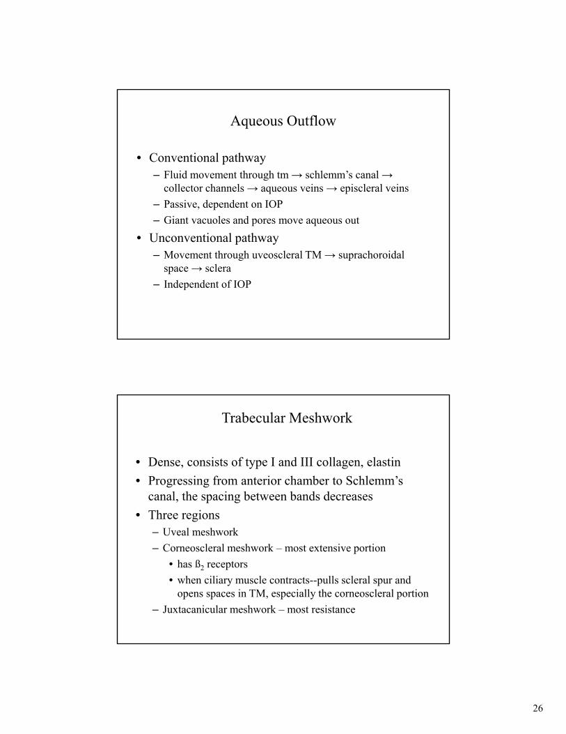
Aqueous Outflow
• Conventional pathway– Fluid movement through tm → schlemm’s canal →
collector channels → aqueous veins → episcleral veins
– Passive, dependent on IOP
– Giant vacuoles and pores move aqueous out
• Unconventional pathway– Movement through uveoscleral TM → suprachoroidal
space → sclera
– Independent of IOP
Trabecular Meshwork
• Dense, consists of type I and III collagen, elastin
• Progressing from anterior chamber to Schlemm’scanal, the spacing between bands decreases
• Three regions– Uveal meshwork
– Corneoscleral meshwork – most extensive portion
• has ß2 receptors
• when ciliary muscle contracts--pulls scleral spur and opens spaces in TM, especially the corneoscleral portion
– Juxtacanicular meshwork – most resistance

27
Trabecular Meshwork
• Aqueous must pass through....– 3 layers
• 1. Endothelial layer of TM
• 2. Collagenous layer of canal wall
• 3. Endothelial wall of canal
• vacuoles
Copyright 2013 wlmillerTrabecular meshwork
Schlemm’s canal
Endothelial wall
Collagenous
Endothelial layer of TM
Trabecular Meshwork is Phagocytic

28
Trabecular Meshwork
• MMPs – matrix metalloproteinases facilitate outflow by regulating synthesis and degradation of ECM
• TIGR – TM glucocorticoid inducible response protein, also called myocilin (Myoc).– Many patients with POAG have TIGR/MYOC mutations,
including children
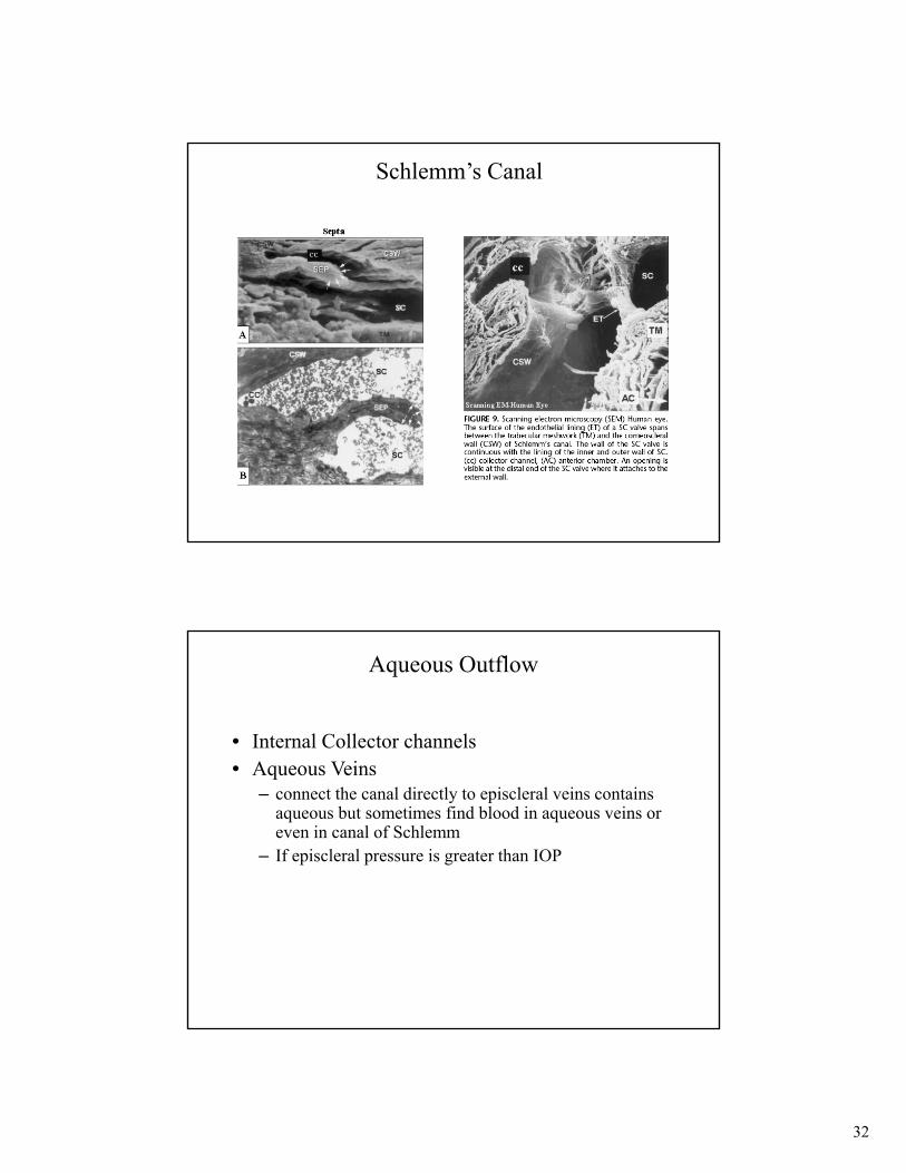
Schlemm’s Canal
• Endothelial layer of juxtacanicular TM comprises inner wall of Schlemm’s Canal
• Lumen diameter is 350 to 500 µm and it undulates. Increases the surface area which aides in aqueous absorption
• Aqueous prior to canal has gone through and irregular path, creates a percolating effect
• Possesses internal collector channels

29
Schlemm’s canal: transport of aqueous via giant vacuoles
Collector Channels
• Intrascleral collector channels– External Collector channels
• 25-35 of them moves to episcleral veins
• connect to episcleral veins or can connect to deep scleral vessels then finally to episcleral veins
• Episcleral veins therefore direct or indirect

30
Identification and Assessment of Schlemm’s Canal by SD-OCT
Kageman L, et al. IOVS 2010
Aqueous Outflow
• routes for flow out:1) pinocytosis2) diverticuli
– outpockets– increase surface area
3) pores and vacuoles– on the inner endothelial wall– relationship between IOP and # of pores– POAG fewer pores

31
Aqueous Outflow in POAG
• pores and vacuoles – on the inner endothelial wall– relationship between IOP and # of pores– POAG fewer pores
• Pore number and size are decreased in glaucoma – 1437/mm2 in normal eyes vs 489/mm2 in glaucoma
– pore larger in normal eyes (0.91µm vs 0.85 µm)
• Outflow facility– 0.25 in normals
– 0.11 in POAG; facility= 1/ resistance
Schlemm’s Canal
32
Schlemm’s Canal
Aqueous Outflow
• Internal Collector channels• Aqueous Veins
– connect the canal directly to episcleral veins contains aqueous but sometimes find blood in aqueous veins or even in canal of Schlemm
– If episcleral pressure is greater than IOP

33
Pressure Gradients
• Ciliary arterial system – 60 mmHg
• IOP – 13-17 mmHg
• Resistance in conventional outflow pathway – 3-4 mmHg
• Episcleral veins – 8-10 mmHg– if episcleral venous pressure increases will increase IOP but
with time adapts and decreases aqueous inflow
– IOP must be greater than episcleral vein pressure for outflow
Unconventional Aqueous Outflow
• Uveoscleral outflow– Another route for aqueous– out through choroid– can go to lymphatics or vitreous– Approximately “10-50%” of aqeuous leaves by this route– pressure independent

34
Fig. 1. IOP before prostaglandin F2α 1-isopropylester (PGF2α-IE) treatment (day 0) and before and after the 7th dose of PGF2α-IE on the 4th day of unilateral topical treatment. Time 0 occurs ∼17 hours after the 6th PGF2α-IE dose (given at ∼3:00 PM on day 3) and immediately before the 7th dose (given at ∼8:30 AM on day 4). Data are mean ± SEM IOP for six animals, each contributing one treated (solid circle) and one untreated (open circle) eye. Symbols on abscissa indicate significant difference between treated and control eyes by the two-tailed two-sample t-test. (Reprinted from Crawford and Kaufman,20 with permission of Investigative Ophthalmology & Visual Science.)
Effects of Prostaglandins on the Aqueous Humor Outflow Pathways
Weinreb, et al, Survey of Ophthalmol 2002
Outflow
• Total flow in and total flow out must = 0• Inflow and outflow operate as parallel flow rather
than in series• Inflow consists of two parts
– one driven by pressure (ultrafiltration, diffusion)– second is active secretion
• Outflow has two parts– through TM which is pressure dependent (Pi = IOP and
Pe)– uveoscleral outflow
35
Copyright 2013 wlmiller
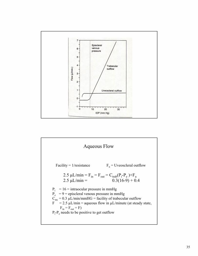
Aqueous Flow
Facility = 1/resistance Fu = Uveoscleral outflow
2.5 μL/min = Fin = Fout = Ctrab(Pi-Pe )+Fu
2.5 μL/min = 0.3(16-9) + 0.4
Pi = 16 = intraocular pressure in mmHg Pe = 9 = episcleral venous pressure in mmHgCtm = 0.3 µL/min/mmHG = facility of trabecular outflowF = 2.5 µL/min = aqueous flow in µL/minute (at steady state,
Fin = Fout = F)Pi-Pe needs to be positive to get outflow

36
Population distribution of IOP
Mangouritsas G, Mourtzoukos S, Mantzounis A, Alexopoulos L - Clin Ophthalmol (2011)
Clinical Applications in Glaucoma
• Beta blockers – suppress aq humor production by decreasing cAMP (timolol, levobunolol)
• Adrenergic (alpha) agonists – suppress production by decreasing cAMP (apraclonidine, brimonidine)
• Carbonic anhydrase inhibitors – suppress production (dorzolamide, brinzolamide)
• Prostaglandin analogs – increases uveoscleral outflow (bimataprost, travaprost, latanaprost)
• Parasympathetic agonists – increases outflow by opening angle (pilocarpine)
• Hyperosmotic agents – decrease vitreous volume (glycerine, mannitol)

37
The Crystalline Lens
The Crystalline Lens
• Transparent biconvex structure behind iris, in front of vitreous
• Contributes about 15D of the total power of the eye, ~58D
• Grows through out life, from 6.5mm diameter at birth to 10mm diameter as adult

38
The Crystalline Lens
• Center point of anterior and posterior surfaces are the anterior and posterior poles, the “axis” is the line joining the poles
• Anterior surface (10mm radius of curvature) is less convex than posterior surface (6mm radius of curvature)
• Outer edges are the equator
Lens Optics
• index of refraction– cortex= 1.386
– n of nucleus= 1.406
– overall= 1.413
• Transparency– regular fiber arrangement
– lamellar style proteins
– avascularity
– thin epithelium
• Absorption– absorbs UV < 360 nm
• Scatter– scatters about 5% of light
due to “n” variation of proteins and membranes.
– Scatter will increase with age due to increase separation of lens fibers and increase in “n”

39
Copyright 2013 wlmiller
20
40
60
80
100
%Trans.
Wavelength (nm)
300 400 600 800 1200 1600
LENS TRANSMITTANCE
_____ 4y.o.------- 53 y.o.------- 75 y.o.
Layers of the Lens
• Lens capsule – outermost elastic layer
• Epithelium – only on the anterior surface
• Lens fibers– Cortex
– Nucleus – innermost

40
Lens Position
• Lens equator is encircled by ciliary processes
• Suspended within the ciliary ring by zonules, suspensoryligaments attached to ciliary processes and lens equator, spanning about 0.5mm space, space decreases as lens grows with age
• Lens is suspended and under tension during distance viewing
• During accommodation the ciliary muscle contract, releases zonular tension, and allows lens to fatten
Lens Composition
• 60% water, 40% proteins (crystallins, increase with age)
• High concentration of glutathione (GSH)– Maintains protein sulfhydryl groups in reduced form
– composed of glycine, cystein and glutamic acid
– Protects from oxidative damage by detoxification of peroxide
– Removes xenobiotics by conjugation with hydrophobic compounds having an electrophilic center
– highest concentration in cortex, lowest in nucleu

41
Lens Biochemistry
• Proteins• Amino acids - glycine, alanine, leucine, taurine,
(actively transported into cells)• Inorganic Ions
– Sodium -17 meq/Kg; low compared to aqueous, actively extruded, concentration high during cataract
– Potassium - 25 meq/Kg, Na in and K out, low in cataract formation
– Calcium and Magnesium– Phosphate– Chloride - 30 meq/Kg– Others: Mn, Cu++, Fe etc
Lens Biochemistry
• Lipids– Cholesterol - concentration increases with cataract formation
– Phospholipids
• mainly sphingomelin- increases with age
• Sphingomyelin and cholesterol
• May have some effect on accommodation

42
Lens Biochemistry
• Carbohydrates• mainly glucose
• If levels in aqueous too high get lens swelling
• May have facilitated transport with unique transporter
• Organic phosphate- ATP– Ascorbic acid - high levels..... eye from UV...
– Inositol - high concentration in lens, actively transported
Lens Capsule
• Consists of about 40 lamella made up of reticular fibers (netlike or entangled) embedded in sulfated glycoaminoglycan
• Composed of collagen type IV
• Forms a barrier to bacteria and inflammatory cells
• Will allow diffusion of smaller molecules
• Acellular basement membrane that completely envelopes lens
• Thinnest at poles and equator
• Thickest in midregions
• Formed by lens epithelium anteriorly and lens fibers posteriorly

43
Lens Epithelium
• Single layer of simple cuboidal epithelium over anterior surface (under capsule), not found posteriorly
• Become columnar towards equator and convert into lens fibers
• Cells are not shed because of capsule
• Has sodium potassium pumps
Lens Epithelium Zones
• Central zone– Cells are flattened and hexagonal
• Germative or proliferative zone– Cells are columnar and smaller in
area, higher in density
– New cells generated here – nuclei divide and migrate posteriorly to become new lens fibers in cortex
– Layers are laid down in close contact through “ball and socket” like joints
• Transition Zone and Equator– Cells elongate and rotate so long axis is parallel to
cortical surface

44
Lens Fibers
lens fibers
• New superficial fibers are nucleated
• Nuclear bow forms as nuclei move anteriorly and fibers pass deeper into the lens to form the cortex
• Deeper fibers are anucleate (true fibers)
• Preceding generations of cells are pushed deeper into center of lens
Lens Fibers

45
Microscopic Anatomy
• Fibers– produced by epithelial cells at
equator– elongation epithelial cell proceed
anteriorly• basal end proceeds posteriorly
– Interdigitations• more like ball and socket• responsible for flexibility of
lens– zonular fibers
• insert into ILM of CB and 1.5 mm on either side of equator
Lens Embryology
• Lens placode noticed at 22 days gestation
• Primary lens fibers - cells of posterior wall of lens elongate, lose nuclei, become transparent lens fibers, make up embryonic nucleus (innermost part of lens), attach to apical surface of anterior lens

46
Lens Embryology
• Secondary lens fibers – new fibers added (throughout life) and grow around embryonic nucleus to make up the fetal nucleus
• Secondary fibers form Y sutures – Formed by fusion of lens fibers
– Anterior Y suture is erect
– Posterior Y suture is inverted
Lens Embryology
• Cortex – after birth new fibers continue to form, produced by mitosis of epithelial cells in equitorialregion
• Fibers elongate to surround existing nucleus
• Development of cortical fibers form the “nuclear bow” or “lens bow”

47
1. Tunica vasculosa lentis2. Vasa Hyaloidea Propria3. Hyaloid artery (glial sheath)
Lens Embryology
• Hyaloid artery supplies lens in developing fetus
• Regresses prior to birth
• Small remnant can remain – Mittendorf’s dot on the posterior lens surface
– Bermeister’s papilla in posterior pole
Abnormalities during Development
• Mittendorf Dot
• Persistent Hyaloid Artery
• Bergmeister’s Papilla

48
Lens Comparative Anatomy
guinea pig lens, Ostrin, et al, IOVS 2014
Lens Growth and Suture Formation

49
Lens Growth and Suture Formation
Lens “Circulation”
• Gap Junctions – connexins, move nutrients between cells
• Aquaporin 0 – important for water transport
• Na/K ATPase pump– in lateral apical membranes
– near equitorial differentiating fibers
– important in maintaining transparency and water balance
– pump leak mechanism

50
Lens Cytoskeleton – Alpha Crystallin
• Horwitz J. Eye 13:403, 1999– major lens protein whose structure helps maintain
transparency
– alpha crystallin is a chaperone
• binds with unfolded/denatured protein
• suppresses aggregation therefore maintains transparency
• subunits alpha A:B in ratio of 3:1
– preserves thermal stability
• chaperone properties are better served in higher temperatures ie 37ºC vs 20ºC
Lens Cytoskeleton
• Quinlan RA et. al. Eye 13:409, 1999– lens contains microtubules, microfilaments and
intermediate filaments
– intermediate filaments
• CP49 and filensin
– found complexed with alpha and beta crystallin
– found in all stages of lens fiber differentiation
– precise role unknown
– implicated in cataract formation

51
Lens Metabolism
• Primarily anaerobic through glycolytic pathway
• Continuous supply of ATP needed, uses only small amount of energy, made in epithelium– Growth
– Synthesis
– Pumping nutrients in and waste out
Lens Metabolism - Glucose
Energy production based on glucose metabolism– glucose enters lens by simple and facilitated diffusion
– rapidly metabolized
1. anaerobic metabolism – 85% of glucose metabolism
2. aerobic metabolism – kreb’s cycle, 3% of glucose metabolism
3. Hexose monophosphate shunt – 5%, source of NADPH for sorbitol and glutathione pathways
4. Sorbitol pathway – 5%, obtained by reduction of glucose, sorbital sets up osmotic gradient

52
K+
Glucose
Na+
Na+
K+
Glucose
InositolGSH
ATP
Water
Glucose
Water
Sorbital SorbitolFructose
InositolGSHATP
Changes during diabetic cataract
Lens
• Key enzymes against oxidative insult
– Glutathione peroxidase• 2 GSH + H2O2 GSSG + 2 H2O
– Catalase• H2O2 + H2O2 2 H2O + O2
– Superoxidase Dismutase• O2· + O2· + 2 H H2O2 + O2
– Glutathione S-Transferase• GSH + RX .......... Mercapturic Acid

53
Lens
• Transport and Permeability• Cation Balance
– lots of Potassium in lens– try to decrease Calcium and Sodium via Na/K pumps– decrease in lens temperature causes Na and K– Electrophysiology
• lens has a -70 mV compared to Aqueous
Copyright 2013 wlmiller
Ca++
AA
3 Na+ 3 Na+
3 Na+
2 K+2 K+
2 K+
Posterior Lens
Anterior Lens
Diffusion Diffusion (H20, Cl-)
Facilitateddiffusion
Na= 25 mMK= 140 mM
Na= 163 mMK= 4 mM
Na= 144 mMK = 8 mM
ATPase
ATPaseGlucose

54
Functions
• Absorption/transmittance
• Transparency
• Refraction
• Accommodation
Age Changes in the Lens
• Water content– nuclear content increases– changes occur around cataracts
• Electrolytes– increase sodium, calcium– decrease potassium– Mg constant
• Protein changes– increase in insoluble and decrease in soluble
• Increase in albuminoid protein

55
Age Changes in the Lens
• Lipid changes– Increase
– Increase in cholesterol
• Metabolic changes– decrease in hexokinase
– increase aldose reductase
– decrease in Na/K ATPase
– Glutathione in oxidized state
• Gross– increase in lens size– increase in lens weight
and thickness– increase in lens nucleus
• Microscopic– Increase in lens density– Optical changes– Zones of discontinuity
not as clearly visible– yellowing - brunescence
Any Questions?