Embed Size (px)
Citation preview

1114
INTRODUCTIONAquaporins are membrane proteins that function in the movementof water and small solutes across the cell membrane. Initiallycharacterized in human red blood cells (Preston et al., 1992) andkidney cells (Nielsen et al., 1993), aquaporins are found in othermammals, anurans, arthropods, plants and yeast (Borgnia et al.,1999). These small pore proteins are highly selective, allowing onlywater and small uncharged solutes, such as glycerol and urea, topassively diffuse through the membrane (Ishibashi et al., 1994).Aquaporins are implicated in various responses to osmoticchallenge, including urine concentration in mammals (Nielsen etal., 1995), water shunting in sap-sucking insects (Beuron et al.,1995) and the tolerance of sea water consumption in birds (Mülleret al., 2006).
Although most insects are freeze intolerant and must rely onsupercooling for winter survival, some are freeze tolerant and canwithstand ice formation within their extracellular fluids (Salt, 1961).During freezing, only water molecules join the growing ice lattice,thus concentrating solutes in the remaining unfrozen fraction ofwater, termed freeze concentration. The resulting osmotic gradientdraws water out of the cells, thereby preventing lethal intracellularfreezing (Lee, 1989).
The inability of most cells to survive freezing is due to aninadequate amount of intracellular water being replaced withcryoprotectant molecules, such as glycerol (Hagedorn et al., 2002).Consequently, efforts to improve cell survival duringcryopreservation have turned to the artificial expression ofaquaporins. RNA insertion and protein overexpression of aquaporinssuccessfully increases the survival of mouse oocytes duringcryopreservation (Edashige, 2003) and the viability of baker’s yeastin bread dough after freezing (Tanghe et al., 2002). In addition, a
recent report by Izumi et al. (Izumi et al., 2006) provides evidencethat aquaporins promote freeze tolerance in a rice stem boring insect.By blocking the functionality of the protein with mercuric chloride,a known inhibitor of some aquaporins (Preston et al., 1993), theyreported a reduction in cell survival during freezing.
Similar to freezing, desiccation places an organism under osmoticstress. In desiccated plants, Smith-Espinoza et al. (Smith-Espinozaet al., 2003) and Barrieu et al. (Barrieu et al., 1999) found an increasein aquaporin transcription and protein concentration. Likewise,mammals increase aquaporin expression in response to dehydration(Ishibashi et al., 1997). The upregulated expression of aquaporinssuggests they play a role in desiccation tolerance.
Overwintering larvae of the goldenrod gall fly, Eurostasolidaginis (Fitch), are both freeze tolerant and desiccation resistant.Larvae acquire freeze tolerance during the autumn, in part throughthe accumulation of glycerol and sorbitol (Morrissey and Baust,1976), and can survive extended periods of subzero exposure totemperatures below –50°C (Storey and Storey, 1988; Lee, 1991).Also, larvae are subjected to desiccating conditions as thesurrounding gall tissues of the goldenrod plant (Solidago sp.) senesceand dry in the autumn (Rojas et al., 1986) and during cold, drywinters; Ramløv and Lee (Ramløv and Lee, 2000) reported that therate of water loss for these overwintering larvae is among the lowestreported for any insect.
We hypothesized that aquaporins play an important role in theability of E. solidaginis to survive freezing and desiccation. To firstdetermine whether aquaporins are present in E. solidaginis, weimmunoblotted soluble proteins of control and desiccated larvaeagainst mammalian anti-AQP2, -AQP3 and -AQP4 antibodies. Totest the role of aquaporins in freeze tolerance, we froze isolated fatbody, midgut and salivary glands in the presence or absence of the
The Journal of Experimental Biology 211, 1114-1119Published by The Company of Biologists 2008doi:10.1242/jeb.016758
Aquaporins play a role in desiccation and freeze tolerance in larvae of the goldenrodgall fly, Eurosta solidaginis
Benjamin N. Philip*, Shu-Xia Yi, Michael A. Elnitsky and Richard E. Lee, JrDepartment of Zoology, Miami University, Oxford, OH 45056, USA
*Author for correspondence (e-mail: [email protected])
Accepted 27 January 2008
SUMMARYSurvival of freezing not only requires organisms to tolerate ice formation within their body, but also depends on the rapidredistribution of water and cryoprotective compounds between intra- and extracellular compartments. Aquaporins aretransmembrane proteins that serve as the major pathway through which water and small uncharged solutes (e.g. glycerol) enterand leave the cell. Consequently, we examined freeze-tolerant larvae of the goldenrod gall fly, Eurosta solidaginis, to determinewhether aquaporins are present and if their presence promotes freeze tolerance of specific tissues. Immunoblotting withmammalian anti-AQP2, -AQP3 and -AQP4 revealed corresponding aquaporin homologues in E. solidaginis, whose patterns ofexpression varied depending on acclimation temperature and desiccation treatment. To examine the role of aquaporins in freezetolerance, we froze fat body, midgut and salivary gland tissues in the presence and absence of mercuric chloride, an aquaporininhibitor. Survival of fat body and midgut cells was significantly reduced when mercuric chloride was present. In contrast, survivalof the salivary gland did not decrease when it was frozen with mercuric chloride. Overall, this study supports our hypothesis thatnaturally occurring aquaporins in E. solidaginis are regulated during desiccation and promote cell survival during freezing.
Key words: aquaporins, osmotic stress, water balance, freeze-tolerant insects.
THE JOURNAL OF EXPERIMENTAL BIOLOGY
1115Aquaporins in desiccation and freezing
aquaporin inhibitor mercuric chloride and assessed cell viability afterfreezing.
MATERIALS AND METHODSCollection of insects
Spherical galls containing the larvae of E. solidaginis (Diptera:Tephritidae) were collected from goldenrod plants (Solidago sp.)at the Miami University Ecology Research Center located nearOxford, Ohio (39°31�57�N, 84°43�23�W) on November 27th, 2006.The galls were kept in plastic bags and acclimated to 4°C or frozenat –20°C until used for experiments (~4·months).
Desiccation treatmentThree replicates of 10 larvae from each acclimation group wereplaced in a Petri dish and dehydrated in a desiccator over freshDrierite (0% relative humidity) for 4·days at 4°C. The larvae lost3–7% of their fresh body mass during this desiccation treatment.
Protein extraction and SDS-PAGEGroups of 10 larvae were homogenized in a 2·ml glass homogenizerwith a buffer containing 150·mmol·l–1 NaCl, 10·mmol·l–1 Tris-HCl(pH 7.2), 0.1% sodium deoxycholate and protease inhibitors[5·�g·ml–1 aprotinin, 5·�g·ml–1 antipain, 5·�g·ml–1 leupeptin and1·mmol·l–1 PMSF (phenylmethanesulfonylfluoride)] (Goel et al.,2006; Yi et al., 2007). After the homogenate was sonicated with anultrasonic processor (Cole Parmer, Vernon Hills, IL, USA) andincubated on ice for 30·min, it was centrifuged twice at 16·000·gfor 20·min at 4°C to remove any insoluble fragments. The resultingsupernatant contained soluble proteins, whose concentration wasdetermined using the Bio-Rad protein assay (Bio-Rad, Hercules,CA, USA) with BSA (bovine serum albumin) as a standard. Proteinsamples (50·�g) were mixed with Laemmli sample buffer containing5% �-mercaptoethanol and incubated at either 95°C for 3·min or60°C for 10·min, and then analysed by SDS-PAGE on a 4–15%gradient gel (Bio-Rad). Precision Plus protein standard kit (Bio-Rad) was used as a reference.
Protein staining and immunoblottingFollowing electrophoresis, proteins were transferred to anitrocellulose membrane (Bio-Rad). The membrane was stained with0.1% Ponceau S staining solution (Sigma Chemical Company, SaintLouis, MO, USA) for 10·min and rinsed with ultrapure water toverify that proteins were transferred. After digitally scanning themembranes, they were destained for 30·s in an aqueous solution of0.1·mol·l–1 NaOH and were used for immunoblotting as describedby Yi et al. (Yi et al., 2007). Non-specific protein antigens wereblocked at 4°C overnight in 10% non-fat milk Western wash buffer(10·mmol·l–1 Tris, 100·mmol·l–1 NaCl and 0.1% Tween 20 at pH7.5). The membranes were incubated with corresponding primaryantibodies in a 5% non-fat milk solution at 21°C for 2·h. Rabbitanti-actin (1:400), anti-AQP2 (1:500), anti-AQP3 (1:200) and anti-AQP4 (1:1000), and goat anti-rabbit IgG-HRP (horseradishperoxidase) conjugates were purchased from Sigma ChemicalCompany. After three 15·min washes in Western wash buffer, themembranes were incubated for 2·h with secondary antibody (goatanti-rabbit IgG-HRP conjugates) diluted 1:1000 in Western washbuffer. Membranes were then washed in Western wash buffer,incubated for 2·min in ECL (enhanced chemiluminescence)detection reagents (Amersham Biosciences, Piscataway, NJ, USA)and exposed to autoradiography film. Although all immunoblotswere replicated to verify findings, only representative Western blotsare included in the results presented here. Bands were normalized
to 4°C samples and semi-quantified using ImageQuant 5.2(Molecular Dynamics, Sunnyvale, CA, USA).
Role of aquaporins in freeze toleranceLarvae acclimated to 4°C were dissected in Coast’s solution (Coast,1988) on a silicone elastomer-filled Petri dish (Yi and Lee, 2003).Fat bodies (~3.5·mg wet mass) and midguts (~0.15·mg wet mass)were dissected while larvae were pinned with their ventral sideuppermost, whereas salivary glands (~0.12·mg wet mass) weredissected while larvae were pinned with their dorsal side uppermost.Dissected tissues were transferred to ~1·ml of fresh Coast’s solutionfor 1·h at 21°C. Tissues were then transferred to 1·ml of Coast’ssolution + 0.25·mol·l–1 glycerol and incubated at 4°C for 1·h.Following the incubation, they were placed into 100·�l of Coast’ssolution, Coast’s solution + 0.25·mol·l–1 glycerol, or Coast’s solution+ 0.25·mol·l–1 glycerol + 0.2·mmol·l–1 mercuric chloride (HgCl2),based on a modified protocol from Izumi et al. (Izumi et al., 2006).
To determine whether the effects of mercuric chloride werepermanent, tissues were exposed to �-mercaptoethanol, a reducingagent, which reverses the inhibitory effect of mercuric chloride(Preston et al., 1992). The dissection and incubation procedureremained the same except for an additional step in which tissueswere incubated in Coast’s solution + 0.25·mol·l–1 glycerol +0.2·mmol·l–1 HgCl2 for 15·min at 21°C before being frozen in 100·�lof Coast’s solution + 0.25·mol·l–1 glycerol + 0.2·mmol·l–1 HgCl2 +2·mmol·l–1 �-mercaptoethanol.
The microcentrifuge tubes containing the tissues were cooled ata rate of 0.2°C·min–1 (4°C to –20°C) over 2·h and kept at –20°Cfor 2·h. When the temperature of the ethanol bath reached –4°C,the microcentrifuge tubes were sprayed with Super FriendlyFreeze’It (Fisher Scientific Company, Hanover Park, IL, USA) toseed ice nucleation and freezing of the treatment solutions. Allcontrol tissues were kept at 4°C for 4·h.
Cell viability assayAll tissues were thawed at 21°C for 1·h prior to the assessment ofcell survival using the Live/Dead sperm viability kit [MolecularProbes, Inc., Eugene, OR, USA (Yi and Lee, 2003)] containingSYBR 14 dye (1·mmol·l–1 in DMSO) and propidium iodide(2.4·mmol·l–1 in water). Working solutions of SYBR (2·�l per100·�l Coast’s solution) and propidium iodide (4·�l per 100·�lCoast’s solution) were prepared for all samples. The tissues wereincubated in 25·�l SYBR stain for 15·min on glass microscopeslides. The addition of 25·�l propidium iodide to the slides wasfollowed by a second 15·min incubation. The SYBR stain ismembrane permeable and can penetrate the nuclei of all cells,whereas propidium iodide can only enter damaged cells that havelost plasma membrane integrity. The slides were covered with acoverslip and examined on a fluorescence microscope (OlympusBX60) (Davis and Lee, 2001; Yi and Lee, 2003). Cell viability wasassessed using the following criteria: live cells fluoresced green anddead cells fluoresced orange to bright red. The percentage survivalof the midgut and salivary gland from each individual was basedon the mean of three counts of 100 cells. There are fewer fat bodycells per larvae, therefore the viability of the fat body was basedsolely on the total count of cells (~130–200) dissected from eachindividual. The individual (N=4) average for each tissue was usedto calculate the mean ± s.e.m. for each treatment.
StatisticsData were compared using an analysis of variance andBonferroni–Dunn post-hoc tests (Statview 4.5, Cary, NC, USA).
THE JOURNAL OF EXPERIMENTAL BIOLOGY

1116
Statistical significance was set at P<0.05. Values are presented asmeans ± s.e.m.
RESULTSImmunoidentification of aquaporins
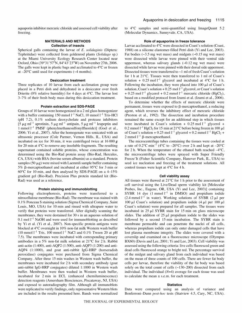
The proteins extracted from E. solidaginis larvae were separatedusing electrophoresis and their banding patterns were compared ona Ponceau S-stained membrane (Fig.·1). We verified that there wasno difference in banding pattern between samples prepared at 95°Cfor 3·min or 60°C for 10·min, therefore 95°C for 3·min was usedin all subsequent experiments. Immunoblotting with antibodiesraised against mammalian aquaporins identified homologues for twowater channel proteins (AQP2 and AQP4) and one glycerol channelprotein (AQP3; Fig.·2). Endogenous actin, a 42·kDa protein, servedas an internal loading control and indicated that the samples wereloaded with the same amount of protein (Fig.·2A).
The antibodies from all three aquaporins reacted with the larvalprotein extracts. The anti-AQP2 antibody detected a group of threemajor protein bands of 26, 29 and 31·kDa (Fig.·2A), which is withinthe previously reported size range of 30·kDa (Chou et al., 2000).Anti-AQP3 identified two pronounced protein bands with apparentmolecular masses of 25 and 75·kDa, and two faint bands of 40 and50·kDa (Fig.·2B). Previous reports suggest that AQP3 is ~26·kDa(Rai et al., 2006) and that any larger immunoidentified bands areeither glycosylated or oligomers (Lu et al., 1996). Therefore, wepropose that the 25·kDa band is a monomer and the 75·kDa bandcorresponds to a trimer of AQP3. Although the expected AQP4 sizeis ~31·kDa (Rash et al., 1998), we observed a single band at 60·kDa(Fig.·2C), which is probably dimeric AQP4 (Neely et al., 1999).
As suggested by semi-quantitative densitometry, the concentrationof all three aquaporin homologues varied depending on temperatureacclimation and/or desiccation treatment when compared with 4°Cacclimated larvae. For AQP2 immunoblots (N=2), the density ofthe 26·kDa band for each of the three treatments (4°C desiccated,–20°C frozen and –20°C frozen + desiccated) was 27–77 % lowerthan that of the 4°C acclimated larvae (Fig.·2A). In contrast, theAQP3 immunoblots (N=3) of treated samples suggest an almost 50%increase in concentration at 25·kDa compared with the 4°C
B. N. Philip and others
acclimated larvae (Fig.·2B). Lastly, the 4°C acclimated larvaeexpressed a slightly higher concentration of AQP4 (N=2) comparedwith the other treatment groups (Fig.·2C).
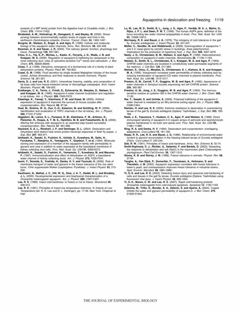
Effect of aquaporins on freeze toleranceA high proportion of the control tissues fluoresced green followingvital dye staining, which indicates survival of the cells(Fig.·3A,G,M). The high survival rate of the fat body (95.7±0.6%),midgut (98.3±0.8%) and salivary gland (97.9±0.7%) control tissuesat 4°C indicates minimal mechanical damage to the tissues duringdissection (Figs·4 and 5).
Unlike the control tissues, many cells fluoresced red after beingfrozen in Coast’s solution, indicating disruption of the plasmamembrane and cell death (Fig.·3C,I,O). As E. solidaginis acquiresfreeze tolerance during the autumn, they accumulate glycerol. Tomimic natural conditions and increase cell survival rates, all tissueswere frozen in the presence of glycerol (Fig.·3B,H,N). In each tissue,there was a significant increase in cell survival compared withsamples frozen only in Coast’s solution; fat body survival increasedfrom 27.8% to 81.6% (Fig.·4A, P<0.0001), midgut survivalincreased from 19.8% to 91.4% (Fig.·4B, P<0.0001) and salivarygland survival increased from 18.9% to 93.7% (Fig.·5, P<0.0001).
Mercuric chloride was used to assess whether aquaporin functionis necessary for larval survival during freezing. We held mercuricchloride controls (Coast’s solution + 0.25·mol·l–1 glycerol +0.2·mmol·l–1 HgCl2) at 4°C for 4·h and did not see a difference insurvival compared with tissues frozen in Coast’s solution and
95°C 95°C 95°C 60°C 60°C
M
250
150
100
75
50
37
25
15 10
20
Desiccated Acclimated Acclimated
–20°C 4°C
60°C
Fig.·1. Overall protein staining with Ponceau S demonstrates that there isno difference in banding pattern between samples prepared at differenttemperatures. Protein samples were treated at either 95°C for 3·min or60°C for 10·min before SDS-PAGE electrophoresis. Markers are in kDa.
Fig.·2. Immunoidentification of (A) AQP2 (N=2), (B) AQP3 (N=3) and (C)AQP4 (N=2) in protein extracts of E. solidaginis larvae using mammalianantisera. Fifty micrograms of protein were loaded onto each lane. Larvaewere acclimated for ~4·months at 4°C or –20°C. Desiccated larvae werekept at 0% relative humidity for 4·days. Below each band on theserepresentative gels is the corresponding average relative abundance (RA),with the RA of 4°C treated samples set to 1.0.
AQP3
25
40
75 50
kDa
AQP2 �Actin 42
26 29 31
4°C –20°C
RA (26 kDa) 1.0 0.73 0.59 0.23
60 AQP4
RA (25 kDa) 1.0 1.53 1.41 1.54
RA (60 kDa) 1.0 0.95 0.86 0.65
Acc
limat
ed
Acc
limat
ed +
D
esic
cate
d
Acc
limat
ed
Acc
limat
ed +
D
esic
cate
d
A
B
C
THE JOURNAL OF EXPERIMENTAL BIOLOGY

1117Aquaporins in desiccation and freezing
glycerol, suggesting that mercuric chloride was not toxic to unfrozentissues (Fig.·3E,K). However, when these tissues were frozen in thepresence of mercuric chloride, there was a significant reduction incell survival. As seen in Fig.·3B,D, survival of fat body wassignificantly lower compared with tissues frozen in Coast’s solutionand glycerol alone (Fig.·4A, P<0.0001). Similarly, Fig.·3H,Jindicates that survival of the midgut was significantly lower whenfrozen in the presence of mercuric chloride (Fig.·4B, P<0.0001).
Tissues were frozen with �-mercaptoethanol to determine whetherthe effects of mercuric chloride were reversible. Neither fat body(Fig.·3F) nor midgut (Fig.·3L) differed in cell survival comparedwith tissues frozen in Coast’s solution and glycerol (Fig.·4A,B). Thissuggests that aquaporin blocking by mercuric chloride is reversible.
Unlike the fat body and midgut, the salivary gland did not exhibita significant reduction in cell survival when frozen with mercuricchloride (Fig.·5). Because there was no difference between salivaryglands frozen in Coast’s solution + 0.25·mol·l–1 glycerol (Fig.·3N)or Coast’s solution + 0.25·mol·l–1 glycerol + 0.2·mmol·l–1 HgCl2(Fig.·3P), neither mercuric chloride controls nor �-mercaptoethanoltreatments were run.
DISCUSSIONImmunoreactivity of aquaporins
The immunoblots for AQP2, AQP3 and AQP4 demonstrate thatthere are aquaporin homologues in E. solidaginis. These antisera,raised against mammalian aquaporins, reacted with E. solidaginis
protein extracts at similar molecular masses suggesting that theyshare conserved antigenic epitopes. Kaufmann et al. (Kaufmann etal., 2005) reported that an aquaporin protein isolated fromDrosophila melanogaster (DRIP) is 44% identical to human AQP4.Aquaporins within the order Diptera are even more closely related,as shown by AeaAQP (from the mosquito Aedes aegypti), whichhas an amino acid sequence that is 65% identical to DRIP(Kaufmann et al., 2005). Thus, even between phylogeneticallydistant species, aquaporin proteins retain conserved elements, whichallowed us to use mammalian antisera to confirm the presence ofaquaporin-like proteins in E. solidaginis.
Desiccation regulates aquaporinsMany organisms adapt to desiccation and osmotic stress byregulating various proteins, such as LEAs in plants [e.g. dehydrins(Close, 1996)] and heat shock proteins in insects (Hayward et al.,2004). Similarly, the aquaporins in our study were either upregulated(AQP3) or downregulated (AQP2 and AQP4) following desiccationof the larvae. Among the three aquaporins characterized, AQP3 isespecially intriguing because it is permeable to water and glycerol(Ishibashi et al., 1994), both of which are important during osmoticstress caused by desiccation and freezing. In our study, soluble AQP3levels from 4°C acclimated larvae increased by 50% with desiccation(Fig.·2B), which is similar to the upregulation previously reportedin kidneys of dehydrated rats (Ecelbarger et al., 1995). Thisupregulation of AQP3 may be part of a coordinated set of
Fig.·3. Fluorescence micrographs of fat body (A–F),midgut (G–L) and salivary gland (M–P) treatments used todetermine the role of aquaporins in freeze tolerance. Theglycerol (A,G,M) and mercuric chloride control tissues(E,K) were kept at 4°C. The treated tissues were cooledfrom 4°C to –20°C over 2·h and left at –20°C for another2·h. All tissues were frozen in Coastʼs solution (C,I,O),Coastʼs solution + 0.25·mol·l–1 glycerol (B,H,N), Coastʼssolution + 0.25·mol·l–1 glycerol + 0.2·mmol·l–1 mercuricchloride (D,J,P) or Coastʼs solution + 0.25·mol·l–1 glycerol+ 0.2·mmol·l–1 mercuric chloride + 2·mmol·l–1 �-mercaptoethanol (F,L). The scale bar is 100·�m for J, M,N and O and 200·�m for all other micrographs.
THE JOURNAL OF EXPERIMENTAL BIOLOGY

1118
physiological changes that increase the permeability of water andglycerol across the cell membrane as larvae prepare for the osmoticstress associated with host plant senescence and extracellular iceformation. To our knowledge, this is the first report of insectaquaporins being regulated in response to desiccation.
Aquaporins promote freeze toleranceThe normally high level of freeze tolerance in E. solidaginis tissueswas significantly reduced when aquaporin channels were blockedwith mercuric chloride. This high mortality in the fat body andmidgut occurred despite tissues being frozen in a solution that alsocontained glycerol, a cryoprotectant that promotes freeze tolerance(Fig.·4). Izumi et al. (Izumi et al., 2006) suggested that cell survivalof freezing depends on a portion of intracellular water beingreplaced by glycerol and demonstrated that mercuric chlorideblocks both water and glycerol movement in the fat body. Similarly,the inability to regulate cell volume was suggested as the cause ofreduced freeze tolerance in an earthworm that was exposed to copper(Bindesbøl et al., 2005). Because copper has been reported as an
B. N. Philip and others
aquaporin inhibitor (Zelenina et al., 2004), the loss of freezetolerance in the earthworm may be a result of blocked aquaporinchannels. We propose that the decrease in cell survival of themercury-exposed fat body and midgut tissues in our study are aresult of obstructed water and/or glycerol flux through aquaporins,which is essential for freeze tolerance.
Just as there is a diversity of aquaporins among differentmammalian tissues (Borgnia et al., 1999), aquaporin isoformsexpressed in E. solidaginis may also vary among tissues. Unlike fatbody and midgut tissues, salivary glands frozen in the presence ofmercuric chloride exhibited high levels of cell survival. This resultwas unexpected because other arthropods, such as the tick, Ixodesricinus, have mercury-sensitive aquaporins in their salivary glands(Bowman and Sauer, 2004). Although we did not directly test thecause of cell survival in this study, our findings may be the resultof an abundance of mercury-insensitive aquaporins in the salivarygland.
Ring and Danks (Ring and Danks, 1994) hypothesized that coldtolerance is linked to (and probably derived from) an organism’sresponse to desiccation stress. Because both stresses require thecontrol of solutes and body water, organisms often employ similarmechanisms to cope with desiccation and freezing. The results fromthis study suggest that aquaporins play a role in both desiccationand freeze tolerance in E. solidaginis.
This research was supported by NSF grant no. IOB-0416720. We thank AndorKiss and Tim Muir for insightful comments on this manuscript and Sara Waits forassistance with Eurosta solidaginis collection.
REFERENCESBarrieu, F., Marty-Mazars, D., Thomas, D., Chaumont, F., Charbonnier, M. and
Marty, F. (1999). Desiccation and osmotic stress increase the abundance of mRNAof the tonoplast aquaporin BobTIP26-1 in cauliflower cells. Planta 209, 77-86.
Beuron, F., Le Caherec, F., Guillam, M., Cavilier, A., Garret, A., Tassan, J.,Delmarche, C., Schultz, P., Mallouh, V., Roland, J.-P. et al. (1995). Structural
Cel
l sur
viva
l (%
)
0
20
40
60
80
100
Contro
l
Mer
curic
chlor
ide co
ntro
l
Coast’
s
Coast’
s + g
lycer
ol
Mer
curic
chlor
ide
�-Mer
capt
oeth
anol
0
20
40
60
80
100
a
a,c
b
b
a
c
a
aa
a
bb
4°C
–20°C
__________ ___________________
A
B
Fig.·4. The survival of (A) fat body and (B) midgut tissue samplesdecreased when they were frozen in the presence of an aquaporininhibitor, mercuric chloride. Controls (held at 4°C for 4·h) were incubated inCoastʼs solution + 0.25·mol·l–1 glycerol with or without 0.2·mmol·l–1 HgCl2.The remaining groups, labelled Coastʼs, Coastʼs + glycerol (Coastʼssolution + 0.25·mol·l–1 glycerol), mercuric chloride (Coastʼs solution +0.25·mol·l–1 glycerol + 0.2·mmol·l–1 HgCl2) and �-mercaptoethanol (Coastʼssolution + 0.25·mol·l–1 glycerol + 0.2·mmol·l–1 HgCl2 + 2·mmol·l–1 �-mercaptoethanol), were cooled from 4°C to –20°C over 2·h and held at–20°C for 2·h before cell viability was assessed (N=4 per treatment).Different letters signify a significant difference between mean cell survivalamong treatments (P<0.05).
Contro
l
Coast’
s
Coast’
s + g
lycer
ol
Mer
curic
chlor
ide
Cel
l sur
viva
l (%
)
0
20
40
60
80
100a
a,c
b
c
4°C –20°C _____ ________________________
Fig.·5. The survival of frozen salivary glands was unaffected by thepresence of an aquaporin inhibitor, mercuric chloride. The control (held at4°C for 4·h) was incubated in Coastʼs solution + 0.25·mol·l–1 glycerol. Theremaining groups, labelled Coastʼs, Coastʼs + glycerol (Coastʼs solution +0.25·mol·l–1 glycerol) and mercuric chloride (Coastʼs solution + 0.25·mol·l–1
glycerol + 0.2·mmol·l–1 HgCl2), were cooled from 4°C to –20°C over 2·hand held at –20°C for 2·h before cell viability was assessed (N=4 pertreatment). Different letters signify a significant difference between meancell survival among treatments (P<0.05).
THE JOURNAL OF EXPERIMENTAL BIOLOGY
1119Aquaporins in desiccation and freezing
analysis of a MIP family protein from the digestive tract of Cicadella viridis. J. Biol.Chem. 270, 17414-17422.
Bindesbøl, A.-M., Holmstrup, M., Damgaard, C. and Bayley, M. (2005). Stresssynergy between environmentally realistic levels of copper and frost in theearthworm Dendrobaena octaedra. Environ. Toxicol. Chem. 24, 1462-1467.
Borgnia, M., Nielsen, S., Engel, A. and Agre, P. (1999). Cellular and molecularbiology of the aquaporin water channels. Annu. Rev. Biochem. 68, 425-458.
Bowman, A. S. and Sauer, J. R. (2004). Tick salivary glands: function, physiology andfuture. Parasitology 129, S67-S81.
Chou, C.-L., Yip, K.-P., Michea, L., Kador, K., Ferraris, J. D., Wade, J. B. andKnepper, M. A. (2000). Regulation of aquaporin-2 trafficking by vasopressin in therenal collecting duct: roles of ryanodine-sensitive Ca2+ stores and calmodulin. J. Biol.Chem. 275, 36839-36846.
Close, T. J. (1996). Dehydrins: emergence of a biochemical role of a family of plantdehydration proteins. Physiol. Plant. 97, 795-803.
Coast, G. M. (1988). Fluid secretion by single isolated Malpighian tubules of the housecricket, Acheta domesticus, and their response to diuretic hormone. Physiol.Entomol. 13, 381-391.
Davis, D. J. and Lee, R. E. (2001). Intracellular freezing, viability, and composition offat body cells from freeze-intolerant larvae of Sarcophaga crassipalpis. Arch. InsectBiochem. Physiol. 48, 199-205.
Ecelbarger, C. A., Terris, J., Frindt, G., Echevarria, M., Marples, D., Nielsen, S.and Knepper, M. A. (1995). Aquaporin-3 water channel localization and regulationin rat kidney. Am. J. Physiol. 269, F663-F672.
Edashige, K., Yamaji, Y., Kleinhans, F. W. and Kasai, M. (2003). Artificialexpression of aquaporin-3 improves the survival of mouse oocytes aftercryopreservation. Biol. Reprod. 68, 87-94.
Goel, M., Sinkins, W. G., Zuo, C.-D., Estacion, M. and Schilling, W. P. (2006).Identification and localization of TRPC channels in the rat kidney. Am. J. Physiol.290, F1241-F1252.
Hagedorn, M., Lance, S. L., Fonseca, D. M., Kleinhans, F. W., Artimov, D.,Fleischer, R., Hoque, A. T. M. S., Hamilton, M. B. and Pukazhenthi, B. S. (2002).Altering fish embryos with aquaporin-3: an essential step toward successfulcryopreservation. Biol. Reprod. 67, 961-966.
Hayward, S. A. L., Rinehart, J. P. and Denlinger, D. L. (2004). Desiccation andrehydration elicit distinct heat shock protein transcript responses in flesh fly pupae. J.Exp. Biol. 207, 963-971.
Ishibashi, K., Sasaki, S., Fushimi, K., Uchida, S., Kuwahara, M., Saito, H.,Furukawa, T., Nakajima, K., Yamaguchi, Y., Gojobori, T. et al. (1994). Molecularcloning and expression of a member of the aquaporin family with permeability toglycerol and urea in addition to water expressed at the basolateral membrane ofkidney collecting duct cells. Proc. Natl. Acad. Sci. USA 91, 6269-6273.
Ishibashi, K., Sasaki, S., Fushimi, K., Yamamoto, T., Kuwahara, M. and Marumo,F. (1997). Immunolocalization and effect of dehydration on AQP3, a basolateralwater channel of kidney collecting ducts. Am. J. Physiol. 272, F235-F241.
Izumi, Y., Sonoda, S., Yoshida, H., Danks, H. V. and Tsumuki, H. (2006). Role ofmembrane transport of water and glycerol in the freeze tolerance of the rice stemborer, Chilo suppressalis Walker (Lepidoptera: Pyralidae). J. Insect Physiol. 52, 215-220.
Kaufmann, N., Mathai, J. C., Hill, W. G., Dow, J. A. T., Zeidel, M. L. and Brodsky,J. L. (2005). Developmental expression and biophysical characterization of aDrosophila melanogaster aquaporin. Am. J. Physiol. 289, C397-C407.
Lee, R. E. (1989). Insect cold-hardiness: to freeze or not to freeze. Bioscience 39,308-313.
Lee, R. E. (1991). Principles of insect low temperature tolerance. In Insects at LowTemperature (ed. R. E. Lee and D. L. Denlinger), pp. 17-46. New York: Chapman &Hall.
Lu, M., Lee, M. D., Smith, B. L., Jung, J. S., Agre, P., Verdijk, M. A. J., Merkx, G.,Rijss, J. P. L. and Deen, P. M. T. (1996). The human AQP4 gene: definition of thelocus encoding two water channel polypeptides in brain. Proc. Natl. Acad. Sci. USA93, 10908-10912.
Morrissey, R. E. and Baust, J. G. (1976). The ontogeny of cold tolerance in the gallfly, Eurosta solidagensis. J. Insect Physiol. 22, 431-437.
Müller, C., Sendler, M. and Hildebrandt, J. (2006). Downregulation of aquaporins 1and 5 in nasal gland by osmotic stress in ducklings, Anas platyrhynchos:Implications for the production of hypertonic fluid. J. Exp. Biol. 209, 4067-4076.
Neely, J. D., Christensen, B. M., Nielsen, S. and Agre, P. (1999). Heterotetramericcomposition of aquaporin-4 water channels. Biochemistry 38, 11156-11163.
Nielsen, S., Smith, B. L., Christensen, E. I., Knepper, M. A. and Agre, P. (1993).CHIP28 water channels are localized in constitutively water-permeable segments ofthe nephron. J. Cell Biol. 120, 317-383.
Nielsen, S., Chou, C., Marples, D., Christensen, E. I., Kishore, B. K. and Knepper,M. A. (1995). Vasopressin increases water permeability of kidney collecting duct byinducing translocation of aquaporin-CD water channels to plasma membrane. Proc.Natl. Acad. Sci. USA 92, 1013-1017.
Preston, G. M., Carroll, T. P., Guggino, W. B. and Agre, P. (1992). Appearance ofwater channels in Xenopus oocytes expressing red cell CHIP28 protein. Science256, 385-387.
Preston, G. M., Jung, J. S., Guggino, W. B. and Agre, P. (1993). The mercury-sensitive residue at cysteine-189 in the CHIP28 water channel. J. Biol. Chem. 268,17-20.
Rai, T., Sasaki, S. and Uchida, S. (2006). Polarized trafficking of the aquaporin-3water channel is mediated by an NH2-terminal sorting signal. Am. J. Physiol. 290,C298-C304.
Ramløv, H. and Lee, R. E. (2000). Extreme resistance to desiccation in overwinteringlarvae of the gall fly Eurosta solidaginis (Diptera: Tephritidae). J. Exp. Biol. 203, 783-789.
Rash, J. E., Yasumura, T., Hudson, C. S., Agre, P. and Nielsen, S. (1998). Directimmunogold labeling of aquaporin-4 in square arrays of astrocyte and ependymocyteplasma membranes in rat brain and spinal cord. Proc. Natl. Acad. Sci. USA 95,11981-11986.
Ring, R. A. and Danks, H. V. (1994). Desiccation and cryoprotection: overlappingadaptations. Cryo-Letters 15, 181-190.
Rojas, R. R., Lee, R. E. and Baust, J. G. (1986). Relationship of environmental watercontent to glycerol accumulation in the freezing tolerant larvae of Eurosta solidaginis(Fitch). Cryo-Letters 7, 234-245.
Salt, R. W. (1961). Principles of insect cold hardiness. Annu. Rev. Entomol. 6, 55-74.Smith-Espinoza, C. J., Richter, A., Salamini, F. and Bartels, D. (2003). Dissecting
the response to dehydration and salt (NaCl) in the resurrection plant Craterostigmaplantagineum. Plant Cell Environ. 26, 1307-1315.
Storey, K. B. and Storey, J. M. (1988). Freeze tolerance in animals. Physiol. Rev. 68,27-84.
Tanghe, A., Van Dijck, P., Dumortier, F., Teunissen, A., Hohmann, S. andThevelein, J. M. (2002). Aquaporin expression correlates with freeze tolerance inbakerʼs yeast, and overexpression improves freeze tolerance in industrial strains.Appl. Environ. Microbiol. 68, 5981-5989.
Yi, S.-X. and Lee, R. E. (2003). Detecting freeze injury and seasonal cold-hardening ofcells and tissues in the gall fly larvae, Eurosta solidaginis (Diptera: Tephritidae) usingfluorescent vital dyes. J. Insect Physiol. 49, 999-1004.
Yi, S.-X., Moore, C. W. and Lee, R. E. (2007). Rapid cold-hardening protectsDrosophila melanogaster from cold-induced apoptosis. Apoptosis 12, 1183-1193.
Zelenina, M., Tritto, S., Bondar, A. A., Zelenin, S. and Aperia, A. (2004). Copperinhibits the water and glycerol permeability of aquaporin-3. J. Biol. Chem. 279,51939-51943.
THE JOURNAL OF EXPERIMENTAL BIOLOGY