Embed Size (px)
Citation preview

1106 | wileyonlinelibrary.com/journal/cep Clin Exp Pharmacol Physiol. 2017;44:1106–1115.© 2017 John Wiley & Sons Australia, Ltd
Received: 20 March 2017 | Revised: 13 June 2017 | Accepted: 22 June 2017
DOI: 10.1111/1440-1681.12812
O R I G I N A L A R T I C L E
AQP4- knockout aggravation of isoprenaline- induced myocardial injury is mediated by p66Shc and endoplasmic reticulum stress
Yusi Cheng1,2 | Jie Chao2 | Dezai Dai3 | Yin Dai3 | Dongdong Zhu1 | Bicheng Liu1
1Institute of Nephrology, Medical School of Southeast University, Nanjing, China2Department of Physiology, Medical School of Southeast University, Nanjing, China3Research Division of Pharmacology, China Pharmaceutical University, Nanjing, China
CorrespondenceBicheng Liu, Institute of Nephrology, School of Medicine, Southeast University, Nanjing, China.Email: [email protected]
Funding informationNational Natural Science Foundation of China, Grant/Award Number: 81400300; Natural Science Foundation of Jiangsu Province, Grant/Award Number: BK20130642; Fundamental Research Funds for the Central Universities, Grant/Award Number:
2242017K40092
SummaryAquaporin 4 (AQP4) is a type of water channel protein that maintains the water balance of cardiomyocytes. However, the physiological role of AQP4 in cardiovascular disease is poorly understood. We wanted to explore whether p66Shc and endoplas-mic reticulum stress participates in AQP4 knockout (KO)- mediated cardiac injury. There were two types of mice: AQP4 knockout and wild- type mice. Each type was randomly divided into three groups: Control group, isoprenaline stimulation group (ISO, 1 mg/kg, s.c., 5 days), and apocynin treatment group (APO, 100 mg/kg, p.o., 3 days). H9c2 rat cardiomyocytes were cultured for RNA interference of AQP4. Results showed increased left ventricular weight index and more severe myocardial inflammation were induced in AQP4 knockout mice relative to wild- type mice, accom-panied by significantly increased levels of the oxidative stress biomarkers MDA and NOX4. In addition, the expressions of p66Shc, ER stress markers PERK, GRP78 and CHOP and proinflammatory factors such as ETA, IL6 and TNFα were upregulated in the myocardium of AQP4 knockout mice or AQP4 siRNA treated cardiomyocytes, whereas CASQ2 was downregulated. ISO stimulation aggravated these abnormalities, which were significantly attenuated by apocynin. This study showed that AQP4 knockout mice were susceptible to cardiac injury induced by ISO. The mechanism was closely connected with p66Shc and proinflammatory factors. Endoplasmic reticulum stress was also involved in the pathological process.
K E Y W O R D S
AQP4, endoplasmic reticulum stress, NOX4, p66Shc, PERK
1 | INTRODUCTION
Water homeostasis is crucial to the survival and adaptation of living cells. Water crosses cell membranes mainly by two parallel pathways: simple diffusion across the hydrophobic bilayer and diffusion through aquaporins (AQPs).1 The AQPs are divided into 13 subtypes, including aquaporin 4 (AQP4). This protein can transport water molecules dual- directionally through the cell membrane. Some researchers have stud-ied the role of AQP4 in some diseases by utilizing AQP4 knockout (KO) mice.2 AQP4 has been identified in both human and mouse hearts at
the mRNA and protein levels.3 However, its regulation and function in cardiomyopathy are largely unknown.
AQP4 knockout may induce oxidative stress in vivo.4 We have pre-viously described upregulation of the NADPH oxidase p67phox subunit in the myocardium of AQP4 knockout mice, which induced increased oxidative stress under isoprenaline (ISO) stimulation.5 Here we hy-pothesized that application of the NADPH oxidase inhibitor apocynin (APO) may reduce oxidative stress and alleviate myocardial oxidative damage caused by AQP4 knockdown or ISO stimulation. p66Shc plays a key role in oxidative stress- induced angiocardiopathy and can

| 1107CHENG Et al.
regulate cell senescence and cell life by controlling the generation of reactive oxygen species (ROS).6 ROS can affect cell multiplication, differentiation and apoptosis. Wang et al. found that p66Shc partic-ipated in cardiomyocyte apoptosis caused by high doses of alcohol,7 and Miao et al. found that p66Shc was upregulated in peripheral blood mononuclear cells in patients with coronary disease and was related to vasodilatation.8 Additionally, p66Shc is known to stabilize formation of NADPH oxidase complexes. So it could be a contributory factor in the mechanism of activation of NADPH oxidase, and could further en-hance oxidative stress induced by AQP4 knockout or ISO stimulation.
Disturbances in cellular redox regulation may contribute to endo-plasmic reticulum (ER) stress, which has been found to be involved in the pathophysiological processes of cardiovascular diseases.9 Excessive ER stress can activate the unfolded protein response (UPR) and induce cell apoptosis and inflammatory reactions. In ER stress, the activation of PKR- like ER kinase (PERK) can phosphorylate serine of eukaryotic translation initiation factor 2 (eIFZα) and downregulate the amount of intracellular synthesized protein in the ER.10 Wu et al. found that the protein lev-els of phosphorylated PERK were increased in type 2 diabetes- induced congenital heart defects in murine embryos.11 Previous work from our group found calcium- handling proteins in endoplasmic reticulum such as FKBP12.6 and SERCA2a were downregulated in myocardium of AQP4 knockout mice before, resulting in an increased risk of cardiac arrhyth-mias.5 This was supported by other reports of disequilibrium of calcium homeostasis in cardiomyocytes leading to ER stress.12,13
The relationship between AQP4 and p66Shc or ER stress in myocar-dial injury is unclear. We hypothesized that p66Shc and oxidative stress participated in AQP4 knockout induced myocardial injury and that ER stress plays a role. The literature had suggested that apocynin attenuated isoprenaline- induced myocardial necrotic lesions, inflammation and fibro-genesis.14 We sought to determine whether apocynin can improve AQP4 knockout induced myocardial injury by inhibiting p66Shc and ER stress.
2 | RESULTS
2.1 | Left ventricular hypertrophy and inflammation
Compared with the control groups, the left ventricular weight index (LVW/BW) in isoprenaline (ISO) treated groups was significantly higher in both types of mice (P < .01). However, no obvious differ-ences were observed between the AQP4- KO and wide- type (WT) mice. APO treatment could partly reduce the increased left ventricle weight index caused by ISO (P < .05). Measurement of cardiomyocyte diameter was also performed in hematoxylin and eosin (HE) stained sections. The results showed cardiomyocyte diameter increased by 42.49% in WT- ISO group and 44.20% in AQP4- KO- ISO group rela-tive to control groups (P < .01), but there was no statistical differences between AQP4 KO- ISO group and WT- ISO group in cardiomyocyte diameter. After treatment with apocynin, cardiomyocyte diameter de-creased 15.94% in WT mice and 12.34% in AQP4 KO mice compared with ISO stimulated groups (P < .01) (Figure 1A,C).
The HE- staining results also showed that some inflammatory cells (leukocytes) appeared in the endocardium of the left ventricle
in AQP4- KO mice. After stimulation by ISO, the inflammatory cells in myocardium of AQP4- KO mice increased by 77.29% (P < .01). APO treatment reduced the inflammatory cells by 43.87% in AQP4- KO mice injected with ISO (P < .01) (Figure 1B,C).
2.2 | Oxidative stress
Dihydroethidium (DHE) oxidation assays were performed to assess ROS levels in H9c2 cells. The results showed that fluorescence in-tensity increased significantly in AQP4 siRNA- Ctrl group compared with Scr- Ctrl group (P < .01), indicating increased oxidative stress was induced by knockdown of AQP4 in H9c2 cells. After treatment with ISO, fluorescence intensity was further enhanced in AQP4 siRNA group (P < .01) (Figure 2A).
Malondialdehyde (MDA) is the final product of lipid peroxidation and can cause the cross- linking of proteins, nucleic acids and other molecules. NOX4 is a subunit of NADPH oxidase, which is the main source of ROS and related to inflammation. The MDA contents in serum and heart tissue increased in AQP4- KO mice, and the mRNA and protein levels of NOX4 were upregulated in the myocardium of AQP4- KO mice relative to WT mice (P < .01). Changes in these bioac-tive molecules were more evident after the injection of ISO (P < .01). APO treatment partly improved these abnormal molecules but did not eliminate the molecular differences between AQP4- KO and WT mice (P < .01) (Figure 2B- E), which indicated that the oxidative stress in AQP4- KO mice increased significantly relative to WT mice.
2.3 | Activated p66Shc
P66Shc is also an important biomarker related to oxidative stress. The mRNA and protein expressions of p66Shc were markedly increased in AQP4- KO mice vs WT mice (P < .01). After stimulation with ISO, the ex-pression of p66Shc increased further (P < .05 or P < .01). Additionally, the abnormal expression of p66Shc was significantly attenuated after APO treatment compared to the ISO group (P < .05 or P < .01) (Figure 3A,B).
Immunohistochemical analysis showed that the region of p66Shc expression was localized in cardiomyocytes and vascular endothelial cells. In the WT- Ctrl group, only a small amount of p66Shc expression was observed in the myocardium. In AQP4- KO mice, the increased expression area of p66Shc was increased (Figure 3C). After stimula-tion with ISO, the expression of p66Shc was significantly enhanced in cardiomyocytes. The degree of enhancement was more pronounced in the AQP4- KO- ISO group. APO treatment reduced the expression area of p66Shc in the myocardium to 68.62% (Figure 3C).
We also found p66Shc was activated in H9c2 cells with AQP4 knockdown, for the ratio of pp6Shc/p66Shc increased significantly in the AQP4 siRNA- ISO group (Figure 3D). To explore the impact of p66Shc to cardiomyocyte injure with AQP4 silence, double knockdown of p66Shc and AQP4 (Double siRNA) was applied in H9c2 cells, and markers of myocardial injury such as lactate dehydrogenase (LDH) and creatine kinase (CK) in the supernatant of the media were measured. Results showed that the levels of LDH and CK in Double siRNA- ISO group were decreased compared to single silence of AQP4- ISO group.

1108 | CHENG Et al.
MDA content was also reduced in Double siRNA- ISO group relative to the single AQP4 siRNA- ISO group (Figure 3E- G). The results indicated p66Shc suppression reduced oxidative stress and the susceptibility of AQP4 silence to isoprenaline- induced cardiomyocyte injury. The upreg-ulated p66Shc played an important role in AQP4 siRNA exacerbated cardiac injury.
2.4 | Increased ER stress
PERK is a biomarker of ER stress. The mRNA and protein expressions of PERK were markedly upregulated in the myocardium of AQP4- KO mice relative to WT mice (P < .01), indicating that ER stress was gener-ated if aqp4 was knocked out. After ISO stimulation, the expression of
PERK in the myocardium of both mouse strains was further increased (P < .01), but this was much more obvious in AQP4- KO mice than in WT mice. PERK expression was downregulated by APO treatment (P < .01), but the difference between WT and AQP4- KO mice was not eliminated (P < .01) (Figure 4A,B).
To study the effect of AQP4 knockout on PERK, we then mea-sured the immunohistochemical expression of PERK in the myocar-dium and found that it was mainly localized in cardiomyocytes and vascular endothelial cells (Figure 4C). The immunohistochemical ex-pression of PERK was significantly increased in both mouse strains after stimulation with ISO compared to controls. Additionally, the im-munohistochemical expression of PERK was increased by 825% in the AQP4- KO- ISO group compared with AQP4- KO- Ctrl group (P < .01).
F IGURE 1 Effects of AQP4 knockout on left ventricular weight index and myocardium histopathology. Changes caused by AQP4 KO and isoprenaline on the myocardium structure were characterized by myocardial hypertrophy and necrocytosis. (A) LVW/BW; (B) myocardium histopathology; (C) diameter of cardiomyocytes and quantity of inflammatory cells. Mean ± SD. *P < .05, **P < .01 vs Ctrl; #P < .05, ##P < .01 vs ISO; +P < .05,++P < .01 vs WT

| 1109CHENG Et al.
APO treatment reduced the immunohistochemical expression of PERK to 70.27% (P < .01) (Figure 4C).
We also detected protein levels of pPERK, PERK, GRP78 and CHOP in H9c2 cells. It was found that AQP4 siRNA induced the activation of endo-plasmic reticulum stress in H9c2 cells. In particular, AQP4 siRNA caused a significant upregulation of proteins expression of pPERK/PERK ratio, GRP78 and CHOP after ISO stimulation (P < .05 or P < .01) (Figure 4D- F).
CASQ2 is a type of calcium- handling protein that is related to cal-cium leakage and arrhythmia. Analysis indicated that the mRNA and protein expression of CASQ2 in the myocardium of AQP4- KO mice was significantly downregulated relative to WT mice and that ISO fur-ther exaggerated the abnormal downward trend (P < .05 or P < .01). Intervention with APO can partly alleviate the abnormal expression of CASQ2 and reduce the risk of arrhythmia (Figure 4G,H).
2.5 | Upregulated proinflammatory factors and pro- apoptotic factor
ETA is the main endothelin receptor related to inflammation. The mRNA and protein expressions of ETA were markedly upregulated in the myocar-dium of AQP4- KO mice vs WT mice (P < .01). ISO administration greatly
enhanced the increase expression of ETA (P < .01) relative to the control groups. The intervention with APO can improve the abnormal expression of ETA (P < .05 or P < .01), but the molecular gap between AQP4- KO and WT mice was not eliminated (P < .05 or P < .01) (Figure 5A,B).
We also detected protein levels of proinflammatory factors such as ETA, IL6 and TNFα in H9c2 cells. Results showed that AQP4 siRNA induced upregulation of proinflammatory factors in H9c2 cells and the trend was more significant after stimulation by ISO (P < .05 or P < .01) (Figure 5C- E).
Bax is a classic pro- apoptotic factor in cells and Bcl2 acts as an anti- apoptotic regulator that is involved in a wide variety of cellular activities. Results showed that the protein expression ratio of Bcl2/Bax decreased significantly in AQP4 siRNA- ISO group compared to Scr- ISO group, indicating that silence of AQP4 induced apoptosis in H9c2 cells (Figure 5F).
3 | DISCUSSION
AQP4 is a type of water channel protein that maintains the water balance of cardiomyocytes. Therefore, the physiological function of
F IGURE 2 Effects of AQP4 knockout on oxidative stress- related molecules on the cardiomyocytes. (A) DHE staining of H9c2 cells; (B) MDA levels in serum; (C) MDA levels in heart tissue; (D) NOX4 mRNA in heart tissue; (E) NOX4 protein in heart tissue. Mean ± SD. *P < .05, **P < .01 vs Ctrl; #P < .05, ##P < .01 vs ISO; +P < .05, ++P < .01 vs WT

1110 | CHENG Et al.
F IGURE 3 Effects of AQP4 knockout on p66Shc in the myocardium and double knockdown of p66Shc and AQP4 (Double siRNA) on H9c2 cell injury. The expression of p66Shc was upregulated in AQP4 KO mice compared to WT mice. ISO treatment exacerbated these abnormalities, which were improved by APO, although this compound was less effective in the AQP4 KO mice. Double knockdown of p66Shc and AQP4 alleviated cardiomyocytes injury. (A) p66Shc mRNA in myocardium; (B) p66Shc protein in myocardium; (C) immunohistochemistry assay of P66Shc; (D) pp66Shc/p66Shc protein in H9c2 cells; (E) effects of double siRNA on LDH; (F) effects of double siRNA on CK; (G) effects of double siRNA on MDA. Mean ± SD. *P < .05, **P < .01 vs Ctrl; #P < .05, # #P < .01 vs ISO or AQP4 siRNA; +P < .05, ++P < .01 vs WT or Scr

| 1111CHENG Et al.
F IGURE 4 Effects of AQP4 knockout or AQP4 siRNA on ER stress sensor PERK, GRP78, CHOP and calcium handling protein CASQ2 in the myocardium or H9c2 cells. The expression of PERK, GRP78 and CHOP was upregulated caused by AQP4 knockout or AQP4 siRNA, whereas CASQ2 was downregulated in AQP4 KO mice compared to WT mice. (A) PERK mRNA in myocardium; (B) PERK protein in myocardium; (C) immunohistochemistry assay of PERK in myocardium; (D) pPERK/PERK protein in H9c2 cells; (E) GRP78 protein in H9c2 cells; (F) CHOP protein in H9c2 cells; (G) CASQ2 mRNA in myocardium; (H) CASQ2 protein in myocardium; Mean ± SD. *P < .05, **P < .01 vs Ctrl; #P < .05, # #P < .01 vs ISO; +P < .05, ++P < .01 vs WT or Scr

1112 | CHENG Et al.
cardiomyocytes may change when the aqp4 gene is deleted or elimi-nated. In this study, when mice were injected with ISO for 5 days, the myocardial remodelling of structure and function occurred in AQP4- KO mice. The left ventricular weight index, for example, in-creased in AQP4 knockout mice. Additionally, ISO aggravated myo-cardial inflammation and downregulated the endoplasmic reticulum calcium regulatory protein CASQ2 in AQP4- KO mice, which was associated with the risk of fatal cardiac arrhythmia.15
Oxidative stress is a major pathological factor of myocardial injury. If large amounts of ROS are produced and exceed the scavenging ca-pacity of cardiomyocytes, they will attack cardiomyocytes and lead to arrhythmia, cell apoptosis and necrosis. Hao et al. found that AQP4- KO enhanced astrocyte toxicity induced by 1- methyl- 4- phenylpyridinium ions and lipopolysaccharide, and increased oxidative stress.4 Their re-port is consistent with the results of this study. In this study, AQP4- KO induced oxidative stress in vivo and in vitro, with increases in the levels of ROS and MDA. NADPH oxidase promotes the production of ROS, which can significantly affect the remodelling process and participate in heart failure. As a subunit of NADPH oxidase, NOX4 may play an ac-tive role in the cardiovascular system. Maalouf et al. found that NOX4- induced ROS generation mediated myocardial injury in rats with type 1 diabetes.16 In this study, NOX inhibitor apocynin attenuated the myocardial hypertrophic response, and improved abnormal mRNA and
protein expressions of NOX4, p66Shc and proinflammatory factors induced by isoprenaline in myocardium. However, apocynin could not make the molecular differences disappear between AQP4 knockout mice and WT mice. It indicated that NADPH oxidase at least partly played a role in AQP4 knockout induced myocardial damage and this was supported by increased NOX4 expressions in myocardium of APQ4 knockout mice compared with WT ones. Increased MDA and NOX4 in AQP4- KO mice indicated that AQP4- KO elevated the level of oxidative stress and production of ROS in myocardium, contributing to myocardial damage. One study showed that AQP4 siRNA caused increased ROS production following S100B exposure.17 Moreover, the expression of AQP4 in physiological balance may reduce oxida-tive stress. As a NADPH oxidase inhibitor, APO can downregulate the expression of NOX4 and alleviate the myocardial damage induced by AQP4- KO.
p66Shc is also an important indicator of oxidative stress, which could be a contributing factor for hypertrophy, and related to apop-tosis.18 Oxidative stress is both an activator and a product of p66Shc. The literature showed that p66Shc knockout markedly inhibited myocardial apoptosis caused by alcohol.7 Additionally, p66Shc is connected with inflammation.19 In this study, ISO aggravated the myocardial inflammation in AQP4- KO mice and disturbed the bal-ance of pro- apoptotic and anti- apoptotic system by decreasing the
F IGURE 5 Effects of AQP4 knockout or AQP4 siRNA on proinflammatory factors and apoptosis related factors. The upregulation of ETA was found in AQP4 KO mice vs WT mice. These changes were exacerbated by isoprenaline and attenuated by APO. Also, expressions of IL6, TNFα and the value of Bcl2/Bax were increased in H9c2 cells treated with AQP4 siRNA. (A) ETA mRNA expression in myocardium; (B) ETA protein expression in myocardium; (C) ETA expression protein in H9c2 cells; (D) IL6 protein expression in H9c2 cells; (E) TNFα protein expression in H9c2 cells; (F) Bcl2/Bax in protein expression of H9c2 cells. Mean ± SD. *P < .05, **P < .01 vs Ctrl; #P < .05, ##P < .01 vs ISO; +P < .05, ++P < .01 vs WT or Scr

| 1113CHENG Et al.
ratio of Bcl2/Bax in protein expression. Further, AQP4 KO upregu-lated the mRNA and protein expressions of p66Shc in myocardium and increased precipitated proteins according to immunohisto-chemical study assay, which was more evident after ISO stimulation. Importantly, double knockdown of p66Shc and AQP4 resulted in remission of myocardial injury and reduction of MDA in vitro, sug-gesting the upregulated p66Shc participated in AQP4 knockdown exacerbated myocardial damage.
Oxidative stress and calcium- handling system disorder in en-doplasmic reticulum can result in ER stress. One report suggested that a knockout of SERCA2a (a type of calcium- handling protein) was associated with ER stress in cardiomyocytes, which induced apoptosis.13 ER stress is closely related to the pathological process of cardiovascular disease.20 Dalal et al. found that β- adrenergic re-ceptor activation can stimulate ER stress in cardiomyocytes and the heart.21 PERK, GRP78 and CHOP are biomarkers of ER stress, and in this study, PERK, GRP78 and CHOP upregulation were involved in AQP4- KO or AQP4 siRNA induced cardiomyocytes injury. ER stress may be involved in cardiomyocyte hypertrophy and remodelling.20 Overloaded intracellular calcium level is an important contributory factor in the process. Given the CASQ2 downregulation in AQP4- KO mice in this study, and earlier results published by our group show-ing increased diastolic calcium levels in this same model,5 we specu-late that ER stress may be driving myocardium hypertrophy in AQP4 knockout mice stimulated by isoprenaline via increasing cytosolic calcium levels.
Proinflammatory factors such as ETA, IL6 and TNFα are also linked with oxidative stress and inflammation, and they are involved in the pathological process of many diseases, including nephropa-thy and cardiovascular complications.22,23 Endothelin is related to AQP4 and the water balance within cells. ET- 1 overexpression in the brain caused cerebral edema in astrocytes, which expressed AQP4.24 Tanaka et al. found that endothelins decreased the expression of aquaporins and plasma membrane water permeability in cultured rat astrocytes.25 In our study, endothelin receptor ETA, IL6 and TNFα were all involved in the pathological process of AQP4- KO or AQP4 siRNA- mediated cardiomyocytes injury. These abnormalities were further aggravated by ISO, which may cause myocardial inflamma-tion and necrocytosis.
In summary, our findings suggested a role for AQP4- KO in the pathological process of myocardial injury and indicated that it was closely related to p66Shc and oxidative stress. ER stress and inflam-matory cytokines appeared to also be involved in this pathological process and can easily lead to myocardial calcium- handling protein disorder, such as CASQ2 downregulation. Finally, APO treatment ameliorated myocardial injury by inhibiting these abnormities.
4 | MATERIALS AND METHODS
4.1 | Animals and drugs
Four- month- old, male, specific- pathogen- free mice (30 ± 2 g) were used for our experiment. Two strains were employed: AQP4- KO and
wide- type (WT) mice. Mice were kept under a stable raising condi-tion. Experimental animals operating procedures were based on the Principles of ARRIVE guidelines, and authorized by the Institutional Animal Care and Use Committee of the Medical School of Southeast University.
Drugs used in the experiment were as follows: Apocynin (APO, NADPH oxidase inhibitor; Sigma- Aldrich Corporation, St. Louis, MO, Lot S43156- 327); Isoprenaline injection (ISO; Shanghai Hefeng Pharmaceutical Co., Ltd, Shanghai, China, Lot 6E20005).
4.2 | Animal treatment
The two types of mice were stochastically separated into three groups, and each group included six mice: Control group, isoprenaline model group (ISO, 1 mg/kg, s.c., 5 days), and group treated with iso-prenaline (1 mg/kg, s.c., 5 days) and apocynin (APO, 100 mg/kg, p.o., from 3 to 5 days). Additionally, 0.5% sodium carboxymethyl cellulose (CMC–Na) was administered to both the control group and the ISO stimulated group.
All mice were killed by intraperitoneal injection with 20% of ure-thane (6 mL/kg) on the sixth day. Blood samples were taken from the ophthalmic venous plexus of mice and were centrifuged (1509.3 g, 10 minutes) to collect serum. Then, the mice were dissected, and the myocardial samples were obtained.
4.3 | Left ventricle weight index
The mouse body weight (BW) and left ventricular weight (LVW) were measured, and the ratio of the left ventricular weight to body weight (LVW/BW) was calculated and compared among the groups.
4.4 | Detections of MDA, LDH and CK
The detections of MDA, LDH and CK were in accordance with the manuals of the kits, produced by Nanjing Jingcheng Bioengineer Institute in China. MDA can react with 2- thiobarbituric acid (TBA) at high temperatures and under acidic conditions to produce a reddish brown product that has an absorption peak at 532 nm. The activities of LDH and CK in culture medium were detected by ELISA at 450 and 660 nm, respectively.
4.5 | Histological examination
The left ventricle specimens were fixed in 10% formalin neutral solution. The fixation time was at least 24 hours. Then, the sam-ples were embedded in paraffin and serially sectioned at a slice thickness of approximately 4 μm. Then, the slice was dewaxed and stained by hematoxylin and eosin. Myocardium inflamma-tion was observed under a light microscope (Nikon TE 2000- U, Tokyo, Japan). HE stained sections were observed under micro-scope at 200×, and the number of inflammatory cells was counted. Diameter of cardiomyocytes was examined to evaluate the degree of cardiac hypertrophy.
1114 | CHENG Et al.
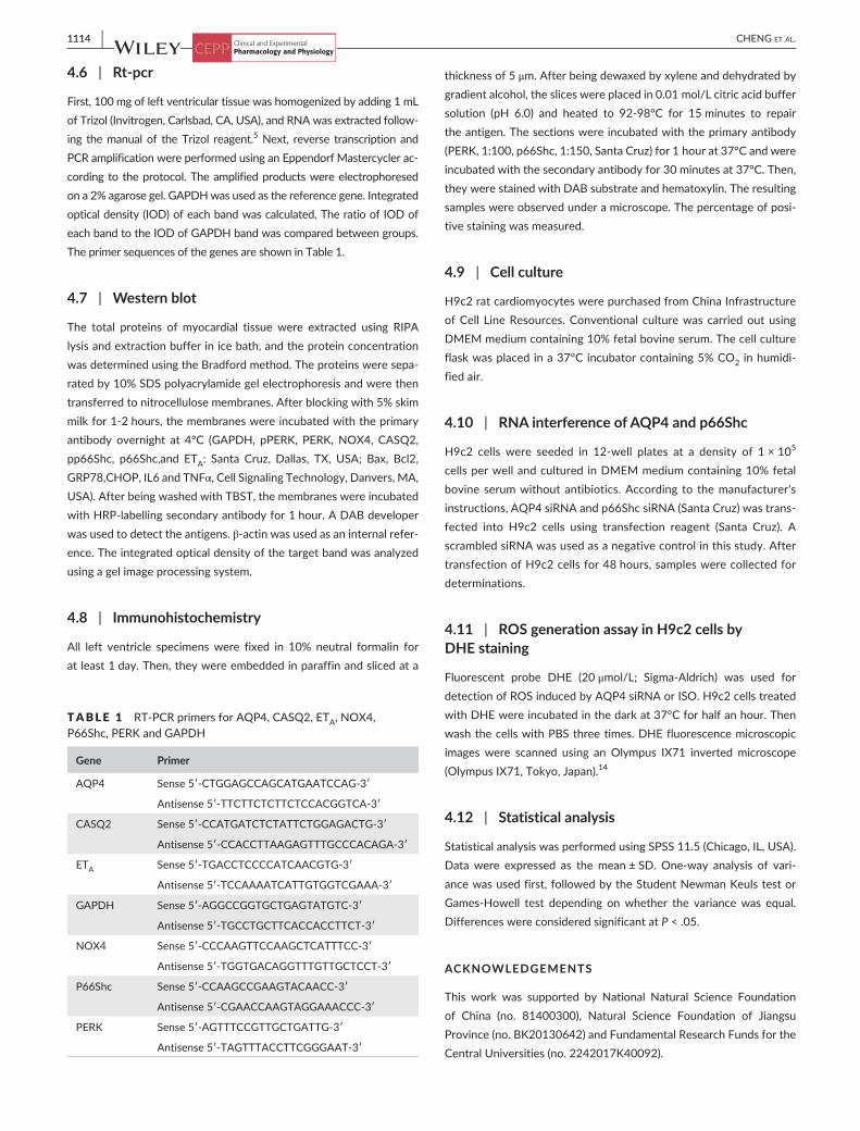
4.6 | Rt- pcr
First, 100 mg of left ventricular tissue was homogenized by adding 1 mL of Trizol (Invitrogen, Carlsbad, CA, USA), and RNA was extracted follow-ing the manual of the Trizol reagent.5 Next, reverse transcription and PCR amplification were performed using an Eppendorf Mastercycler ac-cording to the protocol. The amplified products were electrophoresed on a 2% agarose gel. GAPDH was used as the reference gene. Integrated optical density (IOD) of each band was calculated. The ratio of IOD of each band to the IOD of GAPDH band was compared between groups. The primer sequences of the genes are shown in Table 1.
4.7 | Western blot
The total proteins of myocardial tissue were extracted using RIPA lysis and extraction buffer in ice bath, and the protein concentration was determined using the Bradford method. The proteins were sepa-rated by 10% SDS polyacrylamide gel electrophoresis and were then transferred to nitrocellulose membranes. After blocking with 5% skim milk for 1- 2 hours, the membranes were incubated with the primary antibody overnight at 4°C (GAPDH, pPERK, PERK, NOX4, CASQ2, pp66Shc, p66Shc,and ETA: Santa Cruz, Dallas, TX, USA; Bax, Bcl2, GRP78,CHOP, IL6 and TNFα, Cell Signaling Technology, Danvers, MA, USA). After being washed with TBST, the membranes were incubated with HRP- labelling secondary antibody for 1 hour. A DAB developer was used to detect the antigens. β- actin was used as an internal refer-ence. The integrated optical density of the target band was analyzed using a gel image processing system.
4.8 | Immunohistochemistry
All left ventricle specimens were fixed in 10% neutral formalin for at least 1 day. Then, they were embedded in paraffin and sliced at a
thickness of 5 μm. After being dewaxed by xylene and dehydrated by gradient alcohol, the slices were placed in 0.01 mol/L citric acid buffer solution (pH 6.0) and heated to 92- 98°C for 15 minutes to repair the antigen. The sections were incubated with the primary antibody (PERK, 1:100, p66Shc, 1:150, Santa Cruz) for 1 hour at 37°C and were incubated with the secondary antibody for 30 minutes at 37°C. Then, they were stained with DAB substrate and hematoxylin. The resulting samples were observed under a microscope. The percentage of posi-tive staining was measured.
4.9 | Cell culture
H9c2 rat cardiomyocytes were purchased from China Infrastructure of Cell Line Resources. Conventional culture was carried out using DMEM medium containing 10% fetal bovine serum. The cell culture flask was placed in a 37°C incubator containing 5% CO2 in humidi-fied air.
4.10 | RNA interference of AQP4 and p66Shc
H9c2 cells were seeded in 12- well plates at a density of 1 × 10⁵ cells per well and cultured in DMEM medium containing 10% fetal bovine serum without antibiotics. According to the manufacturer’s instructions, AQP4 siRNA and p66Shc siRNA (Santa Cruz) was trans-fected into H9c2 cells using transfection reagent (Santa Cruz). A scrambled siRNA was used as a negative control in this study. After transfection of H9c2 cells for 48 hours, samples were collected for determinations.
4.11 | ROS generation assay in H9c2 cells by DHE staining
Fluorescent probe DHE (20 μmol/L; Sigma- Aldrich) was used for detection of ROS induced by AQP4 siRNA or ISO. H9c2 cells treated with DHE were incubated in the dark at 37°C for half an hour. Then wash the cells with PBS three times. DHE fluorescence microscopic images were scanned using an Olympus IX71 inverted microscope (Olympus IX71, Tokyo, Japan).14
4.12 | Statistical analysis
Statistical analysis was performed using SPSS 11.5 (Chicago, IL, USA). Data were expressed as the mean ± SD. One- way analysis of vari-ance was used first, followed by the Student Newman Keuls test or Games- Howell test depending on whether the variance was equal. Differences were considered significant at P < .05.
ACKNOWLEDGEMENTS
This work was supported by National Natural Science Foundation of China (no. 81400300), Natural Science Foundation of Jiangsu Province (no. BK20130642) and Fundamental Research Funds for the Central Universities (no. 2242017K40092).
TABLE 1 RT- PCR primers for AQP4, CASQ2, ETA, NOX4, P66Shc, PERK and GAPDH
Gene Primer
AQP4 Sense 5′- CTGGAGCCAGCATGAATCCAG- 3′
Antisense 5′- TTCTTCTCTTCTCCACGGTCA- 3′
CASQ2 Sense 5′- CCATGATCTCTATTCTGGAGACTG- 3′
Antisense 5′- CCACCTTAAGAGTTTGCCCACAGA- 3′
ETA Sense 5′- TGACCTCCCCATCAACGTG- 3′
Antisense 5′- TCCAAAATCATTGTGGTCGAAA- 3′
GAPDH Sense 5′- AGGCCGGTGCTGAGTATGTC- 3′
Antisense 5′- TGCCTGCTTCACCACCTTCT- 3′
NOX4 Sense 5′- CCCAAGTTCCAAGCTCATTTCC- 3′
Antisense 5′- TGGTGACAGGTTTGTTGCTCCT- 3′
P66Shc Sense 5′- CCAAGCCGAAGTACAACC- 3′
Antisense 5′- CGAACCAAGTAGGAAACCC- 3′
PERK Sense 5′- AGTTTCCGTTGCTGATTG- 3′
Antisense 5′- TAGTTTACCTTCGGGAAT- 3′

| 1115CHENG Et al.
DISCLOSURE
The authors declare that they have no conflicts of interest to disclose.
ORCID
Bicheng Liu http://orcid.org/0000-0002-9220-0477
REFERENCES
1. Madeira A, Moura TF, Soveral G. Detecting aquaporin function and regulation. Front Chem. 2016;4:3.
2. Szu JI, Binder DK. The role of astrocytic aquaporin- 4 in synaptic plas-ticity and learning and memory. Front Integr Neurosci. 2016;10:8.
3. Rutkovskiy A, Stenslokken KO, Mariero LH, et al. Aquaporin- 4 in the heart: expression, regulation and functional role in ischemia. Basic Res Cardiol. 2012;107:280.
4. Hao C, Liu W, Luan X, et al. Aquaporin- 4 knockout enhances astrocyte toxicity induced by 1- methyl- 4- phenylpyridinium ion and lipopolysac-charide via increasing the expression of cytochrome P4502E1. Toxicol Lett. 2010;198:225-231.
5. Cheng YS, Tang YQ, Dai DZ, Dai Y. AQP4 knockout mice manifest ab-normal expressions of calcium handling proteins possibly due to ex-acerbating pro- inflammatory factors in the heart. Biochem Pharmacol. 2012;83:97-105.
6. Camici GG, Savarese G, Akhmedov A, Luscher TF. Molecular mecha-nism of endothelial and vascular aging: implications for cardiovascular disease. Eur Heart J. 2015;36:3392-3403.
7. Wang Y, Zhao J, Yang W, et al. High- dose alcohol induces reac-tive oxygen species- mediated apoptosis via PKC- beta/p66Shc in mouse primary cardiomyocytes. Biochem Biophys Res Commun. 2015;456:656-661.
8. Miao Q, Wang Q, Dong L, Wang Y, Tan Y, Zhang X. The expression of p66shc in peripheral blood monocytes is increased in patients with coronary heart disease and correlated with endothelium- dependent vasodilatation. Heart Vessels. 2015;30:451-457.
9. Logue SE, Cleary P, Saveljeva S, Samali A. New directions in ER stress- induced cell death. Apoptosis. 2013;18:537-546.
10. Merlot AM, Shafie NH, Yu Y, et al. Mechanism of the induction of endoplasmic reticulum stress by the anti- cancer agent, Di- 2- pyridylketone 4,4- dimethyl- 3- thiosemicarbazone (Dp44mT): activation of PERK/eI-F2alpha, IRE1alpha, ATF6 and calmodulin kinase. Biochem Pharmacol. 2016;109:27-47.
11. Wu Y, Reece EA, Zhong J, et al. Type 2 diabetes mellitus induces congenital heart defects in murine embryos by increasing oxidative stress, endoplasmic reticulum stress and apoptosis. Am J Obstet Gynecol. 2016;215:366.e1-366.e10.
12. Lin X, Zhao Y, Li S. Astaxanthin attenuates glutamate- induced apop-tosis via inhibition of calcium influx and endoplasmic reticulum stress. Eur J Pharmacol. 2017;806:43-51.
13. Liu XH, Zhang ZY, Andersson KB, et al. Cardiomyocyte- specific dis-ruption of Serca2 in adult mice causes sarco(endo)plasmic reticulum stress and apoptosis. Cell Calcium. 2011;49:201-207.
14. Liu L, Cui J, Yang Q, et al. Apocynin attenuates isoproterenol- induced myocardial injury and fibrogenesis. Biochem Biophys Res Commun. 2014;449:55-61.
15. Basaki M, Asasi K, Tabandeh MR, Aminlari M. Polymorphism iden-tification and cardiac gene expression analysis of the calsequestrin 2 gene in broiler chickens with sudden death syndrome. Br Poult Sci. 2016;57:151-160.
16. Maalouf RM, Eid AA, Gorin YC, et al. Nox4- derived reactive oxygen species mediate cardiomyocyte injury in early type 1 diabetes. Am J Physiol Cell Physiol. 2012;302:C597-C604.
17. Esposito G, Imitola J, Lu J, et al. Genomic and functional profiling of human Down syndrome neural progenitors implicates S100B and aquaporin 4 in cell injury. Hum Mol Genet. 2008;17:440-457.
18. Patrussi L, Giommoni N, Pellegrini M, Gamberucci A, Baldari CT. p66Shc- dependent apoptosis requires Lck and CamKII activity. Apoptosis. 2012;17:174-186.
19. Cong XD, Ding MJ, Dai DZ, Wu Y, Zhang Y, Dai Y. ER stress, p66shc, and p- Akt/Akt mediate adjuvant- induced inflammation, which is blunted by argirein, a supermolecule and rhein in rats. Inflammation. 2012;35:1031-1040.
20. Dickhout JG, Carlisle RE, Austin RC. Interrelationship between cardiac hypertrophy, heart failure, and chronic kidney disease: endoplasmic retic-ulum stress as a mediator of pathogenesis. Circ Res. 2011;108:629-642.
21. Dalal S, Foster CR, Das BC, Singh M, Singh K. beta- Adrenergic recep-tor stimulation induces endoplasmic reticulum stress in adult cardiac myocytes: role in apoptosis. Mol Cell Biochem. 2012;364:59-70.
22. Ioannou K, Stel VS, Dounousi E, et al. Inflammation, endothelial dys-function and increased left ventricular mass in chronic kidney disease (CKD) patients: a longitudinal study. PLoS ONE. 2015;10:e0138461.
23. Li Q, Li J, Shao H, Li XX, Yu F, Xu M. Inhibition of CPU0213, a dual en-dothelin receptor antagonist, on apoptosis via Nox4- dependent ROS in HK- 2 cells. Cell Physiol Biochem. 2016;39:183-192.
24. Lo AC, Chen AY, Hung VK, et al. Endothelin- 1 overexpression leads to further water accumulation and brain edema after middle cerebral artery occlusion via aquaporin 4 expression in astrocytic end- feet. J Cereb Blood Flow Metab. 2005;25:998-1011.
25. Tanaka K, Koyama Y. Endothelins decrease the expression of aquapo-rins and plasma membrane water permeability in cultured rat astro-cytes. J Neurosci Res. 2011;89:320-328.
How to cite this article: Cheng Y, Chao J, Dai D, Dai Y, Zhu D, Liu B. AQP4- knockout aggravation of isoprenaline- induced myocardial injury is mediated by p66Shc and endoplasmic reticulum stress. Clin Exp Pharmacol Physiol. 2017;44: 1106-1115. https://doi.org/10.1111/1440-1681.12812


![St. Catherine · Anglin, Neville George Unlawful wounding T Malcolm, Othniel Stafari Robbery with aggravation M Thompson, Johnoy Famons [o.c. Puru] Robbery with aggravation M R –](https://img.dokumen.tips/doc/110x75/606a2eabed4bc80bc83876ed/st-catherine-anglin-neville-george-unlawful-wounding-t-malcolm-othniel-stafari.jpg)