Embed Size (px)
Citation preview

Approaches to Conventional Mechanical Ventilationof the Patient With Acute Respiratory Distress Syndrome
Dean R Hess PhD RRT FAARC
IntroductionVolume and Pressure Limitation
The ARDS Network Higher Versus Lower Tidal Volume StudyVolume or Pressure Limitation?Potential for Auto-PEEPPressure Controlled Versus Volume Controlled VentilationSynchronyTidal Volume Limitation in Patients Who Do Not Have ALI/ARDSIs Volume and Pressure Limited Ventilation Widely Applied?
Prolonged Inspiratory Time and Inverse Inspiratory-Expiratory RatioSetting PEEP for ALI and ARDS
The Randomized Controlled TrialsPEEP and Potential for Recruitment
The Meta-AnalysesApproaches to Setting PEEP
Stress and StrainSummary
To minimize ventilator-induced lung injury, attention should be directed toward avoidance ofalveolar over-distention and cyclical opening and closure of alveoli. The most impressive study ofmechanical ventilation to date is the Acute Respiratory Distress Syndrome (ARDS) Network studyof higher versus lower tidal volume (VT), which reported a reduction in mortality from 39.8% to31.0% with 6 mL/kg ideal body weight rather than 12 mL/kg ideal body weight (number-needed-to-treat of 12 patients). To achieve optimal lung protection, the lowest plateau pressure and VT
possible should be selected. What is most important is limitation of VT and alveolar distendingpressure, regardless of the mode set on the ventilator. Accumulating observational evidence sug-gests that VT should be limited in all mechanically ventilated patients—even those who do not haveALI/ARDS. Evidence does not support the use of pressure controlled inverse-ratio ventilation.Although zero PEEP is probably injurious, an area of considerable controversy is the optimalsetting of PEEP. Available evidence does not support the use of higher PEEP, compared to lowerPEEP, in unselected patients with acute lung injury (ALI)/ARDS. However, results of a meta-analysis using individual patients from 3 randomized controlled trials suggest that higher PEEPshould be used for ARDS, whereas lower PEEP may be more appropriate in patients with ALI.PEEP should be set to maximize alveolar recruitment while avoiding over-distention. Many ap-proaches for setting PEEP have been described, but evidence is lacking that any one approach issuperior to any other. In most, if not all, cases of ALI/ARDS, conventional ventilation strategies canbe used effectively to provide lung-protective ventilation strategies. Key words: acute lung injury;ALI; acute respiratory distress syndrome; ARDS; lung-protective ventilation strategies; mechanical ven-tilation; PEEP; ventilator-induced lung injury. [Respir Care 2011;56(10):1555–1572. © 2011 DaedalusEnterprises]
RESPIRATORY CARE • OCTOBER 2011 VOL 56 NO 10 1555

Introduction
Many lives have been saved by the use of mechanicalventilation. But after years of targeting ventilation strate-gies to normalize arterial blood gases, it was realized thatthese strategies might result in ventilator-induced lung in-jury (VILI). Lung-protective ventilation strategies haverecently received increasing attention and are now consid-ered standard practice. There are 2 principal causes ofVILI: alveolar over-distention, and cyclical opening andclosing of alveoli. Injurious ventilation strategies causeincreased permeability of the alveolar-capillary membraneand increased production of pro-inflammatory mediatorswithin the lungs. Leakage of the inflammatory mediatorsinto the bloodstream can result in downstream organ sys-tem failures, which can lead ultimately to multiple organfailure and death.1-6 Lung-protective ventilation strategiesare directed primarily toward volume and pressure limita-tion during the inspiratory phase and maintenance of al-veolar recruitment during the expiratory phase. The pur-pose of this paper is to review the evidence supportinglung-protective ventilation using conventional ventilationstrategies.
Volume and Pressure Limitation
The ARDS Network Higher Versus Lower TidalVolume Study
In the late 20th century, many experimental studies werepublished suggesting benefit from tidal volume (VT) lim-itation.7 Several patient studies reported that VT limitationwas safe,8-10 and a single-center study from Brazil foundthat inspiratory volume and pressure limitation with higherPEEP was associated with a survival benefit.11 This led tofunding by the National Institutes of Health for the AcuteRespiratory Distress Syndrome (ARDS) Network to studyventilation with lower VT, as compared with traditionalVT, for patients with acute lung injury (ALI) and ARDS.12,13
Patients were recruited from March 1996 through March1999 at 10 university hospitals in the United States. Vol-ume controlled continuous mandatory ventilation was usedand VT was set based on ideal body weight (IBW). In thegroup treated with traditional VT the target VT was 12 mL/kgIBW. This was subsequently reduced, if necessary, to main-tain plateau pressure (Pplat) � 50 cm H2O. In the grouptreated with lower VT, the target VT was reduced to 6 mL/kgIBW and subsequently reduced, as necessary, to maintainPplat � 30 cm H2O. The minimum VT was 4 mL/kg IBW.If Pplat dropped below 25 cm H2O, VT was increased insteps of 1 mL/kg IBW until Pplat was at least 25 cm H2Oor the VT was 6 mL/kg IBW. For patients with severedyspnea, the VT could be increased to 8 mL/kg IBW.
The results of the ARDS Network study are impres-sive—perhaps the most impressive of any study of me-chanical ventilation to date. The trial was stopped after theenrollment of 861 patients because mortality was lower inthe group treated with lower VT than in the group treatedwith traditional VT (31.0% vs 39.8%, P � .007). Trans-lated to the metrics of evidence-based medicine, the num-ber of patients needed to treat with the lower VT to avoida single death is 12 patients. The number of ventilator-freedays during the first 28 days after randomization was greaterin the lower-VT group (12 � 11 d vs 10 � 11 d, P � .007).The mean Pplat was 25 � 6 cm H2O in the lower-VT groupand 33 � 8 cm H2O in the higher-VT group (P � .001).The results of this study clearly demonstrate that, in pa-tients with ALI/ARDS, mechanical ventilation with a lowerVT and Pplat decreases mortality and ventilator days.
Volume or Pressure Limitation?
Whether the actual VT used should be based on theindividual patient’s lung mechanics, specifically Pplat,rather than a VT of 4–8 mL/kg has been debated.14 Theargument in favor of Pplat rather than VT limitation ques-tions the value of VT reduction in patients with ALI/ARDSwhose Pplat is � 30 cm H2O. To address this question,Hager et al15 performed a secondary analysis of data fromthe ARDS Network. They found a beneficial effect of VT
reduction from 12 mL/kg to 6 mL/kg, regardless of thePplat before the VT reduction (Fig. 1).16 Patients enrolled inthe ARDS Network study were divided into quartiles ofPplat. Using a logistic regression model, lower Pplat quar-tile, and lower Acute Physiology and Chronic Health Eval-uation III score were significant predictors of lower mor-tality. However, the interaction between VT and Pplat
quartile was not significant. Thus, patients in the 12 mL/kgPBW VT group would have benefited from VT reductionin each of the quartiles, even those in which Pplat wasalready � 31 cm H2O.
Terragni et al17 performed computed tomography (CT)at end-expiration and end-inspiration in 30 patients venti-
Dr Hess is affiliated with Respiratory Care Services, Massachusetts Gen-eral Hospital, Harvard Medical School, Boston, Massachusetts.
Dr Hess presented a version of this paper at the 26th New HorizonsSymposium, “ARDS Update,” at the 56th International Respiratory Con-gress of the American Association for Respiratory Care, held Decem-ber 6–9, 2010, in Las Vegas, Nevada.
Dr Hess has disclosed relationships with Philips Respironics, Covidien,ResMed, Pari, and Breathe Technologies.
Correspondence: Dean R Hess PhD RRT FAARC, Respiratory Care,Ellison 401, Massachusetts General Hospital, 55 Fruit Street, Boston MA02114. E-mail: [email protected].
DOI: 10.4187/respcare.01387
APPROACHES TO CONVENTIONAL MECHANICAL VENTILATION OF THE PATIENT WITH ARDS
1556 RESPIRATORY CARE • OCTOBER 2011 VOL 56 NO 10

lated with a VT of 6 mL/kg IBW. They identified 20 ofthese patients in whom tidal inflation occurred largely inthe normally aerated parts of the lungs, and 10 patients inwhom tidal inflation occurred largely within the hyperin-flated compartments. The non-aerated compartment wassmaller and the normally aerated compartment was largerin the more protected patients than in the less protectedpatients. Pulmonary cytokines were lower in the more pro-tected patients than in the less protected patients. Ventila-tor-free days were 7 � 8 days and 1 � 2 days in the moreprotected and less protected patients, respectively. Pplat
was 25–26 cm H2O in the more protected patients and28–30 cm H2O in the less protected patients. These resultssuggest that limiting VT to 6 mL/kg IBW and Pplat
� 30 cm H2O may not be sufficient in patients character-ized by a larger non-aerated compartment.
The findings of Hager et al15 and Terragni et al17 sug-gest that there might not be a safe Pplat in patients withALI/ARDS. To achieve optimal lung protection, the low-est Pplat and VT possible should be selected. This is limitedby the degree of permissive hypercapnia that the clinicianis willing to accept. It has been suggested, primarily fromexperimental models, that permissive hypercapnia may beprotective.18 Kregenow et al19 evaluated the effect of hy-percapnic acidosis in a secondary analysis of the ARDSNetwork database. They found that hypercapnic acidosiswas associated with reduced 28-day mortality in the12 mL/kg IBW VT group, after controlling for comorbidi-ties and severity of lung injury. However, this was not
found when the further ongoing injury was reduced by6 mL/kg predicted body weight VT.
Potential for Auto-PEEP
To prevent acidosis with low-VT ventilation, higher thannormal respiratory rates are set—as high a 35 breaths/minin the ARDS Network study.13 This has raised concernabout the development of auto-PEEP.20 However, whendata from patients enrolled in the ARDS Network studywere examined, the level of auto-PEEP was minimal.18
This, in fact, is predictable. Although a high respiratoryrate is expected to shorten the expiratory time, the inspira-tory time is shorter due to the smaller VT . In addition, thelow compliance (high elastance) in ALI/ARDS results in agreater elastic recoil pressure pushing gas out of the lungsduring exhalation.
Pressure Controlled Versus Volume ControlledVentilation
One of the ongoing debates is whether pressure con-trolled ventilation (PCV) should be used a part of lung-protective ventilation strategy.21 There are advantages anddisadvantages to either PCV or volume controlled venti-lation (VCV) (Table 1). Purists will argue that the ARDSNetwork study was conducted with VCV; to achieve sim-ilar results, one should apply the evidence in the same way
Fig. 1. Mortality versus quartile of day-1 plateau pressure (Pplat) at tidal volume (VT) of 6 mL/kg ideal body weight (IBW) or 12 mL/kg IBW.The bars show the VT, the Pplat range, and the number of patients in each category. ARR � absolute risk reduction. (Data from Reference 76.Adapted from Reference 16, with permission.)
APPROACHES TO CONVENTIONAL MECHANICAL VENTILATION OF THE PATIENT WITH ARDS
RESPIRATORY CARE • OCTOBER 2011 VOL 56 NO 10 1557

as was done in the predicate study. However, others willargue that some advantages might exist with PCV. WithPCV, Pplat can never be greater than the peak inspiratorypressure. The square wave of inspiratory pressure withPCV will result in a higher mean airway pressure, whichmight improve ventilation/perfusion matching and PaO2
.Moreover, the flow pattern is an exponential decay, whichmight improve distribution of ventilation, ventilation/per-fusion matching, and PaO2
. Finally, the variable inspiratoryflow has the potential to improve patient-ventilator syn-chrony.
Despite many clinicians having a bias in favor of PCVin patients with ALI/ARDS, evidence supporting the su-periority of PCV in this setting is weak. For the same VT,the same inspiratory time, and a descending ramp of flowwith VCV, the differences in PaO2
between PCV and VCVare trivial.22 Experimental models of ALI/ARDS suggestthat the high initial flow that occurs with PCV might beinjurious rather than lung-protective.23-25
Whether synchrony is better with PCV than VCV is alsodebatable. Some have reported better synchrony withPCV,26,27 but this has not been confirmed by others.28
Adaptive pressure control modes might be particularlyproblematic, as they can take away support for the patientif respiratory drive is high and the resultant VT exceeds thetarget.29-33 Some clinicians are attracted to PCV because itallows the patient to increase VT if respiratory drive in-creases. However, this may make it difficult to avoid al-veolar over-distention and maintain lung-protective venti-lation.28,34 Indeed, Leray et al35 report a case in which apatient recovering from ARDS was switched from VCV(VT 6 mL/kg IBW) to pressure support ventilation. Withpressure support ventilation the VT increased to 14 mL/kgand the patient developed air leaks. When VCV was re-stored, the air leaks resolved. This makes the point that VT
can become excessive with pressure-targeted modes. Al-though PCV maintains a constant pressure applied to theairway, any additional effort from the patient will lowerthe pleural pressure, and the transpulmonary pressure willincrease (Fig. 2).34
In the hands of a skilled clinician, either VCV or PCVcan probably be applied in a lung-protective manner. Whatis most important is limitation of VT and alveolar distend-ing pressure, regardless of the mode set on the ventilator.
Table 1. Volume Controlled Versus Pressure Controlled Ventilation
Pressure Control Volume Control Adaptive Pressure Control
Pplat
Good Limits Pplat with lowcompliance/high resistance
Reduces Pplat with high compliance/low resistance
Keeps transpulmonary pressure(alveolar stretch) constant withpatient effort
Reduces Pplat with high compliance/low resistance
Bad Maintains Pplat with highcompliance/low resistance
Allows increased transpulmonarypressure (alveolar stretch)with patient effort
Increases Pplat with low compliance/high resistance
Airway pressure decreases witheffort
Increases Pplat with low compliance/high resistance
VT
Good Reduces VT with lowcompliance/high resistance
Maintains VT with high compliance/low resistance
Maintains VT with high compliance/low resistance
Bad Increases VT with highcompliance/low resistance
Increased VT with patient effortHigh initial flow may be
injurious
Maintains VT with low compliance/high resistance
Maintains VT with low compliance/high resistance
High initial flow may be injurious
Patient-Ventilator SynchronyGood Variable flow can help Maintains alveolar ventilation and
avoids asynchrony due to acidosisVariable flow may help
Bad Asynchrony can occur even withvariable flow
Fixed flow can be uncomfortable Asynchrony may occur if ventilatortakes away supportinappropriately
Pplat � plateau pressureVT � tidal volume(Adapted from Reference 21.)
APPROACHES TO CONVENTIONAL MECHANICAL VENTILATION OF THE PATIENT WITH ARDS
1558 RESPIRATORY CARE • OCTOBER 2011 VOL 56 NO 10

Synchrony
It is the perception of many clinicians that patient-ventilator asynchrony ensues when VT is reduced to6 mL/kg. Why this should occur is unclear. A normal VT is6–8 mL/kg, so it would seem that this VT should be com-fortable during mechanical ventilation. There are severalpotential reasons why a VT of 6–8 mL/kg might not becomfortable with ALI/ARDS. First, dead space is increasedwith ALI/ARDS, and thus respiratory acidosis will occurunless minute ventilation is increased.36-40 In the ARDSNetwork study,13 respiratory rates up to 35 breaths/minwere used in an attempt to avoid acidosis. Other potentialreasons for asynchrony are pain and anxiety due to endo-tracheal intubation and the disease process. Thus, adequateattention should be given to address these discomforts.
With low-VT ventilation the inspiratory time may bevery short. This can produce an inspiratory time set on theventilator that is less than the neural inspiratory time of the
patient. The result is that the patient will double-trigger theventilator. During VCV, a double-trigger can result in breathstacking and double the desired VT. This effectively ne-gates the intent of volume and pressure limitation. In onestudy it was reported that double-triggering is commonduring low-VT ventilation.41
A number of strategies can be used to improve patient-ventilator synchrony during lung-protective ventilation (Ta-ble 2).42,43 A recent study44 reported improved outcomeswith paralysis for the first 48 hours following intubation inpatients with ARDS. The mechanism to explain this ben-efit is unclear, but may be related to improved ability toapply lung-protective ventilation strategies in the earlyphase of the disease process. A common perception ofclinicians is that there is a greater requirement for sedationwith low-VT ventilation. That sedative doses were foundto be similar for patients randomized to 6 mL/kg versus12 mL/kg VT in 2 ARDS Network centers supports theconclusion that sedative needs may be largely determinedby clinical factors other than ventilation strategy.45,46
Tidal Volume Limitation in Patients Who Do NotHave ALI/ARDS
Although the evidence is clear and conclusive for vol-ume and pressure limitation in patients with ALI/ARDS,should we limit the VT in all patients? There are currentlyno randomized controlled trials that address this question,but accumulating observational evidence suggests that VT
should be limited in all mechanically ventilated pa-tients.47-51
Yilmaz et al51 reported the results of a quality-assuranceproject in which an interdisciplinary team of intensivistsand respiratory therapists designed a protocol to limit VT
to a maximum of 10 mL/kg IBW in all patients receivinginvasive ventilation, with a recommendation to use6–8 mL/kg for patients at any risk of ALI/ARDS. A chartwith calculated values of IBW was attached to each ven-tilator and it was also provided online. Comprehensivedidactic and Web-based teaching was provided to physi-cians, nurses, and respiratory therapists involved in thecare of mechanically ventilated patients. This protocol wasimplemented along with a protocol to limit transfusions.With implementation of this protocol the frequency of ALIdecreased from 28% to 10%, the duration of mechanicalventilation decreased from a median of 5 days to 4 days,and, in a multivariate logistic regression analysis, the in-tervention was associated with a reduction in the frequencyof new ALI (odds ratio 0.21, 95% CI 0.10–0.40). Theseresults suggest that ALI may be, to a large extent, iatro-genic and might be preventable through relatively simpleinterventions such as limitation of VT. Recent studies havealso reported benefit for VT limitation in patients withsevere brain injury52,53 and in potential organ donors with
Fig. 2. Estimation of transpulmonary pressure during spontaneousbreathing on pressure-targeted ventilation. Note that the pressureacross the alveolus is determined not only by the pressure appliedto the airway, but also by the change in pleural pressure. Paw � prox-imal airway pressure. PS � pressure support. PR � pressure dropdue to airways resistance. PA � alveolar pressure. �PA � trans-alveolar pressure. Patm � atmospheric pressure. Ppl � pleural pres-sure. (Adapted from Reference 34, with permission.)
APPROACHES TO CONVENTIONAL MECHANICAL VENTILATION OF THE PATIENT WITH ARDS
RESPIRATORY CARE • OCTOBER 2011 VOL 56 NO 10 1559

brain death,54 where it increased the number of eligibleand harvested lungs compared with a conventional strat-egy using higher VT.
Is Volume and Pressure Limited Ventilation WidelyApplied?
Volume and pressure limitation, at least for ALI/ARDS,should be standard practice. Some reports published soonafter publication of the ARDS Network study found onlya modest change in the prescribed VT.55,56 However, morerecent studies have not confirmed those findings. Estebanet al57 reported that in 2004, compared with 1998, VT
decreased significantly in patients with ARDS (7.4 mL/kgvs 9.1 mL/kg, P � .001). Checkley et al58 reported a
gradual and ultimately substantial effect on lowering of VT
at ARDS Network hospitals after completion of the study.However, these findings may not be generalizable to non-enrolled patients or to patients from other hospitals.
Adoption of lower-VT ventilation may be improved withfeedback and education on lung-protective mechanical ven-tilation. Even a costly intervention to improve adherenceto low-VT ventilation in patients with ALI reduces deathand is cost-effective by current societal standards.59 Re-cording VT in mL/kg IBW rather than the more traditionalapproach of absolute VT might draw clinician attention toexcessive VT. Similarly, recording Pplat rather than peakinspiratory pressure alone may draw clinicians’ attentionto excessive distending pressure. A recent study describedthe use of a computer system to alert bedside providers
Table 2. Approaches to Patient-Ventilator Asynchrony
1. Sedation, Analgesia, ParalysisAppropriate sedation and analgesia are necessary during mechanical ventilation, regardless of tidal volume. Factors such as agitation, delirium,metabolic acidosis, drug withdrawal, septic encephalopathy, and pain need to be considered. Neuromuscular blocking agents should beconsidered in the 48 hours following intubation; otherwise, paralysis used to achieve patient-ventilator synchrony only if sedation and analgesiaare insufficient and when other methods described here have been exhausted.
2. Respiratory RateAn increase in respiratory rate setting on the ventilator may match the breathing pattern of the patient to the ventilator, thereby enhancingsynchrony. Increasing the respiratory rate setting decreases work of breathing and increases patient comfort. During transition to lower-tidal-volume ventilation, the respiratory rate should be increased as tidal volume is decreased, to maintain constant minute ventilation (maximum35 breaths/min).
3. Tidal VolumeAn increase in tidal volume, if accompanied by an increase in alveolar ventilation, decreases respiratory drive. The ARDS Network protocolallows tidal volume to be increased to 8 mL/kg ideal body weight in the case of asynchrony and severe dyspnea, provided plateau pressure is� 30 cm H2O.
4. Trigger SensitivitySet trigger as sensitive (as low) as possible without causing auto-triggering.
5. Auto-PEEPMinimize the amount of auto-PEEP (set appropriate inspiratory-expiratory ratio, minimize airways resistance).
6. Inspiratory FlowAn increase in set inspiratory flow may better meet the flow demand of the patient and improve patient comfort. A higher inspiratory flow alsodecreases neural inspiratory time, however, resulting in a greater spontaneous breathing frequency, which can further contribute to asynchrony.In this case a lower inspiratory flow may be appropriate.
7. Inspiratory TimeA shorter inspiratory time (higher inspiratory flow during volume controlled ventilation) may improve patient-ventilator synchrony. If theinspiratory time setting on the ventilator is less than the neural inspiratory time, however, double-triggering and worsening asynchrony mayoccur. In this case, a longer inspiratory time may be appropriate.
8. Flow WaveformAsynchrony may improve with a descending flow waveform in some patients. For the same peak flow, inspiratory time is longer with adescending flow, which may achieve the goal of better synchrony because of the higher flow, while avoiding double-triggering secondary to aninspiratory time that is too short.
9. Pressure Controlled Ventilation (PCV)PCV achieves the goals of a descending flow waveform and an adjustable inspiratory time. PCV may result in better synchrony in somepatients. A potential limitation of PCV is the possibility that transpulmonary pressure (an important determinant of volutrauma) may increasebecause of the generation of high negative intrapleural pressure swings, consequently increasing delivered tidal volume. For the same tidalvolume and inspiratory flow, work of breathing is probably the same for PCV and volume controlled ventilation.
10. Pressure Rise TimeWith PCV the clinician can adjust the rate of pressure rise at the onset of the inspiratory phase. Faster rise time may affect work of breathingand patient comfort.
(Adapted from References 42 and 43.)
APPROACHES TO CONVENTIONAL MECHANICAL VENTILATION OF THE PATIENT WITH ARDS
1560 RESPIRATORY CARE • OCTOBER 2011 VOL 56 NO 10

(respiratory therapists and critical care fellows) by textpaging notification about potentially injurious ventilatorsettings. This was associated with decreased patient expo-sure to potentially injurious settings.60
Prolonged Inspiratory Time andInverse Inspiratory-Expiratory Ratio
In the mid-1980s there were reports of improved oxy-genation with pressure controlled inverse-ratio ventilation(PCIRV),61-65 and for some time in the decade thereafterthere was enthusiasm for this method. With PCIRV, aninspiratory time greater than the expiratory time is used toincrease mean airway pressure and thus improve arterialoxygenation. Although inverse-ratio ventilation is mostoften used with PCV, VCV with inverse ratio has alsobeen described.66 Following the initial enthusiasm for thisapproach, a number of subsequent controlled studies re-ported no benefit or marginal benefit of PCIRV over moreconventional approaches to ventilator support in patientswith ARDS.67-71 A disadvantage of this approach is thatthe auto-PEEP that occurs with PCIRV may adverselyaffect hemodynamics. Because this approach is uncom-fortable for the patient, sedation and paralysis are oftenrequired. Based on the available evidence, there is no clearbenefit for PCIRV in the management of patients withARDS. The improvement in oxygenation PCIRV is smalland the risk of auto-PEEP and hemodynamic compromiseis great.72-74
Setting PEEP for ALI and ARDS
Most patients who require mechanical ventilation forALI/ARDS receive PEEP of 5–12 cm H2O.57,75
PEEP � 5 cm H2O is probably harmful, at least in theearly stages of the disease process.76 Higher PEEP mayimprove oxygenation and reduce VILI, due to better alve-olar recruitment. But higher PEEP may also cause lunginjury from over-distention. A matter of controversy ishow to set PEEP for patients with ALI/ARDS.
The Randomized Controlled Trials
To date, 6 clinical trials have assessed the application oflower versus higher PEEP in patients with ALI/ARDS.11,77-81 Of these, only 2 reported a significant mor-tality reduction with higher PEEP.11,81 In both of the stud-ies, however, 2 interventions were applied. That is, a higherPEEP was combined with a lower VT. Therefore, it isunknown whether the mortality benefit was related to thehigher PEEP, lower VT, or a combined effect of the 2interventions.
In an ARDS Network study, Brower et al77 randomlyassigned 549 patients with ALI/ARDS to receive eitherlower or higher PEEP. All patients received volume andpressure limitation according to the original ARDS Net-work study.13 The PEEP was selected according to differ-ent tables of predetermined combinations of PEEP andFIO2
, selected to maintain PaO2in the range of 55–80 mm Hg
or SpO2in the range of 88–95% (Table 3). The mean � SD
PEEP on days 1 through 4 were 8.3 � 3.2 cm H2O in thelower-PEEP group and 13.2 � 3.5 cm H2O in the higher-PEEP group. Not surprising, the PaO2
/FIO2was significantly
greater in the higher-PEEP group (P � .01). The rates ofdeath before hospital discharge were 24.9% and 27.5%,respectively (P � .48). There were 14.5 � 10.4 ventila-tor free days in the lower-PEEP group and 13.8 � 10.6ventilator free days in the higher-PEEP group (P � .50).The data and safety monitoring board stopped the study atthe second interim analysis, after 549 patients had beenenrolled, on the basis of futility. The authors concludedthat, in patients with ALI/ARDS who receive mechanicalventilation with a VT goal of 6 mL/kg of IBW and aPplat � 30 cm H2O, clinical outcomes are similar whetherlower or higher PEEP is used.
Meade et al78 reported a study to compare a low-VT
ventilation strategy with an experimental strategy designedas an open-lung approach, which combines low VT, lung-recruitment maneuvers, and high PEEP. This was a ran-domized controlled trial with concealed allocation andblinded data analysis conducted in 30 intensive care unitsin Canada, Australia, and Saudi Arabia. The study enrolled983 patients with ALI and PaO2
/FIO2� 250 mm Hg; 85%
Table 3. Tables Used to Set Combinations of FIO2and PEEP in the ARDS Network Study
Lower PEEP/Higher FIO2
FIO20.3 0.4 0.4 0.5 0.5 0.6 0.7 0.7 0.7 0.8 0.9 0.9 0.9 1.0
PEEP 5 5 8 8 10 10 10 12 14 14 14 16 18 18–24Higher PEEP/Lower FIO2
FIO20.3 0.3 0.3 0.3 0.3 0.4 0.4 0.5 0.5 0.5–0.8 0.8 0.9 0.9 1.0 1.0
PEEP 5 8 10 12 14 14 16 16 18 20 22 22 22 22 24
(Adapted from Reference 77.)
APPROACHES TO CONVENTIONAL MECHANICAL VENTILATION OF THE PATIENT WITH ARDS
RESPIRATORY CARE • OCTOBER 2011 VOL 56 NO 10 1561

of the subjects met the criteria for ARDS at enrollment.The control strategy included a target VT of 6 mL/kg IBW,Pplat � 30 cm H2O, and a conventional PEEP level(n � 508). The open-lung strategy included target VT of6 mL/kg IBW, Pplat � 40 cm H2O, recruitment maneuvers,and higher PEEP (n � 475). The mean PEEP was14.6 � 3.4 cm H2O in the open-lung group and9.8 � 2.7 cm H2O in the control group during the first72 hours (P � .001). All-cause hospital mortality rateswere 36.4% and 40.4%, respectively (P � .19). Baro-trauma rates were 11.2% and 9.1% (P � .33). The open-lung group had lower rates of refractory hypoxemia (4.6%vs 10.2%, P � .01), death with refractory hypoxemia (4.2%vs 8.9%, P � .03), and use of rescue therapies (5.1% vs9.3%, P � .045). The authors concluded that, for patientswith ALI/ARDS, a ventilation strategy designed to recruitand open the lungs resulted in no significant difference inhospital mortality, compared with an established low-VT
ventilation strategy. This open-lung strategy did appear toimprove secondary end points related to hypoxemia anduse of rescue therapies.
Mercat et al79 designed a study to compare the effect onoutcome of a strategy for setting PEEP aimed at increasingalveolar recruitment while limiting hyperinflation to oneaimed at minimizing alveolar distention in patients withALI. This was a multicenter randomized controlled trial of767 adults conducted in 37 intensive care units in France.The VT was set at 6 mL/kg IBW in both strategies. Patientswere randomly assigned to a moderate PEEP strategy of5–9 cm H2O (n � 382) or to a PEEP set to reach a Pplat of28–30 cm H2O (n � 385). The 28-day mortality rate in theminimal-distention group was 31.2% versus 27.8% in theincreased-recruitment group (P � .31). The hospital mor-tality rate in the minimal-distention group was 39.0% ver-sus 35.4% in the increased-recruitment group (P � .30).The increased-recruitment group, compared with the min-imal-distention group, had a higher median number ofventilator-free days (7 d, interquartile range [IQR] 0–19 dvs 3 d, IQR 0–17, P � .04) and organ-failure-free days(6 d, IQR 0–18 vs 2 d, IQR 0–16, P � .04). This strategyalso was associated with higher compliance values, betteroxygenation, less use of adjunctive therapies, and largerfluid requirements. The authors concluded that a strategyfor setting PEEP aimed at increasing alveolar recruitmentwhile limiting hyperinflation did not significantly reducemortality. However, it did improve lung function and re-duced the duration of mechanical ventilation and the du-ration of organ failure.
The reason why these 3 trials were negative with respectto a survival benefit has been the source of much specu-lation and debate. Perhaps a higher level of PEEP in pa-tients with ALI/ARDS is not effective. Perhaps the meth-ods used to set PEEP were not the correct approaches.Perhaps higher PEEP is not effective in unselected patients
with ALI/ARDS, but might be effective in patients withARDS but not ALI, or in patients with a higher potential forrecruitment.Orperhaps thesestudies,althoughrelatively large,were underpowered to show a difference in outcome.82
PEEP and Potential for Recruitment
An important issue with the use of higher PEEP is theassociated increase in Pplat.43 This is more likely to occurwhen higher PEEP is added to patients who have nonre-cruitable lung regions. Patients who have recruitable lungshave proportionally less increase in Pplat when PEEP israised, and such patients may benefit from PEEP with lessrisk of over-distention. It is likely that the trials of higherversus lower PEEP77-79 enrolled patients who had both alower and a greater potential for alveolar recruitment.
In 68 patients with ALI/ARDS, Gattinoni et al83 eval-uated the relationship between the percentage of poten-tially recruitable lung and the clinical and physiologic ef-fects of PEEP. Patients underwent whole-lung CT at airwaypressures of 5, 15, and 45 cm H2O. The potential forrecruitment was defined as the proportion of lung tissue inwhich aeration was restored at airway pressures between 5and 45 cm H2O. They found that the percentage of poten-tially recruitable lung ranged widely in these patients, witha mean of 13 � 11% of the lung weight. An average of24% of the lung could not be recruited. Patients with ahigher percentage of potentially recruitable lung, greaterthan the median value of 9%, had greater total lung weights,lower PaO2
/FIO2and respiratory-system compliance, higher
dead space, and higher mortality. CT was better than phys-iologic variables for predicting the amount of recruitablelung, Clinically relevant changes in physiologic markerswith higher PEEP, such as reduced physiologic dead space(or a decrease in PaCO2
at a fixed minute ventilation) or anincrease in respiratory-system compliance, may be helpfulindicators of lung recruitability.
To investigate how lung recruitability influences alve-olar strain and cyclical opening and closing after the ap-plication of high PEEP, Caironi et al84 analyzed data fromthe 68 patients with ALI/ARDS in the previously describedpaper by Gattinoni.83 Alveolar strain and opening and clos-ing lung tissue were computed at 5 and 15 cm H2O PEEP.In patients with a higher percentage of potentially re-cruitable alveoli, the increase in PEEP reduced the amountof opening and closing lung tissue (P � .001). However,no differences were observed in patients with a lower per-centage of potentially recruitable alveoli. Alveolar strainsimilarly increased in the 2 groups. Opening and closinglung tissue was distributed mainly in the dependent andhilar lung regions, and it was an independent risk factor fordeath. The authors concluded that, in patients with higheralveolar recruitability, the beneficial impact of reducing
APPROACHES TO CONVENTIONAL MECHANICAL VENTILATION OF THE PATIENT WITH ARDS
1562 RESPIRATORY CARE • OCTOBER 2011 VOL 56 NO 10

cyclical alveolar opening and closing by increasing PEEPprevails over the effects of increasing alveolar strain.
Because the effects of high PEEP depend on lung re-cruitability, which varies widely among patients with ARDS,
increasing PEEP may lead to alveolar over-distention (harm),or to less cyclical alveolar opening and closing (benefit). Thisis illustrated in Figure 3. If the increase in PEEP is greaterthan the increase in Pplat, presumably alveolar recruitment hasoccurred, the respiratory system is improved, and the benefitof higher PEEP may be greater than the potential harm of thesmall increase in Pplat. On the other hand, if the increase inPplat is greater than the increase in PEEP, particularly if theresulting Pplat is � 30 cm H2O, then it can be argued that theharm of over-distention is greater than the benefit of alveolarrecruitment with PEEP. Because the 3 PEEP studies probablyenrolled some patients with higher potential for recruitmentand other patients with a lower potential for recruitment, thismay explain the negative results.
A higher PEEP potentially increases the risks of hemo-dynamic compromise and barotrauma. Pneumothorax ratedoes not appear to be greater in patients receiving higherPEEP.85 The risk of complications is probably related toover-distention rather than PEEP per se.
The Meta-Analyses
The value of a meta-analysis is that, by pooling datafrom several studies, statistical power is improved. Thus,results from smaller negative studies might prove positivewhen combined in a meta-analysis. There are 5 meta-analyses published on the topic of higher versus lowerPEEP in patients with ALI/ARDS (Table 4).85-89 Of these,
Table 4. Meta-analyses of Studies That Compared Higher Versus Lower PEEP
Meta-analysis Studies Included Principal Finding Comments
Oba87 Amato11 Lower hospital mortality with higher PEEP(RR 0.89, 95% CI 0.80–0.99, P � .03)
Analysis included studies that used 2simultaneous interventions: higherPEEP with lower VT vs lower PEEPwith higher VT
Brower77
Meade78
Mercat79
Villar81
Putensen89 Brower77 Higher PEEP did not reduce hospital mortality (OR0.86, 95% CI, 0.72–1.02, P � .08)
Lower VT reduced hospital mortality(OR 0.75, 95% CI 0.58–0.96,P � .02) compared to higher VT atsimilar PEEP
Meade78
Mercat79
Phoenix88 Brower77 Higher PEEP did not reduce hospital mortality in the3 trials that used 2 PEEP levels, but VT was heldconstant (RR 0.90, 95% CI 0.72–1.02, P � .077)
Concluded that high PEEP may have aclinically relevant mortality benefitMeade78
Mercat79
Briel86 Brower77 Patients with ARDS benefited from higher PEEP(RR 0.90, 95% CI 0.81–1.00, P � .049)
Patients without ARDS did not benefit from higherPEEP (RR 1.37, 95% CI 0.98–1.92, P � .07)
Used patient-level data rather thanpooled resultsMeade78
Mercat79
Dasenbrook85 Brower77 No significant difference in 28-day mortality(RR 0.90, 95% CI 0.79–1.02) or in-hospitalmortality (RR 0.94, 95% CI 0.84–1.05) for higherversus lower PEEP
Pneumothorax rate not increased withhigher PEEPMeade78
Mercat79
Talmor80
RR � relative riskOR � odds ratio(Adapted from Reference 82.)
Fig. 3. Potential effects of an increase in PEEP. If the potential forrecruitment is low, an increase in PEEP results in a large increase inplateau pressure (Pplat) (increased driving pressure), to an unsafe level.In this case, the potential harm from over-distention probably out-weighs any benefit resulting from increased alveolar recruitment. Ifthe potential for recruitment is high, an increase in PEEP results inlittle increase in Pplat. In this case, the potential benefit of increasedPEEP probably outweighs the harm due to the small increase in Pplat.
APPROACHES TO CONVENTIONAL MECHANICAL VENTILATION OF THE PATIENT WITH ARDS
RESPIRATORY CARE • OCTOBER 2011 VOL 56 NO 10 1563

4 found no effect on mortality from higher PEEP, com-pared with moderate PEEP, in unselected patients withALI/ARDS.85,87-89
The meta-analysis by Briel et al86 is of particular inter-est. Data from 2,299 individual patients in 3 trials wereanalyzed, using uniform outcome definitions. The mortal-ity rate was 32.9% for patients assigned to treatment withhigher PEEP, and 35.2% for patients assigned to lowerPEEP (adjusted relative risk 0.94, 95% CI 0.86–1.04,P � .25). However, treatment effects varied with the pres-ence or absence of ARDS (PaO2
/FIO2� 200 mm Hg). In
patients with ARDS the mortality was 34.1% in the high-er-PEEP group and 39.1% in the lower-PEEP group (ad-justed relative risk 0.90, 95% CI 0.81–1.00, P � .049). Inpatients without ARDS the mortality was 27.2% in thehigher-PEEP group and 19.4% in the lower-PEEP group(adjusted relative risk 1.37, 95% CI 0.98–1.92, P � .07).The results of this meta-analysis suggest that treatmentwith higher versus lower PEEP was associated with im-proved survival among the subgroup of patients with ARDS(PaO2
/FIO2� 200 mm Hg). Interestingly, there was a trend
toward worse outcomes with higher PEEP in the subgroupwith ALI (PaO2
/FIO2� 200 mm Hg).
Approaches to Setting PEEP
Zero PEEP is probably harmful during mechanical ven-tilation of patients with ALI/ARDS.76 Higher PEEP is prob-ably better in patients with ARDS, and moderate PEEPmay be better for patients with ALI. The challenge at thebedside is to select the PEEP that is appropriate for theindividual patient, and a number of approaches have beendescribed in the literature (Table 5).
In the ARDS Network studies13,77 and the study by Meadeet al,78 PEEP was individually titrated according to a table,with combinations of PEEP and FIO2
selected for a targetarterial oxygenation (see Table 3). These tables have beenwidely criticized as arbitrary and not fitted to the lungmechanics of an individual patient. Despite the criticism ofthese tables, they have been used successfully in all of the
trials conducted by the ARDS Network and thus have highface validity.16 But because these tables do not select PEEPbased on potential for alveolar recruitment, their use mightlead to over-distention in patients who have less potentialfor recruitment.90-92
The method used by Mercat et al79 is simple and attrac-tive. They adjusted PEEP based on airway pressure, andPEEP was set as high as possible without increasing the
Table 5. Methods for Selecting PEEP
Table Use a table of FIO2and PEEP combinations to achieve PaO2
or SpO2in a target range
Maximal PEEP while avoiding overdistention Use highest PEEP with plateau pressure � 30 cm H2OGas exchange Lowest shunt (highest PaO2
), lowest dead space (lowest PaCO2), best oxygen delivery
(CaO2� cardiac output)
Compliance Use the PEEP that results in the highest respiratory-system complianceStress index Observe the pressure-time curve during constant-flow inhalation for signs of tidal
recruitment and over-distentionEsophageal pressure8 Estimate the intra-pleural pressure with an esophageal-balloon measurement of
esophageal pressure, then determine the optimal PEEPPressure-volume curve Set PEEP slightly higher than the lower inflection pointImaging Computed tomography, electrical impedance tomography, ultrasound
Fig. 4. The pressure-volume curve of a normal subject (dashedcurve) and a patient with acute respiratory distress syndrome(ARDS) (solid curve). The pressure-volume curve is shifted down-wards on the volume axis and has a reduced total lung capacity(TLC). The sigmoid shape of the curve is much more evident inARDS. Note the small amount of pressure at the start of the ARDSpressure-volume curve, indicating a small amount of intrinsic PEEP(PEEPI) at end-expiratory lung volume (EELV). Some investigatorsdivide the curve into linear segments: Cstart, Cinf or Clin, and Cend
(thin lines, explained below). Using these segments, the upper andlower Pflex (the pressure at the intersection of 2 lines: a low-com-pliance region at low lung volumes [Cstart] and a higher-compli-ance region at higher lung volumes [Cinf]) were defined by theintersection of these lines. The lower inflection point (LIP) andupper inflection point (UIP) are defined by where the curve firstbegins to deviate from the line Clin. Mathematically these are notinflection points; the true inflection point (where concavity changesdirection) is marked by the solid dot. (Adapted from Reference 94.)
APPROACHES TO CONVENTIONAL MECHANICAL VENTILATION OF THE PATIENT WITH ARDS
1564 RESPIRATORY CARE • OCTOBER 2011 VOL 56 NO 10

Pplat above 28–30 cm H2O. PEEP was individually titratedbased on Pplat, regardless of its effect on oxygenation.Using this approach, higher PEEP will probably be setwhen there is much potential for recruitment, because therewill be less increase in Pplat when the application of PEEPincreases alveolar recruitment. On the other hand, if thereis less potential for recruitment, the Pplat limit will bereached quickly when the PEEP is increased.79
In a classic paper published in 1975, Suter et al93 stud-ied 15 normovolemic patients who required mechanicalventilation for acute pulmonary failure. The PEEP thatresulted in the maximum oxygen delivery and the lowestdead-space fraction also resulted in the greatest respirato-ry-system compliance. The optimal PEEP ranged between0 and 15 cm H2O. Mixed venous PO2
increased between0 PEEP and the PEEP that resulted in maximum oxygendelivery, but then decreased at higher PEEP. The authorsconcluded that respiratory-system compliance can be usedto determine the optimal PEEP. Because compliance � VT
/(Pplat – PEEP), the optimal PEEP is that which results inthe lowest driving pressure (Pplat –PEEP) if VT is keptconstant. What is most attractive about this approach isthat it is easy to evaluate at the bedside.
The pressure-volume (PV) curve has been used to selectoptimal PEEP (Fig. 4).94 With this approach, the relation-ship between volume and pressure as the lungs are inflatedor deflated is evaluated. By assessing the shape of thisrelationship, the pressure at which alveoli are recruited orderecruited is identified. This approach has been used inseveral randomized controlled trials of lung-protective ven-tilation.11,81 Measurement of the PV curve, however, re-quires sedation and often paralysis, because even minimalpatient effort may confound measurements of inflectionpoints. There can be considerable inter-observer variabil-ity in interpreting PV curves.95 Separation of the effect ofthe chest wall from the effect of the lungs on the PV curveis not possible without measuring esophageal pressure.96-98
The shape of the PV curve may be affected by the starting
Fig. 5. Top: Stress index in a patient early in the course of acute respiratory distress syndrome (ARDS) secondary to H1N1 infection. In thiscase the stress index improved as PEEP was increased. Bottom: Stress index (SI) in a patient late in the course of ARDS. In this case thestress index improved as PEEP was decreased.
APPROACHES TO CONVENTIONAL MECHANICAL VENTILATION OF THE PATIENT WITH ARDS
RESPIRATORY CARE • OCTOBER 2011 VOL 56 NO 10 1565

pressure (ie, PEEP).99,100 Finally, it is now accepted thatrecruitment may occur throughout the entire inflation PVcurve. As such, attention has focused more recently on thedeflation limb of the PV curve, where the upper inflectionpoint may represent the beginning of derecruitment.101 Thetraditional method for determining the PV curve is to usea calibrated syringe, with measures of pressure for eachstep change in volume. Dynamic PV curves plotted on theventilator screen are potentially useful only if the lungs areinflated with a constant slow flow.94 Dynamic PV curvesmeasured during PCV or descending-ramp VCV are notuseful. Although automated procedures integral to the ven-tilator system have made it easier to construct a PV curve,102
more evidence is needed before the PV curve can be rec-ommended for routine use to determine optimal PEEP.
The stress index has been proposed to assess the level ofPEEP to maximize recruitment yet avoid over-distention.91,103 This approach uses the shape of the pres-sure-time curve during constant-flow VCV. A linear in-crease in pressure suggests adequate recruitment withoutover-distention. If compliance is worsening as the lungsare inflated (upward concavity, stress index � 1), thissuggests over-distention and the recommendation is to de-crease PEEP. If the compliance is improving as the lungsare inflated (downward concavity, stress index � 1), thissuggests tidal recruitment and potential for additional re-cruitment, and thus a recommendation to increase PEEP.Examples of changes in stress index in patients with ARDSare shown in Figure 5.
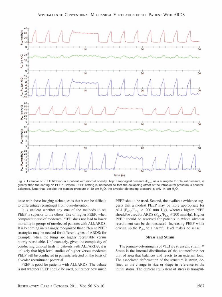
Particularly in patients with extrapulmonary ARDS, thechest-wall compliance may be reduced.104 Chest-wall com-pliance is reduced with abdominal-compartment syndrome,chest-wall edema, pleural effusion, or obesity.105,106 Thiscan result in an increase in pleural pressure and, if pleuralpressure is high relative to alveolar pressure, there may bepotential for alveolar collapse (Fig. 6). In that case it isdesirable to keep PEEP greater than pleural pressure. Theuse of an esophageal balloon to assess intra-pleural pres-sure has been advocated to allow more precise setting ofPEEP (Fig. 7).80,106-108 Unfortunately, artifacts in esopha-geal pressure, especially in supine critically ill patients,make it difficult to measure absolute pleural pressure ac-curately.109 In patients with abdominal-compartment syn-drome, bladder pressure may be useful to assess intra-abdominal pressure, the potential collapsing effect on thelungs, and the amount of PEEP necessary to counterbal-ance this effect.105,107
Some advocate opening the lung with recruitment ma-neuvers, with subsequent stepwise reduction of PEEP untilevidence of derecruitment (eg, respiratory-system compli-ance change) is identified on the deflation limb of thePV curve, with a decremental, rather than an incremen-tal, PEEP trial.110,111 Using this approach, PEEP is set� 20 cm H2O and then decreased to identify the level
that produces the best PaO2and compliance. However, a
recent study was unable to show differences in patientoutcomes when setting PEEP with a table was comparedto a method that used recruitment maneuvers and dec-remental PEEP.112
Dead-space measurements may be helpful to determinethe optimal PEEP.93 As PEEP is increased, the ratio ofdead space to VT (VD/VT) should decrease as alveoli arerecruited. If PEEP results in alveolar over-distention, how-ever, VD/VT should increase. It should be possible, how-ever, to assess these effects by evaluating PaCO2
with fixedminute ventilation; a decrease in PaCO2
is consistent with alower VD/VT, and vice versa. Methods are now availableto measure functional residual capacity at the bedside inpatients with ARDS.113,114 However, it is difficult to knowwhether an increase in lung volume with an increase inPEEP is due to alveolar recruitment or over-distention.
Imaging techniques have been used to evaluate PEEP set-tings. But CT is not practical. Ultrasound115 and electricalimpedance tomography116,117 need additional validation. One
Fig. 6. Effect of a stiff chest wall on transpulmonary pressure. Inthis example, although the plateau pressure (Pplat) is 35 cm H2O,the distending pressure across the alveolus is only 10 cm H2Obecause the pleural pressure is 25 cm H2O. PR � pressure dropdue to airways resistance. PA � alveolar pressure. �PA � trans-alveolar pressure. Patm � atmospheric pressure. Ppl � pleural pres-sure.
APPROACHES TO CONVENTIONAL MECHANICAL VENTILATION OF THE PATIENT WITH ARDS
1566 RESPIRATORY CARE • OCTOBER 2011 VOL 56 NO 10
issue with these imaging techniques is that it can be difficultto differentiate recruitment from over-distention.
It is unclear whether any one of the methods to setPEEP is superior to the others. Use of higher PEEP, whencompared to use of moderate PEEP, does not lead to lowermortality in groups of unselected patients with ALI/ARDS.It is becoming increasingly recognized that different PEEPstrategies may be needed for different types of ARDS; forexample, when the lungs are highly recruitable versuspoorly recruitable. Unfortunately, given the complexity ofconducting clinical trials in patients with ALI/ARDS, it isunlikely that high-level studies of higher versus moderatePEEP will be conducted in patients selected on the basis ofalveolar recruitment potential.
PEEP is good for patients with ALI/ARDS. The debateis not whether PEEP should be used, but rather how much
PEEP should be used. Second, the available evidence sug-gests that a modest PEEP may be more appropriate forALI (PaO2
/FIO2� 200 mm Hg), whereas higher PEEP
should be used for ARDS (PaO2/FIO2
� 200 mm Hg). HigherPEEP should be reserved for patients in whom alveolarrecruitment can be demonstrated. Increasing PEEP whiledriving up the Pplat to a harmful level makes no sense.
Stress and Strain
The primary determinants of VILI are stress and strain.118
Stress is the internal distribution of the counterforce perunit of area that balances and reacts to an external load.The associated deformation of the structure is strain, de-fined as the change in size or shape in reference to theinitial status. The clinical equivalent of stress is transpul-
Fig. 7. Example of PEEP titration in a patient with morbid obesity. Top: Esophageal pressure (Pes), as a surrogate for pleural pressure, isgreater than the setting on PEEP. Bottom: PEEP setting is increased so that the collapsing effect of the intrapleural pressure is counter-balanced. Note that, despite the plateau pressure of 40 cm H2O, the alveolar distending pressure is only 14 cm H2O.
APPROACHES TO CONVENTIONAL MECHANICAL VENTILATION OF THE PATIENT WITH ARDS
RESPIRATORY CARE • OCTOBER 2011 VOL 56 NO 10 1567

monary pressure (�PL), and the clinical equivalent of strainis the ratio of volume change (�V) to the functional re-sidual capacity (FRC):
�PL (stress) � specific lung elastance � �V/FRC (strain)
�V is the change in lung volume above resting FRC withthe addition of PEEP and VT. Specific lung elastance isrelatively constant at about 13.5 cm H2O. A harmful thresh-old of strain is about 2. Thus, the harmful threshold ofstress (transpulmonary pressure) is approximately27 cm H2O. The recommended Pplat below 30 cm H2O isthus reasonable for most patients with ALI/ARDS. How-ever, a higher Pplat may be safe when transpulmonary pres-sure is reduced due to an increase in pleural pressure. Thismakes a case for measurement of esophageal pressure (asa surrogate for pleural pressure) in a patient with a stiffchest wall.
This concept can be illustrated from Figure 7. When thePEEP is set at 24 cm H2O, the end-inspiratory transpul-monary pressure (stress) is 14 cm H2O. From the equationabove, strain is about 1. In this case, stress at 14 cm H2Oand strain at 1 are both in the safe range (� 27 cm H2Oand � 2, respectively), despite the Pplat of 40 cm H2O.
Summary
Lung-protective ventilation strategies are now widelyaccepted in the management of patients with ALI/ARDS.In most, if not all, cases of ALI/ARDS, conventional ven-tilation strategies can be used effectively, as described inthis paper. A suggested approach is given in Table 6.
REFERENCES
1. Slutsky AS. Ventilator-induced lung injury: from barotrauma tobiotrauma. Respir Care 2005;50(5):646-659.
Table 6. Ventilator Settings for ALI/ARDS
1. Choose ventilation modeAlthough any mode may be used, note that the ARDS Network study was performed with volume controlled continuous mandatory ventilation.Regardless of the mode, choose settings that ensure both the volume and pressure limitations. An important caveat is that Pplat may not reflecttranspulmonary pressure in pressure-targeted modes (pressure controlled ventilation, pressure support ventilation, adaptive pressure control) ifthe patient is making active inspiratory efforts that lower the pleural pressure.
2. Calculate ideal body weight (IBW)Males: IBW (kg) � 50.0 � 2.3 �height (inches)–60Females: IBW (kg) � 45.5 � 2.3 �height (inches)–60
3. Choose VT goal of 6 mL/kg predicted body weight4. Set PEEP to � 8 cm H2O5. Measure and record Pplat at least every 4 hours and after any changes in VT or PEEP:
If Pplat � 30 cm H2O, reduce VT to 5 mL/kg IBW, and then to 4 mL/kg IBW, if necessary to decrease Pplat to � 30 cm H2O.If VT � 6 mL/kg IBW and Pplat � 25 cm H2O, increase VT by 1 mL/kg IBW to a maximum of 6 mL/kg.
6. Adjust respiratory rate or VT according to pH goals:If pH � 7.30, consider increasing the respiratory rate to as high as 35 breaths/min while monitoring for development of auto-PEEP.If pH � 7.15 and respiratory rate 35 breaths/min, consider increasing VT and suspending the Pplat limit, depending on patient tolerance of theacidemia.
7. Set inspiratory-expiratory ratio to 1:2. Adjust to avoid auto-PEEP and asynchrony.8. Address asynchrony
If severe asynchrony, increase VT to 7 or 8 mL/kg IBW if Pplat remains � 30 cm H2O.If Pplat � 30 cm H2O on 7 or 8 mL/kg IBW, revert to lower VT, consider ventilator adjustments to improve patient synchrony, and consideradditional sedation.
9. Set PEEP to maximize alveolar recruitment while avoiding over-distention.Increase PEEP in increments of 2–3 cm H2O, allow 5–10 min of consistent measurements of airway pressure and VT. Select the PEEP thatgives the best compliance (lowest driving pressure).When PEEP is decreased, lower PEEP by 2–3 cm H2O and allow at least 30 min of consistent measurements of airway pressure and VT toavoid derecruitment.If reduced chest wall compliance is likely, esophageal pressure measurements may be useful.
10. Adjust FIO2to achieve SpO2
of 88–95% (PaO255–80 mm Hg).
11. Avoid ventilator disconnections and manual ventilation; use in-line suction catheter.
ALI � acute lung injuryARDS � acute respiratory distress syndromePplat � plateau pressureVT � tidal volume(Adapted from Reference 43.)
APPROACHES TO CONVENTIONAL MECHANICAL VENTILATION OF THE PATIENT WITH ARDS
1568 RESPIRATORY CARE • OCTOBER 2011 VOL 56 NO 10

2. Slutsky AS, Tremblay LN. Multiple system organ failure. Is me-chanical ventilation a contributing factor? Am J Respir Crit CareMed 1998;157(6 Pt 1):1721-1725.
3. Tremblay LN, Slutsky AS. Ventilator-induced injury: from baro-trauma to biotrauma. Proc Assoc Am Phys 1998;110(6):482-488.
4. Tremblay LN, Slutsky AS. Ventilator-induced lung injury: from thebench to the bedside. Intensive Care Med 2006;32(1):24-33.
5. Ranieri VM, Giunta F, Suter PM, Slutsky AS. Mechanical venti-lation as a mediator of multisystem organ failure in acute respira-tory distress syndrome. JAMA 2000;284(1):43-44.
6. Ranieri VM, Suter PM, Tortorella C, De Tullio R, Dayer JM,Brienza A, et al. Effect of mechanical ventilation on inflammatorymediators in patients with acute respiratory distress syndrome: arandomized controlled trial. JAMA 1999;282(1):54-61.
7. Dreyfuss D, Saumon G. Ventilator-induced lung injury: lessonsfrom experimental studies. Am J Respir Crit Care Med 1998;157(1):294-323.
8. Stewart TE, Meade MO, Cook DJ, Granton JT, Hodder RV, Lap-insky SE, et al. Evaluation of a ventilation strategy to preventbarotrauma in patients at high risk for acute respiratory distresssyndrome. Pressure- and Volume-Limited Ventilation StrategyGroup. N Engl J Med 1998;338(6):355-361.
9. Brochard L, Roudot-Thoraval F, Roupie E, Delclaux C, Chastre J,Fernandez-Mondejar E, et al. Tidal volume reduction for preven-tion of ventilator-induced lung injury in acute respiratory distresssyndrome. The Multicenter Trail Group on Tidal Volume reductionin ARDS. Am J Respir Crit Care Med 1998;158(6):1831-1838.
10. Brower RG, Shanholtz CB, Fessler HE, Shade DM, White P Jr,Wiener CM, et al. Prospective, randomized, controlled clinical trialcomparing traditional versus reduced tidal volume ventilation inacute respiratory distress syndrome patients. Crit Care Med 1999;27(8):1492-1498.
11. Amato MB, Barbas CS, Medeiros DM, Magaldi RB, Schettino GP,Lorenzi-Filho G, et al. Effect of a protective-ventilation strategy onmortality in the acute respiratory distress syndrome. N Engl J Med1998;338(6):347-354.
12. Kallet RH. What is the legacy of the National Institutes of HealthAcute Respiratory Distress Syndrome Network? Respir Care 2009;54(7):912-924.
13. The Acute Respiratory Distress Syndrome Network. Ventilationwith lower tidal volumes as compared with traditional tidal vol-umes for acute lung injury and the acute respiratory distress syn-drome. N Engl J Med 2000;342(18):1301-1308.
14. Steinberg KP, Kacmarek RM. Respiratory controversies in the crit-ical care setting. Should tidal volume be 6 mL/kg predicted bodyweight in virtually all patients with acute respiratory failure? RespirCare 2007;52(5):556-564; discussion 565-567.
15. Hager DN, Krishnan JA, Hayden DL, Brower RG, Network ACT.Tidal volume reduction in patients with acute lung injury whenplateau pressures are not high. Am J Respir Crit Care Med 2005;172(10):1241-1245.
16. Kallet RH, Branson RD. Respiratory controversies in the criticalcare setting. Do the NIH ARDS Clinical Trials Network PEEP/FIO2
tables provide the best evidence-based guide to balancing PEEPand FIO2 settings in adults? Respir Care 2007;52(4):461-475; dis-cussion 475-477.
17. Terragni PP, Rosboch G, Tealdi A, Corno E, Menaldo E, Davini O,et al. Tidal hyperinflation during low tidal volume ventilation inacute respiratory distress syndrome. Am J Respir Crit Care Med2007;175(2):160-166.
18. Laffey JG, O’croinin D, Mcloughlin P, Kavanagh BP. Permissivehypercapnia: role in protective lung ventilatory strategies. IntensiveCare Med 2004;30(3):347-356.
19. Kregenow DA, Rubenfeld GD, Hudson LD, Swenson ER. Hyper-capnic acidosis and mortality in acute lung injury. Crit Care Med2006;34(1):1-7.
20. de Durante G, Del Turco M, Rustichini L, Cosimini P, Giunta F,Hudson LD, et al. ARDSNet lower tidal volume ventilatory strat-egy may generate intrinsic positive end-expiratory pressure in pa-tients with acute respiratory distress syndrome. Am J Respir CritCare Med 2002;165(9):1271-1274.
21. MacIntyre NR, Sessler CN. Are there benefits or harm from pres-sure targeting during lung-protective ventilation? Respir Care 2010;55(2):175-180; discussion 180-183.
22. Davis K, Jr., Branson RD, Campbell RS, Porembka DT. Compar-ison of volume control and pressure control ventilation: is flowwaveform the difference? J Trauma 1996;41(5):808-814.
23. Fujita Y, Fujino Y, Uchiyama A, Mashimo T, Nishimura M. Highpeak inspiratory flow can aggravate ventilator-induced lung injuryin rabbits. Med SciMonit 2007;13(4):BR95-BR100.
24. Kotani M, Kotani T, Li Z, Silbajoris R, Piantadosi CA, HuangY-CT. Reduced inspiratory flow attenuates IL-8 release and MAPKactivation of lung overstretch. Eur Respir J 2004;24(2):238-246.
25. Rich PB, Reickert CA, Sawada S, Awad SS, Lynch WR, JohnsonKJ, et al. Effect of rate and inspiratory flow on ventilator-inducedlung injury. J Trauma 2000;49(5):903-911.
26. MacIntyre NR, McConnell R, Cheng KC, Sane A. Patient-ventila-tor flow dyssynchrony: flow-limited versus pressure- limited breaths.Crit Care Med 1997;25(10):1671-1677.
27. Yang LY, Huang YC, MacIntyre N. Patient-ventilator synchronyduring pressure-targeted versus flow-targeted small tidal volumeassisted ventilation. J Crit Care 2007;22(3):252-257.
28. Kallet RH, Campbell AR, Dicker RA, Katz JA, Mackersie RC.Work of breathing during lung-protective ventilation in patientswith acute lung injury and acute respiratory distress syndrome: acomparison between volume and pressure-regulated breathingmodes. Respir Care 2005;50(12):1623-1631.
29. Mireles-Cabodevila E, Chatburn RL. Work of breathing in adaptivepressure control continuous mandatory ventilation. Respir Care2009;54(11):1467-1472.
30. Branson RD. Dual control modes, closed loop ventilation, hand-guns, and tequila. Respir Care 2001;46(3):232-233.
31. Sottiaux TM. Patient-ventilator interactions during volume-supportventilation: asynchrony and tidal volume instability - a report ofthree cases. Respir Care 2001;46(3):255-262.
32. Jaber S, Delay J-M, Matecki S, Sebbane M, Eledjam J-J, BrochardL. Volume-guaranteed pressure-support ventilation facing acutechanges in ventilatory demand. Intensive Care Med 2005;31(9):1181-1188.
33. Jaber S, Sebbane M, Verzilli D, Matecki S, Wysocki M, EledjamJJ, et al. Adaptive support and pressure support ventilation behaviorin response to increased ventilatory demand. Anesthesiology 2009;110(3):620-627.
34. Schmidt UH, Hess DR. Does spontaneous breathing produce harmin patients with the acute respiratory distress syndrome? RespirCare 2010;55(6):784-786.
35. Leray V, Bourdin G, Flandreau G, Bayle F, Wallet F, Richard J-C,et al. A case of pneumomediastinum in a patient with acute respi-ratory distress syndrome on pressure support ventilation. RespirCare 2010;55(6):770-773.
36. Kallet RH, Siobal MS. Measuring dead space: does it really matter?Or, what are we waiting for? Respir Care 2010;55(3):350-352.
37. Nuckton TJ, Alonso JA, Kallet RH, Daniel BM, Pittet J-F, EisnerMD, et al. Pulmonary dead-space fraction as a risk factor for deathin the acute respiratory distress syndrome. N Engl J Med 2002;346(17):1281-1286.
APPROACHES TO CONVENTIONAL MECHANICAL VENTILATION OF THE PATIENT WITH ARDS
RESPIRATORY CARE • OCTOBER 2011 VOL 56 NO 10 1569

38. Raurich JM, Vilar M, Colomar A, Ibanez J, Ayestaran I, Perez-Barcena J, et al. Prognostic value of the pulmonary dead-spacefraction during the early and intermediate phases of acute respira-tory distress syndrome. Respir Care 2010;55(3):282-287.
39. Lucangelo U, Bernabe F, Vatua S, Degrassi G, Villagra A, Fernan-dez R, et al. Prognostic value of different dead space indices inmechanically ventilated patients with acute lung injury and ARDS.Chest 2008;133(1):62-71.
40. Kallet RH, Alonso JA, Pittet JF, Matthay MA. Prognostic value ofthe pulmonary dead-space fraction during the first 6 days of acuterespiratory distress syndrome. Respir Care 2004;49(9):1008-1014.
41. Pohlman MC, Mccallister KE, Schweickert WD, Pohlman AS, Ni-gos CP, Krishnan JA, et al. Excessive tidal volume from breathstacking during lung-protective ventilation for acute lung injury.Crit Care Med 2008;36(11):3019-3023.
42. Hess DR, Thompson BT. Patient-ventilator dyssynchrony duringlung protective ventilation: what’s a clinician to do? Crit Care Med2006;34(1):231-233.
43. Ramnath VR, Hess DR, Thompson BT. Conventional mechanicalventilation in acute lung injury and acute respiratory distress syn-drome (abstract). Clin Chest Med 2006;27(4):601-613, viii.
44. Papazian L, Forel J-M, Gacouin A, Penot-Ragon C, Perrin G, Loun-dou A, et al. Neuromuscular blockers in early acute respiratorydistress syndrome. N Engl J Med 2010;363(12):1107-1116.
45. Cheng IW, Eisner MD, Thompson BT, Ware LB, Matthay MA,Network ARDS. Acute effects of tidal volume strategy on hemo-dynamics, fluid balance, and sedation in acute lung injury. Crit CareMed 2005;33(1):63-70; discussion 239-240.
46. Kahn JM, Andersson L, Karir V, Polissar NL, Neff MJ, Ruben-feld GD. Low tidal volume ventilation does not increase seda-tion use in patients with acute lung injury. Crit Care Med 2005;33(4):766-771.
47. Gajic O, Dara SI, Mendez JL, Adesanya AO, Festic E, Caples SM,et al. Ventilator-associated lung injury in patients without acutelung injury at the onset of mechanical ventilation. Crit Care Med2004;32(9):1817-1824.
48. Gajic O, Frutos-Vivar F, Esteban A, Hubmayr RD, Anzueto A.Ventilator settings as a risk factor for acute respiratory distresssyndrome in mechanically ventilated patients. Intensive Care Med2005;31(7):922-926.
49. Li G, Malinchoc M, Cartin-Ceba R, Venkata CV, Kor DJ, PetersSG, et al. Eight-year trend of acute respiratory distress syndrome: apopulation-based study in Olmsted County, Minnesota. Am J Re-spir Crit Care Med 2011;183(1):59-66.
50. Schultz MJ, Haitsma JJ, Slutsky AS, Gajic O. What tidal volumesshould be used in patients without acute lung injury? Anesthesiol-ogy 2007;106(6):1226-1231.
51. Yilmaz M, Keegan MT, Iscimen R, Afessa B, Buck CF, HubmayrRD, et al. Toward the prevention of acute lung injury: protocol-guided limitation of large tidal volume ventilation and inappropri-ate transfusion. Crit Care Med 2007;35(7):1660-1666.
52. Mascia L, Mastromauro I, Viberti S. High tidal volume as a pre-dictor of acute lung injury in neurotrauma patients. Minerva Anes-tesiologica 2008;74(6):325-327.
53. Mascia L, Zavala E, Bosma K, Pasero D, Decaroli D, Andrews P,et al. High tidal volume is associated with the development of acutelung injury after severe brain injury: an international observationalstudy. Crit Care Med 2007;35(8):1815-1820.
54. Mascia L, Pasero D, Slutsky AS, Arguis MJ, Berardino M, GrassoS, et al. Effect of a lung protective strategy for organ donors oneligibility and availability of lungs for transplantation: a random-ized controlled trial. JAMA 2010;304(23):2620-2627.
55. Thompson BT, Hayden D, Matthay MA, Brower R, Parsons PE.Clinicians’ approaches to mechanical ventilation in acute lung in-jury and ARDS. Chest 2001;120(5):1622-1627.
56. Young MP, Manning HL, Wilson DL, Mette SA, Riker RR, LeiterJC, et al. Ventilation of patients with acute lung injury and acuterespiratory distress syndrome: has new evidence changed clinicalpractice? Crit Care Med 2004;32(6):1260-1265.
57. Esteban A, Ferguson ND, Meade MO, Frutos-Vivar F, ApezteguiaC, Brochard L, et al. Evolution of mechanical ventilation in re-sponse to clinical research. Am J Respir Crit Care Med 2008;177(2):170-177.
58. Checkley W, Brower R, Korpak A, Thompson BT; ARDS NetworkInvestigators. Effects of a clinical trial on mechanical ventilationpractices in patients with acute lung injury. Am J Respir Crit CareMed 2008;177(11):1215-1222.
59. Cooke CR, Kahn JM, Watkins TR, Hudson LD, Rubenfeld GD.Cost-effectiveness of implementing low-tidal volume ventilation inpatients with acute lung injury. Chest 2009;136(1):79-88.
60. Herasevich V, Tsapenko M, Kojicic M, Ahmed A, Kashyap R,Venkata C, et al. Limiting ventilator-induced lung injury throughindividual electronic medical record surveillance. Crit Care Med2011;39(1):34-39.
61. Cole A, Weller S, Sykes M. Inverse ratio ventilation compared withPEEP in adult respiratory failure. Intensive Care Med 1984;10(5):227-232.
62. Gurevitch MJ, Van Dyke J, Young ES, Jackson K. Improved ox-ygenation and lower peak airway pressure in severe adult respira-tory distress syndrome. Treatment with inverse ratio ventilation.Chest 1986;89(2):211-213.
63. Tharratt RS, Allen RP, Albertson TE. Pressure controlled inverseratio ventilation in severe adult respiratory failure. Chest 1988;94(4):755-762.
64. Abraham E, Yoshihara G. Cardiorespiratory effects of pressurecontrolled inverse ratio ventilation in severe respiratory failure.Chest 1989;96(6):1356-1359.
65. Lain D, DiBenedetto R, Morris S, Van Nguyen A, Saulters R,Causey D. Pressure control inverse ratio ventilation as a method toreduce peak inspiratory pressure and provide adequate ventilationand oxygenation. Chest 1989;95(5):1081-1088.
66. Marcy TW, Marini JJ. Inverse ratio ventilation in ARDS. Rationaleand implementation. Chest 1991;100(2):494-504.
67. Mercat A, Graïni L, Teboul JL, Lenique F, Richard C. Cardiore-spiratory effects of pressure-controlled ventilation with and withoutinverse ratio in the adult respiratory distress syndrome. Chest 1993;104(3):871-875.
68. Munoz J, Guerrero JE, Escalante JL, Palomino R, De La Calle B.Pressure-controlled ventilation versus controlled mechanical venti-lation with decelerating inspiratory flow. Crit Care Med 1993;21(8):1143-1148.
69. Lessard MR, Guerot E, Lorino H, Lemaire F, Brochard L. Effectsof pressure-controlled with different I:E ratios versus volume-con-trolled ventilation on respiratory mechanics, gas exchange, and he-modynamics in patients with adult respiratory distress syndrome.Anesthesiology 1994;80(5):983-991.
70. Mercat A, Titiriga M, Anguel N, Richard C, Teboul J. Inverse ratioventilation (I/E � 2/1) in acute respiratory distress syndrome: asix-hour controlled study. Am J Respir Crit Care Med 1997;155(5):1637-1642.
71. Zavala E, Ferrer M, Polese G, Masclans JR, Planas M, Milic-EmiliJ, et al. Effect of inverse I:E ratio ventilation on pulmonary gasexchange in acute respiratory distress syndrome. Anesthesiology1998;88(1):35-42.
APPROACHES TO CONVENTIONAL MECHANICAL VENTILATION OF THE PATIENT WITH ARDS
1570 RESPIRATORY CARE • OCTOBER 2011 VOL 56 NO 10

72. Shanholtz C, Brower R. Should inverse ratio ventilation be used inadult respiratory distress syndrome? Am J Respir Crit Care Med1994;149(5):1354-1358.
73. Kacmarek RM, Hess D. Pressure-controlled inverse-ratio ventila-tion: panacea or auto-PEEP? Respir Care 1990;35(10):945-948.
74. Duncan S, Rizk N, Raffin T. Inverse ratio ventilation. PEEP indisguise? Chest 1987;92(3):390-392.
75. Esteban A, Anzueto A, Frutos F, Alía I, Brochard L, Stewart TE, etal. Characteristics and outcomes in adult patients receiving mechan-ical ventilation: a 28-day international study. JAMA 2002;287(3):345-355.
76. Ferguson ND, Frutos-Vivar F, Esteban A, Anzueto A, Alía I, BrowerRG, et al. Airway pressures, tidal volumes, and mortality in patientswith acute respiratory distress syndrome. Crit Care Med 2005;33(1):21-30.
77. Brower RG, Lanken PN, MacIntyre N, Matthay MA, Morris A,Ancukiewicz M, et al. Higher versus lower positive end-expiratorypressures in patients with the acute respiratory distress syndrome.N Engl J Med 2004;351(4):327-336.
78. Meade MO, Cook DJ, Guyatt GH, Slutsky AS, Arabi YM, CooperDJ, et al. Ventilation strategy using low tidal volumes, recruitmentmaneuvers, and high positive end-expiratory pressure for acute lunginjury and acute respiratory distress syndrome: a randomized con-trolled trial. JAMA 2008;299(6):637-645.
79. Mercat A, Richard J-CM, Vielle B, Jaber S, Osman D, Diehl J-L,et al. Positive end-expiratory pressure setting in adults with acutelung injury and acute respiratory distress syndrome: a randomizedcontrolled trial. JAMA 2008;299(6):646-655.
80. Talmor D, Sarge T, Malhotra A, O’Donnell CR, Ritz R, Lisbon A,et al. Mechanical ventilation guided by esophageal pressure in acutelung injury. N Engl J Med 2008;359(20):2095-2104.
81. Villar J, Kacmarek RM, Perez-Mendez L, Aguirre-Jaime A. A highpositive end-expiratory pressure, low tidal volume ventilatory strat-egy improves outcome in persistent acute respiratory distress syn-drome: a randomized, controlled trial. Crit Care Med 2006;34(5):1311-1318.
82. Hess DR. How much PEEP? Do we need another meta-analysis?Respir Care 2011;56(5):710-713.
83. Gattinoni L, Caironi P, Cressoni M, Chiumello D, Ranieri VM,Quintel M, et al. Lung recruitment in patients with the acute respi-ratory distress syndrome. N Engl J Med 2006;354(17):1775-1786.
84. Caironi P, Cressoni M, Chiumello D, Ranieri M, Quintel M, RussoSG, et al. Lung opening and closing during ventilation of acuterespiratory distress syndrome. Am J Respir Crit Care Med 2010;181(6):578-586.
85. Dasenbrook E, Needham DM, Brower RG, Fan E. Higher positiveend-expiratory pressure in patients with acute lung injury: a sys-tematic review and meta-analysis. Respir Care 2011;56(5):568-575.
86. Briel M, Meade M, Mercat A, Brower RG, Talmor D, Walter SD,et al. Higher vs lower positive end-expiratory pressure in patientswith acute lung injury and acute respiratory distress syndrome:systematic review and meta-analysis. JAMA 2010;303(9):865-873.
87. Oba Y, Thameem DM, Zaza T. High levels of PEEP may improvesurvival in acute respiratory distress syndrome: a meta-analysis.Respir Med 2009;103(8):1174-1181.
88. Phoenix SI, Paravastu S, Columb M, Vincent J-L, Nirmalan M.Does a higher positive end expiratory pressure decrease mortality inacute respiratory distress syndrome? A systematic review and meta-analysis. Anesthesiology 2009;110(5):1098-1105.
89. Putensen C, Theuerkauf N, Zinserling J, Wrigge H, Pelosi P. Meta-analysis: ventilation strategies and outcomes of the acute respira-tory distress syndrome and acute lung injury. Ann Intern Med 2009;151(8):566-576.
90. Grasso S, Fanelli V, Cafarelli A, Anaclerio R, Amabile M, AnconaG, et al. Effects of high versus low positive end-expiratory pres-sures in acute respiratory distress syndrome. Am J Respir Crit CareMed 2005;171(9):1002-1008.
91. Grasso S, Stripoli T, De Michele M, Bruno F, Moschetta M, An-gelelli G, et al. ARDSnet ventilatory protocol and alveolar hyper-inflation: role of positive end-expiratory pressure. Am J Respir CritCare Med 2007;176(8):761-767.
92. Grasso S, Stripoli T, Sacchi M, Trerotoli P, Staffieri F, Franchini D,et al. Inhomogeneity of lung parenchyma during the open lungstrategy: a computed tomography scan study. Am J Respir Crit CareMed 2009;180(5):415-423.
93. Suter PM, Fairley B, Isenberg MD. Optimum end-expiratory air-way pressure in patients with acute pulmonary failure. N EnglJ Med 1975;292(6):284-289.
94. Harris RS. Pressure-volume curves of the respiratory system. Re-spir Care 2005;50(1):78-98; discussion 98-99.
95. Harris RS, Hess DR, Venegas JG. An objective analysis of thepressure-volume curve in the acute respiratory distress syndrome.Am J Respir Crit Care Med 2000;161(2 Pt 1):432-439.
96. Mergoni M, Martelli A, Volpi A, Primavera S, Zuccoli P, Rossi A.Impact of positive end-expiratory pressure on chest wall and lungpressure-volume curve in acute respiratory failure. Am J Respir CritCare Med 1997;156(3 Pt 1):846-854.
97. Owens RL, Hess DR, Malhotra A, Venegas JG, Harris RS. Effect ofthe chest wall on pressure-volume curve analysis of acute respiratorydistress syndrome lungs. Crit Care Med 2008;36(11):2980-2985.
98. Ranieri VM, Brienza N, Santostasi S, Puntillo F, Mascia L,Vitale N, et al. Impairment of lung and chest wall mechanics inpatients with acute respiratory distress syndrome: role of ab-dominal distension. Am J Respir Crit Care Med 1997;156(4 Pt1):1082-1091.
99. Dall‘ava-Santucci J, Armaganidis A, Brunet F, Dhainaut JF, NouiraS, Morisseau D, et al. Mechanical effects of PEEP in patients withadult respiratory distress syndrome. J Appl Physiol 1990;68(3):843-848.
100. Puybasset L, Gusman P, Muller JC, Cluzel P, Coriat P, Rouby JJ.Regional distribution of gas and tissue in acute respiratory distresssyndrome. III. Consequences for the effects of positive end-expi-ratory pressure. CT Scan ARDS Study Group. Intensive Care Med2000;26(9):1215-1227.
101. Hickling KG. Reinterpreting the pressure-volume curve in patientswith acute respiratory distress syndrome. Curr Opin Crit Care 2002;8(1):32-38.
102. Grooms DA, Sibole SH, Tomlinson JR, Marik PE, Chatburn RL.Customization of an open lung ventilation strategy to treat a case oflife threatening acute respiratory distress syndrome. Respir Care2011;56(4):514-519.
103. Grasso S, Terragni P, Mascia L, Fanelli V, Quintel M, Herrmann P,et al. Airway pressure-time curve profile (stress index) detects tidalrecruitment/hyperinflation in experimental acute lung injury. CritCare Med 2004;32(4):1018-1027.
104. Gattinoni L, Pelosi P, Suter PM, Pedoto A, Vercesi P, Lissoni A.Acute respiratory distress syndrome caused by pulmonary and ex-trapulmonary disease. Different syndromes? Am J Respir Crit CareMed 1998;158(1):3-11.
105. Hess DR, Bigatello LM. The chest wall in acute lung injury/acuterespiratory distress syndrome. Curr Opin Crit Care 2008;14(1):94-102.
106. Piraino T, Cook DJ. Optimal PEEP guided by an esophageal bal-loon. Respir Care 2011;56(4):510-513.
107. Loring SH, O’Donnell CR, Behazin N, Malhotra A, Sarge T, Ritz R,et al. Esophageal pressures in acute lung injury: do they representartifact or useful information about transpulmonary pressure, chestwall mechanics, and lung stress? J Appl Physiol 2010;108(3):515-522.
APPROACHES TO CONVENTIONAL MECHANICAL VENTILATION OF THE PATIENT WITH ARDS
RESPIRATORY CARE • OCTOBER 2011 VOL 56 NO 10 1571

108. Talmor D, Sarge T, O’Donnell CR, Ritz R, Malhotra A, Lisbon A,et al. Esophageal and transpulmonary pressures in acute respiratoryfailure. Crit Care Med 2006;34(5):1389-1394.
109. Talmor DS, Fessler HE. Are esophageal pressure measurementsimportant in clinical decision-making in mechanically ventilatedpatients? Respir Care 2010;55(2):162-172; discussion 172-174.
110. Girgis K, Hamed H, Khater Y, Kacmarek RM. A decrementalPEEP trial identifies the PEEP level that maintains oxygenationafter lung recruitment. Respir Care 2006;51(10):1132-1139.
111. Hickling KG. Best compliance during a decremental, but not in-cremental, positive end-expiratory pressure trial is related to open-lung positive end-expiratory pressure: a mathematical model ofacute respiratory distress syndrome lungs. Am J Respir Crit CareMed 2001;163(1):69-78.
112. Huh JW, Jung H, Choi HS, Hong S-B, Lim C-M, Koh Y. Efficacyof positive end-expiratory pressure titration after the alveolar re-cruitment manoeuvre in patients with acute respiratory distress syn-drome. Crit Care 2009;13(1):R22.
113. Branson RD, Johannigman JA. Innovations in mechanical ventila-tion. Respir Care 2009;54(7):933-947.
114. Owens RL, Stigler WS, Hess DR. Do newer monitors of exhaledgases, mechanics, and esophageal pressure add value? Clin ChestMed 2008;29(2):297-312, vi-vii.
115. Bouhemad B, Brisson H, Le-Guen M, Arbelot C, Lu Q, Rouby JJ.Bedside ultrasound assessment of positive end-expiratory pressure-induced lung recruitment. Am J Respir Crit Care Med 2011;183(3):341-347.
116. Costa EL, Borges JB, Melo A, Suarez-Sipmann F, Toufen C Jr,Bohm SH, Amato MB. Bedside estimation of recruitable alveolarcollapse and hyperdistension by electrical impedance tomography.Intensive Care Med 2009;35(6):1132-1137.
117. Victorino JA, Borges JB, Okamoto VN, Matos GF, Tucci MR,Caramez MP, et al. Imbalances in regional lung ventilation: a val-idation study on electrical impedance tomography. Am J RespirCrit Care Med 2004;169(7):791-800.
118. Chiumello D, Carlesso E, Cadringher P, Caironi P, Valenza F, PolliF, et al. Lung stress and strain during mechanical ventilation foracute respiratory distress syndrome. Am J Respir Crit Care Med2008;178(4):346-355.
APPROACHES TO CONVENTIONAL MECHANICAL VENTILATION OF THE PATIENT WITH ARDS
1572 RESPIRATORY CARE • OCTOBER 2011 VOL 56 NO 10