Embed Size (px)
Citation preview

F
Nm
MF
a
ARR1AA
KNSNMNE
1
ifbcflldliios
pvbco
h0
Applied Surface Science 399 (2017) 480–490
Contents lists available at ScienceDirect
Applied Surface Science
journa l h om epa ge: www.elsev ier .com/ locate /apsusc
ull Length Article
anosecond laser texturing of uniformly and non-uniformly wettableicro structured metal surfaces for enhanced boiling heat transfer
atevz Zupancic ∗, Matic Moze, Peter Gregorcic, Iztok Golobicaculty of Mechanical Engineering, University of Ljubljana, Askerceva 6, 1000 Ljubljana, Slovenia
r t i c l e i n f o
rticle history:eceived 22 September 2016eceived in revised form1 December 2016ccepted 15 December 2016vailable online 15 December 2016
eywords:
a b s t r a c t
Microstructured uniformly and non-uniformly wettable surfaces were created on 25-�m-thin stainlesssteel foils by laser texturing using a marking nanosecond Nd:YAG laser (� = 1064 nm) and utilizing variouslaser fluences and scan line separations. High-speed photography and high-speed IR thermography wereused to investigate nucleate boiling heat transfer on the microstructured surfaces. The most pronouncedresults were obtained on a surface with non-uniform microstructure and non-uniform wettability. Theobtained results show up to a 110% higher heat transfer coefficients and 20–40 times higher nucleationsite densities compared to the untextured surface. We show that the number of active nucleation sites is
anosecond laserurface texturingon-uniform wettabilityicrocavitiesucleation criterianhanced boiling heat transfer
significantly increased in the vicinity of microcavities that appeared in areas with the smallest (10 �m)scan line separation. Furthermore, this confirms the predictions of nucleation criteria and proves thatstraightforward, cost-effective nanosecond laser texturing allows the production of cavities with diam-eters of up to a few micrometers and surfaces with non-uniform wettability. Additionally, this opens upimportant possibilities for a more deterministic control over the complex boiling process.
© 2016 Elsevier B.V. All rights reserved.
. Introduction
Nucleate boiling is a phase change heat transfer mechanism ands one of the most effective and technically controllable approachesor removal of high heat flux. Its performance is usually quantifiedy two characteristic parameters, heat transfer coefficient and criti-al heat flux (CHF). The first one is defined as the ratio between heatux transmitted from the surface, and the wall superheat, while the
atter represents the upper limit of the nucleate boiling regime. Theesire to increase both the heat transfer coefficient and the CHF has
ed to the development of various surface modification techniquesn the last decades as the surface and its interaction with the work-ng fluid have a profound effect on the boiling phenomenon. Studiesn hydrophobic (water-repellent) and hydrophilic (water-loving)urfaces have already shown some key improvements [1–8].
Hydrophobic surfaces prefer to be in contact with the vaporhase rather than the liquid water and therefore promote the acti-ation of nucleation [1]. At the same time, they provide larger
ubble departure diameters and the bubbles are more likely tooalesce. Reduction of wettability reduces the temperature of thenset of nucleate boiling [2] and increases the heat transfer coef-∗ Corresponding author.E-mail address: [email protected] (M. Zupancic).
ttp://dx.doi.org/10.1016/j.apsusc.2016.12.120169-4332/© 2016 Elsevier B.V. All rights reserved.
ficient at low heat fluxes. As the heat flux increases, the formationof large vapor blankets [3] decreases heat transfer and promotesthe CHF conditions. On the contrary, hydrophilic surfaces increasethe “suction” of the liquid towards the nucleation sites, decreasebubble contact diameters [3,4], and, finally, delay the CHF due tothe water replenishment on dry spots. However, because of theincreased wettability, hydrophilic surfaces require larger super-heat for nucleate boiling to occur and do not necessarily providean enhanced heat transfer coefficient at low heat fluxes [3]. Forfurther enhancements, several authors already suggested simulta-neous implementation of hydrophobic and hydrophilic features ona single surface to produce a so called biphilic surface [3,6,7,9].
Another approach of boiling enhancement is incorporation ofpores or micro cavities onto the surface [10,11]. According to theexisting nucleation criteria [12,13], a conically-shaped cavity on thesurface might serve as an active nucleation site if the diameter ofthis cavity falls within a certain range. This size range depends onthe wall superheat and contact angle. To provide large number ofpotentially active nucleation sites at different wall superheats, thesurface should incorporate various contact angles (e.g. non-uniformwettability [14]) as well as wide range of cavity diameters [15].A recent study of flow boiling in microchannels [15] proved that
porous multi-scale rough surface significantly enhances bubblenucleation and considerably mitigates two-phase flow instabilities.
rface Science 399 (2017) 480–490 481
ditboftgsnanisaafw[
wctocbotaiIistopaOti
2
nIGnsnratuctt(tpefie
θ = 90° (Hsu)θ = 1° (Hsu) θ = 10° (Hsu)
0 5 10 15 20 25 30
θ = 1° (Kandlikar) θ = 90° (Kandlikar)θ = 10° (Kandlikar)
T -T (K)w sat
r (µm
)c
-210
-110
010
110
210
310
M. Zupancic et al. / Applied Su
Mechanical resistance, thermal stability and challenging pro-uction [16] are still the main drawbacks preventing wider
mplementation of hydrophilic and hydrophobic surfaces in heatransfer applications. In our previous study [3], we demonstratedoiling heat transfer enhancement on biphilic surfaces comprisedf polydimethylsiloxane-silica films. The transition of wettabilityrom hydrophobic to hydrophilic was achieved by locally heatinghe coating using a pulsed Nd:YAG laser. However, the coatings ineneral have significant drawbacks for real applications. Modifyingurfaces’ wettability and micro/nanostructure by laser texturing ofon-coated surfaces is a relatively simple, fast, and highly flexiblepproach. It is also more environmentally acceptable and there areo problems with adhering any coating to the surface. In pool boil-
ng, recent study by Kruse et al. [5] on superhydrophilic stainlessteel surfaces, processed by femtosecond laser pulses, showed both
considerable increase of the CHF from 910 kW/m2 to 1420 kW/m2
s well as a significant increase of the heat transfer coefficientrom 23 kW/m2K to 68 kW/m2K. However, most of the reported
ork on laser micro/nanostructuring relies on costly ultrafast lasers5,17,18].
The main objective of this study is to incorporate advantages ofettability modifications together with microstructure and micro
avities to develop surfaces for enhanced nucleate boiling heatransfer. Additional aim is to achieve this by using a nanosec-nd marking laser, which represents a cost-effective alternative tourrently utilized laser-processing technologies for surface wetta-ility and microstructure modifications. Surfaces were developedn stainless steel, which was used due to its widespread applica-ions in heat and process engineering, good resistance to corrosion,nd because the results of previous research indicate the possibil-ty of modifying its wettability using nanosecond laser pulses [19].n order to study the durability of the textured surfaces in a boil-ng heat transfer application, we performed several consecutiveaturated pool boiling experiments. We employed high-speed IRhermography and high-speed recordings to obtain a visualizationf the departing bubbles with the corresponding local wall tem-erature measurements. Surfaces were characterized before andfter boiling experiments through contact angle measurements.ur experiments show that surfaces produced by nanosecond laser
exturing have the potential to be a good alternative to those mod-fied using currently established techniques.
. Nucleation criteria
The impact of surface material, texture and topography onucleate boiling phenomenon has long been investigated [20].
nfluences on bubble growth were first researched by Bankoff [21],riffith and Wallis [22], and Han and Griffith [23]. One of the firstucleation criteria for dimensioning of potential active nucleationites was developed by Hsu [12]. Hsu’s model considers a bubbleucleus sitting at the mouth of a cavity with the radius rc sur-ounded by the liquid in the saturated state. At the beginning of
bubble growth cycle, only the surface is uniformly superheatedo �T above the saturation temperature (Tsat). Gradually, the liq-id is warmed through transient-conduction process, which in turnauses the thermal layer to grow. This growth is limited by a strongurbulence in the liquid. Beyond a certain thickness, the tempera-ure of the liquid is considered to be constant at bulk temperatureTbulk). The model predicts that the bubble will start to grow if fluids’emperature within the thermal layer reaches the saturation tem-erature inside the bubble. By combining the Clausius-Clapeyron
quation for saturated temperature, Gaussian expression for sur-ace tension, Carslaw and Jaeger model for the temperature profilenside the thermal layer [24] and geometrical relations, a quadraticquation can be obtained. Its solutions [Eq. (1)] represent the upperFig. 1. Effective nucleation cavity radius versus wall temperature for surfaces withdifferent wettabilities. The example considers saturated boiling of pure water atatmospheric pressure (� = 72 mN/m).
(rc,max) and the lower (rc,min) limit of the potential active nucleationcavities radii at a given superheat by taking into account the ther-mal properties of the liquid and its interaction with the surfacethrough contact angle �:
{rc,min, rc,max
}= ı
2
(sin
(�)
1 + cos(�))(
1 ∓
√1 −
8�LV Tsat(
1 + cos(�))
hLV�V (Tw − Tsat) ı
)(1)
Eq. (1) accounts for the surface tension between the liquid andthe vaporous phase (�LV), saturation temperature (Tsat), wall tem-perature (Tw), latent heat of vaporization (hLV), and the density ofthe vapor (�V). Thermal layer thickness (ı) is calculated by divid-ing the thermal conductivity of the working fluid (�) with its heattransfer coefficient (�) for natural convection. If the discriminantof the quadratic equation is negative, nucleate boiling is not takingplace on the surface. As the thickness of the thermal layer con-stantly changes due to turbulence, the solutions of the quadraticequation are subject to perpetual change as well.
Kandlikar et al. [13,25] numerically simulated liquid flowaround a growing bubble and derived a nucleation criterion by com-paring the equilibrium pressure corresponding to the radius of theforming bubble with the liquid temperature at the location of thestreamline passing over the top of the forming bubble. The finalequations for the minimum and maximum radii of the potentialactive nucleation sites are remarkably similar to those originatingfrom Hsu’s model but are able to account for the potential subcooledstate of the liquid and use the receding contact angle �r instead ofequilibrium contact angle used in Hsu’s model:
{rc,min, rc,max
}=ı sin
(�r)
2.2
(Tw − TsatTw − Tbulk
)(1 ∓
√1 − 8.8�LV Tsat (Tw − Tbulk)
hLV�V (Tw − Tsat)2ı
)(2)
Possible application of both aforementioned nucleation crite-ria is shown in Fig. 1, where the theoretical results for differentsurface wettabilities are shown with different colors. Saturatedboiling of pure water with a thermal conductivity of 0.679 W/mKand a heat transfer coefficient of 3000 W/m2K is considered; mul-tiple wettabilities of the heating surface are contemplated. At ahigher superheat, the size range of potential active nucleationsites is greater and the desired cavity radii are smaller on surfaceswith higher wettability, i.e. lower contact angles. The differencesbetween the criteria are small for highly wettable surfaces andalmost negligible for surfaces with a contact angle close to 90◦.
The following conclusions can be derived from the presented(Fig. 1) nucleation criteria. First and foremost, non-uniform wet-tability of the surface is desirable since it means that the radii ofthe potential active nucleation sites can be located anywhere in
482 M. Zupancic et al. / Applied Surface Science 399 (2017) 480–490
s used in laser surface texturing.
tnlfdfl
mnrf
3
3
ll1l02s
ufudutis
iz�Fbficri
wp
Fig. 2. Scan lines separation
he size range spanning across at least two to four orders of mag-itude. Furthermore, nucleate boiling will first occur at a slightly
ower superheat on surfaces with a lower wettability. Hence, it iseasible for such areas to be present on the surface despite theirisadvantages in regard to the ability of achieving high critical heatuxes.
To summarize, both non-uniform wettability of the surface andulti-scale cavity radii [15] are desirable in an effort to improve
ucleate boiling heat transfer by surface modification. This is theeason behind our decision to also perform experiments on a sur-ace with non-uniform wettability.
. Experiments and methods
.1. Laser texturing
Surfaces were textured using a marking nanosecond pulsedaser Nd:YAG (LPKF, OK DP10) with a wavelength of 1064 nm. Theaser system was equipped with a scanning head (Scanlab SCANgine4) and an F-Theta focusing lens allowing the delivery of a focused
aser beam over the sample surface with beam spot diameter of.03 mm (defined as 1/e2 of the peak intensity). As samples we used5-�m-thick AISI 316 (EN 1.4401, ISO X5CrNiMo17-12-2) stainlessteel foils (Precision Brand).
Laser pulse duration of 40 ns and repetition rate of 25 kHz weresed for all texturing processes. Microstructuring on the foils’ sur-
aces was performed by scanning the surface with the laser beamsing a constant speed of 150 mm/s, first in the y and then in the xirection (e.g., see Fig. 2). Four different sets of parameters weresed to create surfaces F1–F4 with different micro and nanos-ructures. Here, we varied (i) the average laser power resultingn different fluencies, and (ii) the distance between adjacent lasercanning lines – the so-called scan line separation.
The parameters for laser texturing of surfaces F1–F4 are shownn Table 1. For surfaces F1–F3, the scan line separation in hori-ontal (�x) and vertical (�y) direction was kept constant; it wasx = �y = 50 �m for surface F1, and �x = �y = 10 �m for surfaces
2 and F3 [e.g., see Fig. 2(a)]. Contrarily, surface F4 was processedy a variable scan line separation. The latter changed periodicallyrom 10 �m to 200 �m and then back to 10 �m by a step of 10 �m,.e., �x = �y = {10, 20, . . ., 200, . . . 20, 10} �m as it is schemati-ally shown in Fig. 2(b). Variable scan line separation on surface F4esulted in a heterogeneous surface texture shown in a microscope
mage in Fig. 3.The surfaces were cleaned before laser processing by beingiped clean using 2-propanol (≥99,8%, Riedel-da Haën) and after
rocessing by being dipped into 2-propanol for one minute.
Fig. 3. Microscope image of non-uniformly textured surface F4.
3.2. Pool boiling setup
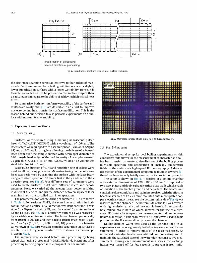
The experimental setup for pool boiling experiments on thinconductive foils allows for the measurement of characteristic boil-ing heat transfer parameters, visualization of the boiling processin visible spectrum, and observation of unsteady temperaturefields on the surface via high-speed IR thermography. A detaileddescription of the experimental setup can be found elsewhere [3];therefore, here we only briefly summarize its crucial components.
The setup is shown in Fig. 4. It consists of a boiling chamberwith external dimensions of 170 × 100 × 100 mm3, comprised oftwo steel plates and double glazed vertical glass walls which enableobservation of the bubble growth and departure. The heater unitconsisting of a ceramic base and stainless steel foil with the effectiveheat transfer area of 17 × 27 mm2 mounted onto nickel plated cop-per electrical contacts (e.g., see the bottom right side of Fig. 4) wasinserted into the chamber. The bottom side of the foil was coveredwith high emissivity paint and the ceramic base had a rectangularhole milled into it, both of which allowed for the use of a high-speed IR camera for temperature measurements and temperaturefield visualization. A golden mirror at a 45◦ angle was used to avoidpositioning the IR camera directly below the chamber.
Double-distilled water was used as the working fluid in allexperiments and was vigorously boiled before each series of mea-surements in order to remove most of the dissolved gases. An
immersed cartridge heater was utilized for preheating and tomaintain the saturated state of the water during consecutive mea-surements. During each measurement in a series, the cartridgeheater was turned off for few seconds to prevent it from influ-
M. Zupancic et al. / Applied Surface Science 399 (2017) 480–490 483
Table 1Parameters of laser texturing.
Sample Parameter
Scan line separation (see Fig. 2)�x (�m) × �y (�m)
Pulse energy(�J)
Pulse fluence(J cm−2)
F1 50 × 50 32 4.0F2 10 × 10 32 4.0F3 10 × 10 48 6.0F4 variable, (10–200) × (10–200) 72 9.0
F l boilit
et(iapf4atflfasca
yc
3
t
ig. 4. Schematics (left) and photography (right) of the experimental setup for poohe bottom right.
ncing movements of the water in the boiling chamber. A T-typehermocouple was used to measure the temperature of the watermeasurement accuracy for class 1 thermocouple is 1.0 K). Vapor-zed water was condensed using a custom-made glass condensernd a separate water cooling circuit. The foil was heated using therinciple of Joule heat generation due to electrical resistance of the
oil when supplied with electrical current through Sorensen SGA0/250 DC power supply. Heat flux was calculated from the volt-ge drop across the foil and the DC current which was determinedhrough the voltage drop on a reference resistor. To obtain the heatux, the heat generation was divided by the effective heat trans-
er surface area of the foil; uniform heat generation was assumedcross the entire foil surface. The heat fluxes used for testing of theurfaces ranged from 0 to 300 kW/m2 or 0–400 kW/m2. Thermo-ouple temperature and voltage drop signals were recorded usingn Agilent 34970A data acquisition unit.
The boiling process was visualized in visible spectrum (for anal-sis of bubbles formation and their dynamics) by using a high-speedamera Casio Exilim EX-F1 (recording at 1200 fps).
.2.1. Measurement uncertaintyA high-speed IR camera FLIR SC6000 with a cooled detec-
or was used for temperature field visualization and wall-
ng experiments. A cross-section view of the thin metal foil heater unit is shown in
temperature measurements. Contactless measurements, highachievable recording speeds and high sensitivities are one of themajor advantages of IR cameras. Therefore, they were successfullyutilized in many pool boiling and flow boiling studies [26–29].In our case average wall temperature was calculated from a 10 srecording of the high-speed IR camera by spatial and temporal aver-aging of the two-dimensional temperature fields. Recording speedwas set to 1000 fps, while spatial resolution was 250 �m per pixel.Raw digitalized IR camera signal was converted to temperatures viathe calibration curve obtained under the same ambient conditionsthat were present during the actual experiments. The expandedabsolute measurement uncertainty of the temperature was deter-mined to be 2.0 K and was practically constant across the entirecalibration range of 80–180 ◦C. We must emphasize that our cam-era provides a noise equivalent differential temperature (NEDT) ofonly 20 mK. The measurement uncertainty of the temperature dif-ference between individual pixels is therefore much lower thanabsolute temperature uncertainty.
The expanded relative measurement uncertainty of the heat flux
was estimated to be 0.5% and results from the combined uncer-tainty of voltage drop across the foil measurements, heater areauncertainty, and electrical current measurement uncertainty. Theuncertainty of the heat transfer coefficient is influenced by both the
484 M. Zupancic et al. / Applied Surface Science 399 (2017) 480–490
Table 2Equipment used for surface analyses.
Analysis Equipment
SEM Scanning electron microscope JEOL JSM-IT100(magnification 5–300,000×)
Optical interferometry 3D optical microscope Bruker ContourGT-K0(white and green light interferometry, lateralresolution 40 nm, vertical resolution 0.1 nm)
Contact angle measurementGoniometer, developed in Laboratory for ThermalTechnology (Faculty of Mechanical Engineering),using IDS UI-3060CP camera equipped with amicroscope objective, and micrometer syringe
uTw
3
taUmslarei
4
4
MraTdrtapttiosp
flawtida
lflsi
Table 3Topographical parameters, measured by optical interferometry. All data [with theexception of the reference surface (SS)] was filtered using a low-pass regressionGaussian filter.
Surface Parameter
Sa(�m) Ssk(/) Sku(/) r (/)
SS 0,07 −1,7 10,6 1,07F1 0,65 −0,7 9,6 1,25F2 0,71 −0,3 3,1 1,25F3 0,78 0,4 3,4 2,02F4 (1 st area) 1,36 −1,1 34,2 2,82
Gilmont GS 1200 with a steel size 22 capillary tube(water droplet volume 10 �L)
ncertainty of the temperature and the heat flux measurements.he maximum expanded uncertainty of the heat transfer coefficientas determined to be 3.4 kW/m2K.
.3. Surface characterization
Laser textured surfaced were characterized using scanning elec-ron microscopy (SEM), optical interferometry and through contactngle measurements. The equipment used is listed in Table 2.sing 3D optical microscope software analysis tools, we deter-ined topographical parameters of the textured surfaces. The 3D
pace parameters Sa, Ssk, Sku and the roughness factor r were calcu-ated. The wettability of the surfaces was measured through contactngles using a goniometer of our own design. Contact angles wereecorded immediately after laser processing, after every boilingxperiment, and irregularly over the course of 40 days after fin-shing the boiling experiments or after laser surface texturing.
. Results and discussion
.1. Surface microstructure
SEM images of the laser textured surfaces are shown in Fig. 5.icro- and nanostructure depend heavily on the scan line sepa-
ation and the laser pulse fluence. Despite low fluence, meltingnd ablation were achieved on surface F1 [e.g., see Fig. 5(a–c)].he damage trail on the surface was narrower than the beam spotiameter on the surface and no splattering of the melted mate-ial was detected. Since the scan line separation was larger thanhe damage trail width, a “mesh” with untextured area appearsfter the laser processing. Additionally, the following interestinghenomenon can be observed in Fig. 5(b). The surface was firstlyextured in the vertical direction and – in the second step – inhe horizontal direction. In places where traces cross each other,t is clearly visible that the initial structure is fully erased by a sec-nd (horizontal) passage and (at these processing parameters) newtructure depends on the direction of the second passage of therocessing beam alone.
Surface F2 [e.g., see Fig. 5(d–f)] was textured using the sameuence as F1 but with a decreased scan line separation. Thusly,pproximately 60% overlap between adjacent laser scanning linesas achieved. As it is clearly visible from Fig. 5(d–f), in this manner
he whole area was structured. The microstructure on surface F2s finer compared to the one on surface F1 with noticeable smallerroplets of melted material. However, some details are quite similars it is visible from a comparison between Fig. 5(c) and (f).
For texturing of the surface F3 [e.g., see Fig. 5(g–i)], the same scan
ine separation was used as for the surface F2, but the laser pulseuence was increased. As it is discernible from SEM micrographs,urface F3 included both micro- and nanostructure. However, exactdentification of the latter proved impossible due to the limitationF4 (2nd area) 3,50 −1,3 7,4 5,87
of the SEM image magnification and resolution. By increasing thefluence, some details were lost [e.g., comparison of Fig. 5(f) and (h)].
In contrast with homogenously processed surfaces F1–F3, sur-face F4 was textured with a variable scan line separation resultingin a non-uniform microstructure. Laser pulse fluence was increasedin comparison with the surface F3 and periodically varied scan lineseparation from 10 �m to 200 �m by a step of 10 �m and then backfrom 200 �m to 10 �m was utilized. The SEM micrographs of theselected F4 area (the region with the smallest scan line separationin the vertical direction) are shown in Fig. 5(j–l). It is evident thatsurface F4 also appears to be slightly porous, while the increasedpulse fluence resulted in the deepest and widest damage trailswith clearly noticeable abundant melted material splashing. Theadjacent laser scanning lines overlapped only in certain areas, i.e.,where scan line separation was small enough. In these regions, var-ious microcavities with the diameter of up to a few micrometerswere detected along with microporosity, as marked on the right-hand side of Fig. 6, where the diameters of microcavities are alsolisted. From Fig. 6 it is clearly noticable that multi-scale microcav-ities appear in the region with the smallest scan line separation(10 �m). These cavities could potentially act as active nucleationsites in accordance with the aforementioned nucleation criteria.Furthermore, their diameter is not uniform and spans across atleast one order of magnitude and it is therefore expedient for heattransfer enhancement.
The topographical parameters measured by optical interferom-etry are listed in Table 3. Here, all the results with the exception ofthe reference surface (SS) were filtered using a low-pass regressionGaussian filter. These results show that the highest roughness wasachieved on surface F4, mainly due to the deepest damage trails oflaser processing (as a result of the highest laser pulse fluence). In thecase of surface F4, which had a non-uniform microstructure, mea-surements are listed for two selected areas. The data for the “1 starea” was obtained by measurements in the area processed by thehighest scanning line separation (200 �m), while the data for the“2nd area” was measured in the area with the smallest scanningline separation (10 �m). Ratio between real surface area and theprojected area, expressed by roughness factor r, could be upwardsof five in certain regions of the surface F4.
4.2. Surface wettability
The measurements of the surface wettability were performedon textured-only surfaces and on textured surfaces (using the samelaser parameters and, therefore, marked with the same labels) thatwere used in boiling experiments. Contact angle of a water dropleton a reference, untextured surface is shown on Fig. 7(a) and was
◦
equal to 88 . Immediately after the processing, surfaces F1 andF2 were slightly hydrophilic [e.g. see Fig. 7(b–c)], surface F3 wassuperhydrophilic [e.g. see Fig. 7(d)], while surface F4 exhibitednon-uniform wettability due to the heterogeneous microstructure.
M. Zupancic et al. / Applied Surface Science 399 (2017) 480–490 485
of the
tTcawtdfcsipa
Fig. 5. SEM micrographs
We also measured the temporal wettability development withhe samples being exposed to the ambient air at room conditions.he results are shown in Fig. 7(e). Here, mark “b” in the legendorresponds to the surfaces that were used in boiling experimentsfter the processing. As it is visible from the presented results, theettability of all of the surfaces decreased over time, similar to
he observations of other authors [18,19,30]. Authors in Ref. [18]etected and increased amount of carbon on the stainless steel sur-
ace after being treated with femtosecond laser. However, the initialarbon amount is not enough to fully cover the structure and entire
urface is superhydrophilic due to combination of underlying polarron oxides and increased surface roughness. Over time, the decom-osition of carbon dioxide into carbon takes place and starts toccumulate on the surface. In combination with dual-scale rough-laser textured surfaces.
ness structure this results in superhydrophobic surface. In theircase, the superhydrophobicity was developed in about 10–30 daysafter the laser treatment. In our case, the contact angle increasedby 20–40◦ depending on the surface-texturing parameters and itstabilized after approximately 15–20 days after the processing. Nosignificant difference is observed between textured-only surfacesand surfaces used in boiling experiments. This result proves thatthe nanosecond laser texturing has a great potential in applicationsfor improved boiling heat transfer, since it can ensure durability ofthe produced surfaces.
Sample F4, which was processed by a variable laser scan lineseparation, exhibits heterogeneous surface texture and therefore
486 M. Zupancic et al. / Applied Surface Science 399 (2017) 480–490
Fig. 6. SEM micrograph of the surface F4 in the region with the smallest scan lineseparation (10 �m); some microcavities are marked with arrows with their diame-ters also listed.
Fig. 7. (a–d) Wettability of bare stainless steel foil (SS) and surfaces F1–F3 imme-d(m
at
4
iardtsi
0 100 200 300 4000
10
20
30
2q (kW/m )
2α
(kW
/mK
)
0 5 10 15 20 25 300
100
200
300
400
T - T (K)w sat
SSF1F2F3F4
PolishedS1S2S3S4
2q
(kW
/m)
(a)
(b)
Kruse et al.2015
.
.
Fig. 8. (a) Heat flux as a function of superheat. (b) Heat transfer coefficient versusheat flux. The results are shown for our, nanosecond-laser textured, surfaces F1–F4
iately after the processing. (e) Temporal development of wettability for boiledmarked with ‘b’) and textured-only surfaces. Number of repeated trials for each
easurement was 10. Error bars are showing.
lso non-uniform wettability. We have measured the contact angleso be in between 66◦ and 142◦.
.3. Pool boiling performance
The pool boiling performance was firstly estimated by measur-ng the heat flux vs. surface overheat and heat transfer coefficient as
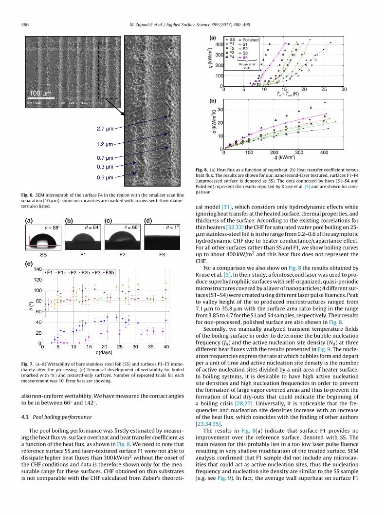
function of the heat flux, as shown in Fig. 8. We need to note thateference surface SS and laser-textured surface F1 were not able to
issipate higher heat fluxes than 300 kW/m2 without the onset ofhe CHF conditions and data is therefore shown only for the mea-urable range for these surfaces. CHF obtained on thin substratess not comparable with the CHF calculated from Zuber’s theoreti-(unprocessed surface is denoted as SS). The dots connected by lines (S1–S4 andPolished) represent the results reported by Kruse et al. [5] and are shown for com-parison.
cal model [31], which considers only hydrodynamic effects whileignoring heat transfer at the heated surface, thermal properties, andthickness of the surface. According to the existing correlations forthin heaters [32,33] the CHF for saturated water pool boiling on 25-�m stainless-steel foil is in the range from 0.2–0.6 of the asymptotichydrodynamic CHF due to heater conductance/capacitance effect.For all other surfaces rather than SS and F1, we show boiling curvesup to about 400 kW/m2 and this heat flux does not represent theCHF.
For a comparison we also show on Fig. 8 the results obtained byKruse et al. [5]. In their study, a femtosecond laser was used to pro-duce superhydrophilic surfaces with self-organized, quasi-periodicmicrostructures covered by a layer of nanoparticles; 4 different sur-faces (S1–S4) were created using different laser pulse fluences. Peakto valley height of the so produced microstructures ranged from7.1 �m to 35.8 �m with the surface area ratio being in the rangefrom 3.85 to 4.7 for the S1 and S4 samples, respectively. Their resultsfor non-processed, polished surface are also shown in Fig. 8.
Secondly, we manually analyzed transient temperature fieldsof the boiling surface in order to determine the bubble nucleationfrequency (fb) and the active nucleation site density (Nd) at threedifferent heat fluxes with the results presented in Fig. 9. The nucle-ation frequencies express the rate at which bubbles form and departper a unit of time and active nucleation site density is the numberof active nucleation sites divided by a unit area of heater surface.In boiling systems, it is desirable to have high active nucleationsite densities and high nucleation frequencies in order to preventthe formation of large vapor covered areas and thus to prevent theformation of local dry-outs that could indicate the beginning ofa boiling crisis [28,27]. Universally, it is noticeable that the fre-quencies and nucleation site densities increase with an increaseof the heat flux, which coincides with the finding of other authors[23,34,35].
The results in Fig. 8(a) indicate that surface F1 provides noimprovement over the reference surface, denoted with SS. Themain reason for this probably lies in a too low laser pulse fluenceresulting in very shallow modification of the treated surface. SEM
analysis confirmed that F1 sample did not include any microcav-ities that could act as active nucleation sites, thus the nucleationfrequency and nucleation site density are similar to the SS sample(e.g. see Fig. 9). In fact, the average wall superheat on surface F1
M. Zupancic et al. / Applied Surface
50 100 3000
10
20
30
40
50
2q (kW/m )
f n (H
z)SSF1F2F3F4
˙
50 100 300
010
110
210
2q (kW/m )
2N
(1/
cm)
a
˙
(a)
(b)SSF1F2F3F4
Fs
i(omattu
mtflmElcmanb
fsaKtmasF
4
litoc
ig. 9. Comparison of (a) nucleation frequencies and (b) active nucleation site den-ities at three different heat fluxes. Error bars in (a) indicate standard deviation.
s higher than for SS sample throughout entire experimental range0–300 kW/m2). This might be contributed to higher wettabilityf F1 (e.g. see Fig. 7) and the fact that higher wettability requireore energy for the boiling incipience [36], which results in higher
ctivation temperatures as well as higher average temperatureshroughout the nucleate boiling regime. One should still be awarehat differences between SS and F1 are within the measurementncertainty.
Surfaces F2 and F3 with more significant, but still uniformicrostructure and wettability, enable enhanced boiling heat
ransfer in regard to the reference surface. In the 0–300 kW/m2 heatux span, both surfaces provided an approximate 40% enhance-ent of the heat transfer coefficient compared to the SS sample.
ven though the surface structure of F2 and F3 samples is simi-ar, the average wall superheat for F3 was more than 1 K higherompared to F2 [see Fig. 8(a)]. Despite the fact that the expandedeasurement uncertainty of IR camera is 2 K, the difference in aver-
ge temperatures can be again contributed to the superhydrophilicature of the F3 sample and therefore higher required energy foroiling incipience compared to F2.
The highest heat transfer enhancement was achieved on sur-ace F4. In this case, we obtained higher heat fluxes at lower walluperheats, not only in comparison with the reference surface, butlso in comparison with the state-of-the-art results presented byruse et al. [5]. Heat transfer coefficient on the surface F4 was more
han two-times higher that on the SS sample [e.g. 110% enhance-ent, as visible from Fig. 8(b)], while nucleation frequency was
lso enhanced by a factor of two [see Fig. 9(a)] and nucleationite density was higher by more than one order of magnitude [seeig. 9(b)].
.4. Control of active nucleation sites by laser texturing
The control of active nucleation sites is one of the most chal-enging issues in boiling heat transfer. Therefore, it is extremely
mportant to correlate the locations of active nucleation sites withhe laser textured microstructure. In order to identify the locationsf active nucleation sites, we performed appropriate image pro-essing and analysis of the high-speed thermography recordingsScience 399 (2017) 480–490 487
for all tested surfaces F1–F4 at the three different heat flux rates:50 kW/m2, 100 kW/m2 and 300 kW/m2. The results are presentedin Fig. 10.
During each individual bubble growth the surface is rapidlycooled down due to the large local heat flux in the moment ofvaporization. This round-shaped cold spot can be easily identifiedand measured with pixel resolution for each time frame based in IRthermographs. Therefore, we decided to add each detected nucle-ation area to some cumulative matrix. This matrix was then plottedas a grayscale image, as shown in each odd line in Fig. 10. Each cir-cular shape on this grayscale image represents maximum bubblecontact diameter, while the number of circles divided by the heaterarea corresponds to the active nucleation site density. To better dis-play the spatial distribution of nucleation sites, we determined thecenter of each detected nucleation. The results are represented asthe blue dots on a gray background in each even line of Fig. 10. Toobtain these results, we analyzed 10 s of a recording (e.g. 10,000frames) for each sample at each individual heat flux.
As it is discernable from the first row in Fig. 10, bubble con-tact diameters are the largest on surface F1 and range from 1.3 mmto 6.3 mm at 300 kW/m2. Similarly to the unstructured stainlesssteel foil [27], F1 sample with shallow microstructure requiredhigh activation temperature, which resulted in large bubble con-tact diameters and a low nucleation frequency. Large bubbles alsolimit the total number of active nucleation sites. On surfaces F2 andF3 (e.g., see 3rd and 5th line in Fig. 10), bubble contact diameters areabout two-times smaller compared to F1. Pronounced microstruc-ture on F2 and F3 enhances potential active nucleation sites. Eventhough the contact angles on F1 and F2 are very comparable, wemust emphasize that pronounced microstructure on F2 results inbetter wickability, which enhances heat transfer performance [37].This confirms that contact angle itself (as a surface-fluid property)is not sufficient parameter to predict boiling behavior. Despite thenucleation site density enhancement on F2 and F3, the nucleationsare still randomly distributed across the entire heater area, as it isclearly distinguishable from the spatial distributions of the activenucleation sites in Fig. 10.
The smallest bubbles were observed on surface F4 where thecontact diameters at 300 kW/m2 ranged from only 0.4 mm to1.8 mm. An even more important result is presented in the lastrow of Fig. 10, where it is clearly visible that the active nucle-ation site density on surface F4 is periodically distributed acrossthe entire surface and exactly matches the spatial distribution ofthe F4 areas with the smallest scan line separations. These areasare covered with multi-scale sized microcavities, as shown throughsurface analysis presented in Fig. 6. This is in agreement with thenucleation criteria (presented in Fig. 1) implying that in order tomaximize the number of possible active nucleation sites the sur-face should provide micrometer and sub-micrometer sized cavitiesand also variable wettability. In addition to increasing active nucle-ation sites, the presence of micro cavities also lowered the onset ofnucleate boiling for more than 5 K compared to bare stainless steel(e.g. see Fig. 8).
Although the resolution of IR thermography is limited and, con-sequently, the position of active nucleation sites should be moreaccurately determined by other state-of-the-art temperature map-ping techniques in the future [38], our results prove that thenumber of nucleation sites was significantly higher in the vicin-ity of microcavities in comparison with other areas. These results,obtained on surface F4, confirm the predictions of the nucleationcriteria and lead to an important conclusion that straightforwardand cost-effective nanosecond laser texturing is able to produce
cavities of diameters up to a few micrometers and surfaces withnon-uniform wettability. This opens up an important possibilityfor controlling the boiling process in terms of defining active nucle-
488 M. Zupancic et al. / Applied Surface Science 399 (2017) 480–490
Fig. 10. Spatial distribution of active nucleation sites.

rface
ad
5
swlpnpsbt
al
•
•
•
amitmae
A
b0
R
[
[
[
[
[
[
[
[
[
[
[
[
[
[
[
[
[
[
[
[
[
M. Zupancic et al. / Applied Su
tion areas, bubble diameters and nucleation frequencies as well asensity of active nucleation sites and finally wall superheat.
. Conclusions
In this study we examined the potential of laser texturedurfaces for enhanced boiling heat transfer. Multiple surfacesere prepared on thin stainless steel foils by using different
aser texturing patterns and different laser pulse fluences. Pre-ared samples were either hydrophilic, superhydrophilic or hadon-uniform wettability depending on the combination of therocessing parameters. The wettability decreased over time andtabilized in approximately two to three weeks. The comparisonetween textured-only surfaces and surfaces used in boiling provedhe stability of laser-textured surfaces in boiling experiments.
Nucleate boiling heat transfer on textured surfaces was evalu-ted by means of high-speed IR thermography. The obtained resultsead to the following important conclusions:
Laser textured surfaces are able to enhance nucleate boiling heattransfer with lower superheats and up to 110% higher heat trans-fer coefficients compared to the untextured surface.Under certain texturing parameters, non-uniformly wettable sur-face structure with multi-scale microcavities is formed throughmelting and setting of the material. Our results show that thesefeatures are responsible for achieving 20–40 times higher activenucleation site density compared to the bare untreated surface.IR thermographs proved that most nucleations happened in thevicinity of the microcavities. This is also in agreement with thedescribed theoretical nucleation criteria.Through the control of texturing parameters, the location ofmicrocavities can be controlled and consequently also the areaswhere the majority of the bubbles form and grow. This suggeststhat laser texturing technique enables some control over the com-plex boiling process.
The presented surface modification technique does not requireny additional coatings and/or post-processing. Results prove thatarking nanosecond laser allows modification of surfaces for an
ncreased boiling heat transfer performance. This is an impor-ant demonstration, since cheaper production (in comparison with
icro- and nanostructuring through the use of ultrafast, i.e., psnd fs laser systems) enables wider dissemination of surfaces withnhanced functionalities in different areas of applications.
cknowledgement
The authors acknowledge the financial support from the stateudget by the Slovenian Research Agency (Programme Nos. P2-223 and P2-0392).
eferences
[1] C.H. Wang, V.K. Dhir, Effect of surface wettability on active nucleation sitedensity during pool boiling of water on a vertical surface, J. Heat Transf. 115(1993) 659–669, http://dx.doi.org/10.1115/1.2910737.
[2] B. Bourdon, P. Di Marco, R. Rioboo, M. Marengo, J. De Coninck, Enhancing theonset of pool boiling by wettability modification on nanometrically smoothsurfaces, Int. Commun. Heat Mass Transf. 45 (2013) 11–15, http://dx.doi.org/10.1016/j.icheatmasstransfer.2013.04.009.
[3] M. Zupancic, M. Steinbücher, P. Gregorcic, I. Golobic, Enhanced pool-boilingheat transfer on laser-made hydrophobic/superhydrophilicpolydimethylsiloxane-silica patterned surfaces, Appl. Therm. Eng. 91 (2015)288–297, http://dx.doi.org/10.1016/j.applthermaleng.2015.08.026.
[4] H.S. Ahn, H.J. Jo, S.H. Kang, M.H. Kim, Effect of liquid spreading due tonano/microstructures on the critical heat flux during pool boiling, Appl. Phys.Lett. 98 (2011) 98–101, http://dx.doi.org/10.1063/1.3555430.
[5] C.M. Kruse, T. Anderson, C. Wilson, C. Zuhlke, D. Alexander, G. Gogos, S. Ndao,Enhanced pool-boiling heat transfer and critical heat flux on femtosecond
[
[
Science 399 (2017) 480–490 489
laser processed stainless steel surfaces, Int. J. Heat Mass Transf. 82 (2015)109–116, http://dx.doi.org/10.1016/j.ijheatmasstransfer.2014.11.023.
[6] A.R. Betz, J. Jenkins, C.J. Kim, D. Attinger, Boiling heat transfer onsuperhydrophilic, superhydrophobic, and superbiphilic surfaces, Int. J. HeatMass Transf. 57 (2013) 733–741, http://dx.doi.org/10.1016/j.ijheatmasstransfer.2012.10.080.
[7] I. Golobic, M. Zupancic, Wall-temperature distributions of nucleate poolboiling surfaces vs boiling curves: a new approach, Int. J. Heat Mass Transf. 99(2016) 541–547, http://dx.doi.org/10.1016/j.ijheatmasstransfer.2016.04.033.
[8] S.K. C.S, S. S, A. C.R, S.K. M.C, P. A.S, R. K, Flow boiling heat transferenhancement on copper surface using Fe doped Al2O3–TiO2 compositecoatings, Appl. Surf. Sci. 334 (2015) 102–109, http://dx.doi.org/10.1016/j.apsusc.2014.08.076.
[9] C.-H. Choi, M. David, Z. Gao, A. Chang, M. Allen, H. Wang, C. Chang, Large-scalegeneration of patterned bubble arrays on printed Bi-functional boilingsurfaces, Sci. Rep. 6 (2016) 23760, http://dx.doi.org/10.1038/srep23760.
10] C.Y. Lee, B.J. Zhang, K.J. Kim, Morphological change of plain and nano-poroussurfaces during boiling and its effect on nucleate pool boiling heat transfer,Exp. Therm. Fluid Sci. 40 (2012) 150–158, http://dx.doi.org/10.1016/j.expthermflusci.2012.02.011.
11] S. Vemuri, K.J. Kim, Pool boiling of saturated FC-72 on nano-porous surface,Int. Commun. Heat Mass Transf. 32 (2005) 27–31, http://dx.doi.org/10.1016/j.icheatmasstransfer.2004.03.020.
12] Y.Y. Hsu, On the size range of active nucleation cavities on a heating surface, J.Heat Transf. 84 (1962) 207–213.
13] S.G. Kandlikar, V.R. Mizo, M.D. Cartwright, E. Ikenze, Bubble nucleation andgrowth characteristics in subcooled flow boiling of water, in: ASME Proc 32ndNatl. Heat Transf. Conf., ASME, 1997, pp. 11–18.
14] S. Gong, P. Cheng, X. Quan, Two-dimensional mesoscale simulations ofsaturated pool boiling from rough surfaces. Part I: bubble nucleation in asingle cavity at low superheats, Int. J. Heat Mass Transf. (2016), http://dx.doi.org/10.1016/j.ijheatmasstransfer.2016.04.085.
15] D. Deng, R. Chen, Y. Tang, L. Lu, T. Zeng, W. Wan, A comparative study of flowboiling performance in reentrant copper microchannels and reentrant porousmicrochannels with multi-scale rough surface, Int. J. Multiph. Flow. 72 (2015)275–287, http://dx.doi.org/10.1016/j.ijmultiphaseflow.2015.01.004.
16] Y.-W. Lu, S.G. Kandlikar, Nanoscale surface modification techniques for poolboiling Enhancement. A critical review and future directions, Heat Transf.Eng. 32 (2011) 827–842, http://dx.doi.org/10.1080/01457632.2011.548267.
17] A.Y. Vorobyev, C. Guo, Multifunctional surfaces produced by femtosecondlaser pulses, J. Appl. Phys. 117 (2015) 33103, http://dx.doi.org/10.1063/1.4905616.
18] A.M. Kietzig, S.G. Hatzikiriakos, P. Englezos, Patterned superhydrophobicmetallic surfaces, Langmuir 25 (2009) 4821–4827, http://dx.doi.org/10.1021/la8037582.
19] V.D. Ta, A. Dunn, T.J. Wasley, J. Li, R.W. Kay, J. Stringer, P.J. Smith, E. Esenturk,C. Connaughton, J.D. Shephard, Laser textured superhydrophobic surfaces andtheir applications for homogeneous spot deposition, Appl. Surf. Sci. 365(2016) 153–159, http://dx.doi.org/10.1016/j.apsusc.2016.01.019.
20] W.M. Rohsenow, A method of correlating heat transfer data for surfaceboiling of liquids, Trans. ASME 74 (1952) 969–976.
21] S.G. Bankoff, Entrapment of gas in the spreading of a liquid over a roughsurface, AIChE J. 4 (1958) 24–26, http://dx.doi.org/10.1002/aic.690040105.
22] P. Griffith, J.D. Wallis, The role os surface conditions in nucleate boiling, Chem.Eng. Prog. Symp. 56 (1960) 49–63.
23] C.-Y. Han, P. Griffith, The mechanism of heat transfer in nucleate poolboiling—part II, Int. J. Heat Mass Transf. 8 (1965) 905–914, http://dx.doi.org/10.1016/0017-9310(65)90074-8.
24] H.S. Carslaw, J.C. Jaeger, Conduction of Heat in Solids, second ed., OxfordUniversity Press, London, 1959.
25] S.G. Kandlikar, Nucleation characteristics and stability considerations duringflow boiling in microchannels, Exp. Therm. Fluid Sci. 30 (2006) 441–447,http://dx.doi.org/10.1016/j.expthermflusci.2005.10.001.
26] H. Kim, J. Buongiorno, Detection of liquid-vapor-solid triple contact line intwo-phase heat transfer phenomena using high-speed infrared thermometry,Int. J. Multiph. Flow 37 (2011) 166–172, http://dx.doi.org/10.1016/j.ijmultiphaseflow.2010.09.010.
27] J. Petkovsek, Y. Heng, M. Zupancic, H. Gjerkes, F. Cimerman, I. Golobic, IRthermographic investigation of nucleate pool boiling at high heat flux, Int. J.Refrig. 61 (2016) 127–139, http://dx.doi.org/10.1016/j.ijrefrig.2015.10.018.
28] T.G. Theofanous, T.N. Dinh, J.P. Tu, A.T. Dinh, The boiling crisis phenomenonpart II: dryout dynamics and burnout, Exp. Therm. Fluid Sci. 26 (2002)793–810, http://dx.doi.org/10.1016/S0894-1777(02)00193-0.
29] J. Yoo, C.E. Estrada-Perez, Y.A. Hassan, An accurate wall temperaturemeasurement using infrared thermometry with enhanced two-phase flowvisualization in a convective boiling system, Int. J. Therm. Sci. 90 (2015)248–266, http://dx.doi.org/10.1016/j.ijthermalsci.2014.12.007.
30] D.V. Ta, A. Dunn, T.J. Wasley, R.W. Kay, J. Stringer, P.J. Smith, C. Connaughton,J.D. Shephard, Nanosecond laser textured superhydrophobic metallic surfacesand their chemical sensing applications, Appl. Surf. Sci. 357 (2015) 248–254,http://dx.doi.org/10.1016/j.apsusc.2015.09.027.
31] N. Zuber, On the stability of boiling heat transfer, Trans. Am. Soc. Mech. Eng.80 (1958) 711–720.
32] I. Golobic, A.E. Bergles, Effects of heater-side factors on the saturated poolboiling critical heat flux, Exp. Therm. Fluid Sci. 15 (1997) 43–51, http://dx.doi.org/10.1016/S0894-1777(96)00170-7.

4 rface
[
[
[
[
[
90 M. Zupancic et al. / Applied Su
33] M. Arik, A. Bar-Cohen, Effusivity-based correlation of surface property effectsin pool boiling CHF of dielectric liquids, Int. J. Heat Mass Transf. 46 (2003)3755–3764, http://dx.doi.org/10.1016/S0017-9310(03)00215-1.
34] N. Zuber, Nucleate boiling. The region of isolated bubbles and the similarity
with natural convection, Int. J. Heat Mass Transf. 6 (1963) 53–78, http://dx.doi.org/10.1016/0017-9310(63)90029-2.35] H.J. Ivey, Relationships between bubble frequency, departure diameter andrise velocity in nucleate boiling, Int. J. Heat Mass Transf. 10 (1967)1023–1040, http://dx.doi.org/10.1016/0017-9310(67)90118-4.
[
Science 399 (2017) 480–490
36] B. Bourdon, R. Rioboo, M. Marengo, E. Gosselin, J. De Coninck, Influence of thewettability on the boiling onset, Langmuir 28 (2012) 1618–1624, http://dx.doi.org/10.1021/la203636a.
37] M.M. Rahman, E. Olc eroglu, M. McCarthy, Role of wickability on the critical
heat flux of structured superhydrophilic surfaces, Langmuir 30 (2014)11225–11234, http://dx.doi.org/10.1021/la5030923.38] I. Sedmak, I. Urbancic, R. Podlipec, J. Strancar, M. Mortier, I. Golobic, Submicronthermal imaging of a nucleate boiling process using fluorescence microscopy,Energy 109 (2016) 436–445, http://dx.doi.org/10.1016/j.energy.2016.04.121.