Embed Size (px)
Citation preview

APPLICATION OF REMOTE ISCHAEMIC PRECONDITIONING TO HUMAN CORONARY ARTERY BYPASS SURGERY by ISHTIAQ ALI RAHMAN A thesis submitted to The University of Birmingham for the degree of DOCTOR OF MEDICINE (MD)
School of Clinical and Experimental Medicine College of Medical and Dental Sciences
January 2010

University of Birmingham Research Archive
e-theses repository This unpublished thesis/dissertation is copyright of the author and/or third parties. The intellectual property rights of the author or third parties in respect of this work are as defined by The Copyright Designs and Patents Act 1988 or as modified by any successor legislation. Any use made of information contained in this thesis/dissertation must be in accordance with that legislation and must be properly acknowledged. Further distribution or reproduction in any format is prohibited without the permission of the copyright holder.

ABSTRACT
This thesis reports a clinical study designed to assess myocardial, renal and
lung outcomes following cardiac surgery. In a single centre, prospective
randomized, placebo intervention-controlled trial the effects of intermittent
upper limb ischaemia (remote ischaemic preconditioning (RIPC)) were
compared in non-diabetic adult patients undergoing on-pump multi-vessel
coronary artery surgery. Patients, investigators, anaesthetists, surgeons and
critical care teams were all blind to group allocation. Subjects were
randomized(1:1) to RIPC(or placebo) stimuli (3x upper limb (or dummy arm) 5
minute cycles of 200mmHg cuff inflation/deflation) during sternotomy and
conduit procurement. Anaesthesia, perfusion, cardioplegia and surgical
techniques were standardized.
Groups were well matched on demographic and operative variables. In
contrast to prior smaller studies, RIPC did not reduce troponin T (48 hour area
under the curve (AUC); 6hour and peak) release, improve post-operative
haemodynamics (cardiac indices; low cardiac output episodes incidence;
IABP usage; inotrope and vasoconstrictor use; M mode, 2D contrast-
enhanced echocardiography and tissue Doppler imaging) or offer anti-
arrhythmic benefit (de novo left bundle branch block or Q waves; ventricular
tachyarrhythmia incidence). RIPC did not afford renal (peak creatinine, AUC
urinary albumin-creatinine ratios, dialysis requirement) or lung protection
(intubation times, 6hour and 12 hour pO2/FiO2 ratios). Case urgency did not
influence RIPC effect.

DEDICATION
I would like to dedicate this work to my mother, Shakeela, and my father,
Altaf, for providing me with so many opportunities; to my wife, Hina, for
helping me to maximise those opportunities; and to my daughter, Tamara, for
being my inspiration.

ACKNOWLEDGMENTS
This work was undertaken between 2006 and 2009 at the University Hospital
Birmingham NHS Foundation Trust (FT) under the guidance of my
educational supervisor Professor Robert Stuart Bonser. My contribution was
funded by a fellowship via a project grant of the British Heart Foundation
acquired by competitive application and I thank the BHF, together with their
sponsors for this support. I would like to thank the Wellcome Trust Clinical
Research Facility and the Cardiac Intensive Care Units, University Hospital
Birmingham NHS FT for clinical support and the Department of Clinical
Biochemistry, Selly Oak Hospital, University Hospital Birmingham NHS FT
and East Kent Hospitals University NHS FT for laboratory support.
I would also like to thank my co-supervisor Professor Michael Frenneaux,
Regius Professor of Medicine and mentor Dr Rick Steeds, Consultant
Cardiologist. Dr Howard Marshall, Consultant Cardiologist, facilitated my
investigations of arrhythmia analysis. The specific help of Peter Nightingale,
Statistician, was crucial to this work. Echocardiography was performed by Ms
Anne Marie Marsh, Dr Lynne Williams and Mr Ador Alvior. Primary work on
myocardial specimens was done by Dr Hussain Contractor.
I would also acknowledge the contributions of the co-authors Mr Jorge
Mascaro, Dr Peter Gosling, Dr Peter Townsend, Dr John Townend and Dr
David Green on our published manuscripts

PERSONAL CONTRIBUTION
The work of this thesis was undertaken during the tenure of a British Heart
Foundation Fellowship (2006-9). I was attached to the Department of Cardiac
Surgery, under the guidance of Professor Robert Stuart Bonser, at the
University Hospital Birmingham NHS Foundation Trust. The thesis was
registered with the University of Birmingham. The Fellowship had clinical
responsibility for all 162 trial patients and additional time was dedicated to
research. This thesis details the clinical trial that was undertaken.
At the time of this work, in coronary artery bypass grafting (CABG), adverse
outcomes predominantly related to cardiomyocyte injury due to myocardial
protection failure presented a current problem that was projected to grow as a
more aged population with increased co-morbidities presented for surgery. A
need to conserve cardiac function and myocyte integrity (myocardial
protection) in the peri-operative period and thereafter had been identified.
Early experimental and clinical evidence suggested that RIPC could attenuate
such injury safely and practically whilst also offering renal and lung protection.
Informed consent of patients, management of the trial, data accrual and
analysis of data was my own responsibility. Echocardiographic image analysis
was performed by myself under the guidance of Dr Rick Steeds. Continuous
ECG monitoring data disks was analysed solely by myself. I observed the
techniques for analysis of myocardial specimens by Dr Hussain Contractor
and was involved in analysis. The writing of this thesis is wholly my own
endeavour.
TABLE OF CONTENTS
CHAPTER 1: THE DEVELOPMENT AND CURRENT STATUS OF
SURGERY FOR ISCHAEMIC HEART DISEASE 1 Section 1: Development of coronary artery surgery
and cardiopulmonary bypass 1 1.0 Introduction 1 2.0 Early history of cardiac surgery 1 3.0 A brief history of coronary artery surgery 4 4.0 The development of cardiopulmonary bypass 7 Section 2: The need for myocardial protection 9
1.0 Introduction 9 2.0 Brief history of myocardial protection 10 3.0 Reperfusion Injury 11 4.0 Cardioplegia 12 4.1 Introduction to cardioplegia 12 4.2 Principles of Cardioplegia 12 4.2.1 Prompt diastolic arrest 13 4.2.1.1 Crystalloid cardioplegia 14 4.2.1.2 Blood cardioplegia 14 4.2.1.3 Miniplegia 14 4.2.2 Reduction in cardiac oxygen demand 15 4.2.3 Temperature 15 4.2.4 Route of delivery 16 4.2.5 Cardioplegia additives 18 4.3 Cardioplegia Strategy 18 4.4 Blood vs. crystalloid cardioplegia 19 4.5 Directed cardioplegia 20 5.0 Volatile anaesthetics 20 5.1 Introduction to volatile anaesthetics 20 5.2 Enflurane, Sevoflurane, Desflurane and 20
Isoflurane 5.3 Volatile vs. non-volatile anaesthesia 21 6.0 Ischaemic conditioning 22 7.0 Nicorandil 23 8.0 Statins 23 9.0 Aprotinin 23 10.0 Conclusions 24
Section 3: The current status of coronary artery bypass graft surgery (CABG) 25
1.0 Adult Cardiac Surgical Database Report 2008 25 1.1 Patient populations treated by CABG 25 1.2 Survival following CABG 26

2.0 Revascularisation vs. medical therapy 29 3.0 Revascularisation vs. percutaneous intervention 30 3.1 CABG vs. balloon angioplasty 30 3.2 CABG vs. coronary stenting 32
CHAPTER 2: ISCHAEMIC PRECONDITIONING (IP)
IN CARDIAC SURGERY: A SYSTEMATIC REVIEW OF EXPERIMENTAL AND CLINICAL LITERATURE 35 1.0 Introduction 35 1.1 Local (or ‘Cardiac) Ischaemic Preconditioning 35 1.2 Remote Ischaemic Preconditioning 37 2.0 Mechanisms Involved in Conditioning 39 2.1.1 Ischaemic Preconditioning 39 2.1.2 Cellular Events 40 2.1.3 cAMP 45 2.2 Remote Ischaemic Preconditioning 48 2.3 Mitochondrial Transition Pore Opening 49 2.4 Postconditioning 52 2.5 Cardiopulmonary bypass 53 2.6 Neural Factors 54 2.7 Humoral Factors 57 3.0 Clinical Benefits of Preconditioning 58 3.1 Clinical Benefits of IP 58 3.2 Clinical Benefits of RIPC 62 4.0 Conclusion 65
CHAPTER 3: CORE METHODOLOGY 66
1.0 Introduction 66
1.1 Study Flow 67 2.0 Recruitment 67 2.1 Patient selection 67 2.2 Inclusion and Exclusion Criteria 68 2.3 Randomisation 68 3.0 Pre-operative medications 69 3.1 Permitted drugs 70 3.1.1 Statins 70 3.1.2 Ca2+ channel antagonists (oral) 70 3.1.3 β-receptor antagonists (oral) 70 3.1.4 Long acting nitrates (oral) 70 3.2 Drugs to be omitted 70 3.2.1 Anti-platelet agents 70 3.2.2 Angiotensin converting enzyme (ACE) inhibitors 71 3.2.3 Angiotensin-II receptor antagonists ‘Sartans’ 71 3.2.4 Diuretics 71 3.2.5 Potassium channel activators 71 3.3 Scheduled drugs 72 3.3.1 Premedication 72

4.0 Treatment Groups 72 4.1 Control Group 72 4.2 RIPC Group 73 5.0 Anaesthetic Protocols 76 5.1 Induction of anaesthesia 76
5.2 Central venous cannulation 76 5.3 Maintenance of anaesthesia 76
5.4 Other medications 77 6.0 Perfusion Protocols 78 6.1 Bypass schedule 78
6.2 Cardioplegia composition 79 6.3 Cardioplegia administration 80 6.4 Discontinuation of CPB 80
7.0 Surgical Protocol 80 7.1 Conduct of operation 80
7.2 Surgical technique 81 7.3 Myocardial protection 81 7.4 Myocardial Biopsy 81
8.0 Operating department practitioner protocol 82 8.1 Checklist prior to commencing 82 8.2 Conduct of ODP 82 9.0 Biochemical monitoring protocol 84 9.1 Cardiac Troponin T 84 9.2 Test Principle – Sandwich principle 85 10.0 Haemodynamic monitoring 86 10.1.1 Initial pre-bypass studies (Baseline/pre-sternotomy) 86 10.2 Post-bypass haemodynamic studies (pre-protamine) 86 10.3 Post-protamine haemodynamic studies 86 10.4 Post-sternal closure haemodynamic studies 87 10.5 Haemodynamic studies on the intensive care unit 87 10.6 Low cardiac output episodes 87 11.0 Protocol for post-operative inotropic / vasoconstrictor 87 12.0 Echocardiography 88 12.1 Contrast Echocardiography for EF 89 12.2 Isovolumic Acceleration 90 12.3 Tissue Velocity Imaging 91 13.0 Arrhythmia Assessment 93 13.1 Continuous Holter ECG recording 93 13.2 12 lead electrocardiograms (ECGs) 96 13.3 Atrial Fibrillation – Treated 96 14.0 Renal outcomes protocol 97 14.1.1 Creatinine 97 14.1.2 Test principle – Kinetic colorimetric assay 98 14.2.1 Albumin 98 14.2.2 Test principle – Immunoturbidimetric assay 99 14.3.1 Urine ACR 100 14.4.1 Urine for α-1 microglobulin 100 14.4.2 Test principle – Rate nephelometry 100 15.0 Lung outcomes protocol 101 15.1 Spirometry 101

15.2 Positive End Expiratory Pressure 101 15.3 Arterial PaO2 / fractional inspired oxygen ratio 102 15.4 Intubation time 102 16.0 Analysis of myocardial samples 102 16.1 Tissue homogenization / protein quantification 103 16.2 Western Blotting 104 17.0 Statistics 107 17.1 Primary outcome measure /estimation of sample size 107 17.1.1 Estimation of sample size using cTnI 107 17.1.2 Estimation of sample size using cTnT 109 17.2 Secondary outcome measures 109 17.3 Statistical analysis cardiac, renal, lung 109 17.4 Statistical analysis echocardiographic parameters 110 18.0 Demographic and intraoperative variables 110
CHAPTER 4: THE EFFECT OF RIPC ON CARDIAC
OUTCOMES IN PATIENTS UNDERGOING CORONARY ARTERY BYPASS SURGERY 112 1.0 Introduction 112 2.0 Methods 114 3.0 Results 114 3.1 Myocardial injury 116 3.2 Haemodynamic effects 117 3.3 Inotrope and Vasoconstrictor requirements 119 4.0 Discussion 121 5.0 Conclusion 125
CHAPTER 5: THE FUNCTIONAL IMPACT OF RIPC ON
PATIENTS UNDERGOING FIRST TIME CABG 126 1.0 Introduction 126 2.0 Effects of coronary artery surgery on myocardial 126
function 2.1 Coronary artery surgery and LV function 126 2.2 Coronary artery surgery and RV function 127 2.3 Effect of IP and RIPC on LV and RV function 129 3.0 Methods 132 4.0 Results 132 5.0 Discussion 141 6.0 Conclusion 144
CHAPTER 6: THE EFFECTS OF RIPC ON ARRYTHMIAS ON
PATIENTS UNDERGOING FIRST TIME CORONARY ARTERY BYPASS SURGERY 145 1.0 Introduction 145 2.0 Methods 147 3.0 Results 147

3.1 Perioperative myocardial infarction outcomes 147 3.2 Continuous ECG recording outcomes 148 3.3 Atrial fibrillation – treated 149 4.0 Discussion 149 5.0 Conclusion 151
CHAPTER 7: THE EFFECT OF RIPC ON RENAL OUTCOMES IN
PATIENTS UNDERGOING FIRST TIME CORONARY ARTERY BYPASS SURGERY 152 1.0 Introduction 152 2.0 Methods 156 3.0 Results 156 4.0 Discussion 157 5.0 Conclusion 159
CHAPTER 8: THE EFFECT OF RIPC ON LUNG OUTCOMES IN
PATIENTS UNDERGOING FIRST TIME CORONARY ARTERY BYPASS SURGERY 160 1.0 Introduction 160 2.0 Methods 164 3.0 Results 164 4.0 Discussion 165 5.0 Conclusion 167
CHAPTER 9: PILOT STUDY INTO THE EFFECTS OF RIPC ON GENES AND
PATHWAYS IN PATIENTS UNDERGOING FIRST TIME CORONARY ARTERY BYPASS SURGERY 168 1.0 Introduction 168 2.0 Methods 171 3.0 Results 171 4.0 Discussion 175 5.0 Conclusion 177
CHAPTER 10: SUMMARY AND DIRECTIONS FOR
FUTURE RESEARCH 178 1.0 Introduction 178 2.0 Summary of effects of RIPC in patients 179
undergoing CABG 2.1 Cardiac outcomes 179 2.2 Functional impact 181 2.3 Arrhythmias 182 2.4 Renal outcomes 182 2.5 Lung outcomes 183 2.6 Genes and Pathways 184 3.0 RIPC from promise to disappointment 185
3.1 Cardiac outcomes 185 3.2 Functional outcomes 187 3.3 Arrhythmias 188 3.4 Renal outcomes 189 3.5 Lung outcomes 189 3.6 Genes and Pathways 190 4.0 Future Direction 191 5.0 Recommendation I: Trial 192 5.1 Aims 192 5.2 Design 192 6.0 Recommendation II: Other future work 194 7.0 RIPC in Transplantation 194 8.0 Limitations 195 9.0 Conclusion 195
REFERENCES 197 APPENDIX A TRIAL DOCUMENTATION 241
1.0 Data collection sheet 241 2.0 Patient information sheet 257 3.0 Consent form 264
APPENDIX B STANDARDISED PROTOCOLS 265 1.0 Preparing the operating schedule 265
2.0 Protamine dose 265 3.0 Placement of epicardial pacing wires 265 4.0 Postoperative medication 266 4.1 Antiplatelet therapy 266 4.2 Angiotensin converting enzyme (ACE) inhibitors 266 4.3 Other cardiovascular medication 266 4.4 Non cardiovascular medication 267 5.0 Protocols for the management of 267
post-operative complications 5.1 Protocol for management of pacing and 267
arrhythmias 5.1.1 Management of ventricular fibrillation post AXC 267
removal 5.1.2 Post-operative pacing management 267 5.1.3 Protocol for the management of suspected 269
atrial fibrillation and other supraventricular tachyarrhythmias
5.1.4 Management of AF or atrial flutter: Scenario 1 269 5.1.4.1Patient unable to take oral medication 269 5.1.4.2Patient able to take oral medication 271 5.1.5 Management of AF or atrial flutter: Scenario 2 272 5.1.5.1Patient unable to take oral medication 272

5.1.5.2Patient able to take oral medication 273 5.1.6 D.C. Cardioversion technique for atrial 274
fibrillation/flutter 5.1.7 Diagnosis and management of other 274
supraventricular tachycardias
5.1.8 Protocol for the management of ventricular 275 dysrhythmias
5.1.8.1Treatment of sustained pulseless ventricular 275 tachycardia or ventricular fibrillation
5.1.9 Protocol for the management of bradycardias 276 (< 60 B.P.M for > 5 minutes)
6.0 The management of post-operative 276 hypertension on ITU
7.0 Assessment and management of post-operative 277 hypotension (i.e. sustained MAP <60mmHg)
8.0 Assessment and management of suspected 278 low cardiac output state
8.1 Low cardiac output state in theatres 278 and anticipatory treatment
8.2 Post-operative low cardiac output state 279 diagnosed in the ITU/HDU
8.3 Management of a ‘Reopening’ Procedure 280 9.0 Protocol for the administration of colloids 280
and blood products 10.0 Protocol for the management of a low urine 281
output 11.0 Protocol for the management of suspected 281
wound infection 11.1 Suspected sternal or leg wound Infection 281 11.2 Other infections 282 12.0 Protocol for management of blood glucose 282
13.0 Post-operative prescription for all ITU trial 283 patients
13.1 Fluids 283 13.2 Potassium 283
13.3 Insulin 283 13.4 Sedation and continuous intravenous analgesia 283 13.5 Antihypertensive medication 283 13.6 Anti-emetic medications 284 13.7 Antibiotics 284 13.8 Heparin DVT prophylaxis 284 13.9 Intravenous diuretic therapy 284 13.10 Inotropes and Vasoconstrictors 284
APPENDIX C PRESENTATIONS AND PUBLICATIONS ARISING
FROM THIS THESIS AND THE RESEARCH FELLOWSHIP 286 1.0 Presentations 286 2.0 Publications 288

LIST OF FIGURES
Figure 1.1: Dr Daniel Hale Williams 2 Figure 1.2: Dr Ludwig Rehn 4 Figure 1.3: Crosscirculation by Lillehi 8 Figure 1.4: Antegrade cardioplegia cannula with a side port 16
for venting Figure 1.5: Retrograde cardioplegia catheter with self-inflating 17
balloon for measuring coronary sinus pressure Figure 2.1 Ischaemic preconditioning cascade 47 Figure 3.1: Tourniquet machine 72 Figure 3.2: Dummy arm and cuff adjacent to cuffed upper limb 73 Figure 3.3: Cuffed left upper limb with attached pulse oximeter 74
probe Figure 3.4: Pulse oximeter monitor concealed below operating table 75 Figure 3.5: Cuffs attached once patient prepped and draped 75 Figure 3.6: Anaesthetic machine 77 Figure 3.7: Cardiopulmonary bypass machine 79 Figure 3.8: Tissue velocity imaging 93 Figure 3.9: Continuous Holter ECG Monitor 94 Figure 3.10: Anterior view of applied Holter leads 95 Figure 3.11: Lateral view of applied Holter leads 95 Figure 3.12: Posterior view of applied Holter leads 96 Figure 3.13: Spirometer 101 Figure 3.14: Gel tank assembly for SDS-polyacrylamide gel 106
electrophoresis Figure 4.1: Consort diagram of trial recruitment 115 Figure 4.2: Cardiac troponin T release over 48 hours 116 Figure 4.3: 6 hour and Peak Cardiac troponin T release 117 Figure 4.4: Serial cardiac index measurements in each group 118 Figure 4.5: Incidence of Low Cardiac Output Episode, Intra-aortic 118
balloon pump and Inotrope usage Figure 4.6: Serial left ventricular stroke work index in each group 119 Figure 4.7: Incidence of individual inotrope usage in the first two 6 120
hour periods Figure 4.8: Cumulative dose (SD) of inotrope received over 6 hours 120
by group Figure 6.1: Rates of detected non-sustained ventricular 148
tachyarrhythmias in each group Figure 9.1: Activation of PI3K-Akt, a part of the Risk-pathway 171 Figure 9.2: Relative expression of p-Akt between groups 173 Figure 9.3: Relative expression of PAN-Akt between groups 173 Figure 9.4: Relative expression of GSK-3β between groups 174 Figure 9.5: Relative expression of p-GSK-3β between groups 174 Figure 9.6: Relative expression of β-catenin between groups 175

LIST OF TABLES Table 1.1: Principles and composition of cardioplegia 13 Table 3.1: Bypass schedule 78 Table 3.2: Cardioplegia protocol 80 Table 3.3: Surgical technique 81 Table 3.4: Myocardial protection 81 Table 3.5: ODP checklist 82 Table 3.6: Antibodies used in Western Blotting 107 Table 3.7: cTnI sample size calculations 108 Table 3.8: Pre-operative and intra-operative patient characteristics 111 Table 4.1: Post-operative data 115 Table 5.1: Comparison for preoperative demographics and 134
intraoperative variables of those who did and did not undergo echocardiography
Table 5.2: Comparison for preoperative demographics and 134 intraoperative variables of Control and RIPC groups of those who underwent echocardiography
Table 5.3: Analysis of preoperative echocardiographic parameters 135 of LV and RV size and function between Control and RIPC groups
Table 5.4: Within Control group analysis preoperative and 136 postoperatively of echocardiographic parameters of LV and RV size and function
Table 5.5: Within RIPC group analysis preoperative and 137 postoperatively of echocardiographic parameters of LV and RV size and function
Table 5.6: Analysis of postoperative echocardiographic 138 parameters of LV and RV size and function between Control and RIPC groups
Table 5.7: Analysis of ∆(Postop-Preop) echocardiographic 139 parameters of LV and RV size and function between Control and RIPC groups
Table 5.8: Correlation of echo injury with 48 hour AUC cTnT 140 release (all patients)
Table 5.9: Correlation of echo injury with 0-6hrs inotrope 140 incidence (all patients)
Table 5.10: Correlation of echo injury with 6-12 hours inotrope 140 incidence (all patients)
Table 6.1: Post-operative dysrhythmias 147 Table 7.1: Post-operative renal outcomes 157 Table 8.1: Post-operative lung outcomes 165

ABBREVIATIONS AND ACRONYMS
A A1M alpha-1-microglobulin A2C apical 2 chamber view A4C apical 4 chamber view ACE-I angiotensin converting enzyme inhibitor ACP antegrade cardioplegia ACR albumin creatinine ratio AF atrial fibrillation AKI acute kidney injury AlRed aldose reductase ALI acute lung injury Angio angiography ANOVA analysis of variance ARDS adult respiratory distress syndrome ARF acute renal failure ARF-D acute renal failure requiring dialytic therapy ARTS Arterial Revascularization Therapies Study ATP adenosine triphosphate AUC area under curve AV atrioventricular AVR aortic valve replacement AXC aortic cross clamp B BARI Bypass Angioplasty Revascularisation Investigation Bcl-2 antiapoptotic protein BHF British Heart Foundation BPM beats per minute BSA body surface area C oC degrees centigrade Ca2+ calcium ions CABG coronary artery bypass grafting CABRI Coronary Angioplasty vs Bypass Revascularisation Investigation cAMP cyclic adenosine monophosphate CAO coronary artery occlusion CASS Coronary Artery Surgery Study CCS Canadian Cardiovascular Society CGRP calcitonin gene-related peptide CHD coronary heart disease CI cardiac index CKMB creatine kinase-MB CNS central nervous system COPD chronic obstructive pulmonary disease COS cardiac output study COX-2 cyclooxygenase 2

CPB cardio-pulmonary bypass CPK creatine phosphokinase cTnI cardiac Troponin I cTnT cardiac Troponin T CPD citrate phosphate dextrose Cr creatinine CrCl creatinine clearance Cryo cryoprecipitate CVA cerebrovascular accident CVP central venous pressure D DAG diacylglycerol DC direct current DHES deferoxamine-conjugated hydroxyethyl-starch DNA deoxyribonucleic acid DoB date of birth Dopa dopamine DoS date of surgery Dr Doctor DVT deep vein thrombosis E e' early myocardial relaxation velocity E/e’ transmitral to mitral relaxation velocity ratio ECG electrocardiogram ECSS European Coronary Surgery Study EF ejection fraction Epi epinephrine ERACI Argentine Estudio Argentino de Angioplastia vs Cirugia Erk extracellular signal regulated kinase EVAR endovascular repair F FEV1 forced expiratory volume in one second FFP fresh frozen plasma FiO2 fraction of inspired oxygen FMD flow mediated dilatation FT Foundation Trust FVC forced vital capacity G GIK glucose insulin potassium GSK-3β glycogen synthase kinase 3β GTN glyceryl trinitate H HB haemoglobin HD haemodilution HDU High Dependency Unit

HMR-1098 specific sarcolemmal KATP channel blocker HO Heme oxygenase HOE-140 inhibitor of bradykinin B2 receptors HP hypoxic preconditioning HSP heat shock protein I IABP intra-aortic balloon pump ICCF intermittent crossclamp fibrillation ICU Intensive Care Unit IKKbeta signalling molecule in the activation of the NF-kappaB pathway IL interleukin iNOS inducible nitric oxide synthase INR international normalised ratio IP ischaemic preconditioning IR ischaemia reperfusion ITU Intensive Therapy Unit iv intravenous IVA isovolumic acceleration IVSd intraventricular septum in diastole K KATP Mito ATP-dependent potassium channel in mitochondria KATP SARC ATP-dependent potassium channel in sarcolemma KCl potassium chloride kD kilo Daltons kDA kilo Daltons kg kilogram L L litre LA left atrium LAD left anterior descending LBBB left bundle branch block LCOE low cardiac output episode LIMA left internal mammary harvest LMS left main stem LOS length of stay LREC local research ethics committee LV left ventricle LVEDV(i) left ventricular end-diastolic volume (index) LVEF left ventricular ejection fraction LVESV(i) left ventricular end-systolic volume (index) LVIDd left ventricular internal dimension in diastole LVPWd left ventricular posterior wall dimension in diastole LVSWI left ventricular stroke work index M MACCE major adverse cardiac or cerebrovascular event MABP mean arterial blood pressure

MAO mesenteric artery occlusion MAP mean arterial pressure MASS-II Medicine Angioplasty or Surgery Study MI myocardial infarction min minute mitoKATP mitochondrial membrane ATP-sensitive K+ channels mL millilitres mmHg millimetres mercury MnSOD manganese superoxide dismutase MOPS 3-(N-morpholino)propanesulfonic acid mPT mitochondrial permeability transition mPTP mitochondrial permeability transition pore Mr Mister MRI magnetic resonance imaging N NFKB nuclear factor kappa-B NKH477 potent activator of adenylyl cyclise NGAL earliest responding biomarker for acute kidney injury NHS National Health Service NIM811 mitochondrial permeability transition inhibitor NSTEMI non-ST elevated myocardial infarction NO nitric oxide NorE norepinephrine NSAID non steroidal anti-inflammatory drug NYHA New York Heart Association O ON-CABG on-pump coronary artery bypass grafting OP-CABG off-pump coronary artery bypass grafting OPCAB off-pump coronary artery bypass ODP operating department practitioner ORT operating room technician P PA preconditioned acceptor PaO2 partial pressure of oxygen in arterial blood PAWP pulmonary artery wedge pressure PCI percutaneous coronary intervention PCWP pulmonary capillary wedge pressure PD preconditioned donor PEEP positive end expiratory pressure PKC protein kinase C PLC phospholipase C PLD phospholipase D PMI perioperative myocardial infarction PO ‘per os’ (by mouth) POD postoperative day Postop postoperative PPM permanent pacemaker

PRBC packed red blood cells Preop preoperative PRN ‘pro re nata’ (as the situation arise) PTCA percutaneous transluminal coronary angioplasty PVDF Polyvinylidene fluoride R RA room air RBC red blood cells RCP retrograde cardioplegia RCT randomised controlled trial RIPC remote Ischaemic preconditioning RISK reperfusion injury salvage kinase RITA Randomised Intervention Treatment of Angina trial RMANOVA repeated measure analysis of variance ROS reactive oxygen species RV right ventricle RVEF right ventricular ejection fraction RVIDd right ventricular internal dimension in diastole S s' systolic myocardial velocity SBP systolic blood pressure SD standard deviation SDS sodium dodecyl sulfate SES Sirolimus eluting stent SNP sodium nitroprusside SoS Stent or Surgery SR sinus rhythm STEMI ST elevated myocardial infarction SVR systemic vascular resistance T T-SOD/MDA superoxide dismutase/malondialdehyde TAPSE tricupsid annular systolic annular plane excursion THAM tris(hydroxymethyl)aminomethane TIA transient ischaemic attack TLR target lesion revascularisation TNF-alpha tumour necrosis factor alpha TRIS Tris(hydroxymethyl)-aminomethane V VA Veterans Administrative Coronary Artery Bypass Surgery Study VAC Vacuum assisted closure VF ventricular fibrillation VT ventricular tachycardia

1
CHAPTER 1: THE DEVELOPMENT AND CURRENT STATUS OF
SURGERY FOR ISCHAEMIC HEART DISEASE
Section 1: Development of coronary artery surgery and
cardiopulmonary bypass
1.0 Introduction
Currently surgery on the heart and/or great vessels (cardiac surgery)
represents an established form of medical practice. Its applications have
extended to include treatment of ischaemic, congenital, valvular heart disease
and transplantation.
In the financial year ending 2008, 22846 patients underwent isolated coronary
artery bypass surgery, on symptomatic and prognostic grounds, with a
mortality rate of 1.5% [1]. Access to the heart is a recent phenomenon and its
history is linked with controversy.
2.0 Early History of Cardiac Surgery
In 1801 Fransciso Romero, a Catalonian physician, performed an open
pericardiostomy to treat a pericardial effusion and became credited with the
title of ‘The First Heart Surgeon’ [2]. He went on to present his work at the
Society of the School of Medicine in Paris in 1815 but considered too
aggressive in his procedure his work was silenced for many years.
The future, at this stage for cardiac surgery, continued to look bleak and in
1881, the founding father of modern abdominal surgery, Theodore Billroth
announced:

2
‘Anyone who would attempt to operate on the heart should loose the
respect of his colleagues‘
By 1896 Stephen Paget, surgeon and son of the distinguished surgeon and
pathologist Sir James Paget, wrote:
‘Surgery of the heart has probably reached the limits set by Nature; no
new methods and no new discovery can overcome the natural difficulties that
attend a wound of the heart’
In 1893, Dr. Daniel Hale Williams (Figure 1.1) a surgeon from Chicago,
successfully operated on a 24-year-old man who had been stabbed during a
fight by suturing an artery and vein inside the chest wall and a tear in the
pericardium [3].
Figure 1.1: Dr Daniel Hale Williams
A wound in the right ventricle was noted but was not bleeding, so Williams did
not place a stitch through the heart wound. The patient recovered and
Williams reported this case four years later [3][4]. This operation was the first
successful surgery involving a documented stab wound to the heart. At the
time, Williams' surgery was considered bold and daring, and although he did
not actually place a stitch through the wound in the heart, his treatment seems

3
to have been appropriate. Under the circumstances, he most likely saved the
patient's life.
On the 4th of September 1895 the first surgery on the heart itself was
performed, albeit unsuccessfully, by the Norwegian surgeon Axel Cappelen at
Rikshospitalet in Kristiania, now Oslo.
A 24 year old man presented in deep shock following a stab injury to the left
axilla. Cappelen accessed the chest through a left thoracotomy and ligated
the bleeding coronary artery. The young man awoke and appeared to respond
well in the first 24 hours but then became pyrexial and passed away from
mediastinitis on the third postoperative day [5].
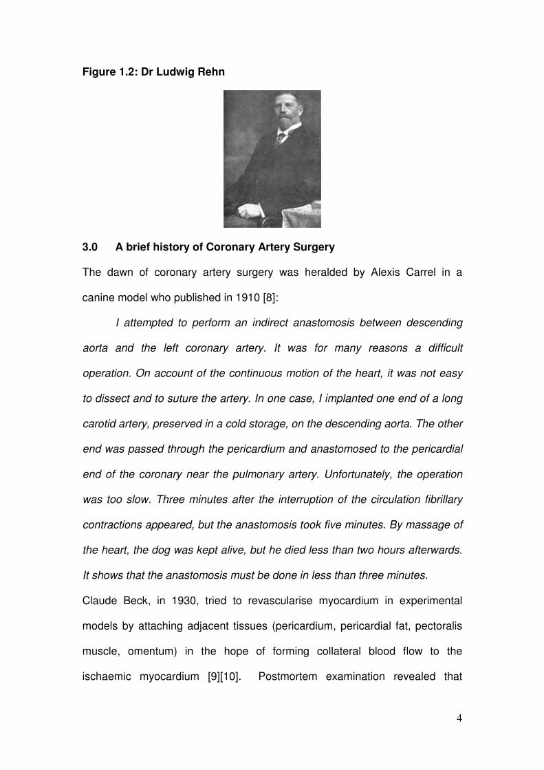
A year on, after suturing a stab wound to the right ventricle without any
complications, Ludwig Rehn (Figure 1.2) had performed the first successful
surgery of the heart. In his own words:
... Today the patient is cured. He looks very good. His heart action is
regular. I have not allowed him to work physically hard. This proves the
feasibility of cardiac suture repair without a doubt! I hope this will lead to more
investigation regarding surgery of the heart. This may save many lives.
Ten years after Rehn's initial repair, he had accumulated a series of 124
cases with a mortality of 60% [6]. His successful work was inspirational to
other pioneers and reversed the prevailing belief of inviolability of the heart
and marked the beginning of cardiac surgery [7].
4
Figure 1.2: Dr Ludwig Rehn
3.0 A brief history of Coronary Artery Surgery
The dawn of coronary artery surgery was heralded by Alexis Carrel in a
canine model who published in 1910 [8]:
I attempted to perform an indirect anastomosis between descending
aorta and the left coronary artery. It was for many reasons a difficult
operation. On account of the continuous motion of the heart, it was not easy
to dissect and to suture the artery. In one case, I implanted one end of a long
carotid artery, preserved in a cold storage, on the descending aorta. The other
end was passed through the pericardium and anastomosed to the pericardial
end of the coronary near the pulmonary artery. Unfortunately, the operation
was too slow. Three minutes after the interruption of the circulation fibrillary
contractions appeared, but the anastomosis took five minutes. By massage of
the heart, the dog was kept alive, but he died less than two hours afterwards.
It shows that the anastomosis must be done in less than three minutes.
Claude Beck, in 1930, tried to revascularise myocardium in experimental
models by attaching adjacent tissues (pericardium, pericardial fat, pectoralis
muscle, omentum) in the hope of forming collateral blood flow to the
ischaemic myocardium [9][10]. Postmortem examination revealed that

5
anastomotic vessels had developed between these tissues and the
myocardium. Beck subsequently performed this operation with modifications
on 16 patients.
In 1946 Arthur Vineberg reported successful implantation of the internal
mammary artery through a tunnel in the myocardium but not actually
anastomosing the left internal mammary artery to the coronary artery [11].
Mason Sones later validated Vineberg's concept by demonstrating
communications between the graft in the myocardium and the coronary
system by angiography in two patients operated on 5 and 6 years earlier. In
the middle 1960s the Vineberg operation with many variations was performed
at many institutions in the United States and Canada [12].
Coronary arterial endarterectomies were also attempted for the treatment of
ischemic coronary disease but mortality was high, and the procedure was
abandoned as an isolated operation [13].
From 1960 to 1967, isolated cases of coronary grafting were reported. None
had an impact on the development of coronary surgery. In 1960 Robert Goetz
performed the first coronary artery bypass operation in a human, which was
successful [14]. The right internal mammary artery was connected to the right
coronary artery using a non suture technique. The patient was asymptomatic
for a year, then developed recurrent angina and died of a myocardial
infarction.
The first clinical case of successful coronary artery bypass surgery using
autogenous saphenous vein was performed by Garett, Dennis and DeBakey
on a 42-year-old man in 1964 [15]. The vein was placed from the aorta to the
left anterior descending. The internal mammary artery had been anastomosed

6
to the left coronary artery in 1952 by Vladimir Demikhov, in a canine model
[16] and Longmire performed the first internal mammary to coronary artery
anastomosis in humans [17].
In 1967 Kolessov reported his experience with mammary artery–coronary
artery anastomoses for treatment of angina pectoris in six patients [18]
through a left thoracotomy without extracorporeal circulation or preoperative
coronary angiography. The following year, Green et al [19] and Bailey and
Hirose [20] separately published reports in which the internal mammary artery
was used for coronary artery bypass in patients. Bailey and Hirose carried out
the anastomosis on the beating heart and then Green et al advocated using
cardiopulmonary bypass, fibrillating the vented heart, cross-clamping the
aorta, and washing all blood from the coronary system while performing the
anastomosis.
Favalaro used saphenous vein for bypassing coronary obstructions [21].
However, the start of modern coronary bypass surgery took place in 1969
when Johnson et al reported a series of 301 patients who had undergone
various operations for coronary disease since early 1967 [22].
Johnson presented guidelines for direct surgery:
One: Do not limit grafts to proximal portions of large arteries....
Two: Do not work with diseased arteries. Vein grafts can be made as
long as necessary and should be inserted into distal normal arteries.
Three: Always do end-to-side anastomosis....
Four: Always work on dry, quiet field. Consistently successful fine
vessel anastomoses cannot be done on a moving, bloody target....
Five: Do not allow the haematocrit to fall below 35.
7
The direct anastomosis between the internal mammary artery and the
coronary artery was not initially as popular as the vein graft technique;
however, due to the persistence of Drs. Green, Loop, Grondin, and others,
internal mammary artery grafts eventually became the conduit of choice when
their superior long-term patency became known [23].
4.0 The development of cardiopulmonary bypass
Successful cardiopulmonary bypass development requires understanding of
cardiovascular physiology, a mechanism to pump blood, and a technique to
ventilate blood whilst preventing it from clotting [24].
Credit for anticoagulation in the heart-lung machine is owed to McLean in
1916 [25][26] who discovered heparin [27][28]. The work of John Gibbon led
to the first successful demonstration that life could be maintained by an
artificial heart and lung and that the native heart and lungs could resume
function [29]. Dennis et al went on to develop a heart-lung machine to support
circulation intraoperatively in a young girl during atrial septal defect closure
[30][31]. Although the machine was successful in purpose the patient did not
survive.
With the help of General Motors in 1952 Dodrill and colleagues developed a
mechanical blood pump [32] that successfully bypassed the left-side of the
human heart. Dodrill went on to successfully perform right-sided heart bypass
[33] and in 1955 transferred use of the oxygenator to human models.
John Gibbon, with the assistance of the International Business Machines
(IBM) Corporation, built a machine which contained a rotating vertical cylinder
oxygenator and a modified DeBakey rotary pump. The machine was
8
successful in canine models and Gibbon went on to develop a machine with a
larger oxygenator planned for human models [34].
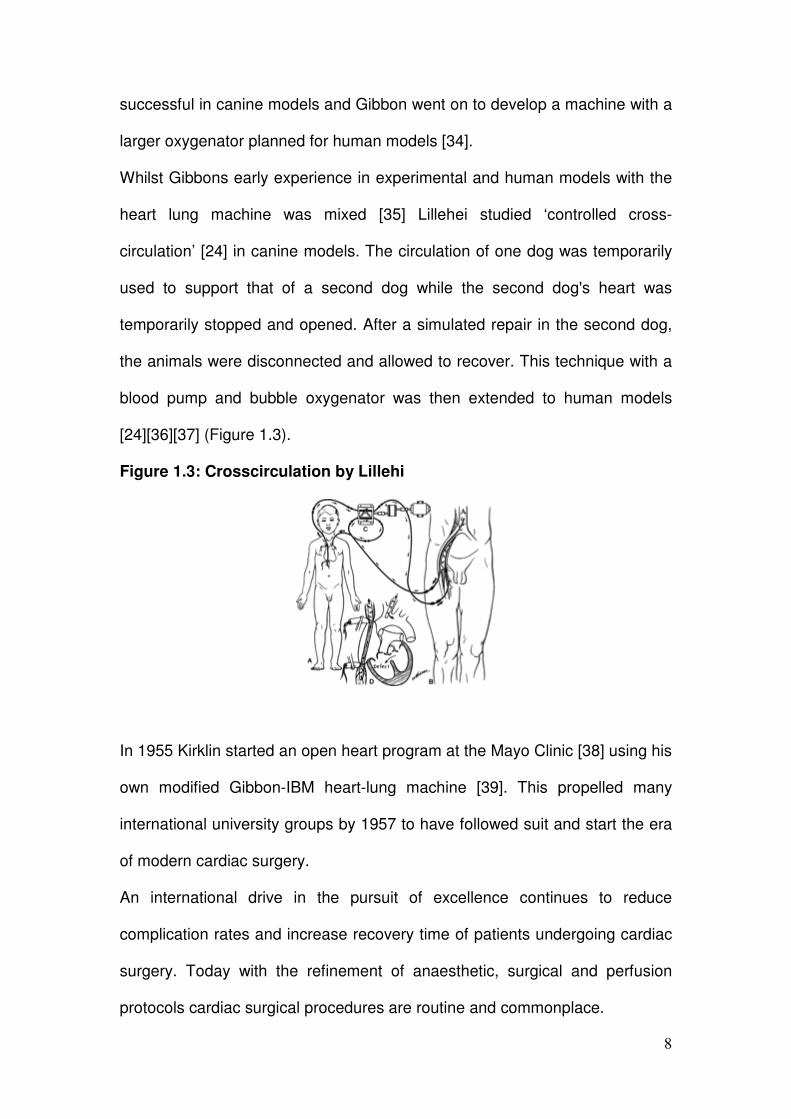
Whilst Gibbons early experience in experimental and human models with the
heart lung machine was mixed [35] Lillehei studied ‘controlled cross-
circulation’ [24] in canine models. The circulation of one dog was temporarily
used to support that of a second dog while the second dog's heart was
temporarily stopped and opened. After a simulated repair in the second dog,
the animals were disconnected and allowed to recover. This technique with a
blood pump and bubble oxygenator was then extended to human models
[24][36][37] (Figure 1.3).
Figure 1.3: Crosscirculation by Lillehi
In 1955 Kirklin started an open heart program at the Mayo Clinic [38] using his
own modified Gibbon-IBM heart-lung machine [39]. This propelled many
international university groups by 1957 to have followed suit and start the era
of modern cardiac surgery.
An international drive in the pursuit of excellence continues to reduce
complication rates and increase recovery time of patients undergoing cardiac
surgery. Today with the refinement of anaesthetic, surgical and perfusion
protocols cardiac surgical procedures are routine and commonplace.

9
Section 2: The need for myocardial protection
1.0 Introduction
Coronary heart disease (CHD) is the leading cause of death world-wide and
can manifest as acute myocardial ischaemia-reperfusion injury with
detrimental effects.
Myocardial injury has been found to occur in 10-40% of cases during coronary
intervention and can be characterized by a slight increase of markers of
myocardial necrosis, without symptoms, electrocardiographic changes or
impairment of cardiac function [40]. Coronary artery bypass graft (CABG)
surgery, the procedure of choice for coronary artery revascularisation in a
large number of patients with severe CHD, can also result in myocardial injury
leading to worse short and long-term clinical outcomes. As a trend in
progressively increased preoperative patient risk profile has been observed
worse outcomes are envisaged in the future.
This projection of poorer outcomes has alerted the medical fraternity to the
need for treatment strategies designed to protect the heart in terms of
reducing myocardial injury and preserving left ventricular systolic function.
Microvascular protection during the acute event has become the focus of a
variety of emerging technologies. The goal of these mechanical and
pharmacologic therapies is the restoration of normal metabolic function at the
myocyte level [41].
Myocardial protection aims at preventing myocardial tissue loss and current
methods include cardioplegia, off-pump coronary artery bypass surgery

10
(OPCAB), ischaemic preconditioning, on-pump beating heart surgery and
intermittent ischaemic arrest.
2.0 Brief history of myocardial protection
Early work on circulatory arrest in 1914 by Alexis Carrel found that:
….it was possible to clamp the pedicle of the heart (aorta and
pulmonary artery) for two and a half or three minutes without any subsequent
trouble. As soon as the clamp was removed, the heart resumed its pulsations,
and after a very short time, the pulsations were again normal. [42]
Hooker in 1929 suggested that potassium inhibition induced by an excess of
potassium chloride could be used to stop the heart leading Melrose in 1955
[43] to present the first experimental study describing induced arrest by
potassium-based cardioplegia. Blood cardioplegia was used:
"to preserve myocardial energy stores at the onset of cardiac ischemia."
Melrose goes on to state that
"... they have succeeded in evolving a reliable method of stopping and
restarting the heart at both normal and reduced body temperatures."
Unfortunately, the Melrose solution proved to be toxic to the myocardium, and
as a result, cardioplegia was not used widely for several years.
Gay and Ebert [44] and Tyres [45] demonstrated that cardioplegia with lower
potassium concentrations was safe. Studies by Kirsch, [46] Bretschneider [47]
and Hearse [48] demonstrated the effectiveness of cardioplegia with other
constituents and renewed interest in this technique.
Gay and Ebert in 1973 demonstrated a significant reduction in myocardial
oxygen consumption during potassium-induced arrest when compared with
11
that of the fibrillating heart [44]. They also showed that the problems in the use
of the Melrose solution in the early days of cardiac surgery probably were due
to its hyperosmolar properties and perhaps not to the high potassium
concentration.
Follette, in 1978, [49] demonstrated in experimental and clinical studies that
hypothermic, intermittent blood cardioplegia provided better myocardial
protection than normothermic, continuous coronary perfusion and/or
hypothermic, intermittent blood perfusion without cardioplegia solution.
Since the 1980s the use of cardioplegia as a form of myocardial preservation
technique has become established and commonplace in surgical practice.
Debate remains over the ideal composition of cardioplegia solution.
Formulations, methods of delivery, and recommended temperature continue
to refine and evolve.
3.0 Reperfusion Injury
An increasing body of evidence suggests a multifactorial mechanism for
myocyte injury and microvascular collapse that is associated with a profound
impact on long-term outcomes.
During cardiac surgery, the reperfusion of blood following prolonged
ischaemia (e.g. aortic crossclamping) provides oxygen and substrate for the
return of oxidative metabolism and rewarming increases the metabolic rate
toward normal. There is potential, with reperfusion, to add further injury to the
ischaemic damage sustained by cells through a localised inflammatory
response whose basic components are:
12
• Free radical injury
• Calcium overload
• Complement activation
• Neutrophil activation
• Cytokine system
• Endothelial response to ischaemia/reperfusion injury
4.0 Cardioplegia
4.1 Introduction to cardioplegia
Over thirty years ago, the development of hyperkalaemic cardioplegic
solutions revolutionised cardiac surgery by offering effective chemically-
induced cardiac arrest and myocardial protection during global ischaemia.
Despite remaining the most widely-used cardioplegic technique,
hyperkalaemia can have detrimental effects due to the Na and Ca loading of
the cardiac cell induced by depolarisation of the cell membrane. Efforts over
the last two decades to establish better cardioplegic agents have mainly
remained limited to animal experiments. The failure of these approaches to
progress to clinical trials may be due to a lack of clear criteria that a
cardioplegic agent should meet at a cellular level and, more importantly, at a
system level [50].
4.2 Principles of Cardioplegia
During the interruption of coronary circulation by the intraoperative cross-
clamping of the aorta cardioplegia is used to arrest the heart and reduce
myocardial metabolism. In the absence of cardioplegia cross-clamping alone

13
would induce anaerobic metabolism, depletion of myocardial energy stores
and cause severe myocardial dysfunction. Cardioplegic solutions today are
able to provide myocardial protection of over 3 hours without adversely
affecting myocardial function [51].
The optimal results from cardioplegia are based on the principles of prompt
diastolic arrest, buffering, reducing calcium levels, an adequate delivery
system, temperature and the addition of substrates designed to optimize
myocardial metabolism or prevent cell damage (Table 1.1).
Table 1.1: Principles and composition of cardioplegia [51]
PRINCIPLE COMPOSITION
Prompt diastolic arrest KCl 20-25 mEq.L-1
Buffering THAM, bicarbonate
Reduction of calcium levels Citrate-phosphate-dextrose (CPD)
Adequate delivery Antegrade+/-retrograde administration
Temperature Cold vs. tepid vs. warm
Substrate additives to optimize
myocardial metabolism or prevent cell
damage
Aspartate glutamate
Na+-H+ exchange inhibitors
Insulin
Magnesium
Procainamide
L-arginine
Calcium channel blockers
THAM = tris(hydroxymethyl)aminomethane; CPD = citrate-phosphate-dextrose
4.2.1 Prompt diastolic arrest
Prompt diastolic arrest of the heart is achieved by administering a solution
containing approx 20-25 mEq/L of potassium chloride (KCl). Three methods of

14
delivery currently employed are crystalloid cardioplegia, blood cardioplegia
and miniplegia.
4.2.1.1 Crystalloid cardioplegia
In crystalloid cardioplegia potassium is added to a crystalloid solution that is
administered undiluted. The resulting solution provides little substrate and no
oxygen to the heart during ischaemic arrest. It functions primarily by arresting
the heart at cold temperatures and has the potential to be oxygenated by
bubbling oxygen through the solution although this is uncommon [51].
4.2.1.2 Blood cardioplegia
When potassium is concentrated in a smaller bag of crystalloid solution and is
administered in a mixture with blood in varying ratios (most commonly 4:1
blood) blood cardioplegia is produced. This form of cardioplegia provides
oxygen, natural buffering agents, antioxidants and free-radical scavengers.
Standard additives include buffers such as THAM to achieve an alkaline pH,
citrate-phosphate-dextrose (CPD) to lower calcium levels and mannitol to
maintain slight hyperosmolarity. The cardioplegia mixture formed then passes
through a separate heater/cooler system in the extracorporeal circuit and
infusion rate and pressure can be controlled [52].
4.2.1.3 Miniplegia
When potassium is added directly to blood to minimize haemodilution
miniplegia is formed.

15
4.2.2 Reduction in cardiac oxygen demand
By arresting the heart at normothermia cardiac oxygen demand is reduced by
nearly 90%. Maintenance of arrest during the cross-clamp period is achieved
by readministering the solution every 15-20minutes so as to deliver potassium
and washout the metabolic byproducts that have been generated. A low
potassium solution (12-15mEq.L-1) is used to maintain the arrest while
avoiding an excess potassium load. A high potassium solution should be used
if the heart resumes any activity [51].
In the case of cold blood cardioplegia alone, this can be given retrograde into
the coronary sinus as an alternative to subsequent doses of cardioplegia to
optimise tissue oxygenation and metabolism while minimising potassium load
[51].
4.2.3 Temperature
The reduction in myocardial metabolism attributable to hypothermia is quite
insignificant compared with that achieved by diastolic arrest. Nonetheless,
systemic hypothermia supplemented by use of topical cold saline and topical
cooling devices that surround the left ventricle and protect the phrenic nerve
from cold injury is routinely used in patients receiving cold cardioplegia [51].
Since enzymatic and cellular reparative processes function better at
normothermia, some surgeons use ‘warm cardioplegia’ for myocardial
protection. However because of the tendency for the heart to resume
electrical activity at normothermia, this must be given continuously or with
only brief interruptions to protect the heart. When given continuously it can
obscure the operative field [51].
16
Warm cardioplegia can be used as an adjunct to cold cardioplegia when given
at the beginning and the end of the period of aortic cross-clamping.
Administering 500mL of warm cardioplegia immediately after aortic cross-
clamping may be beneficial in actively ischaemic hearts with energy depletion
by providing a brief period of time during which oxygen can be used to repair
cell damage and replace energy stores.
Terminal warm blood cardioplegia (‘hot shot’) is commonly given just before
the removal of the aortic cross clamp as it has been shown to improve
myocardial metabolism. The heart tends to remain asystolic for several
minutes after removal of the aortic cross clamp, during which time the heart is
able to ‘repair’ cellular processes or replenish energy stores while the oxygen
demand is low [53][54].
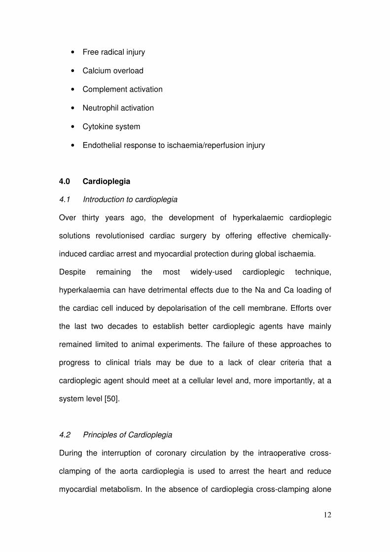
4.2.4 Route of delivery
Cardioplegia is initially administered antegrade (Figure 1.4) into the aortic root
and then may be given retrograde through a catheter placed in the coronary
sinus (Figure 1.5).
Figure 1.4: Antegrade cardioplegia cannula with a side port for venting
17
The efficacy of antegrade cardioplegia (ACP) delivery may be compromised
by severe coronary artery stenosis and is often dependent on collateral flow.
Sufficient root distension may not be achieved in patients with more than mild
aortic insufficiency and it can be cumbersome to readminister ACP during
aortic and mitral valve operations.
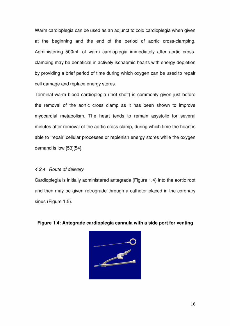
Figure 1.5: Retrograde cardioplegia catheter with self-inflating balloon
for measuring coronary sinus pressure
Retrograde cardioplegia (RCP) is easy to administer, either intermittently or
continuously, and does not interrupt the flow of an operation. RCP does
provide excellent myocardial protection but there is concern about
maldistribution of the solution especially to the right ventricle. Careful
monitoring of coronary sinus pressure during the administration of RCP is
required to avoid coronary sinus rupture.
Routes of delivery of cardioplegia should be complementary and not
exclusive. Contrast echocardiography has demonstrated left ventricular
perfusion to be better with warm antegrade delivery than retrograde delivery.
Delivery to the right ventricle is poor with either approach especially in right
coronary artery occlusion [51].

18
In patients undergoing elective coronary artery bypass grafting, retrograde
continuous infusion of cardioplegia by gravitational force combined with
antegrade cardioplegia has been shown to provide satisfactory myocardial
protection and to eliminate the need for inotropic support when compared with
antegrade delivery alone [55].
4.2.5 Cardioplegia additives
A variety of medications that might potentially be cardioprotective when given
in the cardioplegia solution have been studied. Those showing the greatest
promise include the Na+-H+ exchange inhibitors (such as cariporide),
adenosine and L-arginine. Aspartate and glutamate are Krebs’ cycle
intermidiates that have been used to improve myocardial energy metabolism
with variable success. Other drugs include procainamide, magnesium (which
has been shown to reduce arrhythmias) and free-radical scavengers. The
insulin cardioplegia trial involved use of tepid cardioplegia enriched with
glucose and insulin. This demonstrated metabolic and functional benefits to
patients undergoing elective but not urgent surgery [51].
4.3 Cardioplegia Strategy
Variations in cardioplegia strategies and solutions have made it difficult to
ascertain which specific element provides true benefit. The extent of coronary
disease and coronary flow can significantly impact the efficacy of both
antegrade and retrograde flow and confound any analysis. Clinical studies
have not demonstrated the superiority of one type of cardioplegia in routine
cases but have suggested patients with more advanced left ventricular
19
dysfunction fare better with blood cardioplegia especially given both
antegrade and retrograde.
Studies suggest that in low-risk groups, multidose cold crystalloid, or cold or
warm blood cardioplegia, whether given antegrade and/or retrograde, produce
relatively comparable clinical results. In general, protection of the right
ventricle is suboptimal with all strategies, especially in patients with right
coronary artery disease. In high-risk cases a combination of ACP and RCP
cold or tepid blood cardioplegia may offer the best results [51].
4.4 Blood vs. crystalloid cardioplegia
A meta-analysis of 34 RCTs (18/34 undergoing elective CABG surgery) found
superior myocardial protection provided by blood cardioplegia, deduced from
lower low output syndrome rates and CKMB release after surgery at 24hours,
when compared to crystalloid [56]. Blood cardioplegia provides a closer
approximation to normal physiology and this most likely translates into the
measurable clinical benefits and advantage.
A survey of UK practice has found 56% of surgeons use cold blood
cardioplegia, 14% use warm blood cardioplegia, 14% use crystalloid
cardioplegia, 21% use retrograde infusion and 16% do not use any
cardioplegia [57].
Although blood cardioplegia has been shown to be 'superior' to crystalloid this
advantage is marginal and might explain the continuous use of crystalloid
cardioplegia by some surgeons [58].

20
4.5 Directed cardioplegia
To eliminate the steal of cardioplegia, during nonselective antegrade
cardioplegia, from severely stenosed vessels by other less diseased arteries
‘direct cardioplegia’ is a novel approach that is being developed. To protect
poorly perfused myocardial regions the surgeon occludes the other main
branches and directs a certain volume of cardioplegic solution into the
severely diseased coronary artery. Early results in severe left main coronary
artery disease have been promising [59].
5.0 Volatile anaesthetics
5.1 Introduction to volatile anaesthetics
Volatile anaesthetic agents have been shown to have direct protective
properties against ischaemic myocardial injury by a mechanism termed
'anaesthetic preconditioning' [60][61]. The cardioprotective effects occur at
therapeutic doses and are independent of anaesthetic and haemodynamic
effects [62].
It is suggested that the cardiac depressant effects of volatile anaesthetics
decrease myocardial oxygen demand and may thus improve the myocardial
oxygen balance during ischemia whilst also directly protecting from ischemic
myocardial damage [63].
5.2 Enflurane, Sevoflurane, Desflurane and Isoflurane
Both Enflurane [64] and Sevoflurane have been shown independently to
enhance postischaemic functional recovery in patients undergoing coronary
surgery. Sevoflurane decreases postoperative release of brain natiuretic

21
peptide, the sensitive biochemical marker of myocardial contractile
dysfunction [65].
Preischaemic administration of isoflurane also protects against prolonged
ischaemia with functional recovery after CPB, increases mean cardiac index
and reduces necrosis (cTnI) in patients undergoing coronary artery bypass
graft surgery. Experimentally isoflurane has been shown to provide
myocardial protection through a signal transduction cascade that is similar to
the pathways identified in ischaemic preconditioning [66].
Meta-analysis’ have demonstrated that desflurane and sevoflurane reduce
postoperative mortality and incidence of myocardial infarction following
cardiac surgery with advantages in postoperative cardiac troponin release,
need for inotrope support, time on mechanical ventilation, intensive care unit
and overall hospital stay [61][62][63]. In support of current evidence The
American College of Cardiology/American Heart Association have
recommended volatile anaesthetic agents to be used during non-cardiac
surgery for the maintenance of general anaesthesia in patients at risk for
myocardial infarction [61][62][63].
5.3 Volatile vs. non-volatile anaesthesia
The advantage of volatile anaesthesia, against total intravenous anaesthesia,
on cardiac troponin release in off-pump coronary artery bypass grafting
(OPCAB) has been found in multicenter randomized controlled studies [67].
This benefit has also been shown to extend to patients undergoing CABG with
CPB with reduced cardiac troponin release, reduced need for postoperative
inotropic support and trends toward a reduction in number of Q-wave

22
myocardial infarction, time on mechanical ventilation, intensive care unit and
overall hospital stay [68].
A systematic overview and meta-analysis of all randomized trials comparing
volatile with non-volatile anaesthesia in CABG surgery identified 27 trials that
included 2979 patients. Post-bypass, patients randomized to receive volatile
anaesthetics had 20% higher cardiac indices, lower troponin I serum
concentrations and lesser requirement for inotropic support. Duration of
mechanical ventilation was reduced by 2.7 h and there was a 1 day decrease
in hospital length of stay [69].
6.0 Ischaemic conditioning
‘Conditioning’ of the heart to harness its endogenous cardioprotective
capabilities using either brief ischaemia or pharmacological agents provides a
potentially novel approach to myocardial protection during cardiac surgery by
limiting myocardial injury, preserving left ventricular systolic function and
potentially improving morbidity and mortality in patients with CHD [70][71].
The conditioning stimulus can give be given locally or distant to the organ and
before or after a prolonged period of ischaemia.
Ischemic postconditioning by brief episodes of ischemia performed just at the
time of reperfusion has been shown to reduce infarct size in animal models, in
the clinical settings of percutaneous coronary intervention and in adult
patients undergoing valve replacement [72].
Brief episodes of non-lethal ischaemia and reperfusion to an organ or tissue
remote from the heart, thereby obviating the need to 'condition' the heart
directly has been termed remote ischaemic conditioning and it is suggested

23
there is potential for widespread systemic protection to other organs which are
susceptible to acute ischaemia-reperfusion injury such as the brain, liver,
intestine or kidney [71].
7.0 Nicorandil
This potassium channel activator in intravenous preparation has been shown
to have beneficial effects. When administered during CABG surgery with CPB
lowers concentrations of cTnT were observed [73] suggesting its potential for
myocardial protection.
8.0 Statins
The perioperative use of statin therapy has demonstrated improved short and
long-term cardiac outcomes following noncardiac surgery. Its beneficial
effects have been attributed to its positive effects on plaque stability
(pleiotropic) [74], lipid lowering effects and anti-inflammatory properties [75].
In cardiac surgery, one series found pretreatment with statins to reduce the
incidence of death and myocardial infarction following coronary artery bypass
grafting surgery [76] supporting its role as a cardioprotective agent.
9.0 Aprotinin
Aprotinin, a serine protease inhibitor, can limit systemic inflammation, and has
been associated with myocardial, pulmonary and cerebral protection in
addition to its proven haemostatic efficacy [77]. It has antithrombotic,
antifibrinolytic, and antiinflammatory effects and is effective in reducing
24
bleeding and the need for blood transfusions after cardiac surgery with
cardiopulmonary bypass [77][78].
Recent controversy has started since a large randomized controlled trial
comparing antifibrinolytics in patients undergoing cardiac surgery was
stopped after a preliminary analysis suggested a trend toward an increase in
all-cause 30-day mortality associated with aprotinin [78]. Further investigation
is currently under way.
10.0 Conclusions
The dangers of insufficient myocardial protection during cardiac surgery
represents a real worry to surgeons as our population ages and the patient
presenting have more severe comorbidities. A number of techniques have
been identified to tackle this issue and continue to be tested and refined.
The topic of investigation of this thesis and the consecutive chapters is the
applicability of remote ischaemic preconditioning as a novel technique to aid
in the armamentarium of the surgical team to reduce myocardial injury and
enhance myocardial preservation.

25
Section 3: The current status of coronary artery bypass graft
surgery (CABG)
1.0 Adult Cardiac Surgical Database Report 2008
The Society of Cardiothoracic Surgeons of Great Britain and Ireland 6th
National Adult Cardiac Surgical Database Report (2008) [1] documents the
nature of adult cardiac surgical practice in the United Kingdom and Ireland. It
provides an invaluable resource for the extraction of data, analysis and
comment on the current practice of coronary artery surgery.
1.1 Patient populations treated by CABG
In the financial year ending 2008 22,846 patients underwent isolated CABG.
Analyses of patients' risk profiles show quite marked changes, with surgeons
operating on progressively higher-risk patient year-on-year:
A recent review confirms the feelings that a higher-risk population has started
to develop. The most current data reports 75% of patients undergoing isolated
CABG are overweight & about one-third are obese. The number of elderly
patients presenting for surgery is increasing with the over 75s now making up
more than 20% of all cardiac surgery and the over 80s making up over 5%.
The average age of patients undergoing isolated CABG between 1991and
2007 has increased from 58 to 66 years.
There are marked changes in the proportion of female patients with increasing
age: in patients under the age of 51 years of age 13% of patients are female,
in those over the age of 80 it rises to nearly 30%.
26
The incidence of comorbidities of patients presenting for coronary surgery
between 2001 and 2008 has increased: 33% in diabetics and more than 15%
in hypertensives. While left main stem (LMS) disease rates appear to have
stabilised now, they did increase between 2001 and 2006. Approximately half
the patients have had a previous myocardial infarction (MI) and the proportion
of patients who have suffered a heart attack within the previous 30 days has
gone up from under 19% in 2004 to over 34%in 2008.
An increase to over 8% of patients presenting for CABG have had prior
percutaneous coronary intervention (PCI) with the vast majority having
undergone PCI during a previous hospital admission. There has also been a
small increase in the proportion of patients with preoperative dialysis-
dependent renal failure, extra-cardiac arteriopathy and a greater proportion of
urgent cases.
There is suggestion that an increase in the longevity of conduit used is being
observed. Fewer patients are requiring repeat operations and the time
between first- and second-time operations is increasing.
1.2 Survival following CABG
The outcomes for patients undergoing coronary artery bypass surgery as an
elective operation are excellent, with low mortality, low morbidity and good
medium-term survival. Operative mortality rates for isolated CABG fell from
2.3% in 2001 to 1.5% in 2008. In those patients with no history of previous MI
in the year to March 2008 the mortality rate was 0.8% and for those who had
2 or more previous MIs it was 3.6%.

27
The operative mortality of elective surgical patients under the age of 70 years
is less than 1%. Survival appears to be worse for patients who are older,
female, undergoing urgent surgery, diabetic, suffering from impaired cardiac
function or in renal failure. In terms of improving outcome, surgical procedures
have seen a trend towards increased usage of arterial grafts.
Following surgery body habitus plays a role in outcome as the extremes of
weight (under and morbidly obese) have prolonged in-hospital stays. Those
that are underweight fair poorly with an observed mortality rate of 4.1%.
Obese and morbidly obese patients have the same medium-term survival
rates as patients of normal weight.
As an indicator of improved care and treatment strategies the operative
mortality has continued to fall whilst the proportion of patients over 75 and 80
has continued to increase. The mortality rate for patients over the age of 75
has also fallen from 5.0% in 2004 to 3.4% in 2008. Kaplan-Meier survival rate
at 5 years post-operatively for the under 66 year age group is over 90% and in
the over 80 group is 69%. Gender has been shown to affect outcome as
women have an in-hospital mortality that is nearly twice that of their male
counterparts and worse medium-term survival.
The impact of comorbidities on prognosis remains significant. Patients who
have had a previous heart attack are twice as likely to die at the time of
surgery compared to patients who have not had a heart attack. However, the
in-hospital mortality rate of diabetics has decreased in spite of a 50% increase
in the diabetic proportion between 2001 and 2008. The mortality for diabetics
remains higher than for non-diabetic patients. Diabetics have a longer length-

28
of-stay and a medium-term survival rate of 85% compared to 90% of non-
diabetics.
In the most recent year of study there is no significant increase in operative
mortality on inpatients with hypertension. Hypertension does remain
associated with longer in-hospital stay and worse medium-term survival.
Severe angina remains an important risk factor for operative mortality. Those
with LMS disease have significantly higher rates of mortality. LMS disease is
associated with a significantly greater length of in-hospital stay and a worse
medium-term survival.
Priority is an important predictor of operative mortality with urgent and
emergency cases having mortality rates of 2.2% & 8.3% respectively.
Positively, in-hospital mortality in this group has fallen inspite of an increase in
proportion presenting compared to elective cases. Urgent and emergency
patients consistently stay in hospital longer than elective patients.
Patients undergoing isolated CABG within 24 hours of a PCI have an overall
mortality rate of 7.9%. Moderate and severe dyspnoea and extra cardiac
arteriopathy are associated with an increased operative mortality and the
mortality associated with redo surgery is nearly four times that of first-time
surgery.
Any degree of renal disease is associated with a marked increase in length-
of-stay. Renal disease is a powerful predictor of poor post-operative survival:
for patients on dialysis the medium-term survival rate at 5 years after surgery
is 50% compared to 90% for those without renal disease.

29
2.0 Revascularisation vs. medical therapy
Three multicentre randomised controlled trials demonstrated early on the
benefit of CABG over medical therapy: Veterans Administration Coronary
Artery Bypass Surgery (VA) Study [79], Coronary Artery Surgery Study
(CASS) [80] and the European Coronary Surgery Study (ECSS) [81].
Following these studies, there has been impetus to refine understanding and
identify the best responders to CABG.
In CASS [80] 780 patients with stable ischaemic heart disease were randomly
assigned equally to receive surgical or nonsurgical treatment and quality of
life was assessed. At ten year follow up these patients were found to have no
difference in cumulative survival, percentage free of death and nonfatal
myocardial infarction. Patients with an ejection faction (EF) <50% exhibited a
better survival with initial surgery whilst those with EF ≥50% exhibited a higher
proportion free of death and myocardial infarction with initial medical therapy.
The 10 year follow up results of CASS confirmed the early reports that
patients with left ventricular dysfunction have long-term benefit from surgery
[82].
In the European Coronary Surgery Study 768 men under 65 with mild to
moderate angina, at least two-vessel disease and good left ventricular
function were recruited. CABG was shown to significantly improve survival in
patients with three-vessel disease and in those with stenosis in the proximal
third of the left anterior descending artery constituting a component of either
two or three vessel disease [81][83].
To secure the position of CABG as a beneficial treatment strategy, an
overview of seven randomised trials over ten years found those patients with

30
stable coronary heart disease receiving initial CABG (n=1324) had lower
mortality at 5, 7 and 10 years than those receiving initial medical therapy
(n=1325) [84]. Risk reduction was greater in those with left main artery
disease than in those with three vessel disease or one or two vessels. The
absolute benefits of CABG were more pronounced in patients in the highest
risk categories.
3.0 Revascularisation vs. percutaneous intervention
3.1 CABG vs. balloon angioplasty
Coronary-artery bypass grafting (CABG) and percutaneous transluminal
coronary angioplasty (PTCA) are alternative methods of revascularization in
patients with coronary artery disease. A number of large trials exist comparing
the two treatment strategies:
Randomised Intervention Treatment of Angina (RITA) trial compared the long-
term effects of PTCA with CABG in patients with one, two, or three diseased
coronary arteries and randomised 1011 patients. Although recovery after the
more invasive CABG took longer than PTCA it did result in less risk of angina
and fewer additional diagnostic and therapeutic interventions in the first 2
years with no significant difference in risk of death or myocardial infarction
[85].
The Bypass Angioplasty Revascularisation Investigation (BARI) in patients
with multivessel disease found PTCA did not significantly compromise five-
year survival in patients with multivessel disease, however once again
subsequent revascularization was required more often with this strategy. For
treated diabetics five-year survival was significantly better after CABG [86].

31
The multinational, multicentre Coronary Angioplasty versus Bypass
Revascularisation Investigation (CABRI) in 1054 patients with symptomatic
multivessel coronary disease found PTCA patients more likely to have
reinterventions [87].
In another study the 5-year prognosis of patients with isolated proximal left
anterior descending coronary artery stenosis was good and both PTCA and
CABG improve clinical status, but revascularization was needed more
frequently after PTCA. [88]
To draw together the experiences a meta-analysis of eight randomised trials
including 3371 patients comparing CABG (n=1161) with PTCA (n=1710) was
performed. Overall there was a substantial similarity in outcome across the
trials. The combined evidence showed no difference in prognosis between
these two initial revascularisation strategies. However, the treatments differed
markedly in the subsequent requirement for additional revascularisation
procedures and in the relief of angina [89].
In terms of long-term mortality, pooled patient data (n=7812) from ten
randomised trials with PCI achieved with balloon angioplasty in six trials and
with bare-metal stents in four trials found it to be similar after CABG and PCI
in most patient subgroups with multivessel coronary artery disease [90]. More
recently the SYNTAX trial , a noninferiority comparison of 1800 patients with
three-vessel or left main coronary artery disease for the primary endpoint of a
major adverse cardiac or cerebrovascular event (MACCE) (i.e., death from
any cause, stroke, myocardial infarction, or repeat revascularization) during
the 12-month period after randomization was performed. It was recommended
that CABG remain the standard of care for patients with three-vessel or left

32
main coronary artery disease, since the use of CABG, as compared with PCI,
resulted in lower rates of the combined end point of major adverse cardiac or
cerebrovascular events at 1 year [91].
3.2 CABG vs. coronary stenting
Following the arrival of coronary stenting as a method of coronary
revascularisation studies have been performed to evaluate its efficacy as
compared to the longer established conventional CABG:
ERACI II found in symptomatic 450 patients with multivessel coronary artery
disease randomly assigned to stenting (225) or CABG (225) during the first 30
days, stented patients had lower major adverse events (death, myocardial
infarction, repeat revascularization procedures and stroke [92].
ARTS (Arterial Revascularization Therapies Study) trial assessed the
relationship between completeness of revascularization and adverse events
at one year. In 1,205 randomly assigned patients with multivessel disease,
complete revascularization was found to be more frequently accomplished by
bypass surgery than by stent implantation. One year after bypass, there was
no significant difference in event-free survival between surgically treated
patients with complete revascularization and those with incomplete
revascularization, but patients randomized to stenting with incomplete
revascularization had a greater need for subsequent bypass surgery. [93]
The Stent or Surgery (SoS) trial assessed the effect of stent-assisted
percutaneous coronary intervention (PCI) (n=488) versus CABG (n=500) in
the management of patients with multivessel disease. The use of coronary
stents reduced the need for repeat revascularisation when compared with

33
previous studies that used balloon angioplasty, though the rate remained
significantly higher than in patients managed with CABG [94]. The six-year
follow up from the Stent or Surgery Trial (SoS) demonstrated a continued
survival advantage for patients managed with CABG [95].
It was felt by some clinicians that the above trials and others individually were
underpowered to properly assess safety end points like death, stroke, and
myocardial infarction in stented patients. Since pooling data from randomized
controlled trials increases the statistical power and allows better assessment
of the treatment effect in high-risk subgroups a meta-analysis with 5-year
patient level data from the ARTS, ERACI-II, MASS-II and SoS trials was
performed [96].
The pooled analysis of 3051 patients in 4 randomized trials evaluating the
relative safety and efficacy of PCI with stenting and CABG at 5 years for the
treatment of multivessel coronary artery disease found PCI with stenting was
associated with a long-term safety profile similar to that of CABG. However,
as a result of persistently lower repeat revascularization rates in the CABG
patients, overall major adverse cardiac and cerebrovascular event rates were
significantly lower in the CABG group at 5 years [97].
Stents themselves have evolved since their initial application with the arrival
of drug-eluting stents to add to the armamentarium, along with bare metal
stents, of the Cardiologist.
A randomized, double-blind trial comparing a sirolimus-eluting stent with a
standard stent in 1058 patients found it to reduce the rates of restenosis and
associated clinical events in all subgroups analyzed [97]. A further study has
found that Sirolimus eluting stents (SES) are not inferior to CABG or bare

34
metal stents for the treatment of patients with multivessel coronary disease
including involvement of the proximal LAD [98].
Reports continue to be promising with dug-eluting stents. Paclitaxel when
compared to bare metal stents in a patient population with complex lesions
reduces reduces clinical and angiographic restenosis [99].
A significant body of longer term data comparing revascularisation techniques
is awaited. However some data is becoming available. In a series of patients
undergoing revascularisation between 2000 and 2007 (n=363) for unprotected
left main stem coronary artery stenosis, CABG (n=216), PCI with drug eluting
stents (n=94) and PCI with bare-metal stent (n=53) were compared. It was
found that CABG patients had higher target lesion revascularisation (TLR)-
free survival and revascularisation-free survival than rival PCI groups. Cardiac
death did not significantly differ between the 3 groups [100].
More long term data on the safety and outcomes of these techniques is still
required and will certainly mark a turning point in the future of coronary
revascularisation therapies on offer to specific patient populations.
35
CHAPTER 2: ISCHAEMIC PRECONDITIONING (IP) IN CARDIAC
SURGERY: A SYSTEMATIC REVIEW OF
EXPERIMENTAL AND CLINICAL LITERATURE
1.0 Introduction
1.1 Local (or ‘Cardiac’) Ischaemic Preconditioning
In 1986 Reimer et al found that ATP levels in dog hearts subjected to serial
ischaemia were higher than in those exposed to a single ischaemic event.
This finding was the first description of ischaemic preconditioning (IP) [101].
This initial breakthrough by Reimer was confirmed by the finding by Murray et
al in the same year that paradoxically intermittent ischaemia may not have an
additive effect but rather a protective affect against subsequent ischaemia.
[102]
Murray et al described that myocardial ATP concentrations, on canine hearts,
fell during the first brief episode of ischaemia. These concentrations were then
preserved during further identical coronary artery occlusions and no necrosis
occurred. They demonstrated that pre-treatment with repeated periods of
ischaemia triggered adaptive changes that protected the myocardium from the
effects of subsequent prolonged ischaemic insult “ischaemic preconditioning
(IP)” [102].
Murray et al established that 4 cycles of 5 minutes coronary occlusion and 5
minutes of reperfusion were sufficient to precondition the dog heart. They
reported that the infarct size produced by 40 minutes of sustained coronary
artery occlusion in anaesthetized open-chest dogs averaged approximately
30% of the myocardium at risk. In contrast, in dogs that received 4 cycles of 5
36
minutes episodes of brief ischaemia immediately before the sustained test
occlusion only 7% of the myocardium at risk became necrotic. Thus, despite a
longer cumulative duration of coronary occlusion, infarct size in
preconditioned dogs averaged only one fourth of that seen in controls.
Reduction of infarct size is now considered the “gold standard” of IP.
Yellon et al were the first to report that IP protects the human heart during
cardiac surgery by demonstrating conservation of myocardial adenosine
triphosphate (ATP) content in biopsy specimens. [103] They showed that 2
cycles of 3 minutes ischaemia with 2 minutes reperfusion was effective during
CABG surgery in preconditioning the human myocardium. They found that, as
in the experimental canine model, myocardial ATP stores after 10 minutes of
sustained ischaemia, in this case induced by aortic cross clamping and
fibrillation during coronary artery bypass surgery, were significantly preserved
in patients who first received two 3 minute episodes of intermittent
crossclamping when compared with controls who received the sustained
ischaemia alone. Subsequent studies by these investigators further
documented a reduction in the release of cardiac troponin T (an index of
cellular injury) in patients initially preconditioned before undergoing bypass.
[104]
The majority of surgeons today utilise cardioplegic arrest for cardioprotection.
The first study during cardiac surgery with cardioplegia was performed with
warm blood cardioplegia and the investigators found that preconditioning
increased the early release of creatine kinase MB and the transmyocardial
lactate gradient shifted toward production, this would suggest that
preconditioning may increase the injury. This study initiated several studies,

37
some of which found a positive effect of preconditioning whereas others either
found no effect or even a worsened postoperative outcome indicated by
increased inotropic support. [105]
Perrault et al reported that in the presence of cardioplegic arrest, there was no
difference in the release of biochemical markers (CK-MB) between
preconditioned and control groups. This lack of additional protection conferred
by IP was further confirmed by the absence of difference in the post-arrest
myocardial levels of ribonucleic messengers coding for cardioprotective heat
shock proteins between the 2 groups. Similar negative results were reported
by Kaukoranta, DiSalvo et al, who went on to report that in the presence of
hypothermia, no beneficial effect of preconditioning was observed in similar
patients undergoing surgery with intermittent fibrillation techniques. [106]
1.2 Remote Ischaemic Preconditioning (RIPC)
More than 20 years on since the breakthrough by Reimer et al the effect of
intermittent upper limb ischaemia during coronary artery bypass surgery on
the human heart has been investigated [107]. This technique has been coined
“Remote Ischaemic Preconditioning (RIPC)”.
RIPC was first suggested by Przyklenk et al after demonstrating that brief
circumflex artery occlusion preconditioned the area of myocardium perfused
by the left anterior descending artery. [108]
Subsequent animal studies have expanded on the principle of RIPC to show
that ischaemia of different organs such as the kidney, mesenterium, intestine
and brain can also protect the heart. At the same time all organs tested
demonstrate the presence of the preconditioning response. Furthermore,

38
RIPC of the brain appears not only to protect the heart but also preserves
endothelial function of aortic rings in vitro. [105][109]
IP is a powerful protective mechanism that appears to have immediate and
delayed protective effects, the importance of which varies between species
and organ systems. It is a multifactorial process requiring the interaction of
numerous signals, second messengers and effector mechanisms. Stimuli
other than ischaemia, such as hypoxic perfusion, tachycardia and
pharmacological agents, including isoflurane have preconditioning-like effects.
Currently IP is used during minimally invasive cardiac surgery without
cardiopulmonary bypass to protect the myocardium against ischaemic injury
during the anastomosis. [109]
Other situations in which ischaemia occurs naturally or is induced by
therapeutic manipulations include unstable angina before infarction,
percutaneous transluminal coronary angioplasty and coronary artery bypass
surgery with intermittent cross-clamp fibrillation. [110]
The data accruing thus far for RIPC in both the medical and surgical
cardiological arenas is promising and at present it appears to be one of the
most important potential myocardial protective adjuncts so far identified.[111]
Progress has been made in elucidating the mechanistic pathways underlying
‘conditioning’ cardioprotection, and have resulted in the identification of novel
targets for pharmacological preconditioning.[70] Future clinical applications of
IP and RIPC will depend on the clarification of the underlying biochemical
mechanisms, the development of pharmacological methods to induce
preconditioning, and controlled trials in humans showing improved
outcomes.[109]

39
2.0 Mechanisms Involved in Conditioning
2.1.1 Ischaemic Preconditioning
The underlying mechanism of IP remains to be unravelled in its entirety.
Hypotheses have been postulated and relative mimetics have been identified
but the clear underlying pathway is still not known.
Once the molecular mechanisms of IP and the conditions that confer optimal
protection in the human heart are defined then pharmacological intervention
that mimic IP may be developed and used as an adjunct, rather than
alternative to cardioplegia.[109]
The first reported corollary of infarct size reduction with preconditioning is the
observation by Murray et al that the rate of depletion of adenosine
triphosphate (ATP) during the initial minutes of the sustained test occlusion
was markedly attenuated in preconditioned dogs versus controls.[102]
IP appears to provide ischaemia protection by a variety of physiological
mechanisms. Energy requirements are reduced, conserving substrates and
diminishing metabolism. Acid base and electrolyte homeostasis is therefore
better controlled. Preconditioned tissues also demonstrate reduced oxidative
stress, neutrophil activation and cytokine production and apoptosis.[112]
Prolonged myocardial ischaemia, in addition to inflicting lethal injury on
myocytes, also has a deleterious effect on the coronary vasculature, typically
manifesting as a loss in both endothelium-dependent and endothelium-
independent coronary vasodilator reserve. However, whether the benefits of
IP extend beyond the myocyte and protect the vasculature from sustained
ischaemia is a topic of some controversy.[113]
More relevant to cardiothoracic surgical practice, Yellon et al showed that IP
40
had positive effects on reducing ATP utilisation by cardiomyocytes during
cardiopulmonary bypass.[103] Patients with angina within 24 hours of
myocardial infarction had smaller infarct size. Improved left ventricular
function and enhanced survival after reperfusion therapy.[112]
It is suggested that the cardioprotection achieved with preconditioning is a
receptor-mediated phenomenon. In the simplest terms, brief antecedent
ischaemia is thought to stimulate one or more receptors on the myocyte
membranes, thereby initiating one or more signal transduction pathways that
culminate in the phosphorylation of an effector protein and ultimately render
the myocytes resistant to subsequent sustained ischaemia. However,
definitive identification of the receptor, the end effector, and most notably the
intermediate signal transduction pathway evoked by brief antecedent
ischaemia has proven to be far from simple.[113]
2.1.2 Cellular Events
IP is a powerful mechanism in reducing post-ischaemic myocardial injury.
Brief periods of myocardial ischaemia result in the breakdown of ATP and so
releasing adenosine. Adenosine receptor occupancy has been implicated to
serve as both a trigger and a mediator of infarct size reduction with
preconditioning.[113]
A1 receptors are located on myocardial cells and A2 receptors are located on
coronary endothelial and leucocyte/platelet membranes. Pretreatment of dog
heart with selective A1 receptor agonists prior to an ischaemic event mimics
the protection derived from IP, hence A1 receptors play a role in IP.
Adenosine infusion prior to CABG surgery has been shown to improve

41
haemodynamics after cardiopulmonary bypass and decreases CPK
release.[105] Adenosine has demonstrated a role in infarct size reduction with
preconditioning in the rabbit, dog and swine models, however the rat is the
exception, suggesting adenosine receptor activation is not the sole trigger for
infarct size reduction with preconditioning.[113]
The mechanisms of preconditioning probably vary between acute and delayed
models and between local versus remote.[105] The initial phase of
preconditioning may be similar between classic and delayed models and may
start by release of a trigger substance during the brief episodes of ischaemia
and reperfusion that may bind to surface receptors coupled to G1 proteins.
Preconditioning–induced cardioprotection can be initiated by more than one
receptor. Adenosine, α1adrenergic receptors, muscarinic M2, angiotensin II,
bradykinin B2, σ-opiod receptors have been shown to play a contributory or
additive role, or in some cases redundant roles. The common theme is that all
receptors implicated are coupled to pertussis toxin-sensitive inhibitory G
proteins. It appears that the stimulation of G1 protein-coupled receptors during
brief antecedent ischaemia serves as a trigger for preconditioning-induced
cardioprotection.[113] Other trigger substances have been identified as
prostanoids, nitric oxide and low doses of reactive oxygen intermediates and
natiuretic peptides. Tumour necrosis factor alpha has been indicated as a
trigger of the preconditioning response.[105][113]
The intracellular message in IP is generated through coupling of agonists of
G-proteins that span the cell membrane. Adenosine, norepinephrine and
bradykinin receptors are coupled to G proteins which when stimulated
increase activity of phospholipase C and D. These enzymes catalyse

42
hydrolysis of membrane phospholipids, increasing diacylglycerol and inositol
triphosphate levels.
Diacylglycerol, a second messenger, activates protein kinase C (PKC), an
enzyme believed to play a key role in the biochemistry of IP through its
phosphorylation of effector proteins.[113]
PKC is a hotly debated second messenger implicated to play a role in
preconditioning. Brief episodes of myocardial ischaemia also result in
transient increase in cytosolic calcium concentration and calcium is known to
participate as a second messenger in many signalling pathways.[113]
Tyrosine kinases as well as mitogen activated protein kinases appear
involved in the signalling cascade in several species, although which kinase
cascade is upstream or downstream is currently an issue of debate.[105]
The next step in the signalling pathway is activation of transcription factors by
protein kinases, reactive oxygen species, and nitric oxide, of which particularly
nuclear factor κ-B (NFκB) has been investigated in both classic and delayed
models. NFκB is a redox sensitive transcription factor that regulates
transcription of battery of genes most of which are associated with
proinflammatory effects such as cytokines, chemokines, and leucocyte
adhesion molecules. Some candidate genes for organ protection in
preconditioning are also regulated by NFκB. Pharmacologic blocking of NFκB
translocation inhibits preconditioning in both classic and delayed models.[105]
The importance of transcription factors is easy to grasp in models of delayed
preconditioning in which the time frame for translation of protective
substances is ample. However, the fact that transcription factors appear
important for protection in classic models is less easy to grasp. The

43
cardioprotective effect of NFκB in immediate models may be due to an
upregulation of its own inhibitor IκBα thus contributing to reducing a NFκB-
dependent inflammatory response during sustained ischaemia. However,
abolishing classic preconditioning effects through employing the
transcriptional inhibitor actinomycin D indicates that the process is complex.
Other endogenous cardioprotective substances that are suggested as
upregulated and protective are heat shock proteins of the 70kDA or 27kDA
families. These have their own regulatory factors but do influence the
activation of NFκB. Antioxidants, inducible nitric oxide synthase and inducible
cyclooxygenase, all regulated by NFκB, have furthermore been indicated as
organ effectors of protection. A last possible route of NFκB mediated
cardioprotection is through the antiapoptotic effect of preconditioning. NFκB
regulates several antiapoptotic molecules, such as Bcl-2, surviving, and
inhibitor of apoptosis protein-1, as well as X-linked inhibitor of apoptosis
protein-1.[105]
In 1992 Gross et al proposed that opening of KATP channel may be involved in
the reduction in infarct size seen in preconditioned myocardium. They found
the selective KATP channel antagonist glibencamide in dogs either before, or
immediately after, the brief preconditioning stimulus gave infarct sizes greater
than those observed in preconditioned dogs. They also found that KATP
agonists including bimakalin and aprikalim mimicked, the reduction in infarct
size achieved by preconditioning and demonstrated a connection or synergy
between adenosine receptor activation, opening of KATP channels, and
cardioprotection.[113][114]
Opening of the KATP channel is initiated by low intracellular concentrations of

44
ATP (i.e. as seen during ischaemia). The resultant efflux of potassium from
the myocytes into the interstitium has thus been suggested to protect the cells
via local “cardioplegia”. Opening of the KATP channel also results in membrane
hyperpolarisation and shortening of the action potential duration. These
electrical responses have further been associated with a reduction in calcium
influx via voltage-sensitive calcium channels, and thus may attenuate myocyte
death by attenuating calcium overload. KATP channels thus do not mediate
infarct size reduction with preconditioning solely via more rapid or pronounced
shortening of the action potential.[113] Activation of KATP channel causes
release of reactive oxygen species and one may speculate that their role in
preconditioning is linked to this.[105]
There is only general agreement that G protein-coupled receptors initiate and
KATP channels mediate infarct size reduction with preconditioning. The
sequence of cellular events occurring between receptor activation and
eventual cardioprotection are subject of considerable controversy.[113]
In anaesthetized rabbits, transient systemic ischaemia, followed by HD with
D-HES, can enhance early cardiac functional recovery following coronary
artery occlusion and reperfusion, functioning as a powerful mechanism of IP.
Transient systemic ischaemia is associated with increased endogenous nitric
oxide (NO) production or release during reperfusion, which may have
contributed to the improved early myocardial functional recovery in the
rabbits.[115]
In summary, stimulation of G protein-coupled receptors (most notably the
adenosine A1 and A3 receptors) trigger or initiate the adaptation to later
ischaemia and KATP channels appear to play role in “end effector” (Figure

45
2.1).[113]
2.1.3 cAMP
Reduction of cAMP has been implicated in the protection of IP against
myocardial necrosis. Evidence for this is even in the absence of ischaemia,
elevations in cAMP have been shown to produce myocardial necrosis. High
levels of cAMP increase sarcolemmal calcium entry and increase the
activation of cardiac lipases, effects that are known to aggravate ischaemic
injury.
In a rabbit heart model, 10 to 30 minutes of sustained regional ischaemia is
accompanied by a nearly twofold increase in cAMP levels. IP induced in this
model with a single cycle of transient ischaemia and reperfusion (IR) prior to
sustained ischaemia, caused the increase in cAMP to be reduced.
Furthermore three cycles of IR preceding the sustained ischaemia prevented
the increase in cAMP.[116]
How IP prevents the ischaemia-induced elevation in cAMP levels remains
unclear. It has been suggested that the mechanism of cAMP reduction by IP
does not involve activation of protein kinase C (PKC) or reduced
responsiveness of the β-adrenergic effector pathway. It is suggested that the
lack of elevation in cAMP levels observed during sustained ischaemia in IP
hearts is mediated by an attenuation of norepinephrine release.
Activation of adenylyl cyclase with NKH477 increases cAMP levels during
sustained ischaemia but does not increase necrosis in control hearts or block
the protection of IP against necrosis. It has been shown that the
administration of either adenosine A1 receptor agonists or muscarinic M2

46
receptor agonists can mimic the positive effect of IP. In cardiac myocytes,
both these receptors are coupled to adenylyl cyclase through β-adrenergic G1
proteins and stimulation of these receptors inhibit cAMP formation.[116]
Evidence has emerged that activation of PKC is the final effector pathway
responsible for the protection of IP against necrosis and postischaemic
dysfunction. Activation of PKC is also known to produce several effects on the
cAMP-generating and degrading pathways. However, the net effect of PKC
activation on myocardial cAMP levels is difficult to predict from available
studies, because one of these effects would be expected to decrease cAMP
(e.g. uncoupling of adenylyl cyclase-coupled receptors from Gs protein),
whereas others would be expected to increase cAMP (e.g. ablation of G1
function and direct sensitisation of adenylyl cyclase).[116]
IP has also been shown to protect against arrhythmias in several species,
including the conscious rabbit. High levels of cAMP are casually linked to the
occurrence of arrhythmias and reducing cAMP levels with drugs such as β-
adrenergic receptor blockers have antiarrhythmic effects. Elevating cAMP
levels with NKH477 have been shown to significantly increase the occurrence
of fibrillation in both control and IP groups, thereby suggesting that high levels
of cAMP are proarrhythmic.[116]

47
Figure 2.1: Ischaemic preconditioning cascade. The first window of
protection is shown on the left and the second window of protection on
the right. AlRed = aldose reductase; Bcl-2 = antiapoptotic protein; DAG =
diacylglycerol; COX-2 = cyclooxygenase 2; endothelial nitric oxide
synthase; G-protein = protein that contains cell membrane receptors;
HSP27 and HSP70 = heat shock proteins; iNOS = inducible nitric oxide
synthase; KATP Mito and KATP SARC = ATP-dependent potassium
channels in the mitochondria and sarcolemma respectively; MnSOD =
manganese superoxide dismutase; NFKβ = nuclear factor Kβ; NO = nitric
oxide; PKC = protein kinase C; PLC/PLD = phospholipases C and D;
ROS = reactive oxygen species [334].

48
2.2 Remote Ischaemic Preconditioning
RIPC appears to involve adenosine, bradykinin, nitric oxide, protein kinase C,
and inducible nitric oxide synthase. The apparent general effect of IP may be
mediated by humoral or neurogenic factors or through substances in blood
released from the preconditioned organ.[105]
Previous studies in animals have indicated that remote preconditioning seems
to involve release of adenosine, bradykinin or norepinephrine and activation of
KATP channels and bears mechanistic resemblance to local preconditioning. In
addition to humoral factors, remote preconditioning may also involve the
autonomic nervous system or modulate the functions of circulating cells such
as platelets.[117]
There is evidence that endothelial dysfunction and reduced NO bioavailability
are important components of IR injury, possibly limiting the extent of
reperfusion after ischaemia and amplifying IR injury. RIPC might therefore
reduce tissue injury by limiting endothelial damage during ischaemia
reperfusion. Ventricular function during ischaemia has been seen to improve
in preconditioned animals, suggesting a direct effect on the myocardium,
possibly modifying the metabolic response to ischaemia. Whether endothelial
preconditioning contributes directly to the reduction in myocardial IR injury
remains to be determined, but results suggest that RIPC prevents tissue injury
in animal and human models.[117]
Data from a randomized controlled trial investigating the effects of RIPC on
children undergoing cardiac surgery were consistent with an acute
modification of inflammatory pathways. No significant difference in the mean
level of IL-6, IL-8, IL-10 or TNF-alpha was found but there were significant

49
differences in variance of levels of IL-10 and TNF-alpha. High levels of TNF-
alpha are known to have a deleterious effect e.g. myocardial depression,
capillary leak and pulmonary dysfunction.[118]
In a human forearm model, RIPC has been shown to reduce neutrophil
activation and endothelial dysfunction. Neutrophil activation leads to release
of proteases and oxygen radical species that impair mechanical properties of
the lung.[118]
In a wild-type mouse hind limb model, RIPC protected left ventricular function
and reduced infarct size during reperfusion. It was concluded that delayed
cardioprotection induced by hind limb preconditioning involved signalling
through nuclear factor kappa-B (NF B) and inducible nitric oxide synthase
(iNOS).[119]
Although the mechanism of both IP and RIPC has begun to be unravelled
there still remains much that is not clear or well-defined. This represents an
area that would benefit from large scale studies.
2.3 Mitochondrial Transition Pore Opening
Mitochondrial permeability transition (mPT) is a key event in cell death after
ischaemia-reperfusion. Opening of the non-specific mitochondrial permeability
transition pore (mPTP) in the inner mitochondrial membrane results in the
collapse of the membrane potential, uncoupling of the respiratory chain, and
efflux of cytochrome c and other proapoptotic factors that may lead to either
apoptosis or necrosis. Ca2+ overload and excessive production of reactive
oxygen species (ROS) in the early minutes of reflow trigger opening of the
mPTP and are crucial events in reperfusion injury.[120]

50
A study on anaesthetised open-chest rabbits who underwent ischaemia-
reperfusion and control hearts that underwent no additional intervention found
that postconditioning, preconditioning, and NIM811 (mPTP inhibitor)
significantly limit infarct size.[120]
The Ca2+ load required to open the mPTP in postconditioning, preconditioning
and NIM811 was significantly higher than in controls and the study concluded
that postconditioning inhibits opening of the mPTP and provides a powerful
anti-ischemic protection. The data strongly suggested that mPT is an
important mediator of cardioprotection.[120]
In an adult rat myocyte model, known inhibitors of mPT (cyclosporine A and
N-methyl-4-valine-cyclosporin A) demonstrated an increase in time taken to
induce the mPT compared with control groups. Hypoxic preconditioning (HP)
and pharmacological preconditioning, using diazoxide or nicorandil also
increased the time taken to induce mPT compared with control groups. The
effects of HP, diazoxide and nicorandil were shown to be abolished in the
presence of mitochondrial ATP-sensitive K+ (KATP) channel blockers
glibencamide and 5-hydroxydecanoate but were maintained in the presence
of the sarcolemmal KATP channel blocker HMR-1098.[121]
This evidence suggested that preconditioning protected the myocardium by
reducing the probability of the mPT, which normally occurs during ischaemia-
reperfusion in response to oxidative stress.[121]
Further studies on isolated rat hearts have promoted the theory that IP and
mitochondrial KATP channel activation may protect the myocardium by
inhibiting mPTP opening at reperfusion.[122]
An isolated rat heart study concluded diazoxide (pharmacological

51
preconditioning) and mitochondrial uncoupling requires transient mPTP
opening also and ROS to mediate protection.[123]
Furthermore, in open-chest beagle dogs, mitochondrial and sarcolemmal KATP
channels have been shown to independently play an important role in the
limitation of infarct size in the heart by IP.[124]
However, in one study on male Wistar rat hearts it was found that mPTP
opening in mitochondria occurred more readily than control mitochondria,
implying that mPTP inhibition by IP in situ was secondary to factors such as
decreased calcium overload and oxidative stress. In addition, the hearts
perfused with inhibitors of mPTP, also recovered better from ischaemia than
controls. These agents were less effective than IP at preventing mPTP
opening. The data suggested that protection from reperfusion injury was
better achieved by reducing factors that induce mPTP opening rather than by
inhibiting mPTP directly.[125]
The evidence seems to indicate that the inner mitochondrial membrane ATP-
sensitive K+ channels (mitoKATP) appear to be activated during preconditioning
and the mitochondrial and cellular consequences of this activation promote
ischaemic protection. The effects seen include decreased loss of tissue ATP
through reverse activity of ATP synthase due to increased mitochondrial
matrix volumes and lower transport of adenine nucleotides into the
matrix.[126]
MitoKATP also decreases the release of mitochondrial ROS by promoting mild
uncoupling in concert with K+/H+ exchange. Finally, mitoKATP activity may
inhibit mitochondrial Ca2+ uptake during ischaemia, which, together with
decreased reactive oxygen release, can prevent mPT, loss of organelle

52
function, and loss of physical integrity.[126]
2.4 Postconditioning
Brief episodes of myocardial ischemia-reperfusion employed during
reperfusion after a prolonged ischemic insult may attenuate the total ischemia-
reperfusion injury. This phenomenon has been termed ischemic
postconditioning.
In humans, preservation of flow mediated dilatation by postconditioning is
consistent with increased nitric oxide availability during reperfusion, either due
to decreased nitric oxide inactivation and/or increased nitric oxide synthesis.
In one study IR-induced endothelial injury resolved spontaneously within 60
minutes of reperfusion, consistent with ‘endothelial stunning’ rather than
necrosis in response to 20 minutes of ischaemia.[127]
Postconditioning was initially proposed to act by reducing neutrophil-mediated
damage in postischaemic myocardium but this cannot be the sole mechanism
of action, because it is protective in isolated perfused hearts and cell culture
systems that are neutrophil free. There may be effects to reduce reactive
oxygen series production and calcium overload. Moreover evidence indicates
that protection may be dependent on adenosine receptor stimulation, opening
of mitochondrial KATP channels, activation of the prosurvival kinases PI3K-Akt
and MEK½-Erk½, and inhibition of mitochondrial permeability transition pore
opening, key factors implicated in ischaemic preconditioning-induced
protection. These observations suggest that postconditioning and IP have
common signalling pathways, which may explain why these protective
phenomena are equally effective in protecting against IR injury.[127]
53
2.5 Cardiopulmonary bypass
Cardiopulmonary bypass (CPB) is known to induce a systemic inflammatory
reaction that is believed to be responsible for increased morbidity. More
recent studies have demonstrated that CPB can also trigger preconditioning
and be cardioprotective.
CPB has been shown to trigger an activation of the kinase cascade that is
mechanistically linked to opening of potassium channels.[128] It has been
shown to stimulate alpha adrenergic and adenosine-1 receptors. It is
implicated in a general inflammatory response involving generation of reactive
oxygen species and increase of cytokines such as tumour necrosis factor
alpha that may also be triggers of the preconditioning response.[105]
The generation of free radical species occurs soon after the institution of CPB
and so free radicals have been implicated as the primary cause of
cardioprotection. The administration of oxygen free radicals scavengers
during the first reperfusion period has been shown to block the beneficial
effect of preconditioning on infarct size in dogs. The generation of low
amounts of free radicals during the short ischaemic episode is not sufficient to
cause cell necrosis but enough to modify cellular activity and induce
preconditioning.[129]
The opening of mitochondrial KATP channels triggers protection through the
generation of free radicals that activate protein kinase C, an obligatory step in
the signal transduction mechanism of IP.[130]
Interleukin-1 and tumour necrosis factor-α, the production of which is
increased by CPB, cause an elevation in tissue manganese-superoxide
dismutase, which was demonstrated when brief sublethal ischaemia or anoxic

54
insults were induced. However, this thesis is unlikely, because the production
of cytokines is a late event in response to CPB that requires more than
10minutes.[131][132]
The induction of CPB affects the body haemodynamics that may provoke a
number of tissue responses. The loss of atrial and ventricular filling may
stimulate a sympathetic-receptor-mediated release of local catecholamines,
whereas the interruption of pulsatile systolic and diastolic blood flow to the
adrenal glands may stimulate a systemic catecholamine release. Therefore,
an altered adrenergic state may also be partially responsible for CPB-
associated preconditioning in human myocardium. Several investigators have
observed that norepinephrine or phenylephrine triggers preconditioning and
that this protection is prevented by adrenergic blockade.[133]
Certainly, more studies are required to elucidate the mechanism of
cardioprotection effected by CPB.
2.6 Neural Factors
One of the most intriguing questions regarding RIPC is how the transfer of the
protective signal from the site of preconditioning to remote tissue occurs.
There is evidence for humoral mediators that may include endogenous
opioids. In addition, a neurogenic pathway has also been suggested, with
evidence for involvement of the autonomic nervous system in the mechanism
of the early phase and sensory C-fibres in the late phase.[134]
One study, infusing the NN-cholinergic antagonist trimetaphan camsylate,
demonstrated in humans in vivo that RIPC prevents endothelial injury in
conduit vessels with two temporally distinct phases of protection. An early

55
(short) phase is activated immediately and disappears within 4h; a second
late (prolonged) phase presents 24h after the application of the RIPC stimulus
and is sustained for at least 48h. Both phases of RIPC are dependent on an
intact autonomic function.[134]
Time-control studies confirmed that trimetaphan, an autonomic ganglion
blocker, had no direct effect on FMD, or the endothelial response to IR injury.
However, when administered during RIPC, it blocked early and late protective
effects on endothelial IR injury. It is possible that release of local triggers of IP
(including bradykinin and adenosine) activate the autonomic nervous system
either directly or via sensory nerves, and transfers the signal to the
myocardium or other remote tissues. The present study data does not indicate
which component of autonomic function (muscarinic or adrenergic) is
involved.[134]
One potential limitation of this study was the lack of specificity of trimetaphan
which also has alpha-adrenoceptor blocking properties and induces the
release of histamine. Moreover, trimetaphan has direct vasodilator actions,
although the mechanisms of this effect are not currently known. Although
these additional actions are unlikely to account for these effects described in
the study, unknown effects of the drug that alter the response of the vascular
endothelium to RIPC cannot be excluded.[134]
In a further study to examine if brief anterior mesenteric artery occlusion
(MAO) protects against myocardial infarction, involvement of a neurogenic
pathway was suggested. The effect of ganglion blockade on the protection by
MAO as well as CAO was examined. Ganglion blockade abolished the
protection by 15-minute MAO during both normothermia and hypothermia but

56
had no effect on infarct size produced by 60-minute CAO and protection by IP
at either temperature.[135] In addition, permanent MAO failed to limit
myocardial infarct size produced by 60minute CAO indicating that reperfusion
of the small intestine was mandatory to activate the neurogenic pathway.
These results demonstrated the involvement of a neurogenic pathway in the
protection by 15-minute MAO, indicating that protection by remote organ
ischaemia may be different from that by IP. Because sustained MAO failed to
produce cardioprotection it was suggested that the activation of the
neurogenic pathway occurred during reperfusion after 15-minute MAO. The
data could be interpreted to suggest that at reperfusion, substances released
in the mesenteric bed (e.g. oxygen-derived free radicals, cytokines) stimulate
afferent neurofibers.
A study on male Wister rats treated with an HOE-140(bradykinin B2 receptor
antagonist) demonstrated an important role for bradykinin in RIPC by MAO.
Results supported the hypothesis that RIPC acts through sensory nerve
stimulation in the ischaemic organ.[136]
Further studies to support a neurogenic pathway include a study in rats
preconditioned with a 15-min MAO that reduced infarct size produced by a 60-
min CAO. Pretreatment with a ganglion blocker abolished the protection by
MAO15 and the cardioprotection of an intramesenteric artery infusion of
adenosine. The findings demonstrated that locally released adenosine during
small intestinal ischaemia stimulates afferent nerves in the mesenteric bed
during early reperfusion, initiating a neurogenic pathway that leads to
activation of myocardial adenosine receptors.[137]
A study on rats found that RIPC significantly reduced infarct size which was

57
completely blocked by a PKC inhibitor The study concluded that RIPC
activates myocardial PKCє, an essential mediator of classical IP, through a
neuronal and bradykinin-dependent pathway. It was assumed that PKCє
activation is an important step in cardioprotection induced by remote
preconditioning. [138]
2.7 Humoral Factors
The combined body of knowledge to date suggests that either a neuronal or
humoral trigger signal is released from distant ischaemic tissue, in effect
preparing non preconditioned myocardium of the potential ischaemic insult to
come. Isolation of the trigger signal responsible for preconditioning at a
distance has obvious therapeutic potential as a stimulant of the patients own
protective mechanisms. If the trigger signal responsible for preconditioning at
a distance is humoral in nature then it should be transferable from one animal
to another of similar species.
Humoral and neuronal mechanisms are not necessarily mutually exclusive.
One can envision a combined neuronal/humoral signal such that a humoral
signal released by ischaemic tissue is detected by the CNS, leading to
protective neuronal output.
A study on rabbits paired by crossmatching and placed into preconditioned
(P) or control sets and then further divided into preconditioned donor (PD) and
preconditioned acceptor (PA) animals underwent a whole blood exchange
between PD and PA animals before and after preconditioning [139]. The
control rabbits underwent the same surgical procedures and time-sequenced
transfusion without preconditioning. All animals then underwent prolonged

58
circumflex occlusion (60minutes) followed by reperfusion (30minutes). The
percent infarct within the risk area was significantly lower in the PA and PD
groups as compared with controls. There was no significant difference
between the PA and PD groups. This study demonstrated the IP effect can be
transferred to non-preconditioned animals via whole blood transfusion,
suggesting a humoral mechanism for preconditioning at a distance.
3.0 Clinical Benefits of Preconditioning
The demographics of patients presenting for cardiac surgery is progressively
shifting towards increased risk and so a need for enhanced myocardial
protection has arisen. Due to the potential complications of repeated aortic
cross clamp application the inclusion of classical IP in the armamentarium of
surgical myocardial protection has been frustrated. The possibility of a method
of overcoming the hazards yet still harnessing the potential benefit of
modifying ischaemia-reperfusion injury in the form of RIPC is warmly
welcomed. It is advertised as a cheap, effective, non-invasive, practical
method with the ability to improve outcome and recovery with health resource
incentive and multiple applications.
3.1 Clinical benefits of Ischaemic Preconditioning
The evidence for utilising IP as a myocardial protective regimen in the clinical
cardiothoracic setting began in 1993 when Yellon et al investigated whether it
was possible to precondition the human heart in coronary artery bypass
surgery [103]. 14 patients undergoing CABG were randomised equally into
two groups; IP and control. After establishing cardiopulmonary bypass (CPB),
59
those in the preconditioning group underwent two 3-min periods of
crossclamping interspersed with 2 min of reperfusion. A slowing of the rate of
adenosine triphosphate (ATP) depletion in biopsy specimens of those patients
who had prior controlled IP was observed demonstrating that it may be
possible to precondition and protect the human myocardium with short
controlled periods of intermittent ischaemia and reperfusion.
Further evidence for the myocardial protective properties of IP in humans was
provided by a RCT into 33 patients with three vessel coronary artery disease
and stable angina undergoing elective coronary artery bypass surgery [104].
After instituting CPB the IP group received a stimulus of two three-minute
periods of myocardial ischaemia, by cross clamping the aorta, separated by
two minutes of reperfusion. This was performed before the ischaemic period
for the first coronary artery bypass graft distal anastomosis. All patients had
an undetectable serum troponin T (<0.1 microgram/l) before CPB which was
raised postoperatively. At 72 hours the serum troponin T release was lower
(P=0.05) in the preconditioned group than in controls (median 0.3 vs. 1.4
micrograms.L-1). The direct application of a preconditioning stimulus in clinical
practice had been shown to protect patients against irreversible myocyte
injury.
The purported benefits of IP do not appear limited to coronary artery bypass
surgery alone. In 1997 Lu et al published their study investigating the effects
of preconditioning on myocardial ischemia-reperfusion injury in thirty patients
with rheumatic heart disease requiring both aortic and mitral valve
replacement undergoing prolonged cold crystalloid cardioplegic arrest [140].
Patients were randomly divided into two equal groups; preconditioned and

60
control. Preconditioning was achieved using two cycles of 2-minute occlusion
of the vena cava and aorta followed by 3 minutes of reperfusion under CPB.
All hearts were arrested with 40C St. Thomas' Hospital cardioplegic solution.
IP protected the human ischaemic myocardium by maintaining ATP content
90 minutes after ischemia (p<0.05), reducing the release of the myocardial-
specific isoenzyme of creatine kinase, improving markers of myocardial
contractility: first derivative of left ventricular developed pressure 30 minutes
after reperfusion (1490±102 vs. 1250±97 mmHg.s-1; p<0.05), and reducing
the incidence of ventricular fibrillation, electric defibrillation time and elevation
of ST segments.
More evidence for the benefit of IP during valve surgery was published in
1999 by Li et al. Forty patients requiring double valve replacement were
randomised into two equal groups after the institution of CPB; IP and control.
The IP stimulus was delivered as two cycles of 3 min ischaemia by occlusion
of the vena cava and aortic crossclamping (effective left ventricular
decompression by intracardiac drainage) followed by 2 min of reperfusion
(removing all the occlusions) under CPB [141]. Hearts were arrested with 40C
cold-blood cardioplegia and sampling from the coronary sinus and radial
artery measured calcitonin gene-related peptide (CGRP) and creatine kinase-
MB (CK-MB) levels. Right atrial myocardial tissue was collected to measure
superoxide dismutase/malondialdehyde (T-SOD/MDA) and to observe
myocardial ultrastructure.
IP was observed to enhance cardioplegic protection by decreasing the
production of oxygen free radicals. At 30 minutes after reperfusion IP reduced
myocardial MDA formation (2.6±0.2 vs. 3.8±0.3 nM.mg-1) and the

61
consumption of myocardial T-SOD (13.1±12.1 vs. 9.2±1.2 IU.mg-1) as
compared to controls. The production of the endogenous myocardial
protective substance CGRP (protein kinase C pathway) was increased just
after preconditioning (92.0±4.1 vs. 52.3±4.5 pg.ml-1) and at the beginning of
reperfusion (95.3±3.8 vs. 61.2±4.9 pg.ml-1). Preconditioning reduced the
release of CK-MB at 12 h post-reperfusion (77.5±9.2 vs. 136.5±8.9 IU.l-1) and
improved cardiac function at 30 min and 12 h (cardiac index 2.8±0.3 vs.
2.3±0.2 and 2.9±0.1 vs. 2.4±0.2 L.min-1.m-2).
As the evidence for the benefit of IP accrued, in a bid to refine myocardial
protective strategy, a prospective randomised study compared IP with two
established methods of protection, namely cold crystalloid cardioplegia and
intermittent cross-clamp fibrillation (ICCF) [142]. 30 CABG patients were
randomised to receive: (a) St Thomas' cardioplegia solution no. 2; (b) ICCF;
or (c) IP. After establishing CPB, patients were preconditioned at
normothermia using two 3-min periods of aortic cross-clamping with epicardial
pacing, separated by 2 min of reperfusion (unpaced). The coronary grafts
were then performed as in group (b).
IP was found to be superior at limiting myocardial necrosis but no difference
was found to exist between cold crystalloid cardioplegia and ICCF. Mean
troponin T at 72 h was significantly lower in the IP group compared with the
cardioplegia and ICCF groups (0.5 vs. 2.1 vs. 1.3 microg.L-1; p=0.05).
Another technique designed as a myocardial protection strategy is off-pump
surgery. Ghosh et al investigated whether IP was able to protect against
myocardial tissue damage in patients undergoing CABG with CPB and on the
beating heart [133]. 120 patients were divided into 3 groups: (1) CPB with

62
ICCF; (2) CPB with cardioplegic arrest using cold blood cardioplegia; and (3)
surgery on the beating heart. In each group (n=40) patients were randomly
subdivided (n=20/subgroup) to controls and IP (1 cycle of 5 minutes of
ischaemia and 5 minutes of reperfusion before intervention). IP was induced
by clamping the ascending aorta in groups (1) and (2) or by clamping the
coronary artery in group (3).
IP was found to be protective in patients undergoing CABG on the beating
heart but offered no additional benefit when associated with CPB regardless
of the mode of cardioprotection used. Total troponin T release and the release
profile was similar in the patients undergoing surgery with CPB (groups (1)
and (2)); they were unaffected by IP. In contrast, the total troponin T release
for the first 48 hours was significantly reduced by IP in the patients
undergoing surgery without CPB (group (3)) (3.1±0.1 to 2.1±0.2 ng.h.ml).
Furthermore the release profile that peaked at 8 hours in the control group
shifted to the left at 1 hour.
As further evidence for the lack of additional benefit of IP in patients on-CPB
in vitro studies using atrial muscles obtained before CPB were found to be
protected by IP however muscles obtained 10 minutes after CPB were
already protected and IP did not result in further improvements.
3.2 Clinical Benefits of Remote Ischaemic Preconditioning
Several clinical reports of the potential of RIPC to modify ischaemia-
reperfusion have now been published.
In 37 children undergoing congenital heart defect repairs utilising CPB, lower
limb RIPC (four 5min cycles blood pressure cuff inflation; n=17) has been
63
demonstrated to reduce troponin I release, inotrope requirements and airway
resistance [118].
In abdominal aortic aneurysm surgery (RIPC n=41; Control n=41), RIPC,
induced by 2 cycles of intermittent crossclamping of the common iliac arteries
with 10mins ischaemia followed by 10mins reperfusion reduced the incidence
of myocardial injury (troponin I) by 27% (p<0.01), MI by 22% (p<0.01) and
renal impairment by 23% (p<0.01) [143].
In adults undergoing CABG, intermittent upper limb ischaemia has been
followed by reductions in post-operative release of lactate dehydrogenase
[144] and troponin [103].
Further evidence that RIPC may improve myocardial protection in humans is
provided in a single centre randomised trial investigating RIPC or no
intervention in 45 patients undergoing CABG with or without concomitant
aortic valve replacement (AVR) as an adjunct to antegrade±retrograde blood
cardioplegia myocardial protection [145]. Patients with diabetes, renal, hepatic
or pulmonary dysfunction were excluded as were those with unstable angina
or myocardial infarction within 4 weeks of surgery. The remote preconditioning
stimulus comprised of three 5-minute cycles of forearm ischaemia, induced by
inflating a blood pressure cuff on the upper arm to 200mmHg with an
intervening 5 min reperfusion. The control group had a deflated cuff placed on
the upper arm for 30 min. On parametric analysis, RIPC was found to reduce
AUC serum cTnT release by >40%. The magnitude of the effect was similar to
that observed in a cohort of patients undergoing intermittent ischaemic arrest
as a mode of myocardial protection reported by the same group previously
[107]. Unfortunately, clinical outcomes are not reported.

64
A deeper examination of the study design by Venugopal et al, reveals blinding
of treatment allocation was applied to patients and surgeons only;
anaesthetists (who administer agents capable of preconditioning or affecting
myocardial protection) and investigators were not blinded. Similar proportions
of patients received isoflurane or sevoflurane for anaesthetic maintenance but
dosages are not reported. As such volatile anaesthetic agents may induce a
dose dependent conditioning effect [146] a potential for inadvertent bias
arises. The study was small and contained only half of the estimated number
of patients to detect the initially anticipated difference in AUC cTnT of 15 µg.L-
1.72hrs (standard deviation 25 µg.L-1.72hrs) quoted in the statistical
methodology. Statistical significance was actually attained with a smaller
mean difference and sample size and this is attributable to the lower than
anticipated variance observed in the RIPC group. The study also included
patients requiring AVR; whether RIPC was effective in the CABG alone
patients is not reported. The larger number of combined AVR/CABG cases
contributed to a longer mean bypass time in the control group and bypass
time was an independent predictor of greater troponin release. Despite this
potentially confounding effect, an inter-group statistically significant difference
was maintained after correction for bypass time using a generalised linear
model. Many of the important variables in the study e.g. bypass time, cross-
clamp time and AUC cTnT had unequal variances yet were analysed
parametrically unlike the authors’ previous report. Although the drug history is
reported, whether potentially relevant medications e.g. atorvastatin, potassium
channel blockers [147][148] administered in the 24 hours pre-operatively is
not clear.

65
Nevertheless, the effect of RIPC on troponin release was large and the data
are very encouraging. In particular, the troponin effect was observed despite
the use of halogenated anaesthetic gases in the majority of patients. Several
studies have demonstrated that such volatile anaesthetic agents may reduce
evidence of myocardial injury during CABG via what is thought to be a pre-
conditioning mechanism [66][68][149]. Thus, this study is important as it
suggests that the effect of RIPC is, at least additive to any protective effect
afforded by iso- and sevo-flurane.
On the basis of this and other work, there is now a need to determine (i) the
efficacy of RIPC in promoting protection in other forms of cardiac surgery (ii)
to ascertain whether the changes in troponin release are reproducible in other
studies and (iii) to establish if these changes are reflected in improved clinical
outcomes and that RIPC independently reduces risk.
4.0 Conclusion
With the acknowledged shift to more severe disease within the patient
population undergoing cardiovascular surgery, the cardiothoracic surgeon
must reinforce his armoury with more robust, economically viable methods of
myocardial protection. With the limitations of IP in mind, the initial experience
of RIPC seems to suggest a novel method of protection that could be
realistically harnessed and refined so as to meet the demands posed. A large
randomised controlled study into the effects of RIPC in human cardiac surgery
is thus demanded. In addition, a greater understanding of the mechanisms
involved in IP and RIPC would offer a welcome hope for pharmacological
agents to be developed and assist in the role of myocardial protection.

66
CHAPTER 3: CORE METHODOLOGY
1.0 Introduction
The work of this thesis reports the outcomes of a single centre, prospective,
double blind, randomised, placebo-controlled trial and investigates the effects of
intermittent upper limb ischaemia prior to cardiac ischaemia on the indices of
myocardial injury (biochemical, functional and arrhythmic) and early renal and
lung outcomes in patients undergoing first time isolated coronary artery bypass
grafting (CABG) between January 2007 and March 2009.
This study was conducted at the Department of Cardiothoracic Surgery,
University Hospital Birmingham NHS Trust with the assistance of the Wellcome
Clinical Research Facility and local NHS and University Laboratories. Patient
recruitment was from the operating lists of two Consultant Cardiothoracic
Surgeons.
Research and ethics approval was sought (LREC reference number
06/Q2702/7) and funding was provided by the British Heart Foundation (BHF
Project Grant PG/05/125). The trial was registered with the UKCRN (4659).
A sample of patient data collection, information and consent forms are included
in Appendix A.
Procedures that have been standardised across a number of trials including
protocols for departmental postoperative management of patients are
referenced in Appendix B.
Below follows the study flow and methodology specific to the REMOTE Trial.

67
1.1 Study Flow
1. Informed consent
2. Preoperative investigations (incl. Spirometry; echocardiography)
3. Final consideration of exclusion and inclusion criteria
4. >12 hrs preoperatively Continuous Holter ECG monitor applied. To
continue until 48 hrs post-operatively
5. Randomisation
6. Anaesthesia according to protocol
7. PA catheter flotation in anaesthetic room
8. Baseline cardiac function and biochemical markers pre-sternotomy
9. Remote ischaemic preconditioning (RIPC) stimulus or placebo applied
during the LIMA harvest (approx 45 mins prior to CPB, aortic clamping
and cardioplegia arrest).
10. Cardiopulmonary bypass management according to protocol
11. Coronary artery bypass surgery conducted using intermittent antegrade
cold blood cardioplegia administered according to protocol
12. Serial post-operative studies of haemodynamic performance and
biochemical markers
13. Follow-up of post-operative events and timings
2.0 Recruitment
2.1 Patient selection
All patients undergoing elective or urgent isolated, first time coronary artery
bypass grafts with intention to use cardiopulmonary bypass were considered for
recruitment in this study.
68
Patients were invited to participate in the study at the preadmission clinic or on
the ward in the case of in-patients. Patient Information sheet was provided
(Appendix A) and those that agreed to the trial consented on a standard form
(Appendix A). Patients were under the care of either Surgeon A or B.
Patient demographics and operative details were recorded in a standardised
data collection sheet (Appendix A).
2.2 Inclusion and Exclusion Criteria
Both elective and urgent patients (post-acute coronary syndrome) adult non-
diabetic patients undergoing first time multi-vessel CABG utilising CPB were
included in the trial.
Excluded patients were those >80 years, pregnant, renal impaired requiring pre-
operative renal support therapy, diabetics, intended non-coronary surgery,
episodes of angina or ischaemia within 48 hours of procedure, intention to use
the left radial artery or cephalic vein as a bypass conduit and chronic AF.
Patients with ST-elevation myocardial infarction within 30 days were also
excluded. All referred patients were considered for entry and all eligible
patients were approached for consent.
Pre-medication, anaesthesia, perfusion, cardioplegia and surgical techniques
were standardized.
2.3 Randomisation
Prior to the start of the trial recruitment a computer generated block
randomisation method with blocks of size 6 was used to prepare two boxes of
envelopes labelled to ‘Surgeon A’ and ‘Surgeon B’. Each box contained pre-

69
coded opaque envelopes containing instruction on treatment protocol (RIPC or
control). Subjects were randomised on a 1:1 basis to receive RIPC (3 x 5min
cycles of upper limb (or dummy arm) 9cm cuff inflation to 200mmHg separated
by 5 min periods of cuff deflation. Trial staff, operating surgeons, anaesthetists
and ITU personnel were all blinded to patient group allocation. The envelopes
were accessed confidentially by the operating department practitioner (ODP) at
anaesthetic induction.
The REMOTE technique ensured blinding of trial staff, anaesthetist and
surgeons intraoperatively.
Each envelope contained a form which was signed by the ODP to confirm
treatment had been performed successfully and then was sealed inside a pre-
prepared envelope and returned to the Research Fellow.
3.0 Pre-operative medications
All anti-anginal medication (β-blockers, Ca2+channel blockers, nitrates) were
continued up to and including the morning of surgery to prevent recurrence and
to provide for a more stable anaesthetic course.
Antihypertensive medications were given the morning of surgery to prevent
rebound hypertension and provide for a more stable anaesthetic course. As
patients taking ACE inhibitors tend to have a lower systemic resistance on
bypass and in the immediate postoperative period this medication was withheld
the day prior to surgery. [150]

70
3.1 Permitted drugs
3.1.1 Statins
Patients on a statin(oral) received their standard dose on the evening prior to
surgery because of the beneficial effect (Chapter 1: Section2; 8.0)
3.1.2 Ca2+ channel antagonists (oral)
Patients receiving calcium channel antagonists on admission received a
standard dose on the day of surgery.
3.1.3 β-receptor antagonists (oral)
Patients receiving beta-blockers on admission received a standard dose on the
day of surgery. The use of preoperative β-blockers has been shown to lower the
mortality rate of coronary bypass surgery.[151][152]
3.1.4 Long acting nitrates (oral)
Patients receiving long acting nitrates on admission received a standard dose
on the day of surgery.
3.2 Drugs to be omitted
3.2.1 Anti-platelet agents
Aspirin and other anti-platelet therapy were routinely stopped 7 days before
admission or in the pre-admission clinic. The day that the aspirin was stopped
was recorded. Failure to stop aspirin therapy did not exclude patients from this
study but was recorded for data interpretation purposes.

71
In patient transfers with stable symptoms clopidogrel and aspirin was stopped
for at least 1 week prior to surgery. If symptoms were unstable or there was
significant (>50%) LMS stenosis aspirin was continued (or clopidogrel was
converted to aspirin) for at least 7 days prior to surgery.
Aspirin has been shown to be associated with increased perioperative blood
loss by superimposing impaired platelet function on numerous derangements in
the clotting mechanism caused by CPB. Aspirin irreversibly acetylates platelet
cycloxygenase, impairing thromboxane A2 formation and inhibiting platelet
aggregation for the lifespan of the platelet (7-10 days) [150].
3.2.2 Angiotensin converting enzyme (ACE) inhibitors
ACE inhibitors were stopped 24 hours prior to surgery or on admission in line
with departmental policy.
3.2.3 Angiotensin-II receptor antagonists ‘Sartans’
Angiotensin-II receptor antagonists 'Sartans' were stopped for 24 hours prior to
surgery or on admission.
3.2.4 Diuretics
Diuretics were omitted on the day of surgery.
3.2.5 Potassium channel activators
Nicorandil was not administered for 24 hours pre-operatively so as not to bias
the results of the trial (Chapter 1; Section 2; 7.0).

72
3.3 Scheduled drugs
3.3.1 Premedication
90 minutes before surgery Temazepam 20-30mg (oral), Ranitidine 150 mg
(oral) and Metoclopramide 10 mg (oral) was administered
4.0 Treatment Groups
4.1 Control Group
A dummy arm was placed beside the left upper arm. Both dummy arm and left
upper arm were connected to a tourniquet machine with two independent
tourniquet cuffs (Figures 3.1, 3.2).
Connection of the cuffs was conducted after the patient was anaesthetised,
prepped and draped on the surgical table by the Operating Department
Practitioner (ODP). The ODP would follow the instructions in the randomisation
envelope when connecting the two cuffs and at all times would ensure blinding
of trial staff, anaesthetists, surgeons and theatre staff remained.
Figure 3.1: Tourniquet machine

73
Figure 3.2: Dummy arm and cuff adjacent to cuffed upper limb
Dummy arm cuff inflation was commenced at left internal mammary artery
(LIMA) harvest. 3 cycles of cuff inflation (5minutes) and deflation (5minutes)
were performed.
A pulse oximeter placed on the left middle finger and connected to a concealed
monitor below the draped operating table was checked to ensure no change in
arterial trace took place.
4.2 RIPC Group
Both dummy arm and left upper arm were connected to a tourniquet machine
with two independent tourniquet cuffs.
Connection of the cuffs was conducted after the patient was anaesthetised,
prepped and draped on the surgical table by the ODP. The ODP would follow
the instructions in the randomisation envelope when connecting the two cuffs
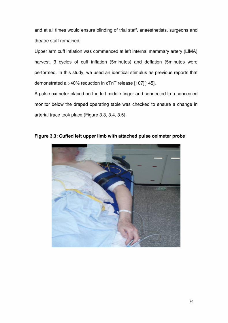
74
and at all times would ensure blinding of trial staff, anaesthetists, surgeons and
theatre staff remained.
Upper arm cuff inflation was commenced at left internal mammary artery (LIMA)
harvest. 3 cycles of cuff inflation (5minutes) and deflation (5minutes were
performed. In this study, we used an identical stimulus as previous reports that
demonstrated a >40% reduction in cTnT release [107][145].
A pulse oximeter placed on the left middle finger and connected to a concealed
monitor below the draped operating table was checked to ensure a change in
arterial trace took place (Figure 3.3, 3.4, 3.5).
Figure 3.3: Cuffed left upper limb with attached pulse oximeter probe

75
Figure 3.4: Pulse oximeter monitor concealed below operating table
Figure 3.5: Cuffs attached once patient prepped and draped

76
5.0 Anaesthetic Protocols
5.1 Induction of anaesthesia
Anaesthesia was induced in the anaesthetic room with the following regimen:
• Fentanyl 10-15 mcg.kg-1
• Etomidate 0.1-0.2 mg.kg-1
• Pancuronium 0.1 mg.kg-1
5.2 Central venous cannulation
All patients received a:
• Quadruple lumen central venous line
• Pulmonary artery catheter and sheath, fully floated into the pulmonary
artery prior to sternotomy.
Baseline haemodynamic measurements were performed prior to sternotomy.
5.3 Maintenance of anaesthesia
• Propofol 2% infused at 4-8 mg.kg-1.hr-1
• Alfentanil 25mg in 50ml used for maintenance infused at 50mcg.kg.h-1
• Enflurane 0-2% or Sevoflurane volatile anaesthesia was used to
supplement the propofol-based anaesthesia (Figure 3.6).
n.b. isoflurane volatile anaesthesia was not permitted to supplement propofol-
based anaesthesia as it has a potential pre-conditioning effect on the
myocardium (Chapter 1: Section 2; 5.2).

77
Figure 3.6: Anaesthetic machine
5.4 Other medications
• Mannitol 20% 0.5g.kg-1 was administered into the central line and not the
heart-lung machine reservoir, prior to cardiopulmonary bypass (CPB)
• GTN (50mg made up to 50ml) was administered intra-operatively at a
minimum dose of 0.1ml.hr-1
• Dopamine (low dose) as “renal dopamine” was not used in this study.
However dopamine (high dose) was the first choice inotropic agent
• Isoflurane was NOT used for anaesthetic maintenance
• Tranexamic acid was used, not aprotinin, as the antifibrinolytic agent.
Aprotinin has beneficial myocardial protective effects (Chapter 1:Section
2;9.0). Initial bolus of 30mg.kg-1; pump prime 2mg.kg-1; infusion

78
16mg.kg.h-1 until the end of surgery. Tranexamic acid has a better risk
benefit profile than aprotinin in low to moderate risk cases.[153]
• Patients with moderately impaired renal function (Cr 130-200) received
frusemide 250mg in 25ml (10mg.ml-1) neat infused at 0.2 ml.hr-1
6.0 Perfusion Protocols
6.1 Bypass schedule
All patients underwent surgery utilising cardiopulmonary bypass (Table 3.1;
Figure 3.7):
Table 3.1: Bypass schedule
Prime 2 L Hartman’s saline + 10000 units heparin
Pump Roller Pump
Oxygenator Capiox.SX Membrane Oxygenator
Flow range 2.4 L.min-1.m-2
Mean perfusion pressure Radial artery 50-60 mmHg
Vasoactive drugs Phenylephrine (1mg in 10ml. N/Saline) administered in 0.5-
1.0ml aliquots and titrated to effect.
Not metariminol (aramine). If phenylephrine fails to
maintain perfusion pressure, noradrenaline may be used
Cooling/rewarming Blood/water temperature gradient to be ≤ 7° C
α-stat standardisation of the blood gases
Cooling active cooling to 34OC in all cases
Protection Antegrade intermittent cold blood cardioplegia
Perfusionists were requested to enter the time and dose of each administration
of phenylephrine onto the perfusion chart. The effect of each test solution on
haemodynamic stability during CPB was assessed as part of the study.

79
Figure 3.7: Cardiopulmonary bypass machine
6.2 Cardioplegia composition
• St. Thomas A stock solution:
1L of Ringer’s compound sodium chloride
102 ml of cardioplegia infusion
16.6 MgCl2.L-1
6.0843 KCl.L-1
Procaine hydrochloride 1391.28 mg.L-1
• St. Thomas B stock solution:
1L of Ringer’s compound sodium chloride
32 ml of cardioplegia infusion
5.2 MgCl2.L-1
1.9 KCl.L-1
Procaine hydrochloride 435.68 mg.L-1

80
6.3 Cardioplegia administration (Table 3.2)
Intermittent antegrade cold blood St Thomas’ cardioplegia (Martindale
pharmaceuticals Essex, UK) was used for myocardial protection with an
induction dose of 12ml.kg-1 followed by maintenance administration each 15-
20 minutes
Table 3.2: Cardioplegia protocol
Temperature 4° C
Delivery Aortic root
Pressure 120-150 mmHg
Induction St Thomas A stock at AXC at 12 ml.kg-1 (800 ml blood + 200 ml stock A
solution)
Maintenance St Thomas B stock after max 30 minutes or between anastomoses at
surgical discretion 300ml doses (400 ml blood + 100 ml stock B
solution). Hot shot was not permitted
6.4 Discontinuation of CPB
On rewarming once the nasopharyngeal temperature reached 35°C the
heater/chiller temperature was set to 37°C
CPB separation occurred at 36oC-37oC nasopharyngeal temperature using
atrial or dual-chamber epicardial pacing to achieve a target heart rate of
90min-1.
7.0 Surgical Protocol
7.1 Conduct of operation
The technique to be adopted is as follows in the operating theatre:
• Institution of cardiopulmonary bypass

81
• Aortic cross-clamping and completion of all proximal and distal
anastomoses during a single clamp period. Planned bypass conduits
would be internal mammary artery and saphenous vein.
• Rewarming should commence during the penultimate anastomosis.
7.2 Surgical technique (Table 3.3)
Table 3.3: Surgical technique
Heparinisation timing Heparin should be requested and administered following
procurement of all conduit just prior to commencing the
aortic purse string insertion
Heparin dose 300 units.kg-1
Cannulation Standard cannulation
Cooling Active cooling to 34°C in all cases
7.3 Myocardial protection (Table 3.4)
Table 3.4: Myocardial protection
Technique Antegrade intermittent cold blood cardioplegia
Induction dose St Thomas ‘A’ stock at AXC at 12 ml.kg-1 (800 ml blood + 200 ml
‘A’ solution)
Maintenance dose St Thomas ‘B’ stock in all cases after 20 minutes or between
anastomoses at surgical discretion (400 ml blood + 100 ml ‘B’
solution) for intermittent administration of 300ml doses.
Hot shot is not permitted
De-airing
7.4 Myocardial Biopsy
Patients underwent myocardial biopsy studies. Biopsies (LV) were full thickness
and taken from a non-scarred area of the anterior wall.
Biopsies (x1 core) were taken from:

82
• Right atrium ↔ at cannulation within boundaries of the
atrial purse string
• LV 1 (pre-ischaemia) ↔ just before AXC on CPB using
fibrillation if preferred
• LV2 (ischaemia) ↔ immediately before AXC release
• LV3 (reperfusion) ↔ 10 minutes after the AXC release
Atrial and trucut biopsies were snap frozen in liquid nitrogen and stored at -
700C. The biopsy sites required 5’O’ prolene suture oversewn to ensure
haemostasis.
8.0 Operating department practitioner protocol
8.1 Checklist prior to commencing (Table 3.5)
Table 3.5: ODP checklist
Anaesthetic drugs as per protocol Placebo group: Dummy arm
Quad-lumen and Swan-Ganz catheter +
injectate coil
Stimulus group: Patient LEFT
arm
Air splitter device Portable saturation monitor
Tourniquet machine with two cuffs Stopwatch
8.2 Conduct of ODP
1. ODP action randomisation - boxes Surgeon A or Surgeon B
2. Prepare anaesthetic drugs as per protocol
3. Insert RIGHT radial arterial line and ABG
4. 20 ml blood for 2x yellow top bottles (cTnT and routine biochemistry),
1x purple and 1x blue (haematological indices) top withdrawn
5. Quad-lumen and Swan-Ganz catheter + injectate coil insert and float

83
prior to draping
6. Urine (for ACR and α-1 microglobulin)
7. Connect air splitter to AIR port in theatre
8. Air splitter device in AIR port in operating theatre to supply
anaesthetic and cuff inflation machine
9. Apply tourniquet cuffs to LEFT arm and dummy arm. Keep tourniquet
hoses detached
10. Apply Pulse oximeter to LEFT index finger from separate monitor
(blinded to all except ODP). Check trace
11. Drape patient
12. Connect tourniquet hose A and hose B to either LEFT arm or dummy
arm (as per randomisation instructions)
13. Ensure RED LIGHT on tourniquet machine (cuff is deflated)
14. Document 1st systolic blood pressure (SBP) from either RIGHT radial
or RIGHT femoral arterial line
15. At beginning of LIMA harvest, blinded anaesthetist to inflate
tourniquet A to 200mmHg (1st cycle of STIMULUS). Start stopwatch
for 5 mins.
16. GREEN LIGHT will show on tourniquet machine (cuff is inflated)
17. ODP check pulse oximeter trace lost if patient arm inflated.
18. After 5 mins of ischaemia deflate tourniquet A. RED LIGHT will show
19. Time 5 mins of reperfusion with stopwatch
20. Document 2nd SBP
21. Reinflate cuff to 200mmHg for 5 mins (2ND cycle of STIMULUS).
GREEN LIGHT will show.

84
22. Deflate cuff for 5 mins reperfusion. RED LIGHT will show
23. Document 3rd SBP
24. Reinflate cuff to 200mmHg for 5 mins (3rd cycle of STIMULUS).
GREEN LIGHT will show
25. Deflate cuff for reperfusion. RED LIGHT will show
26. ODP to check pulse oximeter trace returned once patient arm
deflated and document on sheet
Stimulus now complete.
27. Disconnect tourniquet hoses from cuffs and remove dummy arm
28. On de-draping, safeguard the Holter monitor leads and monitor on the
patient
9.0 Biochemical monitoring protocol
9.1 Cardiac Troponin T [154]
Biochemical estimation of myocyte injury was deduced from cardiac Troponin T
(cTnT). Samples were taken pre-sternotomy (in anaesthetic room), 6h, 12h, 24h
and 48h after aortic cross clamp release and sent for analysis to the
Department of Biochemistry at the University Hospital Birmingham NHS FT. A
commercially available assay (Elecsys 2010; Roche Diagnostics, UK) was
utilised in conjunction with an automated clinical analyser (Roche Modular
E170).
Troponin T (TnT) is a component of the contractile apparatus of the striated
musculature. Although the function of TnT is the same in all striated muscles,
TnT originating exclusively from the myocardium (cardiac TnT, molecular
weight 39.7kD) clearly differs from skeletal muscle TnT. As a result of its high

85
tissue-specificity, cTnT is a cardio specific, highly sensitive marker for
myocardial damage.
The Elecsys Troponin T assay employs two monoclonal antibodies
specifically directed against human cardiac troponin T. The antibodies
recognise two epitopes (amino acid position 125-131 and 136-147) located in
the central part of the cardiac troponin T protein, which consists of 288 amino
acids. Elecsys Troponin T assay detects free troponin T as well as binary and
ternary complexes of troponin.
9.2 Test Principle – Sandwich principle [154]
The total duration of the assay was 18 minutes.
• 1st incubation: 15µl of sample, a biotinylated monoclonal troponin T-
specific antibody and a monoclonal troponin T-specific antibody
labelled with ruthenium complex react to form a sandwich complex.
• 2nd incubation: After addition of streptavidin-coated microparticles, the
complex becomes bound to the solid phase via interaction of biotin and
streptavidin.
• The reaction mixture is aspirated into the measuring cell where the
microparticles are magnetically captured onto the surface of the
electrode. Unbound substances are then removed with ProCell.
Application of a voltage to the electrode then induces
chemiluminescent emission which is measured by a photomultiplier.
• Results are determined via a calibration curve which is instrument-
specifically generated by 2-point calibration and a master curve (5-
point calibration) provided by the reagent barcode.

86
The measuring range defined by the lower detection limit and the maximum of
the master curve is 0.010-25.00 µg.L-1 or ng.mL-1. Values below the detection
limit are reported as <0.010 µg.L-1 (ng.mL-1). Values above the measuring
range are reported as >25.00 µg.L-1 or ng.mL-1 (or up to 250 µg.L-1 or ng.mL-1
for 10-fold diluted samples).
10.0 Haemodynamic monitoring
10.1.1 Initial pre-bypass studies (Baseline/pre-sternotomy)
Time was taken for a good wedge trace. Full cardiac output studies were
obtained prior to sternotomy but after cleaning and draping and with the legs
horizontal and the patient flat in the neutral position.
10.2 Post-bypass haemodynamic studies (pre-protamine)
Cardiac output studies were performed following discontinuation of bypass after
volume and pressor adjustment was complete but prior to protamine
administration.
Volume adjustment was at discretion of the surgeon. The aim was to establish
comfortable filling pressures with a MAP of 65-85mmHg at the time of cardiac
output studies.
10.3 Post-protamine haemodynamic studies
Cardiac output studies were repeated between 5 and 15 minutes after the
completed administration of protamine but at an appropriate time convenient to
the surgeon performing haemostasis. The patient was flat. The chest was
open, preferably without the retractor in position

87
10.4 Post-sternal closure haemodynamic studies
Cardiac output studies were repeated 2 minutes following sternal re-
approximation
10.5 Haemodynamic studies on the intensive care unit
Haemodynamic studies were performed 2, 4, 6, 9 and 12 hours post-
reperfusion.
During this 12 hour period, need for IABP and the usage and dosage of any
inotropic support was noted.
10.6 Low cardiac output episodes
Low cardiac output episodes (defined as a CI ≤2.2 L.min-1.m-2 with CVP
≥12mmHg and/or PCWP ≥14mmHg in the presence of a native or paced
synchronised rhythm and heart rate of 90-110min-1) were recorded.
11.0 Protocol for post-operative inotropic support and vasoconstrictor
The haemodynamic aims were as follows:
• M.A.P. of 65-85 mmHg
• C.I. ≥ 2.2 L.min-1.m-2
• Sinus rhythm (Pacing)
In the ideal situation inotropes were not administered until post-bypass
haemodynamic studies were complete but sometimes this was not possible.
Inotropic support, initially with dopamine (200mg.50 ml-1 D5%W) at 5-10ml.hr-
1 was commenced if the cardiac index(CI) was ≤2.2 L.min-1.m-2 in the

88
presence of a central venous pressure(CVP) of ≥12mmHg, pulmonary
capillary wedge pressure(PCWP) of ≥14mmHg, and heart rate of ≥90min-1 in
a coordinated rhythm(sinus, AAI pacing or DDD pacing in order of
preference). Escalation of inotrope therapy with adrenaline or noradrenaline
or intra-aortic balloon pump (IABP) insertion was then at clinician discretion.
Introduction of support was also permitted if the operating surgeon identified
poor contractility at separation of CPB or if marginal haemodynamics were
noted by attending physicians on the intensive care unit (ICU).
Intra-operative boluses of phenylephrine were used to maintain a mean
arterial pressure of ≥55mmHg during CPB and ≥65mmHg following separation
in the context of a low systemic vascular resistance. Phenylephrine was also
used as a post-operative vasoconstrictor at 0-0.4µg.kg-1.min-1 and substituted
as necessary noradrenaline.
Extubation, ICU and hospital discharge criteria and post-operative atrial
fibrillation management were standardised.
12.0 Echocardiography
A comparison was made between left ventricular function assessed on
echocardiography 1-3 days before surgery with post-operative results 5-7
days after surgery in patients undergoing first time coronary artery bypass
surgery with or without RIPC. Patients were studied using a Vivid 7 ultrasound
machine (GE Vingmed Ultrasound, Horten, Norway) with a multifrequency
transducer (3V2c) and second harmonic imaging with presets available for
contrast and tissue Doppler imaging. Left ventricular dimensions and function
were assessed by standard M-mode and 2D according to current American
89
Society and European Society of Echocardiography Guidelines [155]. Studies
were performed by two accredited echocardiographers but analysed off-line
by a single observer (Ectopic PC Version 108.x.x, GE Medical System,
Horten, Norway).
12.1 Contrast Echocardiography for EF
The current method recommended for quantification of LV volumes and
function is the Simpsons biplane estimation [155]. Accuracy of this technique
is reduced if there is difficulty in endocardial border definition and in obtaining
true perpendicular views of the LV in apical four and two chamber views.
Inter-study variability may be up to 10% ejection fraction points between two
studies as a result of these technical issues [156]. Difficulty in obtaining an
optimal acoustic window becomes more acute in the post-operative period
due to pain from the median sternotomy and as patients may not be able to lie
in a left lateral decubitus position. The use of contrast opacification
overcomes the issue of endocardial border definition and enables the
echocardiographer to identify the true apex of the LV more reliably. As a
consequence, reproducibility of LV volumes and function using contrast
opacification during 2D echocardiography is equivalent to that of
cardiovascular magnetic resonance imaging [157]. Contrast-enhanced 2D
Simpson’s biplane was therefore used as the main method for estimation of
LV volumes and ejection fraction in this study. SonoVue (Bracco, Italy) was
used as the transpulmonary contrast agent, which is a suspension of
microspheres filled with perfluorocarbon gas that has a similar size to red
blood cells. 3D echocardiography was not used in this study as the

90
technology available at our institution requires the stitching of pyramids of
data acquired over 4-7 RR intervals to cover larger volume ventricles, which
may be inaccurate in irregular post-operative rhythms such as atrial fibrillation.
Patients were cannulated before study with a 20 gauge cannula located in the
left antecubital fossa. A three-way tap was attached to the cannula with a Luer
lock syringe containing 10mls saline and another Luer lock syringe containing
1-4 ml Sonovue contrast. After optimisation of the 2D image in the apical four
chamber view, a pre-specified contrast pre-set was activated to deliver low
mechanical index imaging (MI 0.2) and contrast was injected with a prompt
saline flush. Focus, compression and gain settings were optimised to
delineate LV endocardial borders.
12.2 Isovolumic Acceleration
Although widely used, LV ejection fraction is of most use when studying
populations or identifying individual risk at any one point in time. LV ejection
fraction is a method for assessment of LV contractility that is altered
significantly by change in loading conditions. As a result of this, it is less
useful as a method of tracking outcomes in an individual patient before and
after CABG surgery - whether 2D, contrast-enhanced or 3D. In order to
attempt to overcome this, load-independent indices of LV function were
studied. In experimental models, myocardial acceleration during isovolumic
contraction (IVA) is a sensitive index of left ventricular contractile function
which is unaffected by changes in loading conditions within a physiological
range [158].

91
After acquisition of standard 2D and M-mode measurements and before
injection of LV opacification contrast, the tissue Doppler preset was activated.
Tissue Doppler presets are required to introduce low pass filters to exclude
low amplitude, high velocity blood signal, thereby optimising image acquisition
to high amplitude, low velocity signal from myocardium. The image sector was
narrowed to the wall of interest to optimise frame rate. During suspended
respiration following exhalation, images were acquired of the inferoseptal,
anterolateral, inferior and anterior walls after optimisation of baseline and
scale to exclude aliaising. IVA was derived by measurement of the slope of
the time-velocity profile from tissue Doppler data acquired by a sample
volume placed within 1 cm of the insertion point of the mitral valve at the base
of all 4 walls. Images were optimised to minimise the angle between the
direction of annular motion and the ultrasound beam. Post-processing of this
signal enabled calculation of the slope of the acceleration during isovolumic
contraction, that part of the cardiac cycle when the LV is contracting to
increase pressure without change in volume before aortic valve opening. [159]
The ∆isovolumic acceleration (∆IVA) was an average from basal septal (A4C),
basal lateral (A4C), anterior (A2C) and inferior (A2C) tissue Doppler images
demonstrating change postoperatively as compared to before surgery.
12.3 Tissue Velocity Imaging
Left ventricular function is now thought to depend upon both radial and
longitudinal function in a fashion analogous to the wringing of a cloth.
Histopathological studies have clearly demonstrated that longitudinally-
orientated fibres predominate in the endocardium, while both the mid- and

92
epicardial layers share longitudinal, helical and circumferential fibres [160]. As
the myocardium is perfused from epicardium to endocardium, the longitudinal
fibres of the endocardium are most at risk from hypoperfusion. Longitudinal
function therefore declines before fall in radial function in ischaemia [161].
There is good evidence that the fall in longitudinal function with ischaemia is
initially compensated for by an increase in radial contraction, leading to
apparent hyperdynamic EF. Hence both basal systolic myocardial velocities
(s’) and early myocardial relaxation velocities (e’) both provide incremental
prognostic information to ejection fraction in ischaemic heart disease [162].
Moreover, early myocardial relaxation velocities can be used to give an
estimate of LV filling pressure in patients with normal EF and assessment of
transmitral to mitral myocardial relaxation velocity ratio (E/e’) is a strong
prognosticator in ischaemic heart disease.
After acquisition of standard 2D and M-mode measurements and before
injection of LV opacification contrast, the tissue Doppler preset was activated.
The image sector was narrowed to the wall of interest to optimise frame rate.
During suspended respiration following exhalation, the pulsed Doppler sample
volume was placed at the base of the inferoseptal, anterolateral, inferior and
anterior walls after optimisation of baseline and scale to exclude aliaising. The
size of the sample volume was altered to cover longitudinal displacement of
the annulus (5-10mm) and placed within 1 cm of the insertion point of the
mitral valve. Images were optimised to minimise the angle between the
direction of annular motion and the ultrasound beam. Gain was then set to
provide clear tissue velocity traces from which s’, e’ and a’ were recorded
(Figure 3.8).

93
Figure 3.8: Tissue velocity imaging
13.0 Arrhythmia Assessment
Three lines of investigation were employed to assess and compare
perioperative arrhythmias between the groups:
13.1 Continuous Holter ECG recording
A continuous Holter ECG recording device was attached by three leads to the
patient ≥ 12 hours prior to aortic crossclamping and was kept in-situ until 48
hours postoperatively. Leads were applied to the right shoulder, left shoulder
and left lower chest in the anterior axillary line. Leads ran behind the patient,
and not across the chest, so as not to obstruct the operative field (median
sternotomy). (Figures 3.9, 3.10, 3.11, 3.12).

94
Ventricular tachyarrhythmia quantitation was undertaken at the following time
periods; pre-operative 12 hours (Pre-12h), the10 minute period after
reperfusion (Post-10min), the next 2 hours of reperfusion (Post-120min), 2-24
hours post-operative (Post2-24h) and 24-48 hours (Post 24-48h).
Figure 3.9: Continuous Holter ECG Monitor

95
Figure 3.10: Anterior view of applied Holter leads
Figure 3.11: Lateral view of applied Holter leads

96
Figure 3.12: Posterior view of applied Holter leads
Note leads do not obstruct operative (median sternotomy) field
13.2 12 lead electrocardiograms (ECGs)
12 lead ECGs were recorded in patients preoperatively, immediately
postoperatively, on postoperative day 1 and day 4.
Perioperative myocardial infarction (PMI) defined by the presence of new left
bundle branch block (LBBB) or new Q waves of 2mm in depth in 2 contiguous
leads by postoperative day 4 was assessed by an independent cardiologist.
13.3 Atrial Fibrillation – Treated
The incidence of atrial fibrillation (AF) that required treatment with either/or
amiodarone, digoxin, DCCV during the postoperative period was recorded.

97
14.0 Renal outcomes protocol
Changes in serum creatinine, urinary albumin:creatinine ratio, urinary α-1
microglobulin, and requirement for diuretics or haemofiltration were used as
outcome measures to assess the effect of renal protection by RIPC on peri-
operative renal injury in this double-blind, randomised controlled trial.
14.1.1 Creatinine
Post-operative creatinine was measured on days 0 and 4.
In muscle metabolism, creatinine is synthesized endogenously from creatine
and creatine phosphate. Under conditions of normal renal function, creatinine
is excreted by glomerular filtration. Serum creatinine measurements are used
as a marker of glomerular filtration in diagnosis and monitoring of acute and
chronic renal disease as well as for the monitoring the efficacy of renal
replacement therapies such as haemofiltration, haemodialysis and chronic
ambulatory peritoneal dialysis.
Because urine excretion of creatinine is constant unless there is an acute
change in renal function, it can be used to correct for variations in urine flow
rate by expressing analytes such as urine albumin or alpha 1 microalbumin as
the urine protein/creatinine ratio. The method used is based on the Jaffe
reaction as described by Popper et al., Seelig and Wust and modified by
Bartels [163][164]. This modified version has a higher sensitivity and better
precision than the original Jaffe method. Serum and urine creatinine were
measured by the Clinical Biochemistry Department using a kinetic, blanked
and compensated method using Roche Modular automated clinical chemistry
analysers.[165][166][167][168][169]

98
14.1.2 Test principle – Kinetic colorimetric assay
In alkaline solution, creatinine forms a yellow-orange complex with picrate.
The colour intensity is directly proportional to the creatinine concentration and
can be measured photometrically. Assays using rate-blanking minimize
interference with bilirubin.
• Sample and addition of R1(Sodium hydroxide: 0.20 mol/L)
• Addition of R2(Picric acid: 25 mmol.L-1) and start of reaction:
creatinine + picric acid → creatinine-picric acid complex
The reference range for 24 hr urine creatinine is 9-21 mmol.24h-1 (1040-2350
mg.24h-1) and for women is 7-14 mmol.24h-1 (740-1570 mg.24h-1).
14.2.1 Albumin
Albumin is a non-glycosylated protein with a molecular weight of 66,000
daltons. Albumin is quantatively the most important protein component in
plasma normally representing about 50% of the total protein content.
However, in normal urine albumin only contributes <30mg.24h-1 of the total
urine protein which is normally <150 mg.24h-1. Low level but pathologically
significant albuminuria, undetectable by chemical dipstix, called
‘microalbuminuria’ is a sensitive marker of renal injury, and may reflect
glomerular or renal tubular dysfunction.
A variety of methods, such as radial immunodiffusion, nephelometry and
turbidimetry, are available for the determination of albumin.
[170][171][172][173][174] Immunoturbidimetric assay for the in vitro

99
quantitative determination of albumin in human urine on Roche Modular
automated clinical chemistry analyzers was performed in this series.
14.2.2 Test principle – Immunoturbidimetric assay
Urine albumin was measured by automated immunoturbidimetry using an
antibody to human albumin.
• Sample and addition of R1 (buffer; TRIS (Tris(hydroxymethyl)-
aminomethane) buffer: 50mmol.L-1, pH 8.0 (25oC); PEG: 4.2%; EDTA:
2.0mmol.L-1; preservative)
• Addition of R2 (anti-albumin antibodies; Polyclonal anti-human albumin
antibodies (sheep): dependent on titre; TRIS buffer: 100mmol.L-1, pH
7.2 (25oC); preservative) and start of reaction: Anti-albumin antibodies
react with the antigen in the sample to form antigen/antibody
complexes which, following agglutination, are measured
turbidimetrically.
• Addition of R3 (human albumin; Albumin (human): 200mg.L-1;
phosphate buffer: 50mmol.L-1, pH 7.0 (25oC); sodium chloride:
150mmol.L-1; preservatives): Human albumin is added and the
resulting additional turbidity change is evaluated for detection of
antigen excess.
The 24 hr urine expected values in adults are <20 mg.L-1 (0.304 µmol.L-1)
albumin or <30 mg.24h-1 (0.456 µmol.24h-1) albumin.

100
14.3.1 Urine ACR
Urine was collected pre-sternotomy (in anaesthetic room), 4h, 8h, 12h and
24h after aortic cross clamp release and sent to the Department of
Biochemistry for analysis. Urinary albumin-creatinine ratios (ACR) were
assessed at 0, 12 and 24 hours. The normal urine ACR reference range for
women is <3.5 and men is < 2.5 mg.mmol-1.
14.4.1 Urine for α-1 microglobulin
Urine was collected pre-sternotomy (in anaesthetic room), 4h, 8h, 12h and
24h after aortic cross clamp release and sent to the Department of
Biochemistry for storage of aliquots that were then sent to East Kent Hospitals
for analysis.
Measurement of alpha-1-microglobulin in urine aids in the diagnosis of kidney
tubular damage. A1M reagent when used in conjunction with the Beckman
Coulter IMMAGE® Immunochemistry Systems and Urine Protein Calibrator
was utilised for quantitative determination of Alpha-1-Microglobulin (A1M) in
human urine by rate nephelometry.
14.4.2 Test principle – Rate nephelometry
The A1M test measures the rate of increase in light scattered from particles
suspended in solution as a result of complexes formed during an antigen-
antibody reaction.
Alpha-1-microglobulin(sample) + Antibody → [Alpha-1-microglobulin(sample)-
Antibody (aggregates)]
The 24 h urine expected values are <1.28 mg.dL-1.

101
15.0 Lung outcomes protocol
15.1 Spirometry
Patients underwent preoperative simple spirometry to assess FEV1, FVC and
FEV1/FVC ratio (Figure 3.13).
Figure 3.13: Spirometer
15.2 Positive End Expiratory Pressure
As Positive End Expiratory Pressure (PEEP) has been shown to increase right
and left ventricular stroke volume variation both during open and closed chest
conditions in addition to reducing right ventricular end-diastolic volume
indicating a preload reductive effect, [175] its use in ventilator management was
standardised to 5cm H2O. Changes in arterial blood partial pressure of oxygen
(PaO2) / fractional inspired oxygen (FiO2) ratio and intubation times were used
as outcome measures.

102
15.3 Arterial PaO2 / fractional inspired oxygen ratio (PaO2:FiO2 ratio)
Arterial blood was sampled preoperatively from a right sided radial arterial line
in the anaesthetic room prior to induction and assessed for PaO2 (kPa) (Bayer
RapidLab865 Analyser). PaO2:FiO2 ratio was recorded and the process
repeated for measurements at 6hr and 12hr postoperatively.
15.4 Intubation time
Intubation time (or time to extubation) was recorded as the time from arrival to
the Intensive Care Unit to extubation (minutes). Extubation was nurse-led and
performed according to standard departmental criteria (>36oC, normal and
stable acid base balance, chest output <100ml.h-1).
16.0 Analysis of myocardial samples
During a small mechanistic study in patients undergoing on-pump coronary
artery bypass grafting randomised to RIPC (three 5 min cycles left upper limb
ischaemia) and Control groups, left ventricular myocardial Tru-cut biopsies
were collected: (a) ‘pre-ischaemia’ (‘LV1’ – just prior to application of aortic
cross clamp); (b) ‘end-ischaemia’ (‘LV2’ – just prior to removal of aortic cross
clamp); (c) ‘post-reperfusion’ (‘LV3’ – 10 minutes after removal of aortic cross
clamp). Biopsies were snap frozen in liquid N2 and stored at –80OC for batch
analysis using standard techniques.
Myocardial samples were transported on dry ice to the University of Oxford for
analysis.
103
The left ventricular apical biopsy samples, taken 10 minutes after the
cessation of the extra-corporeal circuit, were homogenised and protein
extracted for Immunoblotting (see below). Quantification was done by
densitometry and all samples were normalised to beta-tubulin. Comparisons
between control (n=6) and RIPC (n=7) was by unpaired t-testing.
16.1 Tissue homogenization and protein quantification
All samples were stored at -80ºC until use. Tissue samples were
homogenized using a Polytron rotor (GlenMills) in RIPA buffer (50 mM
TrisHCl pH7.6, 150 mM NaCl, 2 mM EDTA, 1% NP-40, 0.1% SDS) containing
1x complete protease inhibitor tablet (Roche Applied Sciences), 1x PhosSToP
phosphatase inhibitor cocktail tablet (Roche Applied Sciences) and 0.5mM
PMSF.
Protein content was quantified using the colorimetric BCA protein assay kit
(Thermo Scientific). Briefly assay reagents A and B supplied were mixed in a
ratio of 50:1. Bovine serum albumin at concentrations of 0, 50, 100, 200, 400
and 800µg.ml-1 was used to create a standard curve. 10µl of each sample of
homogenate was diluted in 40µl of ddH2O. 1ml of mixed assay reagent was
then added to each sample and then samples were incubated at 37ºC for
30mins.
Samples were transferred to cuvettes and then colorimetric density was
assessed using a spectrophotometer (Ultrospec 2000, Pharmacia Biotech) set
at 562nm. By comparison against the standard curve all samples were
normalised to 200µg.ml-1 before the addition of SDS-Page running buffer (4%
SDS, 20% glycerol, 10% 2-mercaptoethanol, 0.004% bromphenol blue, 0.125

104
M Tris HCl) to give a 6:1 dilution. All samples were subsequently heated to
95ºC for 5 minutes prior to immunoblotting.
16.2 Western Blotting
Protocol for ECL Western blotting
Flow diagram Separate protein sample by electrophoresis
Transfer to membrane
Block non-specific sites
Incubate in primary antibody
Incubate in HRP-labelled conjugate
ECL detection reagents
Expose to film
Pre-cast 4-12% 1.5mm Bis-Tris Gels were utilised (Invitrogen) in a standard
electrophoresis tank containing MOPS buffer (200mM MOPS free acid, 50mM
sodium acetate, 10mM EDTA, and 10mM EGTA) and 500 µl of NuPage
antioxidant (Invitrogen) in the central section only.
Into each well was loaded 20µl of sample with 12 µl of Full Range Rainbow
Marker (GE Healthcare) in the outside lane.
Gels were run at 200V for a minimum of 50mins or until the forward protein
marker was seen to have run off the gel. Once completed, gels were removed
from the tank and placed into cooled transfer buffer (4ºC) (48mM Tris Base,
390mM Glycine, 0.1% SDS, 20% Methanol). Transfer stacks were formed
with a layer of fibre pad and pre-cut blotting paper (both soaked in transfer
buffer) as the base for the gel. A PVDF membrane (Bio-Rad) (activated by

105
soaking in 100% methanol for 10 min prior to rinsing in transfer buffer) was
placed on top of the gel and the stack completed with further layers of blotting
paper and fibre pad, prior to rolling to remove any air bubbles. Transfers were
conducted overnight at 20mA, in a 4ºC cold room with constant stirring using
a magnetic flea.
After transfer PVDF membranes were placed in 50ml Falcon tubes and
incubated with constant rolling for 1 hour at room temperature in 5% fat free
milk in TBST (50 mM tris-HCl, 10 ml 0.5 M tris-HCl, 150 mM NaCl, 75 ml 2 M
NaCl and 0.05 % Tween 20) as a blocking agent. The blocking solution was
then replaced with 10ml of the same solution containing the primary antibody
at a dilution of 1:1000. After incubation at room temperature for 2 hours, the
antibody was removed and the PVDF membrane washed 3 times for 5 minute
periods in 15ml TBST. Secondary antibody in the 5% milk and TBST solution
at a dilution of 1:4000 was then added and the membrane incubated at room
temperature for 40 min, prior to a final 3 washes in TBST as above.
ECL advance solution (Amersham Biosciences) reagents A+B were mixed in
a 1:1 ratio and pipetted onto the PVDF membrane, allowing it to incubate at
room temperature for 5 mins. The resulting chemoluminescence was detected
in a Chemi-Doc molecular imaging system (Bio-Rad) and band densitometry
performed using the QuantityOne software package. (Figure 3.14; Table 3.6).

106
Figure 3.14: Gel tank assembly for SDS-polyacrylamide gel
electrophoresis

107
Table 3.6: Antibodies used in Western Blotting
17.0 Statistics
17.1 Primary outcome measure and estimation of sample size
If RIPC is an effective adjunct to myocardial protection it would be expected to
reduce evidence of myocardial ischaemic injury. The optimal biochemical
measurement of myocyte injury remains unclear [176][177]. It is not known
whether a peak, timed or serial measure (ANOVA, AUC) is the most appropriate
predictor of biochemical injury but serial measures contain greater information
about the total injury and until further comparative studies become available we
believe AUC represents the best predictor of mid-term survival post CABG.
At first, cTnI was considered as the biochemical measurement of myocyte
injury.
17.1.1 Estimation of sample size using cTnI
Single time point comparisons and cumulative area-under-the-curve
comparisons of creatine kinase MB fraction (CK-MB) [149] and Troponin I
(cTnI) [178] are currently accepted standards [179][180][181]. In our previous
studies GIK therapy reduced cTnI by 3.07ng.ml-1. (population SD 6.905) [182].
Antibody Species/Ig class Dilution Manufacturer/catalogue No.
Phospho-Akt (Ser 473) Rabbit/Ig G/polyclonal 1:1000 Abcam/ab28821
Pan-Akt Rabbit/Ig G/polyclonal 1:1000 Abcam/ab8805
Phospho-GSK3 beta (S9) Rabbit/Ig G/polyclonal 1:1000 Abcam/ab30619
GSK3 beta Rabbit/Ig G/polyclonal 1:1000 Abcam/ab31366
β-catenin Rabbit/Ig G/polyclonal 1:1000 Abcam/ab6302
β-tubulin Rabbit/Ig G/polyclonal 1:1000 Abcam/ab6046
Rabbit IgG (HRP) Goat/ Ig G/polyclonal 1:4000 Abcam/ab6721

108
If this difference were replicated in the current study this will be detectable
with a total sample size of 162 patients (α 0.05. 1-β 0.8). Previous studies of
ischaemic preconditioning in myocardial protection have suggested that the
reduction in peak or AUC CK-MB or cTnI approximates ≥0.5SD. For instance,
Belhomme et al. reported a difference of 250ng.ml-1.h-1 (population SD 404)
for AUC CKMB, a difference of 80 ng.ml-1.h-1 (population SD 140) for cTnI
AUC, a difference of 29µg.L-1 (control SD 33) for 6 hour CKMB and a
difference of 1.9ng.ml-1 (population SD 3.64) for 6 hour cTnI [149]. Replication
of these differences for the proposed study with a total sample size of 162
patients would yield the following power and alpha (Table 3.7).
Table 3.7: cTnI sample size calculations
Parameter &
study
Estimated
difference
Population
standard
deviation
Total
sample
size
αααα 1-ββββ
6h cTnI [183] 3.07 6.905 162 0.05 0.8
*6h cTnI [183] 3.453 6.905 162 0.05 0.85
6h cTnI [149] 1.9 3.64 162 0.05 0.9
6h CK-MB [149] 29 33 162 0.01 >0.95
AUC CK-MB [149] 250 404 162 0.025 0.95
AUC cTnI [149] 80 140 162 0.05 0.9
AUC cTnI [183] 105 210 162 0.05 0.85
*GIK study (postulated 0.5SD difference)
Thus, using cTnI the primary end-point of 6 hour and 48 hour AUC cTnI
estimation with sampling at 0, 6, 12, 24 and 48 hours post-operatively
[181][184]would be achieved with a total sample size of 162 patients.

109
17.1.2 Estimation of sample size using cTnT
48h AUC cTnT release was finally selected as the primary endpoint
biochemical measurement of myocyte injury for the following reasons: (1)
more available assay; (2) only one assay exists from a single provider; (3) as
used in previous similar studies this study would then be comparable.
Previous studies suggested a large treatment effect; a standardised difference
of 0.8. We hypothesised that RIPC would reduce cTnT AUC by a
standardised difference of 0.6 at 90% power and 5% alpha. This required a
sample size of 120 subjects which we increased by 33% to accommodate
withdrawal or missing data points.
17.2 Secondary outcome measures
Secondary cardiac end points were 6hour and peak cTnT, incidence of
myocardial injury on 12-lead ECG, serial cardiac and left ventricular stroke
work indices (CI, LVSWI), low cardiac output episodes (LCOE) incidence,
inotrope usage and dosage, reperfusion ventricular fibrillation and peri-
operative ventricular tachyarrhythmia incidence and functional
echocardiographic change, ICU and hospital length of stay(LOS) and the
renal and lung data listed above.
17.3 Statistical analysis for cardiac, renal and lung outcomes
Data were analysed with statistical package (SPSS 15.0, Chicago, Ill).
Categoric or ordinal data were compared by using χ2 tests or Kendall tau b,
respectively. Continuous data are presented as mean±standard deviation or
median(interquartile range). Normally distributed data were compared using

110
independent two-sided t tests. Repeated-measures analysis of variance
(RMANOVA) was used for serial measurements. Skewed data were either
logarithmically transformed or analysed non-parametrically (Mann-Whitney U
test).
To assess cTnT AUC, we estimated missing troponin values (6%
measurements) from the group and sampling time using a general linear
model.
17.4 Statistical analysis of echocardiographic parameters
The primary end-point for the echocardiographic assessment of the functional
outcome from RIPC was IVA. With a sample size of 73 patients in each group,
this study would be able to detect a difference in IVA ≥0.5SD between groups
(α 0.05, 1-β 0.85) [158][159][185][186]. Secondary end-points were change in
contrast-enhanced LVEF and change in myocardial tissue systolic and early
myocardial relaxation velocities.
Data were analysed with statistical package (SPSS 15.0, Chicago, Ill).
Categoric or ordinal data were compared by using χ2 tests or Kendall tau b,
respectively. Continuous data are presented as mean±standard deviation or
median(interquartile range). Normally distributed data were compared using
independent two-sided t tests. Repeated-measures analysis of variance
(RMANOVA) was used for serial measurements. Skewed data were either
logarithmically transformed or analysed non-parametrically (Mann-Whitney U
test).
18.0 Demographic and intraoperative variables
Both groups were well matched (Table 3.8).

111
Table 3.8: Pre-operative and intra-operative patient characteristics. CCS
= Canadian Cardiovascular Society; NYHA = New York Heart
Association; TIA = Transient ischemic attack; CVA = Cerebrovascular
accident; ACE-I = Angiotension converting enzyme inhibitor; IABP =
intra-aortic balloon pump.
Control (n=82)
RIPC (n=80)
p
Age 65(60-73) 63(57-71) 0.19 Male gender (n(%)) 72(88) 71(89) 0.59 CCS Angina Class 2(1-2) 1(1-2) 0.54 NYHA Class 2(1-2) 2(1-3) 0.25 Body mass index (kg.m-2) 28(25-31) 27.5(26-30) 0.80 Elective (n(%)) 38(46) 42(52) 0.53 Urgent (n(%)) 44(54) 38(48) 0.53 Current Smokers (n(%)) 7(9) 12(15)
0.15 Ex-smokers (n(%)) 52(63) 39(49) Never smoked (n(%)) 23(28) 29(36) High risk lung status (n(%)) 17(21) 22(28) 0.36 Hypercholesterolemia (n(%)) 59(72) 61(76) 0.59 Hypertension (n(%)) 52(63) 44(55) 0.34 Past history of TIA (n(%)) 4(5) 3(4) 1.00 Past history of CVA (n(%)) 1(1) 1(1) 1.00 Statins (n(%)) 71(87) 76(95) 0.10 Aspirin (n(%)) 70(85) 73(91) 0.33 Diuretics (n(%)) 16(20) 10(13) 0.29 Potassium channel blocker (n(%)) 18(22) 12(15) 0.31 Angiotensin II Antagonist (n(%)) 7(9) 6(8) 1.00 ACE I (n(%)) 57(70) 48(60) 0.25 β-blocker (n(%)) 68(83) 63(79) 0.55 Oral Nitrates (n(%)) 30(37) 18(23) 0.06
Ca2+ channel blockers (n(%)) 31(38) 26(33) 0.51
Pre-CPB IABP (n(%)) 1(1) 0(0) 1.00 Fasting glucose >7mmol.L-1 (n(%)) 0(0) 3(4) 0.33 Fasting triglycerides (mmol.l-1) 1.4(1.1-1.7) 1.3(1.0-1.7) 0.81 Fasting cholesterol(mmol.l-1) 3.6(2.9-4.3) 3.6(3.2-4.4) 0.20 Creatinine (mg.dL-1 ) 1.09±0.18 1.11±0.18 0.44 Pre-operative pO2:FiO2 ratio 53(47-61) 55(48-63) 0.31 Euroscore 3(2-5) 3(2-4.5) 0.10 Logistic Euroscore 2.5(1.5-4.3) 1.9(1.3-3.4) 0.78 Intra-operative factors Enflurane (n(%)) 65(79) 64(80)
0.46 Sevoflurane (n(%)) 15(18) 15(19) Propofol (n(%)) 2(2) 1(1) Bypass time (min) 96±22 100±23 0.25 Cross-clamp time (min) 71±18 76±21 0.08 Number of grafts 3.5(3-4) 3(3-4) 0.88
112
CHAPTER 4 THE EFFECT OF RIPC ON CARDIAC OUTCOMES IN
PATIENTS UNDERGOING CORONARY ARTERY
BYPASS SURGERY
1.0 Introduction
In remote ischemic pre-conditioning (RIPC) short cycles of repeated limb (or
other organ) ischemia provoke a protective effect that can halve the mass of
infarction caused by substantive vessel occlusion and reperfusion [70]. In
coronary artery bypass grafting (CABG), adverse outcomes predominantly
relate to cardiomyocyte injury due to myocardial protection failure and there is
experimental and clinical evidence that RIPC could attenuate such injury
[187]. The adequacy of myocardial protection can be assessed using an array
of measurements that are indicative of cardiomyocyte death, injury or
dysfunction [188]. These include cardiac troponin (cTn) release, the incidence
of post-operative low cardiac output episodes and ventricular arrhythmias, the
need for inotropic support and functional assessment by haemodynamic
monitoring and echocardiography. These measures contribute to an
assessment of reversible and irreversible myocardial injury.
Experimental models have demonstrated a first window (1-4 hours) followed
by a second window (24-72 hours) of protection against infarction afforded by
ischaemic preconditioning. In rabbit models the second window also protects
against infarct and arrhythmias [189] and this has been corroborated by rat
models where repeated left coronary artery occlusions have been shown to
reduce infarct size by >40% [190] and the incidence of ventricular fibrillation
[191].

113
Human ventricular myocytes [192] and atrial appendages taken during CABG
[193] have also been shown to exhibit protection at both windows with
preconditioning protocols followed by prolonged ischaemia and reperfusion.
Suppression of proinflammatory gene transcription in leukocytes within 15
minutes and 24 hours of an intermittent forearm ischaemic stimulus highlights
evidence for first and second windows in human models following RIPC
protocols [194].
In elective percutaneous coronary intervention(PCI), RIPC has been
associated with reduced cTnI release, less electrocardiographic ST elevation
and a reduced incidence of major adverse cardiovascular events [195]. In
abdominal aortic aneurysm repair, RIPC induced by intermittent iliac artery
occlusion reduces the incidence of myocardial injury and may reduce post-
operative renal impairment [143].
In cardiac surgery, the first clinical report of a protective RIPC effect was in
children. RIPC, induced by four 5-min cycles of lower limb ischemia-
reperfusion resulted in lower cTnI release, reduced inotrope requirements and
lower airway resistance implying a possible protective effect on myocardial
injury, function and bypass-related lung injury, a finding also demonstrated in
experimental models [107][118][196].
Two studies in CABG patients, one investigating RIPC in CABG undertaken
using predominantly intermittent ischemic arrest and a second utilising
cardioplegia have reported [107][145]. Both were single blinded, used a 3-
cycle upper arm RIPC stimulus and both demonstrated a significant reduction
of cTnT area-under-the-curve(AUC) release of over 40%. Clinical and
hemodynamic outcomes were however not reported. Much of the cardiac

114
injury associated with CABG appears to be a reversible phenomenon
manifest as transiently subnormal cardiac output and a temporary need for
inotropic support. The role of RIPC in affording protection against such post-
ischemic dysfunction is far less clear [197].
The aims of this study were to confirm the role of RIPC in reducing troponin
release and to ascertain if this is accompanied by an enhanced protection
against post-operative cardiac, renal and pulmonary dysfunction.
2.0 Methods
Biochemical and haemodynamic assessment of cardiac outcomes followed
methodology set out in Chapter 3; Section 9.0 to 11.0. Statistical analysis was
performed as per Chapter 3; Section 17.0 to 17.2.
3.0 Results
Of 313 possible subjects, 206 met the inclusion criteria, 170 consented and
were initially enrolled, 8 of which were withdrawn pre-randomisation for
logistical reasons (Figure 4.1) leaving a study population of 162. All patients
had the appropriate signal on pulse oximetry during RIPC or placebo stimulus
delivery with no inadvertent crossover. The stimulus was applied in the 74±16
minutes between incision and aortic cross clamping (AXC). Demographic and
intra-operative variables were similar (Table 3.8).

115
Figure 4.1: Consort diagram of trial recruitment.
313 assessed for
eligibility 143 excluded95 diabetics36 refused to participate
4 unstable angina8 other
162 randomly assigned
80 underwent
RIPC on upper arm
82 underwent
Control stimulus on dummy arm
170 consented for study
8 withdrawn pre-randomisation for logistical reasons
All patients survived 30 days. There was one in-hospital death in the control
group at day 109 due to pneumonia, a hospital mortality of 0.6%. There was
no difference in ITU or hospital length of stay (Table 4.1).
Table 4.1: Post-operative data. cTnT = Cardiac Troponin T; IABP = intra-
aortic balloon pump; LCOE = low cardiac output episode; LOS = length
of stay.
Control
(n=82)
RIPC
(n=80)
p=
6 hour cTnT (ng.ml-1) 0.93(0.59-1.35) 1.01(0.72-1.43) 0.522
Cmax cTnT (ng.ml-1) 1.02(0.74-1.44) 1.04(0.78-1.51) 0.789
Postoperative IABP (n(%)) 7(9) 6(8) 0.52
12h LCOE incidence (n(%)) 20(24) 27(34) 0.225
Inotrope usage (n(%)) 32(39) 40(50) 0.155
Vasoconstrictor usage (PhE or NE) (n(%)) 54(66) 51(64) 0.870
ITU LOS (days) 3(2-4); 3(2-5) 0.287
Hospital LOS (days) 8(6-11); 8(6-12) 0.392
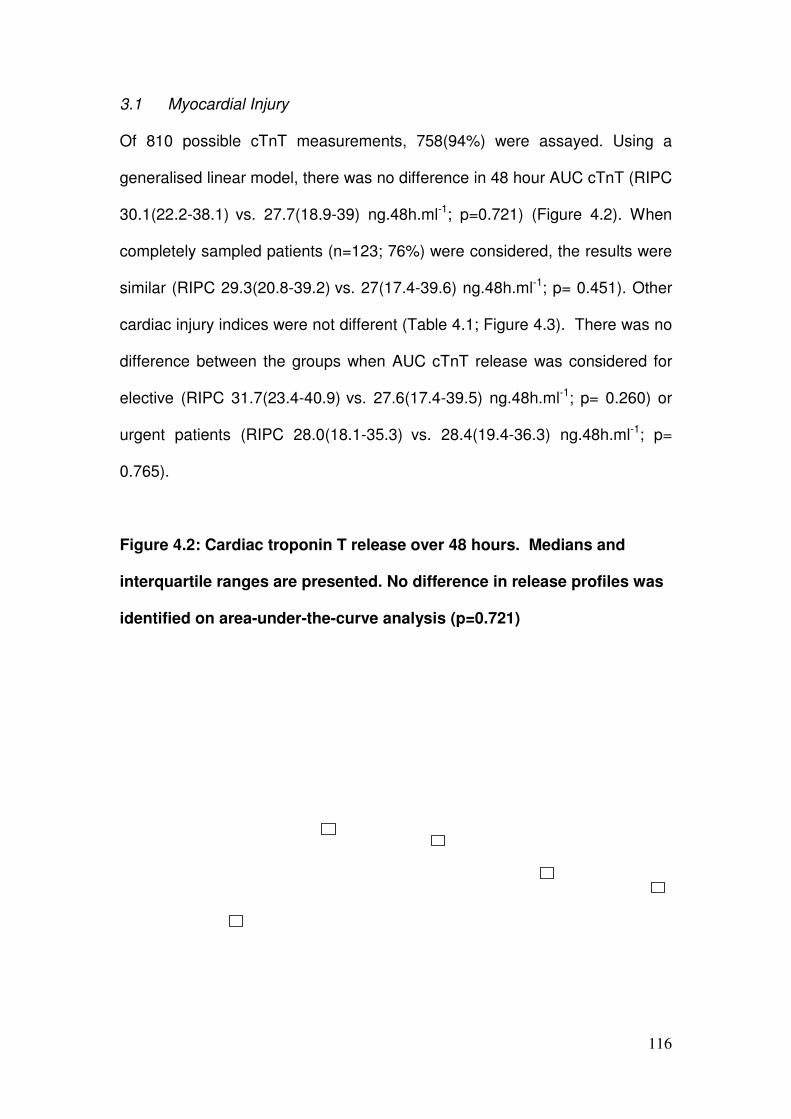
116
3.1 Myocardial Injury
Of 810 possible cTnT measurements, 758(94%) were assayed. Using a
generalised linear model, there was no difference in 48 hour AUC cTnT (RIPC
30.1(22.2-38.1) vs. 27.7(18.9-39) ng.48h.ml-1; p=0.721) (Figure 4.2). When
completely sampled patients (n=123; 76%) were considered, the results were
similar (RIPC 29.3(20.8-39.2) vs. 27(17.4-39.6) ng.48h.ml-1; p= 0.451). Other
cardiac injury indices were not different (Table 4.1; Figure 4.3). There was no
difference between the groups when AUC cTnT release was considered for
elective (RIPC 31.7(23.4-40.9) vs. 27.6(17.4-39.5) ng.48h.ml-1; p= 0.260) or
urgent patients (RIPC 28.0(18.1-35.3) vs. 28.4(19.4-36.3) ng.48h.ml-1; p=
0.765).
Figure 4.2: Cardiac troponin T release over 48 hours. Medians and
interquartile ranges are presented. No difference in release profiles was
identified on area-under-the-curve analysis (p=0.721)

117
Figure 4.3: 6 hour and Peak Cardiac troponin T release. Medians and
interquartile ranges are presented. No difference in release profiles was
identified.
0.0
0.5
1.0
1.5
Card
iac tro
ponin
T (ng.m
l-1)
Control RIPC
6hr Peak
p=0.522 p=0.789
3.2 Hemodynamic Effects
Over the measurement period, the CI increased in both groups but there was
no significant difference between groups (Figure 4.4). Similar numbers in
each group experienced LCOEs or required IABP (Table 4.1; Figure 4.5).
Serial LVSWI was not different (p=0.844; RMANOVA; Figure 4.6).

118
Figure 4.4: Serial cardiac index measurements in each group
(mean±SD). Cardiac index increased from pre-operative values in both
groups (p<0.001) but was not different between groups (p=0.620;
RMANOVA)
Card
iac index (L.m
in-1
.m-2)
2h 4h 6h 9h 12h0
Figure 4.5: Incidence of Low Cardiac Output Episode (LCOE), Intra-
aortic balloon pump (IABP) and Inotrope usage.
0
10
20
30
40
50
60
LCOE IABP usage Inotrope usage
Incidence
Control RIPC%
p=0.225
p=0.519
p=0.155

119
Figure 4.6: Serial left ventricular stroke work index in each group
(mean±SD).
3.3 Inotrope and vasoconstrictor requirements
Intra-operatively all patients required phenylephrine administration at similar
dosage (RIPC 0.36(0.22-0.63) vs. 0.43(0.25-0.67) mg.kg-1; p=0.622). A similar
number of patients in each group required inotropes (Table 4.1; Figure 4.7) at
equivalent dosages (Figure 4.8).
2h 12h9hPost-CPBPre-CPB 4h 6h
Left
ve
ntr
icu
lar
str
oke
w
ork
in
dex
(g
.m.m
-2)

120
Figure 4.7: Incidence of individual inotrope usage in the first two 6 hour
periods. Dopa. = dopamine, Epi.= epinephrine, NorE. = norepinephrine.
Hours 0 - 6 Hours 6 - 12
Perc
enta
ge u
sage o
f in
otropes
p = 0.206
%
Figure 4.8: Cumulative dose(SD) of inotrope received over 6 hours by
group. For presentation, the doses of epinephrine (Epi.) and
norepinephrine(NorE.) are multiplied by 10-1.
Hours 0 - 6 Hours 6 - 12
p=0.245
Inotr
ope d
osage receiv
ed (m
g.k
g-1
.6h
-1)
p=0.246
p=0.806
p=0.458
p=0.430p=0.659

121
4.0 Discussion
This double-blind study using an identical RIPC stimulus and cTnT assay has
not corroborated previous smaller single blind studies that found RIPC to
reduce troponin release following on-pump CABG. This absence of effect on
measurable myocardial injury is matched by a failure to demonstrate any
advantage of RIPC in terms of cardiac performance and inotrope requirement.
Our study suggests that RIPC, like its predecessor classical cardiac
ischaemic preconditioning (IP), has not fulfilled the promise of a practically
useful form of improved myocardial protection. While early studies using
classical IP reported reduced troponin release [103][104], improvement in
high energy phosphate conservation and reduced inotrope requirement these
effects were replicated in some but not all studies [141][142][133][198]. The
clinical effect was less obvious and in some reports detrimental[106] and this
frustrated the use of classical IP as surgical myocardial protective adjunct.
There is debate regarding the site of origin and significance of troponin
release during on-pump CABG. Troponin release may be indicative of
myofibrillar damage and myocyte necrosis or changes in sarcolemmal
permeability with leakage from cytosolic pools [199][200]. RIPC may protect
against necrosis-related but not cytosolic release. CPB may itself affect
sarcolemmal permeability or may have a pre-conditioning effect that
precludes further protection by RIPC [106].
For assessment of perioperative myocardial infarction and heart failure a
range of cardiac biochemical and inflammatory markers have been identified
Although cardiac troponin I (cTnI) is more sensitive and specific than CK-
MB[201][202] it has not shown itself to be a better discriminator when

122
compared to cardiac troponin T (cTnT)[203]. The release of markers in on-
CPB surgery is now well established and known to be higher than those off-
CPB with the associated sequelae[204][205][206][207].
Postoperative cardiac troponin is an independent predictor of in-hospital death
and high concentrations are associated with a cardiac cause of death and
major postoperative outcomes[208][209]. The magnitude of release has been
shown to be related to the type of cardiac surgical procedure[210] In one
series perioperative elevation of cTnI reliably predicted mortality in infants and
children undergoing surgical repair of congenital heart defects.[211]
In this RCT cTnT, as a marker of perioperative myocardial infarction and
myocardial protective intervention efficacy [212][213][214], has shown no
difference between groups when collated up to 48 hours after reperfusion.
Thus, we have no evidence to suggest the benefit of a ‘first window’ of RIPC
protection.
To corroborate the evidence all secondary endpoints of myocardial injury:
peak cTnT, 6hr cTnT; LCOE incidence; serial cardiac indices; IABP usage;
inotrope and vasoconstrictor usage failed to show any advantage for RIPC.
To accommodate for changes in systemic vascular resistance left ventricular
stroke work indices was compared between groups but again failed to show
advantage to those receiving RIPC.
Post-CABG myocardial injury often manifests as a transient period of
reversible dysfunction, treatable by hemodynamic optimisation and temporary
inotropic support. This implies a cardiac stunning phenomenon and the role of
RIPC as a protective measure against stunning is unclear. During clinical PCI,
RIPC has been shown to not attenuate post-ischemic stunning [197]. Our

123
hemodynamic data suggests there is similar absence of effect on reversible
dysfunction in CABG.
In this study, we used an identical stimulus and cTnT assay as previous
reports that demonstrated a >40% reduction in cTnT release [107][145]. To
remove inadvertent bias, we developed a scrupulous technique to blind the
surgeon, anaesthetist and investigators from the group allocation. The
stimulus was administered within an appropriate time-frame and delivery
verified by observing disappearance of the oximetry pulse signal. In the
single-blind studies, statistical significance was attained by the much smaller
variances observed in cTnT levels in the RIPC group. In the current study, the
cTnT release in both groups was similar to that of the control groups in the
previous reports. The study design also had important similarities; most
patients received a volatile halogenated anaesthetic agent peri-bypass and
high percentages of patients received pre-operative statins. Isoflurane, a
known pre-conditioning agent was specifically avoided in the current study.
When re-examining the data in this study no subtle evidence appears to exist
to explain the comparability between the two groups. Volatile anaesthetics
have a known preconditioning effect yet there was no significant difference
between groups in the incidence of Enflurane (RIPC 64/80(80%) v
65/82(79%) or Sevoflurane (RIPC 15/80(19%) v 15/82(18%) used. In three
cases intravenous propofol alone was used as the anaesthetic agent (RIPC
1/80(1%) v 2/82(2%). No difference between the groups was observed when
cTnT AUC was adjusted for calculated CrCl or elective/urgent cases.
Other differences exist between our study and those previously reported. We
noted a higher pre-operative usage of beta-blockers continued until the

124
morning of surgery. Our bypass and cross-clamp times were slightly longer as
we used a single clamp technique and we included non-elective patients who
had angina or an acute coronary syndrome within 30-days of surgery.
However, no patients had experienced angina within 48 hours of surgery and
we observed no differences between those admitted electively and more
urgent cases.
By excluding the harder to precondition group of diabetics the opportunity to
observe a real change from RIPC has been improved. And by further
exclusion of patients who have suffered with recent angina (<48hours) there
can be confidence that changes observed in primary and secondary
endpoints can be attributable to the beneficial effects of our treatment.
As to why the present study did not replicate similar recent studies the answer
is more difficult. An initial challenge may be the ischaemic stimulus itself.
Interestingly not only the same stimulus was used by Haunseloy et al where a
43% decrease in cTnT AUC was observed but also in the present study
verification of the ischaemic stimulus by loss of pulse oximeter trace was
confirmed by the ORT. Differences in protocols such as lack of blinding of
anaesthetists and investigators; lack of data on dosages of volatile
anaesthetics with proven conditioning effects; concomitant AVR with
antegrade and retrograde cardioplegia may offer lines of reasoning
attributable to the purported benefit of RIPC in one study over the other.
Finally, and perhaps most strongly, the statistical difference in the power
generated by a study with nearly four times as many patients investigated
may offer an explanation [111].
125
Thus, we conclude, that RIPC fails to augment myocardial protection in
patients undergoing CABG. Whether it may have a specific role in higher risk
patients undergoing surgery with prolonged cross-clamp times or even
cardiac transplantation warrants further study.
Future clinical applications of IP and RIPC will depend on the clarification of
the underlying biochemical mechanisms, the development of pharmacological
methods to induce preconditioning, and controlled trials in humans showing
improved outcomes.
5.0 Conclusion
In contrast to prior smaller studies, RIPC did not reduce troponin release or
improve post-operative haemodynamics protection in this double-blind study.

126
CHAPTER 5 THE FUNCTIONAL IMPACT OF RIPC ON PATIENTS
UNDERGOING FIRST TIME CABG
1.0 Introduction
Coronary artery bypass grafting (CABG) is highly effective in improving life
expectancy and quality of life in symptomatic patients with multivessel
coronary artery disease [215]. In patients without left main stem stenosis, the
major benefit of surgical revascularisation has been demonstrated in those
with concomitant LV dysfunction. As a result, echocardiographic estimation of
left ventricular ejection fraction (LVEF) has become the main method used in
clinical practice to guide surgical management of patients with coronary artery
disease [216][217].
2.0 Effects of Coronary Artery Surgery on Myocardial Function
2.1 Coronary Artery Surgery and LV Function
Increased survival following CABG has only been observed in those with left
main stem stenosis and in those with three-vessel coronary artery disease,
particularly in the context of impaired LV function [218][219][220][221]. While
reduction in LVEF identifies those with most to benefit from revascularisation,
it is also a predictor both of in-hospital morbidity [222] and mortality [223].
Although LVEF above 40% is associated with reduced peri-operative
complications [224], those with lower values suffer higher in-hospital mortality,
longer cardiopulmonary bypass time, and longer hospital stays.
Surgical revascularisation may improve LV function. Hibernation is the
presence of resting LV dysfunction due to a reduction in myocardial perfusion

127
sufficient to prevent efficient contraction but without cell death. In patients with
impaired LV function and evidence of myocardial hibernation, CABG
increases LVEF and improves regional LV function [225]. Furthermore,
surgical revascularisation of hibernating myocardium reduces mortality
compared to medical therapy [226]. The improvement in LV systolic function
following surgical revascularisation of hibernating myocardium is greatest in
those patients with the lowest preoperative LVEF [227].
Although CABG may result in an improvement in LVEF in those with
preoperative LV dysfunction, standard on-pump CABG for multi-vessel
disease in patients with normal LV function at surgery does not alter LVEF.
This has been established in studies comparing multi-vessel arterial
revascularisation utilising either off-pump CABG (OP-CABG) or on-pump
CABG (ON-CABG). In the standard ON-CABG surgical cases assessed with
cine MRI, mean preoperative LVEF was 62 ±12% and mean postoperative
LVEF was 59 ±11% at a median 6 days following surgery. Surgical technique
however, can affect post-operative LVEF, since there was an absolute
difference in post-operative EF between off-pump and on-pump groups [180].
2.2 Coronary Artery Surgery and RV Function
There is growing evidence that CABG is associated with right ventricular (RV)
dysfunction. RV function is a major determinant of outcome following on-pump
CABG and RV dysfunction is associated with a high mortality [228]. RV
function post-CABG is markedly reduced 3 months after on-pump surgery
compared with preoperative measurements [229]. Reduction in RV systolic
longitudinal function does not improve within the first 3 months after CABG

128
and recovers partially at 1 year, whereas diastolic tissue velocities show no
improvement [230]. This fall in RV long axis contraction occurs in patients who
have no obstructive disease within the right coronary artery and who do not
have surgery on the right heart [229]. RV depression also occurs after bypass
grafting in patients with a moderate stenosis of the right coronary artery that is
not revascularized. Revascularization of a more severe stenosis of the right
coronary artery in this context however, appears to preserve postoperative RV
function [231].
Reduction in RV function does not appear to be related to the type of
cardioplegia used during CABG [230][232][233][234]. The use of on-pump or
off-pump surgical techniques does not reduce the impact of CABG on RV
function. RVEF falls significantly whichever technique is used, with a relative
reduction of 12% in the OPCABG group and 7% in the ONCABG group
compared to pre-operative values without significant intergroup difference.
[235]. In this series, preoperative RVEF was 66±6% in the OPCABG group
and 65±8% in the ONCABG group. After surgery, early RVEF decreased
significantly in both groups (P<0.05) with a relative reduction of 12% in the
OPCABG group and 7% in the ONCABG group, but without significant
intergroup difference (p=0.46). This early reduction in RV function recovered
completely by 6 months. Because of the number of independent variables
involved, a model-building strategy was employed to assess the potential
association between baseline variables and early RV function. First a simple
regression analysis was performed to examine any potential association
between baseline variables (e.g. age, sex, body mass index, preoperative
LVEF and RVEF) and early RV function. Variables with P<0.1 were then

129
included in a multiple linear regression by stepwise selection to assess the
‘best’ subset in predicting early RV function. This analysis indicated the only
predictors of postoperative RV function to be preoperative RV and LV function
[235].
Assessment of RV ejection fraction is difficult using 2D echocardiography due
to the complex anatomical shape, which means that standard geometric
formula for calculation of volumes do not readily apply. Furthermore, the
position of the ventricle immediately behind the sternum means that full 2D
visualisation of the RV endocardial border cannot be achieved from a single
acoustic window. Assessment of RV function has therefore come to rely on
alternative methods. The RV myocardium differs in structure and function to
that of the LV [236]. There is a greater preponderance of longitudinal fibres
within the RV and contraction in this plane is of relatively greater importance
to RV function [160]. Methods that rely on assessment of RV long axis
function such as tricuspid annular systolic plane excursion (TAPSE) and
systolic tissue velocity imaging of the RV lateral annulus (s’) therefore have a
close correlation with gold standard measurement of RV function, including
radionuclide ventriculography and cardiovascular magnetic resonance
[237][238].
2.3 Effect of IP and RIPC on LV and RV function
Early reports of the functional benefit of IP and RIPC have been promising. In
a porcine model (n=17) RIPC induced by intermittent hindlimb ischaemia (four
cycles 5 mins tourniquet inflation and 5 mins deflation; n=9) reduced extent of
myocardial necrosis on histology induced by 40 min balloon occlusion of the

130
LAD compared to controls (n=8) (area of infarction RIPC 26±9% vs. Control
53±8%; p<0.05) and calculated mass of myocardial infarction (RIPC 3.5g vs.
Control 7.5g, p<0.05). The effect on LV function was assessed invasively by
real time left ventricular (LV) pressure-volume loops generated by 8 polar
conductance catheters with integrated micromanometer. During ischaemia,
there was a greater fall in LVEF in controls compared to those with RIPC
(p=0.02) and a greater increase in the time constant of ventricular relaxation
(Tau) in controls (p=0.02). At reperfusion however, this difference was
abolished and late effects were not studied. [117]
Several clinical reports of RIPC in cardiovascular surgery have been
published. In children undergoing congenital heart defect repairs using
cardiopulmonary bypass, lower limb RIPC has been shown to reduce troponin
release and inotrope requirement [118]. In adults undergoing CABG,
intermittent upper limb ischaemia has been followed by reductions in
postoperative release of lactate dehydrogenase [144] and troponin T [107].
None of these studies assessed the functional consequences of RIPC on LV
and RV function assessed by echocardiography. To-date there has been only
one small study of 40 patients listed for CABG for angina assessing the
functional consequence of RIPC, recruiting 20 patients randomised
prospectively to Control and 20 to IP arms. Those patients who had
experienced angina within 48-72 hours (not <48hours) were included [239].
After establishing CPB and running the pump to empty the heart, the
ascending aorta was occluded by cross clamping for 2 min, following 3 min
reperfusion, the procedure being repeated once. In the control group, the
pump was also running for 10min before routine operation. The temperature
131
was kept in normothermia during this period. Blood cardioplegia was delivered
first antegradely and then retrogradely at temperature of 6-90C. Warm
cardioplegia (370C) was given retrogradely before release of the aortic cross
clamp. CI and RVEF were measured using a fast–response volumetric
thermister-tipped pulmonary artery catheter and a microprocessor which
allowed measurement of the diastolic washout plateaus of a thermodilution
cardiac output curve using exponential curve analysis. Prior to surgery there
was no difference in demographics and functional parameters between
groups (IP CI 2.63±0.29 vs. 2.75±0.58 L.min-1.m-2, p=0.51; IP RVEF
(41.54±8.22 vs. 44.11±10.8%, p=0.52). After declamping, CI improved in the
IP group at 1 hour compared to controls (106% vs. 88% of baseline; p=0.03)
but this advantage was not maintained at 6 hours and the 1st postoperative
day (POD) (123% vs. 116% and 121% vs. 114% of baseline). Similarly RVEF
fared better in the IP group at 1 hour (103% vs. 81%; p=0.02) but not at 6
hours and the 1st POD (90% vs. 80%; p=0.27 and 94% vs. 81%; p=0.10).
There was no difference in CI between the groups after the operation (p=0.14)
but the fall in RVEF was less falling CABG in those exposed to IP (p=0.03).
Cardiac troponin I and CK-MB levels remained the same.
In summary, outcome from CABG is dependent upon LV function and RV
function. Surgical revascularization may improve LVEF in those with pre-
operative dysfunction, dependent in part on surgical technique. CABG is
associated with a prolonged fall in RV function, predominantly governed by
pre—operative status. The effect of IP and RIPC on outcome following CABG
has mainly been studied using biochemical surrogate markers and there are
no data on an effect on ventricular function beyond the very early post-

132
operative period. The primary aim of the present study was to assess the
effect of remote ischaemic preconditioning during cardiac surgery on left
ventricular function assessed by transthoracic echocardiography.
3.0 Methods
Echocardiographic assessment of cardiac outcomes followed methodology set
out in Chapter 3; Section 12.0 to 12.3. Statistical analysis was performed as per
Chapter 3; Section 17.4.
4.0 Results
Of the 162 patients (Control n=82; RIPC n=80) recruited to the trial 76
patients underwent preoperative echocardiography, the remaining 86 patients
did not undergo echocardiographic studies for logistical reasons or resource
availability. Of the 76 patients that did undergo preoperative
echocardiography 4 patients did not have post-operative echocardiography
(Prolonged ITU stay n=2; Logistical reasons n=2) and thus preoperative and
postoperative data was available on 72 patients (Control n=38; RIPC n=34)
for comparative analysis. Echocardiography was performed with ventricular
opacification contrast (SonoVue) (Control n=29/38(76%); RIPC
n=26/34(76%)) to assess left and right ventricular size and function. Both
groups who did have (n=72) and did not have echocardiography (n=90) were
well matched for preoperative and intraoperative variables (Table 5.1). Control
(n=38) and RIPC (n=34) groups who did undergo echocardiography were also
well matched at baseline (Table 5.2). There were no significant differences in
LV geometry, LV systolic function or LV diastolic relaxation at baseline

133
between the patients assigned to Control and RIPC (Table 5.3). There were
no differences in RV size and function at baseline between Control and RIPC
(Table 5.3).
Following surgery, there was no change in LV geometry or function within
Control or RIPC groups (Table 5.4 and 5.5). The only difference between pre-
operative and post-operative echocardiography was the fall in TAPSE but with
no other change in RV dimensions or function. There was no difference
between the groups following surgery or difference in the change (postop-
preop) between Control and RIPC groups (Table 5.6 and 5.7).
When examining all patients (Control and RIPC) as a group, no markers of
echo injury was found to correlate with 48 hour cTnT release, (Table 5.8),
incidence of inotrope at 0-6 hours (Table 5.9) and at 6-12 hours
postoperatively (Table 5.10).

134
Table 5.1: Comparison for preoperative demographics and
intraoperative variables of those who did and did not undergo
echocardiography
Echocardiography
(n=72)
No Echocardiography
(n=90)
p
Age 65(61-71) 64(56-72) 0.28
Male gender (n(%)) 82 61 0.31
Body mass index (kg.m-2) 28(26-31) 27(25-30) 0.59
Elective (n(%)) 34 46 0.64
Urgent (n(%)) 38 44 0.64
Euroscore 3(2-5) 3(2-5) 0.27
Logistic Euroscore 2.5(1.5-4.1) 2.1(1.3-3.5) 0.21
Bypass time (min) 98.2±23.5 96.3±19.7 0.58
Cross-clamp time (min) 75.3±18.4 73.4±18.0 0.58
Table 5.2: Comparison for preoperative demographics and
intraoperative variables of Control and RIPC groups of those who
underwent echocardiography
Control (n=38) RIPC (n=34) p
Age 68(63-74) 63(59-68) 0.09
Male gender (n(%)) 33 28 0.75
Body mass index (kg.m-2) 28(25-31) 29(26-30) 0.91
Elective (n(%)) 19 15 0.64
Urgent (n(%)) 19 19 0.64
Euroscore 4(2-5) 3(2-5) 0.33
Logistic Euroscore 2.7(1.5-4.3) 2.1(1.4-3.8) 0.28
Bypass time (min) 98.4±20.1 98.0±26.9 0.93
Cross-clamp time (min) 74.5±14.4 76.2±22.3 0.71

135
Table 5.3: Analysis of preoperative echocardiographic parameters of LV
and RV size and function between Control and RIPC groups. IVSd =
Intraventricular septum in diastole; LVIDd = Left ventricular internal
dimension in diastole; LVPWd = Left ventricular posterior wall
dimension in diastole; LVEDV(i) = Left ventricular end-diastolic volume
(index); LVESVi = Left ventricular end-systolic volume index; LVEF =
Left ventricular ejection fraction; IVA = Isovolumic acceleration; RVIDd =
Right ventricular internal dimension in diastole; TAPSE = Tricuspid
annular plane systolic excursion.
Control RIPC p
LV Size
IVSd (cm) 1.6±0.4 1.5±0.4 0.25
LVIDd (cm) 4.7±0.6 4.8±0.6 0.51
LVPWd (cm) 1.1±0.2 1.1±0.3 0.82
LVEDV Contrast (ml) 117±45 111±40 0.59
LVEDVi Contrast(ml.m-2) 58.5±19.6 55.3±18.4 0.54
LVESVi Contrast(ml.m-2) 22.9±13.8 24.2±15.6 0.75
LV Function
LVEF Contrast (%) 61.6±13.4 58.5±13.3 0.40
Average IVA 2.0±0.7 2.0±0.7 0.56
LVTei 0.6±0.2 0.7±0.3 0.58
E/A 0.9±0.2 1.0±0.3 0.07
Deceleration Time (ms) 227±68 203±46 0.09
e' 0.07±0.04 0.07±0.04 0.37
E/e' 11.6±8.8 10.9±6.2 0.34
LA Volume (ml) 33±19 34±17 0.73
RV Size
RVIDd (cm) 2.7±0.6 2.7±0.4 0.95
RV Function
TAPSE (mm) 21±5 23±5 0.15
RVTei 0.6±0.3 0.5±0.1 0.17

136
Table 5.4: Within Control group analysis preoperative and
postoperatively of echocardiographic parameters of LV and RV size and
function. IVSd = Intraventricular septum in diastole; LVIDd = Left
ventricular internal dimension in diastole; LVPWd = Left ventricular
posterior wall dimension in diastole; LVEDV(i) = Left ventricular end-
diastolic volume (index); LVESVi = Left ventricular end-systolic volume
index; LVEF = Left ventricular ejection fraction; IVA = Isovolumic
acceleration; RVIDd = Right ventricular internal dimension in diastole;
TAPSE = Tricuspid annular plane systolic excursion.
Preop Postop P
LV Size
IVSd (cm) 1.6±0.4 1.7±0.3 0.19
LVIDd (cm) 4.7±0.6 4.7±0.7 0.27
LVPWd (cm) 1.1±0.2 1.2±0.3 0.42
LVEDV Contrast (ml) 117±45 111±38 0.27
LVEDVi Contrast (ml.m-2) 58.5±19.6 55.4±17.1 0.24
LVESVi Contrast(ml.m-2) 22.9±13.8 23.0±15.4 0.24
LV Function
LVEF Contrast (%) 61.6±13.4 61.8±17.0 0.30
Average IVA 2.0±0.7 2.2±0.7 0.24
LVTei 0.6±0.2 0.7±0.2 0.24
E/A 0.9±0.2 1.1±0.3 0.21
Deceleration Time (ms) 227±68 171±48 0.24
e' 0.07±0.04 0.07±0.04 0.12
E/e' 11.6±8.8 12.4±7.6 0.24
LA Volume (ml) 33±19 37±22 0.26
RV Size
RVIDd (cm) 2.7±0.6 2.7±0.5 0.32
RV Function
TAPSE (mm) 21±5 12±3 <0.01
RVTei 0.6±0.3 0.6±0.3 0.25
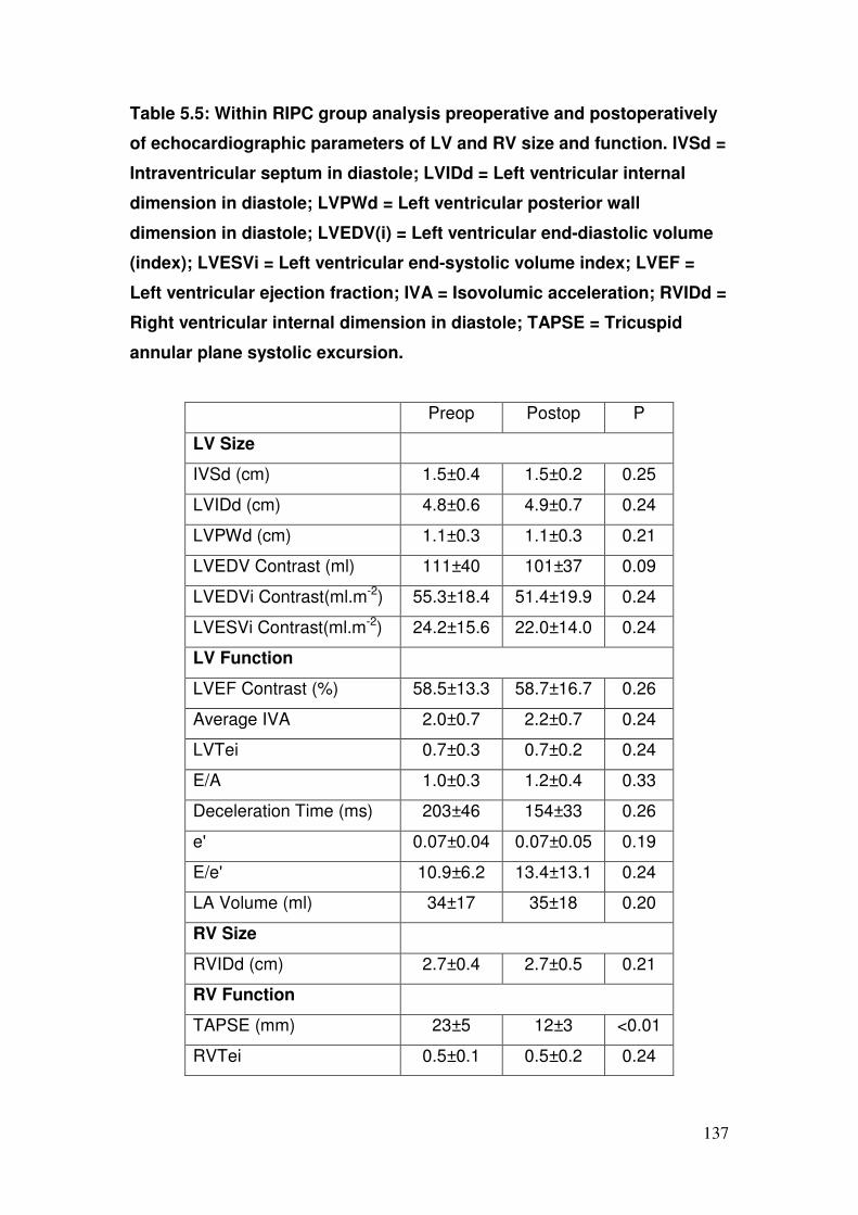
137
Table 5.5: Within RIPC group analysis preoperative and postoperatively
of echocardiographic parameters of LV and RV size and function. IVSd =
Intraventricular septum in diastole; LVIDd = Left ventricular internal
dimension in diastole; LVPWd = Left ventricular posterior wall
dimension in diastole; LVEDV(i) = Left ventricular end-diastolic volume
(index); LVESVi = Left ventricular end-systolic volume index; LVEF =
Left ventricular ejection fraction; IVA = Isovolumic acceleration; RVIDd =
Right ventricular internal dimension in diastole; TAPSE = Tricuspid
annular plane systolic excursion.
Preop Postop P
LV Size
IVSd (cm) 1.5±0.4 1.5±0.2 0.25
LVIDd (cm) 4.8±0.6 4.9±0.7 0.24
LVPWd (cm) 1.1±0.3 1.1±0.3 0.21
LVEDV Contrast (ml) 111±40 101±37 0.09
LVEDVi Contrast(ml.m-2) 55.3±18.4 51.4±19.9 0.24
LVESVi Contrast(ml.m-2) 24.2±15.6 22.0±14.0 0.24
LV Function
LVEF Contrast (%) 58.5±13.3 58.7±16.7 0.26
Average IVA 2.0±0.7 2.2±0.7 0.24
LVTei 0.7±0.3 0.7±0.2 0.24
E/A 1.0±0.3 1.2±0.4 0.33
Deceleration Time (ms) 203±46 154±33 0.26
e' 0.07±0.04 0.07±0.05 0.19
E/e' 10.9±6.2 13.4±13.1 0.24
LA Volume (ml) 34±17 35±18 0.20
RV Size
RVIDd (cm) 2.7±0.4 2.7±0.5 0.21
RV Function
TAPSE (mm) 23±5 12±3 <0.01
RVTei 0.5±0.1 0.5±0.2 0.24
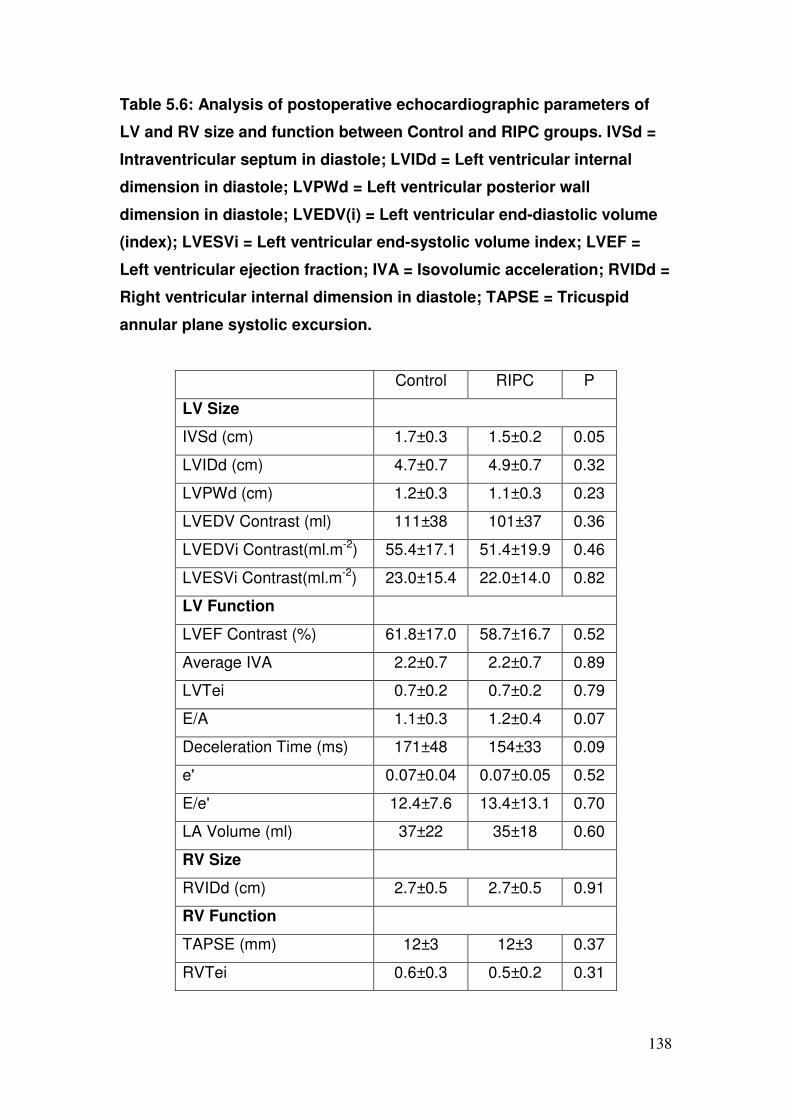
138
Table 5.6: Analysis of postoperative echocardiographic parameters of
LV and RV size and function between Control and RIPC groups. IVSd =
Intraventricular septum in diastole; LVIDd = Left ventricular internal
dimension in diastole; LVPWd = Left ventricular posterior wall
dimension in diastole; LVEDV(i) = Left ventricular end-diastolic volume
(index); LVESVi = Left ventricular end-systolic volume index; LVEF =
Left ventricular ejection fraction; IVA = Isovolumic acceleration; RVIDd =
Right ventricular internal dimension in diastole; TAPSE = Tricuspid
annular plane systolic excursion.
Control RIPC P
LV Size
IVSd (cm) 1.7±0.3 1.5±0.2 0.05
LVIDd (cm) 4.7±0.7 4.9±0.7 0.32
LVPWd (cm) 1.2±0.3 1.1±0.3 0.23
LVEDV Contrast (ml) 111±38 101±37 0.36
LVEDVi Contrast(ml.m-2) 55.4±17.1 51.4±19.9 0.46
LVESVi Contrast(ml.m-2) 23.0±15.4 22.0±14.0 0.82
LV Function
LVEF Contrast (%) 61.8±17.0 58.7±16.7 0.52
Average IVA 2.2±0.7 2.2±0.7 0.89
LVTei 0.7±0.2 0.7±0.2 0.79
E/A 1.1±0.3 1.2±0.4 0.07
Deceleration Time (ms) 171±48 154±33 0.09
e' 0.07±0.04 0.07±0.05 0.52
E/e' 12.4±7.6 13.4±13.1 0.70
LA Volume (ml) 37±22 35±18 0.60
RV Size
RVIDd (cm) 2.7±0.5 2.7±0.5 0.91
RV Function
TAPSE (mm) 12±3 12±3 0.37
RVTei 0.6±0.3 0.5±0.2 0.31

139
Table 5.7: Analysis of ∆(Postop-Preop) echocardiographic parameters of
LV and RV size and function between Control and RIPC groups. IVSd =
Intraventricular septum in diastole; LVIDd = Left ventricular internal
dimension in diastole; LVPWd = Left ventricular posterior wall
dimension in diastole; LVEDV(i) = Left ventricular end-diastolic volume
(index); LVESVi = Left ventricular end-systolic volume index; LVEF =
Left ventricular ejection fraction; IVA = Isovolumic acceleration; RVIDd =
Right ventricular internal dimension in diastole; TAPSE = Tricuspid
annular plane systolic excursion.
Control RIPC P
LV Size
∆IVSd (cm) 0.0±0.4 0.1±0.3 0.89
∆LVIDd (cm) 0.0±0.6 0.0±0.7 0.70
∆LVPWd (cm) 0.1±0.3 0.02±0.3 0.24
∆LVEDV Contrast (ml) -10±30 -14±39 0.69
∆LVEDVi Contrast(ml.m-2) -4.8±15.3 -4.2±16.2 0.90
∆LVESVi Contrast(ml.m-2) -0.4±9.3 -2.4±9.9 0.46
LV Function
∆LVEF Contrast (%) -0.4±16.7 0.0±15.0 0.64
∆Average IVA 0.1±0.7 0.2±0.7 0.74
∆LVTei 0.0±0.3 0.0±0.3 0.42
∆E/A 0.2±0.3 0.2±0.4 0.67
∆Deceleration Time (ms) -63±55 -48±50 0.25
∆e' 0.00±0.03 0.00±0.03 0.73
∆E/e' 0.4±9.0 1.3±9.9 0.36
∆LA Volume (ml) 4±13 -1±20 0.22
RV Size
∆RVIDd (cm) -0.1±0.7 0.0±0.5 0.63
RV Function
∆TAPSE (mm) -9±5 -11±6 0.08
∆RVTei -0.1±0.4 0.0±0.2 0.33

140
Table 5.8: Correlation of echo injury with 48 hour AUC cTnT release (all
patients)
Value Correlation
coefficient
p
∆LVEF(Contrast) -0.2±15.7 -0.13 0.36
∆LV Tei 0.0±0.3 -0.02 0.89
∆RV Tei -0.1±0.3 0.16 0.21
∆TAPSE -10±6 -0.09 0.53
∆Average IVA 0.1±0.7 0.17 0.17
Table 5.9: Correlation of echo injury with 0-6hrs inotrope incidence (all
patients)
Value Correlation
coefficient
p
∆LVEF(Contrast) -0.2±15.7 -0.08 0.49
∆LV Tei 0.0±0.3 -0.01 0.91
∆RV Tei -0.1±0.3 -0.01 0.90
∆TAPSE -10±6 -0.03 0.78
∆Average IVA 0.1±0.7 -0.13 0.20
Table 5.10: Correlation of echo injury with 6-12 hours inotrope incidence
(all patients)
Value Correlation
coefficient
p
∆LVEF(Contrast) -0.2±15.7 -0.03 0.82
∆LV Tei 0.0±0.3 0.10 0.32
∆RV Tei -0.1±0.3 0.08 0.43
∆TAPSE -10±6 0.07 0.55
∆Average IVA 0.1±0.7 -0.14 0.16

141
5.0 Discussion
This double-blind study is the first to demonstrate that no functional benefit on
left or right ventricular function assessed by transthoracic echocardiography
arises from RIPC during on-pump CABG.
Early evidence for a benefit from RIPC on LV function had been promising.
Kharbanda et al in a porcine model undergoing intermittent hindlimb
ischaemia observed a greater fall in LV function, measured invasively, during
ischaemia in controls. Unfortunately this was not maintained at reperfusion
and late effects were not studied perhaps suggesting that benefit in LV
function is short-lived (minutes) and long term functional benefit is unlikely.
In the present study markers of left heart function (LVEF with contrast; LV Tei)
remained comparable between groups when measured following surgery. The
primary endpoint selected was the load-independent IVA which remained
unchanged but there were no changes in LVEF using contrast or in global
measures of systolic and diastolic relaxation. Due to logistical reasons, the
study did not recruit the planned 146 patients (73 patients in each group) so
as to achieve the planned power to detect a difference in IVA ≥0.5SD (α 0.05,
1-β 0.85). Therefore, the lack of effect in this study could have been due to a
lack of statistical power arising from the failure to perform echocardiography in
all subjects recruited. With the achieved sample size of 72 patients however
(Control n=38; RIPC n=34), this study would still have the power to detect a
difference in IVA ≥0.675SD (α 0.05, 1-β 0.80). The observed difference in
∆IVA between the two groups was non-significant (p=0.74) and the 95%
confidence interval of the difference was small (-0.4 to 0.3). From this, it is

142
suggested that should a difference between the two groups exist as a result of
RIPC, this is likely to be small and offer minimal clinical benefit.
Another potential reason for the failure to demonstrate an effect could be the
degree of variability between pre-operative and post-operative measurements
on echocardiography. Image acquisition is dependent not only on observer
seniority but also patient morphological variation. Intra-patient variability exists
particularly in the post-operative setting where “poor acoustic windows” are
commonplace. Poor pain control, less patient compliance, post-operative fluid
and ventilation requirements result in a difficulty in obtaining a “good acoustic
window” postoperatively. This variability may be reflected in the wide standard
deviations for the LVEF values recorded, albeit similar in both groups. The
inter-study variability in our population was minimised as far as possible by
using a single blinded observer to analyse all studies.
Intraoperative tranoesophageal echocardiography was not performed and so
comment on any immediate, albeit short lived, benefit on contractility is not
possible. From a clinical perspective data on longer term functional
improvement is more relevant and it is clear RIPC failed to achieve this. This
failure should not be due to inadequate RIPC stimulus, since an identical
stimulus to that which in previous reports was associated with a >40%
reduction in cTnT release. The RIPC stimulus was administered in this study
within an appropriate time-frame and delivery was verified by observing
disappearance of the oximetry pulse signal. One possibility is that a greater
effect of RIPC would be documented in patients with abnormal LVEF prior to
CABG, in whom improvement following revascularisation of hibernating
segments might be more subject to variation in peri-operative conditions. This
143
study did not directly address the issue of whether RIPC could afford
additional benefit to improvement in LV function in those undergoing CABG
with impaired LV function and hibernation, and was restricted to patients with
normal EF at entry.
At present there is no data in the literature that has investigated the effects of
RIPC on RV function however the effect of IP has been studied. In a clinical
trial Wu et al showed that an IP stimulus (crossclamping of the aorta) with
antegrade and retrograde cold blood cardioplegia followed by a hot shot
improves cardiac index and RVEF 1 hour after declamping. Yet again though
this immediate advantage was not replicated at 6 hours nor on the first
postoperative day. Indeed multiple samples of cardiac troponin I and CKMB,
markers of myocardial necrosis, up to the second postoperative day were
similar between groups. Our study has corroborated the findings that
preconditioning does not preserve late RV function (TAPSE and RV Tei).
Within groups, markers of load-dependent (LVEF with contrast; LV Tei) and
load-independent (IVA) heart function was unchanged following surgery.
Conversely, reduced TAPSE but unchanged RV Tei in both groups would
suggest a reduction in RV longitudinal function that is compensated for
circumferentially as global function remains unchanged. Current myocardial
protection techniques would appear to be effective in the LV but are sub-
optimal in the RV. This reduced RV function was not related to peri-operative
myocardial necrosis or inotrope use. Indeed deleterious echocardiographic
change post-CABG is not predicted by cTnT release or inotrope requirement
up to 12 hours postoperatively.

144
6.0 Conclusion
In this double-blind study, RIPC has failed to afford benefit in LV or RV
contractile function 5-7 days following CABG in spite of manoeuvres designed
to overcome inherent difficulties: intravenous ultrasound contrast;
measurement of load-independent IVA. RV longitudinal function is reduced
post-CABG but overall global RV function is maintained.

145
CHAPTER 6 THE EFFECTS OF RIPC ON ARRYTHMIAS ON
PATIENTS UNDERGOING FIRST TIME CORONARY
ARTERY BYPASS SURGERY
1.0 Introduction
In a porcine model of remote ischaemic preconditioning (RIPC), short cycles
of repeated limb ischaemia during myocardial ischaemia have reduced
myocardial infarction by >20% and protected against malignant arrhythmia
during the reperfusion phase through a K(ATP) channel-dependent
mechanism [240]. Partial hindlimb occlusion has also delayed the
development of ischemia-induced ventricular arrhythmias induced by coronary
artery occlusion [241] in experimental models.
Although as yet no study of the effects of RIPC on arrhythmias in humans has
been reported, IP has been investigated. In one series preinfarction angina, a
clinical form of preconditioning, has been positively associated with protection
from the development of sustained ventricular tachycardia and ventricular
fibrillation (VT/VF) after reperfusion (by PCI) following STEMI. [242] VT/VF is
a significant determinant of long-term major adverse cardiac events.
Continuous Holter ECG monitoring of patients with variant angina has
demonstrated that preconditioning by transient ischemia induces protection
against ischemia-induced complex ventricular arrhythmias [243].
Ischaemic heart disease carries an increased risk of malignant VF and VT
and sudden death [244][245][246][247][248][249] and post-CABG both VF
and VT have been identified as potential life threatening complications. In a
RCT of 86 patients undergoing CABG only (three vessel disease) IP has been
146
shown to reduce postoperative VF (48.8% vs. 79.1%; p<0.01) and VT, the
mechanical ventilation period and need for inotropes [250]. IP has also been
observed to reduce the QT dispersion provoked by coronary artery occlusion
and reperfusion during coronary angioplasty.[251] Preconditioning with
adenosine, which has been implicated in the IP pathway, has been shown to
suppress the frequency of pre- and post-ischaemic tachyarrhythmia’s against
an ischaemia-reperfusion insult in human myocardium harvested from CABG
patients [252].
The timing of the conditioning stimulus does not appear to influence effect. In
a rat model of postconditioning ventricular arrhythmia after regional ischemia
was markedly attenuated [253].
However analyses of the effects of preconditioning on ischaemia and
reperfusion-induced rhythm disturbances have yielded controversy. In a
porcine model no changes were observed in the refractory duration,
ventricular vulnerability or defibrillation energy requirements up to 90 min after
ventricular ischaemic preconditioning [254].
In a prospectively randomised trial (n=35) Ascione et al observed antegrade
cold blood cardioplegia to be associated with less ischaemic stress and
myocardial injury than antegrade warm blood cardioplegia in hypertrophic
hearts of patients undergoing aortic valve surgery. Troponin I release and the
total incidence of postoperative arrhythmias was higher in the warm blood
group. As the evidence pointed towards myocardial injury being responsible
for the occurrence of supraventricular arrhythmias [178], this endpoint
following reperfusion may be an important index of myocardial protection.
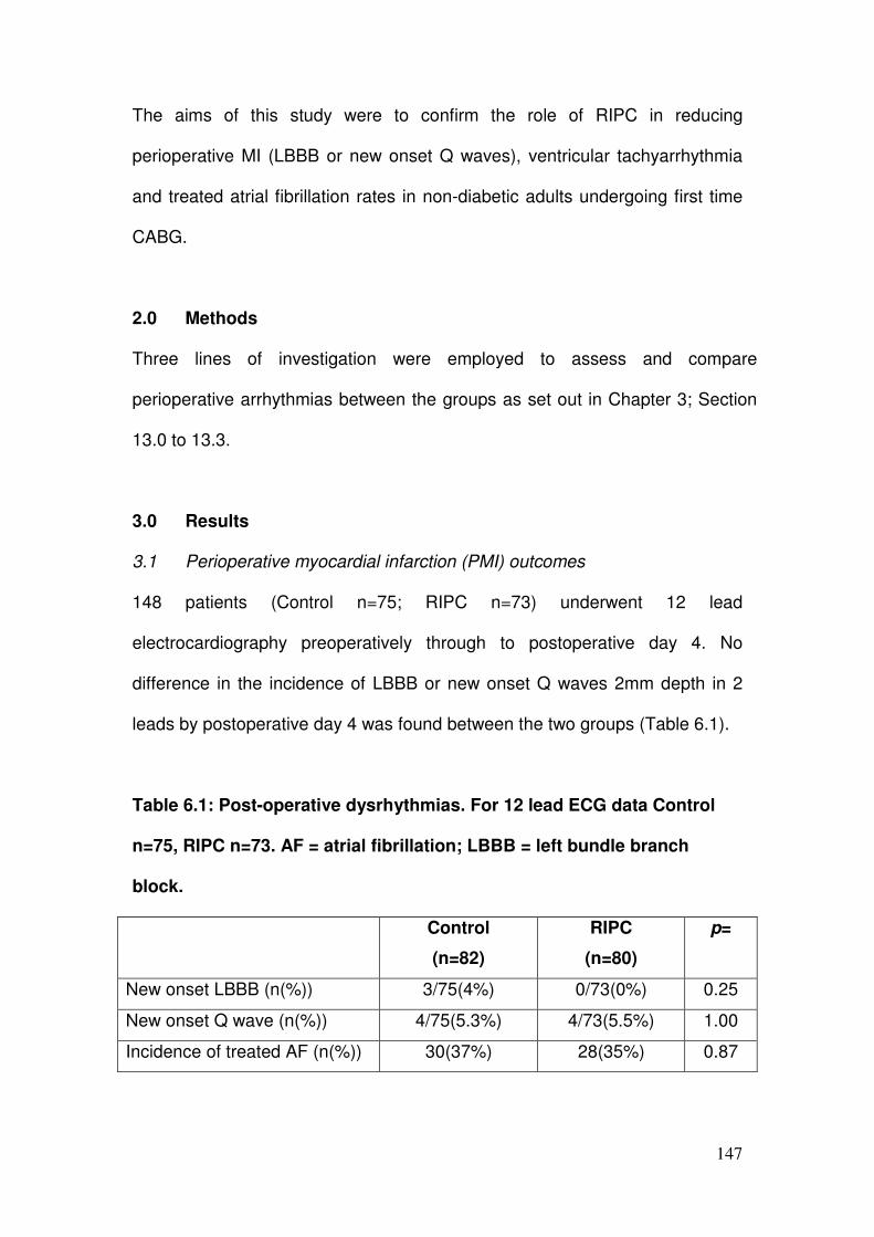
147
The aims of this study were to confirm the role of RIPC in reducing
perioperative MI (LBBB or new onset Q waves), ventricular tachyarrhythmia
and treated atrial fibrillation rates in non-diabetic adults undergoing first time
CABG.
2.0 Methods
Three lines of investigation were employed to assess and compare
perioperative arrhythmias between the groups as set out in Chapter 3; Section
13.0 to 13.3.
3.0 Results
3.1 Perioperative myocardial infarction (PMI) outcomes
148 patients (Control n=75; RIPC n=73) underwent 12 lead
electrocardiography preoperatively through to postoperative day 4. No
difference in the incidence of LBBB or new onset Q waves 2mm depth in 2
leads by postoperative day 4 was found between the two groups (Table 6.1).
Table 6.1: Post-operative dysrhythmias. For 12 lead ECG data Control
n=75, RIPC n=73. AF = atrial fibrillation; LBBB = left bundle branch
block.
Control
(n=82)
RIPC
(n=80)
p=
New onset LBBB (n(%)) 3/75(4%) 0/73(0%) 0.25
New onset Q wave (n(%)) 4/75(5.3%) 4/73(5.5%) 1.00
Incidence of treated AF (n(%)) 30(37%) 28(35%) 0.87

148
3.2 Continuous ECG recording Outcomes
Continuous ECG monitoring was performed in 146 patients (RIPC n=76 vs.
Control n=70). Initial heart rate at the start of monitoring in both groups was
comparable (RIPC 47(43-51) vs. Control 47(43-50) bpm). There was no
difference in the incidence of reperfusion ventricular fibrillation during the 10
minutes after AXC release (RIPC 15.8% vs. Control 11.4%; p=0.43). After the
reperfusion period there were no episodes of sustained ventricular
tachyarrhythmia requiring treatment. Brief non-sustained tachyarrhythmia’s
occurred in 48/76(63.2%) RIPC and 43/70(61.4%) Control patients in the first
post-operative 24 hours. There was no difference in tachyarrhythmia
incidence at different time points between groups (Figure 6.1).
Figure 6.1: Rates of detected non-sustained ventricular
tachyarrhythmia’s in each group: -12h to operation (p=0.489); operation
to reperfusion(p=0.180); post-reperfusion to 2 hours (p=0.124); 2-
24hours(p=0.179); 24-48 hours (p=0.353)
p=0.489
Incid
ence o
f ventr
icula
r arr
hyth
mia
s
-12hTo operation
Operation-reperfusion
Post-reperf. -2 hours
2 hours -24 hours
24 hours -48 hours
p=0.180
p=0.124
p=0.179
p=0.353
149
3.3 Atrial fibrillation - treated
The overall incidence of treated atrial fibrillation was 58(36%) (RIPC 28(35%)
vs. Control 30(37%); p=0.87) (Table 6.1).
4.0 Discussion
This double-blind study is the first to demonstrate that RIPC has no role in
reducing arrhythmia rates during on-pump CABG in non-diabetic adults.
No previous studies have investigated the anti-arrhythmic potential of RIPC in
humans but reports from experimental models had been promising. In a
porcine model four 5 minute cycles of lower limb ischaemia by tourniquet
inflation had reduced myocardial infarction by >20% and protected against
malignant arrhythmia. The present study, however, has failed to replicate this
advantage. All markers of arrhythmia: perioperative MI (12 lead ECG);
ventricular tachyarrhythmia incidence (continuous Holter monitor); treated AF
incidence have afforded no anti- or pro-arrhythmic potential in humans.
A difference in protocols may have prevented change. The porcine model
employed one more cycle of 5 minutes limb ischaemia than the present study
and selected the lower limb. It is possible that this may have conferred extra
benefit but seems unlikely as this study utilised an identical stimulus as
previous reports that demonstrated a >40% reduction in cTnT release on the
upper limb [107][145]. Whether response to RIPC is species-dependent is
possible and identical protocols would have to be used as part of a trial to
deduce the answer.
The benefit afforded by IP in humans has been documented. In a randomised
controlled trial by Wu et al postoperative ventricular tachyarrhythmia’s have

150
been reduced by >30% in patients undergoing on-pump CABG following two
cycles of cross-clamping the aorta for 2 minutes followed by 3 minutes of
reperfusion[250]. The present study however has been unable to replicate
these positive findings.
An explanation in the differences observed may lie in the different cardioplegic
protocol employed by Wu et al. Using both an antegrade and retrograde
delivery system for the cold blood cardioplegia and then completing the
myocardial protection with a further 3 minutes of warm cardioplegia (37oC)
prior to release of the aortic cross clamp may have offered additional
protective benefit. A further possibility for the difference between the IP and
RIPC patients may lie within the level of ischaemia generated by the
preconditioning stimuli. Crossclamping the ascending aorta may generate a
more substantial conditioning stimulus as compared to the 3 cycles of 5min
inflation/deflation peripherally used in the RIPC protocol. This in turn may be
sufficient to generate a functional change which the RIPC stimulus, although
able to activate pathways, is simply not strong enough to match and thus
bring about functional change.
A much lower overall incidence of reperfusion fibrillation and a lower overall
rate in the post-operative 48 hour period was found. This suggests important
differences in protection efficacy between studies but the lower arrhythmia
incidence in the current study makes it under-powered to exclude a lack of
protection by RIPC for this end-point.
Nonetheless at present the evidence is not in favour of any anti-arrhythmic
potential afforded by RIPC to humans. A randomised controlled trial using a

151
different endpoint with higher quantitation of arrhythmia incidence may be
more suitable in detecting change between treatment groups.
5.0 Conclusions
RIPC does not offer any anti-arrhythmic benefit in non-diabetic adult humans
undergoing first time coronary artery bypass surgery. It has not shown any
potential for pro-arrhythmia also.

152
CHAPTER 7 THE EFFECT OF RIPC ON RENAL OUTCOMES IN
PATIENTS UNDERGOING FIRST TIME CORONARY
ARTERY BYPASS SURGERY
1.0 Introduction
Acute kidney injury (AKI) is a common and prognostically important
complication of cardiac surgery. It is independently associated with in-hospital
mortality, even after adjustment for comorbid disease and other
complications, all degrees of AKI are associated with increased mortality and
other adverse outcomes. [255].
In a series of 817 patients undergoing on-pump CABG, a discrete alteration in
renal function (AKI) defined as an increase of 0.3 mg.dL-1 or 50% in baseline
creatinine was found in 52% of patients and independently predicted death
within 30 days (p<0.01). These patients were observed to have higher 30 day
mortality (12.6% vs. 1.4%; p<0.01), longer intensive care unit (ICU) stay
(median 2(2-3) vs. 3(2-5) days; p<0.01) and a larger proportion of patients
with prolonged ICU stay (>14 days) (14 vs. 2%; p<0.01) [256].
In a larger series of 2672 patients undergoing CABG, 7.9% patients
developed acute renal failure(ARF), defined as a rise in serum Cr >1.0mg/dl,
and 0.7% developed acute renal failure requiring some form of dialytic
therapy(ARF-D). ARF and ARF-D patients were observed to have increased
mortality (14% vs. 1%; p<0.01 and 28% vs. 1.5%; p<0.01 respectively).
Increased age, elevated preoperative Cr, duration of CPB, carotid artery bruit
and diabetes were associated with development of AFR and ARF-D by
multivariate analysis. [257] Other risk factors identified for AKI development in

153
a multicenter cohort of 3500 patients undergoing cardiac surgery were left
ventricular dysfunction, presence of IABP, urgent surgery, preoperative
anaemia, perioperative RBC transfusions and postoperative re-
exploration[255].
The aetiology of renal injury has been linked strongly to preoperative and
intraoperative factors, in those presenting for cardiac surgery, that result in
renal cellular ischaemia induced tubular epithelial and vascular endothelial
injury and activation [258][259].
Preoperatively, patients tend to have impaired autoregulation due to pre-
existing comorbidities, (advanced age, atherosclerosis, chronic hypertension
or chronic kidney disease), renal-injurious medication (NSAIDS, ACEI,
Angiotensin receptor blockers) or a pro-inflammatory state. Normal renal
perfusion is autoregulated such that the glomerular filtration rate is maintained
until the mean arterial blood pressure (MABP) falls below 80mmHg [259].
During surgery MABP is often at the lower limits of normal autoregulation and
especially during periods of haemodynamic instability these patients are
vulnerable to ischaemic renal injury.
Intraoperatively, proinflammatory events such as operative trauma, contact of
the blood components with the artificial surface of the CPB circuit, ischaemia-
reperfusion injury, and endotoxaemia [260][261[262] contribute to injury. The
generation of free haemoglobin and iron from haemolysis during CPB [260]
has also been linked.
Early experimental and human models have reported positively to the
application of RIPC in cardiovascular surgery. Temporary infrarenal aortic
occlusion in an animal model, during open thoracoabdominal aneurysm repair

154
has been shown to decrease IR renal injury caused by subphrenic aortic
cross-clamping [263] and patients undergoing elective open abdominal aortic
aneurysm repair demonstrate a 23% reduction in the incidence of renal
impairment with intermittent common iliac artery cross clamping[143].
More recently, in a RCT of patients undergoing endovascular repair (EVAR)
for abdominal aortic aneurysms (n=40), 18 patients undergoing sequential
lower limb ischaemia had a reduced increase in urinary markers of renal injury
(urinary retinol binding protein and urinary albumin:creatinine ratio(ACR) [264]
however no differences in the rates of renal impairment or major adverse
cardiac events were observed.
Reports of ischaemic preconditioning in experimental models have been
equally encouraging. Intermittent renal artery clamping (renal IP) has been
shown to ameliorate ischaemic renal injury as assessed by functional,
metabolic and morphological methods [265][266][267][268][269][270].
Attenuation of NF-kappa B activity, downregulation of IL-18 expression,
attenuation of NGAL and IKKbeta have been implicated in the renoprotective
pathways [159][270]. Ischemic postconditioning has also been shown to
decrease apoptosis and improve renal function, effects possibly related to
activation of Akt and ERK½ activation.[271]
In the assessment of renal injury both proteinuria and microalbuminuria have
an established role in diagnosis and management. The glomerular membrane
selectively allows passage of water and low molecular weight solutes into the
tubule and restricts passage of larger molecular weight plasma proteins,
based on a combination of molecular size, shape and charge. Despite the
protein-retaining properties of the glomerular membrane, some protein does

155
pass into the proximal tubular fluid. Only a very small proportion of plasma
proteins with a molecular weight >50 000 Da normally reaches the urine. This
is primarily due to retention of large molecular weight proteins by the
glomeruli, and tubular reabsorption of lower molecular weight proteins that
pass through the glomerular membrane more easily. Proteins such as
albumin which do reach the glomerular filtrate are reabsorbed in the proximal
tubules by a low capacity high affinity mechanism.
The National Kidney Foundation Guidelines recommends urine albumin as a
sensitive marker for chronic renal disease due to diabetes, glomerular disease
and hypertension. Urinary albumin:creatinine ratio (ACR) may also be used as
a sensitive and early marker of primary and secondary renal
disease[272][273][274]. A reduction in the 24 h urinary ACR by Walsh et al
following intermittent lower limb ischaemia (median 5 vs. 8.8; p=0.06) in
patients undergoing EVAR reconfirms the relevance of this test in a
cardiovascular setting [264].
The glomeruli filter larger amounts of low molecular weight proteins (<50 000
Daltons). Measurement of the urinary content of low molecular weight proteins
such as α1-microglobulin can be used as sensitive markers of proximal
tubular function because. Under normal conditions filtered low molecular
weight proteins are almost completely reabsorbed by the proximal tubule, but
even subtle tubular damage is associated with increased urinary
excretion[275].
The aims of this study were to confirm the role of RIPC in reducing markers of
renal injury (peak creatinine, significant increase in creatinine (>44 µmol.L-1),

156
∆-creatinine (day 0-4), 24h area under the curve (AUC) ACR, 24 h α1-
µglobulin) in non-diabetic adults undergoing first time CABG.
2.0 Methods
Changes in serum creatinine, urinary albumin:creatinine ratio, urinary α-1
microglobulin, and requirement for diuretics or haemofiltration were used as
outcome measures to assess the effect of renal protection by RIPC on peri-
operative renal injury in this double-blind, randomised controlled trial. Protocols
for assessment of renal outcomes are set out in Chapter 3; Section 14.0 to
14.4.2. Statistical analysis was performed as per Chapter 3; Section 17.3.
3.0 Results
Of 162 patients (89% males) 80 were randomized to treatment and 82 to
placebo. The groups were well matched for pre-operative renal function
(Serum creatinine: Control 96±16 vs. RIPC 98±16 µmol.L-1). The mean time
on bypass (96±22 vs. 100±23; p=0.25) mins and aortic cross-clamp times
(71±18 vs. 76±21; p=0.08) mins were not significantly different (Table 3.8).
Peak creatinine, day 4 creatinine levels and ∆-creatinine between days 0-4
were not different. Similar numbers of patients had increases >44 µmol.L-1.
Three RIPC (3.8%) and one (1.2%) control patient required temporary
dialysis. There was no difference in 24h AUC urine ACR or 24 hour urine α-
microglobulin levels (Table 7.1).

157
Table 7.1: Post-operative renal outcomes. For creatinine Control n=77,
RIPC n=75. ACR = albumin:creatinine ratio.
Control
(n=82)
RIPC
(n=80)
p=
Peak creatinine (µmol.L-1) 106(97-123) 106(88-123) 0.61
>44 µmol.L-1 increase in creatinine (n(%)) 8/77(10) 5/75(7) 0.56
∆- creatinine (day 0-4) (µmol.L-1) -0.88(-10.6-7.9) -2.6(-8.8-7.9) 0.67
24h AUC ACR 69(40-112) 58(32-85) 0.08
Pre-op. α1-µglobulin (mg.L-1) 8(4-16) 8(5-13) 0.75
24 h α1-µglobulin (mg.L-1) 51(38-71) 45(28-74) 0.42
Temporary dialysis (n(%)) 1(1) 3(4) 0.36
Intravenous frusemide infusion (n(%)) 6(7) 8(10) 0.59
4.0 Discussion
This double-blind study is the first to demonstrate that RIPC has no role in
reducing rates of acute renal injury following on-pump CABG in non-diabetic
adults.
No previous studies have investigated the renal protective effects of RIPC
following cardiac surgery. Following three relatively small experimental and
human studies: Lazaris et al (n=26); Walsh et al (n=40); Ali et al (n=82), the
evidence for a beneficial effect of RIPC on aortic aneursymal surgery
(including endovascular) has been encouraging.
However, by failing to reduce 24h AUC urinary ACR, RIPC has delivered no
detectable protection at the glomerular level from the insult of cardiac surgery.
There is also no suggestion of protection against damage to tubular
reabsorptive function as 24 h α1-microglobulin levels between the groups
were similar. A difference in protocol between the aneursymal studies and the
present study may offer a clue to the lack of the purported effect. In the rat

158
model of Lazaris et al a vascular clamp was applied on the infra-renal aorta
just above its bifurcation for 15 minutes and in Ali et al’s human model the
right iliac vessel was crossclamped for 10 minutes and then released and
applied to the left iliac vessel for 10 minutes. These invasive methods of
applying a remote ischaemic stimulus were both longer in duration and
arguably more likely to provide a significant ischaemic stimulus. Walsh et al
used two sequential periods of lower limb ischaemia using a tourniquet
inflated for 10 minutes on one leg and then repeated on the other leg.
Whether the three five minute cycles of non-invasive tourniquet cuff inflation
on an upper limb is just as likely to produce an equal strength or sufficient
strength stimulus to effect change is difficult to deduce. Certainly this study
utilised an identical stimulus as previous reports that demonstrated a >40%
reduction in cTnT release on the upper limb [107][145] and so evidence for
robustness of ischaemic stimulus exists. A large scale trial comparing the
effects of intermittent upper limb ischaemia (three 5 min cycles tourniquet
inflation/deflation) on similar aneursymal surgical procedures would be able to
clarify whether the shorter non-invasive stimulus is just as effective as the
reported longer noninvasive and invasive stimuli. Such a trial, if positive,
would also stimulate discussion on whether similar to reports of IP [133] RIPC
offers no additional benefit when associated with CPB regardless of the mode
of cardioprotection used, because CPB per se induces preconditioning.
Other surrogate markers of renal injury: peak serum creatinine concentration,
change in serum creatinine, incidence of intravenous frusemide requirement;
incidence of dialysis requirement also failed to support the improvement of
renal function by intermittent upper limb ischaemia (RIPC).

159
A sub-analysis of the incidence of those patients who suffered a more severe
acute kidney injury as defined by >44 µmol.L-1 increase in creatinine once
again demonstrated no benefit afforded in this group by RIPC (Control
8/77(10%) vs. RIPC 5/75(7%); p=0.56).
In spite of early suggestions that RIPC may offer benefit against IR injury of
the kidneys during coronary surgery this study has failed to offer evidence.
The morbidity and mortality from renal injury post-CABG remains a concern
for the surgical community and the search for a more effective treatment
strategy must continue.
5.0 Conclusion
In this double blind randomised controlled trial RIPC does not offer any renal
protection in non-diabetic adult humans undergoing first time coronary artery
bypass surgery.

160
CHAPTER 8 THE EFFECT OF RIPC ON LUNG OUTCOMES IN
PATIENTS UNDERGOING FIRST TIME CORONARY
ARTERY BYPASS SURGERY
1.0 Introduction
Acute lung injury, presenting as pulmonary oedema and pulmonary gas
exchange abnormalities such as postoperative hypoxaemia is common after
cardiac surgery. Pulmonary dysfunction is a frequent complication of CABG
and is associated with a longer duration of mechanical ventilation, difficulty
weaning the patient, prolonged hospitalization, major morbidity, mortality and
health care expenditure [276][277][278][279]. The pathophysiology reflects the
combined effects of general anaesthesia, surgical injury, median sternotomy
and CPB to produce hypoxia, atelectasis, pleural effusion and dysfunction of
the diaphragm [280].
CPB has been implicated in decreased oxygen and nutrient delivery to the
alveoli due to a number of factors: microemboli from the ascending aorta may
become dislodged during placement of cannula [281][282][283][284];
haemodilution by crystalloid fluids used to prime the pump reduces oxygen
carrying capacity[285][286]; hypothermia increases blood viscosity increasing
the risk of sludging in microvessels and impairing oxygen transfer from
erythrocytes to alveolar cells; nonpulsatile flow, compared to physiologic
pulsatile flow, decreases microvascular blood flow and increases the
expression of inflammatory mediators in blood and vascular cells
[287][288][289].
161
However CPB itself may not be the major contributor to the development of
post-operative pulmonary dysfunction. In a series of 175 patients undergoing
CABG with CPB (n=150) and without CPB (n=25) changes in postoperative
gas exchange was similar between groups. These findings were in spite of
higher levels of markers of systemic inflammatory response in CPB patients
[280]. These findings have been replicated in other studies [276]
In a series of 16,847 patients undergoing on-pump CABG, valve, or CABG-
valve surgery patients receiving transfusion (red blood cells; fresh frozen
plasma) had more risk-adjusted pulmonary complications: respiratory distress,
respiratory failure, longer intubation times, acute respiratory distress
syndrome and reintubation [290].
Early evidence in experimental models has been reassuring for the existence
of lung IP. Clamping of the lung hilum (lung IP) in a rabbit model [291] has
improved markers of lung ischaemia-reperfusion injury. Activation of
adenosine A(1) receptors, alteration of the pulmonary vasodilator response to
histamine, sarcolemmal K(ATP) channels, preservation of endothelial-
dependent vasodilator responses and restoration of surfactant properties
have been identified in the mediation of the protective properties of lung IP on
ischemia-reperfusion injury[292][293][294]. Postconditioning has also been
shown to attenuate lung ischemia-reperfusion injury through upregulating the
protein expression of HO-1 that leads to reduced post-ischemic oxidative
damage[295].
In small scale human models RIPC has been shown to benefit the lungs
whether the upper or lower limbs undergo intermittent ischaemia. In a
prospective RCT of infants undergoing repair of ventricular septal defects

162
those randomised to 3 cycles of 5 min cuff inflation of the left upper limb at 24
h and 1 h before surgery (RIPC n=30; Control n=30) were found to have
higher calculated static lung compliance, dynamic lung compliance and lower
respiratory index than controls postoperatively [296]. An attenuation of the
systemic inflammatory response syndrome and increase in systemic tolerance
to ischaemia-reperfusion injury enhanced lung preservation. A RCT in
children undergoing repair of congenital heart defects with four 5 minute
cycles of tourniquet cuff inflation to the lower limb (RIPC n=17; Control n=20)
resulted in lower airway resistance 6 hours postoperatively [118]. Cardiac
ischemic preconditioning has also been shown to improve lung preservation in
patients having valve replacement, whereby reduction in the accumulation of
polymorphonuclear leukocytes in lung tissue and the formation of oxygen free
radicals has been suggested[297].
Small study experimental models have also produced encouraging results. In
a sheep model of repeated coronary artery occlusion and reperfusion
(mimicking multi-vessel off-pump CABG), three episodes of five-minute
occlusion of the external iliac artery (RIPC n=9; Control n=5) improved gas
exchange (PaO2; PaO2:FiO2 ratio) and preserved pulmonary vascular
resistance [298]. A porcine model (n=18) where the external iliac arteries were
isolated bilaterally and simultaneously clamped for three cycles of 5 minutes
before a prolonged ischemic period reduced markers of systemic
inflammation and acute lung injury [299]. Furthermore lower limb ischaemia
have been observed to reduce lipid peroxidation and lung injury [300][301]
also when CPB has been used[196].

163
The ratio of the partial pressure of oxygen in arterial blood (PaO2) to the
inspired oxygen fraction (FiO2) has been utilised as a tool for quantification of
the degree of abnormality in pulmonary gas exchange[280][302]. The ratio
has been used in experimental models to quantify pulmonary gas exchange
before and after therapeutic intervention [303][304][305] and in clinical models
has aided in the classification of patients’ pulmonary gas exchange status,
including the definitions of acute lung injury (ALI) (27kPa≤PaO2:FiO2<40kPa)
and of adult respiratory distress syndrome (ARDS) (PaO2:FiO2<27kPa)
[306][307]. In the experimental model mimicking CABG by Xia et al RIPC
increased PaO2 and PaO2:FiO2 reconfirming the relevance of this test in the
setting of RIPC. Time to extubation according to standardised departmental
protocols also allows comparative analysis of improvement from lung injury.
Coronary artery bypass grafting (CABG) is established as a highly effective
treatment strategy in improving life expectancy and quality of life of patients
with ischaemic heart disease[215] but its success has been tarnished by a
‘postperfusion syndrome’ implicated in dysfunction of the kidneys, lungs, heart
and clotting system frequently found in patients undergoing extracorporeal
circulation[308]. The activation of the clotting, kallikrein, complement systems
and neutrophils and monocytes together with other blood elements produce
the vasoactive and cytotoxic substances that cause myocardial and
respiratory injury[309].
The aims of this study were to confirm the role of RIPC in reducing markers of
lung injury (6h and 12h PaO2:FiO2 ratio; intubation time) in non-diabetic adults
undergoing first time CABG.

164
2.0 Methods
As Positive End Expiratory Pressure (PEEP) has been shown to increase
right and left ventricular stroke volume variation both during open and closed
chest conditions in addition to reducing right ventricular end-diastolic volume
indicating a preload reductive effect, [175] its use in ventilator management
was standardised to 5cm H2O. Changes in arterial blood partial pressure of
oxygen (PaO2) / fractional inspired oxygen (FiO2) ratio and intubation times
were used as outcome measures as set out in Chapter 3; Section 15.0 to
15.4. Statistical analysis was performed as per Chapter 3; Section 17.3.
3.0 Results
Of 162 patients (89% males) 80 were randomized to treatment and 82 to
placebo. The groups were well matched for incidence of Forced expiratory
volume in one second (FEV1) / Forced vital capacity (FVC) ratio <0.7 (RIPC
12/80(15%) vs. Control 13/82(16%); p=0.51), preoperative PaO2:FiO2 ratio
(RIPC 55(48-63) vs. Control 53(47-61); p=0.31) and smoking history (p=0.15);
current smokers (RIPC 12/80(15%) vs. Control 7/82(9%)), ex-smokers (RIPC
39/80(49%) vs. 52/82(63%)), and never smoked (RIPC 29/80(36%) vs.
Control 23/82(28%)). Mean bypass times (RIPC 100±23 vs. Control 96±22;
p=0.25)mins and aortic cross-clamp times (RIPC 76±21 vs. Control 71±18;
p=0.08)mins were not significantly different (Table 3.8). Postoperatively times
to extubation were not different (RIPC 895(675-1180) vs. 938(766-1403)min;
p=0.28) and PaO2:FiO2 ratios at 6h (RIPC 37(28-45) vs. 36(29-43); p=0.85)
and 12h (RIPC 35(28-41) vs. 33(26-43); p=0.56) were also comparable
(Tables 8.1). Incidence of high risk lung status (preop FEV1:FVC ratio <0.7

165
and/or current smokers) was comparable and no difference postoperatively in
6h (RIPC 36(32-43) vs. Control 30(23-38); p=0.70), 12 h (RIPC 35(27-40) vs.
Control (32(23-40); p=0.50) PaO2:FiO2 ratios and time to extubation (RIPC
900(713-1940) vs. Control (838(574-1112)mins; p=0.29) was observed.
Table 8.1: Post-operative lung outcomes. LOS = length of stay.
Control
(n=82)
RIPC
(n=80)
p=
Extubation time (min) 937(766-1402) 895(675-1180) 0.28
ITU LOS (days) 3(2-4) 3(2-5) 0.29
Hospital LOS (days) 8(6-11) 8(6-12) 0.39
6h PaO2:FiO2 ratio 36(29-43) 37(28-45) 0.85
12h PaO2:FiO2 ratio 33(26-43) 35(28-41) 0.56
4.0 Discussion
This double-blind study is the first to demonstrate that RIPC has no role in
reducing rates of acute lung injury following on-pump CABG in non-diabetic
adults.
No previous studies have investigated the pulmonary protective effects of
RIPC following CABG in humans. In three relatively small human studies,
Wenwu et al (n=60), Cheung et al (n=47) and Li et al (n=40) presented
assuring evidence for the beneficial effect of RIPC in cardiac surgical
procedures. However in this study, RIPC did not improve lung preservation as
assessed by 6 and 12 hour PaO2:FiO2 ratio and time to extubation.
Small sample size, age of population, differences in remote stimulus delivery
and cardioplegia protocols may help explain why RIPC did not produce effect.

166
All three human studies investigated a considerably younger population group
than the present study (median 65 years): Wenwu et al (mean 5 months);
Cheung et al (mean 1 year); Li et al (mean 36 years). Whether the lung is less
likely to respond to RIPC with age would be suggested from the evidence but
could only be truly answered with trials using similar protocols in older age
groups. Certainly this would be less practical in the case of repairs of
congenital defects but may be more so in the case of valve replacements.
Perhaps more significantly a case for the difference in findings attributable to
strength of ischaemic stimulus used could be presented. Wenwu et al used 3
cycles of 5 min cuff inflation of the left upper limb twice (at 24 h and 1 h)
before surgery, Cheung et al employed an extra 5 minute cycle of lower limb
cuff inflation than the three cycles in the present study and Li et al executed
two 3 minute cycles of cross-clamping the aorta. Both non-invasive protocols
were longer in total duration and the invasive stimulus although shorter
arguably produced a more powerful ischaemic stimulus than the present
study. At present no test exists to ascertain the ‘strength’ of a stimulus but
certainly this study utilised an identical stimulus as previous reports that
demonstrated a >40% reduction in cTnT release [107][145] and so evidence
for robustness of ischaemic stimulus exists.
All three positive human studies were conducted on CPB. Cardioplegia
administered may have played an integral part of the purported benefit of
RIPC seen but unfortunately lack of information on protocols makes comment
difficult. Wenwu et al does not qualify what ‘cardioplegic arrest’ was used,
Cheung et al does not expand on ‘blood cardioplegia’ used. Li et al uses a
similar ‘intermittent cold blood (40C) cardioplegic arrest’ in valve replacements

167
as the antegrade cold blood (40C) cardioplegic arrest used in the present
coronary surgery but operative details are limited.
Experimental models also have observed improvement in markers of lung
function. Both Xia et al (n=14) and Harkin et al (n=18) occluded the external
iliac arteries for three 5 minute cycles unilaterally and bilaterally respectively.
Again these invasive protocols may produce stimuli that the present study is
not able to match. However, like repeated cross-clamping of the aorta, it is
unlikely that these invasive procedures will be taken up by the cardiothoracic
surgical community due to the potential for atheroembolism.
A sub-analysis did not observe RIPC to improve lung preservation (6h and
12h PaO2:FiO2 ratio; time to extubation) in those considered as high risk lung
patients (preoperative FEV1:FVC ratio <0.7 and/or current smokers.
In spite of early suggestions that RIPC may offer benefit against IR injury of
the lungs during coronary surgery this study has failed to offer evidence. The
morbidity and mortality from lung injury post-CABG remains a challenge for
the cardiothoracic fraternity and the search for a more effective treatment
strategy must continue.
5.0 Conclusion
In this double blind randomised controlled trial RIPC does not offer any lung
protection in non-diabetic adult humans undergoing first time coronary artery
bypass surgery.

168
CHAPTER 9 PILOT STUDY INTO THE EFFECTS OF RIPC ON
GENES AND PATHWAYS IN PATIENTS
UNDERGOING FIRST TIME CORONARY ARTERY
BYPASS SURGERY
1.0 Introduction
Changes in gene expression and signal transduction pathways have been
reported to be involved in ischaemic conditioning mediated myocardial
protection.
In experimental models RIPC has been shown to modify myocardial gene
expression by upregulating cardioprotective genes and suppressing
proinflammatory genes potentially involved in the pathogenesis of ischemia-
reperfusion injury [311]. Genes encoding key proteins involved in cytokine
synthesis, leucocyte chemotaxis, adhesion and migration, exocytosis, innate
immunity signalling pathways, and apoptosis have been shown to be
suppressed within 15 mins of the RIPC stimulus and these findings are more
pronounced after 24 hours (second window)[194]. In one model, 24 hours
following RIPC, the expression of genes known to be associated with the
stress response, redox regulation, growth and metabolism, DNA repair and
other cellular functions was increased [312].
Hypoxic conditioning of atrial trabeculae harvested from patients undergoing
cardiac surgery has been observed to protect myocardium following
prolonged ischaemia-reperfusion. Protection has been shown to be
dependent on activation of the Reperfusion Injury Salvage Kinase (RISK)
pathway [313].
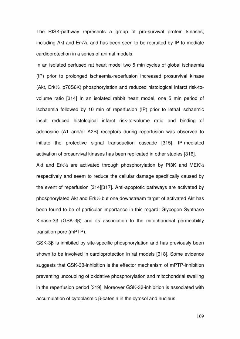
169
The RISK-pathway represents a group of pro-survival protein kinases,
including Akt and Erk½, and has been seen to be recruited by IP to mediate
cardioprotection in a series of animal models.
In an isolated perfused rat heart model two 5 min cycles of global ischaemia
(IP) prior to prolonged ischaemia-reperfusion increased prosurvival kinase
(Akt, Erk½, p70S6K) phosphorylation and reduced histological infarct risk-to-
volume ratio [314] In an isolated rabbit heart model, one 5 min period of
ischaemia followed by 10 min of reperfusion (IP) prior to lethal ischaemic
insult reduced histological infarct risk-to-volume ratio and binding of
adenosine (A1 and/or A2B) receptors during reperfusion was observed to
initiate the protective signal transduction cascade [315]. IP-mediated
activation of prosurvival kinases has been replicated in other studies [316].
Akt and Erk½ are activated through phosphorylation by PI3K and MEK½
respectively and seem to reduce the cellular damage specifically caused by
the event of reperfusion [314][317]. Anti-apoptotic pathways are activated by
phosphorylated Akt and Erk½ but one downstream target of activated Akt has
been found to be of particular importance in this regard: Glycogen Synthase
Kinase-3β (GSK-3β) and its association to the mitochondrial permeability
transition pore (mPTP).
GSK-3β is inhibited by site-specific phosphorylation and has previously been
shown to be involved in cardioprotection in rat models [318]. Some evidence
suggests that GSK-3β-inhibition is the effector mechanism of mPTP-inhibition
preventing uncoupling of oxidative phosphorylation and mitochondrial swelling
in the reperfusion period [319]. Moreover GSK-3β-inhibition is associated with
accumulation of cytoplasmic β-catenin in the cytosol and nucleus.

170
Cytoplasmic β-catenin is a transcriptional regulator of several genes involved
in cell survival and proliferation [320][321] and has been implicated in the
process of preconditioning and healing after myocardial infarction (MI).
Adenovirus mediated gene transfer of β-catenin in a rat model has been
shown to decrease apoptosis in cardiomyocytes and cardiac fibroblasts. It has
been suggested that β-catenin plays an important role in the healing process
after MI by promoting survival and cell cycle in cardiomyocytes and cardiac
fibroblasts with its differentiation into myofibroblasts [322].
Activation of PI3K-Akt, a part of the Risk-pathway, inhibits GSK-3β through
site-specific phosphorylation. GSK-3β inactivation allows β-catenin
accumulation and inhibits opening of the mPTP. Together with Erk½
activation these mechanisms seem to underlie the cytoprotective effects of
preconditioning.
Thus the PI3K/AKT/GSK-3 β/β-catenin cell survival pathway has been
proposed as key in conditioning pathways (Figure 9.1).
Invasive and non-invasive models of RIPC in rats have demonstrated nitric
oxide [323], free radical pathways, heat shock proteins, antioxidant enzymes
[324] and protein kinase C to be involved in these protective pathways [325].
The aim of this pilot study was to generate hypotheses: RIPC exerts its
cardioprotective effect through activation of the PI3K/AKT/GSK-3 β/β-catenin
cell survival pathway in myocardial specimens of non-diabetic adults
undergoing first time CABG.

171
Figure 9.1: Activation of PI3K-Akt, a part of the Risk-pathway, inhibits
GSK-3β through site-specific phosphorylation. GSK-3β inactivation
allows β-catenin accumulation and inhibits opening of the mitochondrial
Permeability Transition Pore (mPTP). Together with Erk½ activation
these mechanisms underlie the cytoprotective effects of
preconditioning.
2.0 Methods
Assessment of acquired myocardial specimens followed methodology set out in
Chapter 3; Section 16.0 to 16.2.
3.0 Results
Adult non-diabetic patients for CABG were recruited into a trial. All patients
had three myocardial biopsies harvested intraoperatively (LV1, LV2, LV3). Of
the 162 patients recruited to the trial 80 were randomised to RIPC and 82 to
Control groups. From the Control arm of the trial 6 ‘post-reperfusion’ biopsy
samples were randomly selected and from the RIPC arm of the trial 7 ‘post-

172
reperfusion’ biopsy samples were randomly selected and analysed (Control
n=6; RIPC n=7). RIPC increased expression of p–Akt (p=0.05; Figure 9.2) and
pan-Akt (p<0.01; Figure 9.3). Total GSK-3β was increased by RIPC (p<0.01;
Figure 9.4) but expression of p-GSK-3β was comparable between groups
(p=0.95; Figure 9.5). β-catenin was significantly increased by RIPC (p<0.01)
(Figure 9.6).

173
Figure 9.2: Relative expression of p-Akt between groups (p=0.05).
Figure 9.3: Relative expression of PAN-Akt between groups (p<0.01).
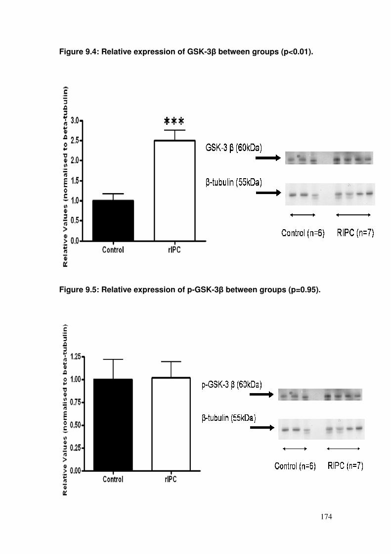
174
Figure 9.4: Relative expression of GSK-3β between groups (p<0.01).
Figure 9.5: Relative expression of p-GSK-3β between groups (p=0.95).

175
Figure 9.6: Relative expression of β-catenin between groups (p<0.01).
4.0 Discussion
The data presented from this small mechanistic sub-study is the first to
demonstrate that RIPC increases myocardial expression of the β-catenin
pathway in humans undergoing CABG.
The identification of a significant increase in the expression of p-Akt, pan-Akt,
total GSK-3β and β-catenin represents an important step in understanding the
cell biology of cardioprotection and the development of clinically viable
therapeutic strategies for reducing IR injury.
The Yellon group observed an increase in the activation of the RISK pathway
in atrium, harvested from patients undergoing cardiac surgery (CABG and/or
valve replacement), subjected to postconditioning protocols. The results in the

176
present study add support to these findings. It appears that the heart globally
(atrium and left ventricle) responds to ischaemic stimuli. As pre- and post-
ischaemic protocols have been successful the timing of stimulus delivery in a
clinical setting offers flexibility and thus could be selected on practicality. A
larger scale study to compare the effects of timing of conditioning stimulus on
magnitude of effect is warranted.
Haunseloy et al, Solenkova et al and Lecour et al observed increased
activation of the prosurvival kinases in experimental IP models and correlated
this with reductions in histological infarct risk-to-volume ratio. Although the
present study can conclude that a remote ischaemic stimulus has been
administered unlike other studies it has not correlated this with a functional
change. In this study cardiac troponin release, a marker of myocardial
necrosis, was unaltered between groups but with the arrival of advanced
imaging techniques whether other endpoints such as infarct risk-to-volume
ratio may show change should be investigated.
The results are in general concordance with previous work showing up-
regulation and phosphorylation of constituents in the pathway by a
cytoprotective stimulus. However, it is rather puzzling that there was no
change in the phosphorylation status of GSK-3β and the reasons for this are
unclear. Indeed this would appear to be a key step in animal models of
preconditioning with GSK-3β inactivation allowing the preservation of β-
catenin. Our data would suggest therefore that the human pathway differs
from that in other mammalian models in this step.

177
Nevertheless this small scale study has shown three 5 min cycles of
intermittent upper limb ischaemia to activate known cardioprotective pathways
in humans following coronary bypass surgery.
5.0 Conclusion
In this small mechanistic study in patients randomised, RIPC increases
activation of cardioprotective pathways in the myocardium of non-diabetic
adult humans undergoing first time coronary artery bypass surgery.

178
CHAPTER 10 SUMMARY AND DIRECTIONS FOR FUTURE
RESEARCH
1.0 Introduction
Ischaemic preconditioning has been recognised as a major cardioprotective
phenomenon for many years [102][326]. Cycles of non-lethal ischaemia and
reperfusion applied to the heart prior to a potentially lethal ischaemic insult
have the capacity to reduce infarct size by >50%. More recently, it became
apparent that the protection generated by this classical form of direct
ischaemic preconditioning could be replicated when the non-lethal ischaemia
was applied to one segment of the heart and the lethal ischaemia applied to a
separate segment [108]. Thereafter, it became established that the same
protection could also occur even if the pre-conditioning ischaemic stimulus
was applied completely distant to the target organ requiring protection, i.e.
transient ischaemia of a remote organ or limb could still generate protection
for the organ being subsequently challenged by lethal ischaemia [327]. There
is now clinical evidence, suggesting that this remarkable remote ischaemic
preconditioning (RIPC) phenomenon may represent a simple, inexpensive,
easily applied method of increasing cardioprotection during an array of
interventional procedures that require a period of cardiac ischaemia to allow
repair or intervention. Moreover, as it is now recognised that such protection
may be achieved by starting the cyclical remote ischaemia and reperfusion
after the period of injurious cardiac ischaemia has commenced; so called
remote post- or peri-conditioning, the potential arises of enhancing protection
in other scenarios, including transplantation [328].

179
Several clinical reports of RIPC in cardiovascular surgery have now been
published. In abdominal aortic aneurysm surgery, RIPC, induced by unilateral
iliac artery clamping has reduced troponin release and renal injury [143]. In
children undergoing congenital heart defect repairs utilising cardiopulmonary
bypass, lower limb RIPC has been demonstrated to reduce troponin release
and inotrope requirements [118]. In adults undergoing coronary artery bypass
surgery (CABG), intermittent upper limb ischaemia has been followed by
reductions in post-operative release of lactate dehydrogenase [144] and
troponin T. In 57 patients, Haunseloy et al observed a >40% reduction in AUC
serum cTnT where each anastomosis was constructed with either intermittent
cross clamp fibrillation or cardioplegia [107]. Venugopal et al went on to
observe 45 patients undergoing CABG (±concomitant aortic valve
replacement) and antegrade±retrograde cold blood cardioplegia produce a
similar magnitude of effect [145].
2.0 Summary of effects of RIPC in patients undergoing CABG
2.1 Cardiac outcomes
Postoperative cardiac troponin is an independent predictor of in-hospital death
and high concentrations are associated with a cardiac cause of death and
major postoperative outcomes. Which troponin measurement; isolated values
at specific time points or area under the curve (AUC) release provides the
most prognostically important information is not yet known. Additionally,
whether troponin release in the first few hours after surgery reflects true
infarction or a change in sarcolemmal integrity or permeability has been
questioned. Previous studies suggested a large treatment effect; a

180
standardised difference of 0.8. The current study hypothesised that RIPC
would reduce 48 h AUC cTnT release by a standardised difference of 0.6 at
90% power and 5% alpha. This required a sample size of 120 subjects which
was increased by 33% to accommodate withdrawal or missing data points.
Sufficient patients with full data sets were recruited.
This double-blind study using an identical RIPC stimulus and cTnT assay has
not corroborated previous smaller single blind studies that found RIPC to
reduce troponin release following on-pump CABG. This absence of effect on
measurable myocardial injury (48h AUC cTnT; peak cTnT; 6h cTnT release)
is matched by a failure to demonstrate any advantage of RIPC in terms of
cardiac performance (serial cardiac indices; IABP usage) and inotrope and
vasoconstrictor requirement. To accommodate for changes in systemic
vascular resistance left ventricular stroke work indices was analysed and was
comparable between groups. Thus, there is no evidence to suggest the
benefit of a ‘first window’ of RIPC protection.
Post-CABG myocardial injury often manifests as a transient period of
reversible dysfunction, treatable by hemodynamic optimisation and temporary
inotropic support. The data has provided no evidence of protection afforded
by RIPC against this cardiac stunning phenomenon.
No subtle differences between the groups exist for known preconditioning
volatile anaesthetic agents (Enflurane or Sevoflurane) usage or AUC cTnT
when adjusted for calculated CrCl or elective/urgent cases which could bias
results.
In conclusion, RIPC in this study has failed to augment myocardial protection
in patients undergoing CABG.
181
2.2 Functional impact
During and following cardiac surgery there is considerable variability in
preload and afterload. Isovolumic acceleration (IVA) was selected as the
primary endpoint of LV contractile function due to its load-independent
qualities. Due to logistical reasons only 72 of the planned 146 patients were
recruited to this sub-study. With the achieved sample size this study would
still have the power to detect a difference in IVA ≥0.675SD (α 0.05, 1-β 0.80).
The observed difference in ∆IVA between the two groups was non-significant
and the 95% confidence interval of the difference was small (-0.4 to 0.3).
From this, it is suggested that should a difference between the two groups
exist as a result of RIPC, this is likely to be small and offer minimal clinical
benefit.
This double-blind study is the first to demonstrate that RIPC offers no long
term functional benefit on LV or RV function assessed by transthoracic
echocardiography during on-pump CABG.
Following surgery RIPC afforded no functional improvement in load-
independent (IVA) and load dependent (LVEF with contrast; LV Tei) markers
of systolic left heart function. Diastolic function (LV Tei; E/A; deceleration time;
E/e’) and RV contractile function (TAPSE; RV Tei) was not improved either.
Within groups, markers of LV function were unchanged following surgery.
Conversely, reduced TAPSE but unchanged RV Tei in both groups would
suggest a reduction in RV longitudinal function that is compensated for
circumferentially as global function remains unchanged. This reduced RV
function was not related to peri-operative myocardial necrosis or inotrope use.

182
Indeed deleterious echocardiographic change post-CABG is not predicted by
cTnT release or inotrope requirement up to 12 hours postoperatively.
In conclusion, following surgery a reduction in RV longitudinal function occurs
although global RV function is preserved. This is the first study to show that
RIPC fails to preserve late LV or RV contractile function.
2.3 Arrhythmias
This double-blind study is the first to demonstrate that RIPC has no role in
reducing arrhythmia rates during on-pump CABG in non-diabetic adults.
All markers of arrhythmia: perioperative MI by 12 lead ECG (new onset LBBB;
Q waves); ventricular tachyarrhythmia incidence by continuous Holter
monitoring; treated AF incidence have found no anti- or pro-arrhythmic
potential in humans.
Wu et al observed a benefit of >30% in postoperative ventricular
tachyarrhythmia’s with IP following CABG. The present study however has
been unable to replicate these positive findings. A much lower overall
incidence of reperfusion fibrillation and a lower overall rate in the post-
operative 48 hour period was found. The lower arrhythmia incidence in the
current study makes it under-powered to exclude a lack of protection by RIPC
for this end-point.
2.4 Renal outcomes
This double-blind study is the first to demonstrate that RIPC has no role in
reducing rates of acute renal injury following on-pump CABG in non-diabetic
adults.

183
No previous studies have investigated the renal protective effects of RIPC
following cardiac surgery. However three relatively small experimental and
human studies have shown a beneficial effect of RIPC on aortic aneursymal
surgery (including endovascular).
Intermittent upper limb ischaemia failed to offer any detectable benefit in
urinary 24h AUC ACR and so at the glomerular level no protection effect from
the insult of cardiac surgery was found. As urinary 24 h α1-microglobulin level
was comparable there is no suggestion of protection by RIPC against damage
to tubular reabsorptive function.
Other surrogate markers of renal injury: peak serum creatinine concentration,
change in serum creatinine, incidence of intravenous frusemide requirement;
incidence of dialysis requirement also failed to support the improvement of
renal function by RIPC.
A sub-analysis of the incidence of those patients who suffered a more severe
acute kidney injury as defined by >44 µmol.L-1 increase in creatinine once
again demonstrated no benefit afforded in this group by RIPC.
In spite of early suggestions that RIPC may offer benefit against IR injury of
the kidneys during on-pump coronary surgery this study has failed to offer
evidence.
2.5 Lung outcomes
This double-blind study is the first to demonstrate that RIPC has no role in
reducing rates of acute lung injury following on-pump CABG in non-diabetic
adults.

184
No previous studies have investigated the pulmonary protective effects of
RIPC following CABG in humans. Small human studies have presented
assuring evidence for the beneficial effect of RIPC in cardiac surgical
procedures in younger patients. However in this study, RIPC did not improve
lung preservation as assessed by 6 and 12 hour PaO2:FiO2 ratio and time to
extubation on the Intensive Care Unit following surgery.
RIPC does not offer benefit against IR injury of the lungs during on-pump
coronary surgery in non-diabetic adults.
2.6 Genes and Pathways
The data presented from this small mechanistic sub-study is the first to
demonstrate that RIPC increases myocardial expression of the β-catenin
pathway in humans undergoing CABG.
The identification of a significant increase in the expression of p-Akt, pan-Akt,
total GSK-3β and β-catenin represents an important step in understanding the
cell biology of cardioprotection and the development of clinically viable
therapeutic strategies for reducing IR injury.
Although studies have observed an increase in the activation of the RISK
pathway in atrium, harvested from patients undergoing cardiac surgery,
subjected to postconditioning protocols this study shows that the heart is
globally affected (left ventricular biopsy) 10 minutes after reperfusion from a
prolonged period of ischaemia when a preconditioning stimulus is used also.
The results are in general concordance with previous work showing up-
regulation and phosphorylation of constituents in the pathway by a
cytoprotective stimulus. However, it is rather puzzling that there was no

185
change in the phosphorylation status of GSK-3β and the reasons for this are
unclear. Indeed this would appear to be a key step in animal models of
preconditioning with GSK-3β inactivation allowing the preservation of β-
catenin. Our data would suggest therefore that the human pathway differs
from that in other mammalian models in this step.
This small scale mechanistic study has shown three 5 min cycles of
intermittent upper limb ischaemia activate known cardioprotective pathways in
humans following on-pump CABG in non-diabetic adults.
3.0 RIPC from promise to disappointment
3.1 Cardiac outcomes
This double-blind study using an identical RIPC stimulus and cTnT assay has
not corroborated previous smaller single blind studies that found RIPC to
reduce troponin release following on-pump CABG.
On initial review the design of Venugopal et al’s investigation into RIPC
effects on patients undergoing CABG using cold blood cardioplegic arrest is
identical to the present study. Closer examination however reveals fewer
similarities which may help explain the difference in results.
Firstly, blinding of treatment allocation was applied to patients and surgeons
only; anaesthetists (who administer agents capable of preconditioning or
affecting myocardial protection) and investigators were not blinded. Similar
proportions of patients received isoflurane or sevoflurane for anaesthetic
maintenance but dosages are not reported. As such volatile anaesthetic
agents may induce a dose dependent conditioning effect [146], a potential for
inadvertent bias arises. In the present study a scrupulous technique to blind

186
the surgeon, anaesthetist and investigators from the group allocation was
developed. The stimulus was administered within an appropriate time-frame
and delivery verified by observing disappearance of the oximetry pulse signal.
Isoflurane, a known pre-conditioning agent was specifically avoided.
Secondly, the Venugopal et al study was small (n=45) compared to the
current study (n=162) and contained only half of the estimated number of
patients to detect the initially anticipated difference in AUC cTnT of 15 µg.L-
1.72hrs (standard deviation 25 µg.L-1.72hrs) quoted in the statistical
methodology. Statistical significance was actually attained with a smaller
mean difference and sample size and this is attributable to the lower than
anticipated variance observed in the RIPC group. Thirdly, the study also
included patients requiring AVR; whether RIPC was effective in the CABG
alone patients is not reported. The larger number of combined AVR/CABG
cases contributed to a longer mean bypass time in the control group and
bypass time was an independent predictor of greater troponin release.
Despite this potentially confounding effect, an inter-group statistically
significant difference was maintained after correction for bypass time using a
generalised linear model. Fourthly, many of the important variables in the
study e.g. bypass time, cross-clamp time and AUC cTnT had unequal
variances yet were analysed parametrically. Lastly, although the drug history
is reported, whether potentially relevant medications e.g. atorvastatin,
potassium channel blockers [147][148] administered in the 24 hours pre-
operatively is not clear. Nevertheless, the effect of RIPC on troponin release
was large and the data encouraging.

187
Differences in other preoperative and intraoperative variables may offer clues
as to why no cardioprotection was observed in the present study.
Preoperatively, non-elective patients who had angina or an acute coronary
syndrome within 30-days of surgery were recruited. However, no patients had
experienced angina within 48 hours of surgery and no differences between
those admitted electively and more urgent cases was observed. A higher
usage of beta-blockers was continued until the morning of surgery.
Intraoperatively bypass and cross-clamp times were slightly longer due to a
single clamp technique. The incidence of volatile anaesthetic (Enflurane or
Sevoflurane) use and AUC cTnT adjusted for calculated creatinine clearance
(CrCl) was similar between treatment and control groups.
3.2 Functional outcomes
Early evidence for a benefit from RIPC on LV function had been promising.
Kharbanda et al in a porcine model undergoing intermittent hindlimb
ischaemia observed a greater fall in LV function during ischaemia in controls,
late effects were not studied. This improvement in contractile function was not
replicated in the present study five to seven days following CABG. Differences
in the designs of the studies: lower limb vs., upper limb ischaemia; invasive
vs. non-invasive measurement of LV function; four vs. three 5 min cycles, in
combination may have increased the likelihood of realisation of
cardioprotection in the Kharbanda et al study.
The lack of effect observed in the current study could have been due to a lack
of statistical power arising from the failure to perform echocardiography in all
subjects recruited. As mentioned above, with the achieved sample size of 72

188
patients this study would still have the power to detect a difference in IVA
≥0.675SD (α 0.05, 1-β 0.80). The observed difference in ∆IVA between the
two groups was non-significant and the 95% confidence interval of the
difference was small (-0.4 to 0.3). From this, it is suggested that should a
difference between the two groups exist as a result of RIPC, this is likely to be
small and offer minimal clinical benefit.
Another potential reason for the failure to demonstrate an effect could be the
degree of variability between pre-operative and post-operative measurements
on echocardiography. Image acquisition is dependent not only on observer
seniority but also patient morphological variation. Intra-patient variability exists
particularly in the post-operative setting where “poor acoustic windows” are
commonplace. Poor pain control, less patient compliance, post-operative fluid
and ventilation requirements result in a difficulty in obtaining a “good acoustic
window” postoperatively. This variability may be reflected in the wide standard
deviations for the LVEF values recorded, albeit similar in both groups. The
inter-study variability in our population was minimised as far as possible by
using a single blinded observer to analyse all studies.
3.3 Arrhythmias
No previous studies have investigated the anti-arrhythmic potential of RIPC in
humans but reports from experimental models had been encouraging. Again
whether the use of the lower limb and an extra cycle of stimulus confer extra
cardioprotective benefit and/or whether RIPC is species-dependent is difficult
to ascertain but may offer reasoning as to why the purported benefit was not
observed.

189
The present study did not replicate the >30% reduction in postoperative
ventricular tachyarrhythmia’s observed by Wu et al, in those receiving IP,
following CABG. A much lower overall incidence of reperfusion fibrillation and
a lower overall rate in the post-operative 48 hour period was found suggesting
important differences in protection efficacy between studies but the lower
arrhythmia incidence in the current study makes it under-powered to exclude
a lack of protection by RIPC for this end-point. Other differences between
studies: central, invasive vs. remote, non-invasive stimulus;
antegrade+retrograde vs. antegrade only cardioplegia; hot shot vs. no hot
shot may have advantaged the study by Wu et al to demonstrate a change.
3.4 Renal outcomes
Although no studies into the renal protective effects of RIPC following cardiac
surgery have been conducted, three small experimental and human studies
have presented evidence for a beneficial effect on aortic aneursymal surgery
(including endovascular). Lazaris et al and Ali et al utilised invasive clamping
of large blood vessels to deliver remote stimuli and Walsh et al although using
a non-invasive protocol did so to both lower limbs and for a longer duration
than the current study. Thus a difference in the ‘strength’ of remote stimulus
may have been able to produce change in these studies.
3.5 Lung outcomes
The current study has not replicated the benefit in lung preservation seen in
three small human studies receiving RIPC in cardiac surgical procedures.

190
Small sample size, age of population, differences in remote stimulus delivery
and cardioplegia protocols may help explain why RIPC did not produce effect.
An age-dependent response to RIPC cannot be ruled out as all three human
studies investigated a considerably younger population group than the
present study. The three studies used either non-invasive protocols that were
longer in duration or an invasive protocol for delivering remote stimuli possibly
providing extra stimulus to cause effect. Although a positive response was
seen with upper limb ischaemia in Wenwu et al’s study, the stimulus was
delivered twice at 24h and 1h before surgery again possibly offering a
synergistic benefit.
In this study PEEP was set to a standardised level of 5cm H2O. Although
PEEP improves arterial oxygenation, it can adversely affect systemic
haemodynamics, reducing venous return and cardiac output. These effects
are proportional to the PEEP level.[175][329] Any future work should address
this potential methodological limitation to ensure unbiased results.
Analysis of bronchoalveolar lavage has been shown to be a sensitive
technique for the diagnosis of acute lung injury in a wide spectrum of clinical
scenarios. [310][330][331][332] Although due to resource availability it was not
employed in this study it may offer useful information in future studies
investigating the efficacy of RIPC on lung preservation post cardiac surgery.
3.6 Genes and Pathways
The data presented from this small mechanistic sub-study is the first to
demonstrate that RIPC increases myocardial expression of the β-catenin
pathway in humans undergoing CABG. The remote stimulus of three 5 min

191
cycles to the upper limb has replicated findings from the Yellon group which
showed activation of the RISK pathway in myocardium subjected to
postconditioning following CABG. IP protocols in three studies have also
demonstrated activation of prosurvival kinases.
In conclusion, the remote stimulus delivered in the current study has been
confirmed to have occurred due to positive changes in the cardioprotective
pathways within the myocardium.
4.0 Future Direction
The activation of cardioprotective pathways within the myocardium by three 5
min cycles of upper limb ischaemia has concurred with previous studies for
the existence of the RIPC phenomenon. However, it is clear that the stimulus
was unable to produce functional beneficial change to the heart, kidneys or
lungs.
A closer look at the body of literature suggests that use of the lower limb and
extra cycles of stimulus may be able to increase the ‘strength’ of the stimulus
itself. Whether this would be able to effect a change in patients undergoing
CABG where other confounding variables such as CPB, cardioplegia and
volatile anaesthesia exist is difficult to predict. Invasive remote stimuli have
also delivered improvement in cardiac, renal and lung preservation but it is
unlikely this method will ever offer a practical solution.
In order to corroborate the findings of this study a further study is
recommended. A more definitive conditioning protocol should be incorporated.
Biochemical markers of preconditioning should be developed to differentiate
those that are preconditioned from those that are not. Population investigated

192
may be those that are higher risk of myocardial injury e.g. left ventricular
hypertrophy. The role of RIPC in transplantation surgery should also be
explored further.
5.0 Recommendation I: Trial
5.1 Aims
To assess whether four 5 min cycles of lower limb ischaemia at two time
points (24h and 1 h before surgery) improves myocardial, renal and lung
outcomes in non-diabetic adult patients undergoing on-pump multi-vessel
coronary artery surgery.
5.2 Design
A large scale single centre, prospective randomised, placebo intervention-
controlled trial. Patients, investigators, anaesthetists, surgeons and critical
care teams will all be blinded to group allocation. Subjects will be randomized
(1:1) to RIPC (or placebo) stimuli (4x lower limb (or dummy leg) 5 minute
cycles of cuff inflation/deflation) 24h and 1h prior to surgery. Anaesthesia,
perfusion, cardioplegia and surgical techniques will be standardized. Patients
will be stratified to Urgent and Elective CABG.
Primary endpoint: 48h AUC cTnT release
Cardiac secondary endpoints: MRI assessed infarct risk-to-volume ratio
6h and peak cTnT release
Perioperative MI (12 lead ECG)
Ventricular tachyarrhythmia’s (Holter)

193
Treated atrial fibrillation incidence
Serial cardiac indices
Serial left ventricular stroke work indices
Low cardiac output episodes
IABP usage
Inotrope and vasoconstrictor usage
Echocardiographic parameters
Renal secondary endpoints: Peak serum creatinine
>44 µmol.L-1 increase in creatinine
∆- creatinine (day 0-4)
Urinary 24h AUC ACR
Urinary 24 h α1-µglobulin
Temporary dialysis incidence
Intravenous frusemide infusion incidence
Lung secondary endpoints: 6h PaO2:FiO2 ratio
12h PaO2:FiO2 ratio
Extubation time
Other endpoints: Infection incidence
ITU length of stay
In-hospital length of stay
194
6.0 Recommendation II: Other future work
There is a need:
(i) to determine the efficacy of RIPC in promoting protection in other forms
of cardiac surgery
(ii) to ascertain whether changes in troponin release reflects true infarction
or a change in sarcolemmal integrity or permeability
(iii) to establish if changes are reflected in improved clinical outcomes and
that RIPC independently reduces risk
(iv) to test if urgent CABG or elective CABG patients are already
preconditioned
7.0 RIPC in Transplantation
The national early mortality for heart transplantation is 12.5% and >60% of
deaths are secondary to dysfunction of the donor heart and so transplantation
represents a specific challenge to a therapeutic role of RIPC. Theoretically,
RIPC could be used in the brainstem dead donor to generate
cardioprotection. However, although the pre-conditioning site would remain
innervated, central neural connection is lost. Moreover, in this circumstance,
reperfusion does not occur in the pre-conditioned environment unless the
stimulus is repeated in the recipient. This is indeed the case, a donor heart,
denervated at transplantation may still be protected from a post-implantation
ischaemic insult by a RIPC stimulus in the recipient [333]. However, whether
such conditioning could protect during the retrieval, transport and implant
period of transplantation is not clear.

195
8.0 Limitations
Despite its true double-blind status, the confirmation of stimulus delivery and
its objective clinical and biochemical end-points our study remains undersized
to detect smaller differences in a protective effect of RIPC and does not
explore effect in a higher risk population. Subtle differences in conditioning
status may occur in patients presenting with urgent coronary syndromes
status that preventing further conditioning in this group.
This study relies heavily on the use of troponin release as a marker of true
infarction but further research is required to confirm changes postoperatively
are not due to change in sarcolemmal integrity or permeability.
9.0 Conclusion
The prospective randomised placebo intervention-controlled trial of RIPC
described in this thesis has, like its predecessor cardiac same-organ IP, not
fulfilled the promise of a practically useful form of improved myocardial
protection. An identical stimulus to smaller positive studies was used with a
robust technique designed to ensure blinding of all team-members. The
stimulus of three 5 min cycles of upper limb ischaemia, administered within an
appropriate time-frame with verified delivery, has been shown to activate
cardioprotective mechanisms within the myocardium but has not been able to
mount a beneficial functional response. An absence of effect on measurable
myocardial injury is matched by a failure to demonstrate any advantage of
RIPC in terms of cardiac performance, inotrope requirement,
echocardiographic function, arrhythmia protection or renal and lung outcomes.

196
As the optimal RIPC stimulus remains elusive further lines of research has
been generated by this study: the significance of troponin release during on-
pump surgery, the conditioning status of those presenting with urgent
coronary syndromes, the efficacy of more cycles of lower limb ischaemia, the
role of RIPC in higher risk and transplantation surgery. The hope is to
establish a novel practical myocardial protective technique with proven benefit
in a range of clinical end-points.

197
REFERENCES
[1] Society of Cardiothoracic Surgeons of Great Britain and Ireland. 6th
National Adult Cardiac Surgical Database Report 2008.
[2] Aris A. Francisco Romero, the first heart surgeon. Ann Thorac Surg
1997 Sep;64(3):870-1.
[3] Williams DH: Stab wound of the heart, pericardium–Suture of the
pericardium–Recovery–Patient alive three years afterward. Med Record 1897;
1–8.
[4] Rehn L: On penetrating cardiac injuries and cardiac suturing. Arch Klin
Chir 1897; 55:315.
[5] Soreide K, Soreide J. Axel H Cappelen (1858-1919): The first suture of
a myocardial laceration from a cardiac stab wound. Journal of Trauma
2006;60(3):653-4
[6] Rehn L: Zur chirurgie des herzens und des herzbeutels. Arch Klin Chir
1907; 83:723; quoted from Beck CS: Wounds of the heart: the technic of
suture. Arch Surg 1926; 13:212.
[7] Blatchford JW 3rd. Ludwig Rehn: the first successful cardiorrhaphy. Ann
Thorac Surg 1985 May 39(5):492-5
[8] Carrel A: On the experimental surgery of the thoracic aorta and the
heart. Ann Surg 1910; 52:83.
[9] Beck CS: The development of a new blood supply to the heart by
operation. Ann Surg 1935; 102:805.
[10] Beck CS: Coronary sclerosis and angina pectoris: treatment by grafting
a new blood supply upon the myocardium. Surg Gynecol Obstet 1937;
64:270.

198
[11] Vineberg AM: Development of an anastomosis between the coronary
vessels and a transplanted internal mammary artery. Can Med Assoc J 1946;
55:117.
[12] Vineberg AM: Medical news section. JAMA 1975; 234:693.
[13] Longmire WP Jr, Cannon JA, Kattus AA: Direct-vision coronary
endarterectomy for angina pectoris. N Engl J Med 1958; 259:993.
[14] Konstantinov IE: Robert H. Goetz: the surgeon who performed the first
successful clinical coronary artery bypass operation. Ann Thorac Surg 2000;
69:1966.
[15] Garrett EH, Dennis EW, DeBakey ME: Aortocoronary bypass with
saphenous vein grafts: seven-year follow-up. JAMA 1973; 223:792.
[16] Demikhov VP: Experimental Transplantation of Vital Organs.
Authorized translation from the Russian by Basil Haigh. New York,
Consultants Bureau, 1962.
[17] Shumaker HB Jr: The Evolution of Cardiac Surgery. Indianapolis,
Indiana University Press, 1992; 141.
[18] Kolessov VI: Mammary artery–coronary artery anastomosis as a
method of treatment for angina pectoris. J Thorac Cardiovasc Surg 1967;
54:535.
[19] Green GE, Stertzer SH, Reppert EH: Coronary arterial bypass grafts.
Ann Thorac Surg 1968; 5:443.
[20] Bailey CP, Hirose T: Successful internal mammary–coronary arterial
anastomosis using a minivascular suturing technic. Int Surg 1968; 49:416.
[21] Favalaro RG: Saphenous vein autograft replacement of severe
segmental coronary artery occlusion. Ann Thorac Surg 1968; 5:334.
199
[22] Johnson WD, Flemma RJ, Lepley D Jr, Ellison EH: Extended treatment
of severe coronary artery disease: a total surgical approach. Ann Surg 1969;
171:460.
[23] Loop FD, Lytle BW, Cosgrove DM, et al: Influence of the internal-
mammary-artery graft on 10-year survival and other cardiac events. N Engl J
Med 1986; 314:1.
[24] Lillehei CW: Historical development of cardiopulmonary bypass.
Cardiopulmonary Bypass 1993; 1:26.
[25] McLean J: The discovery of heparin. Circulation 1959; 19:78.
[26] Letter from Jay McLean to Charles H. Best on November 14, 1940;
quoted from Best CH: Preparation of heparin and its use in the first clinical
case. Circulation 1959; 19:79.
[27] Howell WH, Holt E: Two new factors in blood coagulation: heparin and
pro-antithrombin. Am J Physiol 1918;47:328.
[28] Best C: Preparation of heparin and its use in the first clinical cases.
Circulation 1959; 19:81.
[29] Gibbon JH Jr: Artificial maintenance of circulation during experimental
occlusion of the pulmonary artery. Arch Surg 1937; 34:1105.
[30] Johnson SL: The History of Cardiac Surgery, 1896–1955. Baltimore,
Johns Hopkins Press, 1970; 148.
[31] Dennis C, Spreng DS, Nelson GE, et al: Development of a pump
oxygenator to replace the heart and lungs: an apparatus applicable to human
patients, and application to one case. Ann Surg 1951; 134:709.
[32] Dodrill FD, Hill E, Gerisch RA: Temporary mechanical substitute for the
left ventricle in man. JAMA 1952; 150:642.

200
[33] Dodrill FD, Hill E, Gerisch RA, Johnson A: Pulmonary valvuloplasty
under direct vision using the mechanical heart for a complete bypass of the
right heart in a patient with congenital pulmonary stenosis. J Thorac Surg
1953; 25:584.
[34] Johnson SL: The History of Cardiac Surgery, 1896–1955. Baltimore,
Johns Hopkins Press, 1970; 143.
[35] Gibbon JH Jr: Application of a mechanical heart and lung apparatus to
cardiac surgery. Minn Med 1954; 37:171.
[36] Lillehei CW, Cohen M, Warden HE, et al: The results of direct vision
closure of ventricular septal defects in eight patients by means of controlled
cross circulation. Surg Gynecol Obstet 1955; 101:446.
[37] Lillehei CW, Cohen M, Warden HE, et al: The direct vision intracardiac
correction of congenital anomalies by controlled cross circulation. Surgery
1955; 38:11.
[38] Kirklin JW, DuShane JW, Patrick RT, et al: Intracardiac surgery with
the aid of a mechanical pump-oxygenator system (Gibbon type): report of
eight cases. Mayo Clin Proc 1955; 30:201.
[39] Kirklin JW: The middle 1950s and C. Walton Lillehei. J Thorac
Cardiovasc Surg 1989; 98:822.
[40] Pasceri V, Patti G, Di Sciascio G. Prevention of myocardial damage
during coronary intervention. Cardiovasc Hematol Disord Drug Targets. 2006
Jun;6(2):77-83.
[41] Rizik DG, Klassen KJ, Dowler DA, Villegas BJ, Dixon SR. Promising
though not yet proven: emerging strategies to promote myocardial salvage.
Catheter Cardiovasc Interv. 2006 Oct;68(4):596-606.

201
[42] Carrel, A: Experimental operations on the orifices of the heart. Ann
Surg 1914; 40:1.
[43] Melrose DG, Dreyer B, Bentall MB, Baker JBE: Elective cardiac arrest.
Lancet 1955; 2:21.
[44] Gay WA Jr, Ebert PA: Functional metabolic, and morphologic effects of
potassium-induced cardioplegia. Surgery 1973; 74:284.
[45] Tyers GFO, Todd GJ, Niebauer IM, et al: The mechanism of
myocardial damage following potassium-induced (Melrose) cardioplegia.
Surgery 1978; 78:45.
[46] Kirsch U, Rodewald G, Kalmar P: Induced ischemic arrest. J Thorac
Cardiovasc Surg 1972; 63:121.
[47] Bretschneider HJ, Hubner G, Knoll D, et al: Myocardial resistance and
tolerance to ischemia: physiological and biochemical basis. J Cardiovasc Surg
1975; 16:241.
[48] Hearse DJ, Stewart DA, Braimbridge MV, et al: Cellular protection
during myocardial ischemia. Circulation 1976; 16:241.
[49] Follette DM, Mulder DG, Maloney JV, Buckberg GD: J Thorac
Cardiovasc Surg 1978; 78:604.
[50] Fallouh HB, Kentish JC, Chambers DJ. Targeting for cardioplegia:
arresting agents and their safety. Curr Opin Pharmacol. 2009 Apr;9(2):220-6.
[51] Manual of Perioperative Care in Adult Cardiac Surgery. Bojar RM
p207-214
[52] Beyesdorf F, Buckberg GD. Myocardial protection with blood
cardioplegia during valve operations. J Heart Valve DIs 1994;3:388-403.

202
[53] Barassi A, Merlini G, Finazzi S, Pallotti F, Mantovani V, Sala A, d'Eril
GM. Comparison of three strategies for myocardial protection during coronary
artery bypass graft surgery based on markers of cardiac damage. Clin
Biochem. 2005 Jun;38(6):504-8.
[54] Teoh KH, Christakis GT, Weisel RD et al. Accelerated myocardial
metabolic recovery with terminal warm blood cardioplegia. J Thorac
Cardiovasc Surg 1986;91:888-95.
[55] Radmehr H, Soleimani A, Tatari H, Salehi M. Does combined
antegrade-retrograde cardioplegia have any superiority over antegrade
cardioplegia? Heart Lung Circ. 2008 Dec;17(6):475-7.
[56] Guru V, Omura J, Alghamdi AA, Weisel R, Fremes SE. Is blood
superior to crystalloid cardioplegia? A meta-analysis of randomized clinical
trials. Circulation. 2006 Jul 4;114(1 Suppl):I331-8.
[57] Jacob S, Kallikourdis A, Sellke F, Dunning J. Is blood cardioplegia
superior to crystalloid cardioplegia? Interact Cardiovasc Thorac Surg. 2008
May;7(3):491-8.
[58] Fallouh HB, Chambers DJ. Is blood versus crystalloid cardioplegia
relevant? Significantly improved protection may require new cardioplegic
concepts! Interact Cardiovasc Thorac Surg. 2008 Dec;7(6):1162-3.
[59] Kouerinis IA, Palaiologou MM, Argiriou M, Dedeilias P, Apostolakis E,
Giannakopoulou A, Papadopoulos G, Tsilimingas N. "Directed" cardioplegia: a
new approach in myocardial protection in left main coronary artery disease.
Ann Thorac Surg. 2008 Dec;86(6):2005-7.

203
[60] Minguet G, Joris J, Lamy M. Preconditioning and protection against
ischaemia-reperfusion in non-cardiac organs: a place for volatile
anaesthetics? Eur J Anaesthesiol. 2007 Sep;24(9):733-45.
[61] Landoni G, Bignami E, Oliviero F, Zangrillo A. Halogenated
anaesthetics and cardiac protection in cardiac and non-cardiac anaesthesia.
Ann Card Anaesth. 2009 Jan-Jun;12(1):4-9.
[62] Landoni G, Fochi O, Torri G. Cardiac protection by volatile
anaesthetics: a review. Curr Vasc Pharmacol. 2008 Apr;6(2):108-11.
[63] Landoni G, Fochi O, Tritapepe L, Guarracino F, Belloni I, Bignami E,
Zangrillo A. Cardiac protection by volatile anaesthetics. A review. Minerva
Anestesiol. 2009 May;75(5):269-73.
[64] Peppe A, Polisca P, Tomai F, De Paulis R, Turani F, Zupancich E,
Sommariva L, Pasqualetti P, Chairiello L. Recovery of LV contractility in man
is enhance by preischaemic administration of enflurane. Ann Thorac Surg
1999;68:112-118
[65] Julier K, da Silva R, Garcia C, Bestman L, Frascarolo P, Zollinger A,
Chassot PG, Schmid ER, Turina MI, von Segesser LK, Pasch T, Spahn DR,
Zaugg M. Preconditioning by sevoflurane decreases biochemical markers for
myocardial and renal dysfunction in coronary artery bypass surgery: a double-
blinded, placebo-controlled, multicenter study. Anesthesiology
2003:98(6):1315-27
[66] Lee MC, Chen CH, Kuo MC, Kang PL, Lo A, Liu K. Isoflurane
preconditioning-induced cardio-protection in patients undergoing coronary
artery bypass grafting. Eur J Anaesthesiol. 2006 Oct;23(10):841-7.

204
[67] Guarracino F, Landoni G, Tritapepe L, Pompei F, Leoni A, Aletti G,
Scandroglio AM, Maselli D, De Luca M, Marchetti C, Crescenzi G, Zangrillo A.
Myocardial damage prevented by volatile anaesthetics: a multicenter
randomized controlled study. J Cardiothorac Vasc Anesth. 2006
Aug;20(4):477-83.
[68] Tritapepe L, Landoni G, Guarraciino F, Pompei F, Crivellari M, Maselli
D, De Luca M, Fochi O, D’Avolio S, Bignami E, Calabro MG, Zangrillo.
Cardiac protection by volatile anaesthetics: a multicentre randomised
controlled study in patients undergoing coronary artery bypass grafting with
cardiopulmonary bypass. Eur J Anaesthesiol 2007;24(4):323-31
[69] Symons JA, Myles PS. Myocardial protection with volatile anaesthetic
agents during coronary artery bypass surgery: a meta-analysis. Br J Anaesth.
2006 Aug;97(2):127-36.
[70] Venugopal V, Ludman A, Yellon DM, Hausenloy DJ. 'Conditioning' the
heart during surgery. Eur J Cardiothorac Surg. 2009 Jun;35(6):977-87.
[71] Hausenloy DJ, Yellon DM. Preconditioning and postconditioning:
underlying mechanisms and clinical application. Atherosclerosis. 2009
Jun;204(2):334-41.
[72] Luo W, Li B, Chen R, Huang R, Lin G. Effect of ischemic
postconditioning in adult valve replacement. Eur J Cardiothorac Surg. 2008
Feb;33(2):203-8.
[73] Yamamoto S, Yamada T, Kotake Y, Takeda J. Cardioprotective effects
of nicorandil in patients undergoing on-pump coronary artery bypass surgery.
J Cardiothorac Vasc Anesth. 2008 Aug;22(4):548-53.

205
[74] Dunkelgrun M, Schouten O, Feringa HH, Vidakovic R, Poldermans D.
Beneficial effects of statins on perioperative cardiovascular outcome Curr
Opin Anaesthesiol. 2006 Aug;19(4):418-22.
[75] Howard-Alpe G, Foëx P, Biccard B. Cardiovascular protection by anti-
inflammatory statin therapy. Best Pract Res Clin Anaesthesiol. 2008
Mar;22(1):111-33.
[76] Pascual DA, Arribas JM, Tornel PL, Marín F, Oliver C, Ahumada M,
Gomez-Plana J, Martínez P, Arcas R, Valdes M. Preoperative statin therapy
and troponin T predict early complications of coronary artery surgery. Ann
Thorac Surg. 2006 Jan;81(1):78-83.
[77] Sodha NR, Boodhwani M, Sellke FW. Is there still a role for aprotinin
in cardiac surgery? Drug Saf. 2007;30(9):731-40.
[78] Kristeller JL, Roslund BP, Stahl RF. Benefits and risks of aprotinin use
during cardiac surgery. Pharmacotherapy. 2008 Jan;28(1):112-24.
[79] Murphy ML, Hultgren HN, Detre K, Thomsen J, Takaro T. Treatment of
chronic stable angina. A preliminary report of survival data of the randomised
Veterans Administration Cooperative Study. N Eng J Med 1977;297:621-627
[80] Coronary artery surgery study (CASS): a randomised trial of coronary
artery bypass surgery. Quality of life in patients randomly assigned to
treatment groups. Circulation 1983;68:951-960
[81] Long term results of prospective randomised study of coronary artery
bypass surgery in stable angina pectoris. European Coronary Surgery Study
Group. Lancet 1982;2:1173-1180
[82] Rogers WJ et al. Ten year follow up of quality of life in patients
randomised to receive medical therapy or coronary artery bypass graft

206
surgery. The coronary artery surgery study (CASS). Circulation 1990:82;1647-
1658
[83] Second interim report by the European Coronary Surgery Study Group.
Prospective randomised study of coronary artery bypass surgery in stable
angina pectoris. Lancet 1980;2:491-495
[84] Yusuf S, Zucker D, Peduzzi P et al. Effect of coronary artery bypass
graft surgery on survival: overview of 10-year results from randomised trials
by the coronary artery bypass graft surgery trialists collaboration. Lancet
1994;344:563-70
[85] Coronary angioplasty versus coronary artery bypass surgery; the
Randomized Intervention Treatment of Angina (RITA) trial. Lancet
1993;341:573-580
[86] The Bypass Angioplasty Revascularisation Investigation (BARI)
Investigators. Comparison of coronary bypass surgery with angioplasty in
patients with multivessel disease. N Eng J Med 1996;335:217-225
[87] CABRI Trial Participants. First year results of CABRI (Coronary
Angioplasty versus Bypass Revascularisation Investigation). Lancet
1995;346:1179-1184
[88] Goy JJ et al. Five year outcome in patients with isolated proximal left
anterior descending coronary artery stenosis treated by angioplasty or left
internal mammary grafting. A prospective trial. Circulation 1999;99:3255-3259
[89] Pocock SJ, Henderson RA, Rickards AF et al. Meta-analysis of
randomised trials comparing coronary angioplasty with bypass surgery.
Lancet 1995;346:1184-9

207
[90] Hlatky MA, Boothroyd DB, Bravata DN, Boersma E, Booth J et al.
Coronary artery bypass surgery compared with percutaneous coronary
interventions for multivessel disease: a collaborative analysis of individual
patient data from ten randomised trials. Lancet 2009;373:1190-1197
[91] Serruys PW, Morice MC, Kappetein P et al. Percutaneous coronary
intervention versus coronary artery bypass grafting for severe coronary artery
disease. N Eng J Med 2009;360:961-72
[92] Rodriguez A et al. Argentine Randomised Study: coronary angioplasty
with stenting versus coronary bypass surgery in patients with multiple-vessel
disease (ERACI II): 30 day and one year follow up results. ERACI II
Investigators. J Am Coll Cardiol 2001;37:51-58
[93] Van den Brand MJ et al. The effect of completeness of
revascularisation on event-free survival at one year in the ARTS trial. J Am
Coll Cardiol 2002;39:559-564
[94] Coronary artery bypass surgery versus coronary intervention with stent
implantation in patients with multivessel coronary artery disease (the Stent or
Surgery trial): a randomised controlled trial. Lancet 2002;360:965-970
[95] Booth J, Clayton T, Pepper J et al. Randomised controlled trial of
coronary artery bypass surgery versus percutaneous coronary intervention in
patients with multivessel coronary artery disease: six-year follow up from the
Stent of Surgery Trial (SoS). Circulation 2008;118:381-8
[96] Daemen J, Boersma E, Flather M et al. Long term safety and efficacy
of percutaneous coronary intervention with stenting and coronary artery
bypass surgery for multivessel coronary artery disease: a metaanalysis with

208
5-year patient level data from the ARTS, ERACI-II, MASS-II and SoS trials.
Circulation 2008;118:1146-54
[97] Moses JW et al. Sirolimus-eluting stents versus standard stents in
patients with stenosis in a native coronary artery. N Eng J Med
2003;349:1315-1323
[98] Kukreja N, Serruys PW, De Bruyne B, Colombo A, Macaya C, Richardt
G, Fajadet J, Hamm C, Goedhart D, Macours N, Stoll HP; ARTS-II
Investigators. Sirolimus-eluting stents, bare metal stents or coronary artery
bypass grafting for patients with multivessel disease including involvement of
the proximal left anterior descending artery: analysis of the Arterial
Revascularization Therapies study part 2 (ARTS-II). Heart. 2009
Jul;95(13):1061-6.
[99] Stone GW et al. Comparison of a polymer-based paclitacel stent with a
bare metal stent in patients with complex coronary artery disease: a
randomised controlled trial. JAMA 2005;294:1215-1223
[100] Cheng CI, Lee FY, Chang JP, Hsueh SK, Hsieh YK, Fang CY, Chen
SM, Yang CH, Yip HK, Chen MC, Fu M, Wu CJ. Long-term outcomes of
intervention for unprotected left main coronary artery stenosis: coronary
stenting vs. coronary artery bypass grafting. Circ J. 2009 Apr;73(4):705-12.
[101] Reimer KA, Murry CE, Yamasawa I, Hill ML, Jennings RB. Four brief
periods of myocardial ischaemia cause no cumulative ATP loss or necrosis.
Am J Physiol Heart Circ Physiol 1986;251:H1306-H1315.
[102] Murry CE, Jennings RB, Reimer KA. Preconditioning with ischaemia: a
delay of cell injury in ischaemic myocardium. Circulation 1986;74(5):1124-
1136.
209
[103] Yellon DM, Alkhulaifi AM, Pugsley WB. Preconditioning the human
myocardium. Lancet 1993;342:276-277
[104] Jenkins DP, Pugsley WB, Alkhulaifi AM, Kemp M, Hooper J, Yellon
DM. Ischaemic preconditioning reduces troponin T release in patients
undergoing coronary artery bypass surgery. Heart. 1997 Apr;77(4):314-8.
[105] Vaage J, Valen G. Preconditioning and cardiac surgery. Ann Thorac
Surg 2003;75:S709-S714
[106] Perrault LP, Menasche P, Bel A, de Chaumaray T, Peynet J, Mondry
A, Olivero P, Emanoil-Ravier R, Moalic JM. Ischaemic preconditioning in
cardiac surgery: a word of caution. J Thorac Surg 1996 Nov;112(5):1378-
1386
[107] Hausenloy DJ, Mwamure PK, Venugopal V, Harris J, Barnard M,
Grundy E, Ashley E, Vichare S, Di Salvo C, Kolvekar S, Hayward M, Keogh B,
MacAllister RJ, Yellon DM. Effect of remote ischaemic preconditioning on
myocardial injury in patients undergoing coronary artery bypass graft surgery:
a randomised controlled trial. Lancet 2007;370:575-79
[108] Przyklenk K, Bauer B, Ovize M, Kloner RA, Whittaker P. Regional
ischemic 'preconditioning' protects remote virgin myocardium from
subsequent sustained coronary occlusion. Circulation. 1993 Mar;87(3):893-9.
[109] Hawalaleshka A, Jacobsohn E. Ischaemic preconditioning:
mechanisms and potential clinical applications. Can J Anaesth 1998
Jul;45(7):670-82.
[110] Marber M, Walker D, Yellon D. Ischaemic preconditioning. BMJ Jan 1
1994;308(6920):1-2.
[111] Rahman I, Bonser RS. Remote ischaemic preconditioning: the current
210
best hope for improved myocardial protection in cardiac surgery? Heart
2009;95(15):1553-5
[112] Pasupathy S, Homer-Vanniasinkam S. Surgical implications of
ischaemic preconditioning. Arch Surg 2005 Apr;140(4):405-9.
[113] Przyklenk K, Kloner RA. Ischaemic preconditioning: exploring the
paradox. Progress in Cardiovascular Diseases, 1998;40(6):517-547
[114] Gross GJ, Auchampach JA: Blockade of ATP-sensitive potassium
channels prevents myocardial preconditioning in dogs. Circ Res 1992;70:223-
233
[115] Xia Z, Xia R, Lan HT, Luo T, Tang QZ, Xia ZY, Liu XY. Systemic
ischemic preconditioning plus haemodilution enhanced early functional
recovery of reperfused heart in the rabbits. Interactive Cardiovascular and
Thoracic Surgery 3:528-532(2004).
[116] Sandhu R, Thomas U, Diaz R, Wilson GJ. Effect of ischemic
preconditioning of the myocardium on cAMP. Circ Res 1996;78:137-147.
[117] Kharbanda RK, Mortensen UM, White PA, Kristiansen SB, Schmidt
MR, Hoschtitzky JA, Vogel M, Sorensen K, Redington AN, MacAllister R.
Transient limb ischemia induces remote ischemic preconditioning in vivo.
Circulation 2002;106:2881.
[118] Cheung MM, Kharbanda RK, Konstantinov IE et al. Randomized
controlled trial of the effects of remote ischaemic preconditioning on children
undergoing cardiac surgery: first clinical application in humans. J Am Coll
Cardiol 2006 Jun 6;47(11):2277-82.
[119] Li G, Labruto F, Sirsjo A, Chen F, Vaage J, Valen G. Myocardial
protection by remote preconditioning: the role of nuclear factor kappa-B p105

211
and inducible nitric oxide synthase. Eur J Cardiothorac Surg 2004;26:968-973
[120] Agraud L, Gateau-Roesch O, Raisky O, Loufouat J, Robert D, Ovize M.
Postconditioning inhibits mitochondrial permeability transition pore. Circulation
2005;111:194-197
[121] Hausenloy DJ, Yellon DM, Mani-Babu S, Duchen MR. Preconditioning
protects by inhibiting the mitochondrial permeability transition. Am J Physiol
Heart Circ Physiol 2004;287:H841-849
[122] Hausenloy DJ, Maddock HL, Baxter GF, Yellon DM. Inhibiting
mitochondrial permeability transition pore opening: a new paradigm for
myocardial preconditioning? Cardiovascular Research 2002;55:534-543
[123] Hausenloy D, Wynne A, Duchen M, Yellon D. Transient mitochondrial
permeability transition pore opening mediates preconditioning-induced
protection. Circulation 2004;109:1714-1717
[124] Sanada S, Kitakaze M, Asanuma H, Harada K, Ogita H, Node K,
Takashima S, Sakata Y, Asakura M, Shinozaki Y, Mori H, Kuzuya T, Hori M.
Role of mitochondrial and sarcolemmal KATP channels in ischaemic
preconditioning of the canine heart. Am J Heart Circ Physiol 2001;280:H256-
H263
[125] Javadov SA, Clarke S, Das M, Griffiths EJ, Lim KHH, Halestrap AP.
Ischaemic preconditioning inhibits opening of mitochondrial permeability
transition pores in the reperfused rat heart. J Physiol 2003;549.2:513-524
[126] Carreira RS, Facundo HTF, Kowaltowski AJ. Mitochondrial K+ transport
and cardiac protection during ischaemia/reperfusion. Braz J Med Biol Res
2005;38:345-352
[127] Loukogeorgakis SP, Panagiotidou AT, Yellon DM, Deanfield JE,

212
MacAllister RJ. Postconditioning protects against endothelial ischaemia-
reperfusion injury in the human forearm. Circulation 2006;113:1015-1019.
[128] Pouzet B, Lecharney J, Dehoux M et al. Is there a place for
preconditioning during cardiac operations in humans. Ann Thorac Surg
2002;73:843-8.
[129] Richard VJ, Murry CE, Jennings RB, Reimer KA. Oxygen-derived free
radicals and postischaemic myocardial reperfusion: therapeutic implications.
Fundam Clin Pharmacol. 1990;4:85-103
[130] Pain T, Yang XM, Critz SD, et al. Opening of mitochondrial K(ATP)
channels triggers the preconditioned state by generating free radicals. Circ
Res 2000;887:460-466
[131] Yamashita N, Shiro H, Kinya O, et al. Involvement of cytokines in the
mechanism of whole-body hyperthermia-induced cardioprotection. Circulation
2000;102:452-457
[132] Yamashita N, Nishida M, Hoshida S, et al. Induction of manganese
superoxide dismutase in rat cardiomyocytes increases tolerance to hypoxia
24 hours after preconditioning. J Clin Invest 1994;94:2193-2199
[133] Ghosh S, Galinanes M. Protection of the human heart with ischaemic
preconditioning during cardiac surgery: role of cardiopulmonary bypass. J
Thorac Cardiovasc Surg 2003;126:133-142.
[134] Loukogeorgakis SP, Panagiotidou AT, Broadhead MW, Donald A,
Deanfield JE, MacAllister RJ. Remote ischaemic preconditioning provides
early and late protection against endothelial ischaemia-reperfusion injury in
humans. J Am Coll Cardiol 2005;46:450-6.
[135] Gho B, Schoemaker RG, van den Doel MA, Duncker DJ, Verdouw PD.

213
Myocardial protection by brief ischaemia in noncardiac tissue. Circulation
1996;94:2193-2200
[136] Schoemaker RG, Heihiningen CL. Bradykinin mediates cardiac
preconditioning at a distance. Am J Physiol Heart Circ Physiol
2000;278:H1571-H1576.
[137] Liem DA, Verdouw PD, Ploeg H, Kazim S, Duncker DJ. Sites of action
of adenosine in interorgan preconditioning of the heart. Am J Physiol Heart
Circ Physiol 283: H29-H37
[138] Wolfrum S, Schneider K, Heidbreder M, Nienstedt J, Dominiak P,
Dendorfer A. Remote preconditioning protects the heart by activating
myocardial PKCε-isoform. Cardiovascular Research 2002;55:583-589.
[139] Dickson EW, Reinhardt CP, Renzi FP, Becker RC, Porcaro WA, Heard
SO. Ischaemic preconditioning may be transferable via whole blood
transfusion: preliminary evidence. J Thrombosis and Thrombolysis
1999;8:123-129
[140] Lu EX, Chen SX, Yuan MD, Hu TH, Zhou HC, Luo WJ, Li GH, Xu LM.
Preconditioning improves myocardial preservation in patients undergoing
open heart operations. Ann Thorac Surg 1997;64:1320-1324
[141] Li G, Chen S, Lu E, Li Y. Ischaemic preconditioning improves
preservation with cold blood cardioplegia in valve replacement patients. Eur J
Cardiothorac Surg 1999;15:653-657
[142] Teoh LK, Grant R, Hulf JA, Pugsley WB, Yellon DM. A comparison
between ischemic preconditioning, intermittent cross-clamp fibrillation and
cold crystalloid cardioplegia for myocardial protection during coronary artery
bypass graft surgery. Cardiovasc Surg 2002;10:251-55
214
[143] Ali ZA, Callaghan CJ, Lim E, Ali AA, Nouraei SAR, Akthar AM, Boyle
JR, Varty K, Kharbanda RK, Dutka DP, Gaunt ME. Remote ischaemic
preconditioning reduces myocardial and renal injury after elective abdominal
aortic aneurysm repair: a randomised controlled trial. Circulation 2007:116;I-
98-I-105
[144] Gunaydin B, Cakici I, Soncul H, et al. Does remote organ ischaemia
trigger cardiac preconditioning during coronary artery surgery?
Pharmacological Research 2000;41(4):493-6.
[145] Venugopal V, Hausenloy DJ, Ludman A, et al. Remote ischaemic
preconditioning reduces myocardial injury in patients undergoing cardiac
surgery with cold blood cardioplegia. Heart 2009;95(19):1553-5
[146] Kehl F, Krolikowski JG, Mraovic B, Pagel PS, Warltier DC, Kersten JR.
Is isoflurane-induced preconditioning dose related? Anesthesiology 2002;
96:675-680.
[147] Bell RM, Yellon DM. Atorvastatin, independent of lipid lowering,
protects the myocardium when given at reperfusion by upregulating a
prosurvival pathway. J Am Coll Cardiol 2003; 41:508-515.
[148] Sakai K, Yamagata T, Teragawa H, Mastuura H, Chayama K.
Nicorandil-induced preconditioning as evidenced by troponin T measurements
after coronary angioplasty in patients with stable angina pectoris. Jpn Heart J
2002; 43:443-453.
[149] Belhomme D, Peynet J, Louzy M, Launay J-M, Kitakaze M, Menasche
P. Evidence for preconditioning by isoflurane in coronary artery bypass
surgery. Circulation 1999; 100:II-340-II-344.

215
[150] Bojar RM. Manual of perioperative care in adult cardiac surgery. Fourth
Edition.
[151] ten Broecke PW, De Hert SG, Mertens E, Adriaensen HF. Effect of
preoperative beta-blockade on perioperative mortality in coronary surgery. Br
J Anaesth 2003;90:27-31
[152] Ferguson TB, Coombs LP, Peterson ED. Preoperative beta-blocker
use and mortality and morbidity following CABG surgery in North America.
JAMA 2002;287:2221-7
[153] Karkouti K, Wijeysundera DN, Yau TM, McCluskey SA, Tait G, Beattie
WS. The risk-benefit profile of aprotinin versus tranexamic acid in cardiac
surgery.Anesth Analg. 2010 Jan;110(1):21-9.
[154] cobas®. Elecsys and cobas e analyzers – Troponin T. 2007-09, V4
English
[155] Lang RM, Bierig M, Devereux RB, Flachskampf FA, Foster E, Pellikka
PA, Picard MH, Roman MJ, Seward J, Shanewise JS, Solomon SD, Spencer
KT, Sutton MSJ, Stewart WJ and Members of the Chamber Quantification
Writing Group. Recommendations for Chamber Quantification: A Report from
the American Society of Echocardiography’s Guidelines and Standards
Committee and the Chamber Quantification Writing Group, Developed in
Conjunction with the European Association of Echocardiography, a Branch of
the European Society of Cardiology. J Am Soc Echocardiogr 2005;18:1440-
1463.
[156] Marwick TH. Techniques for comprehensive two dimensional
echocardiographic assessment of left ventricular systolic function. Heart.
2003;89(Suppl III):iii2-iii8

216
[157] Olszewski R, Timperley J, Cezary S, Monaghan M, Nihoyannopoulis P,
Senior R, Becher H. The clinical applications of contrast echocardiography.
Eur J Echocardiogr. 2007 Jun;8(3):S13-23)
[158] Vogel M, Cheung MM, Li J, et al. Noninvasive assessment of left
ventricular force-frequency relationships using tissue Doppler-derived
isovolumic acceleration: validation in an animal model. Circulation. 2003;
107:1647-52
[159] Vogel M, Schmidt R, Kristiansen S, al. e. Validation of myocardial
acceleration during isovolumic contraction as a novel non-invasive index of
right ventricular contractility; Comparison with ventricular pressure-volume
relations in an animal model. Circulation 2002; 105:1693-1699.
[160] Greenbaum RA, Ho SY, Gibson DG, Becker AE, Anderson RH. Left
ventricular fibre architecture in man. British Heart Journal 1981;45:248-63.
[161] Marwick stress echo papers. Staton T, Ingul CB, Hare JL, Leano R,
Marwick TH. Interaction of left ventricular geometry and myocardial ischaemia
in the response of myocardial deformation to stress. Am J Cardiol Oct 2009;
104(7):897-903.
[162] Yu C-M, Sanderson JE, Marwick TH, Oh JK. Tissue Doppler imaging. J
Am Coll Cardiol 2007; 49:1903-14
[163] Popper H et al. Zur Kreatininbestimmung im Blut. Biochem Z
1937;291:354
[164] Seelig HP, Wust H. Arztl Labor 1969;15:34
[165] Foster-Swanson A, Swartzentruber M, Roberts P et al. Reference
Interval Studies of the Rate-Blanked Creatinine/Jaffe Method on BM/Hitachi in
Six US Laboratories. Clin Chem 1994;Abstract No.361

217
[166] Guder WG. Niere und ableitende Harnwege, In: Greiling H, Gressner
AM, ed. Lehrbuch der Klinischen Chemie and Pathobiochemie, 3rd ed.
Stuttgart/New York: Schattauer Verlag; 1995.
[167] Whelton A. Nitrogen metabolites and renal function. In Burtis CA,
Ashwood ER ed. Tietz Textbook of Clinical Chemistry, 2nd ed. Philadeplhia,
PA: WB Saunders Company, 1994.
[168] Thomas L, ed. Labor und Diagnose, 4th ed. Marburg: Die Medizinische
Verlagsgesellschaft, 1992.
[169] Use of Anticoagulants in Diagnostic Laboratory Investigations. WHO
Publication WHO/DIL/LAB/99.1. Rev2. Jan.2002
[170] Workshop Munich. Multicenter study of Tina-quant Albumin in urine
and β-N-acetyl-glucosamidase (β-NAG) in urine. November 29-30, 1990.
Wien klin Wschr. 1991:103, Supplement 189:1-64.
[171] Grelling H, Gressner AM, eds. Lehrbuch der Klinischen Chemie und
Pathobiochemie, 3rd ed. Stuttgard/New York: Schattauer, 1995:223-224, 749-
750.
[172] Rothschild MA, Oratz M, Schreiber SS. Serum albumin. Hepatology
1988;8:385-401
[173] Schaufelberger H, Caduff F, Engler F et al. Evaluation eines
Streifentests (Micral-Test) zur semiquantitativen Erfassung der
Microalbuminurie in der Praxis. Schweiz med Wschr 1992; 122:576-581
[174] Hofmann W, Guder WG. A diagnostic problem for quantitative analysis
of proteinurea. J Clin Chem Cli Biochem 1989;27:589-600
[175] Kubitz JC, Annecke T, Kemming GI, Forkl S, Kronas N, Goetz AE,
Reuter DA. The influence of positive end-expiratory pressure on stroke

218
volume variation and central blood volume during open and closed chest
conditions. Eur J Cardiothorac Surg 2006;30:90-95.
[176] Taggart DP, Hadjnikolaou L, Hooper J, Lincoln C, Yacoub M. Troponin-
T in cardiac surgery. Ann Thorac Surg 1996; 61:1040-1041.
[177] Birdi I, Angelini GA, Bryan AJ. Biochemical markers of myocardial
injury during cardiac operations. Ann Thorac Surg 1997; 63:879-884.
[178] Ascione R, Caputo M, Gomes WJ, et al. Myocardial injury in
hypertrophied hearts of patients undergoing aortic valve surgery using cold or
warm blood cardioplegia. Eur J Cardiothorac Surg 2002; 21:440-446.
[179] Khan NE, De Souza A, Mister R, et al. A randomized comparison of
off-pump and on-pump multivessel coronary artery bypass surgery. New Engl
J Med 2004; 350:21-28.
[180] Selvanayagam JB, Petersen SE, Francis JM, et al. Effects of off-pump
versus on-pump coronary surgery on reversible and irreversible myocardial
injury. Circulation 2004; 109:345-350.
[181] Jacquet L, Noirhomme P, El Khoury G, et a. Cardiac troponin I as an
early marker of myocardial damage after coronary artery bypass surgery. Eur
J Cardiothorac Surg 1998; 13:378-384.
[182] Quinn DW, Pagano D, Bonser RSea. Glucose Insulin Potassium
therapy reduces the incidence of low cardiac output syndrome following
coronary artery surgery. Circulation 2003; 108 (IV):IV-510.
[183] Quinn DW, Pagano D, Bonser RS, et al. Improved myocardial
protection during coronary artery surgery using Glucose-Insulin-Potassium: a
randomised controlled trial. J Thorac Cardiovasc Surg 2006;131:34-42

219
[184] Fellahi JL, Gue X, X. R, al. e. Short- and long-term prognostic value of
post-operative cardiac troponin I concentration in patients undergoing
coronary artery bypass grafting. Anesthesiol 2003; 99:270-274.
[185] Wiederman F, Jamal F, Sutherland GR, al. e. Myocardial function
defined by strain rate and strain during alterations in inotropic state and heart
rate. Am J Physiol Heart Circ Physiol 2002; 283:H792-H799.
[186] Cheung MM, Small horn JF, Vogel M, Van Arsdell GS, Redington A.
Disruption of the myocardial force-frequency relationship following
cardiopulmonary bypass; first human data and implications for postoperative
care. Circulation 2003; 108:IV-575
[187] Kharbanda RK, Nielsen TT, Redington AN. Translation of remote
ischemic preconditioning into clinical practice. Lancet 2009;374:1557-65
[188] Hearse DJ, Braimbridge MV, Jynge P. Protection of ischaemic
myocardium. New York: Raven Press, 1989.
[189] Yang XM, Baxter GF, Heads RJ, Yellon DM, Downey JM, Cohen MV.
Infarct limitation of the second window of protection in a conscious rabbit
model. Cardiovasc Res. 1996 May;31(5):777-83.
[190] Yamashita N, Hoshida S, Otsu K, Taniguchi N, Kuzuya T, Hori M. The
involvement of cytokines in the second window of ischaemic
preconditioning.Br J Pharmacol. 2000 Oct;131(3):415-22.
[191] Yamashita N, Hoshida S, Taniguchi N, Kuzuya T, Hori M. A "second
window of protection" occurs 24 h after ischemic preconditioning in the rat
heart. J Mol Cell Cardiol. 1998 Jun;30(6):1181-9.
[192] Arstall MA, Zhao YZ, Hornberger L, Kennedy SP, Buchholz RA,
Osathanondh R, Kelly RA. Human ventricular myocytes in vitro exhibit both

220
early and delayed preconditioning responses to simulated ischemia. J Mol
Cell Cardiol. 1998 May;30(5):1019-25.
[193] Ghosh S, Standen NB, Galiñanes M. Preconditioning the human
myocardium by simulated ischemia: studies on the early and delayed
protection. Cardiovasc Res. 2000 Jan 14;45(2):339-50.
[194] Konstantinov IE, Arab S, Kharbanda RK, Li J, Cheung MMH,
Cherepanov V, Downey GP, Liu PP, Cukerman E, Coles JG, Redington AN.
The remote ischemic preconditioning stimulus modifies inflammatory gene
expression in humans. Physiol Genomics 2004;19:143-150.
[195] Hoole SP, Heck PM, Sharples L, Khan SN, Duehmke R, Densem CG,
Clarke SC, Shapiro LM, Schofield PM, O’Sullivan M, Dutka DP. Cardiac
remote ischaemic preconditioning in coronary stenting (CRISP Stent) Study: A
prospective, randomized control trial. Circulation 2009;119:820-827
[196] Kharbanda RK, Li J, Konstantinov IE, Cheung MMH, White PA,
Frndova H, Stokoe J, Cox P, Vogel M, Van Arsdell G, MacAllister R,
Redington AN. Remote ischaemic preconditioning protects against
cardiopulmonary bypass-induced tissue injury: a preclinical study. Heart
2006;92:1506-1511
[197] Hoole SP, Khan SN, White PA, Heck PM, Kharbanda RK, Densem CG,
Clarke SC, Shapiro LM, Schofield PM, O'Sullivan M, Dutka DP. Remote
ischaemic pre-conditioning does not attenuate ischaemic left ventricular
dysfunction in humans. Eur J Heart Fail. 2009 May;11(5):497-505.
[198] Illes RW, Sowyer KD. Prospective randomized clinical study of
ischemic preconditioning as an adjunct to intermittent cold blood cardioplegia.
Ann Thorac Surg 1998; 65:748-753.

221
[199] Remppis A, Scheffold T, Greten J, Haass M, Greten T, Kubler W,
Katus HA. Intracellular compartmentation of troponin T: release kinetics after
global ischemia and calcium paradox in the isolated perfused rat heart. J Mol
Cell Cardiol 1995;27:793-803
[200] Fredericks S, Merton GK, lerena MJ, Heining P, Carter ND, Holt DW.
Cardiac troponins and creatine kinase content of striated muscle in common
laboratory animals. Clinica Chimica Acta 2001;304:65-74.
[201] Tzimas PG, Milionis HJ, Arnaoutoglou HM, Kalantzi KJ, Pappas K,
Karfis E, Korantzopoulos P, Drossos G, Papadopoulos GS. Cardiac troponin I
versus creatine kinase-MB in the detection of postoperative cardiac events
after coronary artery bypass grafting surgery. J Cardiovasc Surg (Torino).
2008 Feb;49(1):95-101.
[202] Benoit MO, Paris M, Silleran J, Fiemeyer A, Moatti N. Cardiac troponin
I: its contribution to the diagnosis of perioperative myocardial infarction and
various complications of cardiac surgery. Crit Care Med. 2001
Oct;29(10):1880-6.
[203] Bonnefoy E, Filley S, Kirkorian G, Guidollet J, Roriz R, Robin J,
Touboul P. Troponin I, troponin T, or creatine kinase-MB to detect
perioperative myocardial damage after coronary artery bypass surgery. Chest.
1998 Aug;114(2):482-6.
[204] Chowdhury UK, Malik V, Yadav R, Seth S, Ramakrishnan L, Kalaivani
M, Reddy SM, Subramaniam GK, Govindappa R, Kakani M. Myocardial injury
in coronary artery bypass grafting: on-pump versus off-pump comparison by
measuring high-sensitivity C-reactive protein, cardiac troponin I, heart-type
fatty acid-binding protein, creatine kinase-MB, and myoglobin release. J
222
Thorac Cardiovasc Surg. 2008 May;135(5):1110-9, 1119.e1-10.
[205] Piacenza AE, Cacheda JH, Badaracco JR, Benetti FJ. Troponin as a
marker of myocardial damage during coronary surgery with and without
cardiopulmonary bypass. J Cardiovasc Surg (Torino). 2001 Dec;42(6):709-12.
[206] Koh TW, Carr-White GS, DeSouza AC, Ferdinand FD, Hooper J, Kemp
M, Gibson DG, Pepper JR. Intraoperative cardiac troponin T release and
lactate metabolism during coronary artery surgery: comparison of beating
heart with conventional coronary artery surgery with cardiopulmonary bypass.
Heart.1999 May;81(5):495-500.
[207] Hammer S, Loeff M, Reichenspurner H, Daebritz S, Tiete A, Kozlik-
Feldmann R, Reichart B, Netz H. Effect of cardiopulmonary bypass on
myocardial function, damage and inflammation after cardiac surgery in
newborns and children. Thorac Cardiovasc Surg. 2001 Dec;49(6):349-54.
[208] Hashemzadeh K, Dehdilani M. Postoperative cardiac troponin I is an
independent predictor of in-hospital death after coronary artery bypass
grafting. J Cardiovasc Surg (Torino). 2009 Jun;50(3):403-9.
[209] Lasocki S, Provenchère S, Bénessiano J, Vicaut E, Lecharny JB,
Desmonts JM, Dehoux M, Philip I. Cardiac troponin I is an independent
predictor of in-hospital death after adult cardiac surgery. Anesthesiology. 2002
Aug;97(2):405-11.
[210] Fellahi JL, Hedoire F, Le Manach Y, Monier E, Guillou L, Riou B.
Determination of the threshold of cardiac troponin I associated with an
adverse postoperative outcome after cardiac surgery: a comparative study
between coronary artery bypass graft, valve surgery, and combined cardiac
surgery. Crit Care. 2007;11(5):R106.

223
[211] Montgomery VL, Sullivan JE, Buchino JJ. Prognostic value of pre- and
postoperative cardiac troponin I measurement in children having cardiac
surgery. Pediatr Dev Pathol. 2000 Jan-Feb;3(1):53-60.
[212] Källner G, Lindblom D, Forssell G, Kallner A. Myocardial release of
troponin T after coronary bypass surgery. Scand J Thorac Cardiovasc Surg.
1994;28(2):67-72.
[213] Lehrke S, Steen H, Sievers HH, Peters H, Opitz A, Müller-Bardorff M,
Wiegand UK, Katus HA, Giannitsis E. Cardiac troponin T for prediction of
short- and long-term morbidity and mortality after elective open heart surgery.
Clin Chem. 2004 Sep;50(9):1560-7.
[214] Meng QH, Zhu S, Sohn N, Mycyk T, Shaw SA, Dalshaug G, Krahn J.
Release of cardiac biochemical and inflammatory markers in patients on
cardiopulmonary bypass undergoing coronary artery bypass grafting. J Card
Surg. 2008 Nov-Dec;23(6):681-7.
[215] Yusuf S, Zucker D, Charles TC. Ten-year results of the randomized
controlled trials of coronary artery bypass graft surgery: tabular data compiled
by the collaborative effort of the original trial investigators. I Online J Curr Clin
Trials. 1994;Doc No 145
[216] Shiran A, Adawi S, Dobrecky-Mery I, Halon DA, Lewis BS.
Echocardiographic predictors of late mortality in elderly patients with acute
coronary syndromes. Isr Med Assoc J.2007Apr;9(4):247-51.
[217] Bierig SM, Ryan A, Ziaee A, Kociolek L, Simon J, Herrmann S,
Labovitz AJ. Serial changes in systolic and diastolic echocardiographic
indices as predictors of outcome in patients with decreased left ventricular
ejection fraction. Eur J Echocardiogr. 2007 Oct;8(5):369-74.

224
[218] Shapira I, Isakov A, Yakirevich V, Topilsky M. Long-term results of
coronary artery bypass surgery in patients with severely depressed left
ventricular function. Chest. 1995 Dec;108(6):1546-50.
[219] Hillis GS, Zehr KJ, Williams AW, Schaff HV, Orzulak TA, Daly RC,
Mullany CJ, Rodeheffer RJ, Oh JK. Outcome of patients with low ejection
fraction undergoing coronary artery bypass grafting: renal function and
mortality after 3.8 years. Circulation. 2006 Jul 4;114(1 Suppl):I414-9.
[220] Thielmann M, Hunold P, Böhm C, Massoudy P, Jakob H. Magnetic
resonance imaging in coronary artery bypass surgery--improvement of global
and segmental function in patients with severely compromised left ventricular
function. Vasc Health Risk Manag. 2007;3(5):763-8.
[221] Lee S, Chang BC, Yoo KJ, Hong YS, Kang MS. Clinical results of
coronary revascularization in left ventricular dysfunction. Circ J. 2007
Dec;71(12):1862-6.
[222] Vavlukis M, Georgievska-Ismail LJ, Bosevski M, Borozanov V.
Predictors of in-hospital morbidity and mortality in patients with coronary
artery disease treated with coronary artery bypass surgery. Prilozi. 2006
Dec;27(2):97-113.
[223] Del Rizzo DF, Fremes SE, Christakis GT, Sever J, Goldman BS. The
current status of myocardial revascularization: changing trends and risk factor
analysis. J Card Surg. 1996 Jan-Feb;11(1):18-29.
[224] Wang J, Xiao F, Ren J, Li Y, Zhang ML. Risk factors for mortality after
coronary artery bypass grafting in patients with low left ventricular ejection
fraction. Chin Med J (Engl). 2007 Feb20;120(4):317-22.

225
[225] Erentürk S, Yildiz CE, Gulbaran M. Revascularization in patients with
severe left ventricular impairment who have ischemic heart disease. Saudi
MedJ.2007Jan;28(1):54-9.
[226] Allman KC, Shaw LJ, Hachamovitch R, Udelson JE. Myocardial
viability testing and impact of revascularization on prognosis in patients with
coronary artery disease and left ventricular dysfunction: a meta-analysis. J
Am Coll Cardiol Apr2002;39(7):1151-8
[227] Gasior Z, Krejca M, Szmagala P, Bochenek A. Long term left
ventricular systolic function assessment following CABG. A prospective,
randomised study. Blood versus crystalloid cardioplegia. J Cardiovasc Surg
(Torino). 2000 Oct;41(5):695-702.
[228] Davila-Roman VG, Waggoner AD, Hopkins WE, Barzilai B. Right
ventricular dysfunction in low output syndrome after cardiac operations:
assessment by transoesophageal echocardiography. Ann Thorac Surg
1995;60:1081-1086.
[229] Hedman A, Alam M, Zuber E, Nordlander R, Samad BA. Decreased
right ventricular function after coronary artery bypass grafting and its relation
to exercise capacity: a tricuspid annular motion-based study. J Am Soc
Echocardiogr. 2004 Feb;17(2):126-31.
[230] Alam M, Hedman A, Nordlander R, Samad B. Right ventricular function
before and after an uncomplicated coronary artery bypass graft as assessed
by pulsed wave Doppler tissue imaging of the tricuspid annulus. Am Heart J.
2003 Sep;146(3):520-6.
[231] Schirmer U, Calzia E, Lindner KH, Hemmer W, Georgieff M. Right
ventricular function after coronary artery bypass grafting in patients with and

226
without revascularization of the right coronary artery. J Cardiothorac Vasc
Anesth. 1995 Dec;9(6):659-64.
[232] Hedman A, Alam M, Zuber E, Nordlander R, Samad BA. Decreased
right ventricular function after coronary artery bypass grafting and its relation
to exercise capacity: a tricuspid annular motion-based study. J Am Soc
Echocardiogr Feb2004;17(2):126-31.
[233] Roshanali F, Yousefnia MA, Mandegar MH, Rayatzadeh H, Alinejad S.
Decreased right ventricular function after coronary artery bypass grafting. Tex
Heart Inst J 2008;35(3):250-5.
[234] Diller GP, Wasan BS, Kyriacou A, Patel N, Casula RP, Athanasiou T,
Francis DP, Mayet J. Effect of coronary artery bypass surgery on myocardial
function as assessed by tissue Doppler echocardiography. Eur J Cardiothorac
Surgery 2008;34:995-999.
[235] Pegg TJ, Selvanayagam JB, Karamitsos TD, Arnold RJ, Francis JM,
Neubauer S, Taggart DP. Effects of off-pump versus on-pump coronary artery
bypass grafting on early and late right ventricular function. Circulation. 2008
Apr 29;117(17):2202-10.
[236] Haddad F, Hunt SA, Rosenthal DN, Murphy DJ. Right ventricular
function in cardiovascular disease, part I: Anatomy, physiology, aging and
functional assessment of the right ventricle. Circulation 2008; 117(11):1436-
48
[237] Meluzin J, Spinaroava L, Bakala J, Toman J, Krejci J, Hude P, Kara T,
Soucek M. Pulsed Doppler tissue imaging of the velocity of tricuspid annular
systolic motion: a new rapid and non-invasive method of evaluating right
ventricular systolic function. Eur Heart J 2001;22(4):340-8

227
[238] Ueti OM, Camargo EE, Ueti AA, de Lima-Filho EC, Noquiera EA.
Assessment of right ventricular function with Doppler echocardiographic
indices derived from tricuspid annular motion: comparison with radionuclide
angiography. Heart 2002;88(3):244-8.
[239] Wu ZK, Pehkonen E, Laurikka J, et al. Ischemic preconditioning
protects right ventricular function in coronary artery bypass grafting patients
experiencing angina within 48-72 hours. J Cardiovasc Surg 2002; 43:319-26.
[240] Schmidt MR, Setup M, Konstantinov IE, Shimizu M, Li J, Cheung M,
White PA, Kristiansen SB, Sorensen K, David V, Redington AN, Kharbanda
RK. Intermittent peripheral tissue ischemia during coronary ischemia reduces
myocardial infarction through a KATP-dependent mechanism: first
demonstration of remote ischemic perconditioning. Am J Physiol Heart Circ
Physiol. 2007 Apr;292(4):H1883-90.
[241] Lujan HL, DiCarlo SE. Partial hindlimb occlusion reduced the
susceptibility to sustained ventricular tachycardia in conscious rats. J
Cardiovasc Pharmacol Ther. 2009 Sep;14(3):199-206.
[242] Kaneko H, Anzai T, Naito K, Kohno T, Maekawa Y, Takahashi T,
Kawamura A, Yoshikawa T, Ogawa S. Role of ischemic preconditioning and
inflammatory response in the development of malignant ventricular
arrhythmias after reperfused ST-elevation myocardial infarction. J Card Fail.
2009 Nov;15(9):775-81.
[243] Pasceri V, Lanza GA, Patti G, et al. Preconditioning by transient
myocardial ischemia confers protection against ischemia-induced ventricular
arrhythmias in variant angina. Circulation. 1996;94:1850–1856.

228
[244] Ducceschi V, D’Andrea A, Liccardo B, et al. Ventricular
tachyarrhythmia’s following coronary surgery: predisposing factors. Int J
Cardiol. 2000;73:43–48.
[245] Steinberg JS, Gaur A, Sciacca R, et al. New-onset sustained
ventricular tachycardia after cardiac surgery. Circulation. 1999;99:903–908.
[246] Holman WL, Spruell RD, Vicente WV, et al. Electrophysiological
mechanisms for postcardioplegia reperfusion ventricular fibrillation.
Circulation. 1994;90(suppl II):II-293–II-298.
[247] Pinto RP, Romerill DB, Nasser WK, et al. Prognosis of patients with
frequent premature ventricular complexes and nonsustained ventricular
tachycardia after coronary artery bypass graft surgery. Clin
Cardiol.1996;19:321–324.
[248] Pires LA, Wagshal AB, Lancey R, et al. Arrhythmias and conduction
disturbances after coronary artery bypass graft surgery: epidemiology,
management, and prognosis. Am Heart J. 1995;129:799–808.
[249] Azar RR, Berns E, Seecharran B, et al. De novo monomorphic and
polymorphic ventricular tachycardia following coronary artery bypass grafting.
Am J Cardiol. 1997;80:76–78.
[250] Wu Z-K, Iivainen T, Pehkonen E, Laurikka J, Tarkka MR. Ischaemic
preconditioning suppresses ventricular tachyarrhythmia’s after myocardial
revascularisation. Circulation 2002;106:3091-3096
[251] Okishige K, Yamashita K, Yoshinaga H, et al. Electrophysiologic
effects of ischemic preconditioning on QT dispersion during coronary
angioplasty. J Am Coll Cardiol. 1996;28:70–73.

229
[252] Pomerantz BJ, Joo K, Shames BD, et al. Adenosine preconditioning
reduces both pre and postischaemic arrhythmias in human myocardium. J
Surg Res. 2000;90:191–196.
[253] Kloner RA, Dow J, Bhandari A. Postconditioning markedly attenuates
ventricular arrhythmias after ischemia-reperfusion. J Cardiovasc Pharmacol
Ther. 2006 Mar;11(1):55-63.
[254] Anastasiou-Nana MI, Tsagalou EP, Mavrikakis EM, Siafakas KX,
Tsolakis EJ, Drakos SG, Dalianis A, Karelas J, Terrovitis JV, Nanas JN.
Myocardial ischaemic preconditioning in the pig has no effect on the
ventricular fibrillation and defibrillation thresholds. Resuscitation. 2005
Mar;64(3):373-6.
[255] Karkouti K, Wijeysundera DN, Yau TM, Callum JL, Cheng DC,
Crowther M, Dupuis JY, Fremes SE, Kent B, Laflamme C, Lamy A, Legare
JF, Mazer CD, McCluskey SA, Rubens FD, Sawchuk C, Beattie WS. Acute
kidney injury after cardiac surgery: focus on modifiable risk factors.
Circulation. 2009 Feb 3;119(4):495-502.
[256] Machado Mde N, Miranda RC, Takakura IT, Palmegiani E, Santos CA,
Oliveira MA, Mouco OM, Hernandes ME, Lemos MA, Maia LN. Acute kidney
injury after on-pump coronary artery bypass graft surgery. Arq Bras Cardiol.
2009 Sep;93(3):247-52.
[257] Conlon PJ, Stafford-Smith M, White WD, Newman MF, King S, Winn
MP, Landolfo K. Acute renal failure following cardiac surgery. Nephrol Dial
Transplant 1999; 14:1158-1162.

230
[258] Sutton TA, Fisher CJ, Molitoris BA. Microvascular endothelial injury
and dysfunction during ischaemic acute renal failure. Kidney Int
2002;62:1539-1549.
[259] Abuelo JG. Normotensive ischaemic acute renal failure. N Eng J Med
2007;357:797-805.
[260] Rosner MH, Okusa MD. Acute kidney injury associated with cardiac
surgery. Clin J Am Soc Nephrol 2006;1:19-32.
[261] Paparella D, Yau TM, Young E. Cardiopulmonary bypass induced
inflammation: pathophysiology and treatment: an update. Eur J Cardiothorac
Surg 2002;21:232-244.
[262] Laffey JG, Boylan JF, Cheung DCH. The systemic inflammatory
response to cardiac surgery. Anesthesiology 2002;97:215-252.
[263] Lazaris AM, Maheras AN, Vasdekis SN, Karkaletsis KG,
Charalambopoulos A, Kakisis JD, Martikos G, Patapis P, Giamarellos-
Bourboulis EJ, Karatzas GM, Liakakos TD. Protective effect of remote
ischemic preconditioning in renal ischemia/reperfusion injury, in a model of
thoracoabdominal aorta approach. J Surg Res. 2009 Jun 15;154(2):267-73.
[264] Walsh SR, Boyle JR, Tang TY, Sadat U, Cooper DG, Lapsley M,
Norden AG, Varty K, Hayes PD, Gaunt ME. Remote ischemic preconditioning
for renal and cardiac protection during endovascular aneurysm repair: a
randomized controlled trial. J Endovasc Ther. 2009 Dec;16(6):680-9.
[265] Chen X, Liu X, Wan X, Wu Y, Chen Y, Cao C. Ischemic preconditioning
attenuates renal ischemia-reperfusion injury by inhibiting activation of IKKbeta
and inflammatory response. Am J Nephrol. 2009;30(3):287-94.
[266] Salehipour M, Khezri A, Monabbati A, Jalaeian H, Kroup M, Azizi V,

231
Tanideh N. Ischemic preconditioning protects the dog kidney from ischemia-
reperfusion injury. Urol Int. 2007;79(4):328-31.
[267] Ogawa T, Mimura Y, Hiki N, Kanauchi H, Kaminishi M. Ischaemic
preconditioning ameliorates functional disturbance and impaired renal
perfusion in rat ischaemia-reperfused kidneys. Clin Exp Pharmacol Physiol.
2000 Dec;27(12):997-1001.
[268] Toosy N, McMorris EL, Grace PA, Mathie RT. Ischaemic
preconditioning protects the rat kidney from reperfusion injury. BJU Int. 1999
Sep;84(4):489-94.
[269] Cochrane J, Williams BT, Banerjee A, Harken AH, Burke TJ, Cairns
CB, Shapiro JI. Ischemic preconditioning attenuates functional, metabolic, and
morphologic injury from ischemic acute renal failure in the rat. Ren Fail. 1999
Mar;21(2):135-45.
[270] Cao C, Wang S, Fan L, Wan X, Liu X, Chen X. Renal Protection by
Ischemic Preconditioning Is Associated with p50/p50 Homodimers. Am J
Nephrol. 2009 Oct 23;31(1):1-8
[271] Chen H, Xing B, Liu X, Zhan B, Zhou J, Zhu H, Chen Z. Ischemic
postconditioning inhibits apoptosis after renal ischemia/reperfusion injury in
rat. Transpl Int. 2008 Apr;21(4):364-71.
[272] Gosling P. Microalbuminuria: a sensitive indicator of non-renal
disease? Ann Clin Biochem 1995; 32:439-441.
[273] Riddington D, Venkatesh B, Boivin C, et al. Intestinal permeability,
gastric intramucosal pH and systemic endotoxaemia in patients undergoing
cardiopulmonary bypass. J Am Med Assoc 1996; 275:1007-1012.

232
[274] Gosling P, Czyz J, Manji M. Mortality, IPPV duration and inotropes
compared with admission microalbuminuria in 431 ICU patients. Intensive
Care Med 2004; 30(S1):295.
[275] Boldt J, Brenner T, Lang J, Kumle B, Isgro F. Kidney-specific proteins
in elderly patients undergoing cardiac surgery with cardiopulmonary bypass.
Anesth Analg 2003; 97:1582-1589.
[276] Syed A, Fawzy H, Farag A, Nemlander A. Comparison of pulmonary
gas exchange in OPCAB versus conventional CABG. Heart Lung Circ. 2004
Jun;13(2):168-72.
[277] Scott BH, Seifert FC, Grimson R, Glass PS. Octogenerians undergoing
coronary artery bypass graft surgery: resource utilisation, postoperative
mortality and morbidity. J Cardiothorac Vasc Anesth. 2005;19:583-588.
[278] Wynne R, Botti M. Postoperative pulmonary dysfunction in adults after
cardiac surgery with cardiopulmonary bypass: clinical significance and
implications for practice. Am J Crit Care. 2004;13:384-393.
[279] Zupanich E, Paparella D, Turani F et al. Mechanical ventilation affects
inflammatory mediators in patients undergoing cardiopulmonary bypass for
cardiac surgery: a randomised controlled trial. J thorac Cardiovasc Surg.
2005;130:378-383
[280] Taggart DP. Respiratory dysfunction after cardiac surgery: effects of
avoiding cardiopulmonary bypass and the use of bilateral internal mammary
arteries. Eur J Cardiothorac Surg 2000; 18:31-37.
[281] Mullges W, Franke D, Reents W, Babin-Ebell J, Toyka KV: Reduced
rate of microembolism by optimized aortic cannula position does not influence

233
early postoperative cognitive performance in CABG patients. Cerebrovasc Dis
2003;15:192–198.
[282] Calafiore AM, Di Mauro M, Teodori G, Di Giammarco G, Cirmeni S,
Contini M, Iaco AL, Pano M: Impact of aortic manipulation on incidence of
cerebrovascular accidents after surgical myocardial revascularization. Ann
Thorac Surg 2002;73:1387–1393
[283] Goto T, Baba T, Matsuyama K, Honma K, Ura M, Koshiji T: Aortic
atherosclerosis and postoperative neurological dysfunction in elderly coronary
surgical patients. Ann Thorac Surg 2003;75:1912–1918
[284] Grooters RK, Ver Steeg DA, Stewart MJ, Thieman KC, Schneider RF:
Echocardiographic comparison of the standard end-hole cannula, the softflow
cannula, and the dispersion cannula during perfusion into the aortic arch. Ann
Thorac Surg 2003;75:1919–1923
[285] Engstrom KG, Appelblad M, Brorsson B: Mechanisms behind operating
room blood transfusions in coronary artery bypass graft surgery patients with
insignificant bleeding. J Cardiothorac Vasc Anesth 2002;16:539–544
[286] Groom RC: High or low hematocrits during cardiopulmonary bypass for
patients undergoing coronary artery bypass graft surgery? An evidence-based
approach to the question. Perfusion 2002;17:99–102
[287] Vedrinne C, Tronc F, Martinot S, Robin J, Allevard AM, Vincent M,
Lehot JJ, Franck M, Champsaur G: Better preservation of endothelial function
and decreased activation of the fetal rennin-angiotensin pathway with the use
of pulsatile flow during experimental fetal bypass. J Thorac Cardiovasc Surg
2000;120:770–777

234
[288] Neuhof C, Wendling J, Dapper F, Bauer J, Zickmann B, Jochum M,
Tillmanns H, Neufhoft H: Endotoxemia and cytokine generation in cardiac
surgery in relation to flow mode and duration of cardiopulmonary bypass.
Shock 2001;16(suppl 1):39–43
[289] Undar A, Masai T, Beyer EA, Goddard-Finegold J, McGarry MC,
Fraser CD Jr: Pediatric physiologic pulsatile pump enhances cerebral and
renal blood flow during and after cardiopulmonary bypass. Artif Organs
2002;26:919–923
[290] Koch C, Li L, Figueroa P, Mihaljevic T, Svensson L, Blackstone EH.
Transfusion and pulmonary morbidity after cardiac surgery. Ann Thorac Surg.
2009 Nov;88(5):1410-8.
[291] Li G, Chen S, Lu E, Hu T. Protective effects of ischemic
preconditioning on lung ischemia reperfusion injury: an in-vivo rabbit study.
Thorac Cardiovasc Surg. 1999 Feb;47(1):38-41.
[292] Yildiz G, Demiryürek AT, Gümüşel B, Lippton H. Ischemic
preconditioning modulates ischemia-reperfusion injury in the rat lung: role of
adenosine receptors. Eur J Pharmacol. 2007 Feb 5;556(1-3):144-50.
[293] Kandilci HB, Gümüşel B, Demiryürek AT, Lippton H. Preconditioning
modulates pulmonary endothelial dysfunction following ischemia-reperfusion
injury in the rat lung: role of potassium channels. Life Sci. 2006 Nov
2;79(23):2172-8.
[294] Markart P, Schmidt R, Ruppert C, Höres C, Silber RE, Börgermann J,
Günther A, Friedrich I. Ischemic and endotoxin pre-conditioning reduce lung
reperfusion injury-induced surfactant alterations. J Heart Lung Transplant.

235
2005 Oct;24(10):1680-9.
[295] Xia ZY, Gao J, Ancharaz AK. Protective effect of ischemic
postconditioning on lung ischemia-reperfusion injury in rats and the role of
heme oxygenase-1. Chin J Traumatol. 2009 Jun;12(3):162-6.
[296] Wenwu Z, Debing Z, Renwei C, Jian L, Guangxian Y, Pingbo L, Xinmin
Z. Limb Ischemic Preconditioning Reduces Heart and Lung Injury After an
Open Heart Operation in Infants. Pediatr Cardiol. 2009 Sep 29.
[297] Li G, Chen S, Lu E, Luo W. Cardiac ischemic preconditioning improves
lung preservation in valve replacement operations. Ann Thorac Surg. 2001
Feb;71(2):631-5.
[298] Xia Z, Herijgers P, Nishida T, Ozaki S, Wouters P, Flameng W.
Remote preconditioning lessens the deterioration of pulmonary function after
repeated coronary artery occlusion and reperfusion in sheep. Can J Anaesth.
2003 May;50(5):481-8.
[299] Harkin DW, Barros D'Sa AA, McCallion K, Hoper M, Campbell FC.
Ischemic preconditioning before lower limb ischemia--reperfusion protects
against acute lung injury. J Vasc Surg. 2002 Jun;35(6):1264-73.
[300] Olguner C, Koca U, Kar A, Karci A, Işlekel H, Canyilmaz M, Mavioĝlu
O, Kizildaĝ S, Unlü G, Elar Z. Ischemic preconditioning attenuates the lipid
peroxidation and remote lung injury in the rat model of unilateral lower limb
ischemia reperfusion. Acta Anaesthesiol Scand. 2006 Feb;50(2):150-5.
[301] Waldow T, Alexiou K, Witt W, Albrecht S, Wagner F, Knaut M,
Matschke K. Protection against acute porcine lung ischemia/reperfusion injury
by systemic preconditioning via hind limb ischemia. Transpl Int. 2005
Feb;18(2):198-205.
236
[302] Harrington DK, Rooney SJ, Lilley JP, Bonser RS. Non-neurological
morbidity and profound hypothermia in aortic surgery. Ann Thorac Surg 2004;
78:596-601.
[303] Amato MB, Barbas CS, Medeiros DM, Schettino G, de P, lorenzi Filho
G, Kairalla RA, Deheinzelin D, Morais C, Fernandes E, deO, Takagaki TY.
Beneficial effects of the ‘open lung approach’ with low distending pressures in
acute respiratory distress syndrome. A prospective randomized study on
mechanical ventilation. Am J Respir Crit Care Med 1995;152:1835-1846.
[304] Piccinni P, Dan M, Barbacini S, Carraro R, Lieta E, Marafon S,
Zamperetti N, Brendolan A, D’Intini V, Tetta C et al. Early isovolaemic
haemofiltration in oliguric patients with septic shock. Intensive Care Med
2006;32:80-86.
[305] Demory D, Michelet P, Arnal JM, Donati S, Forel JM, Gainnier M,
Bregeon F, Papazian L. High-frequency oscillatory ventilation following prone
positioning prevents a further impairment in oxygenation. Crit Care Med
2007;35:106-111
[306] Bernard GR, Artigas A, Brigham KL, Carlet J, Falke K, Hudson L,
Lamy M, Legall JR, Morris A, Sprag R. The American-European consensus
conference on ARDS. Definitions, mechanisms, relevant outcomes and
clinical trial coordination. Am J Respir Crit Care Med 1994;149:818-824.
[307] Artigas A, Bernard GR, Carlet J, Dreyfuss D, Gattioni l, Hudson L,
Lamy M, Marini JJ, Matthay MA, Pinsky MR et al. The American –European
Consensus Conference on ARDS, part 2: ventilatory, pharmacologic,
supportive therapy, study design strategies, and issues related to recovery
and remodelling. Am J Respir Crit Care Med 1998;157:1332-1347.

237
[308] Kirklin JK. The postperfusion syndrome: inflammation and the
damaging effects of cardiopulmonary bypass. In: Tinker JH, ed.
Cardiopulmonary bypass: current concept and controversies. Philadelphia:
WB Saunders, 1989:131-146.
[309] Edmunds LH. Inflammatory response to cardiopulmonary bypass. Ann
Thorac Surg 1998;66(Suppl): S12-6
[310] Henderson RF. Use of bronchoalveolar lavage to detect lung damage.
Environ Health Perspect 1984;56:115-29
[311] Konstantinov IE, Arab S, Li J, Coles JG, Boscarino C, Mori A,
Cukerman E, Dawood F, Cheung MMH, Shimizu M, Liu PP, Redington AN.
The remote ischemic preconditioning stimulus modifies gene expression in
mouse myocardium. J Thorac Cardiovasc Surg 2005;130:1326-1332
[312] Huda R, Chung DH, Mathru M. Ischaemic preconditioning at a
distance: Altered gene expression in mouse heart and other organs following
brief occlusion of the mesenteric artery. Heart, Lung and Circulation
2005;14:36-43
[313] Sivaraman V, Mudalagiri NR, Di Salvo C, Kolvekar S, Hayward M, Yap
J, Keogh B, Hausenloy DJ, Yellon DM. Postconditioning protects human atrial
muscle through the activation of the RISK pathway. Basic Res Cardiol. 2007
Sep;102(5):453-9.
[314] Hausenloy DJ, Tsang A, Mihaela M, Yellon DM. Ischemic
preconditioning protects by activating prosurvival kinases at reperfusion. Am J
Physiol Heart Circ Physiol 2005;288(2):H971-976.

238
[315] Solenkova NV, Solodushko V, Cohen MV, Downey JM. Endogenous
adenosine protects preconditioned heart during early minutes of reperfusion
by activating Akt. Am J Physiol Heart Circ Physiol 2006;290(1):H441-449.
[316] Lecour S, Suleman N, Deuchar GA< Somers S, Lacerda L, Huisamen
B, Opie L. Pharmacological Preconditioning With Tumor Necrosis Factor-
{alpha} Activates Signal Transducer and Activator of Transcription-3 at
Reperfusion Without Involving Classic Prosurvival Kinases (Akt and
Extracellular Signal-Regulated Kinase). Circulation 2005;112(25):3911-3918.
[317] Sanada S, Asanuma H, Minamino T, Node K, Takashima S, Okuda H,
Shinozaki Y, Ogai A, Fujita M, Hirata A, Kim J, Asano Y, Mori H, Tomoike H,
Kitamura S, Hori M, Kitakaze M. Optimal Windows of Statin Use for
Immediate Infarct Limitation: 5'-Nucleotidase as Another Downstream
Molecule of Phosphatidylinositol 3-Kinase. Circulation 2004;110(15):2143-
2149.
[318] Tong H, Imahashi K, Steenbergen C, Murphy E. Phosphorylation of
Glycogen Synthase Kinase-3beta During Preconditioning Through a
Phosphatidylinositol-3-Kinase-Dependent Pathway Is Cardioprotective. Circ
Res 2002;90(4):377-379.
[319] Juhaszova M, Zorov DB, Kim SH, Pepe S, Fu Q, Fishbein KW, Ziman
BD, Wang S, Ytrehus K, Antos CL, Olson EN, Sollott SJ. Glycogen synthase
kinase-3beta mediates convergence of protection signalling to inhibit the
mitochondrial permeability transition pore. The Journal of Clinical
Investigation, 2004;113(11):1535-1549.
239
[320] Conacci-Sorrell M, Zhurinsky J, Ben-Ze'ev A. The cadherin-catenin
adhesion system in signalling and cancer. The Journal of Clinical
Investigation, 2002;109(8):987-991.
[321] Gumbiner B. Signal transduction of beta-catenin. Curr Opin Cell Biol,
1995;7(5):634-40.
[322] Hahn JY, Cho HJ, Yuk HS, Kim KI, Park KW, Koo BK, Chae IH, Shin
CS, Oh BH, Choi YS, Park YB, Kim HS. β-catenin overexpression reduces
myocardial infarct size through differential effects on cardiomyocytes and
cardiac fibroblasts. The Journal of Biological Chemistry 2006:281(41);30979-
30989.
[323] Chen XG, Wu BY, Wang JK, Bai T. Mechanism of the protective effects
of noninvasive limbs preconditioning on myocardial ischemia-reperfusion
injury. Chin Med J (Engl). 2005 Oct 20;118(20):1723-7.
[324] Chen YS, Chien CT, Ma MC, Tseng YZ, Lin FY, Wang SS, Chen CF.
Protection "outside the box" (skeletal remote preconditioning) in rat model is
triggered by free radical pathway. J Surg Res. 2005 Jun 1;126(1):92-101.
[325] Weinbrenner C, Nelles M, Herzog N, Sárváry L, Strasser RH. Remote
preconditioning by infrarenal occlusion of the aorta protects the heart from
infarction: a newly identified non-neuronal but PKC-dependent pathway.
Cardiovasc Res. 2002 Aug 15;55(3):590-601.
[326] Yellon DM, Downey JM. Preconditioning the myocardium: from cellular
physiology to clinical cardiology. Physiol Rev 2003; 83:1113-1151.
[327] Pryzklenk K, Darling CE, Dickson EW, Whittaker P. Cardioprotection
'outside the box' - the evolving paradigm of remote preconditioning. Basic Res
Cardiol 2003; 98.

240
[328] Hausenloy DJ, Yellon DM. Remote ischaemic preconditioning:
underlying mechanisms and application. Cardiovasc Res 2008; 79:377-386.
[329] Jardin F, Farcot JC, Boisante L, Curien N, Margairaz A, Bourdarias JP.
Influence of positive end-expiratory pressure on left ventricular performance.
NEJM 1981;304:387-392.
[330] Lin WC, Lin CF, Chen CL, Chen CW, Lin YS. Prediction of outcome in
patients with acute respiratory distress syndrome by bronchoalveolar lavage
inflammatory mediators. Exp Biol Med (Maywood). 2010;235(1):57-65
[331] Synenki L, Chandel NS, Budinger GR, Donnelly HK, Topin J, Eisenbart
J, Jovanovic B, Jain M. Bronchoalveolar lavage fluid from patients with acute
lung injury/acute respiratory distress syndrome induces myofibroblast
differentiation. Crit Care Med 2007;35(3):842-8
[332] Rao PN, Zeevi A, Snyder J, Spichty K, Habrat T, Warty V, Dauber J,
Paradis I, Duncan S, Pham S et al. Monitoring of acute lung rejection and
infection by bronchoalveolar lavage and plasma levels of hyaluronic acid in
clinical lung transplantation. J Heart Lung Transplant 1994;13(6):958-620
[333] Konstantinov IE, Li J, Cheung M, et al. Remote ischaemic
preconditioning of the recipient reduces myocardial ischemia-reperfusion
injury of the denervated heart via a KATP channel-dependent mechanism.
Transplantation 2005; 79:1691-1695.
[334] Pasqualin RC, Auler JOC. Myocardial protection by pre- and post-
anesthetic conditioning. Rev Bras Anestesiol 2008;58(5):506-519

241
APPENDIX A – TRIAL DOCUMENTATION
DATA COLLECTION
SHEET
REMOTE TRIAL
Case ……
If found please contact Mr I A Rahman
Mobile :

242
Demographics REMOTE Code (IR151106)
Hospital Reg No.
Study No. D.o.B. D.o.S.
Surgeon Gender Height
pre-op Weight pre-op
Weight POD 4
BSA pre-op
A - RSB B - JM
M F
Cardiac History CCS NYHA MI
None / Q-wave / Non-Q Intervention
None / PCI / Stent / PPM Cardiac Catheterisation Date:
Vessels diseased: Angio.LVF: Good / Moderate / Poor EF (echo):
Holter monitoring Time monitor ON (12hrs pre-op)
Time Monitor OFF (48 hrs post-op)
Pre-op Rhythm
Sinus AF/Flutter PPM
Admission from Home / In-Patient Transfer

243
Risk Factors Cholesterol / TG
Hypertension
Family Hx
TIA (date)
CVA (date)
Peripheral Vascular disease
Carotid Endaterectomy Date: Other surgery: Date:
Smoking
Never ���� Current ���� No. of pack years………. (20 cig / day / yr= 1 pack yr) Ex Date:
Respiratory disease
None ���� Asthma ���� COPD ���� Bronchodilators ���� Steroids PO ����
Spirometry Pre-op SaO2 >95% on RA
FEV1 FVC FEV1 / FVC Pre-op FEV1 / FVC >70%
Medications

244
Pre op medication
Premed given yes � no �
Aspirin (dose mg) � days stopped prior to surgery
Clopidrogel (dose mg) � days stopped prior to surgery
Clexane (dose mg) � days stopped prior to surgery
IV Nitrates Yes � No �
IV Heparin Yes � No �
IABP Yes � No � Echocardiography PRE_OP ���� POD 6 ���� Operative Details Date and time fasted from Date and time knife to skin
Time on / applied
Time off / released
Reason Re-instituted
1st CPB 2nd CPB 3rd CPB 4th CPB 1st AXC 2nd AXC 3rd AXC
Trucut Biopsies Time taken Atrial LV1 Pre Ischaemia
LV2 Ischaemia AXC removed LV3 Reperfusion

245
Conduit LAD Diagonal Circumflex OM1 OM2 RCA PDA Marginal Cold Blood Cardioplegia Induction: 12mls/kg volume Timing
Maintenance: 300 ml extra Timing
Enflurane: pre CPB � intra CPB �
Additional protamine Yes � No � Time heparin given
Mannitol 20% volume given
CARDIAC OUTPUT & OXYGEN EXTRACTION DATA Pre-
Sternotomy
Pre- Protamine
Post - Protamine
Post-sternal closure
ITU 0 hrs
2 hrs 4 hrs 6 hrs 9 hrs 12 hrs
Time
HR
Rhythm
CO 1
CO 2
CO 3
Average CO

246
Average CI
BP
MAP
PAP
MPAP
PAWP
CVP
Hb
PaO2
SaO2
Temp 1
Temp 2
Mixed Venous SaO2
SV
SVR
SVRI
LCOS e.g. Dopamine 5ml/h started at AXC+2, 5,5,6,7,10

247
Inotropes, Insulin and Electrolytes
KCL Ca Insulin Pre-CPB CPB Post CPB – ITU ITU - 6hrs 6 – 12hrs 12 – 24 hrs 24 – 48 hrs 48 – 72 hrs
Inotrope & Drug Requirements
Time Drug Name Dose Diluent Rate Time Drug Name Dose Diluent Rate
Pre-CPB Dopamine 1 Dopamine Dobutamine Dobutamine Noradrenaline Noradrenaline Adrenaline Adrenaline Phenylephrine Phenylephrine Vasopressin Vasopressin GTN GTN Enoximone Enoximone
CPB Dopamine 2 Dopamine Dobutamine Dobutamine Noradrenaline Noradrenaline Adrenaline Adrenaline Phenylephrine Phenylephrine Vasopressin Vasopressin GTN GTN Enoximone Enoximone
Post-CPB Dopamine 3 Dopamine Dobutamine Dobutamine Noradrenaline Noradrenaline Adrenaline Adrenaline Phenylephrine Phenylephrine Vasopressin Vasopressin GTN GTN Enoximone Enoximone
0 Dopamine 4 Dopamine Dobutamine Dobutamine Noradrenaline Noradrenaline Adrenaline Adrenaline Phenylephrine Phenylephrine Vasopressin Vasopressin GTN GTN

248
Enoximone Enoximone Time Drug Name Dose Diluent Rate Time Drug Name Dose Diluent Rate
5 Dopamine 11 Dopamine Dobutamine Dobutamine Noradrenaline Noradrenaline Adrenaline Adrenaline Phenylephrine Phenylephrine Vasopressin Vasopressin GTN GTN Enoximone Enoximone
6 Dopamine 12 Dopamine Dobutamine Dobutamine Noradrenaline Noradrenaline Adrenaline Adrenaline Phenylephrine Phenylephrine Vasopressin Vasopressin GTN GTN Enoximone Enoximone
7 Dopamine 13 Dopamine Dobutamine Dobutamine Noradrenaline Noradrenaline Adrenaline Adrenaline Phenylephrine Phenylephrine Vasopressin Vasopressin GTN GTN Enoximone Enoximone
8 Dopamine 14 Dopamine Dobutamine Dobutamine Noradrenaline Noradrenaline Adrenaline Adrenaline Phenylephrine Phenylephrine Vasopressin Vasopressin GTN GTN Enoximone Enoximone
9 Dopamine 15 Dopamine Dobutamine Dobutamine Noradrenaline Noradrenaline Adrenaline Adrenaline Phenylephrine Phenylephrine Vasopressin Vasopressin GTN GTN Enoximone Enoximone
10 Dopamine 16 Dopamine Dobutamine Dobutamine Noradrenaline Noradrenaline Adrenaline Adrenaline Phenylephrine Phenylephrine Vasopressin Vasopressin

249
GTN GTN Time Drug Name Dose Diluent Rate Time Drug Name Dose Diluent Rate
17 Dopamine 22 Dopamine Dobutamine Dobutamine Noradrenaline Noradrenaline Adrenaline Adrenaline Phenylephrine Phenylephrine Vasopressin Vasopressin GTN GTN Enoximone Enoximone
18 Dopamine 23 Dopamine Dobutamine Dobutamine Noradrenaline Noradrenaline Adrenaline Adrenaline Phenylephrine Phenylephrine Vasopressin Vasopressin GTN GTN Enoximone Enoximone
19 Dopamine 24-48
Dopamine
Dobutamine Dobutamine Noradrenaline Noradrenaline Adrenaline Adrenaline Phenylephrine Phenylephrine Vasopressin Vasopressin GTN GTN Enoximone Enoximone
20 Dopamine 48 - 72
Dopamine
Dobutamine Dobutamine Noradrenaline Noradrenaline Adrenaline Adrenaline Phenylephrine Phenylephrine Vasopressin Vasopressin GTN GTN Enoximone Enoximone
21 Dopamine Dobutamine Noradrenaline Adrenaline Phenylephrine Vasopressin GTN Enoximone

250
Time Gelofusine HAS Platelets FFP Cryo Pump PRBC
Pre-CPB
CPB
Post-CPB
0
1
2
3
4
5
6
7
8

251
9
Time Gelofusine HAS Platelets FFP Cryo Pump PRBC
10
11
12
13
14
15
16
17
18
19
20

252
21
22
Time Gelofusine HAS Platelets FFP Cryo Pump PRBC
23
24-48
48-72
Blood Results
Pre-op Day 1
Day 2
Day 4
Na K Urea Creatinine PTTR Hb Fasting glucose
Assays - Blood Pre-
sternotomy 6 hours post-XC
12hours post-XC
24 hours post-XC
48 hours post-XC
cTnT
Pre-op 4 hours
post op 8 hours post op
12 hrs post op
24 hours post op
Urine ACR
Urine microglobulin

253
IABP Data
Pre-op IABP Y / N
Date: Reason:
IABP post bypass Y / N
Date: Reason:
Post-op IABP Y / N
Date: Reason
Date and time IABP Removed:
ITU Data
Date and Time Arrived ITU
Date and Time Reached 36 C
Bair Hugger (all patients should have a bair hugger)
Y / N
Total Blood drained at 12hrs or reopening
Total Blood drained at 24 hours or reopening
Date and Time Actual Extubation
Date and Time Actual Discharge
Kidneys Start (date and time) End (date and time) CVVH
Frusemide infusion
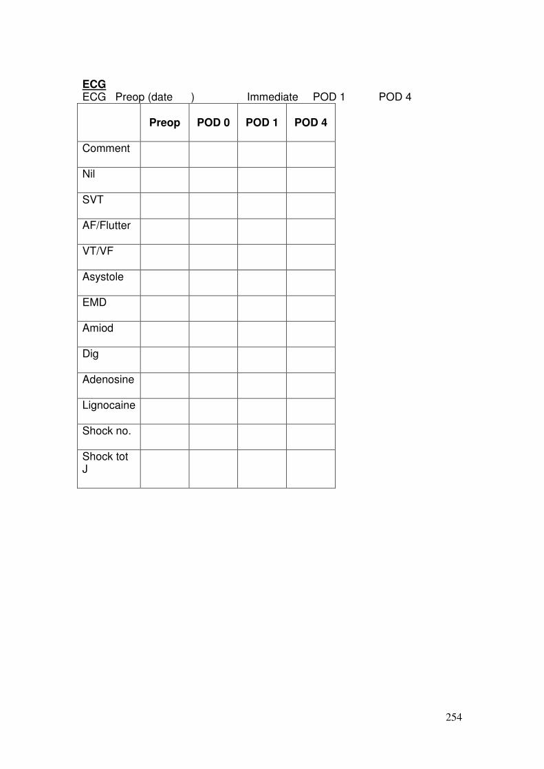
254
ECG ECG Preop (date ) ���� Immediate ���� POD 1���� POD 4����
Preop
POD 0
POD 1
POD 4
Comment
Nil
SVT
AF/Flutter
VT/VF
Asystole
EMD
Amiod
Dig
Adenosine
Lignocaine
Shock no.
Shock tot J
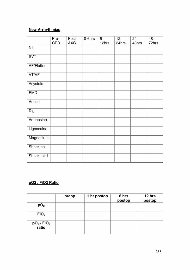
255
New Arrhythmias Pre-
CPB Post AXC
0-6hrs 6-12hrs
12-24hrs
24-48hrs
48-72hrs
Nil
SVT
AF/Flutter
VT/VF
Asystole
EMD
Amiod
Dig
Adenosine
Lignocaine
Magnesium
Shock no.
Shock tot J
pO2 / FiO2 Ratio
preop 1 hr postop 6 hrs postop
12 hrs postop
pO2
FiO2
pO2 / FiO2 ratio
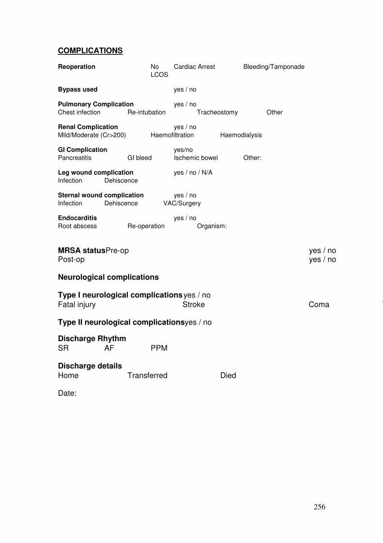
256
COMPLICATIONS
Reoperation No � Cardiac Arrest � Bleeding/Tamponade �
LCOS � Bypass used yes / no Pulmonary Complication yes / no
Chest infection � Re-intubation � Tracheostomy � Other Renal Complication yes / no
Mild/Moderate (Cr>200) � Haemofiltration � Haemodialysis � GI Complication yes/no
Pancreatitis � GI bleed � Ischemic bowel � Other: Leg wound complication yes / no / N/A
Infection � Dehiscence � Sternal wound complication yes / no
Infection � Dehiscence � VAC/Surgery � Endocarditis yes / no
Root abscess � Re-operation � Organism:
MRSA statusPre-op yes / no Post-op yes / no Neurological complications Type I neurological complications yes / no
Fatal injury � Stroke � Coma � TIA Type II neurological complicationsyes / no
Discharge Rhythm
SR � AF � PPM � Discharge details
Home � Transferred � Died � Date:

257
PATIENT INFORMATION SHEETS REC Ref. No. 06/Q2702/7
Application of remote ischaemic pre-conditioning to human coronary
artery bypass surgery (CABG)
AN INVITATION TO PARTICIPATE IN RESEARCH?
The heart surgery team at the Queen Elizabeth Medical Centre is inviting patients to participate in research aimed at making coronary artery surgery safer. We include a simple and non-technical summary of the reasons for the trial and what it involves for patients over and above their routine treatment if they take part. If you are being approached in the pre-admission clinic we would like you to take these information sheets home with you to read and consider participating in this trial between now and your admission date at which time a research doctor will be available to discuss the trial further and answer any questions. You can only be included in this clinical trial if you give your express permission in the form of signed consent. If you are already an inpatient on the ward then a member of the heart surgical team will be happy to discuss the trial with you once you have had at least 24 hours to read through these information sheets.

258
PATIENT INFORMATION SHEETS REC Ref. No. 06/Q2702/7
Application of remote ischaemic pre-conditioning to human coronary artery bypass surgery (CABG) WHAT IS THE STUDY ABOUT? This study is about trying to improve the protection of the heart in patients undergoing
coronary artery bypass surgery (CABG). During CABG the blood flow to the heart is
reduced markedly for 30-90 minutes so that bypass grafts can be constructed and
during this period the heart has to be protected so that it is not injured by the strain of
the operation. Despite using standard techniques to protect the heart, some temporary
injury still occurs from which the heart gradually recovers during the first few hours
and days following operation. We are seeking ways to improve on standard methods
to protect the heart by using a technique called remote ischaemic pre-conditioning
(RIPC).
Experimentally, we know that short periods (minutes) of reduced blood flow
(ischaemia) to the heart can substantially reduce the injury caused by a subsequent
more prolonged episode of reduced blood flow. This phenomenon is called ischaemic
pre-conditioning. This protective pre-conditioning effect has been observed in cardiac
surgery but there are concerns that even the short period of reduced blood flow may
itself cause injury and therefore, this technique is not in general usage. Recently, it
has been demonstrated that the same degree of protection against injury occurs when
a remote organ or limb is pre-conditioned without any reduction in blood flow for the
heart. Thus, three 5 minute periods of reduced blood flow of a limb performed
immediately before a more prolonged period may improve protection of the heart.
This study assesses whether remote pre-conditioning of the arm improves heart
protection during coronary artery surgery. Thus, remote ischaemic pre-conditioning
means that brief episodes of reduced blood supply to a limb can pre-condition or
prime the heart to better cope with any period during which its own blood flow is
reduced.
HOW CAN WE DO THIS?
We seek to investigate whether remote pre-conditioning (RIPC) can improve heart
protection during coronary artery bypass surgery. To do this, we have designed a
randomised control trial in which some patients will be allocated by chance to receive
RIPC or not. Randomized controlled trials are recognized as the very best way of
answering such a question. This study will compare whether RIPC improves heart
protection. The injury that a heart sustains during a heart operation can be detected in
a number of ways. Blood tests may show biochemical abnormalities,
electrocardiograms (ECGs) may show changes indicative of injury, echocardiograms
(ultrasound scans of the heart similar to those used in pregnant women) can show
changes in muscular function of the heart, and monitoring lines can show changes in
the pressures and blood flows generated within the heart. Some patients undergoing
heart surgery require drug therapy to support the heart afterwards. A requirement for
such drugs and the quantity needed is a further index of heart injury. In addition,
analysis of tiny samples of heart muscle can show changes in the heart’s energy
stores. Thus, by performing these investigations in a standard way we can compare
whether RIPC is beneficial or not.

259
HOW HAS THIS CLINICAL TRIAL BEEN DESIGNED?
This study is what is called a double-blind randomized placebo-controlled trial. This
means that if you agree to take part you may be allocated by chance to receive RIPC.
The RIPC is conducted while you are under anaesthetic in the early stages of your
heart surgery. Prior to operation, standard blood pressure cuffs will be applied to your
left arm and to a dummy cylinder. These will be connected by a switch and depending
on your chance allocation, either cuff will be inflated for 3 periods of 5 minutes each,
with 5 minutes of cuff deflation prior to the time when your heart’s blood flow is
reduced to perform the CABG operation. The cuffs will be obscured from view so
that the researchers will not know if you received RIPC or not until such time that we
open the allocation code. Your operation will then proceed in a standard way, you will
be cared for on our Intensive Care Unit and you will be recovered from surgery in the
normal manner. This is done to prevent any bias in the results. By comparing the
results of biochemical, ECG, echocardiographic, muscle analyses and post-operative
drug requirements between those patients receiving RIPC and those not, we will be
able to answer whether RIPC is beneficial.
HOW WILL WE MEASURE THE EFFECTS OF RIPC?
To detect changes in heart function, some measurements need to be taken before,
during and after surgery. Prior to your operation an ECG and echocardiogram will be
performed to compare with those taken after the operation. On the evening before
surgery a portable monitoring ECG will be connected to carefully monitor and record
your heart rhythm before, during and for 48 hours after operation. All these tests are
harmless and cause minimal discomfort if any. When you are placed under
anaesthetic, small monitoring catheters (plastic tubes) are routinely inserted in blood
vessels and are usually removed on the first or second post operative day. We will
record measurements from these catheters throughout this period at specific time
points. These catheters are all a standard part of your heart operation and are removed
in the first few days after operation, usually within 48 hours. From these catheters, we
will draw some additional blood samples for biochemical analysis. During your
operation, your surgeon will take 3 small samples of heart muscle about the size of
short length of pencil lead (actual size ====). Analysis of these samples will enable
us to examine the possible effects of RIPC on the heart’s energy stores or other
chemicals. Also during your heart operation, a small catheter is routinely placed in
your bladder so that your urine output can be measured. The urine is usually discarded
but we will take some timed samples to measure any possible additional protective
effect of RIPC on the kidney.
MEASURING THE POSSIBLE BENEFICIAL EFFECTS OF RIPC
The sampling of additional blood and urine tests: the blood tests performed on
these samples can tell us how well the heart has tolerated the surgery. These blood
samples are removed through the same monitoring lines already mentioned. The total
amount of additional blood taken for this study is about half a cupful (50ml). Samples
of urine will be collected for analysis from the catheter routinely placed under
anaesthesia.
Imaging the heart using a trans-thoracic echocardiogram: An echocardiogram is a
scan that uses ultrasound (sound waves) to produce pictures of the heart. The test is
painless and without side effects. An ultrasound probe and a small amount of gel are
gently placed on your chest and will be moved to different positions. The scan will

260
take approximately 20 –40 minutes to complete and will be performed once before
surgery and once after. The scan will be performed in a special Echo room.
Following all our heart surgery, small wires are left on the surface of the heart so that
we can ensure that your heart rate is satisfactory after the operation. During the
second scan, we will adjust your heart rate between 70 and 120 beats per minute (a
normal range of heart rate) using these wires. This will give us extra information
about heart function. You may be able to feel a change in heart rate. This is harmless,
but if uncomfortable we would stop and complete the scan without varying the heart
rate.
ECG and heart monitoring: ECGs are routinely performed before and several times
after surgery. We will analyse these ECGs carefully as part of the study. Heart injury
may cause some rhythm changes of the heart that are not detectable by standard
ECGs. We will, therefore, connect you to a continuous portable (Holter) monitor on
the evening before your operation which will record your ECG for the pre-operative
period and for 48 hours afterwards. This will then be removed and analysed by
computer. This will be able to tell us how many and what duration of any abnormal
rhythms you had during that period. This test is painless and silent.
Monitoring your heart output
This will be done at specified time points from the lines inserted during your
anaesthetic. The lines themselves are a necessary and standard part of the operation.
We will gather the information from these lines as part of our research.
The removal of tiny samples of the heart muscle, skeletal muscle and fat known
as biopsies: This will tell us whether RIPC protects the heart by conserving its energy
stores. We would aim to obtain 3 heart muscle biopsies all whilst you are asleep under
the anaesthesia, one at the beginning, middle and end of the CABG procedure. A
stitch is placed in the tiny defect left behind and their removal incurs no increased risk
during the surgery nor has any effect on the strength of the heartbeat. We have
performed more than 1500 such biopsies in patients at the time of writing of this
information leaflet with no complications.
The RIPC stimulus
If you have been allocated to the RIPC group, a blood pressure cuff will be inflated on
your left arm for three 5 minute periods at the start of your operation while you are
under anaesthetic. This is exactly the same as having your blood pressure taken but
for a longer period. Previous studies have shown this to be quite safe and harmless
and only modestly uncomfortable even in conscious patients. You should not be
aware of it whatsoever and we would expect that patients will not be able to tell if
they underwent RIPC or not. To be quite sure that this has not caused any problems
whatsoever we will ask you to fill in a questionnaire about any symptoms that may
have arisen in your arm.
WHAT WILL I HAVE TO DO?
If you decide to participate then there are a number of stages to the study. You will be
guided through these stages by us (the research doctors looking after you).

261
You will by now have received the written relevant information and asked to read this
at your leisure at home or on the ward if you are already an inpatient. On the day prior
to your surgery a member of the research team will visit you so you can ask any
questions. If you give signed consent to participate then we will take the opportunity
to perform an echocardiogram (20 minutes) on the day before surgery and also
connect the ECG monitor.
In the anaesthetic room of the operating theatre the anaesthetist will give you a
general anaesthetic. The anaesthetists will insert the pressure measuring catheters
normally inserted at this stage. Once you are asleep, an extra blood pressure cuff will
be applied to your left arm and inflated for three 5 minute periods as above depending
upon your allocation code. The operation will be conducted in a normal and standard
way under the care of the consultant cardiac surgeon, who is in charge of your case.
The blood samples and biopsies will be performed while you are asleep and you will
be unaware of them.
When you awake you will be on the intensive care unit as routine. Blood and urine
tests will be drawn from the lines and catheters already in place. The last blood test
will be performed at 72 hours after the operation. Forty-eight hours after your
operation, the continuous ECG monitor will be removed. On about the fourth day
after your operation we will perform the second echocardiogram. Standard tests will
be performed in the recovery period as for all patients.
In summary we will be taking measurements from pressure measuring catheters that
are routinely used in heart surgery. The entire sampling of blood for the research will
total 50 ml and will not affect your recovery and, because we can use intravenous
pressure measuring lines, there will be no additional need for needles. The biopsies
that we take are removed painlessly during the operation and have no effect on the
heartbeat.
WHAT ARE THE BENEFITS?
We do not know whether RIPC will actually be of benefit. There is sufficient
evidence to believe that it may be but this study is being performed to prove or
disprove this. Therefore, we cannot say if you will benefit as an individual. It is hoped
that this study will provide information that will benefit future populations of patients
undergoing cardiac surgical procedures.
WHAT ARE THE RISKS?
Previous studies of RIPC have shown that it is a safe. There is no reason to suspect
that it would be harmful. Nevertheless the extensive monitoring that is performed as a
matter of routine in a patient undergoing cardiac surgery would be able to detect any
side effects at their earliest stages before a problem developed. In addition, the
questionnaire will help us to establish that it is harmless. All heart operations carry
some risk and these will have already been discussed with you by your surgical team.
For this study we insert routinely used monitoring lines to measure heart function.
These are used in 60% of patients undergoing coronary artery surgery. In the study we
would use them in all patients to more closely monitor heart function. The risks of
insertion of these monitoring lines are minimal and the possibility of a severe
complication is in the region of 1 in 15000. The only additional invasive procedure
performed on you is the biopsy which as stated above has been performed in over 500
patients without ill-effect.
262
WHAT ARE THE ALTERNATIVES?
If you do not wish to take part in the study, your surgery will be undertaken in the
standard manner without any additional measurements, treatments or tests. Your
surgeon and anaesthetist may still use the monitoring lines that we have described if
they feel that their use is in your best interest.
WHAT HAPPENS TO THE INFORMATION?
The information from the study will be collected confidentially and coded for
anonymity. At the end of the study, the information will be analysed. The information
will be presented at scientific meetings and published in scientific journals to inform
other doctors and health professionals of our findings. All data is confidential and
stored by code on secure computers. This ensures that your identity will not be
revealed at any time.
WHO ELSE IS TAKING PART?
We hope that 162 patients will participate in this study. This is the number required
to establish that we are able to detect a true effect of RIPC.
WHAT IF SOMETHING GOES WRONG?
Throughout your surgery, your safety is of paramount concern. If for any reason, your
surgeon and anaesthetist feel that the study should not proceed because of other safety
concerns, the research will not take place. The standard care of patients undergoing
heart surgery involves intensive monitoring. This monitoring allows us to detect any
problems early in their development. We do not expect the study itself to cause any
problems, however as for all heart surgery we are in an ideal position to deal with any
untoward events during your operation and these will be treated in the normal manner,
regardless of the research study. At the time of all the measurements you will be in
the theatre or I.T.U. where trained staff is at hand at all times. Should you become ill
for any reason related to the study, the study would be stopped immediately. Your
safety during and after surgery is paramount, and takes precedence over any research.
263
WHAT HAPPENS AT THE END OF THE STUDY?
The main study is undertaken during the surgery and our collection of additional
information ends on the 4th
or 5th
day after your operation. At the end of the complete
study, the codes of allocation will be broken and the overall results analysed. Your
direct involvement is complete within your stay in hospital. If you would like to
receive summary information about the study results, this will be sent to you once the
study is complete (in approximately 2 ½ years time).
WHAT IF I HAVE MORE QUESTIONS OR DO NOT UNDERSTAND SOMETHING?
Please feel free to ask one of the investigators about any questions or worries that you
may have so that any points can be clarified. You should feel free to ask questions at
any time you like. If you would like more information this can be provided by the
Research Fellow coordinating the study (see below) or from Professor Bonser’s office
extension 2543.
WHAT HAPPENS NOW IF I DECIDE TO TAKE PART?
We will take some details and ask you to sign a consent form that documents your
willingness to participate. You will be listed for surgery as normal. When your
surgery is scheduled we will ask your permission to start the study on the evening
prior to operation and undertake the echocardiogram and ECG monitoring. You are
free to withdraw from this study after initially consenting without prejudice to your
continuing care.

264
CONSENT FORM Application of remote ischaemic pre-conditioning to human coronary
artery bypass surgery (CABG)
This is a consent form that documents your agreement to participate in this particular research. Before signing this form please tick the boxes below. I have read the invitation form I have read the information sheet I have understood the nature of the research I have had time to consider my response/reply I have been able to ask any questions and am satisfied with the answers I understand that I can withdraw at any time without giving explanation The participation or lack of it in this research will not affect my clinical care in any way I would like to receive a summary of the research when it is complete. Please remember that there is no obligation to participate in this study and that even if you do you can withdraw at any time. If you do not consent to this study your operation and care will proceed as planned. There are no rewards or penalties for participating in research although we are grateful for your efforts and inconvenience. If after reading this consent form and the patient information sheet you agree to participate please sign the form below and return to Mr I Rahman. If you have any further questions about any aspect of the research feel free to ask Mr I Rahman. The study has been explained by: Print Name: Signature: Date: Please sign below if you agree to participate in this study I have read and understand the above, and I am happy to take part in the above study. Signed Name (Print) Date

265
APPENDIX B - STANDARDISED PROTOCOLS
Procedures that have been standardised across a number of trials including
protocols for departmental postoperative management of patients are
referenced below:
1.0 Preparing the operating schedule
Operating list for trial patients was annotated thus:
SMITH John DOB 12/12/45 G236819/A CABG (REMOTE TRIAL) E2B
This practice was to ensure ward staff, anaesthetists, ODP, theatre staff,
perfusionists, ITU staff and surgical team were aware of the trial patient and that
all protocols were adhered to.
2.0 Protamine dose
Protamine was administered intraoperatively at a dose of 1mg per 100 units of
heparin once the patient is of CPB.
Additional protamine to cover the pump blood returned to the patient was given
to the following regimen:
• 0-499 ml of pump blood → no additional protamine
• 500-999 ml of pump blood returned → 50 mg protamine
• ≥ 1l of pump blood returned → 100 mg protamine
3.0 Placement of epicardial pacing wires
The placement of atrial and ventricular pacing wires is mandatory. Two
epicardial atrial pacing wires were sited in the region of the atrial cannulation

266
site and two epicardial ventricular wires were placed in the right ventricular
outflow tract.
4.0 Postoperative medication
4.1 Antiplatelet therapy
Patients received on return to the ITU received a dose of aspirin 300mg rectally
and thereafter 150mg orally once a day. In the case of aspirin allergy or other
contra-indications, anti-platelet therapy was at the discretion of the attending
team.
4.2 Angiotensin converting enzyme (ACE) inhibitors
All patients received an ACE inhibitor in the postoperative period (unless
contraindicated). Reinstitution and new prescribing of ACE inhibitor therapy was
not permitted until the fourth postoperative day.
4.3 Other cardiovascular medication
All patients received initial diuretic therapy with oral frusemide 40mg daily.
Long-acting nitrates or nitrate patches were not permitted postoperatively.
Preoperative antihypertensive medications were recommended as clinically
indicated.
β-receptor antagonists, statins and other lipid lowering agents were started as
soon as possible in the postoperative period.
267
4.4 Non cardiovascular medication
Treatment was restarted on as soon as possible after surgery at the discretion
of the caring team.
5.0 Protocols for the management of post-operative complications
5.1 Protocol for management of pacing and arrhythmias
5.1.1 Management of ventricular fibrillation post AXC removal
The management was as follows until the restoration of sinus rhythm:
• 2 x 10 J shock
• Administration lignocaine 100mg IV
All reperfusion VF was recorded, together with shock strengths. Similarly, the
use of induced VF and subsequent defibrillation to facilitate biopsy or other
repairs was recorded.
Treatment beyond this is at the discretion of the patient’s clinical team but was
recorded as part of the results
5.1.2 Post-operative pacing management
At the end of cardiopulmonary bypass all patients had ventricular and atrial
pacing wires in situ.
During weaning from CPB, the target synchronised heart rate was 90bts/min.
This was achieved by:
• native sinus rhythm (± atropine / glycopyrollate)
• AAI pacing (90 bts.min-1)
• DDD (or DVI) pacing (90bts.min-1) with a default AV interval of 160ms.
268
Initial haemodynamic studies were performed in the rhythm or pacing mode
selected by the Consultant Surgeon/Anaesthetist to achieve best overall
haemodynamic performance. These settings were maintained through transfer
to ITU and reviewed by the Research Fellow 2 hours following release of the
aortic cross-clamp.
At this time, prior to the 2 hour haemodynamic assessment:
• in patients in SR with a heart rate of ≥90bts.min-1 and normal AV
conduction no action was required
• In patients in SR with a heart rate <90 bts/min AAI pacing at 90 bts.min-1
was instituted
• In patients DDD or DVI paced (90bts.min-1) the PAUSE button on the
pacing box was depressed briefly:
If this demonstrated persistent AV block, DDD or DVD pacing (default AV
interval 160ms) was continued throughout the study period at 90bts.m-1.
If the PAUSE query demonstrated a rhythm which had AV conduction, the
pacing mode was adjusted. The AV interval setting was increased to a
maximum of 200ms and the rhythm reviewed.
If the rhythm was now atrially paced with native AV conduction, this means
that the native AV interval is <200ms. This rhythm is haemodynamically
preferable and provided there was not a variable native AV interval and thus
intermittent pacing, these pacing settings (DDD AV interval 200ms) were
maintained.
In circumstances in which the AV interval was ≥200ms or in which the native
AV interval was variable with intermittent ventricular pacing, the previous
DDD settings (AV interval 160ms) was resumed.
269
The trial staff would document in the patient notes any pacing changes,
record the data on the study datasheet and proceed with the haemodynamic
studies.
This pacing assessment is the only time when trial staff had a direct clinical
responsibility regarding a patient’s management.
5.1.3 Protocol for the management of suspected atrial fibrillation and other
supraventricular tachyarrhythmias
• Check hypoxia and hypokalaemia (K+ < 4.5 mmol.L-1), record the result
and correct appropriately
• Confirm diagnosis with a12 lead ECG (2 copies named, dated and timed)
• If hypoxia is present a chest radiogram was obtained
5.1.4 Management of AF or atrial flutter: Scenario 1
If the PR interval <180 millisecs, no new LBBB or no new bifasicular block
and ventricular rate >80.min-1 the treatment plan was:
5.1.4.1 Patient unable to take oral medication
If the patient was intubated and ventilated we would strive to ensure oxygen
saturations were greater than 95%, using supplemental oxygen as required. We
aimed to maintain serum K+ greater than 4.0 mmol.L-1
Intravenous Amiodarone 1.2g over 24 hours was administered. The initial bolus
dose of 300mg was administered intravenously over 1 hour followed by 900mg
for the remaining part of 24 hours.

270
Intravenous Amiodarone 1.2g was repeated in the second 24 hours, and
reduced to 600 mg over 24 hours for the next 5 days, and 300mg over 24 hours
thereafter.
Oral Amiodarone was commenced when oral or nasogastric medications were
tolerated.
Prophylactic anti-coagulation was administered using s/c Enoxaparin 40mg
daily for the first 48 hours after the commencement of atrial fibrillation, unless
other anti-coagulation was already ongoing, or unless anti-coagulation was
contra-indicated. If atrial fibrillation persisted for more than 48 hours,
Enoxaparin was increased to 1mg/kg s/c BD, unless contraindicated, until 24
hours after sinus rhythm was restored.
DC cardioversion was considered if sinus rhythm was not restored after 72
hours atrial fibrillation.
If the ventricular capture rate remained high loading with Digoxin 500µg 6
hourly up to 3 doses according to response was considered. Maintenance dose
was then continued at half the recommended dose for age, size and renal
function.
DC cardioversion was also considered, at any stage, if haemodynamic
instability ensued. Patients who failed to cardiovert to sinus rhythm were
warfarinised. Target INR 2.5.
271
5.1.4.2 Patient able to take oral medication
For awake, extubated patients on the ward or ITU we would strive to ensure
oxygen saturation’s were greater than 95%, using supplemental oxygen as
required and aim to maintain serum K+ greater than 4.0 mmol.L-1.
The regimen of oral Amiodarone 400 mg TDS for 48hrs, followed by 200mg
TDS for one week, then 200mg BD for one week and finally 200mg OD for one
week was followed, or until the day of discharge, whichever is the shorter.
Doses were reviewed if patient weight was less than 50Kg.
Prophylactic anti-coagulation was administered using s/c Enoxaparin 40mg
daily for the first 48 hours after the commencement of atrial fibrillation, unless
other anti-coagulation was already ongoing, or unless anti-coagulation was
contra-indicated. If atrial fibrillation persisted for more than 48hours Enoxaparin
was increased to 1mg.kg-1 s/c BD, unless contraindicated, until 24 hours after
sinus rhythm was restored.
DC cardioversion was considered if sinus rhythm was not restored after 72
hours of atrial fibrillation or if at any stage haemodynamic instability ensued.
If the ventricular capture rate remained high loading with Digoxin 500µg 6
hourly up to 3 doses according to response was considered. Maintenance dose
was continued at half the recommended dose for age, size and renal function.
If atrial fibrillation/atrial flutter was intermittent but persistent despite
Amiodarone loading anti-coagulation with Warfarin was commenced prior to
discharge home and continued until review in the outpatient clinic (target INR
2.5). If sinus rhythm was restored when reviewed in the outpatient clinic
Warfarin and Amiodarone was discontinued. If atrial fibrillation/atrial flutter
persisted DC cardioversion was considered.

272
Amiodarone was discontinued on review in the outpatient clinic at 6 weeks if
sinus rhythm persisted.
5.1.5 Management of AF or atrial flutter: Scenario 2
If the PR interval >180 millisecs, or new LBBB or new bifasicular block and
ventricular rate 80.min-1 the treatment plan was:
5.1.5.1 Patient unable to take oral medication
If the patient was unable to take oral medication intravenous Amiodarone 600
mg over 24 hours for 2 days and 300 mg daily thereafter until able to tolerate
oral/NG medication was commenced. We would strive to ensure oxygen
saturations were greater than 95%, using supplemental oxygen as required and
aim to maintain serum K+ greater than 4.0 mmol.L-1.
Prophylactic anti-coagulation was administered using s/c Enoxaparin 40mg
daily for the first 48 hours after the commencement of atrial fibrillation, unless
other anti-coagulation was already ongoing, or unless anti-coagulation was
contra-indicated. If atrial fibrillation persisted for more than 48 hours,
Enoxaparin was increased to 1mg.kg-1 s/c BD, unless contraindicated, until 24
hours after sinus rhythm was restored.
DC cardioversion was considered if sinus rhythm was not restored after 72
hours atrial fibrillation.
If ventricular capture rate remained high loading with Digoxin 500µg 6 hourly up
to 3 doses according to response was considered. Maintenance dose was
continued at half the recommended dose for age, size and renal function.

273
DC cardioversion was also considered, at any stage, if haemodynamic
instability ensued.
Patients who failed to cardiovert to sinus rhythm were warfarinised. Target INR
2.5.
5.1.5.2 Patient able to take oral medication
If the patient was able to take oral medication oral Amiodarone 200 mg TDS for
8 days or until the time of discharge, whichever is the shorter was commenced.
We would strive to ensure oxygen saturations were greater than 95%, using
supplemental oxygen as required and aim to maintain serum K+ greater than
4.0 mmol.L-1.
Prophylactic anti-coagulation was administered using s/c Enoxaparin 40mg
daily for the first 48 hours after the commencement of atrial fibrillation, unless
other anti-coagulation was already ongoing, or unless anti-coagulation was
contra-indicated. If atrial fibrillation persisted for more than 48hours, Enoxaparin
was increased to 1mg.kg-1 s/c BD, unless contraindicated, until 24 hours after
sinus rhythm was restored.
DC cardioversion was considered if sinus rhythm was not restored after 72
hours atrial fibrillation and also at any stage if haemodynamic instability ensued.
If the ventricular capture rate remained high loading with Digoxin 500µg 6
hourly up to 3 doses according to response was considered. Maintenance dose
was continued at half the recommended dose for age, size and renal function.
If atrial fibrillation/atrial flutter was intermittent but persistent despite
Amiodarone loading anti-coagulation with Warfarin was commenced prior to
discharge home and continued until review in the outpatient clinic (target INR

274
2.5). If sinus rhythm was restored when reviewed in the outpatient clinic
Warfarin and Amiodarone was discontinued. If atrial fibrillation/atrial flutter
persisted DC cardioversion was considered. Oral Amiodarone 200 mg daily
thereafter. Amiodarone discontinued on review in the outpatient clinic at 6
weeks if sinus rhythm persists
5.1.6 D.C. Cardioversion technique for atrial fibrillation/flutter
Under anaesthesia the cardiovert protocol followed according to the incremental
energies detailed below:
Monophasic Biphasic
50J x2 attempts 150J x 3
100J x 2 attempts
150J x 2 attempts
360J x 2 attempts
Abandon if unsuccessful. Consider anticoagulation and further management at
team discretion.
5.1.7 Diagnosis and management of other supraventricular tachycardias
In the presence of supraventricular arrhythmias not believed to be AF the aim of
treatment was to restore normal sinus rhythm. Hypokalaemia and hypoxia were
corrected. Carotid sinus massage was attempted and if this failed intravenous
adenosine therapy was instituted as 3 mg in increasing doses of 3 mg (i.e. 3
mg, 6 mg, 9 mg) unless the patient had contraindications to adenosine (asthma,
second or third degree heart block, sick sinus syndrome). Further treatment was
at the discretion of the patient’ consultant/clinical team.

275
5.1.8 Protocol for the management of ventricular dysrythmias
The following ventricular dysrhythmias require attention:
• Bigeminy or Trigeminy
• ‘Salvos” of ventricular tachycardia
• ‘R on T’ phenomenon
• 15 ventricular ectopics/minute for > 5 minutes
• Sustained VT or VF
In all cases hypoxia, hypokalaemia and hypomagnaesemia were corrected, the
diagnosis was confirmed by 12-lead ECG with a lead II rhythm strip unless
patient was in theatres and a chest x-ray was performed (if no obvious cause
for hypoxia).
5.1.8.1Treatment of sustained pulseless ventricular tachycardia or ventricular
fibrillation
The decision to cardiovert, defibrillate or proceed to resternotomy was at the
discretion of the caring team. Defibrillation should be the immediate method of
choice (The Advanced Life Support Guidelines 2000) up to three shocks given
initially with energies of 200 J, 200 J, 360 J (or their equivalent when using
defibrillators with alternative waveforms). Amiodarone or lignocaine could next
be utilised:
• Amiodarone 300 mg (made up to 20 ml with 5% dextrose or from a
prefilled syringe) may be administered. A further dose of 150 mg may be
given for recurrent or refractory VT/VF, followed by an infusion of 1 mg
min-1 for 6 -12 hours then at 0.5 mg min-1.

276
• Lignocaine was given at 1 mg min-1, with a further 1-2 doses of 0.5 mg
min-1 10 minutes apart. A continuous infusion may be considered as
1000 mg of lignocaine to a total volume of 50 ml of 0.9%NaCl (20 mg/ml)
and infused at a rate of 12 ml.hr-1 (4mg.min-1) for 3 hours.
5.1.9 Protocol for the management of bradycardias (<60bpm for >5 minutes)
In the instance of bradycardias we would check initially the ECG monitors scale.
The haemodynamic targets of CVP, MAP and CI would be assessed. A 12lead
ECG would be performed.
Sinus bradycardia, nodal bradycardia or first-degree heart block but with MAP
65-85 mmHg and cardiac index >2.2L.min.-1m-2 required no treatment.
Sinus bradycardia, nodal bradycardia or first degree heart block but with MAP
<65-85 mmHg or CI <2.2 L.min-1.m-2 either glycopyrolate boluses of 0.6 mg up
to a maximum of 1.8 mg or IV atropine boluses of 0.6 mg up to a maximum of
1.8 mg were used. Pacing or isoprenaline infusion was considered.
Second or third degree heart block management was at the discretion of the
managing team. All therapy was recorded.
6.0 The management of post-operative hypertension on ITU
Hypertension is defined as MAP >85mmHg. If a sustained period of
hypertension or repeat temporary episode of hypertension develop then the
patient was managed as below:

277
• Ensure adequate analgesia
• GTN (i.v. 50 mg in 50 ml) titrating the dose to achieve MAP ≤85 mmHg
until the upper limit of infusion of 15 ml.h has been reached.
• If this failed to reduce the blood pressure then SNP (i.v. 50 mg in 50 ml
dextrose 5% solution) at 0-15 ml.h-1 was added as directed by the caring
medical team.
• Once the patient has been extubated, the need for hypertension control
should be re-assessed by the caring medical team. Where appropriate,
intravenous medication should be substituted with either re-
commencement of oral anti-hypertensive therapy or institution of oral
agent is at the discretion of the managing team.
7.0 Assessment and management of post-operative hypotension (i.e.
sustained MAP <60mmHg)
Hypotension is defined as MAP<60mmHg. In the case of hypotension the
following was assessed:
• Arterial line calibration and accuracy
• Cardiac rhythm (if abnormal see treatment of cardiac rhythm
disturbances)
• Filling pressures (CVP and PAWP)

278
• If below the range set by the patient’s consultant/clinical team or < 8 cm
H2O if no range has been set then volume was administered.
• If above the range set by the patient’s consultant/clinical team or > 12 cm
H2O then cardiac output studies were performed.
• If cardiac index was ≤ 2.2 L.min.-1m-2 the protocol for the assessment
and management of low cardiac output state was followed
• If cardiac index was > 2.2 L.min.-1m-2 and a normal SVR (800-
1200dyne.cm.sec-5) associated with hypotension management was
directed by the attending clinical team.
• If cardiac index > 3 L.min.-1m-2 with a low SVR (<800dyne.cm.sec-5) was
the cause of hypotension phenylephrine was prescribed as an infusion of
10mg made to 50 ml with 5% dextrose. The infusion was to start at 2
ml.h-1
8.0 Assessment and management of suspected low cardiac output
state
For the purpose of this study a low cardiac output state is defined as a C.I. of ≤
2.2 L.min.-1m-2 refractory to appropriate intra-vascular volume expansion
following correction or attempted correction of any dysrhythmias including sinus
bradycardia, nodal rhythms, atrial and ventricular tachycardia or dysrhythmias.
8.1 Low cardiac output state in theatres and anticipatory treatment
Whenever possible the surgeon/anaesthetist was to await the results of cardiac
output studies performed and recorded after CPB had been discontinued. In
some cases the surgeon/anaesthetist will anticipate a low cardiac output state

279
and the need for inotropes on subjective and objective features of cardiac
performance before and during surgery. If possible confirmatory cardiac output
studies should be awaited but if not inotropic therapy is started at the discretion
of the managing team. The exact time of commencement and dosage was
recorded.
Low cardiac output state in the presence of adequate ‘filling pressures’ is an
endpoint for this study.
The charts of patients who have had anticipatory inotropic treatment underwent
a review by the low cardiac output committee. The aim of the review was to
confirm that in the absence of C.O.S. data we had a bonafide case of low
cardiac output.
8.2 Post-operative low cardiac output state diagnosed in the ITU/HDU
Any patient having a measured cardiac index of ≤ 2.2 L.min.-1m-2 was assessed
as follows:
• Assess heart rate and rhythm and correct as appropriate
• Measure filling pressures (CVP and PAWP) and correct as appropriate
within targets set by the caring team. Consider overt or covert
haemorrhage
• Consider the possibility of cardiac tamponade
Appropriate management of the above factors is at the discretion of the caring
team. Once these factors were assessed and treated and if the patient still had
a cardiac index of ≤ 2.2 L.min.-1m-2 then the patient was considered to be
suffering from a low cardiac output state and initial management was as follows:

280
• Dopamine (first line inotrope) 200mg in 50ml 5% dextrose commenced at
a dose of 1-10 µg.kg-1.min-1 at the discretion of the caring team.
• All management considered necessary above and beyond dopamine 10µ
g/kg/min is at the discretion of the caring team.
8.3 Management of a ‘Reopening’ Procedure
The decision to reopen a patient is purely a clinical one at the discretion of the
patient’s consultant/team. The intra-operative reopening ‘findings’ were
recorded.
9.0 Protocol for the administration of colloids and blood products
Colloid is prescribed to replace drain losses and maintain adequate filling
pressures to achieve the desired mean arterial pressure (MAP 65-85mmHg)
and cardiac index. The colloid of first choice for this study was Gelofusin©.
The maximum volume of gelofusin© permissible over 12 hours is
• 2 L if the patient is ≥ 70 kg
• 1.5 L if < 70 kg.
If additional filling was required beyond these volumes:
• H.A.S. 4.5% if HB ≥ 8.5
• Packed red cells if HB < 8.5 or active bleeding through drains above
specified rate
Clotting factors, platelets and cryoprecipitate administered to restore clot
formation also served as volume expanders in addition to the Gelofusin@.

281
10.0 Protocol for the management of a low urine output
Low urine output was defined as <0.5ml.kg-1.hr-1. If the urine output fell below
this in any 1 hour the following was checked:
• adequate mean arterial pressure
• adequate CVP and PAWP
• a working urinary catheter. Consider a 50 ml flush
If this checklist is normal then intravenous frusemide 10mg was administered
and response awaited.
If any of the above checklist were not in the target parameters appropriate
colloid was given and cardiac indices repeated considering the use of inotropes
as protocol.
Failure to maintain a satisfactory diuresis would prompt an enquiry regarding a
possible low cardiac output state. If a satisfactory cardiac output is the case but
urine output remains inadequate dopamine (200 mg in a 50ml of 5% dextrose)
run at 3 µg/kg/hr was used supplemented by a low dose frusemide infusion as
necessary. Treatment beyond this was at the discretion of the patient’s clinical
team.
11.0 Protocol for the management of suspected wound infection
11.1 Suspected sternal or leg wound Infection
Bacteriological swabs were taken from sternal or leg wounds in cases of
suspected infection. Treatment was at the discretion of the clinical team.

282
11.2 Other infections
Bacteriological specimens were taken as appropriate in all suspected infection
episodes and treatment was at the discretion of the clinical team.
12.0 Protocol for management of blood glucose
The normal range for random blood glucose was defined as 4 .4-10 mmol.L-1
Post-operative hyperglycaemia and insulin resistance was managed with
standard Trust sliding scale insulin protocols.
3 examples of sliding scale protocols (A, B and C) are given according to
insulin-resistance of the patients below.
Management would start from protocol A and escalate if necessary. The aim
was to maintain blood sugars ≤10mmol/L.
A B C
BM
(mmol.L-1)
Units.h-1 BM
(mmol.L-1)
Units.h-1 BM
(mmol.L-1)
Units.h-1
0 - 7 0 0 - 7 0 0 - 7 0
7.1 – 11 1 7.1 – 11 2 7.1 – 11 4
11.1 – 15 2 11.1 – 15 4 11.1 – 15 8
15.1 – 20 4 15.1 – 20 6 15.1 – 20 10
>20 6 >20 8 >20 14
In more severe cases of insulin-resistance intermittent boluses of insulin of 5-10
units could be administered if serum potassium concentration was ≥4 mmol.L-1

283
13.0 Post-operative prescription for all ITU trial patients
13.1 Fluids
5% Dextrose at 1ml.kg-1h-1 was prescribed
13.2 Potassium
Potassium chloride (KCl) 10-20mmol in 30ml 5% dextrose over 30 minutes was
prescribed as required to maintain K+ 4.5-5.5mmol.L-1.
13.3 Insulin
As per ITU sliding scales
13.4 Sedation and continuous intravenous analgesia
Propofol 10mg/ml at 0-15 ml.h-1
Alfentanil 25mg in 50ml 5% Dextrose at 0-2ml.hr-1
Followed by:
Paracetamol 1q p.o. QDS
Tramadol (PRN) i.v. 50-100 mg 4 hrly or Tramadol p.o. 50-100 mg qds 4hrly
And or Codeine Phosphate 30-60mg 4hrly (max 240mg.24h-1) or
Dihydrocodeine 30-60mg qds (max 240mg.24h-1).
13.5 Antihypertensive medication
Glyceryl trinitrate solution (GTN) 50 mg in 50 ml at 1-15 ml.hr-1 ±±±±
Sodium nitroprusside (SNP) 50mg in 50ml. D5%W at 0-20ml.h-1 or
Nifedipine 10mg sub-lingual PRN for sustained hypertension (>160/90)

284
13.6 Anti-emetic medications
Maxalon 10 mg 8 hrly i.v./i.m./p.o. ±±±±
Cyclizine 50 mg i.v. 8 hrly ±±±±
Ondansetron 4-8 mg IV/IV/PO PRN 6-8 hourly
13.7 Antibiotics
As per the standard unit policy.
13.8 Heparin DVT prophylaxis
Enoxaparin 40mg s/c o.d.
13.9 Intravenous diuretic therapy
Frusemide 10mg i.v. PRN
13.10 Inotropes and Vasoconstrictors
The trial staff would always aim to perform a full series of cardiac output studies
prior to the commencement or escalation of inotropes.
Inotropes would always be prescribed and used in the 50ml 5% dextrose as per
unit policy. The dose of drug required to constitute a single strength solution for
each inotrope is detailed below:
Dopamine 200mg
Dobutamine 250mg
Adrenaline 2mg
Noradrenaline 2mg

285
Phenylephrine should be made up as 10mg / 50mls 5% dextrose.
Enoximone should be made up as 100mg / 40mls N/Saline
The loading dose should be run at 1mg.kg-1 for 1 hour
Enoximone in the pump was not allowed.

286
APPENDIX C - PRESENTATIONS AND PUBLICATIONS ARISING FROM
THIS THESIS AND THE RESEARCH FELLOWSHIP
1.0 PRESENTATIONS
[1] Does remote ischaemic preconditioning during on-pump coronary
artery bypass grafting enhance lung preservation? Rahman IA, Mascaro
JG, Green D, Townsend P, Bonser R. ESCVS International Congress, 2010
(oral presentation).
[2] Does remote ischaemic preconditioning protect the heart and
kidneys in human coronary artery bypass surgery – a randomised
controlled trial. Rahman IA, Mascaro JG, Steeds RP, Frenneaux MP,
Nightingale P, Gosling P, Townsend P, Townend J, Green D, Bonser RS.
SCTS 2010 (oral presentation).
[3] Remote ischaemic pre-conditioning in human coronary artery
bypass surgery, from promise to disappointment. Rahman IA, Mascaro
CJ, Steeds RP, Frenneaux M, Nightingale P, Gosling P, Townsend P,
Townend J, Green D, Bonser RS. American Heart Association Scientific
Session, November 2009 (oral presentation)
[4] Increased beta-catenin pathway expression by a remote
ischaemic preconditioning stimulus: first evidence in humans.

287
Contractor H, Ashrafian H, Rahman IA, Redington A, Frenneaux M, Bonser
RS, Kharbanda R. American College of Cardiology, 2009 (oral presentation)
[5] The functional impact of remote ischaemic preconditioning on
patients undergoing first time coronary artery bypass surgery. Rahman
IA, Nightingale P, Marsh AM, Williams L, Mascaro CJ, Bonser RS, Steeds R.
British Cardiovascular Society, 2009 (poster presentation)
[6] Does remote ischaemic preconditioning during on-pump coronary
artery bypass grafting enhance renal and pulmonary protection?
Rahman IA, Mascaro CJ, Nightingale P, Gosling P, Green D, Townsend P,
Bonser RS. Royal Society Medicine [Cardiothoracic Section].2009 (poster
presentation)
[7] The variability of ejection fraction in patients following on-pump
coronary artery bypass grafting. Rahman IA, Alvior A, Nightingale P, Marsh
AM, Mascaro CJ, Bonser RS, Steeds R. 13th Asia Pacific Congress of Doppler
Echocardiography, 2009 (poster presentation)
[8] Investigating the reduction in right ventricular function following
on-pump coronary artery bypass surgery. Rahman IA, Marsh AM, Williams
L, Mascaro CJ, Bonser RS, Steeds R. 13th Asia Pacific Congress of Doppler
Echocardiography, 2009 (poster presentation)

288
[9] Right ventricular functional change following on-pump coronary
artery bypass grafting and its relationship with perioperative myocardial
necrosis and inotropic support. Rahman IA, Mascaro CJ, Nightingale P,
Green D, Townsend P, Bonser RS. Royal Society Medicine [Cardiothoracic
Section].2008 (poster presentation)
2.0 PUBLICATIONS
[1] Remote ischemic pre-conditioning in human coronary artery
bypass surgery, from promise to disappointment? Rahman IA, Mascaro
JG, Steeds RP, Frenneaux MP, Nightingale P, Gosling P, Townsend P,
Townend J, Green D, Bonser RS.Circulation (Accepted - in print). 2010
[2] Remote ischaemic preconditioning reduces myocardial injury in
patients undergoing cardiac surgery with cold blood cardioplegia: a
randomised controlled trial. Rahman IA, Bonser RS. Heart (Commissioned
Editorial) 2009;95(19):1553-1555
[3] Increased beta-catenin pathway expression by a remote
ischaemic preconditioning stimulus: first evidence in humans.
Contractor H, Ashrafian H, Rahman IA, Redington A, Frenneaux M, Bonser
RS, Kharbanda R. JACC 2009;53:A309

289
[4] The functional impact of remote ischaemic preconditioning on
patients undergoing first time coronary artery bypass surgery. Rahman
IA, Nightingale P, Marsh AM, Williams L, Mascaro CJ, Bonser RS, Steeds R.
Heart 2009;95(Suppl I);A78
[5] The variability of ejection fraction in patients following on-pump
coronary artery bypass grafting. Rahman IA, Alvior A, Nightingale P, Marsh
AM, Mascaro CJ, Bonser RS, Steeds R. Heart, Lung and Circulation Journal
of the Cardiac Society of Australia and New Zealand 2009;18(S1):13-14.
[6] Investigating the reduction in right ventricular function following
on-pump coronary artery bypass surgery. Rahman IA, Marsh AM,
Williams L, Mascaro CJ, Bonser RS, Steeds R. Heart, Lung and Circulation
Journal of the Cardiac Society of Australia and New Zealand 2009;18(S1):14


![Remote Ischemic Preconditioning of the Femoral Artery and ... · ischemic preconditioning procedure on renal pedicle against I/R-induced AKI [2], the species difference between rats](https://img.dokumen.tips/doc/110x75/6005d98ecae0876c03052ae8/remote-ischemic-preconditioning-of-the-femoral-artery-and-ischemic-preconditioning.jpg)