Embed Size (px)
Citation preview

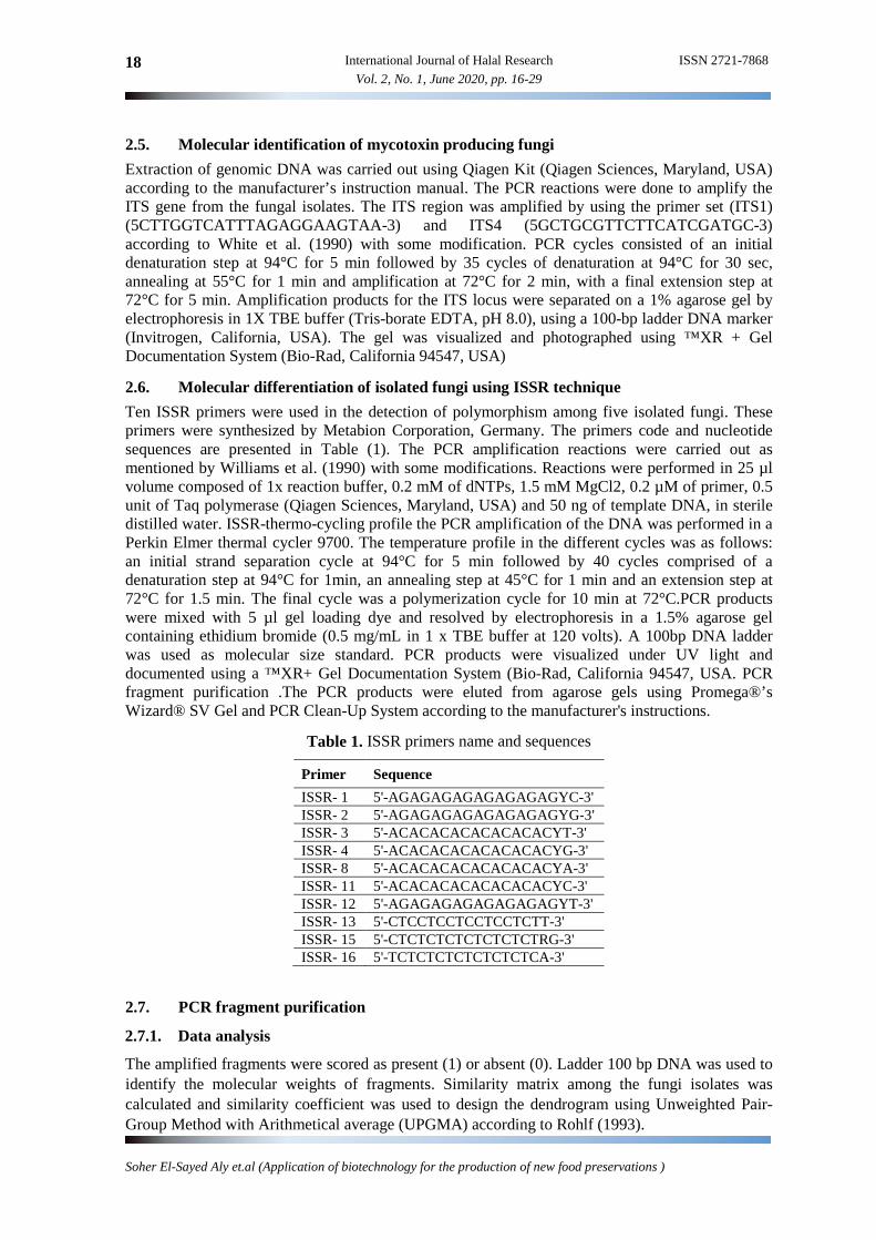
International Journal of Halal Research ISSN 2721-7868 Vol. 2, No. 1, June 2020, pp. 16-29 16
Application of Biotechnology for the Production of New Food Preservations
Soher El-Sayed Alya,1,*, Neven Abdelrahman Emamb
a Food Toxicology and Contaminants Department, National Research Centre, Dokki, Cairo, Egypt b Microbial genetic Department, National Research Centre, Dokki, Cairo, Egypt 1 [email protected] * corresponding author
1. Introduction Different species of pathogenic fungi can produce mycotoxins which contaminate stored different halal agriculture commodity (Sabry et al., 2016). Some of these fungi can produce different mycotoxins as secondary metabolites (such as aflatoxins, fumonisins, ochratoxins, tricothethenes etc…). Aflatoxins are produced from some strains of Aspergillus flavus and Aspergillus parasiticus and contaminated food and agriculture commodity ( Iqbal et al., 2013). Aflatoxin B1 (AFB1) has toxic, carcinogenic, mutagenic and teratogenic effects in laboratory animals (Abdel-Wahhab et al., 2010). In addition, AFB1 and its metabolites can accumulate in animal tissues, and reached to human consumer through the food chain (Bintvihok et al., 2002).
Halal Lactic acid bacteria (LAB) have important role in food industry and that benefit humans and animals health. Moreover, their safety has been proved empirically and scientifically (Hathout and Aly, 2014). Some of probiotic bacterial strains like L. reuteri and L. casei inhibited the fungal growth and used to produce fermented Talpina as cereal- dairy product and Ras cheese (Sahab et al., 2011, Hathout and Aly, 2010). The most important LAB species are of the genera Lactobacillus and Bifidobacterium, due to their probiotic properties exhibited by some strains and generally
AR TIC LE INFO
ABSTRACT
Article history
Received February 25, 2020
Revised June 02, 2020
Accepted June 21, 2020
The presence of pathogenic fungi has been a global concern has economical affects. Recently prevent the fungal growth using biological control using halal source become an important strategy for limiting mycotoxigenic fungi. Therefore the aim of this study that the use of some LAB bacteria and screen their antimicrobial activity against pathogenic fungi. Halal bacteria extracts were studied for the antimicrobial activity. The results indicated that two isolates were identified. Meanwhile a total of 90 fungal isolates were isolated from halal food samples. Aspergillus was the dominant fungi (65%) in five species namely, parasiticus, flavus ochraceus, terreus, and niger. Fusarium (clomorum and graminerum) and Penicillium were in lower frequency. Halal bacterial extracts were reduced the mycelium dry weight of all tested fungi and showed clear zone inhibition. Molecular identification of mycotoxin producing fungi was estimated. In conclusion, the study revealed the probability of using halal Enterococcus isolates in food preservation to ensure halal food safety. The partial or total sequencing of the 16s ribosomal DNA (rRNA) gene showed a fast technique used fungi classification
This is an open access article under the CC–BY-SA license.
Keywords:
Pathogenic fungi
lactic acid bacteria
molecular biology
antimicrobial
halal food
ISSN 2721-7868 International Journal of Halal Research 17 Vol. 2, No. 1, June 2020, pp. 16-29
Soher El-Sayed Aly et.al (Application of biotechnology for the production of new food preservations )
regarded as safe (GRAS) lactic acid bacteria. Abde-Shafi et al. (2014) reported that Enterococcus faecium, Enterococcus faecalis are important species of enterococcus genus with probiotic properties which used to suppress carcinogenesis, reduce cholesterol level, and prevent bacteria-associated diarrhea. So, these bacteria called halal food. LAB isolated from homemade fermented vegetables produce antibacterial substances against both gram-positive and importantly, gram-negative common foodborne bacterial pathogens. Enterococcus faecium and enterococcus faecalis enterococci are singular, double or short chained gram positive cocci. Streptococcus faecalis was defined by Felis and Dellaglio (2007). Halal bacteria in this genus were divided into various species such as; E. faecium, E. durans, E. faecalis, E. casseliflavus, E. malodoratus, E. avium, E. pseudoavium, E. dispar, E. sulfureus, E. flavescens, E. saccharolyticus, E. columbae ve E. cecorum (Giraffa, 2003; Khan et al., 2010).
Adimpong et al. (2012) examined the partial or total sequencing of the 16s ribosomal DNA (rRNA) gene for practical routine purposes. If two organisms present, a 16s rRNAgene sequence identity are higher than 97% they can be considered closely related and thus belonging to the same species (Větrovský et al., 2013). Identification based on the 16S rDNA sequence is of interest because ribosomal exists generally among bacteria and includes regions with species specific predictability. The aim this study was to isolate and identify the non halal isolates of fungi and control its growth using halal bacteria extracts as antimicrobial against pathogenic fungi and bacteria to produce halal food. The use of molecular protocol technique to identified mycotoxigenic fungi
2. Materials and methods
2.1. Samples collection
Twenty samples of different Egyptian cereal and its products were collected from local markets. While LAB bacteria, isolated from ten samples of traditional dairy product were collected.
2.2. Isolation of fungi using agar test
Fungi were isolated from samples using the agar test methods according to Sahab et al. (2014) using solid potato dextrose agar (PDA) halal media. The individual isolates were transferred to new PDA plates in order to obtain pure cultures. All isolates were maintained on PDA and kept at 4°C for further analysis.
2.3. Identification of fungi
The isolated fungi were identified according to colony morphology and microscopic examination according to the keys of Nelson et al. (1983), Barnett and Hunter (1986) and Leslie and Summerell (2006).
2.4. Ability of fungal isolates to produce mycotoxins
One mL of each of the fungal spore suspension was transferred into 250 mL conical flask containing 100 mL broth (yeast extract 2%-Sucrose 20%). The cultures were incubated for 7 days at 28°C. Aflatoxins, Ochratoxin A, were extracted from culture filtrates using chloroform. The culture filtrates were extracted three times with chloroform and the chloroform extracts were evaporated under nitrogen gas, the residue was dissolved in methanol, and then completely passed through immunoaffinity column (C18) at a rate of about 1-2 drops/second. After passing the samples, the immunoaffinity column was washed twice with 10 mL purified water at a rate of about 2 drops/second. Elution was performed with 1.0 mL methanol and then analyzed by HPLC.
The HPLC system used for mycotoxin analyses was an Agilent 1200 series system (Agilent, Berks, UK) with a fluorescence detector (FLD G1321A), an auto sampler ALS G1329A, FC/ALS thermal G1330B, Degasser G1379B, Bin Bump G1312A and a C18 (Phenomonex, Luna 5 micron, 150 × 4.6 mm) column joined to a pre-column (security guard, 4 × 3-mm cartridge, Phenomenex Luna). The mobile phase was water: acetonitrile: methanol (3:1:1, v/v/v) using an isocratic flow rate of 1 ml/min at 360 nm excitation and 420 nm emission wavelengths.
18 International Journal of Halal Research ISSN 2721-7868
Vol. 2, No. 1, June 2020, pp. 16-29
Soher El-Sayed Aly et.al (Application of biotechnology for the production of new food preservations )
2.5. Molecular identification of mycotoxin producing fungi
Extraction of genomic DNA was carried out using Qiagen Kit (Qiagen Sciences, Maryland, USA) according to the manufacturer’s instruction manual. The PCR reactions were done to amplify the ITS gene from the fungal isolates. The ITS region was amplified by using the primer set (ITS1) (5CTTGGTCATTTAGAGGAAGTAA-3) and ITS4 (5GCTGCGTTCTTCATCGATGC-3) according to White et al. (1990) with some modification. PCR cycles consisted of an initial denaturation step at 94°C for 5 min followed by 35 cycles of denaturation at 94°C for 30 sec, annealing at 55°C for 1 min and amplification at 72°C for 2 min, with a final extension step at 72°C for 5 min. Amplification products for the ITS locus were separated on a 1% agarose gel by electrophoresis in 1X TBE buffer (Tris-borate EDTA, pH 8.0), using a 100-bp ladder DNA marker (Invitrogen, California, USA). The gel was visualized and photographed using ™XR + Gel Documentation System (Bio-Rad, California 94547, USA)
2.6. Molecular differentiation of isolated fungi using ISSR technique
Ten ISSR primers were used in the detection of polymorphism among five isolated fungi. These primers were synthesized by Metabion Corporation, Germany. The primers code and nucleotide sequences are presented in Table (1). The PCR amplification reactions were carried out as mentioned by Williams et al. (1990) with some modifications. Reactions were performed in 25 µl volume composed of 1x reaction buffer, 0.2 mM of dNTPs, 1.5 mM MgCl2, 0.2 µM of primer, 0.5 unit of Taq polymerase (Qiagen Sciences, Maryland, USA) and 50 ng of template DNA, in sterile distilled water. ISSR-thermo-cycling profile the PCR amplification of the DNA was performed in a Perkin Elmer thermal cycler 9700. The temperature profile in the different cycles was as follows: an initial strand separation cycle at 94°C for 5 min followed by 40 cycles comprised of a denaturation step at 94°C for 1min, an annealing step at 45°C for 1 min and an extension step at 72°C for 1.5 min. The final cycle was a polymerization cycle for 10 min at 72°C.PCR products were mixed with 5 µl gel loading dye and resolved by electrophoresis in a 1.5% agarose gel containing ethidium bromide (0.5 mg/mL in 1 x TBE buffer at 120 volts). A 100bp DNA ladder was used as molecular size standard. PCR products were visualized under UV light and documented using a ™XR+ Gel Documentation System (Bio-Rad, California 94547, USA. PCR fragment purification .The PCR products were eluted from agarose gels using Promega®’s Wizard® SV Gel and PCR Clean-Up System according to the manufacturer's instructions.
Table 1. ISSR primers name and sequences
Primer Sequence
ISSR- 1 5'-AGAGAGAGAGAGAGAGYC-3' ISSR- 2 5'-AGAGAGAGAGAGAGAGYG-3' ISSR- 3 5'-ACACACACACACACACYT-3' ISSR- 4 5'-ACACACACACACACACYG-3' ISSR- 8 5'-ACACACACACACACACYA-3' ISSR- 11 5'-ACACACACACACACACYC-3' ISSR- 12 5'-AGAGAGAGAGAGAGAGYT-3' ISSR- 13 5'-CTCCTCCTCCTCCTCTT-3' ISSR- 15 5'-CTCTCTCTCTCTCTCTRG-3' ISSR- 16 5'-TCTCTCTCTCTCTCTCA-3'
2.7. PCR fragment purification
2.7.1. Data analysis
The amplified fragments were scored as present (1) or absent (0). Ladder 100 bp DNA was used to identify the molecular weights of fragments. Similarity matrix among the fungi isolates was calculated and similarity coefficient was used to design the dendrogram using Unweighted Pair-Group Method with Arithmetical average (UPGMA) according to Rohlf (1993).

ISSN 2721-7868 International Journal of Halal Research 19 Vol. 2, No. 1, June 2020, pp. 16-29
Soher El-Sayed Aly et.al (Application of biotechnology for the production of new food preservations )
2.7.2. Isolation of lactic acid bacterial
The isolation of the LAB isolates was done according to Lavanya et al. (2011) as followed: 1ml (or 1g) of the product were homogenized with 9 ml of 0.85%w/v NaCl sterile saline. A 0.1 ml sample of suitable dilution was plated onto different types of media, i.e. MRS agar, and KAA agar. The plates were incubated at 30 for mesophilic LAB and 40 °C for thermophilic LAB for 2 to 3 days. Colonies were selected according to their shape. The purity of isolates was checked by continuous streaking on the respective media. The isolates which were considered as already purified was stored in medium containing 10% skim milk and 20% glycerol as isolate stock at freezing temperature (- 20 °C).
2.7.3. Preparation of bacterial culture extracts
The cultures were extracted from liquid cultures of LAB grown in MRS or M17 medium at optimum temperature for 24h.The cells were removed by centrifugation at 4600 rpm /15 min and the cell free supernatant was adjusted to PH7 and brought to final ammonium sulphate (concentration 60%) by and stirred at 4°C for 18 h. the supernatant fraction was decanted and the pellet was air dried.
2.8. Evaluation the Antifungal activity of isolated lactic acid bacteria:
2.8.1. Zone inhibition
The bacterial culture extracts were prepared and filter paper discs (5 mm diameter, Whatman No.1) were saturated with 10 μL from each extract. The paper discs were placed on inoculated agar plates with the tested fungi and incubated at the appropriate temperature five days. The diameter of the growth inhibition zones was measured averaged and the mean values were recorded.
2.8.2. Mycelium dry weight
Fungal spore suspensions (106 spores/mL) were prepared in an aqueous solution of 0.1% Tween 80. The lactic acid bacteria were grown in De Man Regosa Sharpe Broth at 37°C for 12 h. One mL of each of the bacterial suspension for each strain was transferred into 250 mL conical flask containing 100 mL broth (yeast extract 2%-Sucrose 20%) and inoculated with 1 mL fungal spore suspension. The cultures were incubated for 7 days at 28°C. The mycelium mats were collected by filtration through Whatman filter paper No.4, washed twice with water and dried in an oven at 95°C until constant weight and weighed.
3. Results and discussion 3.1. Fungal isolation
Analysis of different species isolated from cereal samples for morphological and cultural characteristics showed that there was variation in the colony color, margins, and texture and colony reverse colors. Results in Table (2) revealed that the percentage of contamination peanut samples reached to 93%. Aspergillus spp were observed on 83.5% of samples followed by Fusarium (10.67) while Penicillum was detected in few samples. Ninety isolates were counted from all samples belonging five genera and identified from different cereal samples.Table (2) also showed that 63 out 90 isolates were identified as Aspergillus species as the most predominant fungi (70%) followed by Fusarium (22.22%). Among the Aspergillus species (Table 3), A. flavus was isolated in higher incidence (18 out of 63) of all Aspergillus isolates followed by A. niger, A. ochraceus, A. parasiticus and A. terreus in descending order. These results were confirmed by Magnoli et al (2007) who reported that A. flavus is generally the most destructive species in Egyptian cereal and

20 International Journal of Halal Research ISSN 2721-7868
Vol. 2, No. 1, June 2020, pp. 16-29
Soher El-Sayed Aly et.al (Application of biotechnology for the production of new food preservations )
cereal products. These fungi as well as other fungal species which increase the respiratory rate and accelerates deterioration in commercial and nutritional value. Nakai et al. (2008) and Fonseca (2012) explained that cereal and especially peanuts which a high‐risk for contamination with these species because its harvest of rainy periods and have a long drying times. Also Marcos (2005) recorded higher incidence of these storage fungi. Moreover Sahab et al. (2014) also isolated Fusarium species in higher occurrence from Egyptian freshly harvested maize. The differences in finding due to the variation of weather conditions zone especially temperature and water humidity. Our results recorded higher occurrence A. flavus and A.parasiticus is vital observation because these species are known to produce deferent types of aflatoxins, (Pildain et al., 2008) especially aflatoxin B1 which the most potent carcinogens and cause mycotoxicosis to human and animals.
Table 2. Percentage of infection and frequency occurrence (%) of fungal isolates
Fungal genera % infection *No of isolates Occurrence (%) Aspergillus 83.50 63 70 Fusarium 10.76 21 22.22 Penicillium 2.70 6 6.66
*Total number of isolates =90
Table 3. Frequency occurrence (%) of fungal species associated
3.2. The Ability of fungal isolates to produce mycotoxins
Data in Table (4) showed that A. parasiticus produced higher concentrations of aflatoxins B1 (10.7 ppb) than Aspergillus flavus (7.142 ppb). On the other hand, A. ochraceus produced ochratoxin A (OTA) at low concentration (5.086 ppb), whereas both isolated A. niger and A. terreus did not produce OTA or territrems respectively. The production high concentrations of aflatoxin B1 consider highly dangerous thus the aflatoxin B1 is the most potent carcinogen that causes mycotoxicoses to human and animals (Pildain et al., 2008). Previously, several researches (Sahab et al., 2011; Sabry et al., 2016) isolated mycotoxigenic fungi from different cereals commodities and identified their associated mycotoxins. They also reported that and Aspergillus genera is the most economically important genera especially those mycotoxin producing species.
Table 4. Ability of isolated fungi to produce mycotoxins
Fungal species No of isolate AflatoxinB1 (ppb) Ochratoxin A (ppb).
A. ochraceus I II
- 0.432 - 5.086
A flavus I II III
7.142 - 4.76 - ND -
A.parasiticus I II III
5.34 - 3.67 - 10.7 -
A.niger I II
ND ND ND ND
Fungal species No of isolates Occurrence (%) A. flavus 18 20.0 A. ochraceus 14 15.55 A. niger 17 18.88 A.parasiticus 8 8.88 A terreus 6 6.66 F. clomorum 12 13.33
F.graminearum 9 10.0
Penicillium spp 6 6.66
ISSN 2721-7868 International Journal of Halal Research 21 Vol. 2, No. 1, June 2020, pp. 16-29
Soher El-Sayed Aly et.al (Application of biotechnology for the production of new food preservations )
3.3. Antibacterial activity of LAB
Twenty local lactic acid bacteria (LAB) were isolated from milk and traditional dairy product samples (no tabulated data) and select two isolates named as isolate E. faecalis and E. faecium . Table (5) showed the antibacterial activity against the four pathogenic bacteria (Escherichia coli, Staphylococcus aureus, salmonella typhi, and Bacillus cereus). The mean values of zone inhibition diameter reached to 19.3 for isolate E. faecalis which consider a higher effect than E. faecium. The contamination food by pathogenic bacteria resulting not halal food and consequences in numerous foodborne diseases (Targowski and Płusa, 2015) which increase the estimated death worldwide especially in many developing countries (Abdalhai et al., 2015). In order to achieve improved food safety must be advanced by more application to produce halal food, without any haram ingredients, no cause any health hazards that applied a new innovation fermented food using LAB.
Table 5. Antibacterial activity of Bacteria extract against various Pathogenic Bacteria
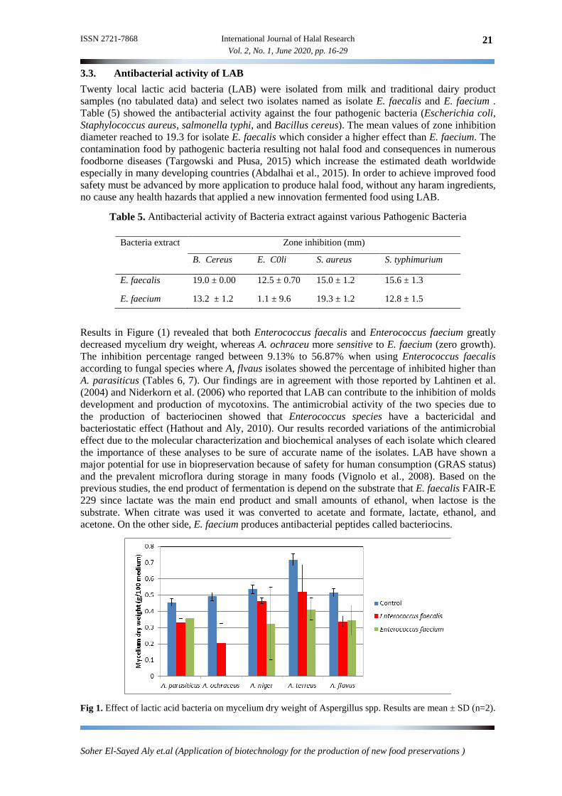
Results in Figure (1) revealed that both Enterococcus faecalis and Enterococcus faecium greatly decreased mycelium dry weight, whereas A. ochraceu more sensitive to E. faecium (zero growth). The inhibition percentage ranged between 9.13% to 56.87% when using Enterococcus faecalis according to fungal species where A, flvaus isolates showed the percentage of inhibited higher than A. parasiticus (Tables 6, 7). Our findings are in agreement with those reported by Lahtinen et al. (2004) and Niderkorn et al. (2006) who reported that LAB can contribute to the inhibition of molds development and production of mycotoxins. The antimicrobial activity of the two species due to the production of bacteriocinen showed that Enterococcus species have a bactericidal and bacteriostatic effect (Hathout and Aly, 2010). Our results recorded variations of the antimicrobial effect due to the molecular characterization and biochemical analyses of each isolate which cleared the importance of these analyses to be sure of accurate name of the isolates. LAB have shown a major potential for use in biopreservation because of safety for human consumption (GRAS status) and the prevalent microflora during storage in many foods (Vignolo et al., 2008). Based on the previous studies, the end product of fermentation is depend on the substrate that E. faecalis FAIR-E 229 since lactate was the main end product and small amounts of ethanol, when lactose is the substrate. When citrate was used it was converted to acetate and formate, lactate, ethanol, and acetone. On the other side, E. faecium produces antibacterial peptides called bacteriocins.
Fig 1. Effect of lactic acid bacteria on mycelium dry weight of Aspergillus spp. Results are mean ± SD (n=2).
Bacteria extract Zone inhibition (mm)
B. Cereus E. C0li S. aureus S. typhimurium
E. faecalis 19.0 ± 0.00 12.5 ± 0.70 15.0 ± 1.2 15.6 ± 1.3
E. faecium 13.2 ± 1.2 1.1 ± 9.6 19.3 ± 1.2 12.8 ± 1.5

22 International Journal of Halal Research ISSN 2721-7868
Vol. 2, No. 1, June 2020, pp. 16-29
Soher El-Sayed Aly et.al (Application of biotechnology for the production of new food preservations )
Table 6. Antifungal activity of Bacteria extracts against various fungal isolates
Bacteria extract
Zone inhibition (mm)
A. flavus
A. Parasiticus
A. ochraceus
A. niger
F. clomorum
F. graminerum
F. oxysporum
E. faecalis 15.6 ± 1.2 16.2 ± 1.54 10.6 ± 1.5 22.3 ± 1.4 11.5 ± 1.4 11.7 ± 1.5 15 ± 2.1
E.faecium 17.4 ± 1.8 19.5 ± 1.19 21.2 ± 1.1 18.9 ± 1.3 14.7 ± 1.8 15.6 ± 1.3 9.2 ± 1.4
Table 7. percentage % inhibition and average growth of different fungal species affected by Enterococcus spp.
Fungal species No of isolates
Enterococcus faecalis
avarege Enterococcus faecium
A.flavus 10 44.78-56.87 50.8 37.56-43.65 40.45 A. ochraceus 14 47.00-55.8 51.4 89.0-100 94.50 A. niger 25 31.24-36.9 34.7 42.73-51.76 47.20 A. parasiticus 8 22.12-34.7 28.4 13.52-21.85 17.68 A. terreus 6 33.36-37.8 35.58 50.17-23.0 36,58 F. clomorum 12 22.86-35.0 28.93 35.33-45.65 40.49 F. graminearum 9 9.13-22.7 15.9 44.7-56.8 50.75 Penicillium spp 6 14.8-34.7 24.75 22.05-34.6 28.32
3.4. Molecular identification of mycotoxin producing fungi
The ITS region is the official DNA barcoding marker for species-level Identification of Fungi. DNA barcoding systems employ a short standardized region (between 400 and 800 base pairs) to identify fungal species. ITS amplification products using the ITS1 and ITS4 primers are a unique band ranging from 500-600 bp was obtained for all isolated fungal strains (Fig 2). The amplicons of ITS regions were column purified and sequenced using a set of primers. The ITS region sequences were aligned using blast algorithm and compared with the published sequences of ITS region gene of different fungi (Table 8). The results revealed that the first fungal isolate of the sequenced 18S rRNA gene was identified as Aspergillus ochraceus and had (98%) similarity with Aspergillus ochraceus isolate Nitaf 24 (Fig 2). The second fungal isolate of the sequenced 18S rRNA gene was identified as Aspergillus flavus and had (99%) similarity with Aspergillus flavus strain UOA/HCPF 5774 (Fig 2). The high similarity between the fungal strains and their closest phylogenetic relative, indicating that 16S rRNA gene sequence data are helpful for identification of fungi. The gene sequence was deposited in GenBank database as; Aspergillus ochraceus Egy2 (Accession No. LC360803.1) and Aspergillus flavus Egy3 (Accession No. LC368455.1). Data in Figs (3,4) showed the phylogenetic tree based on ITS region sequences, the relationship between fungal isolates and other species. The tree was constructed using the neighbor-joining method.
The results indicated the importance of using molecular methods such as DNA barcoding systems (ITS region) for typing newly isolates microorganisms. Phenotypic and genotypic methods are part of the first step of identifications and selection of potential fungal isolates. These results are in agreement with Henry et al. (2000) who also used this method to identify Aspergillus at the species level and differentiate it from other true pathogenic and opportunistic molds using the 18S and 28S rRNA genes for primer binding sites. The contiguous internal transcribed spacer (ITS) region, ITS 1-5.8S-ITS 2, from referenced strains and clinical isolates of Aspergilli and other fungi, were amplified, sequenced, and compared with non-reference strain sequences in GenBank. ITS amplicons from Aspergillus species ranged in size from 565 to 613 bp .

ISSN 2721-7868 International Journal of Halal Research 23 Vol. 2, No. 1, June 2020, pp. 16-29
Soher El-Sayed Aly et.al (Application of biotechnology for the production of new food preservations )
Fig 2. Polymerase chain reaction-amplification of 18S rRNA gene; Lane M: Gene Ruler DNA Ladder 100 bp, Lane 1: 18S rRNA gene fragment of isolate Aspergillus ochraceus, Lane 2: 18S rRNA gene fragment of isolate Aspergillus flavus. DNA molecular weight marker Band sizes: 100, 200, 300, 400, 500, 600 and 700 bp
Fig 3. Phylogenetic tree showing relationship of closely related species constructed using the neighbor-joining method and based on 18S rRNA gene sequences. Isolate is closely related to Aspergillus ochraceus
Fig 4. Phylogenetic tree showing relationship of closely related species constructed using the neighbor-joining method and based on 18S rRNA gene sequences. Isolate is closely related to Aspergillus flavus

24 International Journal of Halal Research ISSN 2721-7868
Vol. 2, No. 1, June 2020, pp. 16-29
Soher El-Sayed Aly et.al (Application of biotechnology for the production of new food preservations )
Table 8. The nucleotide sequence of two fungal isolates
Strain Aligned Sequence Data
Asp
ergi
llus
och
race
us
CGCGGCGCGCCCCCCCCCCGCCCCCCCGATTCACCCATTTATACCTCCAAACACCCCTTGACCCAAAAAATGCGCGCCTTTGTTCCGGGGGGGTGCGCGCGCTCAACTTTCCTTTCCTTAAGGGGAAACCCTGCGGAAGGATCATTACTGAGTGAGGGTCCCTCGGGGCCCCAAACCTCCCCACCCCGTGGTATACCGTACCTTGTTGCTTCGGGCGAGCCCCGCCCCCTTTTTTCTTTTAGGGGGCACAGCGCTCGCCGGAGACACCAACGTGAACACTGTCTGAAGTTTTGTCGTCTGAGTCGATTGTATCGCAATCAGTTAAAACTTTCAACAATGGATCTCTTGGTTCCGGCATCGATGAAGAACGCAGCGAAATGCGATAATTAATGTGAATTGCAGAATTCAGTGAATCATCGAGTCTTTGAACGCACATTGCACCCCCTGGTATTCCGGGGGGTATGCCTGTCCGAGCGTCATTGCTGCCCTCAAGCACGGCTTGTGTGTTGGGTCGTCGTCCCCCCCCAGGGGGACGGGCCCGAAAGGCAGCGGCGGCACCGCGTCCGGTCCTCGAGCGTATGGGGCTTTGTCACCCGCTCTTGTAGCCCGGCCGGCTGCTGGCCGACGCTGAAAAGCAACCAACTATTTTCCAGGGGACCTCGGATCAGGTAGGATACCCGCTGAATTAGG
A
sper
gill
us f
lavu
s
GTGTAACCTGCAGCATGATTCATTACCGAGTGGTAGGGTTCCTTAGCGAGCCCAACCCTCCCCACCCCGTGTTTACTGTACTTTAATTGCTTCGGCGGGCCCCGCCCATTCATGGCCGCCCGGGGGTTTCAGCCCCGGGCCCCGCGCCCCGCCGGAGACACCACGAACTCTGTCTGATCTAGTGAAGTCTGAGTTGATTGTATCGCAATCAGTTAAAACTTTCAACAATGGATCTCTTGGTTCCGGCATCGATGAAGAACGCAGCGAAATGCGATAACTAGTGTGAATTGCAGAATTCCGTGAATCATCGAGTCTTTGAACGCACATTGCGCCCCCTGGTATTCCGGGGGGCATGCCTGTCCGAGCGTCATTGCTGCCCATCAAGCACGGCTTGTGTGTTGGGTCGTCGTCCCCTCTCCGGGGGGGACGGGCCCCAAAGGCAGCGGCGGCACCGCGTCCGATCCTCGAGCGTATGGGGCTTGTCACCCGCTCTGTAGGCCCGGCCGGCGCTTGCCGAACGCAAATCAACTTTCCAGGTTGACCTCGGATCAGGTAGGGAACCCGTGATTAGG
3.5. Molecular differentiation of isolated fungi using ISSR technique
Inter Simple Sequence Repeat (ISSR) represent genome region between microsatellite loci. Sequences amplified by ISSR-PCR can be used for delimiting species. Inter-simple sequence repeat-PCR is a simple, not expensive, robust, multi-locus marker system which has been used in examines genetic variability among fungal pathogens. Primers depend on a sequence repeat and the resultant PCR reaction amplifies the sequence between two SSRs, yielding a multilocus marker system useful for fingerprinting, diversity analysis and genome mapping (Chadha and Gopalakrishna, 2007).
In this study, ISSR PCR technique was used to reveal the genetic diversity among different fungal isolates in order to search the genetic diversity between these isolates. Out of fifty ISSR primers, sixteen primers produced storable and reproducible banding patterns. Sixteen primers produced 388 band positions (loci) and out of these loci, 370 loci amplified were polymorphic. Majority of the primers which produced polymorphic bands had been GA or AG repeats followed by AC or CA repeats. Among the primers used, UBC-842 [(GA)8 YG] produced most number of loci .
A total of 165 full bands were scored from the amplified products with the ten inter-Simple Sequence Repeat (ISSR) primer, 116 bands were polymorphic, 41 unique bands with average range size 143-1939 bp (Table, 9). All primers generated 100% polymorphism except primers ISSR-14 and ISSR-15 which generated 77, and79% respectively. The obtained results revealed that the primers ISSR-1, ISSR-3 have amplified the maximum number of bands 21-23 respectively. On the other hand primers, ISSR-8 and ISSR-14 have amplified the lowest number of bands 13 (Fig 5). These results indicated that the primers ISSR-1 and ISSR-3 are the most repeated sequences in

ISSN 2721-7868 International Journal of Halal Research 25 Vol. 2, No. 1, June 2020, pp. 16-29
Soher El-Sayed Aly et.al (Application of biotechnology for the production of new food preservations )
these fungal isolates than other primers. One the other hand, the forty-one unique bands that were detected among the total bands could be considered for marker-assisted selection. The maximum numbers of unique bands were amplified by primer ISSR-3 which recorded 11 bands, whereas the lowest number of unique bands was amplified by ISSR-2, ISSR-8, ISSR-14, and ISSR-15.
These results showed that ISSR primers are robust, informative maker and would be a better tool for genetic divergence and phylogenetic studies. These data were used to create a similarity matrix for the construction of dendrograms by means of the UPGMA method. Mahmoud et al. (2016) used ISSR markers to analyze the genetic diversity of 30 A. flavus isolates from five agricultural crops and air, suggested that ISSR biotechnology is a highly useful tool for characterizing genetic diversity of A. flavus isolated from different sources. ITS species identification is nowadays performed by traditional techniques combined with molecular markers, resulting in a higher performance of isolate characterization. In the present study, internal transcribed spacer, inter-simple sequence repeats (ISSR), molecular markers were used, with the aim of genetically characterizing strains of Aspergillus species.
Table 9. The ISSR primer names and specific character of ISSR analysis
The similarity matrix between isolated fungi revealed that isolates A. parasiticus and A. niger showed 59% similarity (Table 10). Results also showed that both A. terreus and A. niger had a similarity of 56%, followed by A. flavus and A. niger showing a similarity of 54%. Similarly, Yugander et al. (2015) used ISSR markers to examine the genetic variability of 24 strains of Aspergillus species isolated from paddy. Recently, Adss et al. (2017) used RAPD and ISSR to differentiate between 7 isolates of A. solani and their pathogenic capability. The resultant PCR reaction amplifies the sequence between two SSRs, yielding a multilocus marker system useful for finger printing, diversity analysis and genome mapping (Chadha and Gopalakrishna, 2007).
Table 10. Similarity matrix of isolated fungi based on ISSR analysis
Isolates
Matrix 1 2 3 4 5
A flavus 1
A. parasiticus 45 1
A. Terreus 44 47 1
A.niger 54 59 56 48 1
No Name of primer
Monomorphic bands*
polymorphic bands**
Number of
unique bands
Total bands
Polymorphism (%)
MW range (bp)
Mean of frequency
1 ISSR-1 0 16 5 21 100 214-1939 0.4 2 ISSR-2 0 17 2 19 100 323-997 0.5 3 ISSR-3 0 12 11 23 100 143-1730 0.4 4 ISSR-4 2 9 6 17 88 182-1338 0.5 5 ISSR-8 0 11 2 13 100 227-958 0.5 6 ISSR-11 0 11 3 14 100 145-1365 0.4 7 ISSR-12 0 9 5 14 100 182-1577 0.5 8 ISSR-13 0 14 3 17 100 197-1815 0.4 9 ISSR-14 3 8 2 13 77 278-1434 0.6 10 ISSR-15 3 9 2 14 79 905-1714 0.6 Total 8 116 41 165 94.4

26 International Journal of Halal Research ISSN 2721-7868
Vol. 2, No. 1, June 2020, pp. 16-29
Soher El-Sayed Aly et.al (Application of biotechnology for the production of new food preservations )
Fig 5. ISSR profile for five isolates of Aspergillus species. Lane 1: Aspergillus flavus; Lane 2: Aspergillus parasiticus; Lane 3: Aspergillus terreus; Lane 4: Aspergillus ochraceus; Lane 5: Aspergillus niger.
3.6. Cluster analyses Dendrogame was constructed using UPGMA cluster analyses to reveal the genetic relationships among five fungal isolates. Fig (6) showed four major clusters, the first group included A. flavus isolate; the second group contained A. terreus isolate; the third group included A. parasiticus and A. niger isolates; the fourth group contained A. ochraceus isolate. Similar observations were reported by Abdulateef et al. (2014) who used ISSR-PCR based technology to document genetic diversity among some local Iraqi isolates through the determination of the ability A. flavus isolates to produce aflatoxin B1.

ISSN 2721-7868 International Journal of Halal Research 27 Vol. 2, No. 1, June 2020, pp. 16-29
Soher El-Sayed Aly et.al (Application of biotechnology for the production of new food preservations )
Fig 6. Dendrogram showing the relationships between isolated fungi based on ISSR analysis. Isolate 1: Aspergillus flavus; Isolate 2: Aspergillus parasiticus; Isolate 3: Aspergillus terreus; Isolate 4: Aspergillus ochraceus; Isolate 5: Aspergillus niger
4. Conclusion
These results recorded that the antimicrobial activity of bacterial isolate Enterococcus faecalis was higher than Enterococcus faecium isolate against most of microbial strains under study concluded that these isolated bacteria can be applied as halal food preservation. The present study indicated that ISSR is a suitable and effective tool to evaluate genetic diversity among Aspergillus genera.
References
[1] Abdalhai, M.H., Fernandes, A.M., Xia, X., Musa, A., Ji, J., Sun, X., 2015. Electrochemical Genosensor to detect pathogenic bacteria (Escherichia coli O157:H7) as applied in real food samples (fresh beef) to improve food safety and quality control. J Agric. Food Chem. 63(20), 5017.
[2] Abdel-Shafi, S., Al-Mohammadi, A.R., Negm, S., Enan, G., 2014. Antibacterial activity of Lactobacillus delbreukiisub species bulgaricus isolated from Zabady, Life Sci. J. 11, 264-270.
[3] Abdel-Wahhab, M.A., Hassan, N.S., El-Kady, A.A., Khadrawy, Y.A., El-Nekeety, A.A., Mohamed, S.R., Sharaf, H.A., Mannaa, F.A., 2010. Red ginseng extract protects against aflatoxin B1 and fumonisins-induced hepatic pre-cancerous lesions in rats. Food Chem. Toxicol. 48(2), 733-742.
[4] Abdulateef, S.M., Aljubori, M.H., Abdulbaqi, N.J., 2014. Genetic diversity among some Aspergillus flavus isolates by using Inter-simple sequence repeats (ISSR). Iraqi J. Sci. 55(3), 986-993.
[5] Adimpong, D.B., Nielsen, D.S., Sørensen, K.I., Derkx, P.M., Jespersen, L., 2012. Genotypic characterization and safety assessment of lactic acid bacteria from indigenous African fermented food products. BMC Microbiol. 12. 10.1186/1471-2180-12-75.
[6] Adss, A.A., Abdel-Gayed, M.A., Botros, W., Hafez, E.E., 2017. Multilocus genetic techniques, RAPD-PCR and ISSR-PCR Markers and polygalacturonase activity as tools for differentiation among alternaria solani isolates on tomato fruits and relation to their pathogenicity in Egypt. Asian J. Plant Pathol. 11(1), 18-27.
[7] Aly, S.E., Abo-Sereih, N.A., Salim, R.G., Hathout, A.S., 2018. Isolation and molecular identification of food grade lactic acid bacteria and their antifungal activity. J. Biolo. Sci. 18(6), 260-269.
[8] Barnett, H.L., Hunter, B.B., 1986. Illustrated genera of fungi. 3rd ed. Minneapolis, MN: Burgess Co.
[9] Bintvihok, A., Thiengnin, S., Doi, K., Kumagai, S., 2002. Residues of aflatoxins in the liver, muscle and eggs of domestic fowls. J. Vet. Med. Sci. 64, 1037-1039.
[10] Chadha, S., Gopalakrishna, T., 2007. Comparative assessment of REMAP and ISSR marker assays for genetic polymorphism studies in Magnaporthe grisea. Curr. Sci. 93, 688-692.
[11] Felis, G. E., & Dellaglio, F. (2007). Taxonomy of Lactobacilli and Bifidobacteria. Current Issues Intestinal Microbiology, 8(2), 44-61.

28 International Journal of Halal Research ISSN 2721-7868
Vol. 2, No. 1, June 2020, pp. 16-29
Soher El-Sayed Aly et.al (Application of biotechnology for the production of new food preservations )
[12] Fonseca, H., 2012. A aflatoxina e o amendoim. Boletim Técnico no. 13 [Online]. http://www.micotoxinas.com.br/Boletim13.htm.
[13] Giraffa, G., 2003. Functionality of enterococci in dairy products. Int. J. Food Microbiol. 88(2-3), 215-222.
[14] Hathout, A.S., Aly, S.E., 2010. Role of lactic acid bacteria as a biopreservative agent of Talbina. J. Am. Sci. 6, 889-898.
[15] Hathout, A.S., Aly, S.E., 2014. Biological detoxification of mycotoxins: a review. Ann. Mocrobiol. 64(3), 905-919.
[16] Henry, T., Iwen, P.C., Hinrichs, S.H., 2000. Identification of Aspergillus species using internal transcribed spacer regions 1 and 2. J. Clin. Microbiol. 38, 1510-1515.
[17] Iqbal, S.Z., Asi, M.R., Zuber, M., Akhtar, J., Saif, M.J., 2013. Natural occurrence of aflatoxins and ochratoxin A in commercial chilli and chilli sauce samples. Food Cont. 30(2), 621-625.
[18] Khan, H., Flint, S., Yu, P.L., 2010. Enterocins in food preservation. Int. J.Food Microbiol. 141(1- 2), 1-10.
[19] Lahtinen, C.A., Haskard, A.C., Ouwehand, S.J., Salminen, J., Ahokas, T., 2004. Binding of aflatoxin B1 to cell wall components of Lactobacillus rhamnosus strain GG. Food Add. Contam. 21, 158-164.
[20] Lavanya, R., Sowmiya, S., Balaji, S., Muthuvelan, B., 2011. Screening and characterization of lactic acid bacteria from fermented milk. Brit. J. Dairy Sci. 2(1), 5-10.
[21] Leslie, J.F., Summerell, B.A., 2006. The Fusarium laboratory manual. UK: Blackwell Publishing Ltd.
[22] Magnoli, C, Astoreca, A., Ponsone, M.L., Fernández-Juri, M.J., Barberis, C., Dalcero, A.M., 2007. Ochratoxin A and Aspergillus section Nigri in peanut seeds at different months of storage in Córdoba, Argentina. Int. J. Food Microbiol. 119(3), 213-218.
[23] Mahmoud, M.A., EL-Samawaty, A.M.A., Yassin, M.A., Abd EL-Aziz, A.R.M., 2016. Genetic diversity analysis of Aspergillus flavus isolates from plants and air by ISSR Markers. Gen. Mol. Res. 15(2), 15028.
[24] Marcos Filho, J., 2005. Fisiologia de sementes de plantas cultivadas. Piracicaba: FEALQ.495.
[25] Nakai, V.K., Rocha, L.O., Gonçalez, E., Fonseca, H., Ortega, E.M.M., Corrêa, B., 2008. Distribution of fungi and aflatoxins in a stored peanut variety. Food Chem. 106, 285-290.
[26] Nelson, P.E., Toussoun, T.A., Marasas, W.F.O.., 1983. Fusarium species. An illustrated manual for identification. University Park: Pennsylvania State University Press. 193.
[27] Niderkorn, V., Boudra, H., Morgavi, D.P., 2006. Binding of Fusarium mycotoxins by fermentative bacteria in vitro. J. Appl. Microbiol. 101,849-856.
[28] Pildain, M.B., Frisvad, J.C., Vaamonde, G., Cabral, D., Varga, J., Samson, R.A., 2008. Two novel aflatoxin-producing Aspergillus species from Argentinian peanuts. Int. J. Syst. Evolut. Microbiol. 58,725-735.
[29] Rohlf, F.J., 1993. NTSYS-pc: Em unumerical taxonomy and multivariate system. Version 2.9. New York. Applied Biostatistics. Schimmelcuhves, pp: 116. The Netherlands species using internal transcribed spacer regions 1 and 2. J. Clin. Microbiol. 9, 213.
[30] Sabry, B.A., Hathout A.S., Nooh, A., Aly, S.E., Shehata, M., 2016. The prevalenc of aflatoxin and Aspergillus parasiticus in Egyptian sesame seeds. Int. J. ChemTech Rese. 9(11), 308-319.
[31] Sahab, A.F., Aly, S.E., Nawar, L.S., El-Faham, S.Y., 2011. Fungal occurrence in physic nut (Jatropha curcas) seeds during storage and possibility aflatoxin production by Aspergillus flavus and Aspergillus parasiticus isolates. J. Am. Sci. 21, 342
[32] Sahab, A.F., Hathout, A.S., Sabry, B.A., Aly, S.E., 2014. Application of some plant essential oils to control Fusarium isolates associated with freshly harvested maize in Egypt. J. Essen. Oil Bear. Plants 17(6), 1146 -1155.
[33] Targowski, T., Płusa, T., 2015. Overall principles of treatment in case of 689 toxicological threats. Polski Merkuriusz Lekarski 39 (231), 193-198.

ISSN 2721-7868 International Journal of Halal Research 29 Vol. 2, No. 1, June 2020, pp. 16-29
Soher El-Sayed Aly et.al (Application of biotechnology for the production of new food preservations )
[34] Větrovský, T., Baldrian, P., Neufeld, J., 2013.The variability of the16S rRNA gene in bacterial genomes and its consequences for bacterial community analyses. Plos One 8(2), e57923 doi: 10.1371/journal.pone.0057923.
[35] Vignolo, G., Castellano, P., Belfiore, C., Fadda, S., 2008. A review of bacteriocinogenic lactic acid bacteria used as bio-protective cultures in fresh meat produced in Argentina. Meat Sci. 79(3), 483-99. doi: 10.1016/j.meatsci.2007.10.009.
[36] White, T.J., Bruns, T., Lee, S., Taylor, J.W., 1990. Amplification and direct sequencing of fungal ribosomal RNA genes for phylogenetics. In: PCR Protocols: A Guide to Methods and Applications (Innis MA, Gelfand DH, Sninsky JJ and White TJ, eds.). Academic Press Inc., New York, 315-322.
[37] Williams, J.G., Kubelik, A.R., Livak, K.J., Rafalski, J.A., Tingey S.V., 1990. DNA polymorphisms amplified by arbitrary primers are useful as genetic markers. Nucleic Acids Res. 18, 6531-6535.
[38] Yugander, A., Ladhalakshmi, D., Prakasham, V., Mangrauthia, S.K., Prasad, M.S., Madhav, M.S., Sundaram, R.M., Laha, G.S., 2015. Pathogenic and genetic variation among the isolates of Rhizoctonia solani (AG 1-IA), the Rice sheath blight pathogen. J. Phytopathol. 163(6), 465-474.