Embed Size (px)
Citation preview
Dharmacon™
RNAi, Gene Expression & Gene Editing
Application Note
GE Healthcare
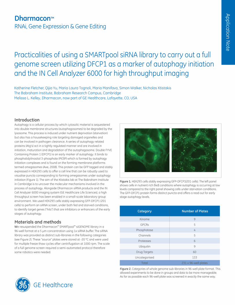
Practicalities of using a SMARTpool siRNA library to carry out a full genome screen utilizing DFCP1 as a marker of autophagy initiation and the IN Cell Analyzer 6000 for high throughput imaging
Katherine Fletcher, Qijia Yu, Maria Laura Tognoli, Maria Manifava, Simon Walker, Nicholas KtistakisThe Babraham Institute, Babraham Research Campus, Cambridge Melissa L. Kelley, Dharmacon, now part of GE Healthcare, Lafayette, CO, USA
IntroductionAutophagy is a cellular process by which cytosolic material is sequestered into double membrane structures (autophagosomes) to be degraded by the lysosome. This process is induced under nutrient deprivation (starvation) but also has a housekeeping role targeting damaged organelles and can be involved in pathogen clearance. A series of autophagy related proteins (Atg’s) act in a tightly regulated manner and are involved in initiation, maturation and degradation of the autophagosome. Double FYVE Containing Protein 1 (DFCP1) is an early marker of autophagy. It binds to phosphatidylinositol 3-phosphate (PI(3)P) which is formed by autophagy initiation complexes and is found on the forming membrane platforms termed omegasomes (Axe, 2008). This protein can be GFP tagged and stably expressed in HEK293 cells to offer a cell line that can be robustly used to visualise puncta corresponding to forming omegasomes under autophagy initiation (Figure 1). The aim of the Ktistakis lab at The Babraham Institute in Cambridge is to uncover the molecular mechanisms involved in the process of autophagy. Alongside Dharmacon siRNA products and the IN Cell Analyzer 6000 imaging system (GE Healthcare Life Sciences), a high throughput screen has been enabled in a small-scale laboratory group environment. We used HEK293 cells stably expressing GFP-DFCP1 (201 cells) to perform an siRNA screen, under both fed and starved conditions, to identify target genes (“hits”) that are inhibitors or enhancers of the early stages of autophagy.
Materials and methods We resuspended the Dharmacon™ SMARTpool™ siGENOME library in a 96-well format at a 5 µm concentration using 1x siRNA buffer. The siRNA library was provided as distinct sub-libraries in the following categories (see Figure 2). These “source” plates were stored at -20 °C and were used for multiple freeze thaw cycles after centrifugation at 1000 rpm. The scale of a full genome screen required a semi-automated protocol therefore some robotics were needed.
Figure 1. HEK293 cells stably expressing GFP-DFCP1(201 cells). The left panel shows cells in nutrient rich (fed) conditions where autophagy is occurring at low levels compared to the right panel showing cells under starvation conditions. The GFP-DFCP1 protein forms distinct puncta and offers a read out for early stage autophagy levels.
Category Number of Plates
Kinome 9
GPCRs 5
Phosphatase 4
Channels 5
Proteases 6
Ubiquitin 9
Drug Targets 60
Uncategorised 133
Total 231 × 96-well plates
Figure 2. Categories of whole genome sub-libraries in 96-well plate format. This allowed experiments to be done in groups and data to be more manageable. As far as possible each 96-well plate was screened in exactly the same way.
201 cells (HEK293 cells stably expressing GFP-DFCP1) were thawed from a common stock of cells dedicated to the screen and were used at least 48 hours after passaging. 201 cells were cultured in DMEM medium containing 10% FBS, 1% penicillin and streptomycin, 800 µg/mL G418.
Cells were reverse transfected using a Biomeck™ 3000 robot (Beckman Coulter) to spot 2.5 µL of siRNA from the source plate onto an empty, custom made glass-bottomed 96-well plate (experiments performed in duplicate). Control siRNAs, including Dharmacon™ ON-TARGETplus Non-targeting Pool (Dharmacon, Cat #D-001810-10-20) and BECN1 (8678) ON-TARGETplus SMARTpool reagent (Dharmacon, Cat #L-010552-00-0020), for the starvation screen were added manually. The DharmaFECT 1 Transfection Reagent (Dharmacon, Cat #T-2001-03) in conjunction with Opti-MEM™ was added on top of the siRNA using a Multidrop™ Combi Reagent Dispenser (Thermo Fisher Scientific, Inc), allowing 20 minutes before adding the cells. Cells were counted using a haemocytometer, calculating 10 000 cells per well and seeded on top of the transfection reaction using the multidrop dispenser.
After 24 hours cells were fed on top with additional medium. At 72 hours after plating, the cells were either starved with medium minus amino acids (140 mM NaCl, 1 mM CaCl2, 1 mM MgCl2, 5 mM glucose, 20 mM Hepes pH 7.4 + 1% BSA) for 1 hour or immediately fixed using 3.7% formaldehyde in 200 mM Hepes pH7.2. The nuclei were stained with Hoechst dye in 2% BSA-PBS-azide at a 1:10 000 dilution and plates were left at -4 °C overnight ready to be imaged and analysed.
Imaging with the IN Cell Analyzer 6000 Before the IN Cell Analyzer 6000 system, imaging used to be the bottleneck of the screening process with each 96-well plate taking more than an hour to image, and a similar amount of time to be run through the attached analysis software. Due to knockdown-induced cell death we found that the automatic focus attached to the microscope was not robust enough to recapture the focus in subsequent wells (Figure 3). This led to additional time for reimaging of plates.
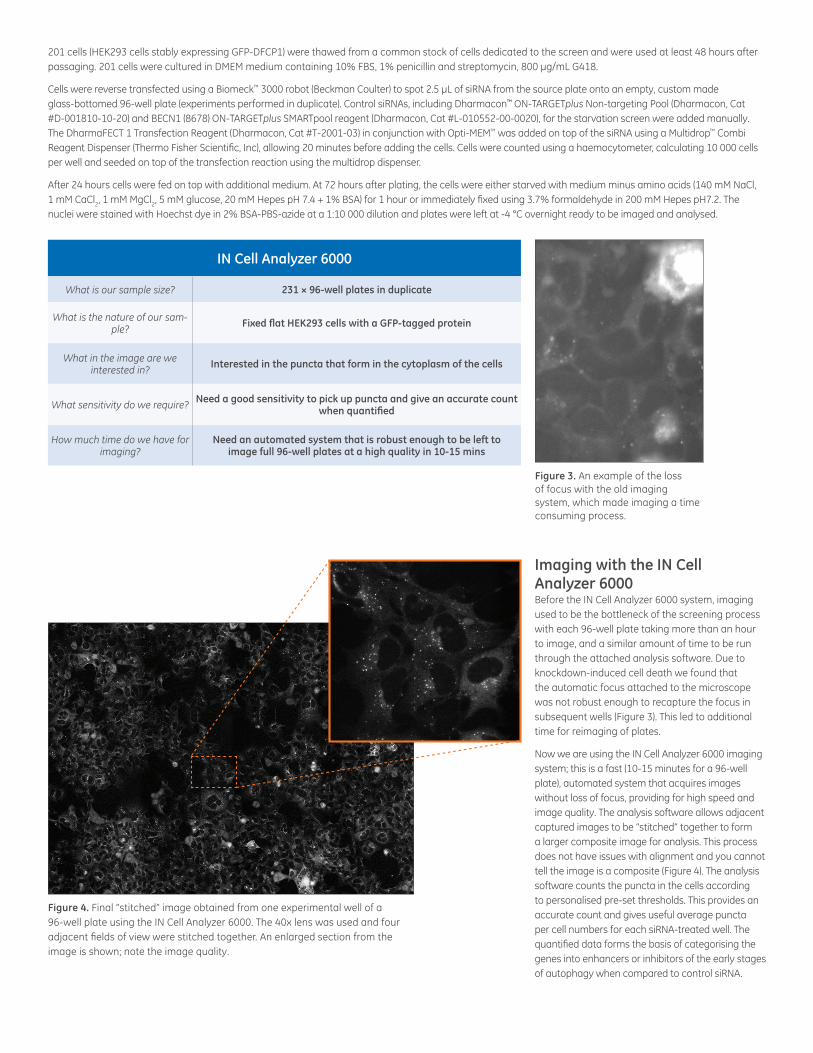
Now we are using the IN Cell Analyzer 6000 imaging system; this is a fast (10-15 minutes for a 96-well plate), automated system that acquires images without loss of focus, providing for high speed and image quality. The analysis software allows adjacent captured images to be “stitched” together to form a larger composite image for analysis. This process does not have issues with alignment and you cannot tell the image is a composite (Figure 4). The analysis software counts the puncta in the cells according to personalised pre-set thresholds. This provides an accurate count and gives useful average puncta per cell numbers for each siRNA-treated well. The quantified data forms the basis of categorising the genes into enhancers or inhibitors of the early stages of autophagy when compared to control siRNA.
Figure 3. An example of the loss of focus with the old imaging system, which made imaging a time consuming process.
IN Cell Analyzer 6000
What is our sample size? 231 × 96-well plates in duplicate
What is the nature of our sam-ple?
Fixed flat HEK293 cells with a GFP-tagged protein
What in the image are we interested in?
Interested in the puncta that form in the cytoplasm of the cells
What sensitivity do we require? Need a good sensitivity to pick up puncta and give an accurate count
when quantified
How much time do we have for imaging?
Need an automated system that is robust enough to be left to image full 96-well plates at a high quality in 10-15 mins
Figure 4. Final “stitched” image obtained from one experimental well of a 96-well plate using the IN Cell Analyzer 6000. The 40x lens was used and four adjacent fields of view were stitched together. An enlarged section from the image is shown; note the image quality.
Orders can be placed at:gelifesciences.com/dharmacon
Customer Support: [email protected] Support: [email protected] or1.800.235.9880; 303.604.9499 if you have any questions.
GE Healthcare
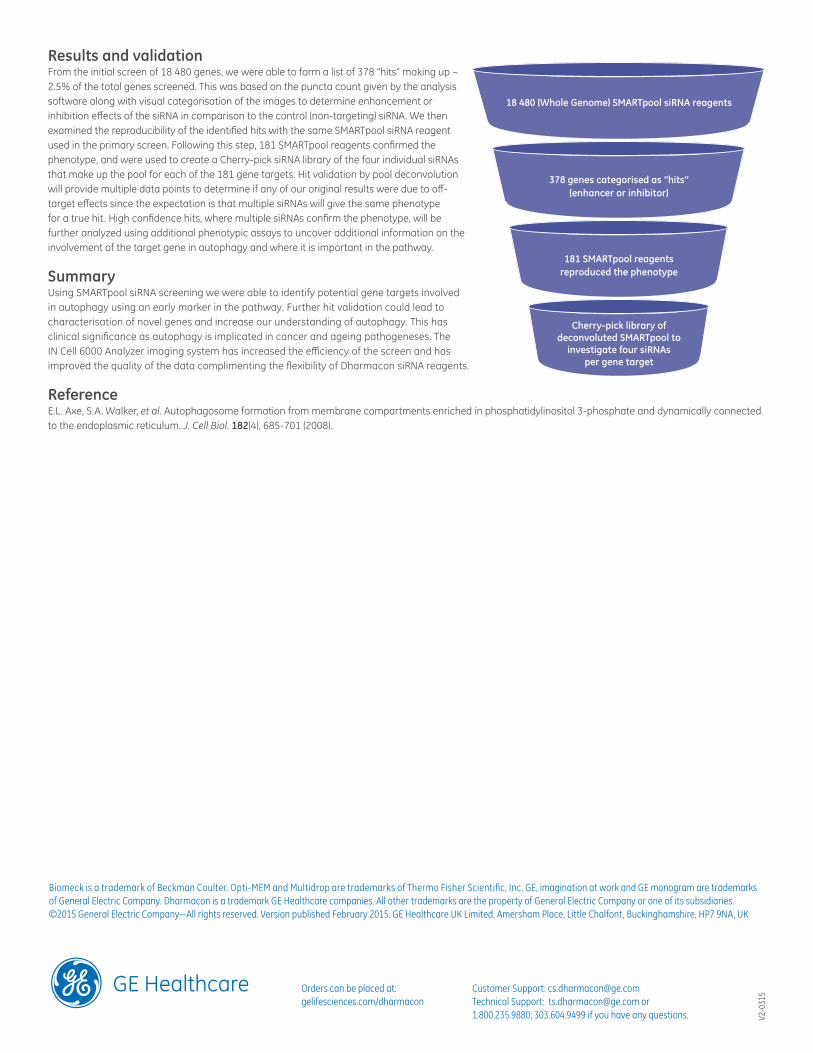
Results and validation From the initial screen of 18 480 genes, we were able to form a list of 378 “hits” making up ~ 2.5% of the total genes screened. This was based on the puncta count given by the analysis software along with visual categorisation of the images to determine enhancement or inhibition effects of the siRNA in comparison to the control (non-targeting) siRNA. We then examined the reproducibility of the identified hits with the same SMARTpool siRNA reagent used in the primary screen. Following this step, 181 SMARTpool reagents confirmed the phenotype, and were used to create a Cherry-pick siRNA library of the four individual siRNAs that make up the pool for each of the 181 gene targets. Hit validation by pool deconvolution will provide multiple data points to determine if any of our original results were due to off-target effects since the expectation is that multiple siRNAs will give the same phenotype for a true hit. High confidence hits, where multiple siRNAs confirm the phenotype, will be further analyzed using additional phenotypic assays to uncover additional information on the involvement of the target gene in autophagy and where it is important in the pathway.
Summary Using SMARTpool siRNA screening we were able to identify potential gene targets involved in autophagy using an early marker in the pathway. Further hit validation could lead to characterisation of novel genes and increase our understanding of autophagy. This has clinical significance as autophagy is implicated in cancer and ageing pathogeneses. The IN Cell 6000 Analyzer imaging system has increased the efficiency of the screen and has improved the quality of the data complimenting the flexibility of Dharmacon siRNA reagents.
Reference E.L. Axe, S.A. Walker, et al. Autophagosome formation from membrane compartments enriched in phosphatidylinositol 3-phosphate and dynamically connected to the endoplasmic reticulum. J. Cell Biol. 182(4), 685-701 (2008).
Biomeck is a trademark of Beckman Coulter. Opti-MEM and Multidrop are trademarks of Thermo Fisher Scientific, Inc. GE, imagination at work and GE monogram are trademarks of General Electric Company. Dharmacon is a trademark GE Healthcare companies. All other trademarks are the property of General Electric Company or one of its subsidiaries. ©2015 General Electric Company—All rights reserved. Version published February 2015. GE Healthcare UK Limited, Amersham Place, Little Chalfont, Buckinghamshire, HP7 9NA, UK
V2-0
315
18 480 (Whole Genome) SMARTpool siRNA reagents
378 genes categorised as “hits” (enhancer or inhibitor)
181 SMARTpool reagents reproduced the phenotype
Cherry-pick library of deconvoluted SMARTpool to
investigate four siRNAs per gene target