Embed Size (px)
Citation preview

CellCelector is protected by US patent 9,822,331 B2
Rev. 20 / 24.09.2018
In-Plate Cell Sorting of Rare Sub Populations
Gentle Single Cell Transfer for Cultivation
Application Note Automated Single Cell PCR Preparation
CellCelector is protected by US patent 9,822,331 B2
Rev. 20 / 24.09.2018
Introduction
Automated Harvesting of Cells utilizing the
CellCelector
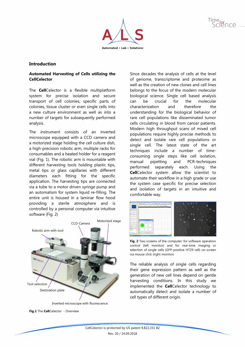
The CellCelector is a flexible multiplatform
system for precise isolation and secure
transport of cell colonies, specific parts of
colonies, tissue cluster or even single cells into
a new culture environment as well as into a
number of targets for subsequently performed
analysis.
The instrument consists of an inverted
microscope equipped with a CCD camera and
a motorized stage holding the cell culture dish,
a high-precision robotic arm, multiple racks for
consumables and a heated holder for a reagent
vial (Fig. 1). The robotic arm is mountable with
different harvesting tools holding plastic tips,
metal tips or glass capillaries with different
diameters each fitting for the specific
application. The harvesting tips are connected
via a tube to a motor driven syringe pump and
an automatism for system liquid re-filling. The
entire unit is housed in a laminar flow hood
providing a sterile atmosphere and is
controlled by a personal computer via intuitive
software (Fig. 2)
Since decades the analysis of cells at the level
of genome, transcriptome and proteome as
well as the creation of new clones and cell lines
belongs to the focus of the modern molecular
biological science. Single cell based analysis
can be crucial for the molecular
characterization and therefore the
understanding for the biological behavior of
rare cell populations like disseminated tumor
cells circulating in blood from cancer patients.
Modern high throughput scans of mixed cell
populations require highly precise methods to
detect and isolate rare cell populations or
single cell. The latest state of the art
techniques include a number of time-
consuming single steps like cell isolation,
manual pipetting and PCR-techniques
performed separately each. Using the
CellCelector system allow the scientist to
automate their workflow in a high grade or use
the system case specific for precise selection
and isolation of targets in an intuitive and
comfortable way.
Fig. 2 Two screens of the computer: for software operation
control (left monitor) and for real-time imaging or
selection of single cells (GFP-positive HT29 cell) on-screen
via mouse click (right monitor)
The reliable analysis of single cells regarding
their gene expression pattern as well as the
generation of new cell lines depend on gentle
harvesting conditions. In this study we
implemented the CellCelector technology to
automatically detect and isolate a number of
cell types of different origin.
Fig.1 The CellCelector - Overview
CCD-Camera
Robotic arm with tool
Tool selection
Motorized stage
Destination plate
Inverted microscope with fluorescence
CellCelector is protected by US patent 9,822,331 B2
Rev. 20 / 24.09.2018
Fig. 4 Cell culture in the imaging software for selection of a
specific target region or cell
Technology and Work Flow
1. Scan & Imaging
The culture dish with the region of interest is
scanned automatically by a high resolution camera
employing the motorized stage (Fig. 3 left). The
entire collection of single images during the scan is
combined by the software into an overview image
(Fig. 3 right). Integrated fluorescence filters can be
very useful in terms of immunochemical labeling
and staining and allows the user in-plate cell sorting
before picking.
2. Selection & Targeting
According to selection parameters (size, diameter
and shape factor of the cell etc.) predefined by the
user the software detects and selects the targeted
cells or colonies (Fig. 4). Selection can be done using
a great variety of analysis methods provided by the
microscope (fluorescence, bright field, phase
contrast) and the Cell*D software providing 3D
imaging, overlays and movies. An overlay of phase
contrast and fluorescence image can be created.
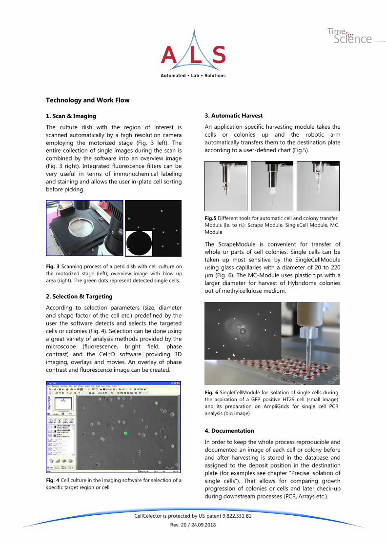
3. Automatic Harvest
An application-specific harvesting module takes the
cells or colonies up and the robotic arm
automatically transfers them to the destination plate
according to a user-defined chart (Fig.5).
The ScrapeModule is convenient for transfer of
whole or parts of cell colonies. Single cells can be
taken up most sensitive by the SingleCellModule
using glass capillaries with a diameter of 20 to 220
µm (Fig. 6). The MC-Module uses plastic tips with a
larger diameter for harvest of Hybridoma colonies
out of methylcellulose medium.
4. Documentation
In order to keep the whole process reproducible and
documented an image of each cell or colony before
and after harvesting is stored in the database and
assigned to the deposit position in the destination
plate (for examples see chapter “Precise isolation of
single cells”). That allows for comparing growth
progression of colonies or cells and later check-up
during downstream processes (PCR, Arrays etc.).
Fig. 3 Scanning process of a petri dish with cell culture on
the motorized stage (left); overview image with blow up
area (right). The green dots represent detected single cells.
Fig.5 Different tools for automatic cell and colony transfer
Moduls (le. to ri.): Scrape Module, SingleCell Module, MC
Module
Fig. 6 SingleCellModule for isolation of single cells during
the aspiration of a GFP positive HT29 cell (small image)
and its preparation on AmpliGrids for single cell PCR
analysis (big image)

CellCelector is protected by US patent 9,822,331 B2
Rev. 20 / 24.09.2018
Software
The CellCelector Software (Fig. 7) contains
numerous features for individual analysis of cells
and cell colonies either automatically or on-screen.
The software combines imaging facilities (camera
control, fluorescence, overlay etc.) with the robot
control for the cell harvest.
The measurement bar provides several tools for
measurement of cells, colonies or structures on-
screen like diameter, area, distance to adjacent cells
etc. (Fig. 8). It is helpful for identification of targets
as well as for the right choice of capillary diameters.
It is possible to select specific cells of interest on-
screen. Driving the microscopic motor stage allows
for searching within the culture dish for target cells
and to mark them by mouse click, either for instant
picking or for collection of multiple targets into a
picking list.
It is possible to select on-screen specific areas out of
the tissue or cell colony. Driving the microscopic
motor stage allows for searching within the culture
dish and then marking single cells, colonies or areas
with a cross-hair, either for instant picking or a
collection of multiple targets.
The well navigator (Fig. 9) indicates where the
camera focuses on. Wells can be addressed for cell
harvest and focus by mouse click and will be shown
in green color. The microscope motor stage is
connected to the navigator and will be driven to the
selected point of view. Special formats of plates and
dishes can be easily configured by the user and
saved for later use.
Fig. 11 Well navigator showing a 6 well plate
Fig. 9 Well navigator showing a 6 well plate
Fig. 8 Measurement bar (left) and close image from
screenshot above showing application of the
measurement tools on-screen (right)
Fig. 7 Screenshot of the software showing software control with well navigator (left) and microscopic image of mouse tumor
cells with measured cell diameter.
CellCelector is protected by US patent 9,822,331 B2
Rev. 20 / 24.09.2018
after picking
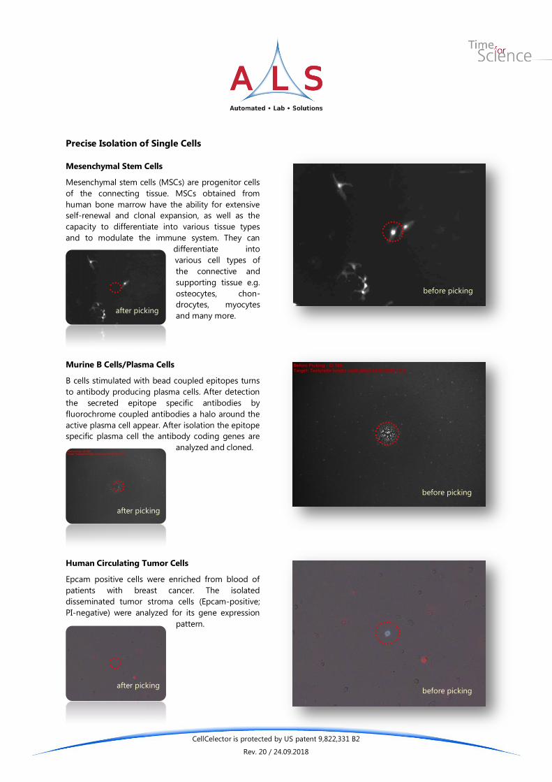
Precise Isolation of Single Cells
Mesenchymal Stem Cells
Mesenchymal stem cells (MSCs) are progenitor cells
of the connecting tissue. MSCs obtained from
human bone marrow have the ability for extensive
self-renewal and clonal expansion, as well as the
capacity to differentiate into various tissue types
and to modulate the immune system. They can
differentiate into
various cell types of
the connective and
supporting tissue e.g.
osteocytes, chon-
drocytes, myocytes
and many more.
Murine B Cells/Plasma Cells
B cells stimulated with bead coupled epitopes turns
to antibody producing plasma cells. After detection
the secreted epitope specific antibodies by
fluorochrome coupled antibodies a halo around the
active plasma cell appear. After isolation the epitope
specific plasma cell the antibody coding genes are
analyzed and cloned.
Human Circulating Tumor Cells
Epcam positive cells were enriched from blood of
patients with breast cancer. The isolated
disseminated tumor stroma cells (Epcam-positive;
PI-negative) were analyzed for its gene expression
pattern.
before picking
before picking
after picking
after picking
before picking

CellCelector is protected by US patent 9,822,331 B2
Rev. 20 / 24.09.2018
Precise Isolation of Single Cells
Schwann Cells (Axolotl)
In this evaluation GFP tagged Schwann cells
prepared from food of Axolotl were isolated. The
tissue was mashed and resuspended in an adequate
dilution. Schwann cells could be easily detect via a
combined Bright Field and Fluorescence
Illumination and isolated precisely from the debris.
Colon Carcinoma Cells (HT29 Cells)
The Human colon adenocarcinoma grade II cell line
HT29 is an often used model system in cancer
research. Here we evaluate the conditions (dilution,
surface of culture plates or picking settings) to
automate the isolation of circulating tumor cells.
GFP-Positive Single Cells from Mosaic Colonies
In this experiment we successfully evaluate the
precise isolation of single cells from cell colonies.
after picking
before picking
after picking
after picking
before picking
before picking

CellCelector is protected by US patent 9,822,331 B2
Rev. 20 / 24.09.2018
Precise Isolation of Single Cells
Human Embryonic Kidney Cells (HEK)
HEK cells have been widely-used in cell biology
research for many years. There are easy to grow and
used for transfections in research and for
therapeutic protein production in the bio-
technological industry.
Human Sperm Cells
The genomic analysis of human sperm cell is of
special interest in the field of criminal forensic.
Fungal Spores
In this case spores of Sordaria macrospora were
picked. In other experiments spores germinating in
agar were isolated.
before picking
after picking
before picking
after picking
before picking
after picking

CellCelector is protected by US patent 9,822,331 B2
Rev. 20 / 24.09.2018
Special Application in Precise Isolation of
Single Cells
Automated Recovery of Single Cells Identified by
Microengraving
A mixture of two mouse cell lines (12CA5 and
HYB099-01) was distributed on micro scale
Polydimethylsiloxane (PDMS) slides carrying 84672
cuboid microwells with an edge length of 50 µm.
Using the unique microengraving technology a
subpopulation from a cell mixture was identified and
isolated with the CellCelector as single cells from
the micro well. Using microengraving technology
single cells can be analyzed for many criteria by high
throughput screening before isolation of the cell sub
population of interest.
Automated Detection, Isolation and Genetic
Analysis of Single Circulating Tumor Cells
CD90 positive cells from patients suffering breast
cancer were detected using several fluorescence
criteria and isolated using glass capillaries with an
inner diameter of 50 µm. Just before picking, PCR
mix was aspirated in the needle. The picked cell was
released on anchor positions at the surface of
Ampligrids (see right figure on the right), the
bottom of PCR plates or the lid of Eppendorf cups.
During releasing the cell was mixed with the PCR
mix and covered with oil to prevent evaporation in a
unique single step manner (see right figure A).
Subsequently performed RT-PCR and q-PCR for 3
genes revealed the reliability of picking and transfer
for 8 picked single cells. The right image B displays
the (dR) values (left) and melting curves (right) for
genes A, B and C from 8 independent single cell
reactions.
after picking
before picking
A
B
A B C

CellCelector is protected by US patent 9,822,331 B2
Rev. 20 / 24.09.2018
A B
C D E
F G H
Selection of Target Single Cells
The automatic detection of single cells requires a set
of target specific characteristic parameters. The
basic detection process is working with grey or color
values. Setting a range of grey values (by definition
light and dark color thresholds fitting for the cells of
interest and visualized as green colored areas on the
reference image) can be sufficient to separate the
target cells from the background – especially for
cells labeled with fluorescence markers (Fig. 10).
After scanning the detected cells are displayed as
overview map (Fig. 11) and listed in a data sheet.
Changing the range of grey
values (Fig. 12 A and B)
allows the user to specify
the areas to detect (Fig. 12
D and E). By combining the
grey value based obtained
signals (Fig. 12 D and E) with
additional parameters like
size, diameter, shape factor,
sphericity etc. (Fig. 12 F) it is
possible to increase the
quality of the scanning
results significantly (Fig 12 G
and H).
Fig. 10 Screenshot of setting grey value thresholds for mesenchymal stem cell detection
Fig. 11: Overview map (top left) after scanning GFP-
expressing H9 cells and enlarged area marked by the
rectangle.
Fig. 12 Setting parameters for scanning human sperms. Different grey value thresholds
(A & B) detect different parts of the sperms (D & E) from the original reference image (C).
Additional parameters (F) improve the scanning result (G & H).

CellCelector is protected by US patent 9,822,331 B2
Rev. 20 / 24.09.2018
To improve the scanning result further it can be
useful to process the original reference image as
first step while scanning. As shown in Fig. 14 single
plasma cells were detected secreting antibodies
which are visualized by fluorochrome labeled
antibodies (Fig. 14 A) and detectable only as cloud-
like conglomerate (Fig. 14 B) holding a number of
picking positions calculated for each dot. Using
morphological filter features (Fig. 13) the dotted
halo surrounding the cell can be processed into a
consistent area (Fig. 14 C) which is detectable as
homogeneous colored area (Fig. 14 D) with a single
picking position in its central part.
A B
C D
Fig. 14 Detection of antibody secreting plasma cells. The plasma cells are located within the central part of the detectable dotted
halo. Original image (A) and its detectable areas (dotted halo (B); processed image (C) and its detectable homogeneous area (D).
Fig. 13 Dialog window to define morphological filter
settings

CellCelector is protected by US patent 9,822,331 B2
Rev. 20 / 24.09.2018
Conclusions
Efficiency
The CellCelector is an efficient and highly selective
tool for a safe transfer of single cells and cell
colonies without interfering with important
properties of the cells such as pluripotency and
viability. Several experiments proofed that an
automated process can improve the quality of
transferred single cells or cell colonies and allow to
set standardized condition for the entire cell picking
process. Of special importance is the cell sorting
capacity in the culture plate directly to target cell
subpopulations before picking the cells.
Sensitivity
An assessment of pluripotency-associated markers
and differentiating cells after automated picking and
replating for several times confirmed their
pluripotency status and a lower number of dead
cells compared to picking by hand. Several
parameters can be combined individually to apply
the right and most gentle resolving aspiration force.
By that and using the heatable destination positions
(37°C) the mechanical stress is reduced and the
viability of cells after transfer is increased. Highly
precise tools allow for a safe transfer of even single
cells provide new possibilities in stem cell research.
With a reproducibly small amount of aspiration
(below 0.1 µl) quantitative single cell RT-PCR and
PCR analysis becomes a standard method.
Flexibility
The CellCelector also enables the scientist to select
cells precisely according to their state of
differentiation using different fluorescence
excitations and markers at the same time. Hence,
the integration of a state-of-the-art microscope
which is widely used in laboratories provides an
innovative and time-saving combination of various
analysis methods and a direct transfer into a new
culture environment or wells for further genetic
analysis (PCR). For working with primary cells and
tissue the CellCelector and the ALS Incubator
FlowBox (Fig. 16) are recommended since
physiological conditions like temperature and CO2
atmosphere can easily and precisely be adjusted.
Security
When working with cells determined for
transmission to patients contamination with
pathogens is an important issue. The complete
automation of the picking process decreases the
need of manual intervention (dish positioning) and
therefore increases the security of valuable cell
material from contamination with retroviruses or
other pathogens.
The CellCelector is placed under a sterile hood and
resistant against intense surface sterilization using
Ethanol and UV-light.
Fig.15 The CellCelector can be placed in a flow box for
increased safety of cell cultures
Fig. 16 The CellCelector is placed in the ALS Incubator
FlowBox with high CO2-atmosphere and heated
environment (37°C), especially useful for long-term
experiments and primary cell cultures.

CellCelector is protected by US patent 9,822,331 B2
Rev. 20 / 24.09.2018
ALS Automated Lab Solutions GmbH headquarters in Jena, Germany
ALS Automated Lab Solutions GmbH is located in Jena, a dynamic city famous for microscopy and
material science. ALS is a specialist for the development of innovative technological solutions for cell
biology research and molecular biology. ALS lifts cell culture to a new level of choice and control on
the leading edge in cell biology, cell therapy research, regenerative medicine and drug discovery. With
automation and standardization of laborious manual procedures, ALS supports science and research
for more efficiency and the creation of new methods for the science of tomorrow.
ALS is partner of:
Please do not hesitate to contact us for further information:
Jens Eberhardt
ALS Automated Lab Solutions GmbH
Otto-Eppenstein-Str. 30
07745 Jena
Germany
Phone: +49 (0) 3641 4820-0
Fax: +49 (0) 3641 4820-11
E-Mail: [email protected]