Embed Size (px)
Citation preview

Asian Pacific Journal of Cancer Prevention, Vol 17, 2016 3185
10.14456/apjcp.2016.73/APJCP.2016.17.7.3185Applicability of Radioguided Occult Lesion Localization in Non-Palpable Benign Breast Lesions
Asian Pac J Cancer Prev, 17 (7), 3185-3190
Introduction
Mammography screening programs are frequently used for the detection of non-palpable clinically occult breast lesions and the widespread use of high-resolution ultrasound has increased the number of detected masses. Although most of the detected masses are benign and do not require surgery, sometimes surgery is needed because of discordance between results of pathologic and radiologic examinations, patient anxiety, positive family history, and patient desire despite benign pathology results. Successful management of these non-palpable non-malignant lesions requires complete surgical excision with optimal cosmetic outcome and minimal morbidity, for which accurate localization of the lesion is essential.
Ultrasound is the most commonly used imaging modality for localization, provided the lesions are visible on ultrasound examination. Mammography and magnetic resonance imaging (MRI) are other methods of 1Cancer Research Center, Cancer Institute of Iran, 2Department of Nuclear Medicine, Vali-Asr Hospital, School of Medicine, 3Division of Surgical Oncology, Department of Surgery, 4Department of Radiology, Medical Imaging Center, Tehran University of Medical Sciences, Tehran, Iran *For correspondence: [email protected]
Abstract
Background: This study was designed to compare radioguided versus routine wire localization of non-palpable non-malignant breast lesions in terms of efficacy for complete excision, ease of use, time saving, and cosmetic outcome. Materials and Methods: Patients with non-palpable breast masses and non-malignant core biopsy results who were candidates for complete surgical lumpectomy were enrolled and randomly assigned to radioguided or wire localization groups. Radiologic, surgical, and pathologic data were collected and analyzed to determine the difficulty and duration of each procedure, ease of use, accuracy, and cosmetic outcomes. Results: This prospective randomized study included 60 patients, randomly divided into wire guided localization (WGL) or radioguided occult lesion localization (ROLL) groups. The mean duration of localization under ultrasound guidance was shorter in the ROLL group (14.4 min) than in the WGL group (16.5 min) (p<0.001). The ROLL method was significantly easier for radiologists (p=0.0001). The mean duration of the surgical procedure was 22.6 min (±10.3 min) for ROLL and 23.6 min (± 9.6 min) for WGL (p=0.6), a non-significant difference. Radiography of the surgical specimens showed 100% lesion excision with clear margins, as proved by pathologic examination, with both techniques. The surgical specimens were slightly heavier in the ROLL group, but the difference was not significant (p=0.06). Conclusions: The ROLL technique provides effective, fast, and simple localization and excision of non-palpable non-malignant breast lesions. Keywords: Breast mass - radioguided localization - wire localization - lumpectomy
RESEARCH ARTICLE
Applicability of Radioguided Occult Lesion Localization for Non-Palpable Benign Breast Lesions, Comparison with Wire Localization, a Clinical Trial
Afsaneh Alikhassi1*, Farzanefar Saeed2, Mehrshad Abbasi2, Ramesh Omranipour3, Habibollah Mahmoodzadeh3, Massoome Najafi3, Masoumeh Gity4, Ali Kheradmand3
localization (Vikram et al., 2009). Preoperative marking of non-palpable lesions is performed by intralesional dye injection, needle wire insertion, or radioguided occult lesion localization (ROLL). In the dye technique, a dye such as methylene blue is injected into the lesion; however, because of diffusion of the dye throughout the breast, the interval between localization and surgery must be very brief (Rose et al., 2003). The current standard method is wire insertion under mammographic or sonographic guidance. Although wire localization is effective, providing complete surgical excision with good cosmetic results (Rissanen et al., 1993; Roos et al., 2013), it is not perfect; procedural failure, wire migration, pneumothorax, and wire fracture are probable (Mohamed et al., 2105). Wire localization is challenging in patients with mammary prosthesis and in dense breast tissue.
The ROLL method, based on intratumoral injection of a radiotracer, was introduced in the late 1990s (Zurrida et al., 1998; Luini et al., 1999). Preoperative scintigraphy

Afsaneh Alikhassi et al
Asian Pacific Journal of Cancer Prevention, Vol 17, 20163186
displays the injection site and surgical excision is performed with the aid of an intraoperative gamma detector probe (Moss et al., 2002; Rampaul et al., 2004; Deepak et al., 2015). The goal of this study was to evaluate the accuracy and effectiveness of ROLL in comparison to standard wire localization technique in non-malignant benign pathology-proven breast masses.
Materials and Methods
This prospective randomized study was performed in the breast clinic of the University of Medical Sciences, Cancer Institute from April 2014 to April 2015. The study was approved by the ethics committee of the university’s deputy of research.
Patients with a non-palpable breast mass who had benign pathology in core needle biopsy but in whom the surgeon decided to perform lumpectomy due to positive family history in first-degree relatives, patient anxiety, and/or radiology-pathology discordance were included.
Informed consent, approved by the ethics committee, was obtained from all patients. The study group was randomized into wire localization or ROLL localization groups.
A total of 60 patients enrolled in this study. Randomization was performed according to a computer-generated list before lesion localization by our statistics consultant and thirty patients were randomized to each of the ROLL and wire groups.
Sonography was the preferred imaging modality for guiding the localization as it is simpler, faster, and more accessible; stereotactic mammography was used for lesions that cannot be visualized by sonography, such as microcalcifications.
ROLL TechniqueImaging guidance was achieved with sonography
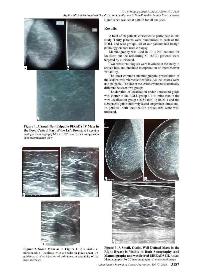
(Esoate My Lab) with a 12-MHz probe or with full digital mammography (Hologic) with a stereotactic system. In all cases, intratumoral radiopharmaceutic injections were performed with a 10-cm long, 22-gauge needle within 3-8 h of the surgical procedure, and the needle insertion site on the skin was marked to accelerate the approach. One milliliter of 99m Tc- Sulfur colloid equal to 0.1 millicurie was injected into the mass. When ultrasound was used for radiotracer injection, intratumoral administration appeared as increased echogenicity within the lesion (Figures 1, 2); in mammography, presence of the needle tip in the mass was satisfactory. The duration of localization was calculated from patient positioning in the ultrasound or mammography suite to radiotracer injection. In order to confirm focal accumulation of radiotracer inside the tumor, radioscintigrams were obtained in all cases.
Wire Localization TechniqueWire localization was also performed approximately
3-8 h before surgery. Non-palpable lesions were targeted under imaging guidance (stereotaxis or ultrasound), then the needle-wire was inserted into the lesion, followed by the localization wire (Matek or Bard 20-gauge). We usually secured the wire at the center of the lesion but
passing through not more than 5mm crossed the lesion was also acceptable. Accurate wire localization was confirmed on additional mammographic images in the craniocaudal and lateral orthogonal planes or by real-time ultrasound imaging (Figure 3, 4). The duration of wire localization was calculated in the same manner as for ROLL.
Surgical ProcedureIn the ROLL group, the patients were transferred
to the operating room after injection of radiotracer by a radiologist. The exact site of the lesion was checked with a gamma probe (Europrobe/France) at its lowest sensitivity setting before general anesthesia to choose the most cosmetically acceptable incision. During surgery, the hot spot area of maximum activity was frequently checked by gamma probe to centralize the lesion while assuring a sufficient margin in the specimen. The lumpectomy cavity was checked for any residual area of high activity before closure.
In the wire localization technique, the wire guides were used for tissue dissection to the point at which the hook end is anchored. The tumor was excised with additional cylindrical dissection. Operating times for both methods were recorded. The duration of surgery was measured from the time of the first incision to complete removal of the specimen.
The lumpectomy specimens obtained by both methods were weighted and sent to the radiology department for specimen mammography. Surgeons scored their work according to ease of the surgery (5=easiest to 1= most difficult).
Radiography of the SpecimenTo confirm removal of the targeted lesion with a secure
surgical margin and measure concentricity of the resection, radiography was performed in all cases (Fig. 5). In specimens that were not discriminated by mammography, post-surgical sonography was performed. A free margin was defined as a distance of at least 1 mm between the border of the benign lesion and the boundary of the specimen upon pathologic and the radiologic examination. The specimens were sent for pathologic assessment.
Statistical AnalysisOur primary goal was to evaluate the effectiveness of
the ROLL method in localization of benign non-palpable breast lesions. We analyzed the localization procedure (method, duration of preoperative localization, ease of use, and complications), surgical data (need for re-excision, time of surgery, evaluation of difficulty of the procedure by the surgeon, and patient cosmetic satisfaction), and pathologic data.
Statistical analysis was performed with the SPSS program (version 18.0, SPSS). A descriptive analysis of the study variables was performed for both groups. To compare the continuous variables in two categorical variables we used Mann-Whitney U test, and to evaluate the relationships between categorical variables we used chi-square or fisher’s exact tests when appropriate. Kruskal-Wallis test was used to compare the continuous variables in more than two categorical variables. Statistical
Asian Pacific Journal of Cancer Prevention, Vol 17, 2016 3187
10.14456/apjcp.2016.73/APJCP.2016.17.7.3185Applicability of Radioguided Occult Lesion Localization in Non-Palpable Benign Breast Lesions
significance was set at p<0.05 for all analyses.
Results
A total of 60 patients consented to participate in this study. Thirty patients were randomized to each of the ROLL and wire groups. All of our patients had benign pathology on core needle biopsy.
Mammography was used in 10 (17%) patients for localization; the remaining 50 (83%) patients were targeted by ultrasound.
Two breast radiologists were involved in the study to reduce bias and preclude interpretation of interobserver variability.
The most common mammographic presentation of the lesions was microcalcifications. All the lesions were non-palpable. The size of the lesions were not statistically different between two groups.
The duration of localization under ultrasound guide was shorter in the ROLL group (14.44 min) than in the wire localization group (16.54 min) (p<0.001) and the stereotactic guide uniformly lasted longer than ultrasound. In general, both localization procedures were well tolerated.
Figure 1. A Small Non-Palpable BIRADS IV Mass in the Deep Central Part of the Left Breast. a) Screening analogue mammography MLO, b) CC view, c) focal compression spot magnification view
Figure 2. Same Mass as in Figure 1. a) is visible in ultrasound; b) localized with a needle in place under US guidance; c) after injection of radiotracer echogenicity of the mass increased
Figure 3. A Small, Ovoid, Well-Defined Mass in the Right Breast is Visible in Both Sonography And Mammography and was Scored BIREADS III. A) Mlo Mammography; b) CC mammography; c) ultrasound image

Afsaneh Alikhassi et al
Asian Pacific Journal of Cancer Prevention, Vol 17, 20163188
Patient discomfort and pain did not differ significantly, and we did not have any complications. Radiologist-defined ease of use was significantly better in the ROLL than in the WGL group (p=0.0001).
Conservative surgery had been scheduled for all patients. Clear margins were considered ≥ 1 mm for benign disease. A numerical rating scale was used to measure ease of surgery, which was more in the ROLL method (p=0.04).The mean duration of the surgical procedure was (22.6±10.3 min) for ROLL and 23.63 min (23.6±9.6 min) for wire localization (p=0.6). Tumor localization and excision were successful in all cases, as confirmed with specimen radiography. Complete tumor excision-i.e., disease-free margins of at least 1 mm after the initial surgical procedure-was achieved in all cases, with no significant differences between techniques. Re-excision was not performed in any patient.
In two cases, pathology was upgraded after surgery; one from fibrocystic change to ductal carcinoma in situ (DCIS) and the other to sclerosis adenosis with DCIS. Two cases had atypia in core needle biopsy but their complete excision related pathology did not upgrade.
The surgical specimens were slightly heavier in the
ROLL group (mean weight 31.5517 gram), than in wire group (mean weight 20.1667 gram) and the differences was not significant (p=0.06).
The patients were asked to score the cosmetic appearance of their lumpectomy after surgery before hospital discharge; patient satisfaction did not significantly differ (p=0.1).
Discussion
Many non-palpable small breast lesions are detected by high-resolution ultrasound and screening mammography (Xi et al., 2102); some of these masses are benign and some malignant.
In some circumstances, surgeons prefer to perform surgery for these benign masses, such as in cases of radiology-pathology discordance (such as when core biopsy result is benign for radiologic BIRADS IVb and above); patient anxiety and desire, in which the patient does not accept follow-up; non- compliant patient for follow up, atypia in core needle biopsy pathology result; or a positive family history of breast cancer in first-degree relatives.
The goal is complete excision of the lesion with the least tissue removal and best cosmetic result. To reach this aim, accurate preoperative localization of non-palpable lesions is essential. Sonography and stereotactic mammography are the most commonly used imaging techniques for guiding localization of these lesions (Vikram et al., 2009). Different dyes have been used for localization of non-palpable lesions (Moss et al., 2002; Rose et al., 2003), but needle wires are most often used for localization (Rissanen et al., 1993; Imrana M et al., 2102; Roos et al., 2013). Several types of wires have been made, differing mainly in the shape of the distal tip and the possibility of repositioning.
Wire localization can have several complications and limitations: patient discomfort, difficult insertion in dense breasts, interference with the surgical approach, wire dislodgment and migration, accidental wire breakage, and rarely, pneumothorax (Imrana et al., 2012; Roos al., 2013; Povoski et al., 2014).
To overcome these disadvantages, a group of investigators (Zurrida et al., 1998) developed the radioguided technique known as ROLL. In this technique, clinically occult breast lesions can be localized by the injection of a radiotracer and then the surgeon will detect them intraoperatively using a gamma probe (Luini et al., 1999; Rampaul et al., 2004; Nadeem et al., 2005).
Luini et al. (Luini A et al., 1999) first compared wire localization and ROLL and showed that ROLL was faster and easier with excision of a smaller volume of tissue with better lesion centering within the excised specimen. Other retrospective studies (Thind et al., 2005; Fraile M et al., 2005; Medina et al., 2008] and a few prospective studies (Rampaul et al., 2004)compared two techniques and concluded that ROLL is safe and effective for the localizing clinically occult breast lesions (Fusco et al., 2014).
Most previous studies evaluated the effectiveness of ROLL in breast cancer. The main objective of our
Figure 4. The Mass in Figure 3 was Localized by Guide Wire under Ultrasound. a) control CC and b) MLO mammography were performed
Figure 5. Sample Mammography of Excised Tissue Showing the Mass, Wire and Radiological-Free Margin

Asian Pacific Journal of Cancer Prevention, Vol 17, 2016 3189
10.14456/apjcp.2016.73/APJCP.2016.17.7.3185Applicability of Radioguided Occult Lesion Localization in Non-Palpable Benign Breast Lesions
prospective investigation was to assess the effectiveness of ROLL in clinically occult benign non-cancerous breast lesions in comparison with the more standard wire localization technique, when surgery and removal of lesion is indicated.
Several factors associated with the effectiveness of the procedures were established, including: ease of use by radiologist, duration of guided localization, accurate localization, ease of performing the surgical procedure, duration of surgery, weight of the surgical specimen, and cosmetic satisfaction of patient.
In our study, the duration of image-guided preoperative localization of the lesion under either stereotactic or ultrasound guidance was significantly shorter in the ROLL group, consistent with prior studies (Medina et al., 2008; Antonio et al., 2009; Deepak et al., 2015), perhaps because placement of the needle wire is more complex than ROLL, taken for granted all the amenities specially radiotracer is promptly available.
The mean duration of the surgical procedure was 22.6 ± 10.3 min for ROLL and 23.63 min ± 9.6 min for wire localization (p=0.6); thus, the time difference was not clinically important. Surgery for ROLL was significantly faster in Peter J. Lovrics’s study, which, however, lacked data for clinical importance (Peter J. et al., 2011). Nadeem et al. (Nadeem et al., 2005) found a significantly shorter (p<0.013) procedure duration with ROLL. In the radiographic features of the specimen, lesion centering was better in the ROLL group, it seems the gamma probe is more helpful, as it provides a three-dimensional approach unlike than wire, which may be displaced and provides only one tract to follow.
In this study, the ROLL method was easier to perform (p=0.04). After radiologic wire localization, the surgeon is obliged to follow the wire, which occasionally does not pass along the best route for cosmetic purposes, nor the shortest or fastest approach; however, ROLL makes the incision independent of the injection site; where it is closest and esthetically more acceptable, the gamma probe provides a greater advantage. These data are compatible with previous findings (Nadeem R et al., 2005; Antonio Mariscal M et al., 2009).
The surgical specimens were slightly heavier in the ROLL group, but the difference was not significant (p=0.06) similar to Postma study (Postma E. et al., 2012). Most previously reported series have not shown significant differences in excised sample size, except Zgajnar, et al. (Zgajnar j et al.,2004), in which ROLL specimens were significantly smaller.
Tumor localization and excision according to specimen radiography were successful in all cases. Complete pathologic tumor excision, with disease-free margins of at least 1 mm, was achieved in all patients in ROLL group and the wire localization group, with no significant difference. Majority of the studies have shown either better or equal complete excision rates with ROLL as compared to WGL (Antonio et al., 2009; Mascaro A et al., 2010). Our study on benign lesions required less spare tissue for the free margin, so our nil re-excision rate is plausible.
Patients’ cosmetic satisfaction did not significantly
differ; thus, method selection does not affect the cosmetic result of surgery.
The limitation of this study is mainly the number of patients, especially patients who had localization under mammography guide. We suggest another study with exact randomized cases selection to increase the number of patients in the near future.
In conclusion, ROLL is somewhat simpler and faster to perform for the radiologist and surgeon. One of the main disadvantages of ROLL is that the radiotracer is not visible on mammograms and needs simultaneous contrast injection to be traceable on mammography, moreover it is not widely available.
We conclude that ROLL can be as effective as the needle-wire technique for localization of non-palpable benign breast lesions and makes surgical excision of these lesions easier.
Both ROLL and wire localization can be selected according to radiotracer availability, amount of experience and the policy in each center-as complementary techniques or as alternatives when one option fails.
References
Antonio Mariscal MA, Sola M, de Tudela AP, et al (2009). Radioguided localization of nonpalpable breast cancer lesions: randomized comparison with wire localization in patients undergoing conservative surgery and sentinel node biopsy. AJR, 193, 1001-9.
Deepak J , Mandeep Singh M. (2015). Radioguided occult lesion localization and sentinel node and occult lesion localization in breast cancer, the future beckons. Asian J Oncol, l 1, 73-6.
Fraile M, Mariscal A, Lorenzo C, et al (2005). Radioguided occult lesion localization combined with sentinel node biopsy in women with breast cancer. Cir ESP, 77, 36 -9.
Fusco R, Petrillo A, Catalano O, et al (2014). Procedures for location of non-palpable breast lesions: a systematic review for the radiologist. Breast Cancer, 21, 522-31.
Imrana M, Shaista A, Gulnaz S, et al (2012). Usefulness of hook wire localization biopsy under imaging guidance for nonpalpable breast lesions detected radiologically. Int J Womens Health, 4, 445-9.
Kolb TM1, Lichy J, Newhouse JH (1998). Occult cancer in women with dense breasts: detection with screening US-diagnostic yield and tumor characteristics. Radiol, 207, 191-9.
Luini A, Zurrida S, Paganelli G, et al (1999). Comparison of radioguided excision with wire localization of occult breast lesions. Br J Surg, 86, 522 -5.
Moss HA, Barter SJ, Nayagam M, et al (2002). The use of carbon suspension as an adjunct to wire localization of impalpable breast lesions. Clin Radiol, 57, 937-44.
Medina H, Abarca-Pérez L, García-Alvarez MN, et al (2008). Radioguided occult lesion localization (ROLL) versus wire-guided lumpectomy for non-palpable breast lesions: a randomized prospective evaluation. J Surg Oncol, 97, 108-11.
Mascaro A, Farina M, Gigli R, et al (2010). Recent advances in the surgical care of breast cancer patients. World J Surg Oncol, 8, 5.
Mohamed A, Alaa Eldin M, Ayman A, et al (2015). Role of imaging guided wire localization of non palpable breast lesions: Effect of localization accuracy on surgical outcome and histopathological safety margins. Biolife, 3, 883-8.
Nadeem R, Chagla LS, Harris O, et al (2005). Occult breast

Afsaneh Alikhassi et al
Asian Pacific Journal of Cancer Prevention, Vol 17, 20163190
0
25.0
50.0
75.0
100.0
New
ly d
iagn
osed
with
out
trea
tmen
t
New
ly d
iagn
osed
with
tre
atm
ent
Pers
iste
nce
or r
ecur
renc
e
Rem
issi
on
Non
e
Chem
othe
rapy
Radi
othe
rapy
Conc
urre
nt c
hem
orad
iatio
n
10.3
0
12.8
30.025.0
20.310.16.3
51.7
75.051.1
30.031.354.2
46.856.3
27.625.033.130.031.3
23.738.0
31.3
0
25.0
50.0
75.0
100.0
New
ly d
iagn
osed
with
out
trea
tmen
t
New
ly d
iagn
osed
with
tre
atm
ent
Pers
iste
nce
or r
ecur
renc
e
Rem
issi
on
Non
e
Chem
othe
rapy
Radi
othe
rapy
Conc
urre
nt c
hem
orad
iatio
n
10.3
0
12.8
30.025.0
20.310.16.3
51.7
75.051.1
30.031.354.2
46.856.3
27.625.033.130.031.3
23.738.0
31.3
lesions: a comparison between radioguided occult lesion localization (ROLL) vs. wire-guided lumpectomy (WGL). Breast, 14, 283-5
Nothacker M, Duda V, Hahn M, et al (2009). Early detection of breast cancer: benefits and risks of supplemental breast ultrasound in asymptomatic women with mammographically dense breast tissue. A systematic review. BMC Cancer, 9, 335.
Peter J. Lovrics, Charlie H. Goldsmith, Hodgson N, et al (2011). A multi-centered, randomized, controlled trial comparing radioguided seed localization to standard wire localization for non-palpable, invasive and in situ breast carcinomas. Ann Surgical Oncol, 18, 3407-14.
Postma E. L, Verkooijen H. M, Van Esser S (2012). Efficacy of ‘radioguided occult lesion localisation’ (ROLL) versus ‘wire-guided localisation’ (WGL) in breast conserving surgery for non-palpable breast cancer: a randomised controlled multicentre trial. Breast Cancer Res Treat, 136, 469-8.
Povoski ST, Jimenez R, Wang W, et al (2014). Use of an intraoperative ultrasonography-guided localization and tissue fixation device demonstrates less margin positivity during breast-conserving surgery for invasive breast cancer than standard preoperative needle-wire localization: a retrospective comparative analysis in a consecutively treated case series. Clinical Breast Cancer, 14, 46-52.
Rissanen TJ, Mäkäräinen HP, Mattila SI, et al (1993). Wire localization biopsy of breast lesions: a review of 425 cases found in screening or clinical mammography. Clin Radiol, 47, 14 -22.
Rose A, Collins JP, Neerhut P, et al (2003). Carbon localisation of impalpable lesions. Breast, 12, 264-9.
Rampaul RS, Bagnall M, Burrel H, et al (2004). Randomized clinical trial comparing radioisotope occult lesion localization and wire-guided excision for biopsy of occult breast lesions. Br J Surg, 91, 1575-7.
Roos M. A. J., Welvaart W. N., Ong K. H. (2013). Should we abandon wire-guided localization for non-palpable breast cancer? a plea for wire-guided localization. Scandinavian J Surgery, 102, 106-9.
Thind CR, Desmond S, Harris O, et al (2005). Radioguided localization of clinically occult breast lesions (ROLL): a DGH experience. Clin Radiol, 60, 681-6.
Vikram S, Wael E. A (2009). Ultrasound-guided procedures. New York, NY: Thieme, 344-6.
Xi Lin, Jianwei W, Feng H, et al (2012). Analysis of eighty-one cases with breast lesions using automated breast volume scanner and comparison with handheld ultrasound. Eur J Radiol, 81, 873-8.
Zurrida S, Galimberti V, Monti S, et al (1989). Radioguided localization of occult breast lesions. Breast, 7, 11-13.
Zgajnar J, Hocevar M, Frkovic-Grazio S, et al (2004). Radioguided occult lesion localization (ROLL) of the nonpalpable breast lesions. Neoplasma, 51, 385-9.