Embed Size (px)
Citation preview

182
APPENDIX A1
Glossary of terms used in biodeterioration Autotroph
An organism that uses carbon dioxid e for its carbon requirement; often used to imply lithoautotrophy , as obligate autotrophs appear to be either chemolithotrophs or photolithotrophs.
Biodeteriogen
An organism or microorganism that causes undesirable changes in the constituent materials of (esp. cultural) objects.
Biofilm
A film of microorganisms, usuall y embedded in extracellular polymers that adhere to surfaces submerged in, or subjected to, aquatic environments.
Chelate A chemical agent that combines with metal ions
Chelation
A chemical process involving the formation of heterocyclic ring compounds containing at least one metal cation or hydrogen ion.
Chemoautotroph
An organism whose energy is derived from endogenous, light-independent chemical reactions (a mode of metabolism termed chemotrophy); a chemotroph that obtains energy by the oxidation of inorgani c substrate(s ) (i.e. , a lithotroph , called chemolithotroph); one that obtains energy by metabolizin g organic substrate(s) (i.e., an organotroph, called a chemoorganotroph).
Chlorophytes
A group of algae characterized by a combination of features (e.g., containing chlorophylls a and b, ß-carotene, and xanthophyll s such as antheraxanthin, lutein , neoxanthin, violax - anthin, and zeaxanthin).
chloroplasts Organisms evolved by a double membrane; the main storage polymer is starch, which is formed within the chloroplast . Motile cells, when formed, bear flagella.
endolithic Growing within rocks, as some algae, lichens, and bacteria do in limestone.
epilithic Growing on a rock surface.
eukaryotes
A group of organisms ; literally those having "nuclei" in their cell s (i.e. , animals , plants , and fungi) in contrast to the prokaryotic cell s of bacteria and blue-green algae; possessing a nucleus bounded by a membranous nuclear envelope and having many cytoplasmic organelles.

183
foliose Leafy, having a body differentiated into stem and leaves; of lichen: having a flattened leaf like thallus more or less firmly attached to the substratum.
fruticose Shrublike; of a lichen: having a thallus that is threadlike (terete) or straplike (more or less flattened), and either erect and shrubby or pendulous , the thallus being attached to the substratum by a holdfast or unattached.
gymnosperms
A group (classified as a division Gymnospermophyta or a class Gymnospermae, or regarded as polyphyletic) containing those seed plants in which the ovules are not enclosed in carpels , the pollen typically germinating on the surface of the ovule.
heterotroph
An organism that uses organic compounds for most or all of its carbon requirements; often used to refer specifically to chemoorganoheterotrophs, although chemolithotrophs and phototrophs may also be heterotrophic.
hyphae Branched or unbranched filaments, many of which together constitute the vegetative form of the organism and (in some species) form the sterile portion of a fruiting body.
metabolite Any of various compounds produced by metabolism.
photoautotroph An autotroph that requires sunlight to provide necessary energy for biosynthesis.
photoheterotroph A heterotroph that requires sunlight to provide necessary energy for biosynthesis
photolithotroph An organism that uses inorganic substrate s (e.g., water, sulfur, sulfide, H2
photoorganotroph
, or thiosulphate) as an electron donor in photosynthesis. An organism that uses an organic substrate as an electron donor in phototrophic metabolism.
prokaryote
A type of microorganism in which the chromosomes are not separated from the cytoplasm by a specialized membrane that is typically devoid of sterol s and in which mitochondria and chloroplast s are absent.
redox reaction
A chemical reaction in which electrons are transferred between atoms, ions, or molecules.
rhizoid
A root like structure consisting of a compact bundle of hyphae; arising mainly from the lower surface of the (usually foliose) thallus and serving to anchor the thallus to the substratum; in some species, may also facilitate the uptake of water and nutrients by thallus.

184
taxonomy
The science of classification as applied to living organisms, including the study of the means of specie s formation
thallus
Plant body not differentiated into leaves, stems, and roots, but consisting of a mycelium or a colony, a filament of cells , a mycelium, or a large branching multicellular structure; the plant body of the algae, fungi, and liverworts.
Microbiologically Influenced Deterioration (MID)
Miro-organisms degrading/ deteriorating non-metals

185
APPENDIX A2
OVERVIEWOF SEVERAL TECHNIQUES USED FOR MATERIALS CHARACTERIZATION AND SURFACE ANALYSIS
Scanning electron microscopy, environmental scanning electron microscopy and energy
dispersion X-ray analysis:
SEM allows the observation and surface characterization of inorganic and organic materials
at a higher resolution and depth of field than that of the conventional optical microscope. In
the SEM analysis, a thermo-ionic emission of an electron beam is directed over the sample to
produce characteristic signals that provide information on crystalline array, chemical
composition, magnetic structure and even on the electric potential of the specimen under
observation. SEM offers a high resolution (approximately 100 Å) and high field depth giving
a 3D effect to the images. Contrary to SEM, ESEM does not operate at high vacuum and
provides fast, accurate images of biological deposits (biofilms), showing their spatial
relationship and surface chemistry without needing extensive manipulation of the samples.
This instrument has a unique secondary electron detector capable of forming high resolution
images at pressures in the range of 0.1–20 torr. At these relatively high pressures, non-
conductive biological specimens are directly observed without metallic coating and any
special preparation. If water vapor is used in the chamber, wet samples can be observed
directly and EDX data can be collected at the same time as sample morphology and
topography are photographed.
In the EDX technique, a solid-state detector is placed close to the sample to emit an electric
pulse when the interaction with the X-rays is produced. The intensity of the electric pulse is
proportional to the energy of the X-ray beam. If the number of pulses and the intensity of the
X-rays are measured, the chemical composition of the sample can be determined. The
wavelength of the radiation is analyzed by a dispersion device which diffracts the
characteristic radiation of the sample by means of a suitable crystal.
X-ray diffraction analysis
XRD is a convenient and useful tool for the identification of crystalline compounds as well as
for assessing the structure of complex natural compounds. The theoretical basis of the method
is the fact that X-ray diffraction patterns are unique for each crystalline structure. When the
X-ray beam passes trough the material, a large number of the particles can be expected to be
aligned in such directions that they can fulfill the Bragg relationship for reflection for each
possible inter-planar spacing.

186
Petrographic analysis
The polarizingmicroscope (also called petrographicmicroscope) is often used to examine thin
sections of constructional materials and it is very useful in the study of rock transformation
due to weathering effects.
Mossbauer spectroscopy
Usually the energy of a beam of γ rays emitted by an atomic nucleus which changes from an
excited level to the ground state is different from the transition energy. Under certain
experimental conditions a fraction of the γ rays beam has a similar energy to the transition
energy (Mossbauer effect). This effect depends on the firmness of the link between the
emitting nucleus and its chemical environment. In the corrosion field themost interesting
nuclei which present the Mossbauer effect are those of Fe, Ni and Zn. It is also used in
corrosion studies where it provides the complete, in situ measurement, non-destructive, three-
dimensional identification of corrosion products and is the only technique able to accurately
measure the fraction of each oxide in a corrosion product layer. Thus, it can be used to study
the electric, magnetic and structural characteristics of metals, alloys, soils andminerals. MS
can be considered as a ‘‘fingerprint’’ technique of varying degrees of sophistication in
mineralogical and geo-chemical studies.
Grazing incident diffraction
GID is a valuable technique that provides very precise information on surface and interface of
atomic arrangement in crystalline structures. By varying the angle of incidence, the
penetration of the X-ray beam into the material can be controlled allowing the analysis of
surfaces and buried interfaces. GID is a powerful tool for surface structural characterization of
properties such as atomic structure and roughness, relaxation and reconstruction. Non-
destructive analysis is a representative application of the GID. The incidence angle was varied
in order to change the penetration depth between few nanometers up to several micrometers.
GID has become well established for the investigation of the structure of films, surfaces and
interfaces.
X-ray photoelectron spectroscopy and reflection electron energy-loss spectroscopy
XPS and REELS are powerful surface-sensitive analytical techniques that are well suited for
the analysis of materials. XPS analysis of the external surface provides valuable information
about the elemental concentrations at the surface and the chemical state. The surface
composition, at the limits of the penetration depth of this

187
technique (w5 nm), can be derived from the intensity of the different photoemission peaks
detected in the general spectra corrected by their respective sensitivity factors.
Raman spectroscopy
The Raman spectroscopy has become increasingly available in the field of cultural heritage
studies. During the last decade, it has gradually become an established technique for the non-
destructive identification and study of ancient materials.
DNA-based molecular techniques
DNA-based molecular techniques allow the direct detection and identification of single
microorganisms in environmental samples. These techniques have been applied to detect
microorganisms in many natural environments since the beginning of the 90s and are also
beginning to be used in biocorrosion studies (corrosion of metallic structures due to
microorganisms). Molecular techniques have been used to characterize the microorganisms
present in biofilms damaging cultural heritage buildings.
These techniques are based on the analysis of ribosomal DNA sequences that are present in
the genome of all living organisms. These sequences can be used as biomarkers for the
accurate identification of microorganisms considering that different microbial species will
have different ribosomal gene sequences (Woese, 1987). Ribosomal genes contain both highly
conserved and highly variable regions where ‘‘signature sequences’’ have been identified for
each domain of life (Bacteria, Eucarya and Archaea) as well as for microbial groups, families,
genera and species. By knowing their ribosomal sequences, the microorganisms there can be
classified and identified.
188
APPENDIX A3
CLASSIFICATION OF ALGAE (V.J. Chapman, 1941)
In the older version of classifications the algae proper were simply divided into four principal
groups, Chlorophyceae or green algae, Cyanophyceae or blue-green algae, Phaeophyceae or
brown algae and Rhodophyceae or red algae. Now, that more is known about the simpler
organisms which were not to be regarded as algae, it has been realized that there is no real
justification for such a distinction, and so the number of algal groups has been increased. This
is because it has become evident that the Flagellata and other simple unicellular organisms
must properly be regarded as algae, even though they are of a very primitive kind. At present
it is most convenient to divide the algae into ten classes, one of which, the Nematophyceae, is
perhaps somewhat speculative. One of the principal bases of this classification is the
difference in pigmentation, and a recent study of this problem shows that it is fully justified.
(1) Cyanophyceae The plants in this group show very little evidence of differentiation,
containing only a very simple form of nuclear material, no proper chromatophore and no
motile cells with cilia or flagellae. The products of photosynthesis are sugars and glycogen.
The colour of the cells is commonly blue-green and hence their name, the colour being due to
the varying proportions of the pigments phycocyanin and phycoerythrin. There is no known
sexual reproduction, propagation taking place by simple division or else by vegetative means.
(2) Chlorophyceae This group used to comprise four great subdivisions, the Isokontae (equal
cilia), Stephanokontae (ringed cilia), Akontae (no cilia) and Heterokontae (unlike cilia). It is
now more in keeping with our present knowledge to place the last section into a separate
class, and this is the procedure adopted in most recent books. The plants of the Chlorophyceae
exhibit a great range of structure from simple unicells to plants with a relatively complex
organization, whilst the chromatophores also vary considerably in shape and size. The final
product of photosynthesis is starch together with oil, and a starch sheath can often be
demonstrated around the pyrenoids. In the bulk of the members of this class the motile cells
are very similar and commonly possesseither two or four flagellae, but in the Oedogoniales
(Stephanokontae) there is a ring of flagellae whilst in the Conjugales (Akontae) there are no
organs of propulsion. Sexual reproduction is of common occurrence and ranges from isogamy
to anisogamy and oogamy. The colour of the cells is usually a grass green because the
pigments are the same as those present in the higher plants and, furthermore, they are present
in much the same proportions.

189
(3) Xanthophyceae (Heterokontae) The plants in this group are usually of a simple nature,
but their lines of development frequently show an interesting parallel or homoplasy with those
observed in the preceding group. The chloroplast is yellow-green owing to an excess of
xanthophyll, one of the four normal constituents of chlorophyll. Oil replaces starch as the
normal storage material, the lack of starch being correlated with the absence or paucity of
pyrenoids. The motile cells possess two unequal flagellae (occasionally only one) arising from
the anterior end. Sexual reproduction is rare and when present is isogamous. The cell wall is
frequently composed of two equal or unequal halves overlapping one another.
(4) Chrysophyceae These form another very primitive group in which the brown or orange
colour of the chloroplasts is determined by the presence of accessory pigments such as
phycochrysin.Most of the forms have no cell wall and hence are “flagellates” in the old sense
of that term, although there are some members which do possess a cell wall and hence are
"algal" in the old sense of the term. Fat and leucosin (a protein-like substance) are the usual
forms of food storage, whilst another marked feature is the silicified cysts which generally
have a small aperture that is closed by a special plug. The motile cells possess one, two or,
more rarely, three equal flagellae attached at the front end, but in one subsection the paired
flagellae are unequal in length. The most advanced habit known is that of a branched filament,
e.g. Phaeothamnion, whilst the palmelloid types attain to a much higher state of
differentiation, e.g. Hydrurus, than in either the Chlorophyceae or the Xanthophyceae. Sexual
reproduction is not certain, and such records as there are point simply to isogamy.
(5) Bacillariophyceae (Diatoms) One of the characteristics of these plants is their cell walls
which are composed partly of silica and partly of pectic material. The wall is always in two
halves and frequently ornamented with delicate markings, which are so fine that microscope
manufacturers make use of them in order to determine the resolving power of their lenses.
The chromatophores are yellow or golden brown containing, in addition to the usual
pigments, accessory brown colouring materials whose nature is only just being established.
One set of forms is radially symmetrical, the other bilaterally so. The presence of flagellate
stages is highly probable in the former whilst there is a special type of sexual fusion in the
latter group.
(6) Cryptophyceae There are usually two large parietal chloroplasts with diverse colours,
though frequently of a brown shade, whilst the product of photosynthesis is starch or a closely
related compound. The motile cells have two unequal flagellae and often possess a complex
vacuolar system. Nearly all the members have a "flagellate” organization and there is no
190
example of the filamentous habit. One type, however, has been described with a tendency
towards the coccoid (non-motile unicell with a cell wall) habit, and so this must be regarded
as the least "algal "-like class. Isogamy has been recorded for one species.
(7) Dinophyceae Most of the members of this class are motile unicells, but there has been an
evolutionary tendency towards a sedentary existence and the development of short algal
filaments, e.g. Dinothrix. Many are surrounded by an elaborate cellulose wall bearing
sculptured plates and inside there are discoid chromatophores, dark yellow or brown in colour
and containing a number of special pigments. The products of photosynthesis are starch and
fat. The motile cells normally possess two furrows, one transverse and one longitudinal,
although they may be absent in some of the more primitive members. The transverse
flagellum lies in the former, and the latter is the starting point for the other flagellum which
points backwards. Sexual reproduction, if it occurs, is isogamous, and it has not been clearly
established in the few cases reported. Characteristic resting cysts are also produced by many
of the forms.
(8) Phaeophyceae This group comprises the common brown algae of the seashore and it is
worth noting that the majority are wholly confined to the sea. The brown colour is due to the
presence of a pigment, fucoxanthin, which masks those other chlorophyll constituents which
are present. The products of photosynthesis are alcohols, fats, polysaccharides and traces of
simple sugars so that there is evidence of some diversity of metabolism. The simplest forms
are filamentous, and there are all stages of development and increasing differentiation up to
the large seaweeds of the Pacific and Arctic shores with their great size and complex internal
and external differentiation. The motile reproductive cells, which possess two flagellae, one
directed forwards and the other backwards, are commonly produced in special organs or
sporangia that are either uni or plurilocular. Sexual reproduction ranges from isogamy to
oogamy, but in the latter case the ovum is normally liberated before fertilization. The life
cycles may be extremely diverse and are perhaps better regarded as race cycles.
(9) Rhodophyceae The members of this class form the red seaweeds, and although most of
them are marine nevertheless a few are fresh-water. Their colour, red or bluish, is caused by
the presence of the pigments phycoerythrin and phycocyanin, whilst the product of
photosynthesis is a material known as "floridean starch". Reproductive stages with locomotor
appendages are not known, even the male reproductive body being without any organ of
locomotion. The simplest members are filamentous, and again all stages of differentiation up
to a complex body can be found, although they do not develop to quite the same degree of
191
complexity as the Phaeophyceae. Very obvious protoplasmic connexions can be distinguished
between the cells of nearly all forms except in the small group known as the Proto-florideae.
Sexual reproduction is oogamous, the ovum being retained upon the parent plant, and
although the subsequent development of the zygote is varied to a certain extent, it usually
gives rise to filaments which bear special reproductive bodies or carpospores, and these latter
are responsible for the production of the tetrasporic diploid plant. Most of the members
exhibit a regular alternation of generations.
(10) Nematophyceae This is a fossil group of which one genus has been known for a long
time (Nematophyton) whilst the other has only recently been described (Nematothallus).
There is still considerable doubt as to their true affinities, but it would seem that a place can
best be found for them as a very highly developed type of alga. Their internal morphology
would ally them closely with the more advanced members of either the Chlorophyceae or the
Phaeophyceae. The only reproduction so far recorded is that of spores which were developed
in tetrads, and therefore may have been akin to the Rhodophycean or Phaeophycean
tetraspores.
Based on the location at which algae colonizes, another classification that is generally
recognized is: (i) marine algae – widely available near sea coasts /sea; (ii) terrestrial algae -
widely available on moist surface in inland regions; (iii) fresh water algae – widely available
on surface of water bodies.

192
APPENDIX B1
Characterization of Three Species of Algae
1. Enteromorpha clathrata (Roth) Plants less than 15cm long, soft, with repeatedly branched
thallus Fig. B2.1. Frequently uniseriate, branchlets in narrow. The cells are more or less
quadrangular in longitudinal and even in transversal rows.
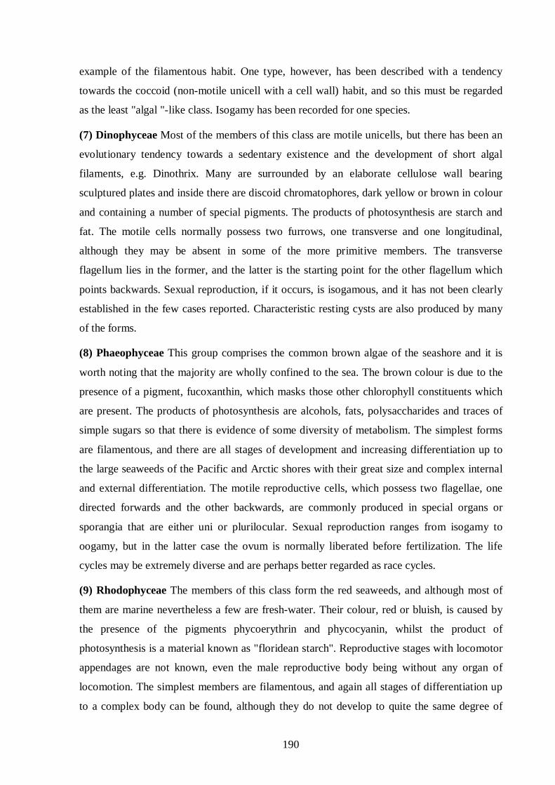
2 Ulva fasciata (Delile) Plants 1-15 cm. tall, the base of the blade cuneate, above expanding
irregularly lobed, generally irregularly or sometimes pinnately divided into ligulate or linear
lobes which may become several decimeters long; in section the cells of the midline region
much taller than those of the margin, the thallus much thicker, 100 µ or somewhat more; the
margins entire to irregularly ruffled and crenate with a somewhat paler central portion Fig.
B2.2.
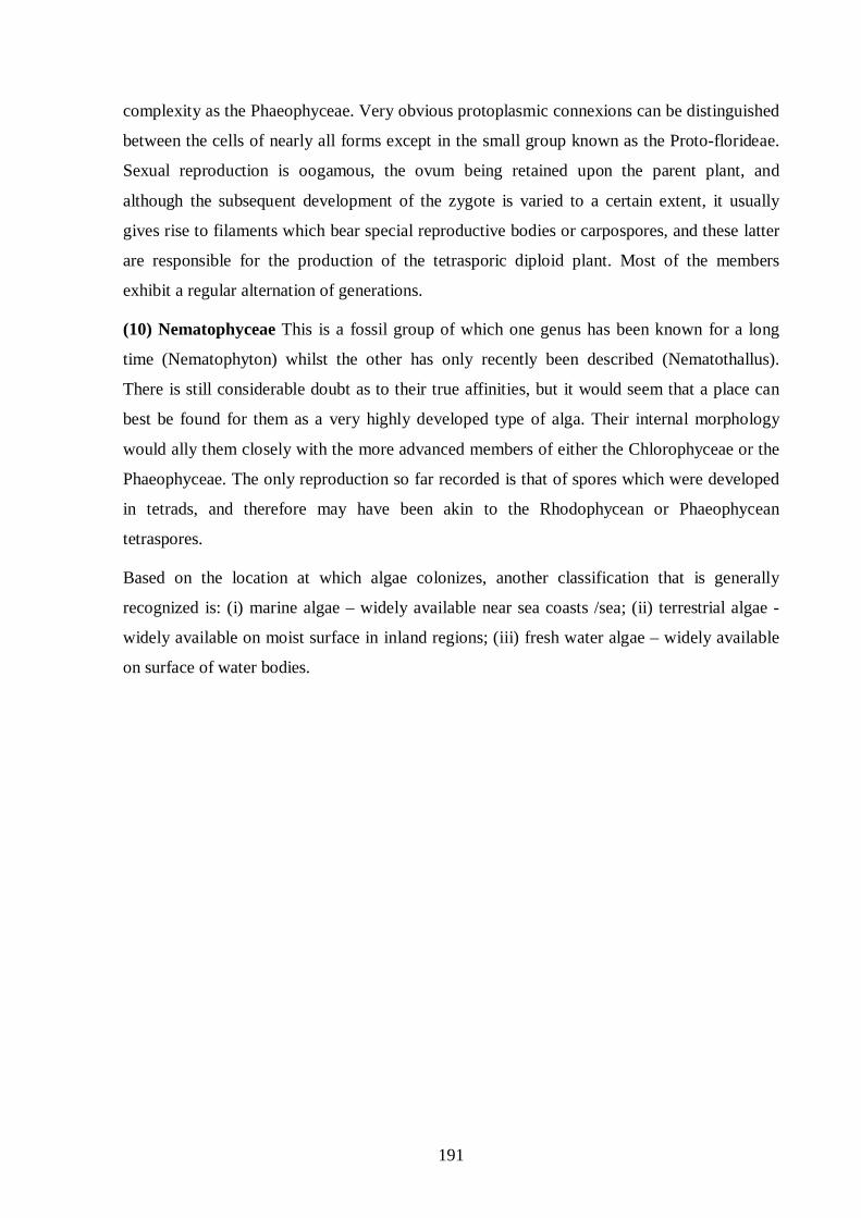
3 Chaetomorpha antennina (Bory) Kutzing
Chaetomorpha antennina forms stiff tufts of unbranched filaments 2 to 15 cm high composed
of large cells. Its overall color is grass green, but close examination will reveal alternating
green and white bands. The tips become pale green or yellowish when bleached by bright
sunlight Fig. B2.3. This seaweed grows intertidally on rocky coastlines exposed to large
breaking wave.
Fig. B2.1 Enteromorpha clathrata Species
193
Fig. B2.2 Ulva fasciata Species
Fig. B2.3 Chaetomorpha antennina Species

194
APPENDIX B2
Mix proportion calculations of M20 and M25 concretes
The mix design was done as per IS 10262- 1982 and the calculation for the two grades of
concrete M20 and M25 is as follows:
Design of M20 grade concrete:
From IS 456-2000, for sever exposure condition of plain concrete the recommended mix is
M20. The cement content used is 310 kg/m3.
Characteristic compressive strength of the concrete at 28 days = 20 N/mm2
Maximum size of aggregate is 10 mm
Degree of quality control is Good
Specific gravity of cement is 3.15
Specific gravity of coarse aggregate is 2.65
Specific gravity of fine aggregate is 2.70
For 10 mm coarse aggregate 3% entrapped air is used
Target mean strength of concrete = 20 + 4.6 x 1.65 = 27.59 N/mm2
For 27.59 N/mm2 from IS 456-2000 water-cement ratio recommended is 0.5.
From IS: 10262 for 20 mm nominal size of aggregates, maximum water content is
186 Kg/ m
ratioCement Water waterofWeight
3
Weight of cement (C) = = 0.5186 = 372 kg
From the total aggregate volume (1m3) deduct 3% for air entrapment. Hence net volume of
solids is 0.97 m3
10001
PCA
PFAW
PC
CAFAc
x
+++
.
TA =
Where TA – Total Aggregate
C is cement content; FA is Fine Aggregate; CA is Coarse Aggregate
PC is specific gravity of cement; PFA is specific gravity of fine aggregate
PCA
10001
65.265.0
70.235.0186
15.3372 xTATA
+++
is specific gravity of coarse aggregate
0.97 =
TA = 1752.37 kg/m
Cement = 372 kg
3
FA = 0.35 x 1752.37 = 613.33 ≈ 613 kg
CA = 0.65 x 1752.37 = 1139.04 ≈ 1139 kg

195
Design of M20 grade concrete:
From IS 456-2000, for sever exposure condition of plain concrete the recommended mix is
M25. The cement content used is 310 kg/m3.
Characteristics compressive strength of the concrete in 28 days = 25 N/mm2
Maximum size of aggregate is 10 mm
Degree of quality control is Good
Specific gravity of cement is 3.15
Specific gravity of coarse aggregate is 2.65
Specific gravity of fine aggregate is 2.70
For 10 mm coarse aggregate 3% entrapped air is used
Target mean strength of concrete = 25 + 4.6 x 1.65 = 32.59 N/mm2
For 32.59 N/mm2 from IS 456-2000 water-cement ratio recommended is 0.43.
From IS: 10262 for 20 mm nominal size of aggregates Maximum Water Content is
186 Kg/ m
ratioCement Water waterofWeight
3
Weight of cement (C) = = 0.43186 = 432.6 ≈ 433 kg
From the total aggregate volume (1m3) deduct 3% for air entrapment. Hence net volume of
solids is 0.97 m3
10001
PCA
PFAW
PC
CAFAc
x
+++
.
TA =
Where TA – Total Aggregate
C is cement conctent; FA is Fine Aggregate; CA is Coarse Aggregate
PC is specific gravity of cement; PFA is specific gravity of fine aggregate
PCA
10001
65.265.0
70.235.0186
15.3433 xTATA
+++
is specific gravity of coarse aggregate
0.97 =
TA = 1701.42 kg/m3
FA = 0.35 x 1701.42 = 595.497 ≈ 596 kg
CA = 0.65 x 1701.42 = 1105.92 ≈ 1106 kg
Cement = 433 kg

196
Table B3.1 Chemical composition of cement
Compound % Silicon-di-oxide (SiO2 20-21 ) Aluminum oxide (Al2O3 5.2-5.6 ) Ferric oxide (Fe2O3 4.4-4.8 ) Calcium oxide (CaO) 6.2-6.3 Magnesium oxide (MgO) 0.5-0.7 Sulfur-tri-oxide (SO3 2.4-2.8 ) Loss on ignition 1.5-2.5
Table B3.2 Grading of coarse and fine aggregate
Coarse Aggregate (mm)
% retained Fine Aggregate % retained
20 0 4.75 0 16 25 2.36 12
12.5 52 0.600 49 10 72 0.300 85
4.75 100 0.150 97 < 0.150 100
Table B3.3 Details of mix proportions
Type of cement
W/C ratio Cement (kg/m3
Water (kg/m) 3
Fine Aggregate
(kg/m)
3
Coarse Aggregate
(kg/m) 3
Target mean
strength (N/mm
) 2)
OPC 0.50 372 186 613 1139 27.59 OPC 0.43 433 186 596 1106 32.59

197
APPENDIX B3
Table B4.1 Temperature, RH and pH prevailed during the study period
Month & Year Temperature RH pH September’07 32.1 71.2 8.0 October’07 31.0 74.4 8.4 November’07 30.4 76.3 8.5 December’07 29.1 75.8 8.2 January'08 26.6 74.8 8.1 February'08 30.8 68.6 7.8 March'08 30.7 72.2 8.0 April'08 32.2 72.7 8.0 May'08 32.4 68.6 8.1 June'08 33.5 70.0 8.0 July'08 32.5 66.0 7.8 August'08 32.5 69.1 7.9 Spetember'08 31.8 69.7 8.3 October'08 31.2 73.9 8.5 November'08 29.2 75.8 8.4 December'08 28.5 76.0 8.3 January'09 28.2 68.6 8.4 February'09 29.9 70.6 8.1 March'09 32.1 70.6 8.2 April'09 32.5 72.1 7.8 May'09 33.2 73.1 7.8 Jun'09 33.6 64.9 7.8 July'09 32.6 62.5 7.6 August’09 33.1 70.1 8.1
Note:
(i) Maximum temperature is 33.6o
(ii) Minimum temperature is 26.6
C o
(iii) Maximum RH is 62.5%
C
(iv) Minimum RH is 76.0%
(v) pH range of actual sea water is 7.6 – 8.5

198
APPENDIX B4
Description of Phycochemical Procedures
1 Extraction
The dried, chopped, milled and weighted algal material was then soaked in methanol (MeOH)
in a large glass jar and was kept in the solvent for one month at room temperature. The extract
of the material thus obtained was then filtered to remove all solid algal particles. It was then
evaporated on a rotary evaporator under reduced pressure. This yielded a dark green, thick
residue, which was then weighed.
2 Saponification
An aliquot of the extract obtained was saponified with 10% KOH in 50% methanol and
refluxed at 100oC for 6 hours. The mixture was then concentrated under reduced pressure and
then H2O and diethyl ester (Et2O) were added. It was then shaken vigorously and the Et2O
layer was separated. The Et2O layer was evaporated and used for fatty acid analysis.
3 Esterification
All the fatty acid fractions obtained were subjected to methylation and 1.5 -2.0 mL ethereal
diazomethane was added to the fatty acid mixture. The reaction mixture was left in the fuming
chamber at room temperature, over night until dissolved. The aliquots were then directly
injected to Gas Chromatograph – Mass Spectrophotometer (GC-MS).
4. Identification
The GC-mass spectra were analytically analyzed for the identification of fatty acids.

199
APPENDIX C1
MASS SPECTROSCOPY OF METHYL ESTERS OF FATTY ACIDS OF ‘CHLOROPHYCEAE’ AT VARIOUS RETENTION TIMES
MS of ‘Chaetomorpha antennina’
Contd…

200
Contd…

201
Contd…

202
Contd…

203

204
MS of ‘Ulva fasciata’
Contd…

205
Contd…

206
Contd…

207
Contd…

208
MS of ‘Enteromorpha clathrata’

209
APPENDIX C2
OXIDATIVE CLEAVAGE OF FATTY ACIDS EXCRETED
DURING METABOLIC ACTIVITY
(A) ‘Enteromorpha clathrata’
7, 10, 13 – hexadecatrienoate CH3. CH2-CH=CH. CH2-CH=CH.CH2.CH=CH. (CH2)5.COOCH
3
Malonic Acid Malonic Acid Pimelic acid
9, 12, 15- hexadecatrienoate H2C=HC-H2C-HC=CH-CH2-CH=CH. (CH2)7.COOCH
3
Malonic Acid Malonic Acid Azelaic acid
9- heptadecenoate CH3.(CH2)6-HC=CH-(CH2)7-COOCH
3
Azelaic acid
5,7 – hexadecadiynoate CH3-(CH2)7-C≡C-C≡C-(CH2)3-COOCH
Oxalic acid Gultaric acid
3
Aerial Oxidation
H2O + ½ O2
H2O + ½ O2
H2O + ½ O2
COOH CH2
COOH
COOH CH2
COOH
COOH (CH2)5
COOH
H2O + ½ O2
H2O + ½ O2
COOH CH2
COOH
COOH (CH2)7
COOH
H2O + ½ O2
COOH (CH2)7
COOH
H2O + ½ O2
H2O + ½ O2
COOH (CH2)3
COOH
COOH
COOH
Aerial Oxidation
Aerial Oxidation
Aerial Oxidation
H2O + ½ O2
COOH CH2
COOH

210
(B) ‘Ulva Fasciata’ 8-heptadecenoate CH3-(CH2)7-CH=CH-(CH2)6-COOCH
CH
3 Suberic acid
9-heptadecenoate
3.(CH2)6.CH=CH-(CH2)7-COOCH
Azelaic acid
3
7- methyl-hexadec-6-enoate CH3-(CH2)8-C=CH-(CH2)4-COOCH
3
Adipic acid 2-trans-octadecenoate (2-18:1)
CH3-(CH2)14 H H COOCH3
Oxalic acid
H2O + ½ O2
Aerial Oxidation
COOH (CH2)6
COOH
H2O + ½ O2
Aerial Oxidation
COOH (CH2)7
COOH
C=C
H2O + ½ O2
Aerial Oxidation
COOH
COOH
H3C
H2O + ½ O2
Aerial Oxidation
COOH (CH2)4
COOH

211
6,9,12-hexadecatrienoate (6,9,12-16:3) CH3-(CH2)-CH=CH-CH2-CH=CH-CH2-CH=CH-(CH2)4-COOCH
CH
3
Malonic acid Malonic acid Adipic acid 9- heptadecenoate
3-(CH2)6-HC=HC-(CH2)7-COOCH
Azelaic acid
3
15-methyl-hexadec-9-enoate CH3-CH-(CH2)-CH=CH-(CH2)-COOCH Azelaic acid 7- methyl-hexadec-6-enoate
3
CH3- (H21C)8-CH = -(CH2)4-COOCH
3
Adipic acid
H2O + ½ O2
Aerial Oxidation H2O
+ ½ O2
H2O + ½ O2
COOH CH2
COOH
COOH CH2
COOH
COOH (CH2)4
COOH
CH3
COOH (CH2)7
COOH
COOH (CH2)7
COOH
H2O + ½ O2
Aerial Oxidation
H3C
H2O + ½ O2
Aerial Oxidation
Aerial Oxidation
COOH (CH2)4
COOH
H2O + ½ O2

212
8- heptadecenoate CH3-(CH2)7-HC=HC-(CH2)6-COOCH
3
Suberic acid 6,9,12,15- hexadecatetraenoate CH2=HC-H2C-HC=HC-CH2-HC=HC-CH2-HC=HC-(CH2)4-COOCH
3
Malonic acid Malonic acid Malonic acid Adipic acid 15-methyl-hexadec-9-enoate H3C-HC-(CH2)4-HC=HC-(CH2)7-COOCH
3
Azelaic acid 4,5-tetradecadienoate (4,5-14:2) CH3-(CH2)7-HC=C=HC-(CH2)2-COOCH Succinic acid
3
H2O + ½ O2
COOH (CH2)2
COOH
H2O + ½ O2
Aerial Oxidation
COOH (CH2)6
COOH
H2O + ½ O2
H2O + ½ O2
H2O + ½ O2
H2O + ½ O2
COOH (CH2)4
COOH
COOH CH2
COOH
COOH CH2
COOH
COOH CH2
COOH
Aerial Oxidation
H3C
Aerial Oxidation
H2O + ½ O2
COOH (CH2)7
COOH
Aerial Oxidation

213
2-trans-octadecenoate CH3. (CH2)14 H
H COOCH
C = C
Oxalic acid 7-methyl-hexadex-6-enoate
3
CH3. (CH2)8-HC = (CH2)4-COOCH
3
Adipic acid 9,12,15-hexadecatrienoate (16:3(n-1)) H2C=HC-H2C-HC=HC-H2C-HC=HC-(CH2)7-COOCH
3
Malonic acid Malonic acid Azelaic acid 4,5-tetradecadienoate (4,5-14:2) CH3.(CH2)7.HC=C=HC-(CH2)2-COOCH Succinic acid
3
H2O + ½ O2
H2O + ½ O2
H2O + ½ O2
COOH CH2
COOH
COOH CH2
COOH
COOH (CH2)7
COOH
H2O + ½ O2
COOH (CH2)2
COOH
Aerial Oxidation
Aerial Oxidation
H2O + ½ O2
Aerial Oxidation
COOH
COOH
Aerial Oxidation
COOH (CH2)4
COOH
H3C H2O + ½ O2

214
9-tetradecenoate (9-14:1) CH3-(CH2)3-HC=HC-(CH2)7-COOCH Azelaic acid
(C) ‘Chaetomorpha antennina’ 2-trans-octadecenoate
3
H COOCH
(CH
3 C = C
2)14
H
Oxalic acid 4,7,10,13- hexadecatetraenoate CH3-CH2-CH=HC-H2C-HC=HC-H2C-CH=CH-CH2-HC=HC-(CH2)2-COOCH3
Malonic acid Malonic acid Malonic acid Succinic acid 7,10,13 – hexadecatrienoate CH3-CH2-HC=HC-H2C-HC=HC-H2C-HC=HC-(CH2)5-COOCH Malonic acid Malonic acid Pimelic acid
3
H2O + ½ O2
COOH (CH2)7
COOH
Aerial Oxidation
H2O + ½ O2
Aerial Oxidation
COOH COOH
H2O + ½ O2
H2O + ½ O2
H2O + ½ O2
H2O + ½ O2
COOH CH2
COOH
COOH CH2
COOH
COOH CH2
COOH
COOH (CH2)2
COOH
H2O + ½ O2
H2O + ½ O2
H2O + ½ O2
COOH CH2
COOH
COOH CH2
COOH
COOH (CH2)5
COOH
Aerial Oxidation
Aerial Oxidation

215
2-trans-octadecenoate H COOCH3 C = C CH3(CH2)14
(CH
H Oxalic acid 15-methyl-hexadec-9-enoate
3)-CH-(CH2)4-HC = HC-(CH2)7-COOCH
3
Azelaic acid 9, 12, 15- hexadecatrienoate (16:3) H2C=HC-H2C-HC=CH-CH2-CH=CH. (CH2)7.COOCH
3
Malonic Acid Malonic Acid Azelaic acid
5,9 – hexadecadienoate CH3-(CH2)5-HC = HC-H2C-CH2-HC = HC-(CH2)3-COOCH
3
Succinic acid Gultaric acid
Aerial Oxidation H2O
+ ½ O2
COOH COOH
H2O + ½ O2
Aerial Oxidation
COOH (CH2)7
COOH
H2O + ½ O2
H2O + ½ O2
COOH CH2
COOH
COOH (CH2)7
COOH
Aerial Oxidation
H2O + ½ O2
COOH CH2
COOH
H2O + ½ O2
H2O + ½ O2
COOH (CH2)2
COOH
COOH (CH2)3
COOH
Aerial Oxidation

216
4,7,10,13- hexadecatetraenoate CH3-CH2-CH=HC-H2C-HC=HC-H2C-CH=CH-CH2-HC=HC-(CH2)2-COOCH3
Malonic acid Malonic acid Malonic acid Succinic acid 9-heptadecenoate (9-17:1) CH3.(CH2)6.CH=CH-(CH2)7-COOCH
Azelaic acid 15-methyl-hexadec-9-enoate
3
(CH3)-CH-(CH2)4-HC = HC-(CH2)7-COOCH
3
Azelaic acid 5,9 – heptadecadienoate CH3-(CH2)6-HC = HC-H2C-H2C-HC = HC-(CH2)3-COOCH
3
Succinic acid Gultaric acid
H2O + ½ O2
H2O + ½ O2
H2O + ½ O2
H2O + ½ O2
COOH CH2
COOH
COOH CH2
COOH
COOH CH2
COOH
COOH (CH2)2
COOH
Aerial Oxidation
H2O + ½ O2
Aerial Oxidation
COOH (CH2)7
COOH
H2O + ½ O2
Aerial Oxidation
COOH (CH2)7
COOH
COOH (CH2)2
COOH
COOH (CH2)3
COOH
H2O + ½ O2
H2O + ½ O2
Aerial Oxidation

217
9- Octadecenoate CH3-(CH2)7-HC = HC-(CH2)7-COOCH
3
Azelaic acid 3,7,11,15- Tetramethylhexadec-trans-2-enoate H3C COOCH
H
3 C = C
3C-CH-(CH2)3-HC-(H2C)3-HC. (CH2)3
Oxalic acid
H
H2O + ½ O2
COOH (CH2)7
COOH
H3C H3C H3C
Aerial Oxidation
Aerial Oxidation
H2O + ½ O2
COOH COOH
218
LIST OF PUBLICATIONS FROM THIS RESEARCH WORK
(A) International Journal/(s)
1. Jayakumar, S. and Saravanane, R. “Detrimental effects on coastal concrete structures by Ulva Fasciata” Accepted for publication in Thomas Telford Journal of Construction Materials
2. Jayakumar, S. and Saravanane, R. (2009) “Biodeterioration of coastal concrete
structures by Macro Algae- Chaetomorpha antennina” Journal of Materials Research, Cubo Multimidia, Brazil, 12(4)
3. Jayakumar, S., Saravanane, R. and Sundararajan, T. “Detrimental effects on coastal
concrete structures by Chaetomorpha antennina” Accepted for publication in ASCE's Journal of Materials in Civil Engineering
(B) International Conference/(s)
1. Jayakumar, S. and Saravanane, R. (2007) “Environmental effects and clean development mechanism on deterioration of structural components” Proceeding of the International Conference on Cleaner Technologies and Environmental Managemnt, Pondicherry Engineering College, Pondicherry, India, January 4-6, 717-720
2. Jayakumar, S. and Saravanane, R. (2008) “Algal detrimental effects on off shore
structural components” 14th
International Biodeterioration and Biodegradation Symposium IBBS-14, Messina, Italy, 6-11 Ottobre, 216
3. Jayakumar, S. and Saravanane, R. (2008) “Biodeterioration of concrete surface by marine chlorophyceae – Chaetomorpha antennina” International Conference on Recent Trends in Materials and Mechanical Engineering, Dr. Mahalingam College of Engineering and Technology, Pollaci, Tamilnadu, India, December 18-20, 84
4. Jayakumar, S., Saravanane, R., Sundararajan, T. and Venkatachalapathy, V.S.K.
(2009) “Biodegradation of concrete surface by marine chlorophycae – Chaetomorpha antennina” Accepted for publication in International Conference on Advances in Mechanical and Building Sciences in the 3rd Millennium, Vellore Institute of Technology, Vellore, Tamilnadu, India, December 14-16
5. Jayakumar, S., Saravanane, R., Sundararajan, T. and Venkatachalapathy, V.S.K. “Detrimental effects on coastal concrete structures by Chaetomorpha antennina” Accepted for publication in 3rd International Perspective on c
urrent & future state of water resources & the environment, Indian Institute of Technology, Chennai, India, January 5-7, 2010

219

220

221