Embed Size (px)
Citation preview
APOPTOSIS, NECROPTOSIS and AUTOPHAGY
Associate Professor Dr. Alexey Podcheko
Spring 2015

OBJECTIVES
1. Define apoptosis
2. Compare and contrast the morphologic changes seen in apoptosis versus necrosis
3.Outline the 4 phases of apoptosis4. Describe the intrinsic and extrinsic pathways of apoptosis

OBJECTIVES
5. List 4 physiologic examples of apoptosis 6. List one role of apoptosis in the Pathology of the immune system
7. Describe the role of p53 in carcinogenesis 8. Describe the role of apoptosis in neurodegenerative diseases9. Define Autophagy10. List 2 diseases where Autophagy play role in pathogenesis

Stages of the cellular response to stress and injurious stimuli

APOPTOSIS
DEFINITION:ENERGY DEPENDENT CONTOLLED PROCESS OF PROGRAMMED CELL DEATH.

ENERGY DEPENDENT CONTOLLED PROCESS OF PROGRAMMED CELL
DEATH


Apoptosis
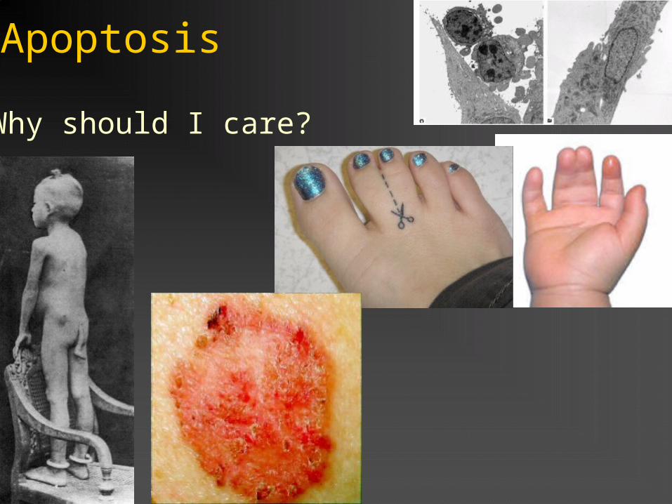
Why should I care?


(maintains normal size of the organ)
(Chronic myelogenous leukemia)
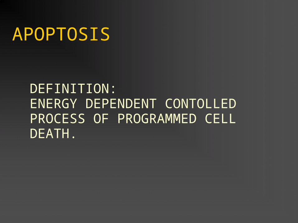
Complexity of Apoptotic pathways
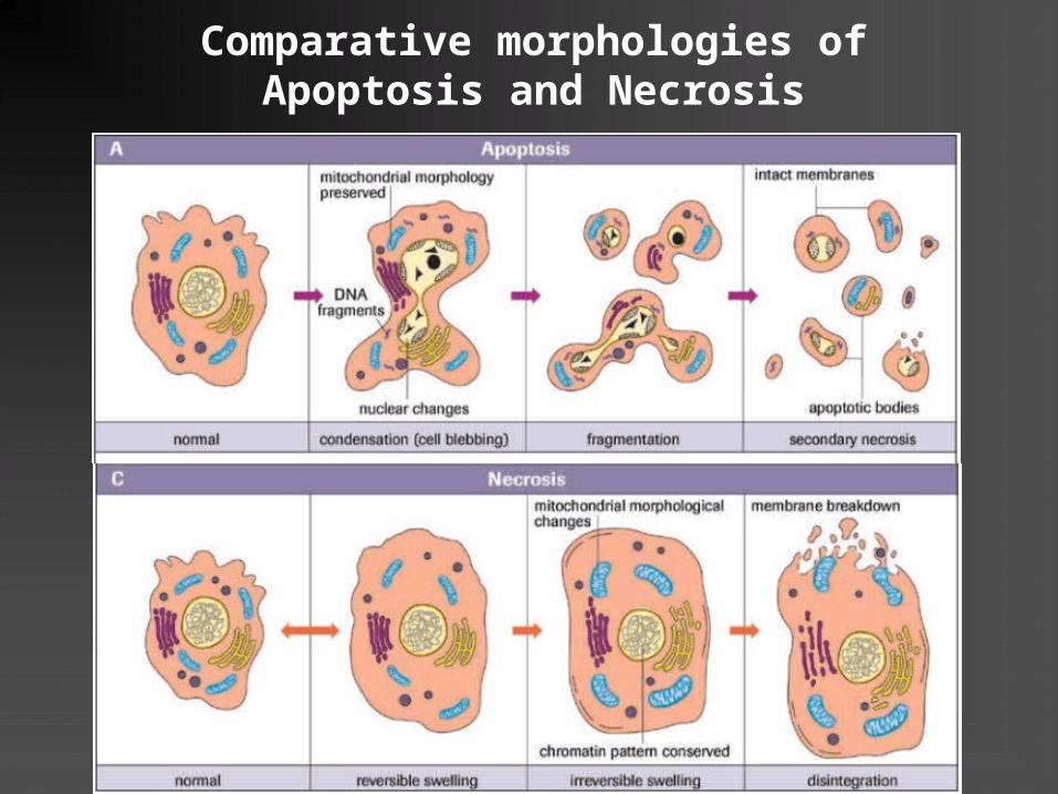
Comparative morphologies of Apoptosis and Necrosis


1
2
3
4

The two major routes to Apoptosis:Intrinsic and Extrinsic


The extrinsic (death receptor–initiated) pathway of apoptosis

The extrinsic (death receptor–initiated) pathway of apoptosis
Click here to see video

The intrinsic or mitochondrial pathway
1. Involves Bcl family of
proteins
2. Both pro and anti
apoptotic members
3. Best known is anti-
apoptotic Bcl2
4. Production of Bcl2
can be induced by
Growth Factors (IGF1)
deprivation or IR
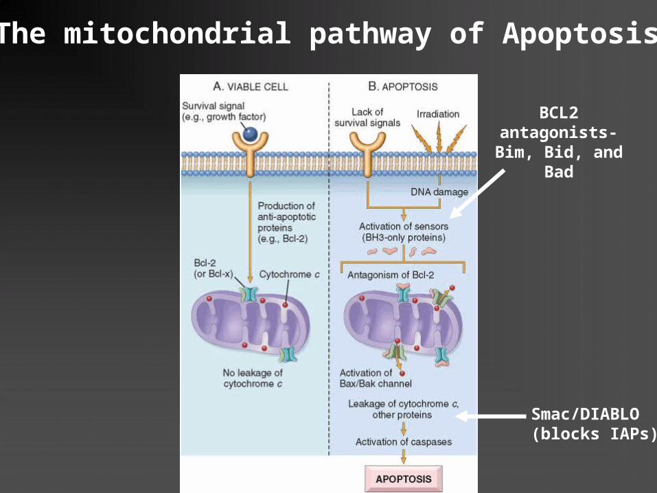
The mitochondrial pathway of Apoptosis
BCL2 antagonists-Bim, Bid, and
Bad
Smac/DIABLO(blocks IAPs)
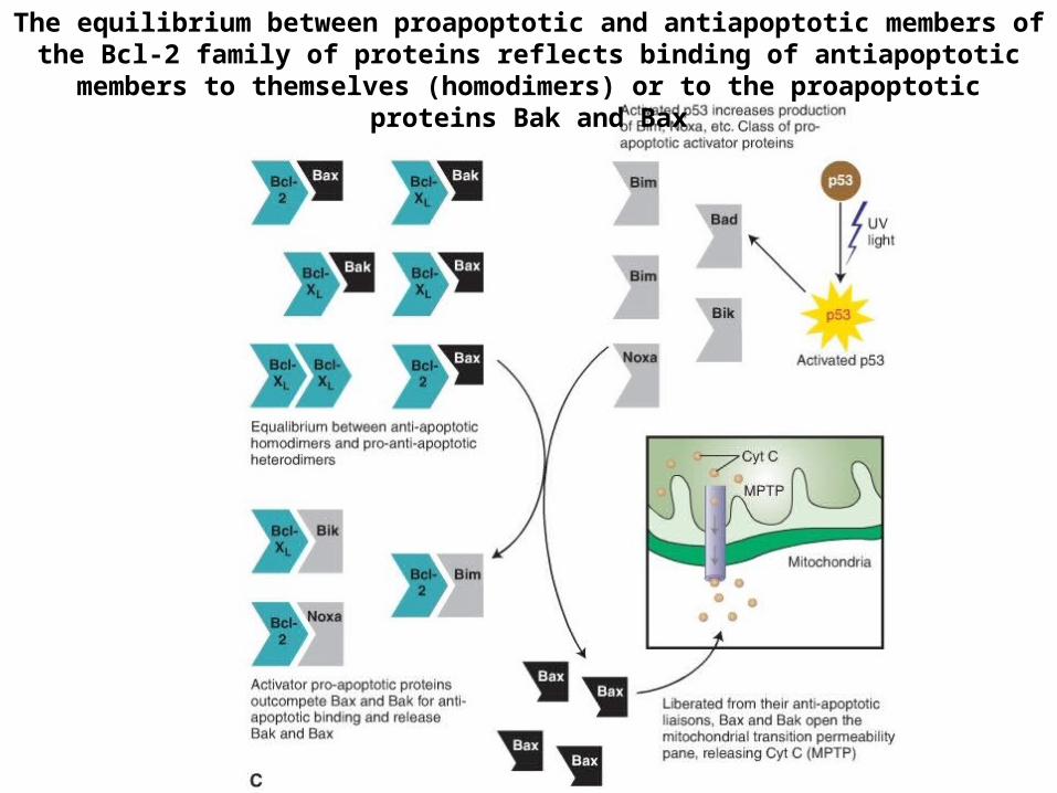
The equilibrium between proapoptotic and antiapoptotic members of the Bcl-2 family of proteins reflects binding of antiapoptotic members to themselves
(homodimers) or to the proapoptotic proteins Bak and Bax

Opening of the mitochondrial permeability transition pore leads to Apaf-1 activation and triggering the apoptotic cascade.
CytC- cytochrome cPTP= permeability transition poresROS = reactive oxygen species
An experiment introduces a “knockout” gene mutation into a cell line. The frequency of shrunken cells with chromatin clumping and cytoplasmic blebbing is increased compared with a cell line without the mutation. Overall survival of the mutant cell line is reduced. Which of the following genes is most likely to be affected by this mutation?(A) BAX(B) p53(C) C-MYC(D) FAS(E) BCL-2
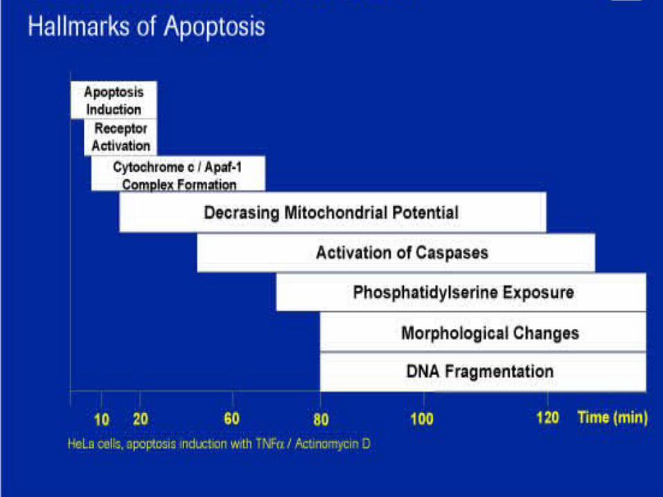
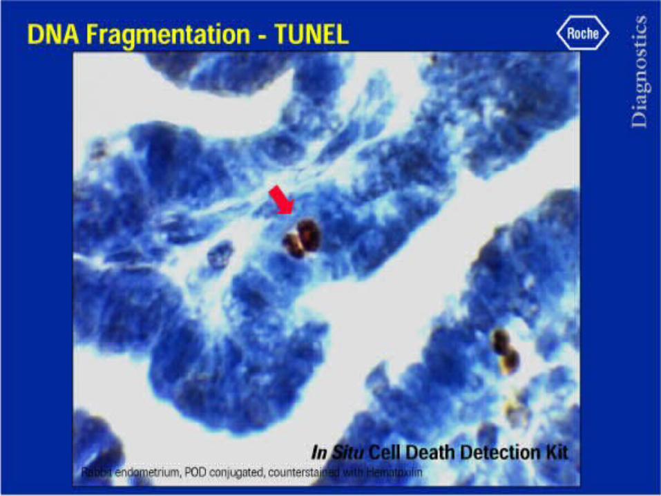
How to detect or measure apoptotic events in the tissues ?

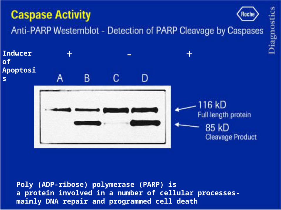
Poly (ADP-ribose) polymerase (PARP) is a protein involved in a number of cellular processes- mainly DNA repair and programmed cell death
Inducer of Apoptosis
- + - +


Phosphatidylserine exposure during apoptosis
Annexin-V-FITC“Eat-me” signal for professional phagocytes

Phosphatidylserine exposure during apoptosis
Propidium Iodide stains in red only cells with damaged membranes

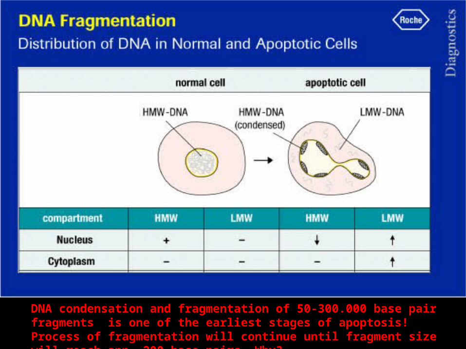
DNA condensation and fragmentation of 50-300.000 base pair fragments is one of the earliest stages of apoptosis! Process of fragmentation will continue until fragment size will reach app. 200 base pairs. Why?
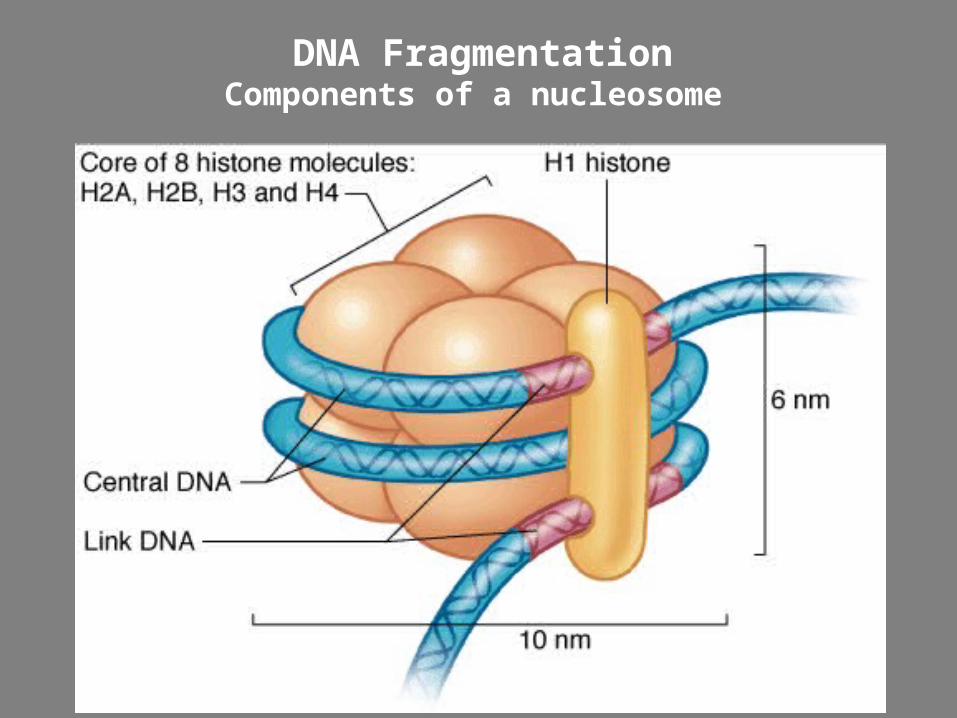
DNA FragmentationComponents of a nucleosome

DNA FragmentationNucleosomal Banding Pattern


Accumulation of cells in sub-G1 phase of cell cycle during apoptosis
G1
SG2/M

How apoptotic cells removed from tissues?
• Phagocytosis is the main mechanism• Signals for phagocytosis are:1. Flip of phosphatidylserine from inner to outer
leaflet of cell membrane (ligand for macrophages receptors)
2. Secretion of soluble factors that recruit phagocytes
3. Production of thrombospondin on cell surface of apoptotic cells
4. Coating of apoptotic cells with C1q complement
Apoptosis
Why should I care?

Role of Apoptosis in the Hand Plate

A 40 y/o male brought to ED post-MVA. Patient has a history of fever, headache, chills, and pain in RUQ 5 days ago. Histologic exam shows liver section w/disruption of normal hepatic lobule. Small shrunken hepatocytes w/intense eosinophilic cytoplasm, fragmented nuclear chromatin, & cytoplasmic bleb formations are noted. Which process is most likely occurring in the hepatocytes described?A) apoptosisB) AtrophyC) Caseous necrosisD) Coagulation necrosisE) DysplasiaF) Fatty changeG) HetrophagyH)Liquefaction necrosisI) metaplasia

CLINICO-PATHOLOGIC CORRELATIONS:
APOPTOSIS AND DISEASE
1.Reduced Apoptosis – Cancer, Autoimmune Diseases (SLE, Rheumatoid Arthritis, etc)
2. Increased Apoptosis – Neurodegenerative Diseases, Myocardial Infarction, Stroke, Viral diseases (Hepatitis, AIDS)

• A 55-year-old woman with colon cancer and a healthy 55-year-old woman are participating a study of colon cancer. Malignant cells are obtained from the tumor in the affected patient, and normal colonic epithelial cells are procured from the healthy subject. After both cell types are treated with transforming growth factor-beta, the number of normal cells decreases, whereas the number of tumor cells remains unchanged. The tumor cells most likely express a mutation that inhibits which of the following physiologic processes?
A) ApoptosisB) Cell cycle progressionC) DNA repairD) MigrationE) Necrosis

• Growth Factor Deprivation: Lymphocytes that are not stimulated by antigens and cytokines, neurons deprived of nerve growth factor, endometrial glands in post menopause
• DNA Damage: Radiation or chemotherapeutic agents induces DNA damage
Mechanisms of increased apoptosis:

Role of P53 Tumor suppressor protein in apoptosis and cancerogenesis
•P53 is a 53kDa protein present mostly in the nuclei of all cells, rapidly accumulates after injury and become phosporylated •Phosphorylation allows to bind to DNA and activate transcription of cell cycle inhibitors or proapoptotic proteins•P53 block transition of cells from G1 to S phase if DNA damage is not repaired

P53 mediated apoptosis
If repair is unsuccessful then P53 will induce expression of BAX. BAX acts against bcl2 and causes apoptosis
Hormonally related cancers seem to particularly involve loss of p53 related apoptosis
Chemo/rad work best when p53 is intact as also act through apoptosis
Role of P53 Tumor suppressor protein in apoptosis and cancerogenesis

Neurodegenerative Diseases and Apoptosis
- Withdrawal of growth factors favors actions of pro-apoptotic members of Bcl family
- For neurons dependent on nerve growth factors the loss of this stimulation can lead to neural apoptosis and degenerative neurologic diseases:
(Alzheimer's disease, Amyotrophic lateral sclerosis, Friedreich's ataxia, Huntington's disease, Lewy body disease, Parkinson's disease, Spinal muscular atrophy)

SOD-1 mutations can activate caspase-1 and caspase-3, and might increase free-radical generation, leading to motor neuron apoptosis. The activation of caspase-1 leads to interleukin-1 production, which can induce a local microglial inflammatory response and increase the number of neurons affected.
Example: SOD-1 mutations activate cell death pathways in familial amyotrophic lateral sclerosis

Mechanisms of IR induced cell death
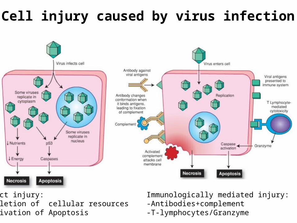
Cell injury caused by virus infection
Direct injury:-Depletion of cellular resources-Activation of Apoptosis
Immunologically mediated injury:-Antibodies+complement-T-lymphocytes/Granzyme

Protein Misfolding and unfolded protein response (aka ER-stress)
• Proteins which called “Chaperones” play crucial role in the proper folding of newly synthesized proteins
• Normally misfolded polypeptides are ubiquitinated and targeted for proteolysis in proteasomes.
• Accumulation of misfolded proteins in the ER trigger a number of cellular responses, collectively called the unfolded protein response
• If cells are unable to cope with the accumulation of misfolded proteins, the cell activates caspases and induces apoptosis.
• Examples: Alzheimer, Huntington, and Parkinson diseases, type 2 diabetes
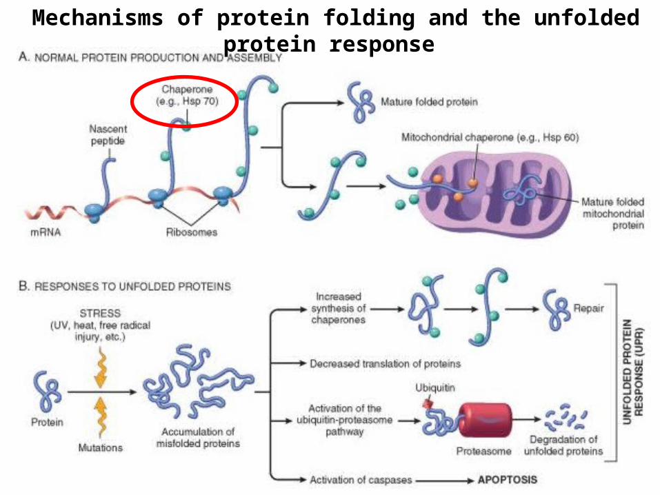
Mechanisms of protein folding and the unfolded protein response

Autophagy
What can activate Autophagy:1. Nutrient Deprivation( low level
of a.acids)2. Accumulation of long-living
proteins
What regulate Autophagy?1. MTOR signaling pathway2. Atgs genes/proteins What Mechanism?1. Atgs proteins create autophagic
vacuoles 2. Autophagic vacuoles fuse with
lysosomes and content is digested
•Definition: Autophagy is the process in which a cell sequesters and recycles damaged organelles and macromolecules ( eats its own contents)

Autophagy and Disease1. Cancer: This is an area of active investigation, autophagy
can both promote cancer growth and act as a defense against cancers.
2. Neurodegenerative disorders: In Alzheimer disease, formation of autophagosomes is accelerated. In Huntington disease, mutant protein huntingtin impairs autophagy.
3. Infectious diseases: Many pathogens are degraded by autophagy; these include mycobacteria, Shigella spp., and HSV-1. This is one way by which microbial proteins are digested and delivered to antigen presentation pathways. Macrophage-specific deletion of Atg5 increases susceptibility to tuberculosis
4. Inflammatory bowel diseases: Studies have linked both Crohn disease and ulcerative colitis to autophagy related genes.

Necroptosis• Necroptosis was first recognized as a caspase-
independent form of cell death that can be triggered by treatment with TNF only in the presence of a pan-caspase inhibitors
• Main executing molecule of the process is called RIPK3 - receptor-interacting protein kinase 3 (RIPK3)
• Necroptosis requires that the function of caspase 8 be inhibited or disrupted.
• Unlike apoptosis, in which several of the highly immunogenic intracellular proteins are sequestered in the dead cell, necroptosis is a strong trigger of innate and adaptive immune responses

Formation of the Necrosome is key
point in the induction of Necroptosis
Molecular mechanism of TNF-mediated necroptosis:Cross-linking of TNFR1 by TNF causes recruitment of RIP1 and RIP3 along with caspase 8. Inhibition of caspase 8, as may occur in some viral infections, allows RIP1 and RIP3 to initiate signals that affect mitochondrial generation of ATP and ROS. This is followed by events typical of necrosis.

Key Concepts of Necroptosis
• Necroptosis resembles necrosis morphologically and apoptosis mechanistically as a form of programmed cell death.
• Necroptosis is triggered by ligation of TNFR1, and viral proteins of RNA and DNA viruses.
• Necroptosis is caspase-independent but dependent on signaling by the RIP1 and RIP3 complex.
• RIP1-RIP3 signaling reduces mitochondrial ATP generation, causes production of ROS, and permeabilizes lysosomal membranes, thereby causing cellular swelling and membrane damage as occurs in necrosis.
• Release of cellular contents evokes an inflammatory reaction as in necrosis.
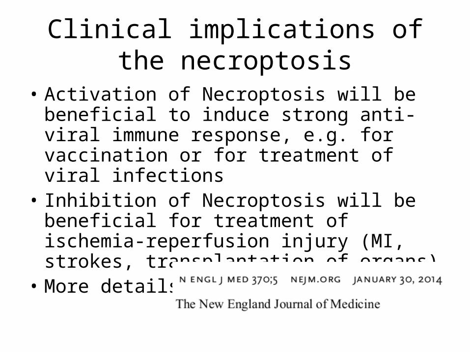
Clinical implications of the necroptosis
• Activation of Necroptosis will be beneficial to induce strong anti-viral immune response, e.g. for vaccination or for treatment of viral infections
• Inhibition of Necroptosis will be beneficial for treatment of ischemia-reperfusion injury (MI, strokes, transplantation of organs)
• More details:

Mediation of Programmed Cell Death by Apoptosis or Regulated Necrosis
mitochondrial permeability
transition
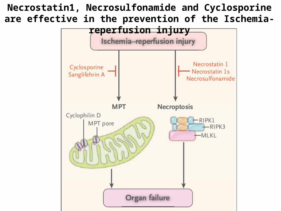
Necrostatin1, Necrosulfonamide and Cyclosporine are effective in the prevention of the Ischemia-reperfusion injury
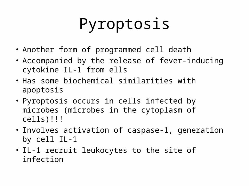
Pyroptosis
• Another form of programmed cell death• Accompanied by the release of fever-inducing
cytokine IL-1 from ells• Has some biochemical similarities with apoptosis• Pyroptosis occurs in cells infected by microbes
(microbes in the cytoplasm of cells)!!!• Involves activation of caspase-1, generation by
cell IL-1• IL-1 recruit leukocytes to the site of infection

From : Cell death in the host response to infection Cell Death and Differentiation (2008) 15, 1339–1349; doi:10.1038/cdd.2008.91; published online 20 June 2008 , K Labbé1 and M Saleh1,2

Pathogen-induced host cell death
The type of death the cell undergoes depends on the nature of the pathogen, pathogen load and site of infection. Pyroptotic, apoptotic, autophagic or oncotic cells display a distinct set of morphological and biochemical characteristicsApoptosis and autophagy do not induce inflammation
Apoptosis, pyroptosis and autophagy are generally beneficial to the host, oncosis favors pathogen dissemination

Please, take your pen and answer on the following questions
You have 80 seconds to answer each question
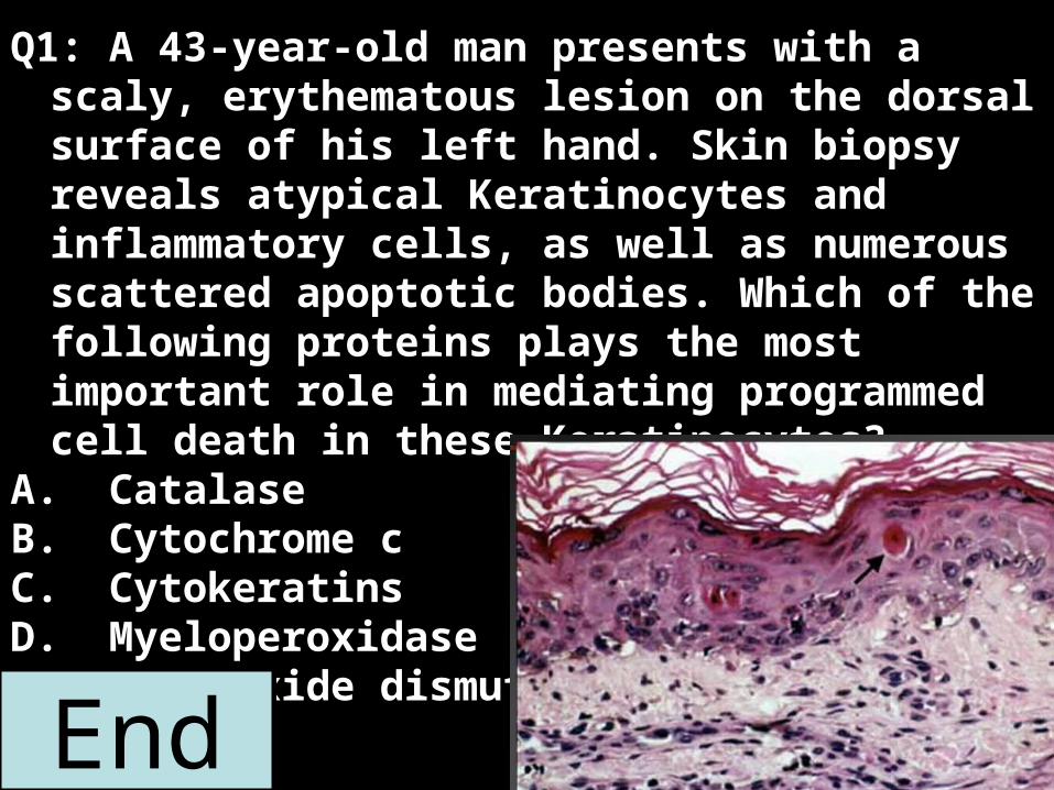
Q1: A 43-year-old man presents with a scaly, erythematous lesion on the dorsal surface of his left hand. Skin biopsy reveals atypical Keratinocytes and inflammatory cells, as well as numerous scattered apoptotic bodies. Which of the following proteins plays the most important role in mediating programmed cell death in these Keratinocytes?
A. Catalase B. Cytochrome cC. CytokeratinsD. MyeloperoxidaseE. Superoxide dismutase
2:001:591:581:571:561:551:541:531:521:511:501:491:481:471:461:451:441:431:421:411:401:391:381:371:361:351:341:331:321:311:301:291:281:271:261:251:241:231:221:211:201:191:181:171:161:151:141:131:121:111:101:091:081:071:061:051:041:031:021:011:000:590:580:570:560:550:540:530:520:510:500:490:480:470:460:450:440:430:420:410:400:390:380:370:360:350:340:330:320:310:300:290:280:270:260:250:240:230:220:210:200:190:180:170:160:150:140:130:120:110:100:090:080:070:060:050:040:030:020:01End

Q2: You observe a slide containing numerous epithelial cells. A colleague looks at the unstained cells under the light microscope and says that most of the cells are undergoing apoptosis. You look at the slide in the microscope and agree. What alterations in the cellular structure did you observe in the light microscope that led to the conclusion that the cells were undergoing apoptosis?A Plasma membrane was intact
B Mitochondria were absent
C Plasma membrane showed blebbing
D Nucleus was absent
E Polysomes were associated with the endoplasmic reticulum 2:001:591:581:571:561:551:541:531:521:511:501:491:481:471:461:451:441:431:421:411:401:391:381:371:361:351:341:331:321:311:301:291:281:271:261:251:241:231:221:211:201:191:181:171:161:151:141:131:121:111:101:091:081:071:061:051:041:031:021:011:000:590:580:570:560:550:540:530:520:510:500:490:480:470:460:450:440:430:420:410:400:390:380:370:360:350:340:330:320:310:300:290:280:270:260:250:240:230:220:210:200:190:180:170:160:150:140:130:120:110:100:090:080:070:060:050:040:030:020:01End
2:001:591:581:571:561:551:541:531:521:511:501:491:481:471:461:451:441:431:421:411:401:391:381:371:361:351:341:331:321:311:301:291:281:271:261:251:241:231:221:211:201:191:181:171:161:151:141:131:121:111:101:091:081:071:061:051:041:031:021:011:000:590:580:570:560:550:540:530:520:510:500:490:480:470:460:450:440:430:420:410:400:390:380:370:360:350:340:330:320:310:300:290:280:270:260:250:240:230:220:210:200:190:180:170:160:150:140:130:120:110:100:090:080:070:060:050:040:030:020:01End
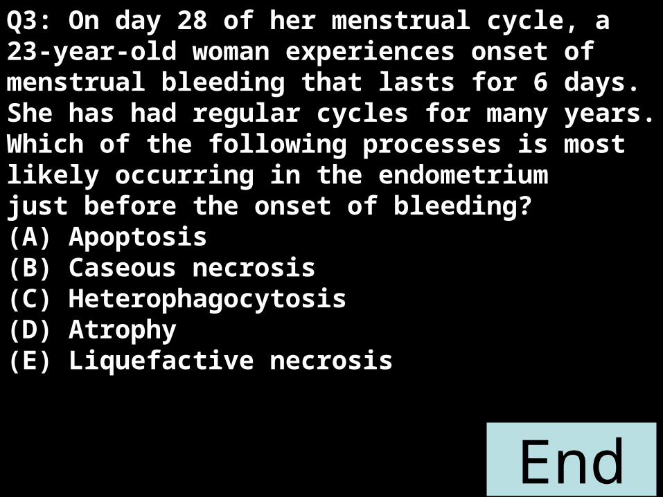
Q3: On day 28 of her menstrual cycle, a 23-year-old woman experiences onset of menstrual bleeding that lasts for 6 days. She has had regular cycles for many years. Which of the following processes is most likely occurring in the endometriumjust before the onset of bleeding?(A) Apoptosis(B) Caseous necrosis(C) Heterophagocytosis(D) Atrophy(E) Liquefactive necrosis

Q4: A cell pathologist performs experiments to examine the biochemical and physiological changes that occur in cells undergoing apoptosis. Which of the following characteristics is associated with the first stage of apoptosis of cells?A DNA ladder formation B Nuclear condensation C Blebbing or "boiling" of the cytoplasmic membrane D Chromatin condensation E Fragmentation of DNA into 50-300 kilobase fragments
2:001:591:581:571:561:551:541:531:521:511:501:491:481:471:461:451:441:431:421:411:401:391:381:371:361:351:341:331:321:311:301:291:281:271:261:251:241:231:221:211:201:191:181:171:161:151:141:131:121:111:101:091:081:071:061:051:041:031:021:011:000:590:580:570:560:550:540:530:520:510:500:490:480:470:460:450:440:430:420:410:400:390:380:370:360:350:340:330:320:310:300:290:280:270:260:250:240:230:220:210:200:190:180:170:160:150:140:130:120:110:100:090:080:070:060:050:040:030:020:01End

A male newborn is found to have syndactyly of three fingers of his right hand. As the attending resident you are called upon to explain to the child's parents as to why this happened to their child. Which of the following cellular processes is most likely to have failed during development in utero and caused the malformation?
A Apoptosis
B Differentiation
C Fusion
D Fission
E Migration
F Proliferation
V