Embed Size (px)
Citation preview

INTRODUCTION
Non-alcoholic fatty liver disease (NAFLD) is the mostcommon liver disease in developed countries (1). NAFLD ischaracterized by fatty infiltration of the liver and increasedendogenous lipogenesis in the absence of chronic alcoholconsumption (2). NAFLD is closely associated with visceraladiposity, insulin resistance and dyslipidemia, and is described asthe hepatic component of metabolic syndrome (3). It is estimatedthat 20% of patients with NAFLD develop non-alcoholicsteatohepatitis (NASH) involving steatosis combined withinflammation, that can progress into fibrosis and cirrhosis (4).
Historically, NAFLD progression was interpreted with “two-hit” hypothesis where the first hit is related to lipid accumulation(1) and the following one with inflammation, fibrosis andcellular death (1, 2). In contrast, a novel multifactor etiologymodel, with a central role for Kupffler cells and oxidized formsof LDL uptake has been recently postulated (5). In this model,lipid-induced cell toxicity and apoptosis are specific to fattyacids present in the circulation and liver (5). There are mainlysaturated fatty acids: palmitic, stearic that promote liver damagevia activation of apoptosis via a caspase-dependent mechanism
(2). Both palmitic and stearic acid activate endoplasmicreticulum stress, which leads to activation of proapoptotic andproinflammatory factors such as C-Jun NH2 terminal kinase,nuclear factor κB, and pro-apoptotic Bcl-2 family members (e.g.Bax) (2, 6). It has been noticed, that patients with NASHexperience significant changes (qualitative and quantitative) inthe lipid composition of plasma and cell membranes comparedto NAFLD patients (4).
5-oxo-6,8,11,14-eicosatetraenoic acid (5-oxo-ETE) is astrong chemoattractant synthesized by proinflammatory cells (7-11). The biological actions of 5-oxo-ETE are mediated by thehighly selective OXE receptors, which are expressed on varietyof inflammatory cells: eosinophil, neutrophils, myeloid cells (8).Synthesis of 5-oxo-ETE (by 5 lipoxygenase andhydroxyeicosanoid dehydrogenase) is regulated by intracellularNADP(+) levels and is dramatically increased under oxidativestress and the respiratory burst in phagocytic cells (restingneutrophils metabolize 5-HETE principally by ω-oxidation to5,20-diHETE) (9, 11).
Amongst other causes, NAFLD can develop (into NASH)due to specific gene variants associated with key elements of lipidmetabolism (4), e.g. the Apo-E gene that encodes apolipoprotein
JOURNAL OF PHYSIOLOGY AND PHARMACOLOGY 2013, 64, 6, 711-717www.jpp.krakow.pl
E. STACHOWSKA1, D. MACIEJEWSKA1, P. OSSOWSKI1, A. DROZD1, K. RYTERSKA1, M. BANASZCZAK1,M. MILKIEWICZ2, J. RASZEJA-WYSZOMIRSKA3,4, M. SLEBIODA5, P. MILKIEWICZ3,4, H. JELEN1
APOLIPOPROTEIN E4 ALLELE IS ASSOCIATED WITH SUBSTANTIAL CHANGESIN THE PLASMA LIPIDS AND HYALURONIC ACID CONTENT IN PATIENTS
WITH NONALCOHOLIC FATTY LIVER DISEASE
1Department of Biochemistry and Human Nutrition, Pomeranian Medical University, Szczecin, Poland; 2Medical Biology Laboratory, Pomeranian Medical University, Szczecin, Poland;
3Liver Unit, Pomeranian Medical University, Szczecin, Poland; 4Liver Unit, Department of General Surgery and Liver Transplantation, Warsaw Medical University, Warsaw, Poland; 5Perlan Technologies Poland, Warsaw, Poland
Fat may affect progression of liver damage in patients with non-alcoholic fatty liver disease (NAFLD). In this study wecharacterize the state of lipid metabolism in 22 patients with NAFLD and different Apo-E variants. Total concentrationof plasma total fatty acids was quantified by gas chromatography, while their derivatives by liquidchromatography/tandem mass spectrometry (LC ESI MS/MS). The ratio of plasma saturated fatty acid tomonounsaturated fatty acid increased, whereas the ratio of polyunsaturated fatty acids to saturated fatty acids wasreduced in Apo-E4 carriers. Simultaneously, the levels of individual plasma linoleic, arachidonic, and alpha linolenicacids significantly increased in subjects with the Apo-E4 allele. The 15-lipoxygenase metabolite, 13-hydroxyoctadecadienoic acid, was significantly higher in Apo-E3 carriers (p<0.006). 5-oxo-6,8,11,14-eicosatetraenoicacid was significantly elevated in Apo-E4 carriers (p<0.009). A significant difference in hyaluronic acid concentration(p<0.0016) as well as predicted advanced fibrosis (using the BARD scoring system) was found in Apo-E4 carriers(p<0.01). We suggest that a distinct mechanism of fibrosis between Apo E alleles. In Apo-E4 carriers, an elevation in 5-oxo-6,8,11,14-eicosatetraenoic acid synthesis and fatty acid dysfunction may induce fibrosis, while an inflammatoryprocess may be the main cause of fibrosis in Apo-E3 carriers.
K e y w o r d s : non-alcoholic fatty liver disease, non-alcoholic steatohepatitis, apolipoprotein E, fatty acids, 5-oxo-6,8,11,14-eicosatetraenoic acid, liquid chromatography/tandem mass spectrometry, gas chromatography
E (Apo-E) - a glycoprotein essential for the metabolism oftriglyceride-rich lipoproteins. An interaction between Apo-E andreceptors determines the removal of Apo-E rich lipoproteins, andhomeostasis of cholesterol and triglycerides (12, 13). There arethree major isoforms of human ApoE (ApoE2, ApoE3, ApoE4)encoded by 3 allels (epsilon 2, 3, and 4) (12). ApoE status isdefined by combination of two SNPs (rs7412 and rs429358).ApoE-ε3 carriers contains rs7412(C) and rs429358 (T) whileApoE-ε4 has both rs7412(C) and rs429358 (C) (14, 15). Invarious combinations, Apo-E alleles are associated withneurodegenerative, coronary and vascular diseases (16-20) aswell as correspond to an altered lipid profile (21, 22).
It was reported that the response of plasma lipids wasdifferent in subjects with Apo-E genotypes (12), but still isunknown whether variants of Apo-E may change plasma lipidcomponents thus predisposing patients to NAFLD progression.Therefore, in this study a lipidomic approach was chosen tocharacterize the state of lipid metabolism in patients with Apo-Evariants. Individuals with Apo-E4 allele were compared to thosewith Apo-E3 - as most frequently reported, wild-type allele. Wequantified the total concentration of plasma total fatty acids bygas chromatography, fatty acids derivatives by liquidchromatography/tandem mass spectrometry (LC ESI MS/MS).ApoE genotype we estimated by real-time PCR, whereas liverfibrosis parameters by ELISA method and BARD scoringsystem. Although all examined subjects in our study werediagnosed with NAFLD, their lipid profiles differed in thecomposition of fatty acids and some of their metabolites.
MATERIALS AND METHODS
Chemicals and materials
Chloroform, methanol and acetic acid were sourced fromMerck (USA). Boron trifluoride in methanol, NaCl and 2,6-di-tert-butyl-4-methylphenol (BHT) were supplied by Sigma-Aldrich (Poland). Double-distilled water was obtained from aMilli-Q Water System (Millipore, Billerica, MA, USA). Fattyacid standards were obtained from Sigma-Aldrich, Neochema(Germany) and Cayman (USA).
Patients
A group of 23 Caucasian individuals diagnosed withNAFLD were prospectively enrolled in the study. The degree ofliver steatosis was assessed by a trained physician according tothe Hamaguchi score (23) using an abdominal ultrasound high-resolution B-mode scanner (Acuson X300). All NAFLD patientsincluded in the study were negative for HBV (hepatitis B virus)and anti-HCV (hepatitis C virus) and had a negative history ofalcohol intake (less than 20 g/d).
All participants underwent Apo-E genotyping for threeisoforms: Apo-E2, ApoE-3, and Apo-E4. Based on Apo-E allelesets, the individuals were divided into two subgroups i.e. Apo-E3(rs429358 [T] and rs 7412 [C]; n=11) or Apo-E4 (rs429358 [C orCC] and rs 7412 [C or CC]; n=11). In ApoE 3 group there are 3woman and 9 man, in Apo E4 group 5 woman 6 man.
After an overnight fasting venous blood for lipid analyseswas collected into tubes with anticoagulant. Whole blood wascollected into ethylenediaminetetraacetic acid (EDTA) tubes.Blood was immediately placed on ice or in a refrigerator, andsamples centrifuged at 3500 rpm for 10 min at 4°C within 2hours of collection. Plasma was then immediately stored underconditions to minimize artificial oxidation (i.e., with anantioxidant cocktail under inert atmosphere). Standard bloodbiochemical analyses were carried out at the University Hospital
Laboratory. Clinical and laboratory data on the analyzed patientsare summarized in Table 1.
BMI was based on an individual’s mass and height. A signedinformed consent was obtained from each patient. The studyprotocol was approved by the ethics committee of PomeranianMedical University and conformed to the ethical guidelines ofthe 1975 Declaration of Helsinki.
ApoE genotyping using real-time PCR
DNA from peripheral blood mononuclear cells was isolatedusing a DNeasy Blood&Tissue Kit (Qiagen). Genotypes weredetermined by real-time polymerase chain reaction (PCR) usingTaqman® SNP genotyping assays for two Apo-Epolymorphisms including rs7412 and rs429358 (AppliedBiosystems, Assay ID: C_904973_10, C_3084793_20,respectively). The fluorescence data were analyzed with allelicdiscrimination 7500 Software v.2.0.2.
Fibrosis parameters
In order to predict advanced fibrosis, the BARD scoringsystem developed and validated by Harrison et al. was used (24).An easily calculated composite score was based on the results offorced entry logistic regression analysis and included a BMI ≥28 = 1 point, AAR ≥ 0.8 = 2 points (AAR: aminotransferase ratioof AST/ALT), and the presence of DM = 1 point (DM: diabetesmellitus). BARD was able to predict advanced fibrosis: a scoreof 2–4 was associated with an OR for advanced fibrosis (F3-F4)(confidence interval 9.2 to 31.9) and a negative predictive valueof 96%. BARD reliably identified patients without significantfibrosis (19, 20) and was also validated in a Polish NAFLDpopulation (25). Hyaluronic acid, a marker of liver fibrosis insubjects with NAFLD (26) was estimated ELISA kit (WuhanEIAab Science).
Eicosanoid analysis by liquid chromatography/tandem massspectrometry (LC ESI MS/MS)
Isolation of fatty acid derivatives from plasma was carriedout using a dual mobile phase gradient (30, 2007). The flow ratewas 1.0 ml/min. The sample injection volume was 60 µl. TheDAD detector monitored peaks by adsorption at 235 nm for 13SHODE, 9S HODE, 15S HETE, 12S HETE and 5S HETE, 280nm for prostaglandin B2 (PGB2) (internal standard) and 5oxoHETE, 210 nm for 16RS HETE and 302 nm for 5 (S),6 (R),15(R) lipoxin A4 and 5(S),6 (R) lipoxin A4.
Analyses were performed using a triple quadrupole massspectrometer (Agilent 1290/Agilent 6400, Santa Clara, CA,USA). The liquid chromatograph was equipped with a binarypump (G4220A), diode array detector (G4212A), well platesampler (G4226A) and thermostated column compartment(G1316C). The mass spectrometer was equipped with an ESI ionsource (XESI with jet stream technology). The column used forcompounds resolution was a Poroshell 120 SBC 18 (3.0×100mm, 2.7 µM, PW. 68-975-302. Liquid chromatographyparameters, in-lab developed, were as follows: flow – 0.8ml/min, solvent A (H2O/0.1% Acetonitrile), solvent B(methanol/0.1% Acetonitrile ), solvent A ratio 90, solvent B ratio10, time 12.5 – solvent ratio B – 85, time 20 – solvent B ratio –100; DAD monitoring wavelengths: 235, 280, 210, 320, 280 nm;injection volume 20 µl; column temperature 25°C.
Spectra were acquired in a negative polarity using MRM.Compounds were detected at the retention times provided belowafter optimization of CE energy and two transitions weremonitored for each compound, where the first was used forquantitation, the other for confirmative purposes: 12HETE
712
(retention time (Rt) 13.34 min; CE(V) 5; 319.2 › 301.2, 319.2 ›179.2); 13HODE (Rt 13.13 min; CE(V) 9; 295.2 › 277.3, 295.2› 195.2); 15HETE (Rt 13.21 min; CE(V) 5; 319.2 › 301.3, 319.2› 257.3); 16HETE (Rt 13.08 min; CE(V) 5; 319.2 › 301.2, 319.2› 257.3); 5HETE (Rt 13.53 min; CE(V); 319.2 › 301.2, 319.2 ›115.0); 5oxoHETE (Rt 13.63 min; CE(V) 9; 317.2 › 203.2, 317.2› 59.1); 5S,6R,15R lipoxin (Rt 10.85 min; CE(V) 9; 351, 2 ›217.2, 351 › 115.1); 5S,6R lipoxin (Rt 10.75 min; CE(V) 9;351.2 › 217.2, 351.2 › 115.1); 9HODE (Rt 13.14 min; CE(V) 9;295.2 › 277.3, 295.2 › 171.1); PGB2 (internal standard; Rt 11.28min; CE(V) 9; 333.2 › 235.2, 333.2 › 175.1). All transitions wereoptimized using an infusion of standards. The operatingparameters for MS were as follows: gas temp – 300°C; gas flow– 5 l/min; nebulizer – 45 psi; sheath gas heater – 250°C; sheathgas flow –– 11 ml/min; capillary voltage – 3500 V.
Acquired data were processed using Mass Hunter software(Agilent Technologies, Santa Clara, CA, USA). Quantitationwas carried out based on MRM with internal standard (PGB2).Calibration curves were determined for a concentration range of0.05–1.00. Linearity for analyzed compounds exceeded 0.993(this value was observed for 16HETE). For the majority ofcompounds linearity was 0.999.
Total fatty acids extraction and gas chromatography analysis
Total fatty acids (FFA) were extracted according to the Folchmethod (27). 1 ml of plasma extract was saponified with 1 ml of2M KOH methanolic solution at 70°C for 20 min and thenmethylated with 2 ml 14% solution of boron trifluoride in methanolunder the same conditions. In the next step, 2 ml of n-hexane and10 ml saturated NaCl solution were added. The samples wereallowed to completely separate into an upper (n-hexane phase) andlower layer. 1 ml of the n-hexane phase was collected.
Gas chromatography was performed using an AgilentTechnologies 7890A GC System. The instrument was equippedwith a SUPELCOWAX™ 10 Capillary GC Column (15 m×0.10mm, 0.10 µm) (Supelco, Bellefonte, PA, USA). Chromatographicconditions were as follows: the initial temperature was 60°C for0 min, increased at a rate of 40°C/min to 160°C (0 min),increased at a rate of 30°C/min to 190°C (0.5 min) and thenincreased at a rate of 30°C/min to 230°C for 2.6 min, where it wasmaintained for 4.9 min. The total analysis time was 8 min and thegas flow rate was 0.8 ml/min with nitrogen as the carrier gas.
Fatty acids were identified by comparison of their retentiontimes with those of commercially available standards.Quantitative analyses were made using ChemStation Software(Agilent Technologies, UK) and were based on peak areas.C17:0 was used as the internal standard for calculation. Resultswere expressed in ug/ml.
Analysis of diet
Diet composition and energy intake were ascertained usingquestionnaires (24-hour food diaries). Each subject wasinterviewed about their dietary pattern in the previous day. Datafrom questionnaires were analyzed using Food compositiontables (IZZ, Poland) and DIETETYK 6.0 software (Jumar,Poland).
Statistical analysis
Statistica 7.1 software was used for the statistical analysisand all results are expressed as median and interquartile range(IQR), and mean ± standard deviation. As the distribution inmost cases deviated from normal (Shapiro-Wilk test), non-parametric tests were used: Mann-Whitney test for comparisonsbetween groups, where p<0.05 was considered significant.
RESULTS
Changes in anthropometry and plasma findings
The baseline characteristics of the study population as wellas laboratory and anthropometry findings in Apo-E carriers areshown in Table 1. There were no significant differences inanthropometric parameters between both groups. In terms ofbiochemical parameters, we found a significant difference in theplasma concentration of postprandial glucose which waselevated in Apo-E4 variant carriers (111.86 ± 36.36 mg/dl versus88.89 ± 5.97, Apo-E3 control, p<0.02) (Table 1). There were nodifferences in insulin, level of enzymes as well as cholesteroland its fractions HDL and LDL (in relation to patients with Apo-E3) (Table 1).
Changes in predicted advanced fibrosis and hyaluronic acidconcentration
A significant difference in predicted advanced fibrosiscalculated using the BARD scoring system was found betweenApo-E variant carriers. Apo-E4 carriers had a score of 2.36 ±1.03 versus 1.17 ± 1.03 in Apo-E3 carriers, p<0.01, associatedwith an increased OR for advanced fibrosis (Table 2). We notedan elevation in plasma hyaluronic acid concentration in Apo-E4carriers compared to Apo-E3 carriers (60.1 ± 11.9 ng/ml versus40.25 ± 6.2 ng/ml). Interestingly, there were no differences inliver steatosis according to the Hamaguchi score (Table 2).
Changes in total fatty acid in plasma
Palmitic acid, stearic acid, oleic acid and linoleic acid werethe major FFA in the circulating plasma pool in both Apo-Evariant carriers (Table 3). Compared with Apo-E3 carriers, theratio of saturated fatty acids (SFA) to monounsaturated fattyacids increased, whereas the ratio of polyunsaturated fatty acids(PUFA) to SFA decreased in Apo-E4 carriers.
Estimated fatty acids ratios:a) ratio all SFA to all PUFA was: 1:2.3 (Apo-E3 alleles)
versus 1: 1.2 (Apo-E4 alleles)b) ratio all SFA to all MUFA was 1.5:1 (Apo-E3 alleles)
versus 2:1 (Apo-E4 alleles)c) ratio all PUFA to all MUFA was 3.25:1 (Apo-E3 alleles)
versus 2.4:1 (Apo-E4 alleles)There was a significant increase in palmitoleic acid (16:1,
n=7) (11.5 ± 4.8 vesus 19.2 ± 13.6; p=0.05), vaccenic acid (18:1,n=7) (12.4 ± 2.8 versus 24.1 ± 14.4; p<0.0004) as well oleic acid(18:1, n=9) (87.2 ± 24.7 versus 151.1 ± 87.6; p<0.006) in Apo-E4 carriers (Table 3). Linoleic acid (18:2, n–6) and α linolenicacid (18:3, n=3) are both essential fatty acids and are sources ofn=6 and n=3 PUFA, respectively. There were increased amountsof the n=6 PUFA family: linoleic acid (18:2, n=6) (85.0 ± 15.6versus 164.4 ± 64.5; p<0.00001), arachidonic acid (20:4, n=6)(0.4 ± 0.5 versus 4.5 ± 6.1, p<0.01) in subjects with the Apo-E4polymorphism (Table 3). The amount of α linolenic acid wasalso significantly increased in subjects with Apo-E4 (3.0 ± 0.8versus 6.1 ± 3.3, p<0.004), followed by docosahesaenoic acid22:6, n=3 (10.6 ± 5.0 vesus 15.4 ± 6.9, p<0.03), a product ofperoxisomal metabolism of PUFA (4) (Table 3).
Changes in plasma inflammatory metabolites of linoleic andarachidonic acid
5-oxo-6,8,11,14-eicosatetraenoic acid (5-oxo-HETE) wassignificantly elevated in Apo-E4 carriers (Table 4). Many productsof the LOX pathway (5-, 12-, 15-HETE) were increased in Apo-E4carriers, however, a significantly higher content of 13-HODE was
713

noted in Apo-E3 carriers. In humans, the conversion of linoleic acidleads to the production of linoleic acid derivatives -HODEs (9, 13-hydroxyoctadecadienoic acids), whereas oxidation of arachidonicacid is a source of HETEs (5, 12, 15-hydroxyeicosatetraenoicacids). These reactions are catalyzed by cellular lipoxygenases(LOXs) as well as reactive oxygen species (4).
Analysis of dietary energy intake among patients
Analysis showed no significant differences in energy intakeand diet composition with regard to key nutrients among theApo-E variants (Table 5).
DISCUSSION
The role of Apo-E genotypes in NAFLD has been previouslyinvestigated with conflicting results. In the last year it wasreported that the Apo-E4 allele showed a statistically significanttwo-fold reduction in NAFLD risk compared with homozygotesApo-E3 (28). A protective effect of the E4 allele towards NAFLDwas related to its role in the regulation of hepatic very low-density lipoproteins (VLDL) secretion (28). Meanwhile VLDLsecreted by the liver may contain different composition andconcentration of fatty acids and their derivatives (it is dependedon diet and liver function). Numerous studies have indicated that
714
Parameters Apo E 3 allele Apo E 4 allele
Median (IQR) Median (IQR)
Mean ± S.D. Mean ± S.D.
Age (year) 51.00 (16.50) 59.00 (14.50)
48.45 ± 11.14 53.36 ± 13.90
Body Mass Index (kg/m2) 31.61 (11.00) 34.22 (6.90)
33.26 ± 7.92 33.80 ± 4.56
Aspartate transaminase (U/l) 31.5 (14.75) 22.0 (7.25)
27.14 ± 9.39 27.14 ± 12.03
Alanine transaminase (U/l) 54.0 (34) 28.5 (15.25)
27.14 ± 9.39 34.86 ± 18.77
Gammaglutamyl trasferase (U/l) 53.5 (54.75) 25.0 (36.5)
56.74 ± 41.55 48.71 ± 51.83
Triacyloglicerols (mg/dl) 106 (33.5) 169 (113)
119.93 ± 90 226.60 ± 264.80
Cholesterol (mg/dl) 191 (43.5) 218 (45.25)
186.34 ± 38.15 246.36 ± 148.38
High density lipoprotein (mg/dl) 50 (6.25) 45 (14.25)
44.9 ± 11.56 47.86 ± 12.40
Low density lipoprotein (mg/dl) 115 (38) 120.5 (55.5)
115.78 ± 41.83 118.4 3± 40.75
Glucose (mg/ml) 95 (4.5) 106.5 (16.75) *
88.89 ± 5.97 111.86 ± 36.36
Insulin (U/ml) 8.8 (8.03) 7.75 (12.83)
12.45 ± 18.91 53.42 ± 159.06
Table 1. Baseline characteristics of the study population. *significant between Apo-E3 versus Apo-E4; p<0.05 versus the control (Apo-E3); U Mann Whitney Test.
Parameters Apo E 3 allele Apo E 4 allele Median (IQR) Median (IQR) Mean ± S.D. Mean ± S.D. Liver steatosis (Hamaguchi score) 3 (2.25) 3 (3) 2.67 ± 1.5 3.46 ± 2 Advanced fibrosis 1 (0.5) 3.00 (1.75)* 1.17 ±1.0 2.36 ± 1.0 Hyaluronic acid (ng/ml) 40 (6) 59 (20)** 40 ± 6 60 ± 12
Table 2. Hepatic parameters of the study population. All values are expressed as the median (IQR) and mean ± S.D. * p<0.05 Apo-E4 versus the control (Apo-E 3); ** p<0.005; (U Mann Whitney Test).

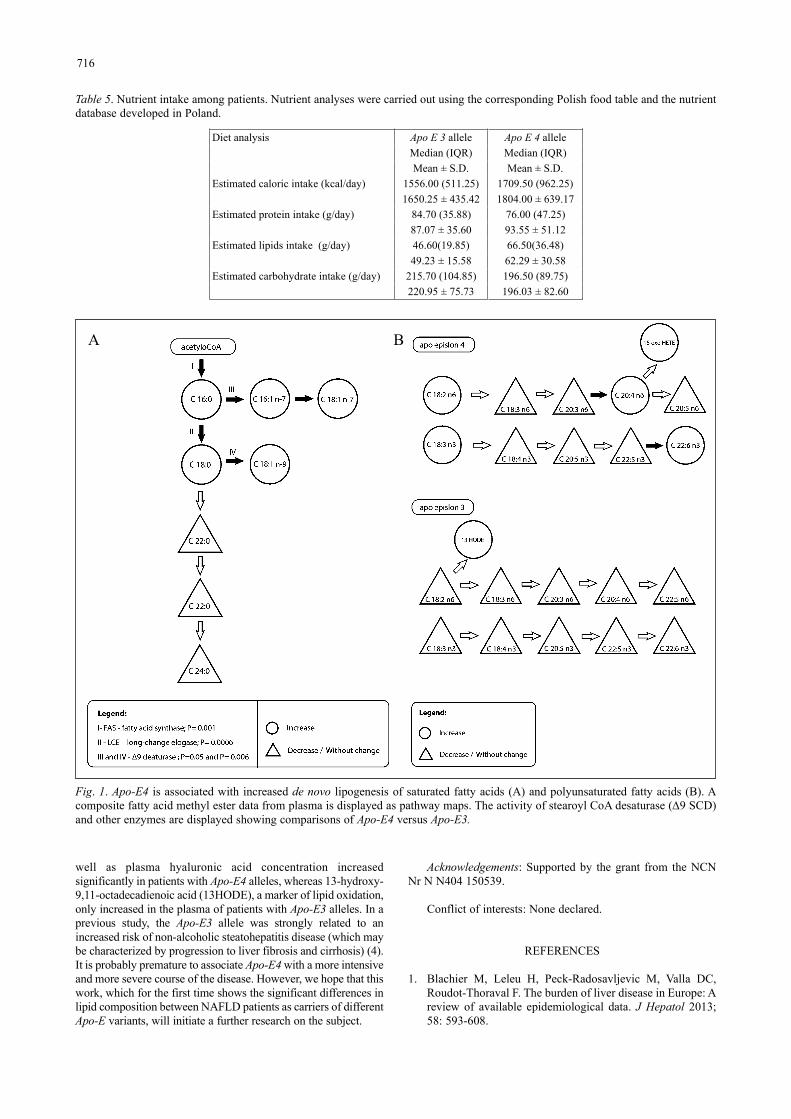
the content of saturated acids in plasma’s VLDL are correlatedwith severity of disease and is more deleterious to hepatocytefunctions than unsaturated fatty acids (2, 5, 29, 30). There is aconcept that elevated fatty acids (and their derivatives) promotehepatotoxicity (4) - especially 18:0 and C16:0 (that were elevatedin Apo-E4 allele carriers in this study, Fig. 1 A) can have variedmetabolic consequences on NAFLD progression (5).
Interestingly, in Apo-E4 carriers in this study an increasedconcentration of some monounsaturated fatty acids such aspalmitoleic, vaccenic acid and oleic acid was noted. These resultsmay connect Apo-E4 polymorphism with with up-regulation(activation) of two main lipogenic enzymes: fatty acid synthase(FAS) and ∆9 stearoyl-CoA desaturase (∆9 SCD). Expression of∆9 SCD is regulated by several transcription factors: peroxisomeproliferator activated receptors (PPAR), liver X receptor (LXR)and sterol regulatory element binding protein-1c (SREBP 1-c)which are up-regulated during inflammation (31) and by anumber of nutritional factors (SFA, glucose, fructose) (5, 32). Asnoted previously, increased ∆9 SCD activity was critical in the
regulation of the synthesis of fatty acids in a mouse model ofNASH (31-33). Interestingly, although the level of the above-mentioned fatty acids increased in Apo-E4 individuals, the intakeof calories (originally from fats) was similar in both groups ofApo-E carriers. Analysis of 24-hour dietary diaries collected fromthe patients showed no significant differences in the intake ofnutrients between individuals from both groups. In addition, Apo-E4 allele carriers (similar to NAFLD individuals) have a negativeratio of SFA to PUFA (5). Furthermore, an increase in SFAconcentration (stearic and palmitic acid) is accompanied byaugmentation of plasma PUFA content: linoleic acid (LA - theprecursor of n=6 PUFAs) and α linolenic acid (αALA a precursorof n=3 PUFAs). The metabolic fate of both LA and αALA are notthe same - only carriers of Apo-E4 showed an increased contentof the selected products of LA and αALA elongation anddesaturation - with greater synthesis of arachidonic acid and itsderivatives (5-oxo-ETE) in individuals with Apo-E4 (Fig. 1B).
In this study, also we showed clearly that Apo-E 4 allele isrelated to the intensification of synthesis 5-oxo-6,8,11,14-eicosatetraenoic acid (5-oxo-ETE). Earlier, elevation 5-oxo-ETE(as well as other biomerkers) among individuals with NASHhave been noticed (29, 30). The increase 5-oxo-ETE content inNASH was associated with progression of inflammatory stressand liver fibrosis (4, 29). It is possible that similar phenomenahas been observed in our study where Apo-E4 carriers werecharacterized by a higher plasma content of docosahexaenoicacid, linoleic acid and alpha linolenoic acid as well asarachidonic acid (Fig. 1B). Both arachidonic acid anddocosahexaenoic acid stimulate 5-oxo-ETE synthesis inneutrophils which triggers the synthesis of proinflammatorycytokines as well as cell proliferation (11). We suppose thatplasma 5-oxo-ETE was elevated by circulating proinflammatoryfatty acids elevated among Apo-E4 allele carriers (Fig. 1 A).
Although all examined subjects in our study were diagnosedwith NAFLD, their lipid profiles differed in the composition offatty acids and some of their metabolites. It seems that Apo-E4favors lipid metabolism disorders and a tendency for fibrosis. Inour study, fibrosis (predicted by the BARD scoring system) as
715
Fatty acids classification Apo E 3 allele Apo E 4 allele Median (IQR) Median (IQR) Mean ± S.D. Mean ± S.D. Saturated fatty acids Lauric acid 0.9 (1.2) 0.0 (1.7) 1.1 ± 1.4 1.1 ± 1.6 Myristic acid 6.2 (3.4) 9.7 (10.6)* 6.6± 2.9 11.8± 6.7 Palmitic acid 117 (18.5) 156.3(115)** 118.5± 25.5 206.3 ± 127.2 Stearic acid 36.0 (9.7) 52.8 (29.0)** 38.9 ± 7.8 66.7± 37.0 Monounsaturated fatty acids Palmitoleic acid 10.0 (4.4) 14.5 (10.2)* 11.5± 4.8 19.2± 13.6 Vaccenic acid 12.3 (3.0) 17.3 (19.0)# 12.4 ± 2.8 24.1± 14.4 Oleic acid 75.4 (31.5) 116.5 (109.4)** 87.2 ± 24.7 151.1 Polyunsaturated fatty acids Linoleic acid 81.4 (19.1) 149.4 (106.9) # 85.0 ± 15.6 164.4 ± 64.5 Gamma linolenic acid 1.9 (1.2) 2.3 (2.3) 1.9 ± 0.9 2.8 ± 2.4 Eicosadienoic acid 0.9 (1.2) 0.0 (0.7) 0.9 ± 0.8 0.4 ± 0.7 Arachidonic acid 0.0 (0.6) 2.1 (5.9)* 0.4 ± 0.5 4.5 ± 6.1 Docosapentaenoic acid 2.2 (1.3) 3.4 (3.6) 2.2 ± 1.4 4.4 ± 4.1 Alpha linolenic acid 3.0 (1.2) 6.3 (5.7)** 3.0 ± 0.8 6.1 ± 03.3 Eicosapentaenoic acid 3.7 (3.3) 6.4 (5.4) 6.8 ± 6.3 7.6 ± 4.9 Docosapentaenoic acid 0.0 (0.9) 0.0 (1.4) 0.4 ± 0.6 1.2 ± 2.2 Docosahesaenoic acid 8.9 (4.2) 13.7 (7.7)* 10.6 ± 5.0 15.4 ± 6.9
Table 3. Fatty acid composition of the plasma fatty acids studypopulation. All values are the Median (IQR ) (µg/ml) mean ±S.D. * p<0.05, ** p <0.005, # p<0.0005, Apo-E 4 versus thecontrol (Apo-E3) (U Mann Whitney Test).
Parameters Apo E 3 allele Apo E 4 allele Median (IQR) Median (IQR) Mean ± S.D. Mean ± S.D. 5S.6RLipoxin 0 (0) 0 (0) 0 ± 0 0.004 ± 0.02 5S.6R.15R Lipoxin 0 (0) 0 (0) 0 ± 0 0 ± 0 13 HODE 0.004 (0.003) 0.003 (0.001)** 0.005 ± 0.002 0.002 ±0.001 9 HODE 0.005 (0.003) 0.004 (0.002) 0.006 ± 0.004 0.004 ± 0.002 15 HETE 0.003 (0.003) 0.003 (0.002) 0.003 ± 0.002 0.003 ± 0.001 12 HETE 0.017 (0.011) 0.013 (0.009) 0.014 ± 0.007 0.014 ± 0.006 5 HETE 0 (0.007) 0.003 (0.005) 0.003± 0.004 0.005 ± 0.006 5 oxoHETE 0 (0) 0.001 (0.003)* 0.00 ± 0.00 0.002 ± 0.002
Table 4. Plasma fatty acid derivatives of the study population.All values are expressed as the median (IQR) and mean ± S.D.;* p< 0.05, ** p<0.005, Apo-E4 versus the control (Apo-E3) (UMann Whitney Test).
well as plasma hyaluronic acid concentration increasedsignificantly in patients with Apo-E4 alleles, whereas 13-hydroxy-9,11-octadecadienoic acid (13HODE), a marker of lipid oxidation,only increased in the plasma of patients with Apo-E3 alleles. In aprevious study, the Apo-E3 allele was strongly related to anincreased risk of non-alcoholic steatohepatitis disease (which maybe characterized by progression to liver fibrosis and cirrhosis) (4).It is probably premature to associate Apo-E4 with a more intensiveand more severe course of the disease. However, we hope that thiswork, which for the first time shows the significant differences inlipid composition between NAFLD patients as carriers of differentApo-E variants, will initiate a further research on the subject.
Acknowledgements: Supported by the grant from the NCNNr N N404 150539.
Conflict of interests: None declared.
REFERENCES
1. Blachier M, Leleu H, Peck-Radosavljevic M, Valla DC,Roudot-Thoraval F. The burden of liver disease in Europe: Areview of available epidemiological data. J Hepatol 2013;58: 593-608.
716
Diet analysis Apo E 3 allele Apo E 4 allele Median (IQR) Median (IQR) Mean ± S.D. Mean ± S.D. Estimated caloric intake (kcal/day) 1556.00 (511.25) 1709.50 (962.25) 1650.25 ± 435.42 1804.00 ± 639.17 Estimated protein intake (g/day) 84.70 (35.88) 76.00 (47.25) 87.07 ± 35.60 93.55 ± 51.12 Estimated lipids intake (g/day) 46.60(19.85) 66.50(36.48) 49.23 ± 15.58 62.29 ± 30.58 Estimated carbohydrate intake (g/day) 215.70 (104.85) 196.50 (89.75) 220.95 ± 75.73 196.03 ± 82.60
Table 5. Nutrient intake among patients. Nutrient analyses were carried out using the corresponding Polish food table and the nutrientdatabase developed in Poland.
Fig. 1. Apo-E4 is associated with increased de novo lipogenesis of saturated fatty acids (A) and polyunsaturated fatty acids (B). Acomposite fatty acid methyl ester data from plasma is displayed as pathway maps. The activity of stearoyl CoA desaturase (∆9 SCD)and other enzymes are displayed showing comparisons of Apo-E4 versus Apo-E3.
A B
2. Gentile CL, Pagliassotti MJ. The role of fatty acids in thedevelopment and progression of nonalcoholic fatty liverdisease. J Nutr Biochem 2008; 19: 567-576.
3. Lomonaco R, Sunny NE, Bril F, Cusi K. Nonalcoholic fattyliver disease: current issues and novel treatment approaches.Drugs 2013; 73: 1-14.
4. Puri P, Baillie RA, Wiest MM, et al. A lipidomic analysis ofnonalcoholic fatty liver disease. Hepatology 2007; 46: 1081-1090.
5. Walenbergh SM, Koek GH, Bieghs V, Shiri-Sverdlov R.Non-alcoholic steatohepatitis: the role of oxidized low-density lipoproteins. J Hepatol 2013; 58: 801-810.
6. Malhi H, Bronk SF, Werneburg NW, Gores GJ. Free fattyacids induce JNK-dependent hepatocyte lipoapoptosis. J BiolChem 2006; 281: 12093-12101.
7. Patel P, Anumolu JR, Powell WS, Rokach J. 5-oxo-15-HETE: total synthesis and bioactivity. Bioorg Med ChemLett 2011; 21: 1857-1860.
8. Grant GE, Rokach J, Powell WS. 5-Oxo-ETE and the OXEreceptor. Prostaglandins Other Lipid Mediat 2009; 89: 98-104.
9. Powell WS, Gravelle F, Gravel S. Phorbol myristate acetatestimulates the formation of 5-oxo-6,8,11,14-eicosatetraenoicacid by human neutrophils by activating NADPH oxidase.J Biol Chem 1994; 269: 25373-25380.
10. Alkhouri N, Morris-Stiff G, Campbell C, et al. Neutrophil tolymphocyte ratio: a new marker for predictingsteatohepatitis and fibrosis in patients with nonalcoholicfatty liver disease. Liver Int 2012; 32: 297-302.
11. Grant GE, Gravel S, Guay J, et al. 5-oxo-ETE is a majoroxidative stress-induced arachidonate metabolite in Blymphocytes. Free Radic Biol Med 2011; 50: 1297-1304.
12. Sazci A, Akpinar G, Aygun C, Ergul E, Senturk O, Hulagu S.Association of apolipoprotein E polymorphisms in patientswith non-alcoholic steatohepatitis. Dig Dis Sci 2008; 53:3218-3224.
13. Zhang H, Wu LM, Wu J. Cross-talk between apolipoproteinE and cytokines. Mediators Inflamm 2011; 94: 9072.
14. Dergunov AD. Apolipoprotein E genotype as a mostsignificant predictor of lipid response at lipid-loweringtherapy: mechanistic and clinical studies. BiomedPharmacother 2011; 65: 597-603.
15. Seripa D, D’Onofrio G, Panza F, Cascavilla L, Masullo C,Pilotto A. The genetics of the human APOE polymorphism.Rejuvenation Res 2011; 14: 491-500.
16. Takeda K, Nagata N. Kamakura K. Alzheimer’s disease withasymmetric parietal lobe atrophy: a case report. J Neurol Sci1998; 160: 96-99.
17. Jofre-Monseny L, de Pascual-Teresa S, Plonka E, et al.Effects of apolipoprotein E3 and E4 on markers of oxidativestatus in macrophages. Br J Nutr 2007; 97: 864-871.
18. Tyynela P, Goebeler S, Ilveskosk E, et al. Age-dependentinteraction of apolipoprotein E gene with eastern birthplacein Finland affects severity of coronary atherosclerosis andrisk of fatal myocardial infarction-Helsinki Sudden DeathStudy. Ann Med 2013; 45: 213-219.
19. Machal J, Vasku A, Hlinomaz O, Linhartova P, Groch L,Vitovec J. Apolipoprotein E polymorphism is associatedwith both number of diseased vessels and extent ofcoronary artery disease in Czech patients with CAD.Biomed Pap Med Fac Univ Palacky Olomouc Czech Repub2012; 156: 151-158.
20. Solanas-Barca M, de Castro-Oros I, Mateo-Gallego R, et al.Apolipoprotein E gene mutations in subjects with mixedhyperlipidemia and a clinical diagnosis of familial combinedhyperlipidemia. Atherosclerosis 2012; 222: 449-455.
21. Papaioannou I, Simons JP, Owen JS. Targeted in situ genecorrection of dysfunctional APOE alleles to produceatheroprotective plasma ApoE3 protein. Cardiol ResPract 2012; 148796.
22. Utermann G, Hees M, Steinmetz A. Polymorphism ofapolipoprotein E and occurrence of dysbetalipoproteinaemiain man. Nature 1977; 269: 604-607.
23. Hamaguchi M, Kojima T, Itoh Y, et al. The severity ofultrasonographic findings in nonalcoholic fatty liver diseasereflects the metabolic syndrome and visceral fataccumulation. Am J Gastroenterol 2007; 102: 2708-2715.
24. Harrison SA, Oliver D, Arnold HL, Gogia S, Neuschwander-Tetri BA. Development and validation of a simple NAFLDclinical scoring system for identifying patients withoutadvanced disease. Gut 2008; 57: 1441-1447.
25. Raszeja-Wyszomirska J, Szymanik B, Lawniczak M,Kajor M, Chwist A, Milkiewicz P, Hartleb M. Validationof the BARD scoring system in Polish patients withnonalcoholic fatty liver disease (NAFLD). BMCGastroenterol 2010; 10: 67.
26. Miele L, Forgione A, La Torre G, et al. Serum levels ofhyaluronic acid and tissue metalloproteinase inhibitor-1combined with age predict the presence of nonalcoholicsteatohepatitis in a pilot cohort of subjects with nonalcoholicfatty liver disease. Transl Res 2009; 154: 194-201.
27. Folch J, Lees M, Sloane SGH. A simple method for theisolation and purification of total lipids from animal tissues.J Biol Chem 1957; 226: 497-509.
28. De Feo E, Cefalo C, Arzani D, et al. A case-control study onthe effects of the apolipoprotein E genotypes in nonalcoholicfatty liver disease. Mol Biol Rep 2012; 39: 7381-7388.
29. Farrell GC, van Rooyen D, Gan L, Chitturi S. NASH is aninflammatory disorder: pathogenic, prognostic andtherapeutic implications Gut Liver 2012; 6: 149-171.
30. Grigorescu M, Crisan D, Radu C, Grigorescu MD, SparchezZ, Serban A. A novel pathophysiological-based panel ofbiomarkers for the diagnosis of nonalcoholic steatohepatitis.J Physiol Pharmacol 2012; 63: 347-353.
31. Gentile CL, Frye MA, Pagliassotti MJ. Fatty acids and theendoplasmic reticulum in nonalcoholic fatty liver disease.Biofactors 2011; 37: 8-16.
32. Miyazaki M, Kim Y.C., Gray-Keller MP, Attie AD, NtambiJM. The biosynthesis of hepatic cholesterol esters andtriglycerides is impaired in mice with a disruption of thegene for stearoyl-CoA desaturase 1. J Biol Chem 2000; 275:30132-30138.
33. Barreyro FJ, Kobayashi S, Bronk SF, Werneburg NW, MalhiH, Gores GJ. Transcriptional regulation of Bim by FoxO3Amediates hepatocyte lipoapoptosis. J Biol Chem 2007; 282:27141-27154.
R e c e i v e d : July 25, 2013A c c e p t e d : October 30, 2013
Author’s address: Prof. Ewa Stachowska, Department ofBiochemistry and Human Nutrition, Pomeranian MedicalUniversity, 24 Broniewskiego Street, 71-460 Szczecin, Poland.E-mail: [email protected]
717


![RESEARCH Open Access Predictors of long-term cognitive ... · age or higher education [15,16], being a carrier of the apolipoprotein E (APOE) ε4 allele [17], or moderate-to-severe](https://img.dokumen.tips/doc/110x75/61007264d8591d5abf4a1f45/research-open-access-predictors-of-long-term-cognitive-age-or-higher-education.jpg)


