Embed Size (px)
Citation preview

HAZD
ATIpcpeAstcAtnige3Av3pr
KA
oU§
r
aAt
mUm0
id
Clinical Research
J
ealing Kinetics of Periapical Lesions Enhanced by thepexum Procedure: A Clinical Trial
vi Metzger, DMD,*† Ronen Huber, DMD,‡ Dragos Slavescu, DMD,§
an Dragomirescu, DMD,� Idan Tobis, BSc Eng,‡ and Hadar Better, MD, DMD‡
Lhpt(
chsb
taceptp
tanibt
h1soctw(
eraadg
omto
bstracthe new Apexum procedure (Apexum Ltd, Or-Yehuda,srael) is based on a minimally invasive removal oferiapical chronically inflamed tissues through a rootanal access. Its goal is to enhance healing kinetics oferiapical lesions. This clinical study was conducted toxplore the safety and efficacy of this procedure. Thepexum procedure was applied, as a supplementarytep, during conventional root canal treatment in 48eeth with periapical lesions. Safety and efficacy werelinically and radiographically assessed and teeth of thepexum-treated group were compared with 39 similar
eeth treated by the same endodontic procedure witho additional intervention. No adverse events occurred
n either the Apexum-treated or conventional treatmentroups. Furthermore, healing kinetics was significantlynhanced in the Apexum-treated group (p � 0.005). Atand 6 months, 87% and 95% of the lesions in the
pexum-treated group, respectively, presented ad-anced or complete healing, whereas only 22% and9% of the lesions in the conventional treatment groupresented this degree of healing at 3 and 6 months,espectively. (J Endod 2009;35:153–159)
ey Wordspexum, healing, minimally invasive, periapical lesions
From the Departments of *Endodontology and †Oral Biol-gy, The Goldschleger School of Dental Medicine, Tel Avivniversity, Tel Aviv, Israel; ‡Apexum Ltd, Or Yehyda, Israel;Department of Stomatology, Tiu Maiorescu University, Buca-est, Romania and �Private practice, Timisoara, Romania.
Supported by Apexum Ltd, Or-Yehuda, Israel.Drs Metzger and Better serve as scientific consultants to
nd Dr Huber and Mr Tobis are employed by Apexum Ltd.pexum Ltd is the manufacturer of the instruments tested in
his study and provided financial support for this study.Address requests for reprints to Dr Zvi Metzger, Depart-
ent of Endodontology, School of Dental Medicine, Tel Avivniversity, Ramat Aviv, Tel Aviv 94778, Israel. E-mail address:[email protected].
099-2399/$0 - see front matter© 2008 Published by Elsevier Inc. on behalf of the Amer-
can Association of Endodontists.oi:10.1016/j.joen.2008.11.019
Y
OE — Volume 35, Number 2, February 2009
esions of apical periodontitis represent an inflammatory response to bacterial in-fection of the root canal. Periapical radiolucency is the most pronounced clinical
allmark of these lesions. Most, but not all, periapical lesions will heal in response toroperly performed endodontic treatment (1–3). However, an evidence-based estima-
ion to assess the healing potential cannot be performed before 12 months after surgery1, 2).
In an extensive study, Ørstavik concluded that (1) at 6 months, only 50% of theases that will eventually heal show clear signs of healing (advanced and completeealing), and (2) at 12 months, 88% of the lesions that will eventually heal show clearigns of healing (1). This may imply that a case should ideally be followed for 12 monthsefore the tooth may be considered a safe abutment (2).
Such a time schedule is difficult to follow in everyday clinical practice because bothhe dentist and patient are eager to finish the case with a permanent restoration as soons possible. Consequently, this essential evidence-based information is not often clini-ally applied. Permanent restoration of the treated tooth as soon as possible is consid-red an unavoidable and thus acceptable practice. This approach will result in a certainercentage of endodontic failures in these recently restored endodontically treated
eeth. Therefore, treatment protocols that may allow early determination of the healingotential of periapical lesions may be of a clinical advantage.
The prolonged healing process of many periapical lesions has been attributed tohe activated macrophages in the lesion that may maintain their state of activation longfter the initial cause of their activation has been eliminated by root canal treatment;amely, the activation state may outlive its useful purpose and become a burden by
nhibiting resolution of the lesion (4, 5). The production of bone-resorbing cytokinesy these cells may persist for many months after the completion of the root canal
reatment, thus preventing resolution of the periapical bone defects (5).The healing of similar lesions after apical surgery is much faster. Kvist and Reit (6)
ave shown that surgically treated lesions of apical periodontitis healed during the first2 months with significantly enhanced kinetics compared with those treated with non-urgical retreatment. This was true even though both groups had similar healing ratesver longer time periods. Fast healing of periapical lesions after apical surgery is aommon clinical observation. These observations may indicate that surgical removal ofhe chronically inflamed periapical tissue may allow a fresh blood clot to form, whichill then organize into an uncommitted granulation tissue and allows for faster healing4, 5).
Performing apical surgery on every case with a periapical lesion will most likelynhance healing kinetics. Nevertheless, it can hardly be justified because surgery hasepercussions for the well-being of the patient; swelling, pain, and discomfort aremong the expected side effects (7). Furthermore, many anatomic locations precludepical surgery either because of inaccessibility or risk to adjacent structures. In accor-ance, the American Association of Endodontists recommends performing apical sur-ery only in cases that cannot be treated otherwise (8).
Recently, a novel method was introduced that allows for the removal or debulkingf periapical tissues without using scalpels, periosteal elevators, or sutures (5). Thisethod is based on a device that removes the chronically inflamed periapical tissues
hrough a root canal access by a procedure that is minimally invasive compared withpen-flap apical surgery. The new technology (Apexum Ablator; Apexum Ltd, Or-
ehuda, Israel) has the possibility of providing some of the benefits of apical surgeryHealing Kinetics Enhanced with Apexum 153

wtpc
e(pmtl
S
bvb
P
fafctcad
mCaf
I
riaw
omatcap
tlttdca
T
aB
wtlhNas(rtNNr
soepts
C
cicpulcaMKadpmpspthaoKgrdkf
AS
t
Clinical Research
1
ithout the drawbacks of the conventional surgical procedure. Thisechnological advancement may allow for the application of such arotocol in many apical periodontitis cases in which healing time is aritical factor.
Recently, preliminary animal trials of this procedure resulted inncouraging results in terms of both safety and efficacy of the procedure9). The present study was conducted to clinically test the new Apexumrocedure as a supplementary stage to conventional root canal treat-ent. It was aimed to assess the safety of the new procedure as well as
o assess its efficacy in enhancing the healing kinetics of periapicalesions in humans.
Materials and Methodstudy Design
The study was designed as a prospective, multicenter, single-linded, and randomized controlled superiority trial. It compared con-entional endodontic treatment with a similar procedure supplementedy the Apexum procedure.
atientsThe patient population consisted of 87 healthy individuals referred
or endodontic treatment in three dental clinics located in Bucharestnd Timisoara, Romania. Patient age was �18 years. Patients sufferingrom uncontrolled hypertension, uncontrolled diabetes mellitus,hronic renal failure, hematologic diseases, HIV, or osteoporosisreated with biphosphonates were excluded. Exclusion criteria also in-luded steroid therapy in excess of 5 mg/d of prednisone, prior headnd neck irradiation therapy, and conditions that require bacterial en-ocarditis prophylaxis.
The study protocol was reviewed and approved by the Ethics Com-ittee of the Ministry of Health of Romania and by the local Ethics
ommittee of the participating clinics or the universities with which theyre affiliated. Each patient agreed to participate in the study and in itsollow-up and signed a detailed informed consent form.
nclusion and Exclusion Criteria and Allocation of TeethTeeth selected for the study were single-rooted teeth with a single
oot canal and a mature, fully formed apex. All teeth had a necrotic andnfected pulp with apical periodontitis, resulting in a radiolucent peri-pical lesion of a 3- to 6-mm diameter. All lesions had a 4 or 5 scorehen evaluated using the periapical index (10).
Teeth with previous root canal fillings, abnormal root canal anat-my, or any other reason that may prevent a common root canal treat-ent were excluded. Teeth located in anatomic areas in which enucle-
tion of the periapical tissues may jeopardize nearby structures, such ashe mandibular nerve, the maxillary sinus, and so on were also ex-luded. Nonrestorable teeth, teeth with advanced periodontal disease,nd those with clinical evidence of a missing buccal bony plate over theeriapical defect were also excluded from this study.
Teeth were randomly allocated to Apexum-treated or conventionalreatment groups. Randomization was done by opening sealed enve-opes with an assignment to either Apexum-treated or conventionalreatment groups and resulted in an approximately 1:1 ratio betweenhe groups. When more than one tooth was in the same patient, ran-omization was done by assigning the right or more mesial tooth to theonventional treatment group, whereas the left or more distal tooth was
llocated to the Apexum-treated group. A54 Metzger et al.
he Apexum DevicesThe Apexum kit consists of two devices, the Apexum NiTi Ablator
nd Apexum PGA Ablator, designed to be used sequentially (Fig. 1).oth instruments are for single use and were provided by Apexum Ltd.
The Apexum NiTi Ablator consists of a specially preshaped Nitinolire. One end is bent and is designed to enter the periapical tissues
hrough the root canal and apical foramen, whereas the other end has aatch-type connector to allow its operation by a low-speed contra-angleandpiece. The bent part is initially concealed in a straight super elasticitinol tube that serves as a sheath allowing its introduction up to thepical foramen (Fig. 1A). When pushed, the wire emerges from itsheath and through the apical foramen and resumes its preshaped formFig. 1B, C). The special retrograde design of the bent part allows it tootate in the periapical soft tissues at 200 to 250 rpm and coarsely grindhem while being deflected from the surrounding bone (Fig. 1C). Theitinol sheath is used first to allow the introduction of the prebentitinol wire to the apical foramen and second to allow unobstructedotation of the wire in the root canal without twisting of the wire.
The second device is the Apexum PGA Ablator, built from a Nitinolhaft, equipped on one end with a latch-type connector to allow itsperation by a low-speed contra-angle handpiece (Fig. 1D). At the othernd, a bioabsorbable filament is attached, which is designed to enter theeriapical bony crypt and rotate at 5,000 to 7,000 rpm, turning the
issue that was initially minced with the NiTi Ablator into a thin suspen-ion that may be flushed through the root canal.
onventional Treatment Group: Endodontic ProcedureTeeth in the conventional treatment group were subjected to a
ommon two-visit root canal treatment. In the first visit, the tooth wassolated, all carious lesions or previous restorations removed, and therown restored using glass-ionomer cement. An access cavity was thenrepared, root canal located, and the root canal cleaned and shapedsing hand and nickel-titanium rotary files (ProFile; Maillefer, Bal-
aigues, Switzerland) with intermittent rinsing with 5.5% sodium hypo-hlorite. Working length was established at 1 mm short of the biologicalpex of the root as determined by using an apex locator (Root ZX,orita, Japan). Root canal preparation was carried out up to a #40
-file, as a master apical file, with a #20 K-file repeatedly used to ensurepical patency. Root canals were then dried with sterile paper points andressed with calcium hydroxide. A sterile cotton pellet was placed in theulp chamber, and the access cavity was sealed with a glass-ionomer ce-ent temporary filling until the next visit. On the second visit, which took
lace7 to24daysafter the first visit, the toothwaschecked forany symptomsuch as pain, sensitivity to percussion, or swelling. If such symptoms wereresent, the first procedure was repeated and a third visit scheduled. When
he tooth was asymptomatic, the dressing was washed out with sodiumypochlorite assisted with hand files. The root canal was checked for thebsence of suppuration or exudate, dried with sterile paper points, andbturated using lateral condensation with AH-26 Plus (Dentsply De Trey,onstanz, Germany) and gutta-percha. The access cavity was sealed with alass-ionomer cement temporary filling followed by taking a post-operativeadiograph. Many of the teeth were permanently restored by the referringentists while in other cases the glass-ionomer temporary restoration wasept through the observation period and its integrity checked when the
ollow-up radiographs were taken.
pexum-Treated Group: Conventional Endodontic Treatmentupplemented With the Apexum Procedure
The treatment protocol in the Apexum-treated group was identicalo that of the conventional treatment group with the addition of the
pexum procedure. More specifically, the first visit procedure was iden-JOE — Volume 35, Number 2, February 2009
tpt
wpw�
sstmtspsswmstnosm
lwssfopcsbmcrwr
FA
FAv
Clinical Research
J
ical in both groups. In the second visit, an identical procedure was alsoerformed until the stage at which the root canal was ready for obtura-
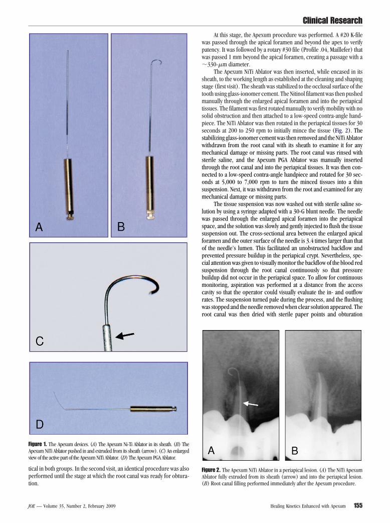
igure 1. The Apexum devices. (A) The Apexum Ni-Ti Ablator in its sheath. (B) Thepexum NiTi Ablator pushed in and extruded from its sheath (arrow). (C) An enlargediew of the active part of the Apexum NiTi Ablator. (D) The Apexum PGA Ablator.
ion. (
OE — Volume 35, Number 2, February 2009
At this stage, the Apexum procedure was performed. A #20 K-fileas passed through the apical foramen and beyond the apex to verifyatency. It was followed by a rotary #30 file (Profile .04, Maillefer) thatas passed 1 mm beyond the apical foramen, creating a passage with a330-�m diameter.
The Apexum NiTi Ablator was then inserted, while encased in itsheath, to the working length as established at the cleaning and shapingtage (first visit). The sheath was stabilized to the occlusal surface of theooth using glass-ionomer cement. The Nitinol filament was then pushed
anually through the enlarged apical foramen and into the periapicalissues. The filament was first rotated manually to verify mobility with noolid obstruction and then attached to a low-speed contra-angle hand-iece. The NiTi Ablator was then rotated in the periapical tissues for 30econds at 200 to 250 rpm to initially mince the tissue (Fig. 2). Thetabilizing glass-ionomer cement was then removed and the NiTi Ablatorithdrawn from the root canal with its sheath to examine it for anyechanical damage or missing parts. The root canal was rinsed with
terile saline, and the Apexum PGA Ablator was manually insertedhrough the root canal and into the periapical tissues. It was then con-ected to a low-speed contra-angle handpiece and rotated for 30 sec-nds at 5,000 to 7,000 rpm to turn the minced tissues into a thinuspension. Next, it was withdrawn from the root and examined for anyechanical damage or missing parts.
The tissue suspension was now washed out with sterile saline so-ution by using a syringe adapted with a 30-G blunt needle. The needleas passed through the enlarged apical foramen into the periapical
pace, and the solution was slowly and gently injected to flush the tissueuspension out. The cross-sectional area between the enlarged apicaloramen and the outer surface of the needle is 3.4 times larger than thatf the needle’s lumen. This facilitated an unobstructed backflow andrevented pressure buildup in the periapical crypt. Nevertheless, spe-ial attention was given to visually monitor the backflow of the blood reduspension through the root canal continuously so that pressureuildup did not occur in the periapical space. To allow for continuousonitoring, aspiration was performed at a distance from the access
avity so that the operator could visually evaluate the in- and outflowates. The suspension turned pale during the process, and the flushingas stopped and the needle removed when clear solution appeared. The
oot canal was then dried with sterile paper points and obturation
igure 2. The Apexum NiTi Ablator in a periapical lesion. (A) The NiTi Apexumblator fully extruded from its sheath (arrow) and into the periapical lesion.
B) Root canal filling performed immediately after the Apexum procedure.Healing Kinetics Enhanced with Apexum 155

cit
pcm
R
ntd
C
mvawtwtlr
T
Amsa
msmaA
T
ATo
tTe“lttlc
egroasg
“tg
S
bwtoa
S
(rmodtaaA6wu
SI
bu
P
stavst
P
mopottN
T
Clinical Research
1
onducted as in the conventional treatment group followed by a glass-onomer cement temporary filling and a postoperative radiograph. Res-orations were performed as in the conventional treatment group.
The Apexum procedure was performed under local anesthesia,rovided in a manner similar to that used for tooth extraction or surgi-al intervention. With some experience, it took an additional 7 to 10inutes compared with a conventional root canal treatment.
adiographic ProceduresAll radiographs were taken using a digital sensor (Schick Tech-
ologies, Long Island City, NY). Two periapical radiographs of eachooth were taken, before, and immediately after the endodontic proce-ure, with follow-up radiographs taken 3 and 6 months thereafter.
linical Follow-upEach patient in both the Apexum-treated and conventional treat-
ent groups was instructed to record pain, swelling, or any other ad-erse event that occurred after treatment and if he/she required anynalgesics or other medication after the endodontic procedure. Patientsere contacted by telephone a week after completion of the root canal
reatment and asked (1) if there was any discomfort or pain, (2)hether analgesics were needed, (3) if there was swelling, and (4) if
here were any other postoperative events. At the 3- and 6-month fol-ow-up visits, the patient was questioned again, and the tooth and sur-ounding tissues were clinically examined and the findings recorded.
he Evaluation of Results: Adverse EventsAdverse events were recorded and used to evaluate the safety of the
pexum procedure as compared with conventional root canal treat-ent. These included swelling, required antibiotics, or having an un-
cheduled appointment or any other events that occurred immediatelyfter the procedure or during the follow-up period.
Analgesics used after surgery with no unscheduled dental appoint-ent were not considered as procedure-related adverse events because
ome pain or discomfort are common after a routine root canal treat-ent of cases with apical periodontitis, and these cases were monitored
nd recorded separately. They also were used for comparison of thepexum protocol to conventional root canal treatment.
he Evaluation of Results: Radiographic RecordsRadiographic follow-up was used to evaluate the efficacy of the
pexum procedure compared with conventional root canal treatment.he radiographic image of the periapical lesion was evaluated from a setf radiographs.
Follow-up radiographs, taken at 3 and 6 months, were viewedogether and compared with those taken immediately after treatment.he change in the radiographic image of the periapical lesion wasvaluated and defined. The following four categories (9) were used: (1)no healing,” no reduction in the size of the lesion or enlargement of theesion; (2) “minor healing,” a clear, but minor, decrease in the size ofhe lesion; (3) “advanced healing,” a substantial decrease in the size ofhe lesion but not a complete healing; and (4) “complete healing,” theesion disappeared completely. Some residual widening of the periapi-al periodontal ligament was also considered as complete healing.
Two reviewers were used, an oral-maxillofacial surgeon and anndodontist. Each reviewer evaluated and scored each set of radio-raphs independently. When both reviewers agreed, then the score wasegistered. When disagreement occurred, then another reviewer (end-dontist) was brought in and the issue was discussed to obtain angreed-on score. All reviewers were initially calibrated by evaluatingets of similar radiographs and were blinded as to the group to which a
iven tooth belonged. t56 Metzger et al.
The scores were later dichotomized (2, 9) so that “no healing” andminor healing” were considered together as “nonhealing,” whereashe “significant healing” and “complete healing” were considered to-ether as “healing.”
ealer ExtrusionSealer extrusion that occurred during root canal obturation was
lindly examined and evaluated from radiographs. Three categoriesere used: (1) no sealer extrusion, (2) a small sealer puff (similar to
hose encountered when patency was preserved throughout the end-dontic procedure), and (3) large sealer puffs (bigger than those thatre common in the technique described earlier).
tatistical AnalysisThe primary safety endpoint was treatment-related adverse events
AEs). The secondary safety endpoint was the occurrence of pain thatequired analgesic. The primary efficacy endpoint was healing at 6onths, defined dichotomously for each periapical lesion as “healing”
r “nonhealing.” The secondary efficacy endpoint was similarly to theichotomized healing scores at 3 months. Healing was also evaluated on
he full four-category healing scale, as described previously, at the 3-nd 6-month follow-up visits. A chi-square and Fisher exact test werepplied to test the significance of healing differences between thepexum-treated and conventional treatment groups at the 3- and-month follow-ups. All tests were two-tailed, and p values of 5% or lessere considered statistically significant. Data analysis was performed bysing SAS software, version 9.1 (SAS Institute, Cary, NC).
Resultsafety: AEs, Pain, and Mechanical Failuresntraoperative AEs
The endodontic treatment per se was uneventful in all cases foroth groups. Periapical tissue removal using the Apexum protocol wasneventful in all 48 cases in the Apexum-treated group (Table 1).
ostoperative AEsSubjects were called within 7 days of the procedure; 46 of the 48
ubjects in the Apexum-treated group and all 39 in the conventionalreatment group were available at this time point. No treatment-relateddverse events were recorded in either the Apexum-treated or the con-entional treatment groups (Table 1). More specifically, there was nowelling and no need for an unscheduled dental appointment in any ofhe patients.
ostoperative PainSome postoperative discomfort or pain within 2 to 3 days of treat-
ent was recorded in 31% of the cases treated by conventional end-dontic treatment. When the Apexum procedure was applied, as a sup-lementary step to conventional endodontic treatment, the occurrencef postoperative discomfort or pain was reduced to 9% of the cases;
hus, the Apexum procedure was significantly less painful postopera-ively than conventional root canal treatment (p � 0.05, Table 1).either swelling nor severe pain were recorded for any of the cases
ABLE 1. AEs and Postoperative Pain
Apexum Procedure Conventional RootCanal Treatment
Intraprocedural AEs 0/48 0/39Postoperative AEs 0/46 0/39Postoperative pain 4/46 (8.7%) 12/39 (30.8%)
reated.
JOE — Volume 35, Number 2, February 2009
M
Ndp
E
wmta
H
wc(wg
p(pc
H
A(
A6
((ifrd
S
glpiih
evtpofairc
eapetagbKssto
if“c
T
Fd
Clinical Research
J
echanical FailuresNo mechanical failure occurred when applying either the Apexum
iTi Ablator or the PGA Ablator in the Apexum-treated group. Eachevice was only used one time in a total of 48 procedures during theresent study.
fficacyOf the 48 periapical lesions treated with the Apexum protocol, 46
ere available for radiographic evaluation at 3 months and 42 at 6onths. Of the 39 periapical lesions treated by conventional endodontic
reatment, 37 were available for radiographic evaluation at 3 monthsnd 31 at 6 months.
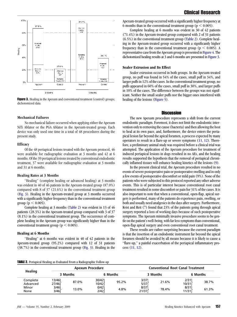
ealing Rates at 3 Months“Healing” (complete healing or advanced healing) at 3 months
as evident in 40 of 46 patients in the Apexum-treated group (87.0%)ompared with 8 of 37 (21.6%) in the conventional treatment groupFig. 3). Healing in the Apexum-treated group at 3 months occurredith a significantly higher frequency than in the conventional treatmentroup (p � 0.005).
Complete healing at 3 months (Table 2) was evident in 13 of 46atients (28.3%) in the Apexum-treated group compared with 3 of 378.1%) in the conventional treatment group. The occurrence of com-lete healing in the Apexum group was significantly higher than in theonventional treatment group (p � 0.005).
ealing at 6 Months“Healing” at 6 months was evident in 40 of 42 patients in the
pexum-treated group (95.2%) compared with 12 of 31 patients38.7%) in the conventional treatment group (Fig. 3). Healing in the
ABLE 2. Periapical Healing as Evaluated From a Radiographic Follow-up
HealingApexum Procedure
3 Months 6 Month
Complete 13/46 87.0% 30/42Advanced 27/46 10/42Minor 3/46 13.0% 0/42
igure 3. Healing in the Apexum and conventional treatment (control) groups;ichotomized data.
None 3/46 2/42
OE — Volume 35, Number 2, February 2009
pexum-treated group occurred with a significantly higher frequency atmonths than in the conventional treatment group (p � 0.005).
Complete healing at 6 months was evident in 30 of 42 patients71.4%) in the Apexum-treated group compared with 2 of 31 patients6.5%) in the conventional treatment group (Table 2). Complete heal-ng in the Apexum-treated group occurred with a significantly higherrequency than in the conventional treatment group (p � 0.005). Aepresentative case from the Apexum group is presented in Figure 4. Theichotomized healing results at 3 and 6 months are presented in Figure 3.
ealer Extrusion and Its EffectSealer extrusion occurred in both groups. In the Apexum-treated
roup, no puff was found in 54% of the cases, small puff in 34%, andarger puffs in 12% of the cases. In the conventional treatment group, nouffs appeared in 60% of the cases, small puff in 30%, and larger puffs
n 10% of the cases. The difference between the groups was not signif-cant. Neither the small sealer puffs nor the bigger ones interfered withealing of the lesions (Figure 5).
DiscussionThe new Apexum procedure represents a shift from the current
ndodontic paradigm. Foremost, it does not limit the endodontic inter-ention only to removing the cause (bacteria) and then allowing the hosto heal at its own pace, and, furthermore, the device enters the peria-ical lesion far beyond the apical foramen, a process expected by manyperators to result in a flare-up or severe symptoms (11, 12). There-
ore, a preliminary animal study was required before a clinical trial wasttempted. The application of the Apexum procedure for treatment ofnduced periapical lesions in dogs resulted in no AEs, and the healingesults supported the hypothesis that the removal of periapical chroni-ally inflamed tissues will enhance healing kinetics of the lesions (9).
In the present clinical trial, the Apexum procedure resulted in novents of severe postoperative pain or postoperative swelling and in onlyfew events of postoperative discomfort or mild pain (9%). None of theatients who were subjected to this protocol reported any other adversevents. This is of particular interest because conventional root canalreatment resulted in some discomfort or pain for 31% of the cases. It islso important to note that when a conventional, open-flap, apical sur-ery is performed, many of the patients do experience pain, swelling, oroth and usually need analgesics in the days after surgery. Furthermore,vist and Reit (7) found that 23% of the patients going through apicalurgery reported a loss of working days because of such postoperativeymptoms. The Apexum minimally invasive procedure seems to be gen-le on the patient’s well-being, with far less symptoms than conventional,pen-flap apical surgery and even conventional root canal treatment.
These results are rather surprising because the current paradigms that the insertion of an endodontic instrument far beyond the apicaloramen should be avoided by all means because it is likely to cause aflare-up,” a painful exacerbation of the periapical inflammatory pro-ess (11, 12).
Conventional Root Canal Treatment
3 Months 6 Months
% 3/37 21.6% 2/31 38.7%5/37 10/31
% 8/37 78.4% 8/31 61.3%
s
95.2
4.8
21/37 11/31Healing Kinetics Enhanced with Apexum 157
dTighofAdnT
ftetmv
rbp
mshrct
paAwdBmm
fc(taTrsafbm2pTs
Ff
Fiot
Clinical Research
1
It should be noted that the Apexum procedure is substantiallyifferent from simple overinstrumentation during root canal treatment.he last traumatizes the tissue and may also introduce bacterial antigensnto a tissue containing immunoglobulins directed against these anti-ens and that is primed to respond to them (5, 13, 14). When thisappens, acute inflammatory response with resulting edema is likely toccur in the periapical tissue, resulting in a flare-up (5, 11, 12). Such alare-up did not appear in a single case of the 48 subjected to thepexum procedure. It is most likely that with the removal or majorebulking of the periapical chronically inflamed tissues, the mecha-isms that could otherwise lead to such a flare-up were also removed.his may explain the quite uneventful postoperative clinical behavior.
Healing kinetics (the progress of healing with time), as judgedrom follow-up radiographs, were enhanced in the group treated withhe Apexum procedure. At 3 months, 87% of the periapical lesions wereither completely healed or in advanced stages of healing, whereas inhe conventional treatment group only 22% showed such a trait. At 6
onths, 95% of the lesions in the Apexum-treated group showed ad-anced or complete healing, whereas conventional root canal treatment
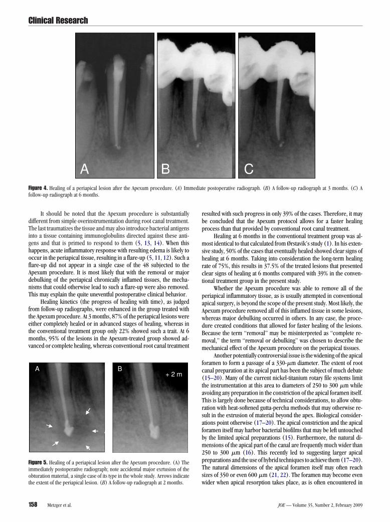
igure 4. Healing of a periapical lesion after the Apexum procedure. (A) Imollow-up radiograph at 6 months.
igure 5. Healing of a periapical lesion after the Apexum procedure. (A) Themmediately postoperative radiograph; note accidental major exrtusion of thebturation material, a single case of its type in the whole study. Arrows indicate
whe extent of the periapical lesion. (B) A follow-up radiograph at 2 months.
58 Metzger et al.
esulted with such progress in only 39% of the cases. Therefore, it maye concluded that the Apexum protocol allows for a faster healingrocess than that provided by conventional root canal treatment.
Healing at 6 months in the conventional treatment group was al-ost identical to that calculated from Ørstavik’s study (1). In his exten-
ive study, 50% of the cases that eventually healed showed clear signs ofealing at 6 months. Taking into consideration the long-term healingate of 75%, this results in 37.5% of the treated lesions that presentedlear signs of healing at 6 months compared with 39% in the conven-ional treatment group in the present study.
Whether the Apexum procedure was able to remove all of theeriapical inflammatory tissue, as is usually attempted in conventionalpical surgery, is beyond the scope of the present study. Most likely, thepexum procedure removed all of this inflamed tissue in some lesions,hereas major debulking occurred in others. In any case, the proce-ure created conditions that allowed for faster healing of the lesions.ecause the term “removal” may be misinterpreted as “complete re-oval,” the term “removal or debulking” was chosen to describe theechanical effect of the Apexum procedure on the periapical tissues.
Another potentially controversial issue is the widening of the apicaloramen to form a passage of a 330-�m diameter. The extent of rootanal preparation at its apical part has been the subject of much debate15–20). Many of the current nickel-titanium rotary file systems limithe instrumentation at this area to diameters of 250 to 300 �m whilevoiding any preparation in the constriction of the apical foramen itself.his is largely done because of technical considerations, to allow obtu-ation with heat-softened gutta-percha methods that may otherwise re-ult in the extrusion of material beyond the apex. Biological consider-tions point otherwise (17–20). The apical constriction and the apicaloramen itself may harbor bacterial biofilms that may be left untouchedy the limited apical preparations (15). Furthermore, the natural di-ensions of the apical part of the canal are frequently much wider than
50 to 300 �m (16). This recently led to suggesting larger apicalreparations and the use of hybrid techniques to achieve them (17–20).he natural dimensions of the apical foramen itself may often reachizes of 350 or even 600 �m (21, 22). The foramen may become even
te postoperative radiograph. (B) A follow-up radiograph at 3 months. (C) A
mediaider when apical resorption takes place, as is often encountered in
JOE — Volume 35, Number 2, February 2009

r(t
cpm
bmtctthaNrh
etbihAtrppc
ptvccph
1
1
1
1
1
1
1
1
1
1
2
2
2
2
2
2
Clinical Research
J
oots with apical periodontitis, such as those treated in the present study23, 24). The similar patterns of sealer extrusion that were observed inhe present study may express this well-established phenomenon.
Considering all the previously mentioned items, enlarging the api-al foramen to a diameter of 330 �m, as required by the Apexumrocedure, probably does not result in a major change to its size inany or even most cases of apical periodontitis.
Passing through the apical foramen with a #30 rotary file followedy the repeated passage of the Apexum devices and the irrigation needleay have yet another potential outcome. Bacterial biofilms that poten-
ially reside in this area may potentially remain undisturbed by theurrent minimal apical intervention concept of many of the rotary nickel-itanium file systems. The Apexum procedure is more likely to eliminatehem or at least mechanically disturb them to the extent of disrupting theost-bacteria equilibrium in favor of the host. Such processes may havelso contributed to the enhanced healing observed in the present study.evertheless, additional studies focusing on this specific issue will beequired before the contribution of this process to the total enhancedealing may be estimated.
At the present stage, with a follow-up period of 6 months, it is tooarly to predict if the final healing rate at 48 months (25) (as opposedo healing kinetics) will also be affected, which is likely the case. It haseen well documented that 15% to 25% of periapical lesions fail to healn response to adequate endodontic treatment. Some of these failuresave been attributed to factors that are most likely to be affected by thepexum procedure, such as extraradicular infections or cystic forma-ions within the periapical lesion. Nevertheless, evaluation of the healingate (as opposed to healing kinetics) will call for longer follow-uperiods and much larger groups of patients. Such studies will soon be inrogress. These may verify whether the healing rate of periapical lesionsan also be affected by the new Apexum procedure.
ConclusionsThe Apexum procedure resulted in no adverse events. The Apexum
rocedure resulted in significantly less postoperative discomfort or painhan conventional root canal treatment or than that reported for con-entional apical surgery. The Apexum procedure resulted in a signifi-antly faster periapical healing as compared with conventional rootanal treatment (p � 0.005). The removal or debulking of the peria-ical inflamed tissues, using the Apexum procedure, seems to enhanceealing kinetics with no adverse events.
References1. Ørstavik D. Time course and risk analysis of the development and healing of chronic
apical periodontitis in man. Int Endod J 1996;29:150 –5.
OE — Volume 35, Number 2, February 2009
2. Friedman S. Prognosis of initial endodontic treatment. Endod Topics 2002;2:59 – 88.3. Figdor D. Apical periodontitis: a very prevalent problem. Oral Surg Oral Med Oral
Pathol Oral Radiol Endod 2002;94:651–2.4. Metzger Z. Macrophages in periapical lesions. Endod Dental Traumatol 2000;
16:1– 8.5. Metzger Z, Abramovitz I. Periapical lesions of endodontic origin. In: Ingle JI,
Bakland LK, Baumgartner JC, eds Ingle’s Endodontics. 6th ed. Hamilton, Canada:BC Decker;2008: 494 –519.
6. Kvist T, Reit C. Results of endodontic retreatment: a randomized clinical study com-paring surgical and nonsurgical procedures. J Endod 1999;25:814 –7.
7. Kvist T, Reit C. Postoperative discomfort associated with surgical and nonsurgicalendodontic retreatment. Dental Traumatol 2000;16:71– 4.
8. Appropriateness of Care and Quality Assurance Guidelines. 3rd ed. Chicago, IL:American Association of Endodontists; 1998.
9. Metzger Z, Huber R, Tobis I, Better H. Enhancement of healing kinetics of periapicallesions in dogs by the Apexum procedure. J Endod (in press).
0. Ørstavik D, Kerekes K, Eriksen HM. The periapical index: a scoring system for ra-diographic assessment of apical periodontitis. Endod Dental Traumatol 1986;2:20 –34.
1. Siqueira J. Reaction of periradicular tissues to root canal treatment: benefits anddrawbacks. Endod Topics 2005;10:123– 47.
2. Baumgartner JC, Rosenberg PA, Hoen MM, Lin LM. Treatment of endodontic infec-tions, cysts, and flare-ups. In: Ingle, JI Bakland, LK Baumgartner, eds. JC Ingle’sEndodontics. 6th ed. Hamilton, Canada: BC Decker; 2008:690 –712.
3. Baumgartner JC, Falkler WAJ. Reactivity of IgG from explant cultures of periapicallesions with implicated microorganisms. J Endod 1991;17:207–12.
4. Kettering JD, Torabinejad M, Jones SL. Specificity of antibodies present in humanperiapical lesions. J Endod 1991;17:213– 6.
5. Nair PNR. Light and electron microscopic studies of root canal flora and periapicallesions. J Endod 1987;13:29 –39.
6. Kerekes K, Tronstad L. Morphological observations of root canals of human molars.J Endod 1977;3:114 – 8.
7. Spangberg LS. The wonderful world of rotary root canal preparation. Oral Surg OralMed Oral Pathol Oral Radiol Endod 2001;92:479.
8. Card SJ, Sigurdsson A, Ørstavic D, Trope M. The effectiveness of increased apicalenlargement in reducing intracanal bacteria. J Endod 2002;28:779 – 83.
9. Walsch H. The hybrid concept of nickel-titanium rotary instruments. Dent Clin NorthAm 2004;48:183–202.
0. Peters OA. Current challenges and concepts in the preparation of root canal systems:a review. J Endod 2004;30:559 – 67.
1. Morfis A, Sylaras SN, Georgopoulou M, Kernani M, Prountzos F. Study of the apices ofhuman permanent teeth with the use of scanning electron microscope. Oral Surg OralMed Oral Pathol 1994;77:172– 6.
2. Briseno-Marroquin B, El-Syed MAA, Willershausen-Zonnchen B. Morphology of thephysiological foramen: I. Maxillary and mandibular molars. J Endod 2004;30:321– 8.
3. Furusawa M, Asai Y. SEM observations of resected root canal ends following apico-ectomy. Bull Tokyo Dent Coll 2002;43:7–12.
4. Vier FV, Figueiredo JAP. Internal apical resorption and its correlation with the type ofapical lesion. Int Endod J 2004;37:730 –7.
5. Strindberg LD. The dependence of the results of pulp therapy on certain factors. Ananalytic study based on radiographic and clinical follow up examination. Acta Od-
ontol Scand 1956;14(suppl):21.Healing Kinetics Enhanced with Apexum 159