Embed Size (px)
Citation preview

1
Aortic Valve What the Nurse Caring for a Patient with Congenital Heart Disease Needs to Know
Amy Donnellan, DNP, CPNP-AC,
Cardiac Intensive Care Unit Nurse Practitioner,
Cincinnati Children’s Hospital Medical Center
Lindsey Justice, DNP, RN, CPNP-AC,
Cardiac Intensive Care Unit Nurse Practitioner,
Cincinnati Children’s Hospital Medical Center
Svetlana Streltsova, MSN, RN, CNE, CCRN,
Clinical Nurse III, Pediatric Cardiac Intensive Care Unit,
Morgan Stanley Children's Hospital of New York Presbyterian
Louise Callow, MSN, RN, CPNP,
Pediatric Cardiac Surgery Nurse Practitioner,
University of Michigan, CS Mott Children’s Hospital
Mary Rummell, MN, RN, CPNP, CNS, FAHA,
Clinical Nurse Specialist, Pediatric Cardiology/Cardiac Services,
Oregon Health & Science University (Retired)
Embryology
Occurrence:
o Defects of cardiac valves are the most common subtype of cardiac malformations
o Account for 25% to 30% of all congenital heart defects
o Most costly and relevant CHD
o Wide spectrum of congenital defects in aortic valve
Development of the heart valves occurs during the fourth to eighth weeks of gestation-
after tubular heart looping
o Walls of the tubular heart consist of an outer lining of myocardium and an inner
lining of endocardial cells
o Cardiac jelly, extensive extracellular matrix (ECM), separates the two layers
o Cardiac jelly expands to form cardiac cushions at the sites of future valves
Outflow track (OT) valves = aortic and pulmonic valves
Final valves derived from endothelial-mesenchymal cells with
neural crest cells from the brachial arches
Valves (Semilunar) have 3 equal cusp-shaped leaflets
Aortic valve incorporates coronary arteries
Atrioventricular (AV) valves = mitral and tricuspid
Final valves derived entirely from endocardial cushion tissue
Leaflet formed without a cusp
Two leaflets associated with left ventricle (mitral)
Three leaflets associated with right ventricle (tricuspid)
2
Coordinated by complex interplay of:
o Genetics
o Signaling pathways that regulate cell apoptosis and proliferation
o Environmental factors
Maternal hyperglycemia
Acidosis
Blood flow through developing heart
Anatomy
Clinical spectrum varies from presence of a malformed bicuspid aortic valve that
functions normally to severe aortic stenosis (AS)
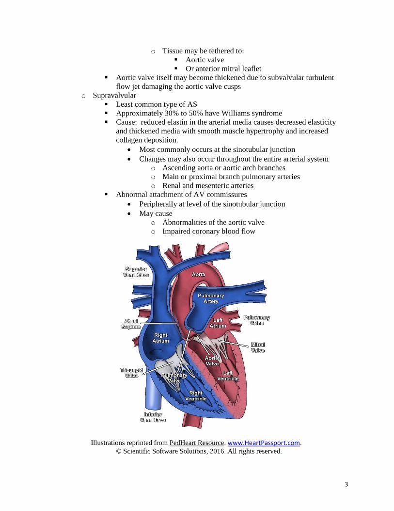
Types/anatomic location of stenosis (See illustration below for intracardiac position of
aortic valve and relation of other structures involved in anatomic locations of stenosis.)
o Valvular
Seventy to 80% of all AS
Decrease in orifice size
Results from thickening and increased rigidity of valve leaflets.
Most common defect
Bicuspid aortic valve
Only two valve cusps present
Results from partial or complete fusion of two of the aortic valve
cusps
Conjoined vs. nonconjoined cusps may be equal or asymmetric
Valve orifice may be central or non-central.
Other forms
Unicuspid valve
o Fusion of more than one cusp
o Results in a single slit like opening that extends to the
annulus
Partial fusion of all three cusps with small central orifice
Hypoplasia of annulus
o Rare
o Aortic valve cusps relatively normal
o Subvalvular
Ten to 20% of all AS
Common associated defects:
Ventricular septal defect
Coarctation of the aorta
Atrioventricular septal defect
Valvular aortic stenosis
Mitral valve anomalies.
Obstruction
Ridge of membranous and/or fibrous tissue
o Encircles left ventricular outflow tract (LVOT)
o Or diffuse and form a tunnel
3
o Tissue may be tethered to:
Aortic valve
Or anterior mitral leaflet
Aortic valve itself may become thickened due to subvalvular turbulent
flow jet damaging the aortic valve cusps
o Supravalvular
Least common type of AS
Approximately 30% to 50% have Williams syndrome
Cause: reduced elastin in the arterial media causes decreased elasticity
and thickened media with smooth muscle hypertrophy and increased
collagen deposition.
Most commonly occurs at the sinotubular junction
Changes may also occur throughout the entire arterial system
o Ascending aorta or aortic arch branches
o Main or proximal branch pulmonary arteries
o Renal and mesenteric arteries
Abnormal attachment of AV commissures
Peripherally at level of the sinotubular junction
May cause
o Abnormalities of the aortic valve
o Impaired coronary blood flow
Illustrations reprinted from PedHeart Resource. www.HeartPassport.com.
© Scientific Software Solutions, 2016. All rights reserved.

4
o Cross sectional illustration shows aortic valve cusps in relation to other heart
valves and coronary arteries.
Illustrations reprinted from PedHeart Resource. www.HeartPassport.com.
© Scientific Software Solutions, 2016. All rights reserved.
Other pathologic features that impact aortic valve dysmorphology and dysfunction
include:
o Calcification (rare in childhood and adolescence, but common in adults)
o Fibrosis
o Lipid accumulation
o Inflammatory changes
o Myxomatous degeneration
o Annular dilation
o Acquired fibrotic fusion of true commissures
Congenitally abnormal aortic valves may result in weakening of ascending aorta
o May result in:
Annular dilation, as well as
Dilation or aneurysm of ascending aorta
At risk for aortic dissection or rupture
May develop left ventricular (LV) hypertrophy and myocardial fibrosis.
Physiology
In utero presence of moderate to severe aortic stenosis
o Increases LV pressure

5
o May lead to:
LV hypertrophy
Decreased LV compliance
Decreased flow through the left heart
May result in hypoplasia of the LV, mitral valve, aortic valve annulus, and
LV outflow tract.
Valvar AS
o Causes obstruction of LV outflow,
o Increases LV afterload
o LV pressure > aortic pressure during ejection due to decreased effective area of
the valve orifice
o With normal stroke volume, the pressure gradient reflects severity of the stenosis
o Neonatal critical aortic stenosis:
Limited antegrade flow across the LV outflow tract
Requires a patent ductus arteriosus (PDA) to provide adequate
systemic perfusion
Results from closure of the PDA
Cardiogenic shock
Severe hypoperfusion
Profound acidosis
o Pediatric AS (Patients present after one year of age)
See compensatory LV hypertrophy
Maintains normal LV wall stress despite elevation in peak systolic
pressure
Maintains cardiac output
Increases LV end-diastolic volume and pressure
Valvular/subvalvular AS
o LV subendocardial ischemia and infarction from:
Imbalance in coronary blood flow to the hypertrophied left ventricle
Increased myocardial oxygen demand
Ventricular pressure overload
o Severe aortic stenosis
Little coronary reserve during stress
Exercise
Minimally increases stroke volume
Results in:
o Increased heart rate
o Shortened systole and diastole
o Decreased time for ejection
o Increased LV systolic pressure
o Increased oxygen demand
o Decreased coronary perfusion from shorter diastole
Increases systemic vasodilation
o Further decreases diastolic blood pressure
o May impair coronary perfusion
o Supravalvular aortic stenosis

6
Physiology similar to valvar and subvalvular
Effect on coronary arteries
Proximal to the obstruction
Exposed to high pressure during systole
o Leads to changes in coronary vasculature
o Limited diastolic flow
Results in inadequate oxygen supply to meet
demand
Leads to ischemia, infarction, and sudden death
Procedures/Interventions
Medical Treatment
o Bacterial endocarditis prophylaxis
o Exercise restrictions
o Periodic follow-up evaluations to monitor progression of valve dysfunction
Indications
o Adults - intervention is recommended only when symptoms develop
o Children and adolescents
Earlier intervention even in asymptomatic patients
Relief of obstruction
Reduces the risk of sudden cardiac death
Decreases the extent of subtle and progressive myocardial injury
Catheter Intervention
o Balloon Valvuloplasty
Children and adolescents
Initial treatment
Re-stenosis or regurgitation may occur
Valve replacement may become necessary for definitive treatment
Not indicated if aortic valve regurgitation
Adults
Not indicated if cusp calcification develops
Not indicated with significant aortic valve regurgitation
American College of Cardiology/American Heart Association guidelines:
Patient population Intervention
Asymptomatic children and young adults with
peak Doppler gradients ≥ 70 mmHg or peak-
to-peak gradient > 60 mmHg.
Consider cardiac cath and
possible balloon valvuloplasty
Patients who play competitive sports or may
become pregnant, with peak Doppler gradients
50-70 mmHg or peak-to-peak gradient > 50
mmHg
Consider cardiac cath and
possible balloon valvuloplasty

7
Patients with symptoms (angina, syncope,
dyspnea on exertion) or ischemic changes at
rest or on exercise ECG
Valvuloplasty if peak-to-peak
gradient is > 50 mmHg, other
symptoms should be sought if
gradient does not meet criteria
Asymptomatic patients with peak-to-peak
gradients < 50 mmHg
Valvuloplasty not
recommended unless cardiac
output is impaired (gradient
underestimates true severity of
obstruction in this setting)
o Transcatheter aortic valve replacement (TAVR)
Minimally invasive catheter placement of a bioprosthetic, expandible
aortic valve within a native, calcified, severely stenotic aortic valve
Inserted in a hybrid (surgical suite with bi-plane imaging) lab
Inserted by a multidisciplinary team
Interventional cardiologist
Cardiothoracic surgeon
Inserted by an atrial or transthoracic approach
Involves sheaths and catheters > 16Fr
Too large for pediatric arteries
Consider in severe symptomatic aortic valve stenosis
Available for patients with severe symptoms at increased surgical risk
Advanced age
Multiple comorbidities
Surgical options determined to be contraindicated or at extreme
risk by two cardiovascular surgeons
Currently (2015) not available for congenital unicuspid or bicuspid
aortic valves
o Surgical Treatment
Indications
Development of progressive aortic valve regurgitation
Recurrent stenosis refractory to balloon valvuloplasty
Mechanical prosthesis
Requires long-term anticoagulation
No potential for valve growth
Limited availability of small sizes
Bioprosthetic valves (homograft or heterograft)
Avoids the need for anticoagulation
No potential for valve growth
Longevity less than mechanical, especially in small children
Ross procedure
May be preferred in infants and small children
Translocation of semi-lunar valves (See cross sectional illustration
for proximity of semi lunar valves and similar structure.)

8
o Native pulmonary valve translocated to the aortic position
o Pulmonary homograft implanted in the pulmonary valve
position
o Native aortic valve may be surgically revised and inserted
into pulmonary position (Double Switch)
Benefits:
o No need for anticoagulation
o Potential autograft (neoaortic valve) growth
Disadvantage:
o May develop pulmonary homograft dysfunction
o May require additional procedures
Pulmonary homograft replacement
Neoaortic valve dilation
Stenosis of both pulmonary and aortic suture lines
Surgical resection of subaortic stenosis
Indications:
o Progression of subaortic obstruction
o Development of aortic regurgitation
Procedure
o LV muscle resection
o Membrane excision
o Potential surgical correction of mitral valve abnormalities
that contribute to the subaortic obstruction, such as
anomalous papillary muscle insertion
Complications:
o Recurrent subaortic stenosis in 20% of patients
o Heart block
o Worsening of aortic or mitral valve regurgitation
o Inadvertent creation of a ventricular septal defect
Severe subaortic stenosis with a small aortic annulus
May require more extensive tissue resection
Ross/Konno procedure
Konno procedure
Indications
o Treat all levels of LVOTO
o Tunnel like subaortic stenosis
o Diffuse obstructive hypertrophic cardiomyopathy
o Congenital aortic valve stenosis
o Proximal ascending aorta stenosis
May be combined with Ross procedure (Ross/Konno) specifically
for tunnel subaortic stenosis and aortic annular hypoplasia
Procedure: Konno operation with Aortic valve replacement
o Longitudinal incision on anterior aspect of ascending aorta
distal to the aortic valve

9
o Continue incision to left of right coronary artery and into
the right ventricle and down into the interventricular
septum below any subvalvar stenosis
o Patch sutured to left ventricular side of the VSD and
continued across the aortic annulus and onto the aorta
enlarging the aortic annulus
o Prosthetic valve placed in the enlarged aortic root
o RVOT reconstructed with patch
Procedure: Modified Konno
o Enlargement of the LVOT without replacement of the
aortic valve (normal size aortic annulus but presence of
subaortic stenosis)
o Right ventricular incision into the ventricular septum and
into the LVOT up to the aortic valve
o Patch placed on the right ventricular side of the surgically
created VSD
Complications
o Complete heart block and requirement for pacemaker
o Incomplete relief of LVOTO
o Residual VSD
o Damage to the mitral valve apparatus
Surgical repair of supravalvular aortic stenosis
Patch enlargement of the sinotubular junction above the
noncoronary sinus
Extended aortoplasty with a patch into the noncoronary and right
coronary sinuses
Insertion of separate patches in all three sinuses after transecting
the aorta (Brom’s technique)
Surgical correction of any obstruction to coronary blood flow
Patch augmentation of the ascending or transverse aorta as
necessary
Specific considerations and routine care
AS pre-procedure management depends on the degree of obstruction to forward flow
from the left ventricle and the presence of systemic hypoperfusion
Neonates with critical AS
o Important to determine if the left heart and aortic structures are compatible with a
two ventricle repair
Based on echocardiographic information
Considerations
Ventricular size, end diastolic volume
Left sided lesions
o Mitral stenosis, adequacy of mitral valve annulus
o Coarctation of the aorta
Neonatal Critical AS pre-procedure Management
o Prostaglandin infusion

10
Establishes ductal-dependent systemic flow
Alleviates pulmonary hypertension seen with severe LV dysfunction
o Vasoactive support
Resuscitate
Support LV
Increase contractility
o Intubation and mechanical ventilation
Correct severe acidosis
Reduce metabolic demand
Control pulmonary hypertension
Afterload reduce left ventricle
o May require emergent atrial septostomy or even management with ECMO.
o Monitor for signs of end-organ compromise
Procedures: (See Peds/Neo Guidelines for Post-operative Care)
o Key post-procedure management points
o All procedures
Continual monitoring of cardiac rhythm for arrhythmias and ST segment
changes
Systolic and diastolic blood pressure, widening pulse pressure
Low cardiac output syndrome
o Balloon valvuloplasty
Impacted by the degree of severity of critical AS.
Low cardiac output syndrome
Best managed by inotropes to improve cardiac function
Ventilation with high concentrations of inspired oxygen, normal
pH to treat pulmonary vascular reactivity
Significant pulmonary hypertension
Manage ventilation, consider use of nitric oxide
Continue prostaglandin infusion to maintain a patent ductus
o Maintain systemic perfusion
o Decompress the pulmonary artery hypertension
Monitor for aortic insufficiency
Monitor for bleeding
Catheter insertion site
Retroperitoneal
o Surgical valvotomy
Monitor for hypertension
LV function usually preserved
Often hyperdynamic due to long standing stenosis and high
afterload results in hypertension
Low cardiac output syndrome (LCOS)
May be prolonged due to the hypertrophied LV
Positive pressure ventilation may reduce LV afterload and improve
cardiac output
Monitor for arrhythmias and changes in coronary artery perfusion
Left bundle branch block or complete heart block

11
ST segment changes
Ventricular arrhythmia
o Surgical aortic valve replacement
Valve selection depends on:
Patient age
Size of aortic valve annulus
Anticoagulation necessary if a prosthetic mechanical valve used (See both
Ped/Neo and Adult Anticoagulation Guidelines.)
o Ross Procedure – (Pulmonary Autograft)
Increased bleeding risk
Re-exploration may occur in a small amount of patients
Dehiscence of aortic root anastomosis
Increased risk of arrhythmias
Complete heart block
o May require a permanent pacemaker
Compromised coronary artery perfusion
o Surgical Repair of Subvalvular Stenosis
Increased sub aortic resection
Increased risk for third degree/complete heart block
Increased risk for significant ventricular dysfunction
Inadvertent creation of a ventricular septal defect
Surgical disruption of mitral valve apparatus and resultant mitral
regurgitation
o Surgical Repair of Supravalvular Stenosis
Risk of pulmonary hypertension
Associated CHD in genetic syndromes such as William’s syndrome
Main pulmonary artery stenosis
Branch pulmonary artery stenosis
Risk of suicide RV if relief supravalvar AS results in
suprasystemic RV pressure due to unrelieved PA stenosis
Long-term problems/complications and routine care
Endocarditis prophylaxis is necessary
Patients with bicuspid aortic valve are at risk for progressive aortic root dilation and
aortic dissection, even in the absence of stenosis or significant regurgitation
o Most patients with aortic dissection have hypertension, so medical management
of hypertension is crucial
o Careful surveillance with serial echocardiograms is warranted to detect aortic root
dilation
Long-term monitoring
o Aortic valve function and gradient
o Development of LV hypertrophy or dysfunction
Anticoagulation (See both Ped/Neo and Adult Guidelines for Anticoagulation)
o Prosthetic mechanical valve replacement

12
o Bioprosthetic valve replacement may require anticoagulation with added risk
factors
Atrial fibrillation
LV dysfunction
Hypercoagulable state
o Recommendations during pregnancy
See Adult Guidelines on Pregnancy in Adult Congenital Heart Disease for
more specific considerations and anticoagulation management
Following intervention, either surgical or by catheterization, patients must be monitored
for re-stenosis or development of valve regurgitation
References:
Armstrong, E.J., & Bischoff, J. (2004). Heart valve development. Endothelial cell signaling and
differentiation. Circulation Research. Published online at http://www.circresaha.org. doi:
10.1161/01.RES.0001411.95728.da Accessed 8/2015.
Assadi, R. (2013) Transcatheter aortic valve replacement. Medscape Reference. Published online
at http://emedicine.medscape.com/article/2039348-overview. Accessed 9/2015.
Biechler, S.V., et al. (2014). The impact of flow-induced forces on the morphogenesis of the
outflow tract, Frontiers in Physiology, 5(225). Published online at http://wwwlfrontiersin.org
doi:10.3389/fphys.2014.00225. Accessed 9/2015
Brown, D.W., et al. (2010). Aortic valve reinterventions after balloon aortic valvuloplasty for
congenital aortic stenosis. Journal of the American College of Cardiology, 56(21), 1740-1749.
Moore, K.L. & Persaud, T.V.N. (2008). The cardiovascular system: in The Developing Human.
Clinically Oriented Embryology (8th ed). Philadelphia, PA: Saunders, an imprint of Elsevier Inc.
Prapa, S., Dimopoulos, K., Shore, D. F., Petrou, M., & Gatzoulis, M. A. (2013). Adult
Congenital Heart Disease. Pediatric Cardiac Surgery (4th ed). 898-899, Wiley-Blackwell
Schneider, D.J., & Moore, J.W. (2008). Left ventricular outflow abnormalities: Aortic stenosis.
In Moss and Adams’ Heart Disease in Infants, Children, and Adolescents (7th ed). Philadelphia,
PA: Lippincott Williams & Wilkins.
Siddiqui, J., et al. (2013). Surgical valvotomy and repair for neonatal and infant congenital aortic
stenosis achieves better results than interventional catheterization. Journal of the American
College of Cardiology, 62(22), 2134-2140.
St. Louis, J.D. & Jaggers, J. (2006). Left ventricular outflow tract obstruction. In Critical Heart
Disease in Infants and Children (2nd ed). Philadelphia, PA: Mosby.
Illustrations reprinted from PedHeart Resource. www.HeartPassport.com. © Scientific Software
Solutions, 2016. All rights reserved. 12/2015