Embed Size (px)
DESCRIPTION
antivirus journal
Citation preview

©2010 International Medical Press 1359-6535 (print) 2040-2066 (online) 53
Antiviral Chemistry & Chemotherapy 2010; 21:53–70 (doi: 10.3851/IMP1684)
Nanomedicine opens new therapeutic avenues for attacking viral diseases and for improving treatment success rates. Nanoparticulate-based systems might change the release kinetics of antivirals, increase their bioavailability, improve their efficacy, restrict adverse drug side effects and reduce treatment costs. Moreover, they could permit the delivery of antiviral drugs to spe-cific target sites and viral reservoirs in the body. These features are particularly relevant in viral diseases where high drug doses are needed, drugs are expensive and
the success of a therapy is associated with a patient’s adherence to the administration protocol.This review presents the current status in the emerging area of nanoparticulate delivery systems in antiviral therapy, providing their definition and description, and highlighting some peculiar features. The paper closes with a discussion on the future challenges that must be addressed before the potential of nanotechnology can be translated into safe and effective antiviral formulations for clinical use.
The global impact of viral infections, the development of resistance to current drugs and the emergence of new viruses all translate into the incessant scientific challenge of drug discovery and formulation development. Over the past 3 decades, many researchers have focused on devel-oping new antivirals that are able to target important ther-apeutic processes. By 1990, just 5 drugs had been licensed as antiviral agents [1], whereas approximately 20 years later more than 40 were on the market. Most of these agents were developed for the treatment of HIV infection, whereas others were active against various herpesviruses (herpes simplex virus [HSV], varicella zoster virus [VZV] and human cytomegalovirus [HCMV]), hepatitis B and C viruses, and influenza A and B viruses.
In 2009, the global market for antiviral drugs reached total sales of approximately USD 28 billion. Sales of antivirals increased by approximately 20% from 2004 to 2006, and a continuing growth trend has been esti-mated until 2011. Moreover, the market is likely to wit-ness even further future growth because of the existence of unmet needs, expanding populations, better diagnos-tics, innovative drugs and new therapeutics; however, developing a safe and effective antiviral drug is a dif-ficult task, and the list of viral diseases for which anti-viral therapies are available is still relatively short.
Several factors hinder the development of antiviral drugs. Viruses are obligate intracellular parasites that
largely depend on the host cell biosynthetic machinery for their replication; therefore, only a limited number of virus-specific metabolic functions can be targeted by antiviral drugs without harming the host. Ideally, these targets are viral proteins essential for viral rep-lication and pathogenesis that are sufficiently different to any host protein to allow selectivity. Moreover, most of these functions are specific for each virus, making it difficult to develop broad-spectrum antivirals that are active against diverse viruses that cause similar symp-toms. The antivirals developed against some viruses (for example, HSV and HIV) treat the acute disease but do not cure the latent infection. This results in recurrent or chronic diseases that require treatment for longer peri-ods of time. These and other issues represent a major challenge in antiviral research and development.
A second key challenge of antiviral therapeutics regards the development of new drug formulations. This involves changing the physicochemical and biopharma-ceutical properties of antiviral molecules using techno-logical strategies during the preparation of their dosage forms. For example, the reformulation of an antiviral drug already present on the market might be performed in order to modify its bioavailability and pharmaco-kinetics. Further improvements to a therapy can also be obtained through the use of innovative delivery sys-tems for antiviral administration; for example, the use
Review
Nanoparticulate delivery systems for antiviral drugsDavid Lembo1, Roberta Cavalli2*
1Dipartimento di Scienze Cliniche e Biologiche, Università degli Studi di Torino, Orbassano Torino, Italy2Dipartimento di Scienza e Tecnologia del Farmaco, Università degli Studi di Torino, Torino, Italy
*Corresponding author e-mail: [email protected]
Introduction

D Lembo & R Cavalli
©2010 International Medical Press54
of nanotechnology has led to the development of nano-particulate carriers. Nanotechnological approaches can be used to improve the design, formulation and delivery of antiviral drugs.
This relatively new class of therapeutic nanomateri-als, also called nanopharmaceuticals, displays unique properties that arise because of their small sizes, high surface-to-volume ratios and their modifiable surfaces. Nanoparticulate carriers are able to incorporate small molecules, as well as proteins and nucleic acids, thus bestowing nanomaterials with a broad spectrum of pro-spective therapeutic applications and the potential to target specific tissue sites where the antivirals are needed. This review describes the current and future generations of nanoparticulate delivery systems and their use as car-riers for the transport of antiviral drugs.
Current antiviral therapies
The antiviral therapies currently approved are based on the use of small molecular weight drugs or proteins that stimulate the innate immune response (interferon). In addition, an antisense oligonucleotide (fomivirsen) has also been approved for the therapy of retinitis caused by strains of HCMV resistant to conventional drugs [2].
Table 1 and Table 2 report the antiviral agents present on the market and used in clinical practice. The approved antiviral drugs for HIV infections are summarized in Table 1 and other antiviral agents are listed, according to the viral infection, in Table 2. As shown, the majority of antiviral drugs are adminis-tered orally, although some are delivered via parenteral (subcutaneous, intravenous and intra vitreal) or topi-cal routes.
Many antiviral drugs present problems that reduce their efficacy, such as limited solubility, a short half-life or slow, incomplete or highly variable absorption. Consequently, high doses and frequent administration are required that, in turn, can negatively affect patient compliance, causing severe side effects.
Many antivirals, such as the antiretrovirals acyclovir and ganciclovir, show low bioavailability when admin-istered orally. An adequate bioavailability (that is, adequate absorption by the gastrointestinal tract that depends on solubility and permeability) is fundamental for the success of an antiviral. Good solubility and per-meability are considered as markers of adequate oral bioavailability and are essential prerequisites for anti-viral drugs. Based on their solubility and permeability, Amidon et al. [3] classified all the orally administered drugs into four classes (I, II, III and IV) according to decreasing solubility and permeability values using the Biopharmaceutics Classification System (BCS). Accord-ing to the BCS, a molecule is considered ‘highly solu-ble’ when its highest dose solubilizes in ≤250 ml of an
aqueous medium over the pH range 1–7.5 at 37°C. By contrast, a molecule is considered ‘highly permeable’ when the extent of intestinal absorption in humans is >90% of the administered dose based on the mass-bal-ance determination or in comparison to an intravenous reference dose. Besides solubility and permeability, other factors are also able to affect the oral bioavail-ability of an antiviral, including the action of intestinal metabolizing enzymes, efflux transporters and food. Consequently, oral absorption is variable and depends on many conditions. The oral administration of an anti-viral with a low or variable bioavailability thus requires the use of higher doses and prolonged treatment dura-tions in order to eradicate the virus. For example, most of the HIV protease inhibitors are of high molecular weight (>500 Da) and possess pH-dependent solubility
Table 1. Approved antiviral drugs for HIV infections
Drug class Route ofand name administration
Nucleoside reverse transcriptase inhibitors Abacavir: 2-amino-6-cyclopropylaminopurin- Oral9-yl-2-cyclopenteneDidanosine: 2’,3’-dideoxyinosine OralEmtricitabine: (-)-β-l-3’-thia-2’,3’-dideoxy- Oral5-fluorocytidineLamivudine: (-)-β-l-3’-thia-2’,3’-dideoxycytidine OralStavudine: 2’,3’-dideoxy-2’,3’-didehydrothymidine OralZalcitabine: 2’,3’-dideoxycytidine OralZidovudine: 3’-azido-2’,3’-dideoxythymidine OralNucleotide reverse transcriptase inhibitors Tenofovir disoproxil fumarate: bis(isopropoxy- Oralcarbonyloxymethyl)ester of (R)-9-(2-phosphonyl-methoxypropyl)adenineNon-nucleoside reverse transcriptase inhibitors Delavirdine OralEfavirenz OralEtravirine OralNevirapine OralIntegrase inhibitors Raltegravir OralProtease inhibitors Amprenavir OralAtazanavir OralDarunavir OralFosamprenavir (a prodrug of amprenavir) OralIndinavir OralLopinavir OralNelfinavir OralRitonavir OralSaquinavir OralTipranavir OralFusion/entry inhibitors Enfuvirtide (T-20) SubcutaneousMaraviroc Oral

Nanoparticulate drug delivery for antivirals
Antiviral Chemistry & Chemotherapy 21.2 55
(that is, they are more soluble at low pH) and high lipophilicity, properties that each could adversely affect oral bioavailability [4]. These properties are classified as both III and IV, according to the BCS system. The majority of nucleoside reverse transcriptase inhibitors show good systemic absorption, although didanosine (BCS class III) and zidovudine exhibit variable bioavail-ability. The bioavailability of the commercially available dosage forms of antiretroviral drugs were recently sum-marized by Sharma and Garg [5] who showed that the majority of these drugs undergo limited absorption.
Acyclovir, used in different dosage forms to treat HSV and VZV infections, has a low oral bioavail-ability (15–20%) because of its slow and incomplete absorption in the gastrointestinal tract (BCS class III); high doses (up to 1,200 mg/day) are therefore required for this antiviral agent. Approximately 80% of the administered dose of acyclovir is never absorbed. To overcome this problem, derivatives and prodrugs have been synthesised, such as valaciclovir, the l-valine ester of acyclovir, and famciclovir, a prodrug of penciclovir, which show improved oral absorptions in comparison with the parent drug. Topical acyclovir therapy has low efficacy because of the low penetration of acyclo-vir in the basal epidermis, and topical formulations of the drug (ointments or creams) need to be applied 5–6 times per day.
The anti-HCMV drug ganciclovir represents another example of an antiviral with very poor oral bioavail-ability (6–9%), requiring the daily administration of a dose >1 g. Moreover, the oral administration of other antivirals is impossible; for example, foscarnet and cidofovir require intravenous administration because of their extremely low oral absorption and their gas-trointestinal toxicity.
The intravitreal administration of ganciclovir and fomivirsen were demonstrated to be more effective than intravenous administration for the local treatment of the posterior segment of the eye for some ocular pathologies, including retinitis, but high doses or the administration of several frequent doses are required, and intraocular injections are poorly tolerated and run associated risks.
Another problem of antiviral agents is that the chronic treatment with such drugs can produce moder-ate levels of drug toxicity, which might lead to serious complications in the patient. Moreover, prolonged anti-viral therapy increases the likelihood that drug-resistant strains of the virus will emerge [6–8].
To improve the therapeutic activity of antivirals present on the market it is possible to change the con-ventional dosage forms. Radically modified formulation of drug dosage forms, such as depot-like injectables, modified release tablets and improved topical delivery systems, have been developed and are currently under investigation by many pharmaceutical companies for their use in the administration of the antiviral drugs already on the market. This type of approach can be useful to increase the BCS score of antivirals, particu-larly if their solubility and dissolution rate are improved with the reformulation.
Such new formulations of conventional dosage forms, which can modify the residence time and reduce the administered dose, aim at overcoming the prob-lems of non-compliance brought about by side effects associated with a drug and difficult dosing regimens.
Route ofDrug name administration
Approved for HBV Adefovir dipivoxil OralEntecavir OralInterferon-α2b SubcutaneousLamivudine OralPegylated interferon-α2a SubcutaneousTelbivudine OralTenofovir disoproxil fumarate OralApproved for HCV Ribavirin OralPegylated interferon-α SubcutaneousApproved for HSV and VZV Acyclovir Intravenous, oral or topicalBrivudin OralFamciclovir OralIodoxuridine (prodrug of penciclovir) IntravenousPenciclovir TopicalTrifluridine Eye dropsValaciclovir OralApproved for HCMV Nucleoside DNA polymerase inhibitors
Ganciclovir Intravenous, oral or intravitrealCidofovir IntravenousValganciclovir (prodrug of ganciclovir) Oral
Non-nucleoside DNA polymerase inhibitors Foscarnet Intravenous
Antisense oligonucleotide-gene expressioninhibitors
Fomivirsen IntravitrealApproved for influenza M2 inhibitors
Amantadine OralRimantadine Oral
Neuraminidase inhibitors Oseltamivir OralZanamivir Inhalation
Table 2. Approved antiviral drugs for HBV, HCV, HSV, VZV, HCMV and influenza virus infections
HCMV, human cytomegalovirus; HSV, herpes simplex virus; VZV, varicella zoster virus.
D Lembo & R Cavalli
©2010 International Medical Press56
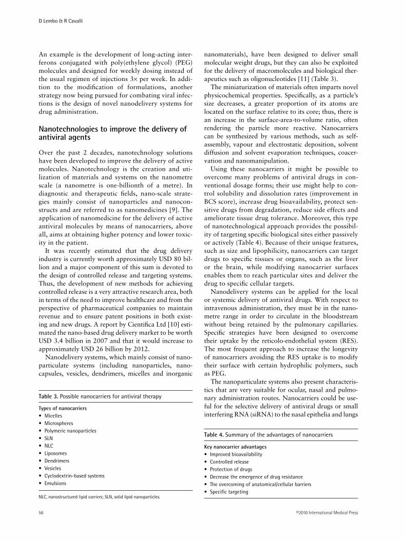
An example is the development of long-acting inter-ferons conjugated with poly(ethylene glycol) (PEG) molecules and designed for weekly dosing instead of the usual regimen of injections 3× per week. In addi-tion to the modification of formulations, another strategy now being pursued for combating viral infec-tions is the design of novel nanodelivery systems for drug administration.
Nanotechnologies to improve the delivery of antiviral agents
Over the past 2 decades, nanotechnology solutions have been developed to improve the delivery of active molecules. Nanotechnology is the creation and uti-lization of materials and systems on the nanometre scale (a nanometre is one-billionth of a metre). In diagnostic and therapeutic fields, nano-scale strate-gies mainly consist of nanoparticles and nanocon-structs and are referred to as nanomedicines [9]. The application of nanomedicine for the delivery of active antiviral molecules by means of nanocarriers, above all, aims at obtaining higher potency and lower toxic-ity in the patient.
It was recently estimated that the drug delivery industry is currently worth approximately USD 80 bil-lion and a major component of this sum is devoted to the design of controlled release and targeting systems. Thus, the development of new methods for achieving controlled release is a very attractive research area, both in terms of the need to improve healthcare and from the perspective of pharmaceutical companies to maintain revenue and to ensure patent positions in both exist-ing and new drugs. A report by Cientifica Ltd [10] esti-mated the nano-based drug delivery market to be worth USD 3.4 billion in 2007 and that it would increase to approximately USD 26 billion by 2012.
Nanodelivery systems, which mainly consist of nano-particulate systems (including nanoparticles, nano-capsules, vesicles, dendrimers, micelles and inorganic
nanomaterials), have been designed to deliver small molecular weight drugs, but they can also be exploited for the delivery of macromolecules and biological ther-apeutics such as oligonucleotides [11] (Table 3).
The miniaturization of materials often imparts novel physicochemical properties. Specifically, as a particle’s size decreases, a greater proportion of its atoms are located on the surface relative to its core; thus, there is an increase in the surface-area-to-volume ratio, often rendering the particle more reactive. Nanocarriers can be synthesized by various methods, such as self- assembly, vapour and electrostatic deposition, solvent diffusion and solvent evaporation techniques, coacer-vation and nanomanipulation.
Using these nanocarriers it might be possible to overcome many problems of antiviral drugs in con-ventional dosage forms; their use might help to con-trol solubility and dissolution rates (improvement in BCS score), increase drug bioavailability, protect sen-sitive drugs from degradation, reduce side effects and ameliorate tissue drug tolerance. Moreover, this type of nanotechnological approach provides the possibil-ity of targeting specific biological sites either passively or actively (Table 4). Because of their unique features, such as size and lipophilicity, nanocarriers can target drugs to specific tissues or organs, such as the liver or the brain, while modifying nanocarrier surfaces enables them to reach particular sites and deliver the drug to specific cellular targets.
Nanodelivery systems can be applied for the local or systemic delivery of antiviral drugs. With respect to intravenous administration, they must be in the nano-metre range in order to circulate in the bloodstream without being retained by the pulmonary capillaries. Specific strategies have been designed to overcome their uptake by the reticolo-endothelial system (RES). The most frequent approach to increase the longevity of nanocarriers avoiding the RES uptake is to modify their surface with certain hydrophilic polymers, such as PEG.
The nanoparticulate systems also present characteris-tics that are very suitable for ocular, nasal and pulmo-nary administration routes. Nanocarriers could be use-ful for the selective delivery of antiviral drugs or small interfering RNA (siRNA) to the nasal epithelia and lungs
Types of nanocarriersMicelles•Microspheres•Polymeric nanoparticles•SLN•NLC•Liposomes•Dendrimers•Vesicles•Cyclodextrin-based systems•Emulsions•
Table 3. Possible nanocarriers for antiviral therapy
NLC, nanostructured lipid carriers; SLN, solid lipid nanoparticles.
Table 4. Summary of the advantages of nanocarriers
Key nanocarrier advantagesImproved bioavailability•Controlled release•Protection of drugs•Decrease the emergence of drug resistance•The overcoming of anatomical/cellular barriers•Specific targeting•

Nanoparticulate drug delivery for antivirals
Antiviral Chemistry & Chemotherapy 21.2 57
in order to target viruses that infect the respiratory tract, such as influenza viruses, respiratory syncytial virus and rhinoviruses, to name just a few.
Promising compounds shown to have antiviral effects in vitro, but that are not currently being administered in vivo because of solubility and bioavailability prob-lems, could be administered using nanocarriers that can permit their administration; this can include peptides and nucleic acid delivery [12]. Certain nanodelivery systems might be suitable for the delivery of peptides and proteins protecting them from degradation.
In the past few years, RNA interference (RNAi) has emerged as a promising antiviral strategy that acts by silencing the gene expression of human viral pathogens, including that of influenza viruses, severe acute respira-tory syndrome virus, flaviviruses, HIV, HCV and HBV [13–16]. Very recently, a study by DeVincenzo et al. [17] provided a unique proof-of-concept for an RNAi-based therapy in humans directed against respiratory syncytial virus [17]. Nevertheless, there are still many obstacles that impede the translation of RNAi into a potential therapeutic platform, and the most important obstacle regards the delivery of siRNA in vivo. Tar-geting the action of siRNA to specific tissues and cells could minimize toxic side effects and improve their ther-apeutic efficacy. However, even if siRNA reach the cor-rect cellular target, their size and their negative charge make it difficult for them to cross the cell membrane, and many primary cell types are highly recalcitrant to siRNA uptake. Consequently, many delivery strategies based on nanotechnology are currently under develop-ment to address these challenges [18]. In addition to the advantages described above, the submicron size range of these delivery systems can also render intracellular uptake and transport of active compounds possible. In particular, the delivery of macromolecules into the cytoplasm is limited by their low membrane perme-ability and their degradation in the endosomal environ-ment after uptake by endocytosis. Their incorporation into a nanoparticulate system could promote cell inter-nalization and protect the molecules from degradation. This is an important feature because most antiviral drugs, like nucleoside analogues, target viral functions that are carried out within a cell. Various mechanisms govern the entry of nanoparticulates into cells, includ-ing caveolae-mediated endocytosis, clathrin-mediated endocytosis, phagocytosis and macropinocytosis [19]. Fluorescent-labelled nanoparticles can be used to study particle uptake by cells and their cellular trafficking. Another relevant feature of nanocarriers is the ability to overcome the physiological barriers. Nanomedicine is able to promote the delivery of drugs to the central nervous system. Various studies using different nanocar-riers report enhanced in vitro and in vivo blood–brain barrier (BBB) permeability and drug accumulation in
the brain [20]. Nanocarriers can enhance brain delivery by three major approaches: increasing the local drug concentration gradient at the BBB by passive targeting, allowing drug trafficking by non-specific or receptor-mediated endocytosis and blocking drug efflux trans-porters at the BBB. By selecting the components and the formulation parameters it is possible to prepare nano-carriers with physicochemical properties that allow delivery to the brain.
Three categories of nanocarriers have been investigated for the delivery of antiretrovirals to the central nerv-ous system: polymer/dendrimer-based, lipid-based and micelle-based systems [20]. The blood–retinal barrier, the anatomical barrier that protects the eyes, could also be overcome using nanocarriers. Moreover, using nanotech-nology-based systems it could be possible to reach ana-tomical compartments or cellular viral reservoirs that are not easily accessible to drugs in their current dosage form. For instance, the central nervous system, the cerebrospi-nal fluid, the lymphatic system, the macrophages and the semen are almost completely inaccessible to drugs, and are therefore compartments where HIV is harboured and evolves independently despite a successful highly active antiretroviral therapy [21–23]. Suboptimal drug penetra-tion into these compartments complicates the treatment of HIV infection and the eradication of viral reservoirs from the patient. Similar issues apply to herpesviruses, which latently infect particular cells and tissues.
The administration of antivirals in nanoparticles might affect the therapeutic efficacy inhibiting efflux transport-ers. Drug efflux transporters, such as P-glycoprotein (P-gp) play an important role in limiting the transport of xenobiotic molecules through various critical barriers in the body. Many orally administered drugs must cross the basolateral membrane in the intestinal epithelium to reach the blood. P-gp could drive compounds from inside the cells back into the intestinal lumen preventing their absorption. In cancer cells P-gp enables the development of resistance to anticancer drugs [24]. The activity of efflux transporters, which expel drugs from cells, leads to subtherapeutic drug concentrations. Indeed, P-gp inhibi-tion represents one potential strategy for the improvement of antiviral intestinal absorption. It has been previously demonstrated that the absorption of acyclovir in vitro is increased in the presence of P-gp-specific inhibitors, but this inhibition can increase side effects [25,26]. Another strategy is the use of nanoparticulate systems to deliver the drug into the cells favouring the absorption.
Another advantage of nanoparticles is that multi-functional systems can be obtained by engineering their surfaces. The advantageous characteristics resulting from such modifications, including longevity, target-ability and stimuli sensitivity, thus combine to pro-duce multifunctional nanocarriers that can simulta-neously perform more than one useful function [27].

D Lembo & R Cavalli
©2010 International Medical Press58
Such multifunctional nanocarriers could significantly enhance the efficacy of many therapeutic protocols.
Another emerging area of research is the development of integrated multifunctional nanosystems for diagnosis and therapy. These novel systems, called theranostics, are designed specifically for the simultaneous diagnosis and treatment of cancer. The nanosystem must be able to biomark cancer cells in order to achieve simultaneous and targeted imaging and treatment [28]. In the future, these integrated medical nanosystems could prove to be useful for the molecular diagnosis, treatment and moni-toring of viral infections at the cellular level.
Nanoparticles have similar nanometre dimensions to viruses. This feature led several researchers to inves-tigate the physical interaction of nanoparticles with viruses and to explore whether this interaction could be exploited as an antiviral strategy. Indeed, silver nanopar-ticles with mean particle diameters ranging from 10 to 50 nm have been shown to inhibit infection by various viruses including HIV, HBV, respiratory syncytial virus and monkeypox virus [29–33]. All of these studies con-cluded that the direct interaction between the nanoparti-cles and the virus was responsible for the antiviral activ-ity observed. It seems that nanoparticles exert antiviral activity at an early stage of viral replication, most likely as a virucidal agent or as an inhibitor of viral attachment and entry. Baram-Pinto et al. [34] further developed this strategy by designing silver nanoparticles capped with mercaptoethane sulphonate in order to target HSV and to compete for its binding to cell-surface heparan sul-phates. This strategy resulted in effective inhibition of HSV type-1 infection in cell culture and led the authors to propose capped nanoparticles as active ingredients of topical microbicides for the prevention of viral infections that depend on heparan sulphates for entry.
Targeted delivery of antiviral agents
The concept of targeted drugs was first suggested by Paul Ehrlich in 1906 who postulated the magic bullet theory. One century after this intuition, tar-geted drug delivery by functionalized nanocarriers has become one of the most attractive and promising areas of research in nanomedicine. However, it should be pointed out that some key challenges must be addressed before achieving quantitative delivery and targeting in vivo [35]. Site-specific drug delivery could be obtained with different types of nanocarriers.
The majority of studies performed to date have focused on developing systems that improve the biodis-tribution of anticancer drugs and their accumulation in specific tissues. Three distinct strategies exist for drug targeting: direct injection to a specific site, passive tar-geting and active targeting. Passive targeting means the nanoparticulate carrier can reach a given organ by the
virtue of its intrinsic properties, such as particle size or lipophilicity, whereas active targeting involves the presence of a ‘homing device’ that guides the carrier to its target site. Passive targeting associated with nano-carrier size permits the penetration of nanoparticles into tumour tissues because of the presence of leaky vasculature. This effect referred to as the ‘enhanced permeability and retention (EPR) effect’ results in nanoparticle accumulation within the tumours as dem-onstrated by Maeda et al. [36] (Figure 1). Because of their small sizes and surface characteristics, nanoparti-cles can be taken up by the lymphatic tissue in the gut (that is, the Peyer’s patches containing M cells) after oral administration.
Lymphatic targeting has increased the amount of attention directed at nanopharmaceuticals because of the prospect of directly targeting lymphocytes with immunomodulators, resident HIV viruses with anti-viral agents and disseminated tumour metastasis [37]. In stark contrast to molecularly dissolved drugs, nano-carriers can be designed for targeting the lymphatic cir-culation. With regards to injectable systems, although the particles must be large enough to drain, preferen-tially through the lymphatics, they must also be small enough to diffuse through the interstitial space away from the injection site. Sizes in the range of 10–100 nm are optimal. Moreover, hydrophilic nanoparticles clear more rapidly than hydrophobic nanoparticles follow-ing interstitial injection.
To date, lymphatic uptake has been widely inves-tigated in relation to the oral administration of medicines. Desai et al. [38] studied the influence of poly(d,l- lactide-co-glycolide) nanoparticle sizes on gas-trointestinal uptake in rats. Depending on the nanopar-ticle size, the Peyer’s patch tissue showed a 2–200-fold higher uptake than non-patch tissue. The use of nano-particle systems for oral drug delivery to the lymphatic system is rendered possible because of physiological particulate uptake mechanisms in the gut, especially the transcellular pathway involving vesicular transport through the M cells of Peyer’s patches. Nanoparticles are taken up by M cells in a size-dependent manner and transported to lymphocytes in the form of vesi-cles. The lymphatic absorption of a drug can prevent its systemic metabolism by the liver and permit target-ing to the lymphatic system. This peculiar particulate behaviour permits the lymphatic system to be reached and could be exploited to target viral reservoirs held within this compartment.
Active targeting can be accomplished by different strategies all consisting of surface modifications, in particular via a specific ligand-receptor-like mecha-nism. The primary strategy uses monoclonal antibodies raised against specific cells or tissues. Other molecules, such as sugars, polymers, proteins, vitamins, lectins

Nanoparticulate drug delivery for antivirals
Antiviral Chemistry & Chemotherapy 21.2 59
and aptamers, can also be used as homing devices as depicted in Figure 2 [39].
Another approach to target specific body areas or intracellular compartments is the use of stimuli- sensitive nanocarriers. This strategy exploits either intrinsically abnormal pH, redox and temperature values of patho-logical sites and intracellular organelles (that is, the endo-somes) or externally applied stimuli, such as a magnetic field, temperature and ultrasounds. All of these stimuli are expected to dissolve, to modify or to guide the sensi-tive nanocarriers, resulting in the release of the loaded drug in a particular region, such as tumours, inflamma-tion sites, infarcts or endosomes [27].
pH-sensitive nanocarriers are of particular interest in the area of therapeutic applications. The concept of pH-sensitive systems emerged from the knowledge that cer-tain enveloped viruses (for example, the influenza virus) lose their envelope in the acidic environment of the endosomal lumen thereby infecting the cells, and that some pathological tissues, as in tumours, inflammations and infections, exhibit a relatively more acidic environ-ment than normal tissues. Different classes of pH-sen-sitive systems have been proposed, such as liposomes, polymeric micelles and nanogels [9]. These pH-sensitive carriers can promote the intracellular release of the encapsulated drug when the pH changes. pH- sensitive liposomes are stable at physiological pH levels (7.4) but become unstable and fusogenic at acidic conditions (that is, in a lysosomal environment), releasing their aqueous content in the intracellular compartment.
External stimuli can also be used in combination with labelled nanocarriers that are externally guided (for example, by a magnetic field) or with specific deliv-ery systems activated by the application of a physical stimulus, such as temperature or ultrasounds.
In magnetic drug delivery, an external magnet is used to guide the drug-loaded nano- or microparticles to the targeted organ and to hold them there. The carrier is therefore magnetically concentrated in the target organ, but the subsequent release of the drug is a passive proc-ess affected by the properties of the particulate system. By contrast, the use of ultrasound permits the activation of the drug release at the site of action. Unlike the various targeted systems developed for anticancer therapy, few examples have been reported in the literature until now for targeted antiviral therapy. Most of these are listed in Table 5 [40–51] and mainly concern liposomes or nano-particles designed for the HIV treatment. An example of an external stimulus approach is that of magnetic micro-spheres containing interferon to achieve targeting using an external magnetic field [46].
Macrophages can act as a virus reservoir and sustain replication of HIV [52]. Macrophage targeting using nanoparticulate systems can be a therapeutic strategy because macrophages easily phagocytose foreign nano-particles [53]. The size, composition and surface proper-ties of nanoparticles can all affect macrophage uptake. For example, polyhexylcyanoacrylate nanoparticles with a diameter of approximately 200 nm were found to be the most useful for targeting antiviral substances to
Normal vasculature Leaky vasculatureDrug/drug carrier penetrates
into interstitium
300–700 nm
Role of ‘cutoff’ size
Figure 1. Schematic representation of passive targeting of tumour tissues associated with the enhanced permeability and retention effect
Tumour-specific vascular pathophysiology, with defective architecture and impaired lymphatic drainage, provides an increased permeability to macromolecules and nanoparticulates. This phenomenon has become a gold standard for the delivery of drugs to solid tumours.

D Lembo & R Cavalli
©2010 International Medical Press60
Drug
Matrix
Targetingmolecule
Cell penetratingpeptide
Image contrastagent
Poly(ethylene glycol)
Magnetic probe
Figure 2. Schematic representation of a functionalized nanoparticle
Poly(ethylene glycol) chains act as stealth agents. Targeting molecules can be attached to the surface of the functionalized nanoparticle as depicted. The presence of the contrast agent could illuminate the interaction of the nanoparticle with a target cell. The magnetic probe could permit the nanoparticle localization using an external magnetic field.
Drug Virus Nanodevice Targeting Targeted tissue In vivo studies Reference
siRNA HCV Cationic liposomes Apolipoprotein A1 Liver Yes Kim et al. [40]AZT HIV Albumin nanoparticles Transferrin Brain Yes Mishra et al. [41]Protease inhibitor HIV Pegylated liposomes Monoclonal antibody HIV-positive cells No Clayton et al. [42] against gp120siRNA HIV Immunoliposomes Antibody against LFA1 Lymphocytes Yes Kim et al. [43]Nosiheptide HBV Recombinant HDL Recombinant HDL Liver Yes Feng et al. [44] Acyclovir HBV Recombinant HDL Recombinant HDL Liver Yes Feng et al. [45]Interferon Magnetic microspheres External magnetic field – No Zhou et al. [46] Saquinavir HIV Nanoparticles Transferrin Brain No Mahajan et al. [47]gp120 Folding HIV Liposomes CD4 antigen HIV-positive cells No Pollock et al. [48]inhibitorInterferon-α Nanoparticles Digalactosyl diacyl glycerol Liver No Chiellini et al. [49]Indinavir HIV Immunoliposomes Antibodies against human and Lymphoid tissues Yes Gagné et al. [50] murine HLA-DR and CD4 antigenProtease inhibitor HIV Liposomes CD4 antigen Lymphocytes Yes Düzgünes et al. [51]
Table 5. Targeted delivery systems developed for antiviral drugs
AZT, zidovudine; HDL, high-density lipoprotein; HLA, human leukocyte antigen; LFA1, lymphocyte function-associated antigen 1; siRNA, small interfering RNA.

Nanoparticulate drug delivery for antivirals
Antiviral Chemistry & Chemotherapy 21.2 61
macrophages [54]; the same study demonstrated a good level of nanoparticle incorporation in macrophages obtained from HIV-infected patients. Additional target-ing moieties can be added to nanoparticles to enhance the level of macrophage uptake. Mannan-coated nano-particles containing didanosine were found to undergo greater targeting to macrophages by exploiting manno-syl receptor- mediated endocytosis.
Drug targeting using surface-modified nanocarriers is a strategy that permits delivery at the organ or even cell level. It was recently shown that the intracellular dis-tribution of nanoparticles could be controlled by cou-pling TAT peptide or cell penetrating peptides to the
nanoparticle surface in order to facilitate endosomal escape. This is particularly important for drugs that act within the cytosol or that must reach the nucleus [55]. The various nanoparticle surface modification strate-gies used for targeting purposes are listed in Table 6.
Overview of particulate carriers
The need for the development of new formulations for HIV, HBV, HCV and HSV antiviral treatments has been the major driving force in antiviral research. In this review, an up-to-date summary of the new formulations of HIV drugs will not be given because this task was recently completed by Sosnik et al. [56]. Moreover bio-conjugate systems and films have not been considered because this review was focused on particulate delivery systems. An overview of the most studied drug delivery systems proposed for use in antiviral therapies is, how-ever, reported below.
LiposomesLiposomes were the first vesicular carriers, proposed by Gregoriadis [57], to be used as drug delivery sys-tems. Liposomes are lipid concentric vesicles in which an aqueous volume is completely enclosed in a lipid bilayer composed mainly of phospholipids and choles-terol (Figure 3). Liposomes can vary in diameter, from 20 to 30 nm up to microns, depending on their chemi-cal composition and the preparation method used. Structurally, they can be classified as either small uni-lamellar vesicles or large unilamellar vesicles. They are able to encapsulate hydrophilic drugs within their inner aqueous phase and lipophilic drugs within their lipid bilayers. Liposomes are recognized as foreign matter by RES. Because HIV resides in macrophages, liposomes have been studied as promising carriers for anti-HIV drugs [58].
The liposome surface can be modified for different purposes. The incorporation of PEG molecules into the liposome bilayer prevents its interaction with plasma proteins and can consequently retard the recognition and removal of liposomes by RES. Functionalization of the liposome surface in order to achieve specific target-ing has also been studied. These vesicular carriers also present certain disadvantages, such as poor stability both in vitro and in vivo, low encapsulation efficiency and high cost of production.
Some liposomal formulations are in clinical practice for the intravenous administration of anticancer or anti-fungal drugs. Liposomal formulations for cancer therapy currently on the market are Doxil® (pegylated liposomal doxorubicin; Ortho Biotech Products, Bridgewater, NJ, USA), Myocet® (non-pegylated liposomal doxorubicin; Cephalon, Frazer, PA, USA) and DaunoXome® (non-pegylated liposomal daunorubicin; Nextar, Boulder,
Surface modificationPresence of surface charge•Surface coating•PEG coating•Antibody binding•Antibody fragment conjugation•CD4-derived peptide conjugation•Mannose conjugation•Galactose conjugation•Transferrin•Apolipoprotein•Recombinant HDL•
Table 6. Summary of possible surface modifications of nanoparticle-based systems for targeting purposes
HDL, high-density lipoprotein; PEG, poly(ethylene glycol).
Figure 3. Structure of a liposome and schematic representation of possible drug incorporation
Hydrophilic head
Hydrophobic tail
Aqueoussolution
Hydrophobic drug
Hydrophilic drug

D Lembo & R Cavalli
©2010 International Medical Press62
CO, USA); Ambisome® (Gilead Sciences, Foster City, CA, USA) is a liposomal formulation of amphotericin.
A vaginal liposomal delivery system for acyclovir has been designed for the local treatment of genital herpes [59]. A bioadhesive hydrogel consisting of Carbopol 974P was used as vehicle for the liposome containing acyclovir. In vitro release studies showed that the sys-tem can be considered for the vaginal sustained release of acyclovir. Liposomes have also been studied for the oral administration of interferon-α [60], the intravitreal administration of antisense oligonucleotides using lipo-somes for the treatment of CMV have been designed [61] and positively charged liposomes for the topical administration of acyclovir have been prepared [62].
The in vitro corneal penetration and in vivo corneal absorption of acyclovir from acyclovir-loaded lipo-somes have been investigated. The extent of absorp-tion with positively charged liposomes was higher than those from negatively charged liposomes. These were probably able to bind to the corneal surface, leading to an increased residence time favouring the acyclovir absorption [63]. Chetoni et al. [64] confirmed the lipo-somal efficacy for the ocular delivery of acyclovir.
Another form of vesicle proposed for drug delivery is the niosome, a vesicle similar to a liposome but formed with non-ionic surfactant instead of lipids [65]. The incorporation of acyclovir into liposomes and niosomes was recently compared [66]: niosomes were found to perform as better carriers for acyclovir because of their superior loading and slower release of the drug com-pared to that obtained with liposomes. Acyclovir-loaded in niosomes consisting of Span 60 (Merck, Frankfurt, Germany), cholesterol and dicetylphosphate have been investigated to improve the oral bioavailability of the drug. In vivo studies revealed that the niosomal disper-sion enhanced, by >2-fold, the oral bioavailability of acyclovir in relation to the free solution [67].
MicellesMicelles are colloidal structures (with particle diameters normally within the 5 to 100 nm range) belonging to a group of association or amphiphilic colloids (molecules that consist of two clearly distinct regions with oppo-site affinities towards water), which form spontane-ously at certain concentrations and temperatures from amphiphilic molecules or surfactants. At low concentra-tions in an aqueous medium, such amphiphilic mole-cules exist separately; however, as their concentration is increased, aggregation takes place, although only within a rather narrow concentration interval. The concentra-tion of a monomeric amphiphile at which micelles appear is called the critical micelle concentration, whereas the temperature below which amphiphilic molecules exist as unimers and above which they appear as aggre-gates is called the critical micellization temperature.
The formation of micelles is driven by the decrease of free energy in the system because of the removal of its hydrophobic fragments from the aqueous environment and the re- establishment of a hydrogen bond network in water. The hydrophobic fragments of amphiphilic mol-ecules form the core of a micelle, whereas hydrophilic fragments form the micelle’s shell. When used as drug carriers in aqueous media, micelles are able to solubi-lize poorly soluble lipophilic agents within its core, and polar molecules can be adsorbed onto the micelle’s sur-face [68] (Figure 4).
Polymeric micelles are nanostructures used to improve aqueous solubility, intestinal permeability and site targeting of several drugs. Compared to conven-tional surfactant-based micelles, polymeric micelles are composed of block copolymers and show greater stability in vivo. Micelles of PEG-polylactide copoly-mer surface modified with galactose units can interact with lectins [69]. Lectin receptors are present on HIV viral reservoirs, such as T-lymphocytes and macro-phages; thus, these copolymer micelles can be used as an approach for targeting viral reservoirs.
Block-copolymers of polyethylene-oxide–polypropyl-ene-oxide, known as Pluronics® (BASF, Florham Park, NJ, USA), have been proposed to enhance the intesti-nal permeability of antiretroviral drugs. Amphiphilic molecules can also be used to obtain self-assembled nanoparticles in water. Self-assembled delivery systems using cholesteryl derivatives as prodrugs for antiviral therapy have also been studied. Cholesteryl derivatives of acyclovir have been synthesised and show a typical
Figure 4. Schematic illustration of a micelle
Hydrophobic tail
Hydrophilic head
Aqueous solution
Hydrophobic tail
Hydrophilic head
Aqueous solution
Hydrophobic drug

Nanoparticulate drug delivery for antivirals
Antiviral Chemistry & Chemotherapy 21.2 63
amphiphilic structure with the lipid as hydrophobic tail and the antiviral nucleoside as the polar head [70]. Self-assembled drug delivery systems have also been designed to obtain nanoparticles from amphiphilic molecules. A lipid derivative of acyclovir has been synthesized show-ing the ability to form nanoparticles that were rapidly removed from blood circulation by macrophage uptake after their injection in rabbits [71].
MicrospheresMicrospheres are particulate carriers within the micron size range and are generally constituted of biodegrad-able polymers. They could be monolithic-type (matrix-type) or reservoir-type (capsular), the latter of which are called microcapsules (Figure 5). A wide range of techniques has been developed for their preparation to date. Different microsphere formulations have been studied as drug delivery systems for antivirals. Specifi-cally, biodegradable particles could be suitable for anti-viral administration via the intraocular route. Poly-d,l-lactide and poly(d,l-lactide-co-glycolide) microcapsules have been prepared by the spray-drying technique, and in the case of acyclovir were found to achieve high encapsulation efficiency. The microspheres were tested in vivo by intravitreal administration in rabbits and showed a prolonged release of acyclovir [72]. Micro-spheres of poly(d,l-lactide-co-glycolide) have been pro-posed for the intravitreal administration of acyclovir that aim at sustaining the release of the drug in order to minimize the dose as much as possible [73]. These
acyclovir-loaded microspheres were prepared by the solvent evaporation method and a factorial design was applied to reduce particle sizes to values suitable for injection through a 27G needle and to increase drug loading. The same author proposed the successful application of biodegradable microspheres of poly(d,l-lactide-co-glycolide) containing the combination of acyclovir and vitamin A palmitate to treat herpes simplex and Epstein–Barr viruses. Acyclovir loading increased when vitamin A palmitate was added to the microspheres and the in vitro acyclovir release was sub-sequently prolonged for 50 days [74].
Semi-interpenetrating polymer networks of acry-lamide grafted onto dextran and chitosan were pre-pared using an emulsion cross-linking method, with glutaraldehyde as the cross-linker for the encapsula-tion of acyclovir [75]. Microspheres of approximately 300 µm were obtained showing prolonged release kinetics of acyclovir.
To increase the oral bioavailability of acyclovir, mucoadhesive microspheres have been investigated as gastroretentive delivery systems. Dhaliwal et al. [76] evaluated different polymers and showed that the thi-olated chitosan mucoadhesive microspheres improved the acyclovir oral bioavailability because of the enhanced retention in the upper gastrointestinal tract. Recently, mucoadhesive acyclovir-loaded microspheres were developed using ethylcellulose as matrix and Carpobol 947 (Lubrizol, Wicklife, OH, USA) as the mucoadhesive polymer, with the purpose of improving
A B
Capsule shell
Inner cavity
Core
DrugDrug
Matrix
Figure 5. Schematic morphologies of the two types of particulate material
(A) Core-shell nanomicrocapsule or reservoir and (B) matrix nanomicroparticle.

D Lembo & R Cavalli
©2010 International Medical Press64
oral bioavailability of acyclovir [77]; the results of a mucoadhesion study [77] showed a prolonged residence time of the drug in rat gastrointestinal tracts.
Polymeric microspheres were also designed for the topical application of acyclovir in order to increase the drug concentration in the basal epidermis, which is the site of HSV infections [78]. The microspheres increased the retention of the drug in comparison with a drug suspension, and consequently allowed a decrease of the topical administration of acyclovir. A delayed release of acyclovir was also obtained by cross-linked malonyl-chitosan microspheres obtained by coacervation–phase separation [79]. The same research group also proposed acyclovir-loaded chitosan microspheres obtained by the spray-drying technique [80]. Microspheres loaded with interferon have been proposed for oral delivery [60]. Microspheres have also been proposed for use in sus-tained delivery systems for vaccines.
NanoparticlesNanoparticles are solid colloidal particles <1 micron in diameter and can be created using polymers, lipids, proteins or other substances, such as inorganic materi-als. They can have a matrix-like or capsule-like struc-ture (Figure 5) as described above for microspheres, obtaining nanoparticles and nanocapsules, respectively. The active molecules can be dissolved or encapsulated within the nanoparticles. Because of their small sizes they can be administered intravenously. As for lipo-somes, opsonization of nanoparticles in the blood can be prevented by the presence of hydrophilic moieties on their surface, such as PEG chains. These are known as stealth nanoparticles [81].
Polymeric nanoparticles are formulated using either natural or synthetic polymers with a high level of bio-compatibility to reduce cytotoxicity and maximize tis-sue compatibility. The only polymers that have been approved by the US Food and Drug Administration for human use are poly-d,l-lactic acid (PLA), polyglycolic acid, poly(lactic-co-glycolic acid), poly-e-caprolactone and poly(methylmethacrylate). Nanospheres made of PLA containing acyclovir were prepared by a nanopre-cipitation process. To obtain PEG-coated nanospheres 1,2-distearoyl-β-phosphatidylethanolamine was also incorporated. In vitro experiments were carried out to compare the ocular pharmacokinetics of the two types of acyclovir-loaded nanospheres compared with an aqueous suspension of the free drug. A PEG-coated microsphere significantly improved the aqueous level of acyclovir and could be suitable for the treatment of ocular viral infections.
Poly(hexylcyanacrylate) nanoparticles were pre-pared by an emulsion polymerization process as anti-sense oligonucleotide carriers for antiviral therapy [82]. Cidofovir, a nucleotide analogue active against
HCMV and smallpox virus, was encapsulated in poly(isobutyl cyanoacrylate) nanocapsules with an aqueous core by Hillaireau et al. [83].
Acyclovir-Eudragit nanoparticles were prepared using different charge density Eudragit® (Röhm, Darm-stadt, Germany), copolymers of poly(ethylacrylate, methacrylate and chlorotrimethyl methacrylate), and the formulation bioavailability was assessed in human volunteers compared with commercial products. The polymeric nanoparticles increase the oral bioavailabil-ity and prolonged the activity of acyclovir [84].
The majority of lipid nanoparticles can be classified as either solid lipid nanoparticles (SLN) or nanostructured lipid carriers (NLC) and they are made from lipids that are solid at body temperature with a mean diameter generally within the 50–500 nm range. SLN were first introduced by Gasco [85], and Müller and Lucks [86].
SLN possess several advantageous properties, includ-ing the solid state of their particle matrix, their ability to protect chemically-labile ingredients against chemical decomposition and their potential use in the modulation of drug release. Upon parenteral administration of SLN, improved bioavailability, targeting and enhanced cyto-toxicity against cancer cells have been observed [81].
NLC are composed of a solid lipid matrix with a certain liquid lipid phase content. SLN and NLC might be able to overcome the problems of membrane stabil-ity and drug leaching that are associated with liposomes and conventional emulsions [87,88]. Acyclovir-loaded SLN have been prepared showing a good incorporation [89], and those with diameters of approximately 400 nm showed greater in vitro activity than free drug [90]. Ade-fovir dipivoxil has been formulated in SLN composed of monostearin for use in HBV therapy.
Protein nanoparticles have also been developed as drug carriers. Albumin nanoparticles for ganciclovir delivery were prepared by coacevation and chemical cross-linking with glutaraldehyde. Depending on the step in which the glutaraldehyde was added in the prep-aration method, nanoparticles between 200–400 nm with a different drug incorporation and release profile were obtained [91].
Albumin nanoparticles have been studied as a delivery system for antisense oligonucleotides [92]. Phosphodiester and phosphorothioate oligonucleotides were adsorbed onto the surface or incorporated into the matrix of nanoparticles. The antiviral activity was eval-uated in fibroblasts infected with HCMV. Both types of nanoparticle formulations protected the oligonu-cleotides from enzymatic degradation, thus increasing their antiviral activity. Hillaireau et al. [83] encapsu-lated cidofovir in poly(isobutyl cyanoacrylate) nano-capsules with an aqueous core for the encapsulation of azidothymidine-triphosphate and cidofovir showing a high incorporation efficiency of the two drugs. Cationic

Nanoparticulate drug delivery for antivirals
Antiviral Chemistry & Chemotherapy 21.2 65
gelatin nanoparticles increased the immunostimulatory effects of CpG oligonucleotides [93].
DendrimersDendrimers are perfectly ordered, nanostructured polymers, characterized by a branching structure emanating from a central core (Figure 6). Their small sizes (<100 nm in diameter) and the possibility of binding targeting ligands to them renders dendrim-ers attractive for use in drug delivery [94]. They have been proposed as carriers for DNA, siRNA and anti-viral drugs. Acyclovir-terminated thiophosphate den-drimers have been synthesised [95]: dendrimer drug polyconjugates that are soluble in water and that can act as a macromolecular prodrug of acyclovir have been obtained. Peptide-derivatized dendrimers have been found to inhibit HCMV replication by blocking virus binding to cell-surface heparan sulphates [96]. VivaGel™, a dendrimer-based formulation developed by Starpharma (Melbourne, Australia) with activity against HIV and HSV [97] has successfully completed Phase I clinical trials [98] and is expected to be avail-able on the market soon as a microbicide for the pre-vention of sexually-transmitted HSV infections.
Emulsion-based delivery systemsEmulsions are heterogeneous systems generally consist-ing of an aqueous phase and an oil phase. In oil-in- water emulsions, the oil is dispersed as droplets within the water; the reverse is also possible and is called a
water-in-oil emulsion. Emulsions with droplet sizes <100 nm are called microemulsions and are transpar-ent systems. These microemulsions differ fundamen-tally from emulsions. Microemulsions are thermody-namically stable systems displaying indefinite stability, whereas emulsions are merely kinetically stabilized and thermodynamically instable, which means that emul-sions will tend to separate into oil and water phases. Microemulsions are systems consisting of water, oil, surfactants and cosurfactants, and they are useful for drug delivery because of their capacity to solubilize both water-soluble and oil-soluble compounds.
The potential applications of emulsions and micro-emulsions include their use as carriers for drugs with poor water solubility, sustained-release systems and site-specific drug delivery achieved by binding ligands for various cell-surface receptors to the particle surface.
Emulsion formulations can be used as vehicles for delivering antivirals within the human body. A micro-emulsion drug delivery system of acyclovir has been designed with the aim of improving oral bioavailability. An in vitro intraduodenal diffusion and in vivo study revealed an increased bioavailability of approximately 13% after the oral administration of an acyclovir micro-emulsion formulation compared with that achieved with commercially available tablets [99]. Positively charged microemulsions have been formulated for the topical application of acyclovir. A two-fold increase in acyclovir accumulation was obtained in comparison with a drug suspension as control [100].
Figure 6. Schematic illustration of the synthesis of a fourth-generation dendrimer
Focal pointactivation
B.
Branchedmonomer
A.
Surfacesubunit
Growth ofhypercore
Core Hypercore Fourth-generation dendrimer
Wedge

D Lembo & R Cavalli
©2010 International Medical Press66
HydrogelsNano- and microhydrogels (that is, nanogels and microgels) form another type of particulate polymeric material. They are formed of cross-linked polymeric networks ranging in size from 10 to 1,000 nm swollen by a good solvent. Compared with soluble polymers, they show different properties, such as rheological behaviour, resistance against degradation, high drug loading and the possibility of carrying structural resi-dues sensitive to external stimuli, such as pH, tempera-ture, light and redox reactions. Hydrophilic natural or synthetic polymers can be transformed in particulate systems using different technological processes, such as emulsion polymerization, chemical cross-linking and physical cross-linking [101]. Thermosensitive hydro-gels have been designed using a carboxymethylderiva-tive of scleroglucan for topical application of antivirals [102]. Ferruti et al. [103] developed nanohydrogels by cross-linking copolymers based on poly(amidoamine) s (PAAs) and β- or γ-cyclodextrin (CD), capable of simul-taneously solubilizing and selectively delivering drugs without the formation of covalent polymer– drug link-ages. These constructs form nanogel in aqueous sys-tems with high swelling capacity [103]. The PAA/CD nanoparticles can have a CD content of 10–35% on a weight-to-weight basis. They showed the capacity to incorporate molecules, such as drugs and proteins, and then slowly release them. Acyclovir-loaded PAA/CD nanoparticles showed an incorporation of the drug of approximately 15%, which is released with sustained kinetics in vitro.
Cyclodextrin-based delivery systemsCDs are cyclic oligosaccharides derived from starch and shaped like truncated cones because of the chair conformation of the glucopyranose units. The most common CDs are α-, β- and γ-CDs, possessing six, seven and eight glucopyranose units, respectively. CDs are able to form non-covalent inclusion complexes (or host–guest complexes). CD complexes are mainly used in the pharmaceutical field to improve aqueous solubil-ity, bioavailability and stability of drugs.
The CD complexation of acyclovir and ganciclovir has been studied in order to increase the drugs’ solubili-ties [104]. The in vitro antiviral activity of ganciclovir against several strains of HCMV was enhanced by com-plexation with β-CD [105]. Derivatives of CDs have been synthesised to improve the complexation capacity and the technological properties of parent CDs.
A new PAA copolymer with β-CD was obtained by polyaddition reaction of 6-deoxy-6-amino-β-CD (β-CD-NH2) and 2-methylpiperazine to 2,2-bis(acrylamido)acetic acid in aqueous medium. This β-PAA/CD copoly-mer bears β-CD units along the macromolecular chain, is water-soluble and non-cytotoxic. This copolymer
showed a good acyclovir complexation capacity. The antiviral activity of the acyclovir–β-PAA/CD complex was evaluated against HSV in cell cultures exhibiting a higher antiviral activity than the free drug [106].
Amphiphilic CD derivatives are of much interest for pharmaceutical applications because of their ability to self-organize in water [107]. Sulphated and non-sul-phated amphiphilic α-, β- and γ-CDs show the ability to complex with acyclovir [108].
Novel polymeric nanoparticles based on a β-CD-poly(4-acryloylmorpholine) monoconjugate (β-CD-PACM), a tadpole-shaped polymer in which the β-CD ring is the hydrophilic head and the PACM chain the amphiphilic tail, were prepared by the solvent injec-tion technique. Acyclovir-loaded nanoparticles were obtained from inclusion complexes of acyclovir with β-CD-PACM. The antiviral activity of acyclovir loaded into β-CD-PACM nanoparticles against two clinical isolates of HSV type-1 was evaluated in comparison with that of both the free drug and the soluble β-CD-PACM complex. When carried in the nanoparticles, acyclovir showed an enhanced antiviral activity com-pared with the other formulations tested [109]. Acy-clovir nanoparticles with other types of amphiphilic CDs have been designed.
Nanoparticles made of amphiphilic perfluoroalkyl α-CD have been prepared for the transport of acyclo-vir [110]. These fluorinated amphiphilic CD nanopar-ticles encapsulated acyclovir with an efficiency of about 40% and allowed its sustained release. Nanosponges are a new class of material consisting of nanoporous solid particles. Recently, CD-based nanosponges have been prepared [111,112]. They are nanoparticles with a rather spherical shape consisting of hyper cross-linked CDs (β or γ) nanostructured in a tridimensional net-work. Nanosponges can be synthesised as neutral or acid, and can be swellable according to the agent used as cross-linker. The cross-linking-to-CD ratio can be varied to improve the drug loading.
Nanosponges showed the capacity of encapsulating various types of molecules in their structure based on the formation of inclusion and non-inclusion com-plexes [113]. The acyclovir loading capacity in car-boxylated nanosponges was approximately 60% w/w and the in vitro release profile showed a prolonged release over time. The acyclovir loaded in nanosponges exhibited a higher antiviral activity than the free drug evaluated against HSV in cell cultures [114].
NanocrystalsNanocrystals of pure drugs can improve their dissolu-tion rate and bioavailability. They can be formulated as aqueous nanosuspensions in the presence or absence of stabilizers, such as non-ionic surfactants. Nano crystals are suitable for nanoparticulate formulation of drugs

Nanoparticulate drug delivery for antivirals
Antiviral Chemistry & Chemotherapy 21.2 67
with properties such as an insolubility in both water and oil, a high melting point, a high log P and high dose. Following the Noyes–Whitney equation, pro-gressive size reduction of the drug particles leads to an increase in the surface area resulting in an increased dissolution rate. Additionally, particle size reduction results in a decrease of the diffusion layer thickness surrounding the particles and an increased concentra-tion gradient between the surface of the particle and bulk solution, which facilitates particle dissolution by increasing dissolution velocity. Therefore, nanosizing is a suitable approach for increasing bioavailability of those drugs where dissolution is the rate-limiting step in systemic absorption. Van Eerdenbrugh et al. [115] investigated the dissolution and in vitro absorption of a poorly water soluble non-nucleoside reverse tran-scriptase inhibitor, loviride (water solubility 0.1 mg/l), after nanonization. Nanocrystals can be obtained by different technological processes.
The media-milling technology, developed by Merisko- Liversidge et al. [116], is a proprietary wet milling technology (Nanocrystal®) of the Elan Corporation (Athlone, Ireland), and there are at least four oral dos-age forms currently in the market containing pure drug nanoparticles (that is, Rapamune®; Wyeth, Princeton, NJ, USA).
Conclusions and future perspectives
The nanomedicine approach opens new therapeutic strategies for attacking viral diseases and for improving treatment success rates. Innovative nanomedicine solu-tions are expected to have great effects in the treatment as well as the eradication of infectious diseases. Their role could be important in prevention, early diagnosis, more effective drug delivery systems, specific targeting and personalized therapy (Table 7).
In particular, nanoparticulate-based systems could improve the efficacy of antivirals, restrict adverse drug side effects and reduce treatment costs. These features are particularly relevant in viral diseases where high drug doses are needed, drugs are expensive and the success of a therapy is associated with patient adherence to the administration protocol. This latter issue is very impor-tant in viral treatments where complicated or chronic reg-imens are common. Nanotechnology can reduce intake frequency and shorten the time of treatments, potentially rendering the treatment more cost-effective.
In addition, nanomedicine can enhance the effec-tiveness of approved antiviral drugs and extend their applicability by overcoming limitations, such as low bioavailability and cellular barriers. However, several important issues must be addressed before the potential of nanotechnology can be translated into safe and effec-tive antiviral formulations for clinical use.
Nanotoxicology is a relatively new discipline and requires further development [117–119]. A deep exploration of the toxicological and the bioelimina-tion aspects of nanocarriers is required to ensure safe manufacture and use of nanomaterials. Note, however, that the risk–benefit assessment of an antiviral formu-lation should be based on more stringent criteria than that for anticancer formulations, for example, because many viral infections are not life-threatening.
Virologists should be directly involved in the devel-opment of antiviral nanocarriers. Besides discovering new antiviral molecules to be delivered by nanocarri-ers, virologists should also be addressing other impor-tant matters; for instance, the selective targeting of a nanocarrier to infected tissues requires the identifica-tion of molecules or functions that are differentially expressed or carried out by a virus-infected cell. Ide-ally, this knowledge should be provided for each virus against which an antiviral drug is available. So far, we do not know whether it is possible to exploit passive targeting for antiviral therapy in a way analogous to that in anticancer therapy. Nanocarriers loaded with anticancer drugs could be passively targeted to a tumour because of the enhanced EPR effect. Virolo-gists should explore the possibility that virus-infected tissues or cells could be more susceptible (or recalci-trant) to nanodelivery systems.
From a technological point of view, the main objec-tives of future antiviral therapy research will be the identification of new technologies for the characteriza-tion of nanoscale materials, the development of nan-odelivery systems devoid of cytotoxicity and with high biocompatibility and biodegradability, the functionali-zation of nanocarriers for effectively targeting specific sites of viral infection in order to reduce drug-related toxicity in other tissues, the design of molecules that, as well as acting as the active carriers, also possess intrinsic antiviral therapeutic properties (for example, dendrim-ers and metallic nanoparticles), the optimization and scale-up of the production procedures in good manufac-turing practice (GMP), the development of regulatory guidelines suitable for nanocarriers, the development of
Table 7. Role of nanomedicine in antiviral therapy
Role of nanomedicineIncreased bioavailability•Improved antiviral delivery•Targeting specific body sites•Control of adverse side effects•Monitoring of antiviral therapy•Viral diagnostics•Patient compliance•Nanomicrobicides in prevention therapy•Personalized therapies•

D Lembo & R Cavalli
©2010 International Medical Press68
cost-effective nanotechnology-based formulations and, finally, making them available to developing countries. In conclusion, the main ethical and scientific challenges of research into antiviral nanomedicines are to produce safe and high quality therapies at reasonable costs
Acknowledgements
This research was supported by grants from Regione Piemonte (Ricerca Finalizzata 2008-bis and 2009). We thank Agnese Bisazza and Andrea Civra for their excel-lent assistance.
Disclosure statement
The authors declare no competing interests.
References1. Milroy D, Featherstone J. Antiviral market overview. Nat
Rev Drug Discov 2002; 1:11–12.
2. Roehr B. Fomivirsen approved for CMV retinitis. J Int Assoc Physicians AIDS Care 1998; 4:14–16.
3. Amidon GL, Lennernas H, Shah VP, Crison JR. A Theoretical basis for a biopharmaceutic drug classification: the correlation of in vitro drug product dissolution and in vivo bioavailability. Pharm Res 1995; 12:413–420.
4. Williams GC, Sinko PJ. Oral absorption of the HIV protease inhibitors: a current update. Adv Drug Deliv Rev 1999; 39:211–238.
5. Sharma P, Garg S. Pure drug and polymer based nanotechnologies for the improved solubility, stability, bioavailability and targeting of anti-HIV drugs. Adv Drug Deliv Rev 2010; 62:491–502.
6. Emery VC. Progress in understanding cytomegalovirus drug resistance. J Clin Virol 2001; 21:223–228.
7. Fridland A, Connelly MC, Robbins BL. Cellular factors for resistance against antiretroviral agents. Antivir Ther 2000; 5:181–185.
8. Liaw YF. The current management of HBV drug resistance. J Clin Virol 2005; 34 Suppl 1:S143–S146.
9. Arshady R, Kono K (Editors). Smart nanoparticles in nanomedicine – the MML series. Vol 8. London: Kentus Books 2006.
10. Sosnik A, Amiji M. Nanotechnology solutions for infectious diseases in developing nations. Adv Drug Deliv Rev 2010; 62:375–377.
11. Mishra B, Patel BB, Tiwari S. Colloidal nanocarriers: a review on formulating technology, types and applications toward targeted drug delivery. Nanomedicine 2010; 6:9–24.
12. Ketzinel-Gilad M, Shaul Y, Galum E. RNA interference for antiviral therapy. J Gene Med 2006; 8:933–950.
13. Kumar P, Sood V, Vyas R, Gupta N, Banerjea AC, Khanna M. Potent inhibition of influenza virus replication with novel siRNA-chimeric-ribozyme constructs. Antiviral Res 2010; 87:204–212.
14. Subramanya S, Kim SS, Abraham S, et al. Targeted delivery of small interfering RNA to human dendritic cells to suppress dengue virus infection and associated proinflammatory cytokine production. J Virol 2010; 84:2490–2501.
15. Singh SK. RNA interference and its therapeutic potential against HIV infection. Expert Opin Biol Ther 2008; 8:449–461.
16. Huang DD. The potential of RNA interference-based therapies for viral infections. Curr HIV/AIDS Rep 2008; 5:33–39.
17. DeVincenzo J, Lambkin-Williams R, Wilkinson T. A randomized, double-blind, placebo-controlled study of an RNAi-based therapy directed against respiratory syncytial virus. Proc Natl Acad Sci U S A 2010; 107:8800–8805.
18. Tiemann K, Rossi JJ. RNAi-based therapeutics – current status, challenges and prospects. EMBO Mol Med 2009; 1:142–151.
19. Hillaireau H, Couvreur P. Nanocarriers’ entry into the cells: relevance to drug delivery. Cell Mol Life Sci 2009; 66:2873–2896.
20. Wong HL, Chattopadhyay N, Wu XY, Bendayan R. Nanotechnology applications for improved delivery of antiretroviral drugs to the brain. Adv Drug Deliv Rev 2010; 62:503–517.
21. Strain MC, Letendre S, Pillai SK, et al. Genetic composition of human immunodeficiency virus type 1 in cerebrospinal fluid and blood without treatment and during failing antiretroviral therapy. J Virol 2005; 79:1772–1788.
22. Pillai SK, Good B, Pond SK, et al. Semen-specific genetic characteristics of human immunodeficiency virus type 1 env. J Virol 2005; 79:1734–1742.
23. Alexaki A, Liu Y, Wigdahl B. Cellular reservoirs of HIV and their role in viral persistence. Curr HIV Res 2008; 6:388–400.
24. Chan LMS, Lowes S, Hirst BH. The ABCs of drug transport in the intestine and liver: efflux proteins limiting drug absorption and bioavailability. Eur J Pharm Sci 2004; 21:25–51.
25. Salama NN, Scott KR, Eddington ND. DM27, an enaminone, modifies the in vitro transport of antiviral therapeutic agents. Biopharm Drug Dispos 2004; 25:227–236.
26. Yang ZG, Meng H, Zhang X, et al. [Effect of quercetin on the acyclovir intestinal absorption]. Beijing Da Xue Xue Bao 2004; 36:309–312. Chinese.
27. Torchilin V. Multifunctional and stimuli-sensitive pharmaceutical nanocarriers. Eur J Pharm Biopharm 2009; 71:431–444.
28. Riehemann K, Scheider SW, Luger TA, Godin B, Ferrari M, Fuchs H. Nanomedicine – challenge and perspectives. Angew Chem Int Ed Engl 2009; 48:872–897.
29. Lara HH, Ayala-Nuñez NV, Ixtepan-Turrent LL, Rodriguez-Padilla C. Mode of antiviral action of silver nanoparticles against HIV-1. J Nanobiotechnology 2010; 8:1–10.
30. Elechiguerra JL, Burt JL, Morones JR, et al. Interaction of silver nanoparticles with HIV-1. J Nanobiotechnology 2005; 3:6–12.
31. Lu L, Sun RW, Chen R. Silver nanoparticles inhibit hepatitis B virus replication. Antivir Ther 2008; 13:253–262.
32. Sun L, Singh AK, Vig K, Pillai SR, Singh SR. Silver nanoparticles inhibit replication of respiratory syncytial virus. J Biomed Nanotechnol 2008; 4:149–158.
33. Rogers JV, Parkinson CV, Choi YW, Speshock JL, Hussain SL. A preliminary assessment of silver nanoparticles inhibition of monkeypox virus plaque formation. Nanoscale Res Lett 2008; 3:129–133.
34. Baram-Pinto D, Shukla S, Perkas N, Gedanken A, Sarid R. Inhibition of herpes simplex virus type 1 infection by silver nanoparticles capped with mercaptoethane sulfonate. Bioconjug Chem 2009; 20:1497–1502.
35. Ruenraroengsak P, Cook JM, Florence AT. Nanosystem drug targeting: facing up to complex realities. J Control Release 2010; 141:265–276.
36. Maeda H, Wu J, Sawa T, Matsumura Y, Hori K. Tumour vascular permeability and the EPR effect in macromolecular therapeutics: a review. J Control Release 2000; 65:271–284.
37. Gupta U, Kompella U. Nanotechnology in drug delivery. New York: Taylor & Francis 2006.
38. Desai MP, Labhasetwar V, Amidon GL, Levy RJ. Gastrointestinal uptake of biodegradable microparticles: effect of particle size. Pharm Res 1996; 13:1838–1845.

Nanoparticulate drug delivery for antivirals
Antiviral Chemistry & Chemotherapy 21.2 69
39. Sanvicens N, Marco PM. Multifunctional nanoparticles-properties and prospects for their use in human medicine. Trends Biotechnol 2008; 26:425–432.
40. Kim SI, Shin D, Lee H, et al. Targeted delivery of siRNA against hepatitis C virus by apolipoprotein A-I-bound cationic liposomes. J Hepatol 2009; 50:479–488.
41. Mishra V, Mahor S, Rawat A, et al. Targeted brain delivery of AZT via transferrin anchored pegylated albumin nanoparticles. J Drug Target 2006; 14:45–53.
42. Clayton R, Ohagen A, Nicol F, et al. Sustained and specific in vitro inhibition of HIV-1 replication by a protease inhibitor encapsulated in gp120-targeted liposomes. Antiviral Res 2009; 84:142–149.
43. Kim SS, Peer D, Kumar P, et al. RNAi-mediated CCR5 silencing by LFA-1-targeted nanoparticles prevents HIV infection in BLT mice. Mol Ther 2010; 18:370–376.
44. Feng M, Cai Q, Shi X, Huang H, Zhou P, Guo X. Recombinant high-density lipoprotein complex as a targeting system of nosiheptide to liver cells. J Drug Target 2008; 16:502–508.
45. Feng M, Cai Q, Huang H, Zhou P. Liver targeting and anti-HBV activity of reconstituted HDL-acyclovir palmitate complex. Eur J Pharm Biopharm 2008; 68:688–693.
46. Zhou S, Sun J, Sun L, et al. Preparation and characterization of interferon-loaded magnetic biodegradable microspheres. J Biomed Mater Res B Appl Biomater 2008; 87:189–196.
47. Mahajan SD, Roy I, Xu G, et al. Enhancing the delivery of anti retroviral drug ‘saquinavir’ across the blood brain barrier using nanoparticles. Curr HIV Res 2010; 8:396–404.
48. Pollock S, Dwek RA, Burton DR, Zitzman N. N-Butyldeoxynojirimycin is a broadly effective anti-HIV therapy significantly enhanced by targeted liposome delivery. AIDS 2008; 22:1961–1969.
49. Chiellini EE, Chiellini F, Solaro R. Bioerodible polymeric nanoparticles for targeted delivery of proteic drugs. J Nanosci Nanotechnol 2006; 6:3040–3047.
50. Gagné JF, Désormeaux A, Perron S, Tremblay MJ, Bergeron MG. Targeted delivery of indinavir to HIV-1 primary reservoirs with immunoliposomes. Biochim Biophys Acta 2002; 1558:198–210.
51. Düzgünes N, Pretzer E, Simões S, et al. Liposome-mediated delivery of antiviral agents to human immunodeficiency virus-infected cells. Mol Membr Biol 1999; 16:111–118.
52. Bergamaschi A, Pancino G. Host hindrance to HIV-1 replication in monocytes and macrophages. Retrovirology 2010; 7:31.
53. Gunaseelan S, Gunaseelan K, Deshmukh M, Zhang X, Sinko PJ. Surface modifications of nanocarriers for effective intracellular delivery of anti-HIV drugs. Adv Drug Deliv Rev 2010; 62:518–531.
54. Schäfer V, Von Briesen H, Andreesen R, et al. Phagocytosis of nanoparticles by human immunodeficiency virus (HIV)-infected macrophages: a possibility for antiviral drug targeting. Pharm Res 1992; 9:541–546.
55. Zhang X, Jin X, Plummer MR, et al. Endocytosis and membrane potential are required for HeLa cell uptake of R.I.-CKTat9, a retro-inverso Tat cell penetrating peptide. Mol Pharm 2009; 6:836–848.
56. Sosnik A, Chiappetta D, Carcaboso A. Drug delivery systems in HIV pharmacotherapy: what has been done and the challenges standing ahead. J Control Release 2009; 138:2–15.
57. Gregoriadis G. Overview of liposomes. J Antimicrob Chemother 1991; 28:39–48.
58. Gupta U, Jain N. Non-polymeric nano-carriers in HIV/AIDS drug delivery and targeting. Adv Drug Deliv Rev 2010; 62:478–490.
59. Pavelic Z, Škalko-Basnet N, Filipovic-Grcic J, Martinac A, Jalšenjak M. Development and in vitro evaluation of a liposomal vaginal delivery system for acyclovir. J Control Release 2005; 106:34–43.
60. Thitinan S, McConville JT. Interferon alpha delivery systems for the treatment of hepatitis C. Int J Pharm 2009; 369:121–135.
61. Bochot A, Couvreur P, Fattal E. Intravitreal administration of antisense oligonucleotides: potential of liposomal delivery. Prog Retin Eye Res 2000; 19:131–147.
62. Peira E, Trotta M, Carlotti ME, Gallarate M, Chirio D. Elastic positively-charged liposomes for topical administration of acyclovir. J Drug Deliv Sci Technol 2007; 17:321–324.
63. Law SL, Huang KJ, Chiang CH. Acyclovir-containing liposomes for potential ocular delivery: corneal penetration and absorption J Control Release 2000; 63:135–140.
64. Chetoni P, Rossi S, Burgalassi S, et al. Comparison of liposome encapsulated acyclovir with acyclovir ointment: ocular pharmacokinetics in rabbits. J Ocul Pharmacol Ther 2004; 20:169–177.
65. Fang JY, Hong CT, Chiu WT, et al. Effect of liposomes and niosomes on skin permeation of enoxacin. Int J Pharm 2001; 219:61–72.
66. Mukherjee B, Patra B, Layek B, et al. Sustained release of acyclovir from nano-liposomes and nano-niosomes: an in vitro study. Int J Nanomedicine 2007; 2:213–225.
67. Attia IA, El-Gizawy SA, Fouda MA, Donia AM. Influence of a niosomal formulation on the oral bioavailability of acyclovir in rabbits. AAPS PharmSciTech 2007; 8:E106.
68. Torchilin VP. Micellar nanocarriers: pharmaceutical perspectives. Pharm Res 2007; 24:1–16.
69. Nagasaki Y, Yasugy K, Yamamoto Y, et al. Sugar-installed block copoloimer micelles: their preparation and specific interaction with lectin molecules. Biomacromolecules 2001; 2:1067–1070.
70. Jin Y, Xin R, Ai P, et al. Cholesteryl derivatives of antiviral nucleoside analogues: synthesis, properties and the vesicle formation. Int J Pharm 2008; 350:330–337.
71. Jin Y, Tong L, Ai P, et al. Self-assembled drug delivery systems: 1. Properties and in vitro/in vivo behavior of acyclovir self-assembled nanoparticles (SAN). Int J Pharm 2006; 309:199–207.
72. Conti B, Bucalo C, Giannovola C, et al. Biodegradable microspheres for the intravitreal administration of acyclovir: in vitro/in vivo evaluation. Eur J Pharm Sci 1997; 5:287–293.
73. Martinez-Sancho C, Herrero-Vanrell R, Negro S, et al. Study of gamma-irradiation effects on aciclovir poly(d,l-lactic-co-glycolic) acid microspheres for intravitreal administration. J Control Release 2004; 99:41–52.
74. Martinez-Sancho C, Herrero-Vanrell R, Negro S, et al. Vitamin A palmitate and acyclovir biodegradable microspheres for intraocular sustained release. Int J Pharm 2006; 326:100–106.
75. Rokhade AP, Patil SA, Aminabhavi TM. Synthesis and characterization of semi-interpenetrating polymer network microspheres of acrylamide grafted dextran and chitosan for controlled release of acyclovir. Carbohydr Polym 2007; 67:605–613.
76. Dhaliwal S, Jain S, Singh P, et al. Mucoadhesive microspheres for gastroretentive delivery of acyclovir: in vitro and in vivo evaluation. AAPS J 2008; 10:322–330.
77. Tao Y, Lu Y, Sun Y, et al. Development of mucoadhesive microspheres of acyclovir with enhanced bioavailability. Int J Pharm 2009; 378:30–36.
78. de Jalon EG, Blanco-Prìeto MJ, Ygartua P, Santoyo S. Topical application of acyclovir-loaded microparticles: quantification of the drug in porcine skin layers. J Control Release 2001; 75:191–197.
79. Stulzer HK, Lacerda L, Tagliari MP, et al. Synthesis and characterization of cross-linked chitosan microspheres for controlled release of acyclovir. Carbohydr Polym 2008; 73:490–496.
80. Stulzer HK, Tagliari MP, Parize AL, et al. Evaluation of cross-linked chitosan microparticles containing acyclovir obtained by spray-drying. Materials Science and Engineering: C 2009; 29:387–392.

D Lembo & R Cavalli
©2010 International Medical Press70
81. Owens DE, Peppas NA. Opsonization, biodistribution, and pharmacokinetics of polymeric nanoparticles. Int J Pharm 2006; 307:93–102.
82. Zimmer A. Antisense oligonucleotide delivery with polyhexylcyanoacrylate nanoparticles as carriers. Methods 1999; 18:286–295.
83. Hillaireau H, Le Doan T, Besnard M, Chacun H, Janin J, Couvreur P. Encapsulation of antiviral nucleotide analogues azidothymidine-triphosphate and cidofovir in poly(iso-butylcyanoacrylate) nanocapsules. Int J Pharm 2006; 324:37–42.
84. Elshafeey AH, Kamel AO, Awad GAS. Ammonium methacrylate units polymer content and their effect on acyclovir colloidal nanoparticles properties and bioavailability in human volunteers. Colloids Surf B Biointerfaces 2010; 75:398–404.
85. Gasco MR, inventor. Method for producing solid lipid mirospheres having narrow distribution. 1993 US patent 5,250,236. 1993.
86. Müller RH, Lucks JS. Arzneistoffträger aus festen Lipidteilchen, Feste Lipidnanosphären (SLN). [Medication vehicles made of solid lipid particles (solid lipid nanospheres – SLN)]. European patent 0605497. 1993.
87. Gasco MR. Solid lipid nanoparticles for drug delivery. Pharm Technol Eur 2001; 13:32–41.
88. Muller RH, Mader K, Gohla S. Solid lipid nanoparticles (SLN) for controlled drug delivery-A review of the state of art. Eur J Pharm Biopharm 2000; 50:161–177.
89. Lukowski G, Pflegel P. Electron diffraction of solid lipid nanoparticles loaded with aciclovir. Pharmazie 1997; 52:642–643.
90. Zhang X, Miao J, Li M, et al. Solid lipid nanoparticles loading adefovir dipivoxil for antiviral therapy. J Zhejiang Univ Sci 2008; 9:506–510.
91. Merodio M, Arnedo A, Renedo MJ, Irache JM. Ganciclovir-loaded albumin nanoparticles: characterization and in vitro release properties. Eur J Pharm Sci 2001; 12:251–259.
92. Arnedo A, Irache JM, Merodio M, et al. Albumin nanoparticles improved the stability, nuclear accumulation and anticytomegaloviral activity of a phosphodiester oligonucleotide. J Control Release 2004; 94:217–227.
93. Zwiorek K, Bourquin C, Winter G, Endres S, Hartmann G, Coester C. Delivery by cationic gelatin nanoparticles strongly increases the immunomodulatory effects of CpG oligonucleotides. Pharm Res 2008; 25:551–556.
94. Gajbhiye V, Palanirajan VK, Tekade RK, et al. Dendrimers as therapeutic agents: a systematic review. J Pharm Pharmacol 2009; 61:989–1003.
95. Salamonczyk GM. Acyclovir terminated thiophosphate dendrimers. Tetrahedron Lett 2003; 44:7449–7753.
96. Luganini A, Giuliani A, Pirri G, Pizzuto L, Landolfo S, Gribaudo G. Peptide-derivatized dendrimers inhibit human cytomegalovirus infection by blocking virus binding to cell surface heparan sulfate. Antiviral Res 2010; 85:532–540.
97. Rupp R, Rosenthal SL, Stanberry LR. VivaGel (SPL7013 gel): a candidate dendrimer-microbicide for the prevention of HIV and HSV infection. Int J Nanomedicine 2007; 2:561–566.
98. O’Loughlin J, Millwood IY, McDonald HM, et al. Safety, tolerability, and pharmacokinetics of SPL7013 gel (VivaGel): a dose ranging, Phase I study. Sex Transm Dis 2010; 37:100–104.
99. Ghosh PK, Majithiya RJ, Umrethia ML, Murthy RS. Design and developement of microemulsion drug delivery system of acyclovir for improvement of oral bioavailability. AAPS PharmSciTech 2006; 7:77.
100. Peira E, Chirio D, Carlotti ME, Spagnolo R, Trotta M. Formulation studies of microemulsions for topical applications of acyclovir. J Drug Deliv Sci Technol 2009; 19:191–196.
101. Coviello T, Matricardi P, Marianecci C, Alhaique F. Polysaccharide hydrogels for modified release formulations. J Control Release 2007; 119:5–24.
102. Feeney M, Casadei MA, Matricardi P. Carboxymethyl derivative of scleroglucan: a novel thermosensitive hydrogel forming polysaccharide for drug delivery applications. J Mater Sci Mater Med 2009; 20:1081–1087.
103. Ferruti P, Ranucci E, Cavalli R, Trotta F, inventors; Hyperbranched polymers based on cyclodextrins and poly(amidoamines) for the controlled release of insoluble drugs. European patent PCT EP 004624. 2008.
104. Luengo J, Aranguiz T, Sepulveda J, Von Plessig C. Preliminary pharmacokinetic study of different preparations of acyclovir with beta-cyclodextrin. J Pharm Sci 2002; 91:2593–2598.
105. Nicolazzi C, Venare V, La Faou A. In vitro antiviral efficacy of the ganciclovir complexed with beta-cyclodextrin on human cytomegalovirus clinical strains. Antiviral Res 2002; 54:121–127.
106. Bencini M, Ranucci E, Ferruti P, et al. Preparation and in vitro evaluation of the antiviral activity of the acyclovir complex of a beta-cyclodextrin/poly(amidoamine) copolymer. J Control Release 2008; 126:17–25.
107. Uekama K, Hirayama F, Irie T. Cyclodextrin drug carrier systems. Chem Rev 1998; 98:2045–2076.
108. Dubes A, Degobert G, Fessi H. Synthesis and characterization of sulfated amphiphilic alpha-, beta-, gamma-cyclodextrins: application to the complexation of acyclovir. Carbohydr Res 2003; 338:2185–2193.
109. Cavalli R, Donalisio M, Civra A, et al. Enhanced antiviral activity of acyclovir loaded into beta-cyclodextrin-poly(4-acryloylmorpholine) conjugate nanoparticles. J Control Release 2009; 137:116–122.
110. Ghera BB, Perret F, Chevalier Y, Parrot-Lopez H. Novel nanoparticles made from amphiphilic perfluoroalkyl alpha-cyclodextrin derivatives: preparation, characterization and application to the transport of acyclovir. Int J Pharm 2009; 375:155–162.
111. Trotta F, Cavalli R. Characterization and applications of new hyper-cross-linked cyclodextrins. Compos Interfaces 2009; 16:39–48.
112. Swaminathan S, Pastero L, Serpe L, et al. Cyclodextrin-based nanosponges encapsulating camptothecin: physicochemical characterization, stability and cytotoxicity. Eur J Pharm Biopharm 2010; 74:193–201.
113. Swaminathan S, Vavia P, Donalisio M, et al. Characterization of antiviral activity of acyclovir-loaded carboxylated nanosponges. 2nd Italian Cyclodextrin Symposium. 3–5 May 2009, Asti, Italy. Abstract P8.
114. Cavalli R, Trotta F, Tumiatti W. Cyclodextrin-based nanosponges for drug delivery. J Incl Phenom Macrocycl Chem 2006; 56:209–213.
115. Van Eerdenbrugh B, Froyen L, Martens JA. Characterization of physico-chemical properties and pharmaceutical performance of sucrose co-freeze-dried solid nanoparticulate powders of the anti-HIV agent loviride prepared by media milling. Int J Pharm 2007; 338:198–206.
116. Merisko-Liversidge E, Liversidge GG, Cooper ER. Nanosizing approach for poorly-water-soluble compounds. Eur J Pharm Sci 2003; 18:113–120.
117. Aillon KL, Xie Y, El-Gendy N, Berkland CJ, Forrest ML. Effects of nanomaterial physicochemical properties on in vivo toxicity. Adv Drug Deliv Rev 2009; 61:457–466.
118. Nel A, Xia T, Mädler L, Li N. Toxic potential of materials at the nanolevel. Science 2006; 311:622–627.
119. De Jong WH, Borm PJ. Drug delivery and nanoparticles: applications and hazards. Int J Nanomedicine 2008; 3:133–149.
Received 3 June 2010; accepted 27 July 2010; published online 3 November 2010