Embed Size (px)
Citation preview

Draft
Antimicrobial properties of cultivable bacteria associated
with seaweeds in Gulf of Mannar of South East Coast of
India
Journal: Canadian Journal of Microbiology
Manuscript ID cjm-2015-0769.R2
Manuscript Type: Article
Date Submitted by the Author: 31-Mar-2016
Complete List of Authors: Thilakan, Bini; Central Marine Fisheries Research Institute, Marine
Biotechnology Division Chakraborty, Kajal; Central Marine Fisheries Research Institute, Marine Biotechnology Division Chakraborty, RekhaDevi; Central Marine Fisheries Research Institute
Keyword: seaweed, nonribosomal peptide synthetase, polyketide synthetase, marine bacteria, antibacterial activity
https://mc06.manuscriptcentral.com/cjm-pubs
Canadian Journal of Microbiology

Draft
1
Antimicrobial properties of cultivable bacteria associated with seaweeds in Gulf of Mannar of 1
South East Coast of India 2
3
B. Thilakan*, K. Chakraborty1,*, and R. D. Chakraborty 4
5
6
7
8
Running head: Seaweed associated bacteria from Gulf of Mannar 9
10
11
12
13
14
15
B. Thilakan, and K. Chakraborty.1 Marine Biotechnology Division, Central Marine Fisheries Research Institute, 16
Ernakulam North P.O., P.B. No. 1603, Cochin-682018, Kerala, India 17
18
R. D. Chakraborty. Crustacean Fisheries Division, Central Marine Fisheries Research Institute, Ernakulam North 19
P.O., P.B. No. 1603, Cochin-682018, Kerala, India 20
21
1 Corresponding author (e-mail: [email protected]). 22
* Equal contribution 23
Page 1 of 34
https://mc06.manuscriptcentral.com/cjm-pubs
Canadian Journal of Microbiology

Draft
2
Abstract: In this study, 234 bacterial strains were isolated from 7 seaweed species in the Gulf-of-Mannar South-24
East coast of India. The strains having consistent antimicrobial activity were chosen for further studies, and this 25
constituted about 9.8% of the active strains isolated. Phylogenetic analysis using 16S rDNA sequencing, assisted 26
with classical biochemical identification indicated the existence of two major phyla, Firmicutes and Proteobacteria. 27
Antimicrobial activity analysis combined with the results of amplifying genes encoding for polyketide synthetase 28
and nonribosomal peptide synthetase showed that seaweed-associated bacteria had broad-spectrum antimicrobial 29
activity. These epibionts might be beneficial to seaweeds by limiting or preventing the development of competing or 30
fouling bacteria. Phylogenetic analysis of ketosynthase regions with respect to the diverse range of ketosynthase 31
(KS) domains showed that the KS domains from the candidate isolates were of Type I. The bacterial cultures 32
retained their antimicrobial activities after plasmid curing, which further suggested that the antimicrobial activity of 33
these isolates was not encoded by plasmid, and the genes encoding the antimicrobial product might be present 34
within the genome. Seaweed-associated bacteria with potential antimicrobial activity suggested the seaweed species 35
as an ideal ecological niche harboring specific bacterial diversity representing a largely underexplored source of 36
antimicrobial secondary metabolites. 37
38
Key words: seaweed, nonribosomal peptide synthetase, polyketide synthetase, marine bacteria, antibacterial activity. 39
40
Résumé: Dans cette étude, 234 souches bactériennes ont été isolées à partir de 7 espèces d'algues dans la côte du 41
Golfe-du-Mannar Sud-Est de l'Inde. Les souches ayant une activité antimicrobienne cohérente ont été choisis pour 42
des études ultérieures, et ce qui constituait environ 9.8% des souches actives isolées. L'analyse phylogénétique 43
utilisant 16S séquençage de l'ADNr, aidé à l'identification biochimique classique indiqué l'existence de deux 44
embranchements majeur, Firmicutes et Proteobacteria. Analyse de l'activité antimicrobienne combinée avec les 45
résultats de l' amplification des gènes codant pour des polycétide synthétase et peptides non ribosomiques synthétase 46
ont montré que les bactéries d'algues associées aux algues ont un large spectre d' activité anti-microbienne. Ces 47
épibiontes pourrait être bénéfique pour les algues en limitant ou en empêchant le développement de la compétition 48
ou l'encrassement des bactéries. L'analyse phylogénétique des régions de cétosynthase par rapport à la grande 49
Page 2 of 34
https://mc06.manuscriptcentral.com/cjm-pubs
Canadian Journal of Microbiology

Draft
3
diversité de cétosynthase (KS) des domaines a montré que les domaines KS provenant des isolats candidats étaient 50
de type I. Les cultures bactériennes ont conservé leurs activités antimicrobiennes après plasmide durcissement, ce 51
qui suggère en outre que l'activité antimicrobienne de ces isolats a pas codés par le plasmide, et les gènes codant 52
pour les produits antimicrobiens peuvent être présents au sein du génome. Bactéries d'algues associée avec une 53
activité antimicrobienne potentiel suggéré les espèces d'algues comme une niche écologique idéal abritant la 54
diversité bactérienne spécifique représentant une source largement sous de métabolites secondaires antimicrobiens. 55
56
Mots-clés: algues, peptides non ribosomiques synthétase, polycétide synthétase, bactéries marines, une activité 57
antibactérienne. 58
59
60
61
62
63
64
65
66
67
68
69
70
71
Page 3 of 34
https://mc06.manuscriptcentral.com/cjm-pubs
Canadian Journal of Microbiology
Draft
4
Introduction 72
The emergence of antibiotic-resistant bacteria and the need for novel, antimicrobial compounds led to the 73
investigation of new habitats to screen the production of bioactive substances (Meklat et al. 2011; Gram et al. 2010). 74
Every surface immersed in the sea, including those of seaweeds and sponges, provide an organic material-rich 75
habitat for the microorganisms to thrive. They are regularly exposed to the high concentrations of potentially 76
harmful microbes, even though they suffer remarkably low levels of microbial infection, despite lacking the cell-77
based immune systems (Gram et al. 2010; Kubaneck et al. 2003). Antimicrobial defenses of marine organisms are 78
largely uncharacterized, although from a small number of studies it appeared that chemical defenses might play a 79
predominant role in the establishment of cross relationships between the marine surface-associated microorganisms 80
and their eukaryotic host (Imhoff et al. 2011; Kubaneck et al. 2003). Marine microbial symbionts are possibly the 81
true producers or take part in the biosynthesis of bioactive marine natural products isolated from the eukaryotic hosts 82
as reported in similar studies (Li 2009; Kubanek et al. 2003; Zhang et al. 2009). Studies on the sponges and their 83
associated microbiota validated this hypothesis (Quevrain et al. 2014). 84
A greater percentage of epiphytic isolates possessing antimicrobial activities also highlighted the 85
biotechnological potential for targeted isolation of marine eukaryote-associated bacteria (Zeng et al. 2005; 86
Kanagasabhapathy et al. 2006; Penesyan et al. 2009; Ali et al. 2012). A few interesting studies have been presented 87
on associates of macroalgae, but a detailed knowledge of the interaction of algae with their associated microbes, and 88
among the microbes on algal surfaces and tissues is still lacking (Kubaneck et al. 2003; Goecke et al. 2010; Wiese et 89
al. 2009, Ali et al. 2012). Investigation of the pharmaceutical metabolites may reveal the biosynthesis mechanisms 90
of related natural products, and solve the current problem of supply limitation in marine drug development (Li 91
2009). 92
Polyketide synthetases (PKS) and non-ribosomal peptide synthetases (NRPS) are multifunctional enzymes 93
catalyzing the biosynthesis of structurally diverse bioactive natural products (Hutchinson 2003), which has been 94
commonly employed for designing molecular tools to assess the metabolically active bacterial groups (Ayuso-95
Sacido and Genilloud 2005; Zhang et al. 2009; Kennedy et al. 2009). There are several reports regarding the 96
screening of the polyketide synthetase (pks) and nonribosomal peptide synthetase (nrps) genes from sponge-97
Page 4 of 34
https://mc06.manuscriptcentral.com/cjm-pubs
Canadian Journal of Microbiology

Draft
5
associated and soil-borne microrganisms, such as bacteria and fungi (Meklat et al. 2011; Zhou et al. 2011). Despite 98
being very important marker gene systems, little is known about the presence of nrps and pks-I in the diverse 99
seaweed-associated microbiota. 100
In this study, we have adopted a culture-dependent method to assess the cultivable antimicrobial 101
heterotrophic bacterial communities associated with seven species of intertidal seaweeds collected from the Gulf of 102
Mannar on the south-east coast of India, and to explore them as a source for potentially useful antimicrobial 103
substances. The potential of the seaweed-associated microbiota to produce secondary metabolites was analyzed by 104
polymerase chain reaction employing degenerate primers of polyketide synthetase (pks-I) and nonribosomal peptide 105
synthetase (nrps) genes exploiting their conserved nature. 106
107
Materials and methods 108
Specimen collection and processing to isolate seaweed-associated bacteria 109
Intertidal seaweeds belonging to Phaeophyceae and Rhodophyceae were collected by scuba diving from the 110
intertidal zone of Mandapam situated at 9° 17' 0" North, 79° 7' 0" East, Gulf of Mannar region in the south-east 111
coast of India. The brown seaweeds collected were Anthophycus longifolium, Sargassum myriocystum, Padina 112
gymnospora, Turbinaria ornata and Dictyota dichotoma, whereas red seaweeds were Hypnea valentiae and 113
Laurencia papillosa. The seaweed samples were placed in a sterile polythene bag filled with seawater. The samples 114
were kept in the dark at 4°C until further processing. The specimens of seaweed samples were thoroughly washed 115
with sterile seawater to remove the loosely attached bacteria from the surface. For isolation of seaweed-associated 116
bacteria, the specimen samples (10 g) were suspended in sterile seawater (10 mL), and aseptically homogenized by 117
using a pestle and mortar in a laminar air flow hood. The suspension was serially diluted in sterile seawater (9 mL), 118
and different dilutions were plated on the aseptically prepared isolation media. The isolation media used in this 119
study was nutrient agar supplemented with sodium chloride (NA, NaCl, 1% w/v), Zobell marine agar (ZMA), 120
seawater agar (SWA) and nutrient agar (half strength). Incubation was performed in the dark at 30°C for a period of 121
7 days. The pure cultures were obtained by respective isolation and purification steps on nutrient agar medium 122
Page 5 of 34
https://mc06.manuscriptcentral.com/cjm-pubs
Canadian Journal of Microbiology

Draft
6
supplemented with sodium chloride (NA, NaCl, 1% w/v) (Wiese et al. 2009; Wilson et al.2010; Kanagasabhapathy 123
et al. 2006; Lemos et al. 1985; Ali et al. 2012; Boyd et al. 2009). 124
Antimicrobial assay 125
The pathogenic organisms used in the study were Vibrio harveyi, Aeromonas hydrophilla, Vibrio 126
alginolyticus, Vibrio angullarum, Vibrio parahaemolyticus MTCC451, Vibrio vulnificus MTCC1145, and Vibrio 127
parahaemolyticus ATCC17802. For preliminary characterization of antimicrobial bacteria, an inhibition test on the 128
solid media was carried out by a spot over lawn assay (Chakraborty et al. 2014). The pathogens were grown to the 129
log phase in nutrient broth supplemented with 1 % sodium chloride for 18–24 h (108 CFU/mL). They were thereafter 130
swabbed on the Mueller–Hinton agar (Himedia, India) with 1 % sodium chloride/50 % seawater over which the 131
purified isolates (5 per dish) were spotted (3 mm diameter) using sterile toothpicks. The plates were incubated at 132
25°C for 24–72 h. The clearing zones in the turbid growth of pathogen were scored as antibacterial activity. A 133
measure of antimicrobial activity was recorded as the diameter of inhibition zones determined as a distance of ≥1 134
mm between the circular area (=lawn of the isolate) and the end of the clear zone bounded by the lawn of the test 135
strain (Gram et al. 2010; Lemos et al. 1985). The live cells were stained with 3-(4, 5-dimethylthiazol-2-yl)-2, 5-136
diphenyltetrazolium bromide (MTT) to visualize the growth inhibition around the cultured isolates. 137
138
Biochemical identification and 16S rRNA-based phylogeny 139
Bacteria with antimicrobial properties were identified using the biochemical methods. The colony 140
morphology was analyzed on agar plates and gram staining was performed. The strains were identified by carrying 141
out biochemical and physiological tests as described in the Bergey’s Manual of Determinative Bacteriology (Krieg 142
and Holt 1984). Conventional tests, such as motility, gas from glucose, starch hydrolysis, indole, nitrate reduction, 143
Vogues-proskauer, citrate utilization, gelatin hydrolysis, esculin hydrolysis, growth at various temperatures and 144
NaCl concentrations etc. were used. The results were further confirmed by 16S rRNA gene sequence-based 145
phylogeny. Total genomic DNA was extracted from the bacterial cultures grown in nutrient broth by using phenol–146
chloroform extraction method (Sambrook and Russell 2001), and quantified using a Biophotometer (Eppendorf, 147
Germany). The molecular characterization of the cultured bacterial strains was performed by 16s rRNA gene 148
Page 6 of 34
https://mc06.manuscriptcentral.com/cjm-pubs
Canadian Journal of Microbiology

Draft
7
sequencing assisted with BLAST similarity search. The primer sequences used for the PCR reaction were presented 149
in Table 1. PCR was performed in a total volume of 25 µL containing 1x reaction buffer with MgCl2 (Sigma), 0.25 150
mM of each dNTP (Fermentas), 0.5 mM of each primer (Sigma), 1 ng DNA, and 0.3 U Taq DNA polymerase 151
(Sigma). The following cycling conditions were used: initial denaturation at 94°C for 5 min, followed by 30 cycles 152
of 95°C for 1 min, 58°C for 1 min, and 72°C for 2 min, with a final extension at 72°C for 5 min (Biorad, USA). The 153
molecular sizes of the amplified fragments were estimated by comparing with a 1 Kb ladder on a 1.5% (w/v) 154
agarose gel using 1X TAE buffer at 80V. The fragments of the expected size were gel-purified using GelEluteTM
gel 155
extraction kit (Sigma) following the manufacturer’s protocol and sequenced. The sequence data were deposited in 156
GenBank, and compared with the existing sequences using blastn search program. Sequences were aligned against 157
the reference sequences with CLUSTALW software of Bioedit program, and the aligned dataset was used as input 158
for phylogenetic analysis program. The evolutionary history was inferred by using the Maximum Likelihood method 159
based on the Kimura 2-parameter model (Kimura et al. 1980), and the bootstrap analysis with 1000 replications. 160
Evolutionary analyses were conducted in MEGA5 (Tamura et al. 2011). 161
Antagonism assays among the isolated marine bacteria and their auto inhibition 162
Marine bacteria were grown in broth for a period of 18 h, and a swab of the organism has been employed as 163
test cultures (108
CFU/mL). Marine bacterial cells were scraped off with a loop from a pre-inoculated plate, and 164
antibacterial activities of the test organisms were determined following established method (Lemos et al. 1985). The 165
plates were incubated overnight to analyze inhibitory zone. All the experiments were carried out in triplicates. 166
Plasmid profiling 167
The plasmid of the strain was extracted by the alkaline method, and identified by agarose gel 168
electrophoresis. Briefly, the cells were grown overnight in the Luria Bertane broth containing sodium chloride (LBS, 169
NaCl, 2% w/v) and incubated at 37oC in a shaker incubator (120 rpm) for 16-18 h. The culture (1.5 mL) was used 170
for plasmid preparation following the method of alkaline lysis (Sambrook et al. 1989; Devi et al. 2009). The nucleic 171
acid was re-dissolved in Tris-EDTA buffer (50 µL,10 mM Tris-HCl, 1 mM Na2EDTA, pH 8.0) containing DNAase 172
free RNAase (20 mg/mL), vortexed briefly, before being stored at -20°C for further use. Electrophoresis was 173
Page 7 of 34
https://mc06.manuscriptcentral.com/cjm-pubs
Canadian Journal of Microbiology

Draft
8
performed using 0.8% agarose gel system in the Tris-borate buffer. The gels were stained with ethidium bromide 174
(0.5 µg/mL). The resolved bands were visualized on a UV transilluminator at a wavelength of 360 nm and 175
photographed using a UV gel documentation system (Biorad, USA). 176
Plasmid curing 177
Plasmid curing was carried out to determine whether the antibacterial substance was plasmid-encoded. For 178
the curing experiment, a chemical agent, sodium dodecyl sulfate (SDS) was used. Each bacterial isolate was 179
inoculated into the LBS broth and incubated at 37°C under shaking in an incubator. Thereafter, the cultured strain 180
(50 µL, 10%) was added into 5 mL of fresh LB medium containing 0.005% SDS with three consecutive transfers for 181
every 24 h. Every day a portion of each culture was withdrawn and checked for the presence of plasmid in the 182
agarose gel (0.8% w/v) electrophoresis. The antimicrobial activities of the colonies were determined after plasmid 183
curing by the established method (Hu et al. 2010). 184
Screening of polyketide synthetase I (pks-I) and nonribosomal peptide synthetase (nrps) genes 185
The highly conserved sequences of β-ketoacyl synthase (KS) domains are shared among all PKSs, and 186
therefore, the KS domains are useful in the screening for PKS genes in bacteria. Similarly, the most conserved 187
Adenylation (A) domain can be used for the design of the PCR primer, and also to survey the NRPS gene diversity. 188
Different sets of degenerate primers targeting genes encoding pks-I and nrps were used to screen the biosynthetic 189
potential of the bacterial isolates to elicit bioactive polyketide compounds characterized by the repetitive occurrence 190
of ketide (-CH2-CO-) moieties and non-ribosomal peptides. The primers have been listed in the Table 1. PCR was 191
performed in a total volume of 25 µL containing 1X reaction buffer with MgCl2 (Sigma), 0.25 mM of each dNTP 192
(Fermentas), 0.5 mM of each primer (Sigma), 1 ng DNA, and 0.3 U Taq DNA polymerase (Sigma). The PCR 193
process was set as initial denaturation time of 5 min at 94°C followed by 35 cycles of 1 min at 95°C, 1 min at 45°C, 194
1 min at 72°C, and a final extension of 5 min at 72°C. The amplified products were examined by 1.5% agarose gel 195
electrophoresis, and bands of 700 to 800 bp and 1000 to 1,400 bp were classified as products of pks-I and nrps 196
genes, respectively. The polymerase chain reaction amplicons were separated by agarose gel electrophoresis. The 197
bands of the expected size (pks-I 700-800 bp and nrps 1000-1400 bp) were purified using Gel EluteTM
Gel 198
Page 8 of 34
https://mc06.manuscriptcentral.com/cjm-pubs
Canadian Journal of Microbiology

Draft
9
Extraction Kit (Sigma) before being cloned into the pGET-Blunt M13 vector (Fermentas) following the 199
manufacturer’s instructions and transformed into chemocompetent E. coli cells. The positive recombinants were 200
screened by ampicillin resistant recombinant selection method assisted by colony PCR with M13 specific primers. 201
The positive clones were used for plasmid isolation using GenJETTM
Plasmid isolation kit (Fermentas) and 202
sequenced using the M13 F and M13 R primers. 203
Table 1 204
Sequence analysis 205
Forward and reverse sequences of the PKS gene amplified product were assembled using Dna Baser v2.exe 206
and the vector sequences were removed using Vescreen (NCBI). The sequence data were subjected to GenBank 207
searches with blastx algorithm. The nucleotide sequences were translated into peptide sequences EMBOSS Transeq 208
(EMBL-EBI), and blastp searches of deduced amino acid sequences were also performed. Multiple alignments of 209
amino acid sequences with reference sequences of the GenBank were carried out by CLUSTALW software of 210
Bioedit program, and then the aligned data set was used as input for the phylogenetic analysis program (Zhu et al. 211
2009). The evolutionary history was inferred by using the Maximum Likelihood method based on the Whelan And 212
Goldman model (Whelan and Goldman 2001) and the bootstrap analysis with 1000 replications in MEGA5 213
(Tamura et al. 2011). Multiple alignments of active sites of type I KS domains with reference sequences was also 214
performed to verify the conserved sequence motifs. 215
216
Accession numbers 217
Partial 16S rRNA gene sequences were deposited under the Accession numbers KC559432-KC559434; 218
KC510279-KC510286; JX203227-JX203230, and the partial PKS sequences were deposited under the Accession 219
numbers KC589396- KC589400; KC607821- KC607823. 220
221
Results 222
Page 9 of 34
https://mc06.manuscriptcentral.com/cjm-pubs
Canadian Journal of Microbiology

Draft
10
Isolation and antimicrobial screening 223
Among the total number of 234 isolates screened during isolation, 53, i.e. about 22% were found to be 224
active against at least one test pathogen used in the preliminary screening (Figure 1). From the above stated 53 225
isolates, only 23 were succeeded in overcoming further laboratory subculturing strategies, and could withstand the 226
activity. The bactericidal activity was ensured by spraying MTT, which stained the live cells as blue (Figure 1.4). 227
The antimicrobial patterns of these isolates against the test pathogens used in the study have been summarized in 228
Table 2. 229
Figure 1 230
Among different seaweed species considered in the present study, L. papillosa contributed a share of 27% 231
of the active bacterial isolates, followed by A. longifolium (22% of the total seaweed-associated bacterial isolates). 232
P. gymnospora and H. valentiae contributed equally (17% each) towards the total number of bacterial isolates with 233
potentially greater antibacterial properties against the pathogens. The cumulative share of the seaweeds S. 234
myriocystum, D. dichotoma, and T. ornata towards the total number of active isolates was found to be significantly 235
lesser (17% of the total seaweed-associated bacterial isolates). The contribution of active isolates from the different 236
seaweed host species was presented as a pie diagram (Figure 2A). It is intriguing to note that the representatives of 237
the γ-Proteobacteria were most abundant (40% of the active isolates) as seaweed association, the majority of which 238
were affiliated with the Shewanellae algae (Figure 2B). The predominant bacterial groups belonging to Firmicutes 239
was found to be Bacillus subtilis followed by Bacillus amyloliquefaciens (31 and 17% of the active isolates, 240
respectively). Bacillus cereus, Pseudomonas putida, and Vibrio alginolyticus contributed the individual share of 4% 241
of the aggregate number of seaweed-associated active isolates (Figure 2B). 242
Figure 2 243
Table 2 244
Biochemical identification and 16S rRNA-based phylogeny 245
Page 10 of 34
https://mc06.manuscriptcentral.com/cjm-pubs
Canadian Journal of Microbiology

Draft
11
The bacteria with antimicrobial properties were characterized using different biochemical tests 246
(Chakraborty et al. 2014). Among the selected bacterial strains, a total of 12 strains were classified as Gram-247
positive, and the remaining 11 belonged to Gram negative as determined by Gram staining and KOH screening 248
experiments. Biochemical and molecular analysis had clustered the promising bacterial isolates into two major phyla 249
Firmicutes and Proteobacteria. The 16S rRNAs of the isolated strains were compared with the closest relatives in 250
the GenBank, and a phylogenetic tree was constructed by comparing the sequences of the 23 isolates with their 251
closest relatives (Figure 3). The isolates were identified as B. subtilis, B. amyloliquefaciens, V. alginolyticus, S. 252
algae and P. putida. 253
Figure 3 254
Antagonism assays among the isolated marine bacteria and their autoinhibition 255
The autoinhibition and mutual inhibition patterns of the antimicrobial isolates associated with seaweed 256
revealed that some of the isolated strains with antibacterial activity did not exhibit autoinhibition, although a few 257
bacterial strains showed inhibition towards other antibacterial isolates considered in the present study. The 258
autoinhibition and mutual inhibition patterns of antimicrobial isolates used in the study have been summarized in 259
Table 3. 260
261
Table 3 262
Plasmid profiling, curing and antibacterial activity 263
The Shewanellae sp were found to possess a plasmid above 10 kb, whereas V. alginolyticus showed two 264
plasmids sized greater than 10 kb and 1 kb, respectively. Among the Bacillus strains, SWI1 (~ >10 kb) and SWI8 265
showed the presence of three plasmids namely, 6 kb, 8 kb and above 10 kb. However, no bands appeared after 266
plasmid curing (Figure 4), and the cultures retained their antimicrobial activity even after the process of plasmid 267
curing. 268
Figure 4 269
Page 11 of 34
https://mc06.manuscriptcentral.com/cjm-pubs
Canadian Journal of Microbiology

Draft
12
Analysis of pks and nrps sequences 270
The PKS specific primers were successful in obtaining the PCR amplicons showing significant homology 271
to the sequences deposited in the GenBank. No correct products were detected when the NRPS primers were used 272
for PCR. The negative results from the initial NRPS and PKS PCRs were also confirmed through subsequent 273
annealing-temperature gradient NRPS and PKS PCRs. The primers used in the study were shown in the Table 1. 274
Among the 23 candidate antimicrobial isolates, 8 were found to be PKS positive with an amplicon of PKS gene (~ 275
0.7 kb). All the PKS positive isolates were found to be the genus Bacillus. The PKS primers could not amplify the 276
Shewanella isolates. To verify the amplified product, the sequenced results obtained through cloning were subjected 277
to blastx analysis in the GenBank. Further, the DNA sequences were translated, and the deduced amino acid 278
sequences were also analyzed through blastp program of NCBI. From the blast analysis report, it was found that 4 of 279
the pks-I positive isolates had sequence similarity with B. subtilis (99%) PKS, and the remaining 4 sequences were 280
similar to the gene sequences reported from B. amyloliquefaciens (99%). All of the 8 bacterial isolates with an 281
amplified pks-I gene product exhibited antibacterial activities against multiple aquaculture pathogens, which 282
demonstrated their broad spectra of antimicrobial potential (Table 2). 283
Phylogeny of KS domain sequences 284
On the basis of the architecture and mode of action of the enzyme assembly lines, the PKSs have been 285
classified into Type I, Type II and Type III. The Type I PKSs refer to linearly arranged and covalently fused 286
catalytic domains within the large multifunctional enzymes, whereas the term Type II indicates a dissociable 287
complex of discrete and usually monofunctional enzymes. Furthermore, the third group of multifunctional enzymes 288
of the chalcone synthase type is denoted as Type III PKSs (Hertweck 2009). The phylogeny of the sequenced KS 289
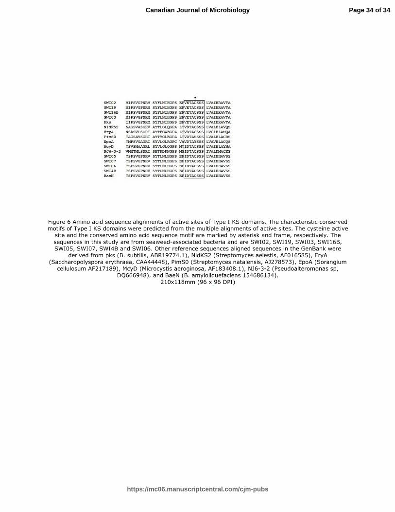
domains characterized the deduced amino acid sequences as Type I bacterial PKSs (Figure 5). The analysis of 290
deduced amino acid sequences demonstrated that the KS domain shared the conserved motif TACSSSLVA (Figure 291
6). 292
293
Figure 5 294
Page 12 of 34
https://mc06.manuscriptcentral.com/cjm-pubs
Canadian Journal of Microbiology

Draft
13
Figure 6 295
296
Discussion 297
Antimicrobial assay 298
Seaweeds were proposed to have chemical defense strategies against targeted microorganisms (Kubanek et 299
al. 2003), and the high proportion of antimicrobial isolates from seaweeds further validated this hypothesis (Lemos 300
et al. 1985; Burgess et al. 1999; Wiese et al. 2009). In the present study, 22 percent of cultured bacterial isolates 301
associated with seaweeds were found to have antimicrobial activity during the preliminary screening. About 9 302
percent of the isolates showed consistent results on further screenings, and these results were found to be similar to 303
those observed by other researchers (Burgess et al. 1999; Lemos et al. 1985). Earlier reports have demonstrated that 304
the proportion of bacteria with inhibitory activity associated with seaweeds and invertebrates was greater (11%) than 305
that of seawater and sediments (Zeng et al. 2005). Bacteria associated with live or inert surfaces were more likely to 306
display antibacterial properties (Gram et al. 2010). There were several reports demonstrating that compounds of the 307
associated bacterium help the host in certain ways to deter the pathogenic microbial flora, and that the bioactive 308
compounds isolated from host have structural similarities to the compounds of the microbial origin (Kubanek et al. 309
2003; Zhang et al. 2009). Bacterial origin of sponge-derived metabolites has been validated in earlier studies 310
(Schneemann et al. 2010; Quevrain et al. 2014). Epimanzamine D, which was originally isolated from a Palaun 311
sponge, and was produced by Streptomyces fulvorobeus strain HB113, and coproporphyrin, produced by the strain 312
HB100, was previously described as products of sponges (Schneemann et al. 2010). Similarly, the bacteria affiliated 313
to the genus Pseudoalteromonas were found to possess the similar spectrum of activity as that of C. clathrus that 314
was reported to produce the compounds monopalmitin and monostearin, which were previously isolated from the 315
whole sponge (Quevrain et al. 2014). The bacterial isolates with antibiotic activity in the present study were 316
pigmented, which further corroborate the observation of the earlier study that antibiotic-producing bacteria were 317
pigmented (Lemos et al. 1985). 318
319
Biochemical identification and 16S rRNA gene-based phylogeny 320
Page 13 of 34
https://mc06.manuscriptcentral.com/cjm-pubs
Canadian Journal of Microbiology

Draft
14
The results obtained by biochemical identification were further confirmed by 16S rRNA gene phylogeny. 321
The 16S rRNA gene sequence-based homology searches showed that most of the isolates of the present study were 322
closely related to each other (>99 percent similarity). However, every individual isolate was included in the study as 323
separate as they were found to be different in their inhibition and growth patterns. In the present survey of 324
antimicrobial bacteria associated with seaweeds, the representatives from two bacterial phyla, Firmicutes and 325
Proteobacteria were found. These results found harmony with earlier findings (Wiese et al. 2009; Kennedy et al. 326
2009). The bacteria isolated from brown seaweed Laminaria saccharina were found to be affiliated to the bacterial 327
domain of the Gram-positive Firmicutes, the Gram-negative Proteobacteria and Bacteroidetes. Representatives of 328
the Proteobacteria were found to be most abundant, the majority of which were affiliated with the γ-subgroup 329
(Wiese et al. 2009). The γ-proteobacterial phylum was demonstrated to be the most dominant cultivable group in a 330
recent study of sponge-associated bacteria from Haliclona simulans collected from the Irish waters (Kennedy et al. 331
2009). The predominant bacterial group found in H. simulans (Kennedy et al. 2008) and on the surface of J. rubens 332
(Ali et al. 2012) (about 73%) was Proteobacteria as determined by the total 16S rRNA gene library (77 percent of 333
clones). The γ-proteobacterial S. algae, however, would be less likely to be enriched by the selection process, and 334
are probably the dominant group of cultivable bacteria from the seaweeds. The entire set of isolates of Firmicutes 335
belonged to the genus Bacillus. A large number of Bacillus isolates found in this study might be due to the 336
selectivity of the media used in the present study as also supported by published literature (Zhang et al. 2009; 337
Kanagasabhapathy et al. 2006). Within the Firmicutes, especially strains belonging to the genus Bacillus are 338
common producers of antimicrobial compounds. Approximately 800 bacterial metabolites with antibiotic activity 339
have been isolated from Bacillus spp (Wiese et al. 2009). The Bacillus clade in the present study had representatives 340
of the species B. cereus, B. subtilis and B. amyloliquefaciens. The DNA similarity searches of the partial 16S rRNA 341
gene sequences of the bacterial strains isolated in the study with the GenBank database had shown that the strains 342
that were phylogenetically associated with B. amyloliquefaciens and B. subtilis have been clustered as one, and 343
therefore, difficult to distinguish. B. amyloliquefaciens and B. subtilis were reported to harbor several rRNA gene 344
clusters in which 16S rDNA sequence variation was found to exist (Hu et al. 2010). However, we could not 345
differentiate them with the sequence results of the present study, possibly because the regions amplified with our 346
primers do not belong to those clusters with sequence variation. 347
Page 14 of 34
https://mc06.manuscriptcentral.com/cjm-pubs
Canadian Journal of Microbiology

Draft
15
Antagonism assays among the isolated marine bacteria and their autoinhibition 348
None of the cells were found to be autoinhibitory. The absence of inhibitory activity of the producer strains 349
against other epiphytic producers indicated that the production of inhibitors could be of great importance in 350
microhabitats, such as an algal surface, where competition for an attachment site is surely a frequent event. In 351
general, a few producer strains inhibited the growth of the other producers, while the activity among the majority of 352
the strains was non-inhibitory. Only one strain, SWI 1 was found to be inhibited by all the producers tested. 353
Interestingly, on the other hand, in contradiction to the Lemos’s (1985) observation, a greater number of 354
antimicrobial relationships between producer strains isolated from the algae were not observed. Most of the bacterial 355
strains considered under the present study were not inhibitory to each other except SWI1. Detailed autoinhibition 356
and mutual inhibition patterns were shown in the Table 3. These results suggested that these beneficial populations 357
coexisted in the seaweed biofilm, and might significantly contribute in protecting the host seaweeds from the 358
deleterious pathogenic microbial populations or other colonizers. 359
360
Plasmid profiling, curing and antibacterial activity 361
The Shewanellae sp were found to possess a plasmid of molecular size greater than 10 kb, and among 362
different Bacillus strains, SWI1 (~>10 kb) and SWI8 (~>10 kb, ~>8 kb, ~>1 kb) showed the presence of plasmids. 363
V. alginolyticus isolated in the present study manifested the occurrence of two plasmids. However, no bands 364
appeared on the electrophorized gel after plasmid curing. The cultures retained their antimicrobial activities even 365
after plasmid curing, suggesting that the antimicrobial activities of the bacterial isolates, which have been considered 366
in the present study, was not encoded by plasmid, and the genes encoding the antimicrobial product might be present 367
within the genome. The antibiotic whose biosynthesis was determined by the SCPI plasmid of Streptomyces 368
coelicolor had been characterized as methylenomycin A (2-methylene-cyclopentan-3-one-4, 5-epoxy-4, 5-dimethyl-369
carboxylic acid) (Wright 1975). Plasmid linkage of bacteriocin activity was reported in an earlier literature 370
(Schillinger 1989). In contrast, chromosomally encoded class II bacteriocin LCI protein of Bacillus 371
amyloliquefaciens Bg-C31 was also reported (Hu et al. 2010). It is of note that several gene clusters aid in 372
biosynthesizing the bioactive peptides and polyketides by enzymes, for example, the non-ribosomal peptide 373
Page 15 of 34
https://mc06.manuscriptcentral.com/cjm-pubs
Canadian Journal of Microbiology

Draft
16
synthetase and polyketide synthetase. In addition to this, bacteriocins are ribosomally synthesized proteins that elicit 374
bactericidal activity. The genetic determinants for bacteriocins are either chromosomal or plasmid encoded as 375
demonstrated previously (Hu et al. 2010). The results obtained in the present study confirmed that the antibacterial 376
activities of the bacterial strains were not due to a plasmid-encoded bacteriocin. 377
378
Analysis of pks-I and nrps gene sequences 379
Polyketides and nonribosomal peptides became immensely important over the past few decades, and the 380
numbers of various novel polyketide and non-ribosomal peptide compounds have been found from marine-derived 381
microbes, most of which showed different biological activities and ecological functions (Zhou et al. 2011). 382
Polyketides, nonribosomal peptides, and PKS/NRPS hybrid compounds are important classes of natural products 383
and include many important drugs. Phycochemical studies showed the ability of algae to produce and store 384
polyketides as polycyclic ether macrolides and open chain polyketides. Although macrolides produced by the 385
terrestrial microorganisms have been used for long in human therapeutics, microlides from marine algae is a recent 386
addition (Cardozo et al. 2007). Compounds of polyketide origin, with potential bioactivities, have been isolated from 387
seaweeds, and were reported to have structural similarity to the known compounds of terrestrial cyanobacteria. It is 388
apparent that the seaweeds use targeted antimicrobial chemical defense strategies, and that the secondary 389
metabolites important in the ecological interactions between marine macroorganisms and microorganisms could be a 390
promising source of novel bioactive compounds, but this hypothesis has rarely been tested (Kubanek et al. 2003). In 391
support, it was found that the deduced amino acid sequence of Type III PKS (SbPKS) from a brown seaweed, 392
Sargassum binderi, shared a higher sequence similarity with bacterial PKSs (38% identity) than plant PKSs 393
(Baharum et al. 2011). This further strengthens the hypothesis of ecological interactions between the seaweed host 394
and their associated bacterial flora. 395
In the present report, 23 antimicrobial isolates with broad spectrum activities against aquaculture pathogens 396
were screened for the presence of secondary metabolite genes, and among those only 8 were able to amplify the 397
desired genes. These results provided us with the basic information on the presence of metabolite genes, but the 398
correlation to specific metabolites was found to be limited due to the lengths of the PCR fragments (Schneemann et 399
Page 16 of 34
https://mc06.manuscriptcentral.com/cjm-pubs
Canadian Journal of Microbiology

Draft
17
al. 2010). The possible absence of the screened amplicons might be due to the chosen primer system, albeit 400
favorable for the great majority of known metabolite genes, are not the working polyketides with unusual molecular 401
constructions (Schirmer et al. 2005; Schneemann et al. 2010) or the coding genes for the antimicrobial products of 402
the screened strains might be different from that of the pks or nrps genes. Our results ratified the statement that there 403
were examples of strains possessing the functional genes with no inhibitory activity and vice versa (Zhao et al. 404
2011). Further, the existence of cultured isolates with the potential to synthesize bioactive compounds, and without a 405
metabolite gene amplified product showed that culturing remains a powerful tool for exploring the bioactive 406
metabolites of bacterial origin (Penesyan et al. 2009). Even though the cultivation-based studies possess some 407
limitations, it remains essential as it provides opportunities to study and understand the microbial ecology, 408
physiology, and to design antibiotic screening assays (Ali et al. 2012). Further, the absence of screened metabolite 409
gene products in the tested active strains demonstrated the possibility of other biosynthetic genes. We, therefore, 410
suggest that the biosynthetic gene-guided screening of bioactive bacterial population also needs to consider the 411
conserved gene sequences of other biosynthetic pathways (Zhu et al. 2009). 412
A phylogeny based on the KS domain sequences from other well-described organisms can be employed to 413
determine the structural similarity of the obtained KS domain sequences (Zhu et al. 2009). The deduced amino acid 414
sequences obtained in the study were aligned with the relative sequences of the GenBank (Figure 3). The 415
phylogenetic study showed that amplified gene products of the present study were of bacterial Type I PKSs. Earlier 416
studies in sponge-associated bacteria too found that the bacterial strains harbor Type I bacterial PKSs (Zhu et al. 417
2009; Zhang et al. 2009). In the present study, KS domain sequence-based phylogeny further clustered four 418
sequences with B. subtilis, and the remaining four with B. amyloliquefaciens. Hence, the KS domain sequences 419
enabled us to differentiate B. subtilis from the B. amyloliquefaciens strains, which we could not do through 16S 420
rRNA gene-based phylogenetic approach. B. cereus strain isolated in the present study was unable to amplify the 421
metabolite gene product. Multiple sequence alignment of the sequenced data with the known sequences from the 422
GenBank further enabled to identify the conserved sequence motif TACSSSLVA. The KS domain conserved 423
residues from the sponge, Hymeniacidon perleve associated bacteria was reported as VDTACSSSLVA (Zhu et al. 424
2009). In our study, this sequence motif amino acids showed some variations at certain specific locations. In B. 425
subtilis, the acidic amino acid, asparte, located at the third position from the cysteine active site in the N-terminal 426
Page 17 of 34
https://mc06.manuscriptcentral.com/cjm-pubs
Canadian Journal of Microbiology

Draft
18
has been replaced with glutamate. Likewise, in B. amyloliquefaciens the amino acid, valine, located at the fourth
427
position from the cysteine active site in the N-terminal was replaced by isoleucine. However, due to the structural 428
similarities of acidic amino acids, aspartate (2-aminosuccinic acid) with glutamate (2-aminopentanedioic acid), and 429
isoleucine (2-amino-3-methylpentanoic acid) with valine (2-amino-3-methylbutanoic acid), it is apparent that the KS 430
domains of the bioactive Bacillus strains in the present study shared a common catalytic mode of action. Due to their 431
versatile assemblage mechanism, polyketides exhibit remarkable diversity both in terms of structure and biological 432
activities. To date, it is estimated that only a small fraction of the antimicrobial molecules potentially produced by 433
the Gram-positive bacteria has been identified. The recent advances in genome sequencing highlighted the genus 434
Bacillus as a potentially important source of antibiotic-like compounds (Fickers 2010). 435
In summary, the seaweeds possess greater potential as potential sources for screening bioactive bacterial 436
isolates that facilitate novel natural product discovery from the marine environment. These epibionts might be 437
beneficial to the seaweeds by limiting or preventing the development of competing, pathogenic and fouling bacteria. 438
Our results further suggest that the antimicrobial activity cannot be solely assessed by metagenomic studies as some 439
strains may escape the amplification of the desired genes. In that case, the antimicrobial potential may only be 440
assessed by screening the inhibition potential of the desired indicator organisms. Further, we recommend metabolite 441
gene-based screening of bioactive organisms should also exploit biosynthetic gene clusters of biosynthetic pathways 442
other than PKSs and NRPSs. 443
Acknowledgements 444
The Authors thank the Ministry of Earth Science, New Delhi for funding under the project “Drugs from the 445
sea” (grant number MoES-2/DS/6/2007 PC-IV) and Indian Council of Agricultural Research, New Delhi for 446
providing the necessary facilities. The authors thank the Director, Central Marine Fisheries Research Institute for his 447
guidance and support. Thanks are due to the Head, Marine Biotechnology Division, Central Marine Fisheries 448
Research Institute for facilitating the research activity. B.T. acknowledges ICAR Outreach Activity-3 for a 449
fellowship. 450
451
Conflict of interest: The authors declare that there is no conflict of interest. 452
Page 18 of 34
https://mc06.manuscriptcentral.com/cjm-pubs
Canadian Journal of Microbiology

Draft
19
453
References 454
Ali, A.I.B., Bour, M.E., Ktari, L., Bolhuis, H, Ahmed, M., Boudabbous, A., and Stal, L.J. 2012. Jania rubens-455
associated bacteria: molecular identification and antimicrobial activity. J. Appl. Phycol. 24(3): 525–534. 456
doi: 10.1007/s10811-011-9758-0. 457
Ayuso-Sacido, A., and Genilloud, O. 2005. New PCR primers for the screening of NRPS and PKS-I systems in 458
actinomycetes: detection and distribution of these biosynthetic gene sequences in major taxonomic groups. 459
Microb. Ecol. 49(1): 10–24. doi: 10.1007/s00248-004-0249-6. PMID: 15614464. 460
Baharum, H., Schlinglo, A., Morita, H., Omitsuka, A., Lee, F.C., Kim-Yong, N.G., Rahim, R.A., Abe, I., and Ho, 461
C.L. 2011. Molecular cloning, modeling, and site-directed mutagenesis of Type III polyketide synthase 462
from Sargassum binderi (Phaeophyta). Mar. Biotechnol. 13(5): 845–856. doi: 10.1007/s10126-010-9344-5. 463
PMID: 21181422. 464
Boyd, K.G., Adams, D.R., and Burgess, J.G. 2009. Antibacterial and repellent activities of marine bacteria 465
associated with algal surfaces. Biofouling. 14(3): 227-236. doi: 10.1080/08927019909378414. 466
Burgess, J.G., Jordan, E.M., Bregu, M., Mearns-Spragg, A., and Boyd, K.G. 1999. Microbial antagonism: a 467
neglected avenue of natural products research. J. Biotechnol. 70(1-3): 27–32. doi: 10.1016/S0079-468
6352(99)80094-0. PMID: 10412203. 469
Cardozo, K.H., Guaratini, T., Barros, M.P., Falcao, V.R., Tonon, A.P., Lopes, N.P., Campos, S., Torres, M.A., 470
Souza, A.O., Colepicolo, P., and Pinto, E. 2007. Metabolites from algae with economical impact. Comp. 471
Biochem. Physiol. C. Toxicol. Pharmacol. 146(1-2): 60-78. doi: 10.3390/s140917725. PMID: 16901759. 472
Chakraborty, K., Thilakan, B., and Raola, V.K. 2014. Polyketide family of novel antibacterial 7-O-methyl-5'-473
hydroxy-3'-heptenoate macrolactin from seaweed associated Bacillus subtilis MTCC 10403. J. Agric. Food 474
Chem. 62: 12194-12208. doi: 10.1021/jf504845m. PMID: 25420039. 475
Devi, R., Surendran, P.K., and Chakraborty, K. 2009. Antibiotic resistance and plasmid profiling of Vibrio 476
parahaemolyticus isolated from shrimp farms along the southwest coast of India. World. J. Microbiol. 477
Biotechnol. 25: 2005–2012. doi: 10.1007/s11274-009-0101-8. 478
Page 19 of 34
https://mc06.manuscriptcentral.com/cjm-pubs
Canadian Journal of Microbiology

Draft
20
Fickers, P. 2012. Antibiotic compounds from Bacillus: why are they so amazing? Am. J. Biochem. Biotechnol. 8 479
(1): 40-46. doi: 10.3844/ajbbsp.2012.40.46. 480
Goecke, F., Labes, A., Wiese, J., and Imhoff, J.F. 2010. Chemical interactions between marine macroalgae and 481
bacteria. Mar. Ecol. Prog. Ser. 409: 267–300. doi: 10.3354/meps08607. 482
Gram, L., Melchiorsen, J., and Bruhn, J.B. 2010. Antibacterial activity of marine culturable bacteria collected from a 483
global sampling of ocean surface waters and surface swabs of marine organisms. Mar. Biotechnol. 12(4): 484
439-451.doi: 10.1007/s10126-009-9233-y. PMID: 19823914. 485
Hu, H.Q., Li, X.S., and He, H. 2010. Characterization of an antimicrobial material from a newly isolated Bacillus 486
amyloliquefaciens from mangrove for biocontrol of capsicum bacterial wilt. Biol. Control. 54(3): 359 -365. 487
doi: 10.1016/j.biocontrol.2010.06.015. 488
Hutchinson, C.R. 2003. Polyketide and non-ribosomal peptide synthases: falling together by coming apart. Proc. 489
Natl. Acad. Sci. USA. 100(6): 3010–3012. doi: 10.1073/pnas.0730689100. PMID: 12631695. 490
Imhoff, J.F., Labes, A., and Wiese, J. 2011. Bio-mining the microbial treasures of the ocean: new natural products. 491
Biotechnol. Adv. 29(5): 468-82. doi: 10.1016/j.biotechadv.2011.03.001. PMID: 21419836. 492
Kanagasabhapathy, M., Hideaki, S., Haldar, S., Shinji, Y., and Shinichi, N. 2006. Antibacterial activities of marine 493
epibiotic bacteria isolated from brown algae of Japan. Ann. Microbio. 56(2): 167-173. doi: 494
10.1007/BF03175000. 495
Kennedy, J., Baker, P., Piper, C., Cotter, P.D., Walsh, M., Mooij, M.J., Bourke, M.B., Rea, M.C., Connor, P.M.O., 496
Paul Ross, R., Hill, C., and Gara, F.O. 2009. Isolation and analysis of bacteria with antimicrobial activities 497
from the marine sponge Haliclona simulans collected from Irish waters. Mar. Biotechnol. 11(3): 384-96. 498
doi: 10.1007/s10126-008-9154-1. PMID: 18953608. 499
Kennedy, J., Codling, C.E., Jones, B.V., Dobson, A.D.W., and Marchesi, J. 2008. Diversity of microbes associated 500
with the marine sponge, Haliclona simulans, isolated from Irish waters and identification of polyketide 501
synthase genes from the sponge metagenome. Environ. Microbiol. 10(7): 1888-902. doi: 10.1111/j.1462-502
2920.2008.01614.x. PMID: 18430018. 503
Kimura, M. 1980. A simple method for estimating evolutionary rate of base substitutions through comparative 504
studies of nucleotide sequences. J. Mol. Evol. 16(2): 111-120. doi: 10.1007/BF01731581 PMID: 7463489. 505
Page 20 of 34
https://mc06.manuscriptcentral.com/cjm-pubs
Canadian Journal of Microbiology

Draft
21
Krieg, N.R., and Holt, J.G. 1984. Bergey’s manual of systematic bacteriology. Williams and Wilkins, Baltimore. 506
Kubanek, J., Jensen, P.R., Keifer, P.A., Sullards, M.C., Collins, D.O., and Fenical, W. 2003. Seaweed resistance to 507
microbial attack: a targeted chemical defense against marine fungi. Proc. Natl. Acad. Sci. USA. 100(12): 508
6916–6921. doi: 10.1073/pnas.1131855100. PMCID: PMC165804. 509
Lemos, M.L., Toranzo, A.E., and Barja, J.L. 1985. Antibiotic activity of epiphytic bacteria isolated from intertidal 510
seaweeds. Microb. Ecol. 11(2): 149-63. doi: 10.1007/BF02010487. PMID: 24221303. 511
Li, Z. 2009. Advances in marine microbial symbionts in the China Sea and related pharmaceutical metabolites. Mar. 512
Drugs 7(2): 113-129. doi: 10.3390/md7020113. PMCID: PMC2707038. 513
Meklat, A., Sabaou, N., Zitouni, A., Mathieu, F., and Lebrihi, A. 2011. Isolation, taxonomy, and antagonistic 514
properties of halophilic actinomycetes in Saharan soils of Algeria. Appl. Environ. Microbiol. 77(18): 6710-515
4. doi: 10.1128/AEM.00326-11. PMID: 21764956. 516
Penesyan, A., Marshall-Jones, Z., Holmstrom, C., Kjelleberg, S., and Egan, S. 2009. Antimicrobial activity observed 517
among cultured marine epiphytic bacteria reflects their potential as a source of new drugs. FEMS 518
Microbiol. Ecol. 69(1): 113-24. doi: 10.1111/j.1574-6941.2009.00688.x. PMID: 19453738. 519
Sambrook, J., and Russell, D.W. 2001. Molecular cloning: a laboratory manual. Cold Spring Harbor Laboratory 520
Press, New York, 2100 pp. 521
Sambrook, J., Fritsch, E.F., and Maniatis, T.1989. Molecular cloning: a laboratory manual, 2nd edn. Cold Spring 522
Harbor Laboratory Press, Cold Spring Harbor, New York, 1626 pp. 523
Schillinger, U., and Lücke, F.K. 1989. Antibacterial Activity of Lactobacillus sake isolated from Meat. Appl. 524
Environ. Microbiol. 55(8): 1901-1906. PMCID: PMC202976, PMID: 2782870. 525
Schirmer, A., Sabaou, N., Gadkari, R., Reeves, C.D., Ibrahim, F., Delong, E.F., and Hutchinson, C.R. 2005. 526
Metgenomic analysis reveals diverse polyketide synthase gene clusters in microorganisms associated with 527
the marine sponge Discodermia dissoluta. Appl. Environ. Microbiol. 71(8): 4840-4849. doi: 528
10.1128/AEM.71.8.4840-4849.2005. PMID: 16085882. 529
Schneemann, I., Nagel, K., Kajahn, I., Labes, A., Wiese, J., and Imhoff, J.F. 2010. Comprehensive investigation of 530
marine Actinobacteria associated with the sponge Halichondria panacea. Appl. Environ. Microbiol. 531
76(11): 3702-3714. doi:10.1128/AEM.00780-10. PMCID: PMC2876447. 532
Page 21 of 34
https://mc06.manuscriptcentral.com/cjm-pubs
Canadian Journal of Microbiology
Draft
22
Tamura, K., Peterson, D., Peterson, N., Stecher, G., Nei, M., and Kumar, S. 2011. MEGA5: Molecular evolutionary 533
genetics analysis using maximum likelihood, evolutionary distance, and maximum parsimony methods. 534
Mol. Biol. Evol. 28: 2731-2739. doi: 10.1093/molbev/msr121. PMID: 21546353. 535
Whelan, S., and Goldman, N. 2001. A general empirical model of protein evolution derived from multiple protein 536
families using a maximum-likelihood approach. Mol. Biol. Evol. 18(5): 691-699. doi. 537
10.1093/oxfordjournals.molbeva003851. PMID: 11319253. 538
Wiese, J., Thiel, V., Nagel, K., Staufenberger, T., and Imhoff, F.J. 2009. Diversity of antibiotic-active bacteria 539
associated with the brown alga Laminaria saccharina from the Baltic Sea. Mar. Biotechnol. 11(2): 287-540
300. doi: 10.1007/s10126-008-9143-4. PMID: 18855068. 541
Weisburg, W.G., Barns, S.M., Pelletier, D.A., and Lane, D.J. 1991. 16S ribosomal DNA amplification for 542
phylogenetic study. J. Bacteriol. 173(2): 697-703. PMID: 1987160. 543
Wilson, G.S., Raftos, D.A., Corrigan, S.L., and Nair, S.V. 2010. Diversity and antimicrobial activities of surface-544
attached marine bacteria from Sydney Harbour, Australia. Microbiol. Res. 165(4):300-11. doi: 545
10.1016/j.micres.2009.05.007. PMID: 19656668. 546
Wright, L.F., and Hopwood, D. A.1976. Identification of the Antibiotic Determined by the SCPl Plasmid of 547
Streptomyces coekolor A3(2). J. Gen. Microbiol. 95: 96-106. doi: 10.1099/00221287-95-1-96. 548
PMID:822125. 549
Zeng, L., Han, X., Chen, H., Lin, W., and Yan, X. 2005. Marine bacteria associated with marine macro organisms: 550
the potential resource for antimicrobial agents. Ann. Microbiol. 55(2): 119-124. 551
Zhang, W., Li Z.F., Miao, X., Meng, Q., and Zang, X. 2009. Investigation of bacteria with polyketide synthase 552
genes and antimicrobial activity isolated from South China Sea sponges. J. Appl. Microbiol. 107(2):567-75. 553
doi: 10.1111/j.1365-2672.2009.04241.x. PMID: 19302490. 554
Zhao, K., Zheng, Y., Penttinen, P.,Guan, T., Xiao, J., Chen, Q., Xu, J., Lindstrom, K., Zhang, L., Zhang, X., and 555
Strobel, G.A. 2011. The diversity and antimicrobial activity of endophytic actinomycetes from medicinal 556
plants in Panxi Plateau, China. Curr. Microbiol. 62(1):182-190. doi: 10.1007/s00284-010-9685-3. PMID: 557
20567975. 558
Page 22 of 34
https://mc06.manuscriptcentral.com/cjm-pubs
Canadian Journal of Microbiology
Draft
23
Zhu, P., Zheng, Y., You, Y.,Yan, X., and Shao, J. 2009. Molecular phylogeny and modular structure of hybrid 559
NRPS/PKS gene fragment of Pseudoalteromonas sp.NJ6-3-2 isolated from marine sponge Hymeniacidon 560
perleve. J. Microbiol. Biotechnol. 19 (3): 229-237. doi: 10.4014/jmb.0804.282.PMID:19349747. 561
Zhao, J., Yang, N., and Zeng, R. 2008. Phylogenetic analysis of type I polyketide synthase and nonribosomal 562
peptide synthetase genes in Antarctic sediment. Extremophiles 12(1):97-105.doi. 10.1007/s13131-011-563
0167-7. PMID: 17726573 564
Zhou, K., Zhang, X., Zhang, F., and Li Z. 2011. Phylogenetically diverse cultivable fungal community and 565
polyketide synthase (PKS), non-ribosomal peptide synthase (NRPS) genes associated with the South China 566
Sea sponges. Microb. Ecol. 62(3):644-654. doi: 10.1007/s00248-011-9859-y. PMID: 21519913 567
568
Page 23 of 34
https://mc06.manuscriptcentral.com/cjm-pubs
Canadian Journal of Microbiology

Draft
24
Table 1. Polymerase chain reaction (PCR) primersa used in this study 569
Primer Targetb Sequence (5’-3’) References
fD1 16SrRNA AGAGTTT GATCCTGGCTCAG Weisburg et al, 1991
rP2 16SrRNA ACGGCTACCTTGTTACGACTT Weisburg et al, 1991
GBF PKS RTRGAYCCNCAGCAICG Zang et al, 2009
GBR PKS VGTNCCNGTGCCRTG Zang et al, 2009
GCF PKS GCSATGGAYCCSCARCARCGSVT Schirmer et al, 2005
GCR PKS GTSCCSGTSCRTGSSCYTCSAC Schirmer et al, 2005
KSDPOOF PKS MGNGARGARGCNNWNSMNATGGAYCCNCARCANMG Zang et al, 2009
KSHGTGr PKS GGRTCNCCNARNSWNGTNCCNGTNCCRTG Zang et al, 2009
KS11f PKS GCIATGGAYCCICARCARMGIVT Schirmer et al, 2005
KS12r PKS GTICCIGTICCRTGISCYTCIAC Schirmer et al, 2005
MTF NRPS GCNGGYGGYGCNTAYGTNCC Zhao et al, 2008
MTR NRPS CCNCGDATYTTNACYTG Zhao et al, 2008
570
a The primer sequences used for the PCR reaction have been represented. PCR was performed in a total volume of 25 571
µL containing 1x reaction buffer with MgCl2 (Sigma), 0.25 mM of each dNTP (Fermentas), 0.5 mM of each primers 572
(Sigma), 1 ng DNA and 0.3 U Taq DNA polymerase (Sigma). 573
574
b Phylogenetic analysis was carried out using 16S rRNA sequencing. Different sets of degenerate primers targeting 575
genes encoding pks-I and nrps were used as described in the text. All of the amplification products were examined by 576
agarose gel electrophoresis, and bands of 700 to 800 bp and 1000 to 1,400 bp were classified as products of pks-I and 577
nrps genes, respectively. 578
579
580
Page 24 of 34
https://mc06.manuscriptcentral.com/cjm-pubs
Canadian Journal of Microbiology

Draft
25
Table 2. Antimicrobial activity of the seaweed-associated bacteria against pathogenic organisms and their screening for secondary metabolite genes 1
(pks and nrps) responsible for bioactivity 2
Strains Seaweed host Seaweed-associated
strains
Antimicrobial activity (mm) against test pathogens Presence of
indicated
gene#
V.
alg
ino
lyti
cus
V.p
ara
ha
emo
lyti
cus
17
802
V.p
ara
hem
oly
ticu
s
45
1
V.v
uln
ific
us
MT
CC
11
45
V.
an
gu
lla
rum
A.
hyd
roph
illa
V.
ha
rvey
i
pks nrps
SWI 1 Anthophycus longifolium B. cereus ++ ++ - ++ - +++ - N N
SWI 2 Anthophycus longifolium B. subtilis +++ +++ ++ +++ ++ +++ ++ Pa
N
SWI 3 Padina gymnospora B. subtilis +++ +++ +++ +++ ++ +++ ++ Pb
N
SWI 4A Laurencia papillosa B. subtilis ++ ++ +++ +++ ++ +++ ++ N N
SWI 4B Laurencia papillosa B. amyloliquefacens - ++ ++ +++ - ++ ++ Pc
N
SWI 5 Turbinaria ornata B. amyloliquefacens +++ +++ +++ +++ ++ +++ +++ Pd
N
SWI 6 Hypnea valentiae B. amyloliquefacens ++ ++ +++ ++ ++ +++ ++ Pe
N
SWI 7 Padina gymnospora B. amyloliquefacens ++ +++ +++ +++ ++ +++ ++ Pf
N
SWI 8 Laurencia papillosa B. subtilis - - ++ ++ - - - N N
SWI 9 Hypnea valentiae S. algae - +++ ++ - - - +++ N N
SWI 10 Laurencia papillosa S. algae - +++ ++ - - - - N N
SWI 11 Padina gymnospora S. algae - +++ ++ - - - +++ N N
SWI 12 Hypnea valentiae S. algae - +++ - - - - ++ N N
SWI 12B Hypnea valentiae V. alginolyticus - - - - - - ++ N N
SWI 13 Padina gymnospora S. algae - ++ + - - - +++ N N
SWI 14 Anthophycus longifolium
S. algae - +++ + - - - +++ N N
SWI 16A Anthophycus longifolium B. subtilis - ++ - ++ - - - N N
SWI 16B Anthophycus longifolium B. subtilis ++ +++ ++ +++ ++ +++ ++ Pg
N
SWI 17 Dictyota dichotoma S. algae - +++ + +++ - - +++ N N
SWI 18 Dictyota dichotoma S. algae - +++ + +++ - - - N N
SWI19 Sargassum myriocystum B. subtilis + +++ ++ - ++ +++ +++ Ph
N
SWI20 Laurencia papillosa S. algae - +++ - - - - - N N
SWI21 Laurencia papillosa P. putida - - - +++ - - ++ N N
# A total of 23 antimicrobial isolates with broad spectrum activities against aquaculture pathogens were screened for the presence of secondary 3
metabolite genes, and among those only 8 were able to amplify the desired genes. 4
GenBank No for the sequenced amplicons a KC589397;
b KC607823;
c KC607821;
d KC589396;
e KC607822;
f KC589396;
g KC589400;
h KC589398 5
(-) signifies no inhibition; (+) signifies the inhibition zone as less than 10 mm; (++) inhibition zone 10-15 mm; (+++) inhibition zone >15mm 6
(P) signifies positive and (N) as negative. 7
Page 25 of 34
https://mc06.manuscriptcentral.com/cjm-pubs
Canadian Journal of Microbiology

Draft
26
Table 3. Autoinhibition and mutual inhibition pattern (expressed in mm) of antimicrobial isolates associated with the 1
seaweeds 2
SWI1 SWI2 SWI3 SWI4A SWI4B SWI5 SWI6 SWI7 SWI8
SWI1 ND 10.3 (1.5) 10.3 (1.53) 12.7 (0.58) ND 9.0 (1.00) 9.33 (1.15) 10.0 (1.0) ND
SWI2 3.0 (2.0) ND ND ND ND ND ND ND ND
SWI3 3.7 (1.5) ND ND ND ND ND ND ND ND
SWI4A 2.3 (0.6) ND ND ND ND ND ND ND ND
SWI4B ND ND ND ND ND ND ND ND ND
SWI5 ND ND ND ND ND ND ND ND ND
SWI6 ND ND ND ND ND ND ND ND ND
SWI7 ND ND ND ND ND ND ND ND 3.33 (0.58)
SWI8 0.7 (0.6) 2.67 (0.51) 10.7 (1.15) 9.67 (0.58) 11.3 (1.15) 10.3 (0.58) 10.7 (1.15) 2.7 (0.60) ND
SWI9 ND ND ND ND ND ND ND ND ND
SWI10 ND ND ND ND ND ND ND ND ND
SWI11 ND ND ND ND ND ND ND ND ND
SWI12 ND ND ND ND ND ND ND ND ND
SWI12B ND ND ND ND ND ND ND 9.3 (1.20) ND
SWI13 1.0 (1.7) ND 9.33 (0.58) 11.3 (1.15) 14.0 (1.0) 11 (1.0) ND 2.0 (0.00) ND
SWI14 0.7 (1.2) 2.33 (0.19) 1.33 (0.58) 1.67 (0.58) ND ND ND 9.7 (0.60) ND
SWI16A 0.7 (1.2) 9.33 (0.19) 11.7 (1.53) 11.0 (1.00) 13.0 (1.0) 11.7 (0.58) ND 12.0 (0.60) 2.67 (0.58)
SWI16B ND ND ND ND ND ND ND ND ND
SWI17 ND ND ND ND ND ND ND ND ND
SWI18 10 (1.5) 12.0 (1.00) 14.3 (0.58) 12 (1.0) 11.7 (1.53) 17.7 (0.58) 15 (1.0) ND ND
SWI19 ND ND ND ND ND ND ND ND ND
SWI20 ND ND 1.67 (0.58) 2.33 (0.58) 1.33 (0.58) ND ND ND ND
SWI21 ND ND ND ND ND ND ND ND ND
3
ND: No autoinhibition recorded 4
Figures in parentheses indicate the mutual inhibition pattern 5
6
7
8
9
10
11
12
13
14
Page 26 of 34
https://mc06.manuscriptcentral.com/cjm-pubs
Canadian Journal of Microbiology

Draft
27
Figure captions 15
Figure 1 Indicative photograph showing the inhibitory zones exhibited by the seaweed-associated 16
antimicrobial bacterial flora during antibacterial screening against the listed pathogens. (1) 17
Seaweed material A. longifolium used for isolation, (2) Antibacterial activities of seaweed-18
associated bacterium SWI 2 against V. vulnificus MTCC1145, (3) Gram staining 19
photomicrograph of SWI2 (B. subtilis), (4) Antibacterial activities of seaweed-associated 20
bacterium SWI 14 against V. parahaemolyticus 17802, whereas the bactericidal zones were 21
yellow, whereas live cells were blue in color, (5) Gram staining photomicrograph of SWI 14 (S. 22
algae). The clearance zones realized by the isolates signify the antibacterial activity. 23
Antimicrobial activity was recorded as the diameter of inhibition zones determined as a 24
distance of ≥1 mm between the circular area (=lawn of the isolate) and the end of the clear zone 25
bounded by the lawn of the test strain. 26
Figure 2 Pie diagrams showing the (A) distribution of seaweed-associated active isolates contributed by the 27
representative seaweed hosts screened under the study, (B) contribution (as percent share towards 28
the total number of bacterial isolates with potentially greater antibacterial properties against the 29
pathogens) of the individual representative bacterium as seaweed association. 30
Figure 3 Phylogenetic tree derived from nearly complete 16S rRNA gene sequences, showing relationships 31
between the antimicrobial bacterial isolates associated with seaweeds and their phylogenetic 32
neighbors. The evolutionary history was inferred by using the Maximum Likelihood method based 33
on the Kimura 2-parameter model. Evolutionary analyses were conducted in MEGA5. 34
Figure 4 Plasmid profiles of the antimicrobial isolates before and after curing. (A) Plasmid profiles of Gram 35
negative isolates of the study 4th
lane (down) V. alginolyticus, M (Molecular marker) (B) Plasmid 36
profiles of Bacillus strain Lane 1 (SWI1), 12th
Lane (SWI8), M (Molecular marker) (C) Plasmid 37
profiles of the strains after curing, M (Molecular marker). Molecular marker used is Gene 38
RulerTM
1kb DNA Ladder (Thermo scientific, 250bp-10,000bp). 39
Figure 5 Molecular phylogeny analysis of ketosynthase regions with respect to the diverse range of 40
ketosynthase domains including. Type I, II and III. The evolutionary history was inferred by using 41
Page 27 of 34
https://mc06.manuscriptcentral.com/cjm-pubs
Canadian Journal of Microbiology

Draft
28
the Maximum Likelihood method based on the Whelan and Goldman model. A discrete gamma 42
distribution was used to model evolutionary rate differences among sites. The tree with the highest 43
log likelihood is shown. The sequences in the experiment are preceded by circles. 44
Figure 6 Amino acid sequence alignments of active sites of Type I KS domains. The characteristic 45
conserved motifs of Type I KS domains were predicted from the multiple alignments of active 46
sites. The cysteine active site and the conserved amino acid sequence motif are marked by asterisk 47
and frame, respectively. The sequences in this study are from seaweed-associated bacteria and are 48
SWI02, SWI19, SWI03, SWI16B, SWI05, SWI07, SWI4B and SWI06. Other reference sequences 49
aligned sequences in the GenBank were derived from pks (B. subtilis, ABR19774.1), NidKS2 50
(Streptomyces aelestis, AF016585), EryA (Saccharopolyspora erythraea, CAA44448), PimS0 51
(Streptomyces natalensis, AJ278573), EpoA (Sorangium cellulosum AF217189), McyD 52
(Microcystis aeroginosa, AF183408.1), NJ6-3-2 (Pseudoalteromonas sp, DQ666948), and BaeN 53
(B. amyloliquefaciens 154686134). 54
55
56
57
58
Page 28 of 34
https://mc06.manuscriptcentral.com/cjm-pubs
Canadian Journal of Microbiology

Draft
Figure 1 Indicative photograph showing the inhibitory zones exhibited by the seaweed-associated antimicrobial bacterial flora during antibacterial screening against the listed pathogens. (1) Seaweed
material A. longifolium used for isolation, (2) Antibacterial activities of seaweed-associated bacterium SWI 2
against V. vulnificus MTCC1145, (3) Gram staining photomicrograph of SWI2 (B. subtilis), (4) Antibacterial activities of seaweed-associated bacterium SWI 14 against V. parahaemolyticus 17802, whereas the
bactericidal zones were yellow, whereas live cells were blue in color, (5) Gram staining photomicrograph of SWI 14 (S. algae). The clearance zones realized by the isolates signify the antibacterial activity.
Antimicrobial activity was recorded as the diameter of inhibition zones determined as a distance of ≥1 mm between the circular area (=lawn of the isolate) and the end of the clear zone bounded by the lawn of the
test strain. 308x231mm (300 x 300 DPI)
Page 29 of 34
https://mc06.manuscriptcentral.com/cjm-pubs
Canadian Journal of Microbiology

Draft
Figure 2 Pie diagrams showing the (A) distribution of seaweed-associated active isolates contributed by the representative seaweed hosts screened under the study, (B) contribution (as percent share towards the total
number of bacterial isolates with potentially greater antibacterial properties against the pathogens) of the individual representative bacterium as seaweed association.
32x33mm (600 x 600 DPI)
Page 30 of 34
https://mc06.manuscriptcentral.com/cjm-pubs
Canadian Journal of Microbiology

Draft
Figure 3 Phylogenetic tree derived from nearly complete 16S rRNA gene sequences, showing relationships between the antimicrobial bacterial isolates associated with seaweeds and their phylogenetic neighbors. The
evolutionary history was inferred by using the Maximum Likelihood method based on the Kimura 2-
parameter model. Evolutionary analyses were conducted in MEGA5. 274x243mm (96 x 96 DPI)
Page 31 of 34
https://mc06.manuscriptcentral.com/cjm-pubs
Canadian Journal of Microbiology

Draft
Figure 4 Plasmid profiles of the antimicrobial isolates before and after curing. (A) Plasmid profiles of Gram negative isolates of the study 4th lane (down) V. alginolyticus, M(Molecular marker) (B) Plasmid profiles of Bacillus strain Lane 1 (SWI1), 12th Lane (SWI8), M (Molecular marker) (C) Plasmid profiles of the strains
after curing, M (Molecular marker). Molecular marker used is Gene RulerTM1kb DNA Ladder (Thermo scientific, 250bp-10,000bp). 20x15mm (600 x 600 DPI)
Page 32 of 34
https://mc06.manuscriptcentral.com/cjm-pubs
Canadian Journal of Microbiology

Draft
Figure 5 Molecular phylogeny analysis of ketosynthase regions with respect to the diverse range of ketosynthase domains including. Type I, II and III. The evolutionary history was inferred by using the
Maximum Likelihood method based on the Whelan and Goldman model. A discrete gamma distribution was used to model evolutionary rate differences among sites. The tree with the highest log likelihood is shown.
The sequences in the experiment are preceded by circles. 325x241mm (72 x 72 DPI)
Page 33 of 34
https://mc06.manuscriptcentral.com/cjm-pubs
Canadian Journal of Microbiology
Draft
Figure 6 Amino acid sequence alignments of active sites of Type I KS domains. The characteristic conserved motifs of Type I KS domains were predicted from the multiple alignments of active sites. The cysteine active
site and the conserved amino acid sequence motif are marked by asterisk and frame, respectively. The
sequences in this study are from seaweed-associated bacteria and are SWI02, SWI19, SWI03, SWI16B, SWI05, SWI07, SWI4B and SWI06. Other reference sequences aligned sequences in the GenBank were
derived from pks (B. subtilis, ABR19774.1), NidKS2 (Streptomyces aelestis, AF016585), EryA (Saccharopolyspora erythraea, CAA44448), PimS0 (Streptomyces natalensis, AJ278573), EpoA (Sorangium
cellulosum AF217189), McyD (Microcystis aeroginosa, AF183408.1), NJ6-3-2 (Pseudoalteromonas sp, DQ666948), and BaeN (B. amyloliquefaciens 154686134).
210x118mm (96 x 96 DPI)
Page 34 of 34
https://mc06.manuscriptcentral.com/cjm-pubs
Canadian Journal of Microbiology