Embed Size (px)
Citation preview

Antimicrobial potentialities of mushroom-based selenium biocomposites
O. M. Tsivileva*,1, A. I. Perfileva2,T. P. Nguyen3, A. M. Zakharevich4, and P. A. Poluboyarinov5 1 Laboratory of Microbiology, Institute of Biochemistry and Physiology of Plants and Microorganisms, Russian Academy
of Sciences, 13 Prospekt Entuziastov,410049 Saratov, Russia 2 Laboratory of Plant-Microbial Interactions, Siberian Institute of Plant Physiology and Biochemistry, Siberian Branch,
Russian Academy of Sciences, 132 Lermontov st., 664033 Irkutsk, Russia 3 Department of Botany, Southern Institute of Ecology, Vietnam Academy of Science and Technology, Add: 01 Mac Dinh
Chi, Dist 1, Hochiminh City, Vietnam 4 Educational Research Institute of Nanostructures and Biosystems, N.G. Chernyshevskii Saratov State National Research
University, 83 Astrakhanskaya st., 410012 Saratov, Russia 5Institute of Engineer Ecology, State University of Architecture and Construction, 28 Titova st., 440028 Penza, Russia
Contemporary biotechnological applications of Se are undoubtedly broad. Elemental selenium and chemically synthesized Se-conjugates are known to possess antimicrobial properties. However, more ecologically safe and beneficial approaches to manufacture the Se-based antibacterial agents are current challenge. In this relation, of especial interest are the selenium-enriched preparations of higher-fungal origin owing to their availability, biocompatibility, and proved biological activity. Wood-decaying higher fungi attract attention as the possible participants of the plant wastes biodestruction processes, as well as the producers of unique complex of biologically active substances. The approach developed in our works recently would allow the bioproduction of submicrostructured elemental selenium-based composites using the edible mushrooms cultures to be put into practice. We demonstrated the occurrence of bacteriostatic and bactericidal effects of the agents under study, and the results favor the supposition on advisability of further research into the selenium bionanocomposites as the agents for agricultural recovery from the bacterial pathogens.
Keywords: antibacterial activity; biocomposites; selenium; mushrooms; culture characteristics; chemical composition
1. Introduction
Wood-decaying higher fungi attract attention as the possible participants of the plant wastes biodestruction processes, as well as the producers of unique complex of biologically active substances. Mushrooms are recognized to be promising ecologically pure raw material for developing the medicinal preparations for care and prophylaxis with wide spectrum of action. The trace element selenium (Se) is essential nutrition mineral. Selenium deficiencies in the human and animal organism are recognized worldwide to be related to a number of pathologies. However, at higher Se concentrations, harmful consequences occur. The contradictions found in the course of Se studies might be related to poor understanding of controversial mechanisms involved in selenium biochemistry. Therefore, a rich area of selenium explorations could be considered as two fields: Se as dietary component and Se as toxic agent. The possible mechanism of toxicity appears to be production of free radicals, adhesion to proteins and uncontrollable accumulation, induction of apoptosis, growth arrest, and cell death. Contemporary biotechnological applications of Se are undoubtedly broad. Elemental selenium and chemically synthesized Se-conjugates are known to possess antimicrobial properties. However, more ecologically safe and beneficial approaches to manufacture the Se-based antibacterial agents are current challenge. In this relation, of especial interest are the selenium-enriched preparations of higher-fungal origin owing to their availability, biocompatibility, and proved biological activity. Interest in using mushrooms as a Se carrier increases. The approach developed in our works recently would allow the bioproduction of submicrostructured elemental selenium-based composites using the edible mushrooms cultures to be put into practice. We demonstrated the occurrence of bacteriostatic and bactericidal effects of the agents under study, and the results favor the supposition on advisability of further research into the selenium bionanocomposites as the agents for agricultural recovery from the bacterial pathogens. Analyzing the selenium content of mushrooms and its utilization, the contribution of mushrooms to the human's selenium demand, selenium content of mycelium cultivated under different conditions, effect of technology (growing) on the selenium content of mycelia, selenium species occurring in mushrooms, bioavailability of selenium in different oxidation states, one could conclude on the significantly positive trends in edible and medicinal mushrooms' implementation in this area. The importance and possibilities for increasing the Se pool of mushroom culture at submerged cultivation, a fate of organoselenium xenobiotics in macrobasidiomycetes for elaborating upon the "green" techniques of submicrostructured Se-containing biomaterials production and evaluating their antimicrobial potentialities are discussed. The favorable profile of newly synthesized organoselenium compounds including those explored in our research warrants their recognition as a promising option for fortification purposes. Further thorough investigation should be focused on the
Antimicrobial research: Novel bioknowledge and educational programs (A. Méndez-Vilas, Ed.)
108
_____________________________________________________________________________

mechanism of Se-containing compounds' toxicity to take that into account when using the various Se sources in biotechnological fields, including the production of ecologically safe antibacterial agents.
2. Fungal component of antimicrobial biocomposites
2.1 Mushrooms, promising ecologically pure multipurpose material
Defining the exact number of fungi on the earth has always been a point of discussion, and several studies have been focused on enumerating the world’s fungal diversity [1]. Wild mushroom harvesting and mushroom cultivation provide a much-needed alternative source of income for rural households. The greatest threat for many mushroom species is that of habitat loss and over-harvesting of wild stocks, thus, by creating awareness of these issues, one enables a more sustainable use of these natural products [2]. Since the wild fungi play an important role to maintain the health of forests besides their medicinal importance and nutritional value in most of the cases, therefore it becomes quite necessary to explore, document and conserve this natural wealth. Mushrooms are ubiquitous organisms found in almost every ecosystem and play central roles in the recycling of organic matter. A considerable amount of literature has been published on the ecology, physiology, genetics, and biotechnology of mushrooms. Edible mushrooms are readily available at any food market owing to their commercial cultivation [3]. The mushrooms do not merely constitute a highly nutritious source of food. More recently, attention has focused on a second area of exploitation following the discovery that many of these fungi produce a range of metabolites of intense interest to the pharmaceutical and food (e.g. flavour compounds) [4, 5]. Relatively low levels of commercial cultivation of the mushrooms limit their availability for use as food and medicine [6]. A good alternative to mushrooms' fruit bodies production is provided in this respect by the submerged fermentation. The process offers several advantages including a fast growth and high biomass productivity [7], compact and controlled environment and shortened production time [8]. This resourceful biotechnological approach in the mushrooms application has been used widely to yield bioactive compounds (polysaccharides, glycoproteins, selected low-molecular substances) in different basidiomycetes [9, 10], as well as mycelial biomass itself. The latter is valuable not only as food and fodder supplement, but also as the intermediate product, seeding material, for obtaining fruiting bodies [11]. Mycelia formed by growing pure cultures under the submerged conditions are high-quality, consistent, safe, predictable and economical mushroom products [12, 13], and a suitable alternative to yield mushroom product fortified with selenium.
2.2 Choice of fungal material with special attention to Ganoderma mushrooms
A part of contemporary researches in mycology is encouraged by strategies for drug discovery as well as for monitoring and managing diseases caused by Ganoderma in woody crops and forest ecosystems [14]. The genus Ganoderma was established by Finnish mycologist Peter Adolf Karsten in 1881 for the Polyporus lucidus W. Curt. In non-edible medicinal species, Ganoderma, which belongs to the polypores (mushrooms that contain pores that hold reproductive spores, rather than gills), is the leader in terms of production [15]. The discovery of Tomophagus cattienensis sp. nov. was reported as earlier as in 2012 by the authors of [16]. Those xylotrophic mushrooms degrade the wood over time and produce a fruiting body (or conk) on the surface of the wood. The extremely important biologically active extracts and compounds from these mushrooms exert the pharmacological effects on tumor cell. Extensive research over the last 10 years has provided evidence of the anticancer activities of both the triterpenoids isolated from Ganoderma [17] and the carbohydrate-enriched crude extracts from these fungi [18]. Ganoderma species exhibit a broad spectrum of antibacterial, antiviral [19], immunostimulatory [18], cytotoxic [20], antifungal [21] activities. It appears that both polysaccharides and triterpenoids are the major antiviral constituents of these mushrooms, polysaccharides playing a more important role for their antibacterial properties. The studies of antibacterial action of Ganoderma extracts does not restricted by G. lucidum [22], other species are also the active players [18, 20, 23]. Ganoderma species are among those fungi that can thrive under hot and humid conditions and are usually found in subtropical and tropical regions [24, 25]. There are numerous Ganoderma species that are native to Vietnam. Most likely that Vietnamese Ganoderma contains not only similar substances to this genus mushrooms isolated elsewhere, but also several unusual or even unique secondary metabolites. G. lucidum species complex is composed of several species that can be difficult to distinguish from one another. There is nevertheless a strong consensus about the true identity of G. lucidum among contemporary European mycologists, that is G. lucidum is probably restricted to western parts of Europe [14]. G. valesiacum Boud. distribution range includes the areas of Siberia along with Europe, China and Japan [26]. Comparative studies of Vietnamese Ganoderma (5 species) and species cultured in Russia were performed within a framework of the joint research project [27]. Those 5 species from Ganoderma genus, including rare G. colossus, G. neo-japonicum, G. cattienensis along with the strains of G. lucidum, G. applanatum intrinsic for tropical area, were explored with the purposes of the subsequent comparative studies with the herbarium strains of G. lucidum, G. applanatum, and G. valesiacum (IBPPM RAS and Irkutsk State University).This research was aimed at elucidating and
Antimicrobial research: Novel bioknowledge and educational programs (A. Méndez-Vilas, Ed.)
109
_____________________________________________________________________________

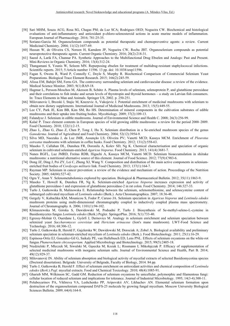
systematizing of physiological-biochemical differences of Ganoderma species by the properties of the set of secondary metabolites, stress-resistance, and at the development of fundamental basis of species specificity within Ganoderma genus and practical recommendations on optimization of the directive cultivation of the selected species. Morphological features, culture and developmental characteristics of species and chemical components such as secondary metabolites contribute greatly to the traditional identification of Ganoderma species [28]. The primary tasks posed for the work were to determine suitable storage and growth conditions of the mushrooms, and to research into their low-molecular composition. Mycelial cells can degenerate due to lack of nutrients or oxygen, infections (viruses), a change in substrate pH or the accumulation of unfavourable metabolites. [29]. Most fungal cultures can be maintained on agar by subculturing at two to six month intervals [30]. "Agar to agar" is a relatively cheap method of storage, with little time required for the production of new mycelial growth. A disadvantage of this technique is that the mycelium under refrigeration may result in degeneration of the mycelium, since the fungus can still be growing under these low temperature conditions [29]. We used "agar to agar" method for short term storage the cultures strains in laboratory. The importance of selecting the suitable short term and long term storage of the fungi was to ensure the fungi remained viable throughout this investigation, as well as for future reference and use. It was also desirable to obtain maximum growth of the fungal mycelium in a liquid cultivation medium so that sufficient material was available to pursue further studies on its biological activity. The conditions of solid-phase cultivation (thermal optima) and liquid-phase submerged cultivation (growth media formulation, temperature, culture duration) were selected by the criteria of highest mycelial biomass accumulation and primordia formation under a laboratory conditions. Optimal growth of mycelia was searched by variation of the environmental parameters, as the nutrient medium composition, carbon and nitrogen sources' kinds and proportion, thermal conditions of culture. Besides, the efficiency of cultivating the seeding mycelia of Ganoderma using the wastes of agricultural production characteristic for tropical zone, as well as using the relatively cheep food components, was shown. Contemporary physicochemical methods (scanning electron microscopy (SEM), gas chromatography coupled with mass spectrometric detection (GC-MS), gas-liquid chromatography) were applied for further characterization of the cultures. The key objective of this research step was to generate the base-line information on Ganoderma species of the largest protection areas in Vietnam, and to compare those with the species or strains of distinct geographical sampling. The comparative analysis by the scanning electron microscopy method was attempted to follow the fruit bodies morphology and mycelia microstructures. The presence of salt crystals on hyphae in mushroom cultures was reported earlier [e.g., 31]. As a rule, crystals cover the hyphae and were rarely found separated from the cells. Usually it is calcium oxalate crystals that are formed on hyphae. Crystals occurred to appear at some mushrooms' culture under different nutrient media (agar and liquid media, grain, compost, etc.) and represented a relatively stable characteristic of the cultures [31]. The density of crystals on the surface of hyphae, as well as the morphology of crystals may vary with a fungus systematic position. In the samples of G. colossus pileus and mycelium on wort agar, calcium salt microparticles were observed (Fig. 1). The presence of calcium was proved by EDX analysis. Near-cubic shape and smooth surface of the particles allowed classifying of crystal structure as calcite, the most stable crystalline structure of calcium carbonate. Back-scattered electron images of the samples of G. valesiacum 120702 mycelium on wort agar showed the presence of small areas inside the samples containing atoms heavier than carbon, oxygen or nitrogen, and formation of calcite particles in the samples can be supposed as well (Fig. 2).
a b Fig. 1. Ganoderma colossus SIE1301 calcium salt crystals in: a - pileus tissue; b - mycelium on wort agar (full-length scale bars are 50 μm)
Fig. 2. Ganoderma valesiacum 120702 calcium salt crystal in the mycelium (scale bar is 10 μm)
Antimicrobial research: Novel bioknowledge and educational programs (A. Méndez-Vilas, Ed.)
110
_____________________________________________________________________________

The presence of yellow pigment is thought to be among the features suitable for characterizing the differentiation rate of Ganoderma species [32]. Using the isolates of Ganoderma strains under study as the inoculum, the cultures were tested for their ability to produce yellow extracellular pigment on media plates. Some relevant peculiarities were observed in our studies, being much more profound for G. colossus. One could notify that the unfavorable culture conditions facilitate the vegetative mycelium pigmentation. Brown color occurrence could be stimulated by a limited variety of carbohydrates (potato-glucose-based formulation). Relatively low culture temperature is a further contributory factor to G. colossus mycelium becoming yellow. Pigmented mycelia of Ganoderma species appear to contain valuable compounds. 1-Octen-3-ol and 3-octanol were found as main volatile flavor compounds in our work, in compliance with the earlier few works dealt with the volatile compounds in Ganoderma lucidum [33] and G. sinense mycelia [34]. Different liquid crystalline systems are based on natural low-molecular substances, which are fatty acids derivatives. Those could serve as chemical stability enhancers for protect drugs from chemical instability reactions [35], as coatings for microspheres in the process of gastroretentive drug delivery [36], as subjects for broad discussion on biodiesel [37, 38]. Monoglycerides are also among the detected compounds. Further studies should be driven by a wide range of promising biotechnological applications based on the mycelial chemical components of lipid nature. Thus, by applying common mycological, SEM, and current chromatographic methods, we have studied micromorphological, cultural and selected biochemical characteristics that might provide a routine basis for identifying the most promising strains as biotechnological subjects. Ganoderma species were under the comparative study in respect to Ganoderma strains from European and Siberian regions' mushroom collections.
3. Mineral component of antimicrobial biocomposites
3.1 Selenium, essential mineral
The trace mineral selenium (Se) is an essential element for human and animal nutrition. Selenium deficiencies in the human and animal organism are recognized worldwide to be related to a number of pathologies [39]. However, at higher Se concentrations, harmful consequences occur. It has been hypothesized that the intake of excessive doses of selenium may cause oxidative damage, leading to genomic instability [40]. The contradictions found in the course of Se studies might be related to poor understanding of controversial mechanisms involved in selenium biochemistry. Therefore, a rich area of selenium explorations could be considered as two fields: Se as dietary component and Se as the agent toxic for organisms at different developmental steps, therewith the toxicity being severely dependent on the dose and chemical essence of Se-containing compound. The possible mechanism of toxicity appears to be production of free radicals, adhesion to proteins and uncontrollable accumulation, induction of apoptosis, growth arrest, and cell death.
3.2 Inorganic and organic Se: dramatically different biological effects
Recommended dietary intakes are not currently met by most diets, unless Se-rich foods are included. Therewith one should take into consideration a poor bioavailability of the most common inorganic forms of selenium. Selenium content in a foodstuff critically influence Se bioactivity to humans and animals. Foodstuffs processing and treatments, along with foodstuff-matrix major and minor components, affect Se bioavailability [41]. A great deal of information has been accumulated indicating that dietary form of Se is a major determinant of its efficiency [42], and the chemical form of Se plays a very important, if not a decisive role in its bioavailability [43]. A growing body of evidence indicates dramatically different biological effects of inorganic and organic chemical forms of selenium, which may explain apparent inconsistencies across studies by inadequate assessment of health risk [44]. The human or animal exposure to selenium in different chemical forms leads to not only different, but in some cases opposite nutritional and toxicological consequences [45, 46]. The worse cases are Se compounds having lower concentrations but the highest toxicological activity such as the inorganic forms selenite and selenate. [47-49]. At the same time, it has been shown repeatedly that Se is more bioavailable to animals and humans in organic forms than in inorganic forms [39, 50, 51], and toxicity of inorganic (tetravalent) Se greatly exceeds that of organic Se. When comparing the bioactivity of Se-containing agents, C.S. Hoefig et al. [52] explored a number of different selenocompounds known from nutrition, supplements or pharmacological use, and tested their ability to support and increase selenoprotein P (SEPP) production by human and murine hepatocytes in culture. Upon comparison of the two inorganic Se compounds, sodium selenite Na2SeO3 was found to be more toxic, and decreased viability was already observed in the micromolar concentration range. LD50 of 2 μM selenite solution was more than 30 times lower than the corresponding value of selenate. The investigations aimed at the development of novel synthetic organoselenium compounds and at the discovery of naturally occurring selenium compounds that are more effective and less toxic than inorganic forms of selenium were initiated at the beginning of the 1980s. Important aspects of the modern organoselenium chemistry are the use of organoselenium reagents as catalysts (organocatalysis), green chemistry, bioinspiration, antioxidant activity. The
Antimicrobial research: Novel bioknowledge and educational programs (A. Méndez-Vilas, Ed.)
111
_____________________________________________________________________________

classical synthetic application of organoselenium reagents are electrophilic, nucleophilic and free radical reagents. Organoselenium compounds find applications in organic synthesis, materials synthesis, ligand chemistry [53-55], antioxidative agents [56-58]. The synthesis and the synthetic applications of some emerging classes of selenium compounds such as hypervalent selenium species and selenoamides, address some biological aspects such as the antimicrobial activity of organoselenium derivatives and the biochemistry of selenoproteins, along with biologically relevant processes as potent therapeutic and chemopreventive agents [59-61] known to be clinically safe and to possess a well-established pharmacology profiles [62]. Today this area of organoselenium research is growing rapidly, and the outcomes of these investigations are highly promising. Exciting studies performed in vitro with respect to cellular responses showed that the dose and form of selenium compounds are critical experimental parameters. Inorganic (at doses up to 10 μM) and organic selenium compounds (at doses equal to or greater than 10 μM) elicit distinctly different cellular responses [40]. One should take into account that toxicity of Se-containing compound is rather a compound-specific property, and is not related directly to the biosynthesis of enzymatically active selenoproteins. Thus, secretion of hepatically derived SEPP, the central selenoprotein in blood controlling Se transport and distribution, is crucial to convert nutritional sources into serum Se, supporting Se status and selenoprotein biosynthesis in other tissues. The selenocompounds different in chemical nature, although showing a positive effect on SEPP production in the cell culture medium, failed to support cell viability in vitro. Sodium selenite, methylseleninic acid, L-selenocystine, and selenodiglutathione caused the increased SEPP concentrations in murine and two human liver cell lines, but induced cell death in micromolar concentrations, whereas the less effective with SEPP selenomethionine (SeMet) or synthetic preparation ebselen were not toxic within the concentration range tested [52].
4. Mushrooms as a Se carrier
4.1 Selenized mycelia
Interest in using mushrooms as a Se carrier increases. Analyzing the selenium content of mushrooms and its utilization, the contribution of mushrooms to the human's selenium demand, selenium content of mycelium cultivated under different conditions, effect of technology (growing) on the selenium content of mycelia, selenium species occurring in mushrooms, bioavailability of selenium in different oxidation states, one could conclude on the significantly positive trends in edible and medicinal mushrooms' implementation in this area. Mushrooms and yeasts have attracted a number of researchers in food and pharmaceuticals. Mushroom-based foods enriched with selenocompounds could be a convenient source of Se to balance the deficiency. Therewith the safety and efficacy factors favor the organic forms of Se. The addition of selenium to the diet through dietary supplements or fortified food/feed becomes increasingly common owing to the frequently suboptimal level of this microelement in standard nutrition in many countries [63]. One of the basic questions arising in relation to Se-fortified food is on the threshold quantities of selenium causing unexplored harmful consequences of using common food. The content of selenium in food of not-fungal-origin, i.e. plants and animals, depends critically on the selenium content in environment. Thus, the selenium concentration in such nutrient products is highly variable [64]. The bioavailability of selenium from fish can be modified by the presence of various contaminants, including arsenic and mercury [65]. Much attention should be paid to the biotechnological processes with the implements of selenium-enrichment of mushroom growing mycelium and post-harvest treatment thereafter, because of the ambiguous influence of elemental selenium upon human and animals. Fortification of edible mushroom cultures with the selenium-containing compounds has proven to be an effective and cost saving strategy for the prevention of Se deficiency. Satisfaction of the human selenium requirements can be considerably contributed from mushrooms, since the selenium enriched mushroom mycelia are valuable functional foods. It is obvious that risks and benefits of Se intakes should be quantified and balanced. The mushrooms are commonly used food product and dietary supplement convenient to apply in the selenium-fortified form. Possibilities for increasing selenium content in foods by increasing selenium content of mushrooms fruit bodies and vegetative mycelia are explored from the end of 20th century up to now. Mycelia of many tested mushroom species at submerged growing are satisfactory Se-sources due to the fact that Se-concentrations absorbed from the sodium selenite-enriched medium could achieve tens percent of its content in the medium (e.g., 62.5% for P. ostreatus) [66]. The mycelial selenium content could be several times higher than in fruiting bodies. In order to optimize fortification process and yields, selenium enrichment in the cultivation substrate can be an approach to increase the Se concentration in fruiting bodies of mushrooms. Popular mushrooms with high commercial values and thus cultivated world wide, e.g. Agaricus bisporus, Lentinula edodes, Pleurotus spp., Flammulina velutipes, Hypsizigus marmoreus appeared to contain nutritionally significant but yet insufficient amounts of Se [67]. Most of edible mushroom species examined are selenium-poor (< 1 microg Se/g dry weight) [68]. These values can be considerably, even by order of magnitude, increased in mushrooms picked in polluted areas. Moreover, some species have accumulating and even hyperaccumulating ability for Se [69]. So, the solution is artificial growing of basidiomycetes. A particularly rich source of selenium could be obtained from selenium-enriched mushrooms that are
Antimicrobial research: Novel bioknowledge and educational programs (A. Méndez-Vilas, Ed.)
112
_____________________________________________________________________________

cultivated on a solid media fortified with selenium (as inorganic salt or selenized-yeast). Addition of Se oxyanions in the form of selenite or selenate to the substrate for growing basidiomycetes results in an increase of the Se content in their fruiting bodies [70, 71]. Growth-compost irrigated with sodium selenite solution appeared to cause the increase in the selenium level in button mushroom, Agaricus bisporus by tens times compared to the control mushroom irrigated solely with water [72]. The enrichment of Lentinula edodes (shiitake mushroom) fruit bodies with Se could be performed by adding the sodium selenite to the cold-shock water used to induce primordial formation in artificial logs [73]. As a result, shiitake absorbs and accumulates Se in carpophores, therewith the color, moisture, and the protein content of mushroom being unaffected. Thus, selenium-enriched fruit bodies are industrially cultivated as functional food or medicinal food in China and Southeast Asia and could provide an efficient way in delivering functional organic Se. However, the composition of selenium substances, as well as the distribution of the main bioactive components, remain still unknown [74]. Several data testify to the greater efficiency of selenized mushroom compared to yeasts. Thus, the high level of SeCys in several Se-enriched mushrooms [72] could provide greater biological benefits than Se-enriched yeast which contains mainly SeMet, since the SeCys residue functions as an active-site amino acid in selenoenzymes, e.g. glutathione peroxidase and thioredoxin reductase [75], and provides antioxidant properties in non-enzymatic selenoproteins [76]. Indeed, the effect of dietary supplementation with Se-enriched Agaricus bisporus on different selenoproteins expression in rats showed that the selenized button mushroom can positively increase GPx-1 and GPx-2 gene expression and GPx-1 enzyme activity in rat colon [77]. The extracts from Lentinula edodes mycelia rich in the organic forms of selenium possess putative cancer-preventive properties more effective than those of selenized-yeast [78].
4.2 Speciation of selenium in Se-enriched mycelial cultures
An increasingly growing database on chemical forms of selenium in mushrooms indicates that the seleno-compounds identified in carpophore include selenocysteine, selenocystine, selenomethionine, Se-methylseleno-L-cysteine, inorganic selenium [79] and several unidentified seleno-compounds; their proportions vary widely [68]. For mycelia at submerged cultivation, the increased mycelial concentration of the above selenoamino acids is feasible by optimizing the concentration of sodium selenite in the liquid medium, e.g. of Se-methylseleno-L-cysteine from essentially non-detectable levels to 120 µg/g dry weight [80], the much higher levels of SeCys (detected as (SeCys)2) compared to SeMet and MeSeCys were found for Agaricus bisporus grown on compost enriched with inorganic Se [72]. Study by Jadwiga Turlo et al. [78] indicate that the shiitake mushroom Lentinula edodes cultivated in media enriched with sodium selenite.is capable of incorporating selenium into the proteins, mainly in the form of selenomethionine. The amount of selenomethionine in the cultivated biomass increased in proportion to the selenium content of the mycelium, but the percentage of selenium accumulated as the selenoamino acid decreased. For greater selenium concentrations, the selenocysteine content of the mycelial biomass does not depend directly on the concentration of selenium in the medium and was essentially unchanged. Thus, a fate of a considerable portion of the introduced selenium is unknown. A sample of Hericium erinaceus (lion's mane mushroom) belonging to Agaricomycetes was examined in the study [81] after moderate selenium enrichment from inorganic selenium source. It was revealed that nearly 50% of the selenium was in organic forms: selenomethionine and Se-methylseleno-L-cysteine. Three Se-adenosyl-compounds were identified: Se-methyl-5-selenoadenosine, Se-methyl-5-selenoadenosine-Se-oxide and the previously unreported Se-dimethyl-5-selenonium-adenosine. Evaluation of the efficacy of supplementation of several medicinal mushrooms with inorganic selenium salts (Na2SeO3 and Na2SeO4) attracted the attention of many researchers in 21st century. Submerged cultivated mycelium of Lentinula edodes accumulated selenium from the cultivation medium very effectively. Selenium was well bioavailable from the mycelial preparations in in vitro and in vivo tests, for which different preparations of selenated mycelium were compared [82]. The speciation of selenium in Se-enriched mycelial cultures testified to the fact that the main, inorganic, part of Se in the tested mycelium was in the zero (elemental selenium) and IV oxidation states. The response of Phanerochaete chrysosporium to the aforementioned selenium oxyanions showed a high sensitivity to selenium, particularly selenite. The latter inhibited the fungal growth and substrate consumption at a level of 10 mg/l in the growth medium, whereas Se(VI) of selenate did not have such a strong influence on the mushroom [83]. P. chrysosporium, too, was found to be a selenium-reducing organism, capable of synthesizing elemental selenium from selenite but not from selenate. Studies with Ganoderma lucidum, Agrocybe aegerita, and Hericium erinaceus showed that the growth of Ganoderma lucidum fruit bodies was observed with up to 0.8 mM Se accompanied by the highest total Se content, macroscopic changes in the fruiting bodies of the examined mushrooms, and color changes of fruiting bodies [84]. In a work by Ivan N. Milovanović [85], such biotechnologically important mushrooms fortified with inorganic Se as Ganoderma lucidum (Leyss.:Fr.) Karst., Pleurotus ostreatus (Jacq.: Fr.) Kumm., Pleurotus eryngii (DC.:Fr.) Quél. var. eryngii, Pleurotus pulmonarius (Fr.) Quél., Flammulina velutipes (Curt.: Fr.) Sing., Ganoderma applanatum (Pers.:Wallr.) Pat., Lenzites betulinus (L.:Fr.) Fr., Trametes hirsuta (Wulf.:Fr.) Pil. were explored in respect to their morpho-physiological characteristics, as well as biological activities. During cultivation on selenite-enriched medium, the appearance of mycelium of brick-red color with significant morphological and ultrastructural changes in
Antimicrobial research: Novel bioknowledge and educational programs (A. Méndez-Vilas, Ed.)
113
_____________________________________________________________________________

comparison with the control was observed. Hyphal density was lower, the cell wall was thick with more expressed extracellular matrix, septa were abundant, and branch frequency and occurrence of clamp-connections were rare. Cytological analysis demonstrated that the majority of selenium was accumulated in cell membrane and vacuoles, while changes taking place in a cell wall were insignificant [85]. The latter study along with a relatively large number of others are focused on the effect of inorganic selenium salts' concentrations on mycelium morphological and ultrastructural features. At a relatively high selenite concentration in Lentinula edodes liquid nutrient medium, the excess selenium is eliminated via its reduction to elemental Se [86]. As for another very popular cultivated mushroom, Pleurotus ostreatus, the studies also showed that higher selenite concentrations caused firstly Se accumulation in P. ostreatus mycelium, and during the suppression growth phase, selenite was reduced to amorphous Se in zero oxidation state and this gave the mycelium and medium a reddish color [87, 88]. Hyphal morphology of P. ostreatus was dependent on Se concentration in the liquid medium. Electron-dense spots, visible in both the control and Se-enriched samples, were described [89] as proteinaceous bodies, since lipid bodies would be extracted during preparation for transmission electron microscopy. In the presence of Se, the number of these bodies increased, and changes in their shape, color, and size were slight. It was shown that the reduction of the ionic Se and production of amorphous Se(0) are really occurring round the bodies [89]. Selenite stress exerts a significant effect on ultra-architectural features of the fungal hyphae and spores of mushroom cultures, e.g., Ganoderma lucidum [90]. A number of works deals with the Se distribution among different cellular compartments, and, in particular, polysaccharide structures contained in fungal cell walls. Se-enriched submerged mycelia of Pleurotus ostreatus were explored in respect to the incorporation of selenium from the growth medium to mushroom [91]. A polysaccharide-containing fraction of mycelia was treated alternatively with Tris-HCl or with chitinase. Better solubility and increased contribution of low molecular mass compounds were observed in chitinase extracts (UV detection), confirming the degradation of polysacharides by the enzyme. The results obtained suggest selenium binding to chitin-containing polysaccharide structures in fungal cell walls [91]. Selenite influenced the pellet morphology of Phanerochaete chrysosporium by reducing the size of the fungal pellets and inducing their compaction and smoothness [83]. Analysis of P. chrysosporium mycelia with transmission electron microscopy, electron energy loss spectroscopy, and a 3D reconstruction showed that elemental selenium was produced intracellularly as nanoparticles.
5. Se-containing antibacterial agents of fungal origin
Almost all of the published works dealt with the Se-fortified fungal cultures are concerned with selenium exclusively in the form of inorganic substances, sodium selenite Na2SeO3 or selenate Na2SeO4. The source of selenium should reasonably be the organic substance 1,5-diphenyl-3-selenopentanedione-1,5 (synonyms - diacetophenonylselenide, bis(benzoylmethyl)selenide, preparation DAPS-25) [92, 93], since its low toxicity at physiological concentrations in combination with high efficiency (compared to, e.g., selenites) has been proved earlier for various living organisms. It is the source of selenium we use in our work. The growth parameters and the dynamics of lectin activity of Lentinula edodes (shiitake mushroom) on liquid media enriched with selenium in the organic form have been studied. Diacetophenonylselenide exerts the appreciably positive effect upon the vital processes of L. edodes [94]. The role of spatial and electron structure, hydrophobic properties and concentration of organoselenium compounds on their interaction with fungal metabolites - extracellular lectins of shiitake mushroom has been considered. Along with DAPS-25, several other compounds of the 1,5-di(4-R-phenyl)-3-selenopentanediones-1,5 series interaction with the basidiomycete L. edodes lectins were explored involving both computations and experiment [95]. Appreciable effect of the selenium-containing preparation DAPS-25 on the vital activity of L. edodes exhibited as the change in mushroom growth parameters and lectin activirty on various organic and mineral, agar and liquid media has been expanded in our studies toward the elemental selenium accumulation at the definite DAPS-25 concentration in culture liquid. For the first time, the intensive red pigmentation of mycelium caused by the elemental selenium accumulation resulted from the organoselenium compound destruction by the mushroom L. edodes has been revealed. The biotransformation of 1,5-diphenyl-3-selenopentanedione-1,5 at the growth of shiitake mushroom under the liquid-phase and solid-phase culture conditions has been studied. At the initial DAPS-25 concentration equal to or higher than 1·10−4 mol/l in the synthetic liquid medium, a red color of L. edodes mycelium develops, the intensity and initiation time of which being related to this Se-additive concentration. The semi-quantitative data obtained by physicochemical methods on comparative selenium level and acetophenone production in the samples allow us to conclude on the L. edodes submerged culture capability of sorbing and/or destructing a xenobiotic of organoselenium nature [96]. Starting from the results of qualitative reaction, the data of X-ray fluorescence, X-ray phase and GC-MS analyses, we should conclude on the L. edodes submerged culture capability of destructing the organoselenium xenobiotic to occur red modification of elemental selenium and to evolve acetophenone. The process of elemental selenium elimination was followed by its precipitation onto gyphae [97]. Within the framework of more recent studies, the growth parameters of more than twenty strains of xylotrophic basidiomycetes belonging to 8 genera, 13 species on liquid media enriched with selenium in organic form were studied.
Antimicrobial research: Novel bioknowledge and educational programs (A. Méndez-Vilas, Ed.)
114
_____________________________________________________________________________

The effect of 1,5-diphenyl-3-selenopentanedione-1,5 within a wide concentration range (1·10–4 − 1·10–8 mol/l) on the mycelial growth was observed. The phenomenon of mycelium growth stimulation was assumed to be related to the selenium antioxidant properties, due to which the negative consequences of exhausting the nutrient components of medium were smoothed over. DAPS-25 concentrations exceeding 5·10–5 mol/l inhibited considerably the mycelium growth. At the initial DAPS-25 concentrations in the synthetic medium not lower than 5·10-5 mol/l, we observed a red pigmentation of mycelium caused by the elemental Se in line with the results we obtained earlier for the shiitake mushroom [97]. The color intensity and time of appearance were dependеnt on the selenium additive's concentration, on the given fungal species, and its growth external conditions. The culture liquids of the fungal species under study were successfully tested for their reducing and stabilizing properties toward organic selenide and elemental selenium, respectively. The results of studying the effect of biologically obtained selenium nanocomposites on the bacterium Clavibacter michiganensis ssp. sepedonicus (Cms) have been obtained [98]. Cms, a Gram-positive bacterium, causes ring rot, which is one of the most dangerous potato diseases. The effective alongside ecologically safe methods for combating Cms are lacking. As the agents feasible for use in this purpose, we examined the selenium biocomposites obtained from the submerged cultures of mushrooms. For exploring the biocomposites effect on Cms, several microbiological techniques were implemented. The results showed that all the biocomposites under study lowered the absorption values of bacterial suspensions, in comparison with the reference specimen, that testified to the bacteriostatic effect of the agents under question. The suppression action of biocompostes in respect to Cms was also revealed. Thus, we demonstrated the occurrence of bacteriostatic and bactericidal effects of the agents under study, and the results favor the supposition on advisability of further research into the selenium bionanocomposites as the agents for agricultural recovery from the bacterial pathogens [98]. Recently the impact of Se-containing biocomposites based on Ganoderma mushroom isolates grown in the presence of oxopropyl-4-hydroxycoumarins, on the bacterial phytopathogens has been examined [99]. The effect of selenium biocomposites obtained by using the submerged cultures of macrobasidiomycetes Ganoderma applantum, G. cattienensis, G. colossus G. lucidum, G. neojaponicum, G. valesiacum, on the phytopathogenic bacteria Clavibacter michiganensis ssp. sepedonicus (Cms), Micrococcus luteus, Pectobacterium atrosepticum, Pectobacterium carotovorum subsp. сarotovorum, Pseudomonas fluorescens, Pseudomonas viridiflava, Xanthomonas campestris has been studied. These bacterial strains used in our work were kindly provided by the Collection of Rhizosphere Microorganisms of IBPPM RAS (http://collection.ibppm.ru). Oxopropyl-4-hydroxychromenones have been used as the components of fungal nutrient media. By means of such methods as the colony-forming units count, the agar well diffusion method, and the bacterial suspension turbidity measurement, the bacteriostatic and bactericidal activity of the Se-containing substances of fungal origin in respect to phytopathogenic bacteria has been elucidated. The composites produced from the extracellular metabolites of G. cattienensis SIE1302 with 4-hydroxy-3-(3-oxo-1,3-diphenylpropyl)-chromen-2-one (S(45)), and of G. lucidum SIE1303 with 4-hydroxy-3-(3-oxo-1-(3-nitrophenyl)-3-phenylpropyl)-chromen-2-one (S(NO2)) possessed the most profound antibacterial action against Cms. The composites produced from the isolates of G. valesiacum 120702 with S(NO2) showed the maximal antibacterial activity vs. Xanthomonas campestris B-610. High antimicrobial effect of G. lucidum 1315 with S(NO2), and of G. colossus SIE1301 against Xanthomonas campestris B-610 and Pseudomonas fluorescens EL-2.1, respectively, has been revealed. The pioneering information on the biological activity of coimarin series compounds in their application for producing the substances of fungal origin has been provided [99].
6. Conclusive remarks
The novel aspects concerned with the essential nutrient and antioxidant Se properties discovered in the 2nd half of XX century and more recently, have changed the views on selenocompounds. Different chemical forms of selenium possess excellent biochemical properties and have been implicated for use in biotechnology. The favorable profile of newly synthesized organoselenium compounds including those explored in our research warrants their recognition as a promising option for fortification purposes. Further thorough investigation should be focused on the mechanism of Se-containing compounds' toxicity to take that into account when using the various Se sources in biotechnological fields, including the production of ecologically safe antibacterial agents. As for the future perspectives of research in this area the following should be noted. Mushrooms are recognized to be promising ecologically pure raw material for developing the medicinal preparations for care and prophylaxis with wide spectrum of action. Contemporary biomedical applications of selenium are undoubtedly broad. In this relation, of especial interest are the selenium-enriched preparations of higher-fungal origin owing to their availability, biocompatibility, and the proved biological activity. The development of elemental selenium should be based on natural edible and medicinal products, e.g., mushroom isolates, and considered to be appropriate "green" method. As being originated from the biotransformed organoselenium compound, the selenium submicroparticles incorporated in or separated from the submerged mycelium possess the benefit of their non-toxic source and provide the potential multipurpose use [27, 100].
Antimicrobial research: Novel bioknowledge and educational programs (A. Méndez-Vilas, Ed.)
115
_____________________________________________________________________________

The approach developed in perspective research should allow the bioproduction of submicrostructured elemental selenium using the edible mushrooms cultures to be put into practice.
Acknowledgements This work was financially supported in part by Russian Foundation for Basic Research (RFBR), research project 16-08-01170 -a. The authors thank the researchers kindly provided the bacterial strains from the Collection of Rhizosphere Microorganisms of IBPPM RAS (http://collection.ibppm.ru).
References [1] Crous PW, Rong IH, Wood A, Lee S, Glen H, Botha W, Slippers B, de Beer WZ, Wingfield MJ, Hawksworth DL. How many
species of fungi are there at the tip of Africa? Studies in Mycology. 2006; 55:13-33. [2] Mortimer PE, Karunarathna SC, Li Q, Gui H, Yang X (Xueqing), Yang X (Xuefei), He J, Ye L, Guo J, Li H, Sysouphanthong
P, Zhou D, Xu J, Hyde KD. Prized edible Asian mushrooms: ecology, conservation and sustainability. Fungal Diversity. 2012. 56(1):31-47.
[3] Kim S, Ha BS, Ro HS. Current Technologies and Related Issues for Mushroom Transformation. Mycobiology. 2015; 43(1):1-8. [4] Buswell JA, Cai YJ, Chang ST, Peberdy JF, Fu SY, Yu HS. Lignocellulolytic enzyme profiles of edible mushroom fungi.
World Journal of Microbiology and Biotechnology. 1996; 12(5):537-42. [5] Ashraf J, Ali MA, Ahmad W, Ayyub CM, Shafi J. Effect of different substrate supplements on Oyster Mushroom (Pleurotus
spp.) Production. Food Science and Technology. 2013; 1(3):44-51. [6] Anike FN, Isikhuemhen OS, Blum D, Neda H. Nutrient Requirements and Fermentation Conditions for Mycelia and Crude
Exo-Polysaccharides Production by Lentinus squarrosulus. Advances in Bioscience and Biotechnology. 2015; 6(8):526-36. [7] Muhammad BL, Suleiman B. Global Development of Mushroom Biotechnolgy. International Journal of Emerging Trends in
Science and Technology. 2015; 2(6):2660-9. [8] Tang YZ, Zhu LW, Li HM, Li DS. Submerged culture of mushrooms in bioreactors - challenges, current state-of-the-art, and
future prospects. Food Technology and Biotechnology. 2007; 45(3):221-9. [9] Chen W, Zhao Z, Li Y. Simultaneous increase of mycelial biomass and intracellular polysaccharide from Fomes fomentarius
and its biological function of gastric cancer intervention. Carbohydrate polymers 2011, 85(2): 369-75. [10] Ruthes AC, Smiderle FR, Iacomini M. D-Glucans from edible mushrooms: A review on the extraction, purification and
chemical characterization approaches. Carbohydrate polymers. 2015; 117:753-61. [11] Tsivileva OM, Pankratov AN, Nikitina VE. Extracellular protein production and morphogenesis of Lentinula edodes in
submerged culture. Mycological Progress. 2010; 9(2):157-67. [12] Wasser SP. Current findings, future trends, and unsolved problems in studies of medicinal mushrooms. Applied Microbiology
and Biotechnology. 2011; 89(5):1323-32. [13] Corrêa RCG, Brugnari T, Bracht A, Peralta RM, Ferreira IC. Biotechnological, nutritional and therapeutic uses of Pleurotus
spp. (Oyster mushroom) related with its chemical composition: A review on the past decade findings. Trends in Food Science & Technology. 2016; 50:103-17.
[14] Moncalvo JM. Molecular Systematics of Ganoderma: What Is Reishi? International Journal of Medicinal Mushrooms. 2005; 7(3):353-4.
[15] Chang ST, Miles PG. Mushrooms: Cultivation, nutritional value, medicinal effect, and environmental impact. 2nd ed. New York: CRC Press, 2004.
[16] Le XT, Le QHN, Pham ND, Dentinger BT, Moncalvo JM. Tomophagus cattienensis sp. nov., a new Ganodermataceae species from Vietnam: Evidence from morphology and ITS DNA barcodes. Mycological progress. 2012; 11(3):775-80.
[17] Wu GS, Guo JJ, Bao JL, Li XW, Chen XP, Lu JJ, Wang YT. Anti-cancer properties of triterpenoids isolated from Ganoderma lucidum-a review. Expert opinion on investigational drugs. 2013; 22(8):981-92.
[18] Osińska-Jaroszuk M, Jaszek M, Mizerska-Dudka M, Błachowicz A, Rejczak TP, Janusz G, Wydrych J, Polak J, Jarosz-Wilkołazka A, Kandefer-Szerszeń M. Exopolysaccharide from Ganoderma applanatum as a Promising Bioactive Compound with Cytostatic and Antibacterial Properties. BioMed Research International. 2014; Article ID 743812: 10 p.
[19] Gao Y, Zhou S, Huang M, Xu A. Antibacterial and Antiviral Value of the Genus Ganoderma P. Karst. Species (Aphyllophoromycetideae): A Review. International Journal of Medicinal Mushrooms. 2003; 5(3):1-12.
[20] De S. Pereira-Jr JA, Rodrigues DP, Peixoto-Filho RC, Bastos IVGA, De Oliveira GG, Araújo JM, Melo SJ. Contribution to Pharmacognostic and Morphoanatomical Studies, Antibacterial and Cytotoxic Activities of Ganoderma parvulum Murrill (Basidiomycota, Polyporales, Ganodermataceae). Latin American Journal of Pharmacy. 2013; 32(7):996-1003.
[21] Sivaprakasam, E., Balakumar, R., Kavitha, D. (2011) Evaluation of antibacterial and antifungal activity of Ganoderma lucidum (curtis) P. Karst fruit bodies extracts. World Journal of Science and Technology, 1 (6): 8-11.
[22] Prasad Y, Wesely WEG. Antibacterial activity of the bio-multidrug (Ganoderma lucidum) on Multidrug resistant Staphylococcus aureus (MRSA). Advanced Biotech. 2008; 10:9-16.
[23] Ofodile LN, Uma N, Grayer RJ, Ogundipe OT, Simmonds MSJ. Antibacterial Compounds from the Mushroom Ganoderma colossum from Nigeria. Phytotherapy Research. 2012; 26(5):748-51.
[24] Adaskaveg JE, Gilbertson RL, Dunlap MR. Effects of Incubation Time and Temperature on In Vitro Selective Delignification of Silver Leaf Oak by Ganoderma colossum. Applied and Environmental Microbiology.1995; 61(1):138-44.
[25] Moncalvo J.M. Systematics of Ganoderma 2. In: Ganoderma diseases of perennial crops, 23. 2000. [26] Hong SG, Jung HS. Phylogenetic analysis of Ganoderma based on nearly complete mitochondrial small-subunit ribosomal
DNA sequences. Mycologia. 2004; 96(4):742-55. [27] Tsivileva OM, Nguyen TP, Vu LN, Lyubun EV, Voronin SP, Gumenyuk AP, Nikitina VE. Biotechnologically valuable species
as subjects of Russian-Vietnamese research cooperation in 2012-2014 dealt with mycology. Proceedings of Higher School. Applied Chemistry and Biotechnology. 2014; 3(8):75-7.
Antimicrobial research: Novel bioknowledge and educational programs (A. Méndez-Vilas, Ed.)
116
_____________________________________________________________________________

[28] Roberts LM. Australian Ganoderma: Identification, growth and antibacterial properties (Doctoral dissertation, Ph. D. Thesis). Swinburne University of Technology, Australia, 2004.
[29] Oei P. Mushroom cultivation: appropriate technology for mushroom growers. Leiden, Netherlands: Backhuys Publishers,. 2003. 429 p.
[30] Ando K. XII. Preservation of mushroom cultures. Food Reviews International. 1997; 13(3):423-9. [31] Buchalo AS., Wasser SP, Mykhaylova OB, Bilay VT, Lomberg ML. Taxonomical significance of microstructures in pure
cultures of macromycetes. In: Mushroom biology and mushroom products. Proceedings of the 7th International Conference on Mushroom Biology and Mushroom Products, Arcachon, France, 4-7 October, 2011. Volume 2. Poster session. Institut National de la Recherche Agronomique (INRA), 2011. p. 50-57.
[32] Szedlay G., Jakucs E., Boldizsar I., Boka K. Basidiocarp and mycelium morphology of Ganoderma lucidum Karst. strains isolated in Hungary. Acta Microbiologica et Immunologica Hungarica. 1999; 46(1):41-52.
[33] Chen ZJ, Yang ZD, Gu ZX. Determination of volatile flavor compounds in Ganoderma lucidum by HS-SPME-GC-MS. Food Research and Development. 2010; 31(2):132-5.
[34] Wang XL, Han WJ, Zhou GY. Analysis of Volatile Flavor Compounds in Submerged Cultured Ganoderma sinense Mycelium by Headspace Gas Chromatography. In: 2nd International Conference on Biomedical Engineering and Informatics (BMEI'09). IEEE, 2009. p. 1-4.
[35] Sadhale Y, Shah JC. Glyceryl monooleate cubic phase gel as chemical stability enhancer of cefazolin and cefuroxime. Pharmaceutical Development and Technology. 1998; 3(4):549-56.
[36] Liu Y, Chen T (Ting), Yang M, Wang C, Huo W, Yan D, Chen J (Jinjin), Zhou J, Xing J. Analysis of mixtures of fatty acids and fatty alcohols in fermentation broth. Journal of Chromatography A. 2014; 1323:66-72.
[37] Di Nicola G, Pacetti M, Polonara F, Santori G, Stryjek R. Development and optimization of a method for analyzing biodiesel mixtures with non-aqueous reversed phase liquid chromatography. Journal of Chromatography A. 2008; 1190(1):120-6.
[38] Likozar B, Levec J. Transesterification of canola, palm, peanut, soybean and sunflower oil with methanol, ethanol, isopropanol, butanol and tert-butanol to biodiesel: Modelling of chemical equilibrium, reaction kinetics and mass transfer based on fatty acid composition. Applied Energy. 2014; 123:108-20.
[39] Hartikainen H. Biogeochemistry of selenium and its impact on food chain quality and human health. Journal of Trace Elements in Medicine and Biology. 2005; 18(4):309-18.
[40] El-Bayoumy K. The protective role of selenium on genetic damage and on cancer. Mutation Research/Fundamental and Molecular Mechanisms of Mutagenesis. 2001; 475(1-2):123-39.
[41] Moreda-Piñeiro J; Moreda-Piñeiro A; Bermejo-Barrera P. In vivo and in vitro testing for selenium and selenium compounds bioavailability assessment in foodstuff. Critical Reviews in Food Science and Nutrition. 2017; 57(4):805-33.
[42] Surai PF, Fisinin VI. Selenium in poultry breeder nutrition: An update. Animal Feed Science and Technology. 2014; 191,1-15. [43] Thiry C, Ruttens A, De Temmerman L, Schneider YJ, Pussemier L. Current knowledge in species-related bioavailability of
selenium in food. Food Chemistry. 2012; 130(4):767-84. [44] Vinceti M, Crespi CM, Malagoli C, Del Giovane C, Krogh V. Friend or foe? The current epidemiologic evidence on selenium
and human cancer risk. Journal of Environmental Science and Health, Part C: Environmental Carcinogenesis and Ecotoxicology Reviews. 2013; 31(4):305-41.
[45] Borella P, Bargellini A, Medici CI. Chemical form of selenium greatly affects metal uptake and responses by cultured human lymphocytes. Biological Trace Element Research. 1996; 51(1):43-54.
[46] Hazane-Puch F, Champelovier P, Arnaud J, Garrel C; Ballester B; Faure P; Laporte F. Long-term selenium supplementation in HaCaT cells: importance of chemical form for antagonist (protective versus toxic) activities. Biological Trace Element Research. 2013; 154(2):288-98.
[47] Soares FA, Farina M, Böettcher AC, Braga AL, Rocha JBT. Organic and inorganic forms of selenium inhibited differently fish (Rhamdia quelen) and rat (Rattus norvergicus albinus) δ-aminolevulinate dehydratase. Environmental Research. 2005; 98(1):46-54.
[48] Xiao R, Qiao JT, Zhao HF, Liang J, Huan LY, Liang J, Liu J, Guo AM, Wang W. Sodium selenite induces apoptosis in cultured cortical neurons with special concomitant changes in expression of the apoptosis-related genes. Neurotoxicology. 2006; 27(4):478-84.
[49] Ayaz M, Dalkilic N, Tuncer S, Bariskaner H. Selenium-induced changes on rat sciatic nerve fibers: compound action potentials. Methods and Findings in Experimental and Clinical Pharmacology. 2008; 30(4):271-5.
[50] Daniels LA. Selenium metabolism and bioavailability. Biological Trace Element Research. 1996; 54(3):185-99. [51] Ortman K, Pehrson B. Selenite and selenium yeast as feed supplements for growing, fattening pigs. Journal of Veterinary
Medicine Series A. 1998; 45(1-10):551-7. [52] Hoefig CS, Renko K, Köhrle J, Birringer M, Schomburg L. Comparison of different selenocompounds with respect to
nutritional value vs. toxicity using liver cells in culture. The Journal of Nutritional Biochemistry. 2011; 22(10):945-55. [53] Mugesh G, Singh HB. Synthetic organoselenium compounds as antioxidants: glutathione peroxidase activity. Chemical Society
Reviews. 2000; 29(5):347-57. [54] Mandal A, Sahoo H, Baidya M. Copper-Catalyzed 8-Aminoquinoline-Directed Selenylation of Arene and Heteroarene C–H
Bonds. Organic Letters. 2016; 18(13):3202-5. [55] Akhoon SA, Naqvi T, Nisar S, Rizvi MA. Synthetic Organo-Selenium Compounds in Medicinal Domain. Asian Journal of
Chemistry. 2015; 27(8):2745-52. [56] Petronilho F, Michels M, Danielski LG, Goldim MP; Florentino D; Vieira A; Mendonça MG; Tournier M; Piacentini B,
Giustina AD; Leffa DD; Pereira GW; Pereira VD; Rocha JBTD. Diphenyl diselenide attenuates oxidative stress and inflammatory parameters in ulcerative colitis: a comparison with ebselen. Pathology-Research and Practice. 2016; 212(9):755-760.
[57] Borges R, Andrade FC, Schwab RS, Sousa FS; de Souza MN; Savegnago L; Schneider PH. Straightforward synthesis and antioxidant studies of chalcogenoaziridines. Tetrahedron Letters. 2016; 57(31):3501-4.
Antimicrobial research: Novel bioknowledge and educational programs (A. Méndez-Vilas, Ed.)
117
_____________________________________________________________________________
[58] Sari MHM, Souza ACG, Rosa SG, Chagas PM; da Luz SCA; Rodrigues OED; Nogueira CW. Biochemical and histological evaluations of anti-inflammatory and antioxidant p-chloro-selenosteroid actions in acute murine models of inflammation. European Journal of Pharmacology. 2016; 781:25-35.
[59] Soriano-Garcia M. Organoselenium compounds as potential therapeutic and chemopreventive agents: a review. Current Medicinal Chemistry. 2004; 11(12):1657-69.
[60] Hassan W, de Oliveira CS, Noreen H, Kamdem JP, Nogueira CW, Rocha JBT. Organoselenium compounds as potential neuroprotective therapeutic agents. Current Organic Chemistry. 2016; 20(2):218-31.
[61] Saeed A, Larik FA, Channar PA. Synthetic Approaches to the Multifunctional Drug Ebselen and Analogs: Past and Present. Mini-Reviews in Organic Chemistry. 2016; 13(4):312-24.
[62] Thangamani S, Younis W, Seleem MN. Repurposing ebselen for treatment of multidrug-resistant staphylococcal infections. Scientific reports. 2015; 5:Article number 11596. 13 pp. doi: 10.1038/srep11596.
[63] Fagan S, Owens R, Ward P, Connolly C, Doyle S, Murphy R. Biochemical Comparison of Commercial Selenium Yeast Preparations. Biological Trace Element Research. 2015; 166(2):245-59.
[64] Alissa EM, Bahijri SM, Ferns GA. The controversy surrounding selenium and cardiovascular disease: a review of the evidence. Medical Science Monitor. 2003; 9(1):RA9-18.
[65] Hagmar L, Persson-Moschos M, Akesson B, Schütz A. Plasma levels of selenium, selenoprotein P, and glutathione peroxidase and their correlations to fish intake and serum levels of thyrotropin and thyroid hormones – a study on Latvian fish consumers. In: Trace Elements in Man and Animals. Springer US, 2002. p. 250-251.
[66] Milovanovic I, Brceski I, Stajic M, Knezevic A, Vukojevic J. Potential enrichment of medicinal mushrooms with selenium to obtain new dietary supplements. International Journal of Medicinal Mushrooms. 2013; 15(5):449-55.
[67] Lee CY, Park JE, Kim BB, Kim SM, Ro HS. Determination of mineral components in the cultivation substrates of edible mushrooms and their uptake into fruiting bodies. Mycobiology. 2009; 37(2):109-13.
[68] Falandysz J. Selenium in edible mushrooms. Journal of Environmental Science and Health C. 2008; 26(3):256-99. [69] Kalač P. Trace element contents in European species of wild growing edible mushrooms: a review for the period 2000–2009.
Food Chemistry. 2010; 122(1):2-15. [70] Zhao L, Zhao G, Zhao Z, Chen P, Tong J, Hu X. Selenium distribution in a Se-enriched mushroom species of the genus
Ganoderma. Journal of Agricultural and Food Chemistry. 2004; 52(12):3954-9. [71] Silva MD, Naozuka J, da Luz JMR, Assunção LS; Oliveira PV; Vanetti MCD; Kasuya MCM. Enrichment of Pleurotus
ostreatus mushrooms with selenium in coffee husks. Food Chemistry. 2012; 131(2):558-63. [72] Maseko T, Callahan DL, Dunshea FR, Doronila A, Kolev SD, Ng K. Chemical characterisation and speciation of organic
selenium in cultivated selenium-enriched Agaricus bisporus. Food Chemistry. 2013; 141(4):3681-7. [73] Nunes RGFL, Luz JMRD, Freitas RDB, Higuchi A, Kasuya MCM, Vanetti MCD. Selenium bioaccumulation in shiitake
mushrooms: a nutritional alternative source of this element. Journal of Food Science. 2012; 77(9):C983-6. [74] Dong JZ, Ding J, Pei ZY, Lei C, Zheng XJ, Wang Y. Composition and distribution of the main active components in selenium-
enriched fruit bodies of Cordyceps militaris link. Food Chemistry. 2013; 137(1):164-7. [75] Rayman MP. Selenium in cancer prevention: a review of the evidence and mechanism of action. Proceedings of the Nutrition
Society. 2005; 64(04):527-42. [76] Ogra Y, Anan Y. Selenometabolomics explored by speciation. Biological & Pharmaceutical Bulletin. 2012; 35(11):1863-9. [77] Maseko T, Howell K, Dunshea FR, Ng K. Selenium-enriched Agaricus bisporus increases expression and activity of
glutathione peroxidase-1 and expression of glutathione peroxidase-2 in rat colon. Food Chemistry. 2014; 146:327-33. [78] Turło J, Gutkowska B, Malinowska E. Relationship between the selenium, selenomethionine, and selenocysteine content of
submerged cultivated mycelium of Lentinula edodes (Berk.). Acta Chromatographica. 2007; 18:36-48. [79] Gergely V, Kubachka KM, Mounicou S, Fodor P, Caruso JA. Selenium speciation in Agaricus bisporus and Lentinula edodes
mushroom proteins using multi-dimensional chromatography coupled to inductively coupled plasma mass spectrometry. Journal of Chromatography A. 2006; 1101(1):94-102.
[80] Klimaszewska M, Górska S, Dawidowski M, Podsadni P, Turło J. Biosynthesis of Se-methyl-seleno-L-cysteine in Basidiomycetes fungus Lentinula edodes (Berk.) Pegler. SpringerPlus. 2016; 5(1):733-40.
[81] Egressy-Molnár O, Ouerdane L, Győrfi J, Dernovics M. Analogy in selenium enrichment and selenium speciation between selenized yeast Saccharomyces cerevisiae and Hericium erinaceus (lion's mane mushroom). LWT-Food Science and Technology. 2016; 68:306-12.
[82] Turło J, Gutkowska B, Herold F, Gajzlerska W; Dawidowski M; Dorociak A; Zobel A. Biological availability and preliminary selenium speciation in selenium-enriched mycelium of Lentinula edodes (Berk.). Food Biotechnology. 2011; 25(1):16-29.
[83] Espinosa-Ortiz EJ, Gonzalez-Gil G, Saikaly PE, van Hullebusch ED, Lens PNL. Effects of selenium oxyanions on the white-rot fungus Phanerochaete chrysosporium. Applied Microbiology and Biotechnology. 2015; 99(5):2405-18.
[84] Niedzielski P, Mleczek M, Siwulski M, Gąsecka M; Kozak L; Rissmann I; Mikołajczak P. Efficacy of supplementation of selected medicinal mushrooms with inorganic selenium salts. Journal of Environmental Science and Health, Part B. 2014; 49(12):929-37.
[85] Milovanović IN. Ability of selenium absorption and biological activity of mycelial extracts of selected Basidiomycotina species (Doctoral dissertation). Belgrade: University of Belgrade, Faculty of Biology, 2014. 84 pp.
[86] Turło J, Gutkowska B, Herold F. Effect of selenium enrichment on antioxidant activities and chemical composition of Lentinula edodes (Berk.) Pegl. mycelial extracts. Food and Chemical Toxicology. 2010; 48(4):1085-91.
[87] Gharieb MM, Wilkinson SC, Gadd GM. Reduction of selenium oxyanions by unicellular, polymorphic and filamentous fungi: cellular location of reduced selenium and implications for tolerance. Journal of Industrial Microbiology. 1995; 14(3-4):300-11.
[88] Poluboyarinov PA, Vikhreva VA, Leshchenko PP, Aripovskii AV, Likhachev AN. Elemental selenium formation upon destruction of the organoselenium compound DAFS-25 molecule by growing fungal mycelium. Moscow University Biological Sciences Bulletin. 2009; 64(4):164-8.
Antimicrobial research: Novel bioknowledge and educational programs (A. Méndez-Vilas, Ed.)
118
_____________________________________________________________________________

[89] Milovanović I, Brčeski I, Stajić M, Korać A, Vukojević J, Knežević A. Potential of Pleurotus ostreatus Mycelium for Selenium Absorption. The Scientific World Journal. 2014; 2014:Article ID 681834, 8 pp.
[90] Goyal A, Kalia A, Sodhi HS. Selenium stress in Ganoderma lucidum: A scanning electron microscopy appraisal. African Journal of Microbiology Research. 2015; 9(12):855-62.
[91] Serafin Muñoz AH, Kubachka K, Wrobel K, Gutierrez Corona JF; Yathavakilla SK; Caruso JA; Wrobel K. Se-enriched mycelia of Pleurotus ostreatus: distribution of selenium in cell walls and cell membranes/cytosol. Journal of Agricultural and Food Chemistry. 2006; 54(9):3440-4.
[92] Drevko BI, Antipov VA, Zhukov OI, Fomenko LA, Markova LI, Drevko RI, Rodionova TN, Efremov VI, Kharchenko VG. Remedy for Treatment and Prophylactics of Diseases Caused by Selenium Deficiency in Organisms of Agricultural Animals and Poultry (in Russian), Patent no. 2051681 of the Russian Federation, MPK 6 A 61 K 33/04, Filed 24.09.1993, no. 93045743/15, Published 10.01.1996, 12 pp., Inventions (Claims and Patents). 1996; Bull. no. 1 (II part):161.
[93] Drevko BI, Drevko RI, Antipov VA, Chernukha BA, Yakovlev AN. Remedy for Treatment and Prophylactics of Infectious Diseases and Poisonings of Animals and Poultry Enhancing Their Productivity and Vitality (in Russian), Patent no. 2171110 of the Russian Federation, MPK 7 A 61 K 33/04, Filed 26.05.1999, no. 99111064/13, Published 27.07.2001, 16 pp., Inventions. Useful Models. 2001; Bull. no. 21 (II part):219.
[94] Tsivileva OM, Nikitina VE, Pankratov AN, Drevko BI, Loshchinina EA, Garibova LV. Effect of a selenium-containing preparation DAPS-25 on growth and lectin activity of Lentinus edodes. Biotechnology in Russia. 2005; 2:70-9.
[95] Pankratov AN, Tsivileva OM, Drevko BI, Nikitina VE. Compounds of the 1,5-di(4-R-phenyl)-3-selenopentanediones-1,5 series interaction with the Basidiomycete Lentinula edodes lectins: Computations and Experiment. Journal of Biomolecular Structure & Dynamics. 2011; 28(6):969-74.
[96] Pankratov AN, Loshchinina EA, Tsivileva OM, Burashnikova MM; Kazarinov IA; Bylinkina NN; Nikitina VE. Effects of xenobiotic organoselenium compound on the growth and metabolism of basidiomycete Lentinula edodes culture. Izvestiya of Saratov University. New Series. Series: Chemistry. Biology. Ecology. 2012; 12(1):11-7.
[97] Tsivileva OM, Loshchinina EA, Pankratov AN, Burashnikova MM, Yurasov NA, Bylinkina NN, Kazarinov IA, Nikitina VE. Biodegradation of an Organoselenium Compound to Elemental Selenium by Lentinula edodes (Shiitake) Mushroom. Biological Trace Element Research. 2012; 149(1):97-101.
[98] Perfileva AI, Tsivileva OM, Koftin OV. Growth behavior of phytopathogen Clavibacter michiganensis ssp. sepedonicus treated with selenium biocomposites of mushroom origin. Journal of Stress Physiology & Biochemistry. 2016; 12(1):13-20.
[99] Perfileva AI, Tsivileva OM, Ibragimova DN, Koftin OV, Fedotova OV. Effect of selenium-containing biocomposites based on Ganoderma mushroom isolates grown in the presence of oxopropyl-4-hydroxycoumarins, on bacterial phytopathogens. Microbiology. 2017; 86(2):183-91.
[100] Tsymbal OA, Tsivileva OM, Pankratov AN, Markin AV, Atkin VS. Promısıng bıomedıcal potentıal of the mushrooms mycelıa enrıched wıth selenıum. Russian Journal of Immunology. 2015; 9(18):767-9.
Antimicrobial research: Novel bioknowledge and educational programs (A. Méndez-Vilas, Ed.)
119
_____________________________________________________________________________