Embed Size (px)
Citation preview
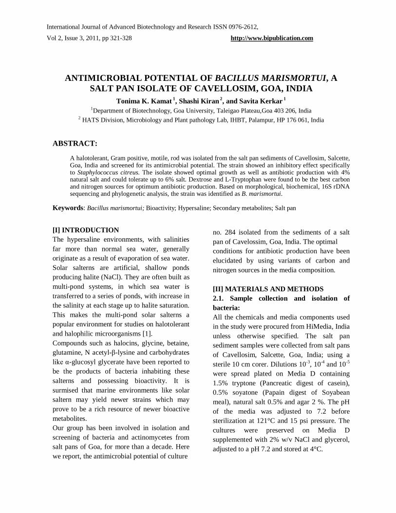
International Journal of Advanced Biotechnology and Research ISSN 0976-2612,
Vol 2, Issue 3, 2011, pp 321-328 http://www.bipublication.com
ANTIMICROBIAL POTENTIAL OF BACILLUS MARISMORTUI, A SALT PAN ISOLATE OF CAVELLOSIM, GOA, INDIA
Tonima K. Kamat 1, Shashi Kiran 2, and Savita Kerkar 1 1Department of Biotechnology, Goa University, Taleigao Plateau,Goa 403 206, India
2 HATS Division, Microbiology and Plant pathology Lab, IHBT, Palampur, HP 176 061, India
ABSTRACT:
A halotolerant, Gram positive, motile, rod was isolated from the salt pan sediments of Cavellosim, Salcette, Goa, India and screened for its antimicrobial potential. The strain showed an inhibitory effect specifically to Staphylococcus citreus. The isolate showed optimal growth as well as antibiotic production with 4% natural salt and could tolerate up to 6% salt. Dextrose and L-Tryptophan were found to be the best carbon and nitrogen sources for optimum antibiotic production. Based on morphological, biochemical, 16S rDNA sequencing and phylogenetic analysis, the strain was identified as B. marismortui.
Keywords: Bacillus marismortui; Bioactivity; Hypersaline; Secondary metabolites; Salt pan [I] INTRODUCTION The hypersaline environments, with salinities far more than normal sea water, generally originate as a result of evaporation of sea water. Solar salterns are artificial, shallow ponds producing halite (NaCl). They are often built as multi-pond systems, in which sea water is transferred to a series of ponds, with increase in the salinity at each stage up to halite saturation. This makes the multi-pond solar salterns a popular environment for studies on halotolerant and halophilic microorganisms [1]. Compounds such as halocins, glycine, betaine, glutamine, N acetyl-β-lysine and carbohydrates like α-glucosyl glycerate have been reported to be the products of bacteria inhabiting these salterns and possessing bioactivity. It is surmised that marine environments like solar saltern may yield newer strains which may prove to be a rich resource of newer bioactive metabolites. Our group has been involved in isolation and screening of bacteria and actinomycetes from salt pans of Goa, for more than a decade. Here we report, the antimicrobial potential of culture
no. 284 isolated from the sediments of a salt pan of Cavelossim, Goa, India. The optimal conditions for antibiotic production have been elucidated by using variants of carbon and nitrogen sources in the media composition. [II] MATERIALS AND METHODS 2.1. Sample collection and isolation of bacteria: All the chemicals and media components used in the study were procured from HiMedia, India unless otherwise specified. The salt pan sediment samples were collected from salt pans of Cavellosim, Salcette, Goa, India; using a sterile 10 cm corer. Dilutions 10-3, 10-4 and 10-5 were spread plated on Media D containing 1.5% tryptone (Pancreatic digest of casein), 0.5% soyatone (Papain digest of Soyabean meal), natural salt 0.5% and agar 2 %. The pH of the media was adjusted to 7.2 before sterilization at 121°C and 15 psi pressure. The cultures were preserved on Media D supplemented with 2% w/v NaCl and glycerol, adjusted to a pH 7.2 and stored at 4°C.

ANTIMICROBIAL POTENTIAL OF BACILLUS MARISMORTUI, A SALT PAN ISOLATE
Savita Kerkar, et al. 322
2.2. Characterisation of culture no. 284: The strain was characterized by morphological, biochemical and phylogenetic analysis. Micro morphology of culture was examined by photomicrography (Gram’s staining method) and Scanning electron microscopy (SEM). Sodium chloride tolerance of the strain was also determined. The utilization of carbon and nitrogen source was carried out by Arahal et. al. [2] method. Various biochemical tests performed for the identification of culture no. 284 are listed in Table 1 and Table 2. The ability of strain 284 to produce enzymes and enzyme inhibitors was also assessed. 2.3. Antimicrobial susceptibility test: A lawn of the culture no. 284 seeded on Muller Hinton agar plates was checked for sensitivity with 29 different antibiotic discs (HiMedia, India). The plate was incubated for 16-24 h at 37°C. After the incubation, the zones of inhibition were measured using a zone measurement scale. The sensitivity or resistance of culture no. 284 to the antibiotic was interpreted with reference to the table provided. The data was compared with Bergey’s manual of Determinative Bacteriology to identify the isolate. 2.4. Phylogenetic analysis: Genomic DNA of the strain was isolated [3] and 16S rDNA sequencing was carried out using eubacterial universal primers. The sequence alignment was done using Clustal W software and a phylogenetic tree was prepared using Neighbour Joining plot [4]. 2.5. Screening of culture no. 284 for antimicrobial activity: The screening method consisted of two steps: Primary and secondary screening. Primary screening was carried out by ‘microbial inhibition spectrum’ [5] on Media D. The test organisms used were Citrobacter freundii, Citrobacter diversus, Acinetobacter baumanii, Salmonella typhimurium, Salmonella paratyphi
A, Proteus mirabilis, Candida albicans, Klebsiella pneumoniae, Morganella morganii, Staphylococcus citreus, Escherichia coli ATCC 25922, Pseudomonas sp. (pigmented), Methicillin resistant Staphylococcus aureus (MRSA), Shigella boydii, Salmonella typhi, Pseudomonas ATCC 27855, Staphylococcus sp. ATCC 25923, Shigella flexineri, Methicillin sensitive Staphylococcus aureus (MSSA) and Vibrio cholerae. Secondary screening was performed by disc diffusion method using media D broth against the test organism Staphylococcus citreus. Sterile filter paper discs (Whatman#1, 5mm diameter) were dipped in Media-D culture broth and allowed to absorb. The dried discs were then placed on actively growing lawn of S. citreus. The inhibition zones were measured after an incubation period of 16-24 h at 37°C using Hi media zone scale. 2.6. Evaluation of salt, carbon and nitrogen source for optimal antibiotic production: Single colony of culture no. 284 was inoculated into media D with 0, 3.5, 5 and 10% natural salt in distilled water at pH 7.2 and incubated at room temperature (28±2°C) on rotary shaker at 150 rpm. Growth was recorded at 600 nm at every 6 h interval. Optimization of carbon source with 1% Dextrose, Ribose and Malonate as sole carbon source in Minimal media [K2HPO4 0.2 g, KH2PO4 0.2 g, MgSO4 0.06 g, natural salt 2g, Distilled water 100 ml (pH 7.0-7.5)] was carried out. The best Nitrogen source was discerned from amino acids viz. L-Tyrosine, L-Tryptophan, L- Asparagine, L- Lysine and L- Glutamic acid in Minimal media supplemented with 3.5 % natural salt. The best carbon and nitrogen source was subsequently used in the antibiotic production media. The pH was maintained between 7.0 to 7.5 and the culture was incubated at room

ANTIMICROBIAL POTENTIAL OF BACILLUS MARISMORTUI, A SALT PAN ISOLATE
Savita Kerkar, et al. 323
temperature (28±2°C) on rotary shaker at 150 rpm. The antibiotic production in the newly designed media incorporating the optimal natural salt concentration, carbon and nitrogen source was assayed by agar disc diffusion method on a lawn of S. citreus, with a media control. The inhibition zones were measured after an incubation period of 16-24 h at 37°C using Hi media zone scale. 2.7. Partial purification of bioactive compound: The bioactive compound produced by culture no. 284 was partially purified by organic solvent extraction method using petroleum ether, chloroform & ethyl acetate [6]. These extracts were individually checked for inhibitory properties using S. citreus as test culture and were also scanned from 190 to 1100 nm (UV mini 1240, UV-Vis Spectrophotometer, Shimadzu) and the peaks were recorded. 2.8. Strain improvement study: Strain improvement was done using Ultraviolet light (UV) mutagenesis to check an increase in the antibiotic production. The culture grown in phosphate buffer saline was exposed to UV light at intervals of 10, 15, 30 and 45 seconds. After exposure the resistant cells were further checked for an increase in antibiotic activity. [III] RESULTS AND DISCUSSION The culture no.284 isolated from the salt pan sediments of Cavellosim, Goa, India exhibited good growth on Media D, with a light brown pigmentation. The morphological & physiological characteristics of the isolate are presented in Table 1. Carbohydrate utilization tests are shown in Table 2. Culture no. 284 was a Gram positive, motile rod shaped bacterium (Figure 1). The isolate was quite specific in its carbohydrate utilization pattern. Out of 33
carbohydrates tested only 3 were utilized. It utilized ribose very scantily but could ferment dextrose and malonate. These 3 sugars were used as carbon sources to optimize the antibiotic potential of culture no.284. Dextrose, at a concentration of 0.5%, was found to be the best carbon source as compared to Malonate and Ribose. It also utilized citrate as a sole carbon source. The isolate seemed to be a rich source in producing significant enzymes viz. Protease, Catalase, Oxidase, Urease and Lysine and Ornithine decarboxylase. The antimicrobial spectrum of culture no. 284 is shown in Table 3. The culture was resistant towards 6 antibiotics and sensitive towards 21 antibiotics. It also showed intermediate sensitivity to Lincomycin and Gentamicin.
Table: 1: Biochemical characterization of
culture no. 284
Sr.No. Characteristics Results 1 Gram staining + 2 Pigment Light
brown 3 ONPG - 4 Lysine decarboxylase + 5 Ornithine
decarboxylase +
6 Nitrate reduction - 7 H2S production - 8 Casein hydrolysis + 9 Starch hydrolysis - 10 Citrate utilization + 11 Phenylalanine
deamination -
12 Catalase + 13 Oxidase + 14 Motility + 15 Amylase inhibitor - 16 Protease inhibitor - 17 Urease +
+ Positive - Negative

ANTIMICROBIAL POTENTIAL OF BACILLUS MARISMORTUI, A SALT PAN ISOLATE
Savita Kerkar, et al. 324
Fig: 1. Scanning Electron micrograph of Bacillus
marismortui
Table: 2. Carbohydrate utilization of culture no. 284
Sr.No. Carbohydrate Results 1 Lactose - 2 Xylose - 3 Maltose - 4 Fructose - 5 Dextrose + 6 Galactose - 7 Raffinose - 8 Trehalose - 9 Melibiose - 10 Sucrose - 11 L-Arabinose - 12 Mannose - 13 Inulin - 14 Sodium gluconate - 15 Glycerol - 16 Salicin - 17 Glucosamine - 18 Dulcitol - 19 Inositol - 20 Sorbitol - 21 Mannitol - 22 Adonitol - 23 α-methyl-D-glucoside - 24 Ribose -/+ 25 Rhamnose - 26 Cellobiose - 27 Melezitose - 28 α-methyl-D-mannoside - 29 Xylitol - 30 Esculin - 31 D-Arabinose - 32 Malonate + 33 Sorbose -
+ Positive - Negative -/+ Scanty utilization
The 16S rDNA sequence of culture no.284 was 1533 bases and showed 99% similarity to Bacillus marismortui. The percentage similarity was on the basis of homology studies done using NCBI BLAST database. A phylogenetic tree, as represented in Figure 2, was prepared on the basis of similarity index. Based on its morphological, chemical properties and phylogenetic analysis, culture no.284 was classified as Bacillus marismortui. Bacillus marismortui was first reported from the Dead Sea [7]. Arahal et. al. [8] proposed the transfer of Bacillus marismortui to the genus Salibacillus as Salibacillus marismortui comb. nov. Later it was proposed by Heyrman et. al. [9] to combine Virgibacillus and Salibacillus in a single genus, Virgibacillus and Bacillus marismortui was renamed as Virgibacillus marismortui comb. nov. The antibiotic produced by B. marismortui, in the present study, was very specifically active against Staphylococcus citreus among the 20 pathogenic cultures tested. The antibiotic did not react with any of the Gram negative organisms tested, nor with Candida albicans and the other three species of Staphylococcus viz. Methicillin resistant Staphylococcus aureus, Methicillin sensitive Staphylococcus aureus and Staphylococcus sp. ATCC 25923. The species specific antagonistic effect of the antibiotic produced was intriguing. Bacitracin, a cyclic polypeptide, is the most common antibiotic produced by Bacillus sp. Few reports are available on marine Bacillus sp. producing antimicrobial compounds [10,11,12,13,14]. But to the best of our knowledge, B. marismortui has not yet been reported of producing any antibacterial compounds.

ANTIMICROBIAL POTENTIAL OF BACILLUS MARISMORTUI, A SALT PAN ISOLATE
Savita Kerkar, et al. 325
Fig: 2. Phylogenetic neighbour-joining tree based on 16S
rDNA sequences showing the relationship between culture
no. 284 and related members of the genus. A.globiformis
was used as an outgroup. Numbers at nodes indicate levels
of bootstrap support ≥ 50% based on a neighbour-joining
analysis of 1000 re-sampled data sets. The tree is based on
neighbour-joining inferences for only complete or nearly
complete sequences. GenBank accession numbers are
given in parentheses. Bar 1% sequence variation.
Fig. 3A. Growth curve of Bacillus marismortui in 0 %
salt.
ANTIMICROBIAL POTENTIAL OF BACILLUS MARISMORTUI, A SALT PAN ISOLATE
Savita Kerkar, et al. 326
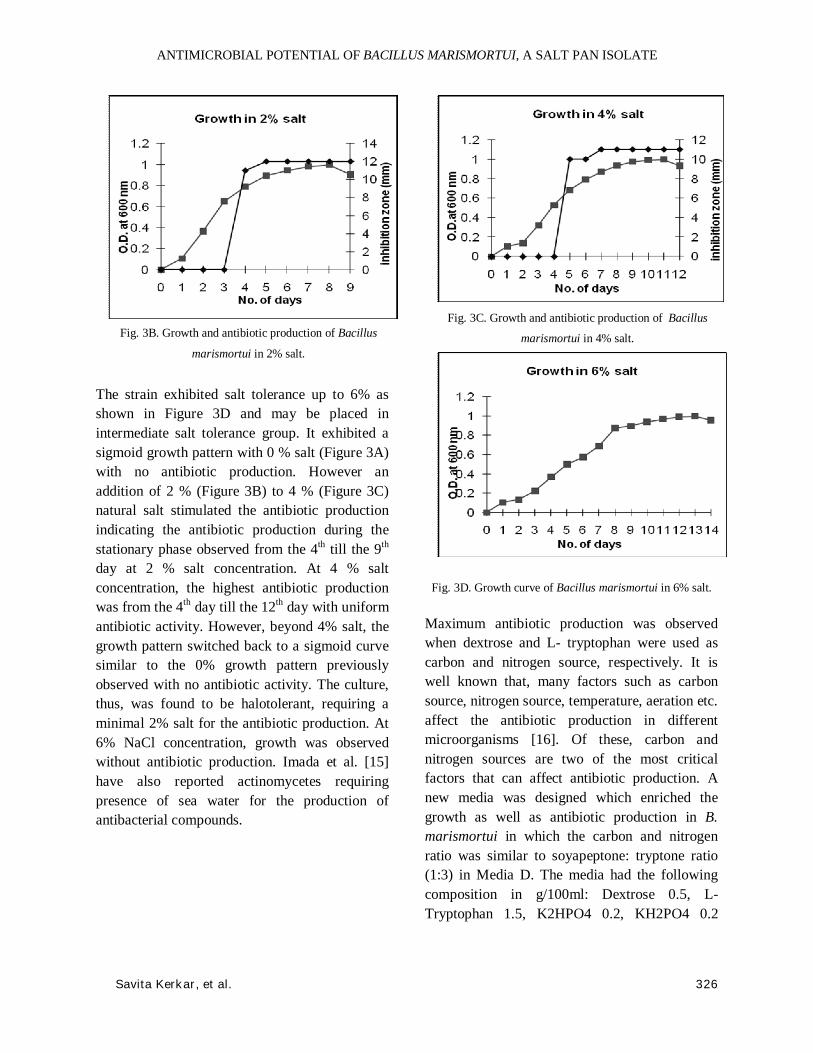
Fig. 3B. Growth and antibiotic production of Bacillus
marismortui in 2% salt.
The strain exhibited salt tolerance up to 6% as shown in Figure 3D and may be placed in intermediate salt tolerance group. It exhibited a sigmoid growth pattern with 0 % salt (Figure 3A) with no antibiotic production. However an addition of 2 % (Figure 3B) to 4 % (Figure 3C) natural salt stimulated the antibiotic production indicating the antibiotic production during the stationary phase observed from the 4th till the 9th day at 2 % salt concentration. At 4 % salt concentration, the highest antibiotic production was from the 4th day till the 12th day with uniform antibiotic activity. However, beyond 4% salt, the growth pattern switched back to a sigmoid curve similar to the 0% growth pattern previously observed with no antibiotic activity. The culture, thus, was found to be halotolerant, requiring a minimal 2% salt for the antibiotic production. At 6% NaCl concentration, growth was observed without antibiotic production. Imada et al. [15] have also reported actinomycetes requiring presence of sea water for the production of antibacterial compounds.
Fig. 3C. Growth and antibiotic production of Bacillus
marismortui in 4% salt.
Fig. 3D. Growth curve of Bacillus marismortui in 6% salt.
Maximum antibiotic production was observed when dextrose and L- tryptophan were used as carbon and nitrogen source, respectively. It is well known that, many factors such as carbon source, nitrogen source, temperature, aeration etc. affect the antibiotic production in different microorganisms [16]. Of these, carbon and nitrogen sources are two of the most critical factors that can affect antibiotic production. A new media was designed which enriched the growth as well as antibiotic production in B. marismortui in which the carbon and nitrogen ratio was similar to soyapeptone: tryptone ratio (1:3) in Media D. The media had the following composition in g/100ml: Dextrose 0.5, L-Tryptophan 1.5, K2HPO4 0.2, KH2PO4 0.2

ANTIMICROBIAL POTENTIAL OF BACILLUS MARISMORTUI, A SALT PAN ISOLATE
Savita Kerkar, et al. 327
MgSO4 0.06, NaCl 2, Distilled water 100 ml (pH 7.0-7.5). The partial purification of the antibiotic, showed an activity in the petroleum ether fraction suggesting a hydrophobic nature of the antibacterial compound. The partially purified fraction was scanned from 190 to 1100 nm and a sharp peak, with an absorbance of 1.349, was obtained at 301 nm in 1/100 dilution, as shown in Figure 4.
Fig. 4. Spectral analysis of partially purified compound.
The antibacterial compound was produced extracellularly and easily diffused in to the medium, confirmed initially with an antibiogram and later by disc diffusion method as seen in Figure-5. The isolate showed insignificant change in growth rate as well as amount of antibiotic production subsequent to UV treatment.
Fig. 5. Culture no. 284 showing antibacterial activity against Staphylococcus citreus.
[V] CONCLUSION In conclusion, the results from present study are promising and need further studies with respect to purification, characterisation and identification of the antibacterial compound produced by Bacillus marismortui.
ACKNOWLEDGEMENT Authors wish to thank the Head, Department of Biotechnology, Goa University for the facilities provided. The authors Shashi Kiran and T Kamat acknowledge the fellowships from DBT and CSIR respectively.
REFERENCES
[1] Kerkar, S. [2004] Ecology of Hypersaline
Microorganisms. In Marine microbiology: Facets &
Opportunities, ed. by N. Ramaiah, pp.53-67.
[2] Arahal, D. R., Dewhirst, F.E., Paster, B.J.,
Volcani, B.E. & Ventosa, A. [1996] Phylogenetic
analysis of some extremely halophilic archaea isolated
from the Dead sea water, determined on the basis of
their 16s r RNA sequences. Appl. Environ. Microbiol.,
62:3779-3786.
[3] Sambrook, J. and Russell, D. W. [2001]
Molecular cloning: A laboratory manual, 3rd edition,
Volume 3, Cold spring Harbor Laboratory Press, Cold
Spring Harbor, New York, pp. A8.9-A8.11.
[4] Saitou, N. and Nie, M. [1987] The neighbor-
joining method: a new method for reconstructing
phylogenetic trees. Mol. Biol. Evol., 4:406-425.
[5] Casida, L. E. [2001] Industrial Microbiology,
New Age International (P) Limited, New Delhi, pp.55-
75.
[6] Riguera, R. [1997] Isolating bioactive
compounds from marine organisms. J. Mar.
Biotechnol., 5:187-193.
[7] Arahal, D.R., Marquez, M.C., Volcani, B.E.,
Schleifer K.H. and Ventosa, A. [1999] Bacillus
marismortui sp. nov., a new moderately halophilic

ANTIMICROBIAL POTENTIAL OF BACILLUS MARISMORTUI, A SALT PAN ISOLATE
Savita Kerkar, et al. 328
species from the Dead sea. Int. J. Sys. Bacteriol.,
49:521-530.
[8] Arahal, D. R., Ma!rquez, M. C., Volcani, B.
E., Schleifer, K. H. and Ventosa, A. [2000]
Reclassification of Bacillus marismortui as
Salibacillus marismortui comb. nov. Int. J. Syst. Evol.
Microbiol., 50:1501–1503.
[9] Heyrman, J., Logan, N. A., Busse, H-J.,
Balcaen, A., Lebbe, L., Rodriguez-Diaz, M., Swings,
J. and De Vos, P. [2003] Virgibacillus carmonensis sp.
nov., Virgibacillus necropolis sp. nov. and
Virgibacillus picturae sp. nov., three novel species
isolated from deteriorated mural paintings, transfer of
the species of the genus Salibacillus to Virgibacillus,
as Virgibacillus marismortui comb. nov. and
Virgibacillus salexigens comb. nov., and emended
description of the genus Virgibacillus. Int. J. Syst.
Evol. Microbiol., 53:501–511.
[10] Nithya, C., Gokila Devi, M. and Pandian, S.
K. [2011] A novel compound from the marine
bacterium Bacillus pumilus S6-15 inhibits biofilm
formation in Gram-positive and Gram-negative
species. Biofouling, 27:5, 519–528.
[11] Musthafa, K. S., Saroja, V., Pandian, S. K.
and Ravi, A. V. [2011] Antipathogenic potential of
marine Bacillus sp. SS4 on N-acyl-homoserine-
lactone-mediated virulence factors production in
Pseudomonas aeruginosa (PAO1). J. Biosci., 36(1),
55–67.
[12] Mondol, M. A. M., Kim, J. H., Lee, H-S.,
Lee, Y-J. and Shin, H. J. [2011] Macrolactin W, a new
antibacterial macrolide from a marine Bacillus sp.
Bioorg. Med. Chem. Lett., 21:3832–3835.
[13] Berrue, F., Ibrahim, A., Boland, P. and Kerr,
R. G. [2009] Newly isolated marine Bacillus pumilus
(SP21): A source of novel lipoamides and other
antimicrobial agents. Pure Appl. Chem., 81:6, 1027–
1031.
[14] Jaruchoktaweechai, C., Suwanborirux, K.,
Tanasupawatt, S., Kittakoop, P. and Menasveta, P.
[2000] New Macrolactins from a Marine Bacillus sp.
Sc026. J. Nat. Prod., 63:984-986.
[15] Imada, C., Koseki, N., Kamata, M.,
Kobayashi, T., and Hamada-Sato, N. [2007] Isolation
and characterization of antibacterial substances
produced by marine actinomycetes in the presence of
sea water. Actinomycetologica, 21:27-31.
[16] Gupte, M.D. and Kulkarni, P.L. [2002] A
study of antifungal antibiotic production by
Streptomyces chattanoogensis MTCC 3423 using full
factorial design. Lett. Appl. Microbiol., 35:22–26.


![Antagonistic activity of Bacillus amyloliquefaciens subsp ...towards other bacteria [16]. In recent years, many studies have been emerged the antimicrobial properties of the genus](https://img.dokumen.tips/doc/110x75/5ec5147a9443b84e7b42b041/antagonistic-activity-of-bacillus-amyloliquefaciens-subsp-towards-other-bacteria.jpg)