Embed Size (px)
Citation preview
International Journal of
Molecular Sciences
Review
Antimicrobial Peptides as Anti-Infective Agents inPre-Post-Antibiotic Era?
Tomislav Roncevic 1,2,* , Jasna Puizina 1 and Alessandro Tossi 3
1 Department of Biology, Faculty of Science, University of Split, 21000 Split, Croatia; [email protected] Laboratory for Aquaculture, Institute of Oceanography and Fisheries, 21000 Split, Croatia3 Department of Life Sciences, University of Trieste, 34127 Trieste, Italy; [email protected]* Correspondence: [email protected]; Tel.: +385-21-619-274
Received: 16 October 2019; Accepted: 11 November 2019; Published: 14 November 2019 �����������������
Abstract: Resistance to antibiotics is one of the main current threats to human health and everyyear multi-drug resistant bacteria are infecting millions of people worldwide, with many dyingas a result. Ever since their discovery, some 40 years ago, the antimicrobial peptides (AMPs) ofinnate defense have been hailed as a potential alternative to conventional antibiotics due to theirrelatively low potential to elicit resistance. Despite continued effort by both academia and start-ups,currently there are still no antibiotics based on AMPs in use. In this study, we discuss what weknow and what we do not know about these agents, and what we need to know to successfullytranslate discovery to application. Understanding the complex mechanics of action of these peptidesis the main prerequisite for identifying and/or designing or redesigning novel molecules with potentbiological activity. However, other aspects also need to be well elucidated, i.e., the (bio)syntheticprocesses, physiological and pathological contexts of their activity, and a quantitative understandingof how physico-chemical properties affect activity. Research groups worldwide are using biological,biophysical, and algorithmic techniques to develop models aimed at designing molecules with thenecessary blend of antimicrobial potency and low toxicity. Shedding light on some open questionsmay contribute toward improving this process.
Keywords: antimicrobial peptides; antimicrobial resistance; AMP identification and design;biosynthesis; mode of action; physico-chemical properties; therapeutic potential
1. Antibiotics and Antimicrobial Resistance—History is Important
Despite a common misconception, exposure to antibiotics is not confined to the modern antibioticera starting from the early 20th century. Ancient civilizations used antibiotics to treat bacterialinfections, with topical applications of moldy bread to wounds being well documented in ancientEgypt, China, Greece, and the Roman empire [1]. Traces of molecules related to modern day antibiotics,the tetracyclines, have been found in skeletal remains from ancient Sudanese Nubia (350–550 A.D.) andfrom the Roman period in Egypt [2–4]. In both cases, it is presumed that they were ingested with grainscontaminated by Streptomycetes [3,4] and most likely had a preventive role rather than a “systemic”effect on infection. Remedies used in traditional Chinese medicine have been the source of potentanti-infective agents for millennia, with artemisinin or “qinghaosu” which is a potent anti-malarialdrug, being one of the best known examples [5].
The modern antibiotic era begins with Paul Erlich’s concept of the “magic bullet” at the beginningof the 20th century. Together with the chemist Alfred Bertheim and bacteriologist Sahachiro Hata,he discovered the arsenic dye arsphenamine, which was later called Salvarsan or 606 (it was the606th compound tested) as a potent anti-syphilis drug [2,6]. This was known as antimicrobialchemotherapy, and the first widely available antibiotic, introduced in 1935 by Gerhard Domagk, was
Int. J. Mol. Sci. 2019, 20, 5713; doi:10.3390/ijms20225713 www.mdpi.com/journal/ijms

Int. J. Mol. Sci. 2019, 20, 5713 2 of 32
sulfonamidochrysoidine or Prontosil, with antibacterial activity occurring in a number of infectiousdiseases. It was soon determined that Prontosil is a precursor to p-aminophenylsulfonamide, alreadydiscovered in 1908 and not patentable, but which led to easily modified derivatives and the era ofsulfonamide antibiotics [7]. Penicillin, which is one of the best known antibiotics, was discovered in1928 by Alexander Fleming, even though mass production started more than 15 years later during WorldWar II, following synthesis and purification work carried out by Howard Florey and Ernest Chain [2,8].In 1944, Selman Waksman (considered “the father of antibiotics” and who first coined the term)discovered an aminoglycoside antibiotic from Streptomyces griseus and named it streptomycin [9,10].This marked the beginning of a golden age, which led to the discovery of more than 20 different groupsof antibiotics in the following decades, of several different types: sulfonamides, β-lactams (with penicillin,cephalosporin, monobactam and carbapenem subclasses), aminoglycosides, quinolones, cyclic peptides(including gramicidins, polymyxins, glycopeptides, lipopeptides and lipoglycopeptides), tetracyclines(including glcyclines), macrolides (including ketolides), amphenicols, nitroimidazoles, dihydrofolate reductase(DHFR) inhibitors, lincosamides, oxazolidinones, and ansamycins (see Figure 1).
There is no doubt that antibiotics have changed the course of medicine and saved untold millionsof lives worldwide since they were first introduced. Infectious diseases that could not be previouslytreated, and their catastrophic effects, could now be easily controlled. However, resistance startedto emerge rapidly. Resistance of Staphylococcus to penicillin had already emerged by 1940, beforemass production had even begun, while resistance toward methicillin was first reported only twoyears after its introduction (see Figure 1). Fortunately, during the golden age, novel antibiotics keptbeing discovered and developed for clinical use. The glycopeptide vancomycin, in use from 1958,was widely believed to be resistance-proof, but, by 1986, a resistant Enterococcus had appeared, andresistant staphylococci emerged before the end of the century (see Figure 1). Multi-drug-resistant(MDR) bacteria have, by now, become a major concern, with the emergence of pan-drug resistant (PDR)or extensively-drug resistant strains (XDR, see Figure 1) such as Mycobacterium tuberculosis resistant tofluoroquinolones and all second-line injectable drugs (capreomycin, kanamycin, or amikacin) [11]. It isconservatively estimated that, in the US and Europe, 2.5 million people are affected by such infectionseach year, and approximately 50,000 people die as a result of the infection [11].
A related major concern is the drying up of the pipeline over the last 30 years, and novel classesof antibiotics entering into it (see Figure 1). Among the limited number of promising compoundsat various stages of clinical investigations and development [12], few, if any, represent a truly newclass, with a completely different mode-of-action to previously approved drugs. Such an example areoxazolidinones, with linezolid being introduced in 2000, but, unfortunately, staphylococcal resistanceemerged shortly after (see Figure 1) [13]. In this period, ketolides, glycylcyclines, and lipopeptides havealso been introduced (see Figure 1). However, telithromycin (ketolide) and tigecycline (glycylcycline)showed adverse side effects, which prompted the Food and Drug Administration (FDA) to labelthe products with their strongest form of warning known as the “black box warning” [14,15]. As aconsequence, telithromycin was discontinued from further use. Daptomycin, which is a lipopeptide,was introduced in 2003 and showed success, but was removed from the World Health Organization(WHO) List of Essential Medicines in 2019 [16].

Int. J. Mol. Sci. 2019, 20, 5713 3 of 32Int. J. Mol. Sci. 2019, 20, x FOR PEER REVIEW 3 of 32
Figure 1. Timeline of antibiotic development as released and in parallel with the timeline for emergence of drug-resistant bacteria [2,6,11,13,17–34]. All antibiotic
classes are represented by at least one antibiotic. For figure clarity, not all antibiotics nor all antibiotic-resistant bacteria are represented in the timeline. (*) Date of
discovery not of release, (**) date of discovery, never introduced due to instability in an aqueous solution [35].
Figure 1. Timeline of antibiotic development as released and in parallel with the timeline for emergence of drug-resistant bacteria [2,6,11,13,17–34]. All antibioticclasses are represented by at least one antibiotic. For figure clarity, not all antibiotics nor all antibiotic-resistant bacteria are represented in the timeline. (*) Date ofdiscovery not of release, (**) date of discovery, never introduced due to instability in an aqueous solution [35].

Int. J. Mol. Sci. 2019, 20, 5713 4 of 32
The drying pipeline has resulted in some shelved antibiotics returning to clinical practice despiteinadequate pharmacological properties, such as colistin (polymyxin E). Discovered in 1950 by Japaneseresearchers [21], it was abandoned shortly afterward due to its nephrotoxicity [36] and the abundanceof other equally potent antibiotics with less pronounced side effects. It is now being used as a “drug oflast resort” against Gram-negative bacterial infections [37]. However, cases of resistance to it haverecently been reported [38], with plasmid-mediated dissemination of the mcr-1 gene was reported inEscherichia coli in 2016 [24], of mcr-8 in Klebsiella pneumoniae [25], and mcr-9 in Salmonella enterica [26]soon after (see Figure 1).
It is, therefore, evident that novel anti-infective agents with alternative modes of action areurgently required to battle the ever evolving, multi-drug resistant bacteria. In 2017, the WHO posteda shortlist of the critical strains to combat: carbapenem- and 3rd generation cephalosporin-resistantAcinetobacter baumannii, Pseudomonas aeruginosa, and Enterobacteriaceae [39]. Many research groupsworldwide are now devoted to solving this crisis and, among other molecules being considered [40,41],antimicrobial peptides (AMPs) are hailed as possible alternatives to conventional antibiotics for sometherapeutic uses, which have a relatively low potential to elicit resistance [42]. Few believe thatcompletely resistance-free antimicrobials can be developed any more.
2. Antimicrobial Peptides—What Are They?
“I Like the Dreams of the Future Better than the History of the Past.”
-Thomas Jefferson (1743–1826)
There is no univocal answer to this question, but we can find a consensus of what the antimicrobialpeptides research community says that they are: multifunctional effector molecules, often gene encoded,produced by almost all organisms, and having a direct antimicrobial activity and/or immunomodulatoryproperties [43,44]. In light of the burgeoning resistance problem, peptides with a direct antimicrobialactivity (AMPs) and often immune-regulatory capacities are easy to fit to the “dream (molecules) ofthe future” definition, to paraphrase Thomas Jefferson. Many AMPs have been discovered to date (seebelow), and they are reported to be active against Gram-negative and Gram-positive bacteria, againstinfective fungi and sometimes with antiviral, antiparasitic, or antiprotozoal properties [45]. Many ofthese have also been shown to modulate host immunity by activating immunocytes, and modulatinginflammation, alternatively suppressing or promoting it [46]. To emphasize their pleiotropic naturein higher organisms, natural AMPs are often referred to as ‘host defense peptides’ (HDP), or, morespecifically, as ‘innate defense regulatory (IDR) peptides,’ since reports on their immunomodulatoryactivities have mostly been confirmed at the level of innate immunity [46,47]. Their impact andimportance to innate immunity is supported by their abundance in all eukaryotic organisms (fungi,algae, plants, invertebrate, and vertebrate animals), and distribution in cells and tissues at the front lineof host defense against infection (mainly circulating immunocytes and epithelia) (see Figure 2). Thededicated Collection of Anti-Microbial Peptides (CAMPR3) database currently contains 8164 entries forpeptides, most of which (~74%) come from animals [48]. A particularly abundant source are anuranspecies, with almost two thousand peptide sequences reported in the dedicated Database of AnuranDefense Peptides (DADP) [49]. AMPs are, however, also well represented in prokaryotes, produced byboth Gram-negative and Gram-positive bacteria, with one abundant class being the bacteriocins [50].In this case, however, their role is somewhat different to that in eukaryotes, as they are principallyused to clear the immediate environment of producer bacteria from competition by closely related,bacterial strains [50].

Int. J. Mol. Sci. 2019, 20, 5713 5 of 32Int. J. Mol. Sci. 2019, 20, x FOR PEER REVIEW 5 of 32
Figure 2. Distribution of AMPs across kingdoms based on sequences in the CAMPR3 database.
(http://www.camp.bicnirrh.res.in/dbStat.php) [48].
3. Ribosomal vs. Non-ribosomal Synthesis and Antimicrobial Peptide Precursors
AMPs are often gene encoded and ribosomally synthesized (this is, by far, the most common
case in eukaryotes), or can be assembled by large multi-functional enzymes known as non-ribosomal
peptide synthetases (NRPSs) [51,52]. The latter process is used by bacteria and fungi [52] and allows
incorporation of non-proteinogenic amino acids into the peptides (often the D-enantiomers of natural
residues) and to further modify the peptides with ring formation, glycosylation, hydroxylation, or
acylation [53,54]. There are ~500 non-proteinogenic amino acids known today, which possess added
structural and functional features that can contribute significantly to a peptide’s potency. In fact, the
cyclic peptide antibiotics polymyxin B, gramicidin S, and vancomycin are synthesized in this manner
[54] and all contain some non-proteinogenic amino acids in their sequences [53,55].
Gene encoded, ribosomally synthesized peptides are produced by almost all forms of life,
including bacteria [42,48,56]. Quite often, multiple AMP genes are clustered at a single chromosomal
locus, which is the case with α- and β-defensins [57] and can be co-expressed. They are, furthermore,
frequently expressed as inactive precursors, containing a signal peptide region and a pro-piece that
can serve to keep the mature peptide inactive until it is conveyed to the site of infection, where it is
proteolytically released (see Figure 3). For this reason, the pro-piece is often anionic to complement
the mature peptide, which is generally cationic. In most cases, the pro-region precedes (it is N-
terminal to) the AMP sequence, but cases are known where it is the C-terminal ( such as for some fish
and plant peptides) [58,59].
Figure 3. Representation of AMP expression. Peptides are cleaved at dibasic cleavage sites (-KR, -RR,
in red). The proregion has a distinctly anionic nature. Depicted peptide sequences belong to anuran
AMPs from the Ranidae family [60–63].
Figure 2. Distribution of AMPs across kingdoms based on sequences in the CAMPR3 database.(http://www.camp.bicnirrh.res.in/dbStat.php) [48].
3. Ribosomal vs. Non-ribosomal Synthesis and Antimicrobial Peptide Precursors
AMPs are often gene encoded and ribosomally synthesized (this is, by far, the most commoncase in eukaryotes), or can be assembled by large multi-functional enzymes known as non-ribosomalpeptide synthetases (NRPSs) [51,52]. The latter process is used by bacteria and fungi [52] and allowsincorporation of non-proteinogenic amino acids into the peptides (often the D-enantiomers of naturalresidues) and to further modify the peptides with ring formation, glycosylation, hydroxylation, oracylation [53,54]. There are ~500 non-proteinogenic amino acids known today, which possess addedstructural and functional features that can contribute significantly to a peptide’s potency. In fact,the cyclic peptide antibiotics polymyxin B, gramicidin S, and vancomycin are synthesized in thismanner [54] and all contain some non-proteinogenic amino acids in their sequences [53,55].
Gene encoded, ribosomally synthesized peptides are produced by almost all forms of life, includingbacteria [42,48,56]. Quite often, multiple AMP genes are clustered at a single chromosomal locus, whichis the case with α- and β-defensins [57] and can be co-expressed. They are, furthermore, frequentlyexpressed as inactive precursors, containing a signal peptide region and a pro-piece that can serve tokeep the mature peptide inactive until it is conveyed to the site of infection, where it is proteolyticallyreleased (see Figure 3). For this reason, the pro-piece is often anionic to complement the maturepeptide, which is generally cationic. In most cases, the pro-region precedes (it is N-terminal to) theAMP sequence, but cases are known where it is the C-terminal ( such as for some fish and plantpeptides) [58,59].
Int. J. Mol. Sci. 2019, 20, x FOR PEER REVIEW 5 of 32
Figure 2. Distribution of AMPs across kingdoms based on sequences in the CAMPR3 database.
(http://www.camp.bicnirrh.res.in/dbStat.php) [48].
3. Ribosomal vs. Non-ribosomal Synthesis and Antimicrobial Peptide Precursors
AMPs are often gene encoded and ribosomally synthesized (this is, by far, the most common
case in eukaryotes), or can be assembled by large multi-functional enzymes known as non-ribosomal
peptide synthetases (NRPSs) [51,52]. The latter process is used by bacteria and fungi [52] and allows
incorporation of non-proteinogenic amino acids into the peptides (often the D-enantiomers of natural
residues) and to further modify the peptides with ring formation, glycosylation, hydroxylation, or
acylation [53,54]. There are ~500 non-proteinogenic amino acids known today, which possess added
structural and functional features that can contribute significantly to a peptide’s potency. In fact, the
cyclic peptide antibiotics polymyxin B, gramicidin S, and vancomycin are synthesized in this manner
[54] and all contain some non-proteinogenic amino acids in their sequences [53,55].
Gene encoded, ribosomally synthesized peptides are produced by almost all forms of life,
including bacteria [42,48,56]. Quite often, multiple AMP genes are clustered at a single chromosomal
locus, which is the case with α- and β-defensins [57] and can be co-expressed. They are, furthermore,
frequently expressed as inactive precursors, containing a signal peptide region and a pro-piece that
can serve to keep the mature peptide inactive until it is conveyed to the site of infection, where it is
proteolytically released (see Figure 3). For this reason, the pro-piece is often anionic to complement
the mature peptide, which is generally cationic. In most cases, the pro-region precedes (it is N-
terminal to) the AMP sequence, but cases are known where it is the C-terminal ( such as for some fish
and plant peptides) [58,59].
Figure 3. Representation of AMP expression. Peptides are cleaved at dibasic cleavage sites (-KR, -RR,
in red). The proregion has a distinctly anionic nature. Depicted peptide sequences belong to anuran
AMPs from the Ranidae family [60–63].
Figure 3. Representation of AMP expression. Peptides are cleaved at dibasic cleavage sites (-KR, -RR,in red). The proregion has a distinctly anionic nature. Depicted peptide sequences belong to anuranAMPs from the Ranidae family [60–63].

Int. J. Mol. Sci. 2019, 20, 5713 6 of 32
The activity of AMPS is, therefore, regulated not only by the expression level but also by thepresence and abundance of appropriate proteases at the right place and the right time to cleave thepeptide, which is generally at dibasic cleavage sites (see Figure 3) [57,64]. The signal peptide is acommon feature of prokaryotic and eukaryotic proteins that need to enter secretory pathways [65]. Avery useful aspect of antimicrobial pro-peptides is that signal regions for a given class can be much moreevolutionarily conserved than the mature peptides themselves (see Figure 3) [58,66–68]. This providesa very useful handle for sequence mining in databases. The diversity of the mature AMP sequencemost likely occurs as species’ adaptation to specific microbial communities in a particular environment.While there is still no solid explanation for the phenomenon of signal sequence conservation, it givesvaluable insights into the evolution of some AMP families, as in the case with the anuran ones [69].
Lastly, it is worth noting that a majority of gene encoded AMPs undergo post-translationalmodifications, currently classified into more than 15 types, including disulfide-bridge formation,N-terminal or C-terminal capping (acetylation, pyroglutamic acid formation, amidation), halogenation,hydroxylation, phosphorylation, glycosylation, etc. Peptides can be modified to a greater or lesserextent, which contributes to peptide potency and/or stability [70].
4. Physico-Chemical Properties
Certain physico-chemical properties of AMPs are undeniably related to their interaction with lipidmolecules that make up the bilayer system in the membrane of the target cells and correlate directlywith the peptides’ biological activity and specificity. In fact, the same consideration could be made forother components of the microbial cell wall, but the data is less consistent. However, even limiting one’sfocus to the lipid bilayer, there is still an imperfect understanding of the complex relationship betweenthe AMP and membrane, since even peptides with very similar structures can have remarkably differentreported mechanisms of action (e.g., buforin and magainin 2, or LL-37 and the rhesus orthologueRL-37) [71–73]. A better understanding of the relationship between physico-chemical properties andbiological activity is, therefore, required to identify features that are responsible for potency andspecificity of AMPs.
4.1. Molecular Type, Size, and Structure
AMPs have been divided into several categories, according to particular features of their secondarystructure. The simplest and most widely used classification divides them into extended structures,linear α-helical peptides and peptides with β-sheet or hairpin-like structures, generally braced bydisulfide bridges [74]. Guha et al. [75] have, however, proposed a more elaborate taxonomy based onsecondary motifs, with six different groups including i) α-helix, ii) 3/10 helix, iii) pi-helix, iv) β-strand, v)β-turn, and vi) disordered coil, which may concern the entire peptide or only parts of it, where somescaffolds combine to make different structural motifs. Essentially, this classification still depicts threemajor classes of AMPs: helical, β strand, and extended. In this review, the latest Wang terminology willbe used to classify peptides in four classes [76]: α helical (e.g., human LL-37), β sheet (e.g., the human α
defensin HD-4), αβ peptides (e.g., the human β defensin hBD-2), and non αβ peptides (e.g., the small,extended Trp-rich peptide indolicidin) (see Figure 4).

Int. J. Mol. Sci. 2019, 20, 5713 7 of 32
Int. J. Mol. Sci. 2019, 20, x FOR PEER REVIEW 7 of 32
Figure 4. An overview of major structural classes of antimicrobial peptides. The structures taken as
an example were solved either by nuclear magnetic resonance spectroscopy or X-ray diffraction and
coordinates were downloaded from Protein Data Bank (PDB) (https://www.rcsb.org/) [77]. PDB IDs:
LL-37 (2k6o), human α-defensin-4 (1zmm), human β-defensin-2 (1fd3), and indolicidin (1g89).
Visualization was done using PyMOL 1.8 [78] and amino acids colored according to a normalized
Eisenberg hydrophobicity scale (light grey – polar, red - hydrophobic) [79].
The “α−helical” peptides, which are among the most studied and, consequently, the better
understood, generally exhibit little or no structuring in a bulk aqueous solution, but only adopt this
defined secondary structure in the presence of a bacterial membrane or some other type of anisotropic
environment (sodium dodecyl sulfate micelles or water/trifluoroethanol mixtures) [46]. The adoption
of this active structure is aided by:
• the presence of helix-stabilizing residues distributed throughout the sequence (e.g., Leu, Ala,
Lys),
• the clustering hydrophobic residues on one side of the helix when it forms, which allows
insertion into the membrane bilayer (or, seen the other way round, the presence of a lipid
layer that induces appropriately distributed hydrophobic residues to cluster into a well-
defined sector of the helix by interacting with it),
• salt-bridging between oppositely charged residues placed next to each other when the helix
forms (normally, but not necessarily, when these residues are spaced three or four positions
apart [80]).
It should be noted that increased helix stability correlates with increased potency, but only to a
certain extent [81]. In fact, an increased propensity to transit from a coiled to helical conformation on
passing from bulk solution to the membrane surface will generally positively affect activity. On the
other hand, an increased propensity for helix formation in bulk solution will result in ‘sticky’
molecules (due to formation of a hydrophobic sector exposed to an aqueous environment), that tend
to oligomerize or interact with other hydrophobic molecular surfaces. In the first case, this will alter
the mode of the membrane interaction, which inevitably affects activity. In the second case, it leads
to sequestration, with a negative effect on potency [72,82].
Figure 4. An overview of major structural classes of antimicrobial peptides. The structures takenas an example were solved either by nuclear magnetic resonance spectroscopy or X-ray diffractionand coordinates were downloaded from Protein Data Bank (PDB) (https://www.rcsb.org/) [77]. PDBIDs: LL-37 (2k6o), human α-defensin-4 (1zmm), human β-defensin-2 (1fd3), and indolicidin (1g89).Visualization was done using PyMOL 1.8 [78] and amino acids colored according to a normalizedEisenberg hydrophobicity scale (light grey—polar, red—hydrophobic) [79].
The “α−helical” peptides, which are among the most studied and, consequently, the betterunderstood, generally exhibit little or no structuring in a bulk aqueous solution, but only adopt thisdefined secondary structure in the presence of a bacterial membrane or some other type of anisotropicenvironment (sodium dodecyl sulfate micelles or water/trifluoroethanol mixtures) [46]. The adoptionof this active structure is aided by:
• the presence of helix-stabilizing residues distributed throughout the sequence (e.g., Leu, Ala, Lys),• the clustering hydrophobic residues on one side of the helix when it forms, which allows insertion
into the membrane bilayer (or, seen the other way round, the presence of a lipid layer that inducesappropriately distributed hydrophobic residues to cluster into a well-defined sector of the helixby interacting with it),
• salt-bridging between oppositely charged residues placed next to each other when the helix forms(normally, but not necessarily, when these residues are spaced three or four positions apart [80]).
It should be noted that increased helix stability correlates with increased potency, but only to acertain extent [81]. In fact, an increased propensity to transit from a coiled to helical conformationon passing from bulk solution to the membrane surface will generally positively affect activity. Onthe other hand, an increased propensity for helix formation in bulk solution will result in ‘sticky’molecules (due to formation of a hydrophobic sector exposed to an aqueous environment), that tend tooligomerize or interact with other hydrophobic molecular surfaces. In the first case, this will alter themode of the membrane interaction, which inevitably affects activity. In the second case, it leads tosequestration, with a negative effect on potency [72,82].
The size of helical peptides (sequence length) is also an important feature, especially in the contextof peptide activity, since a minimum of seven to eight amino acids are needed to form an amphipathic
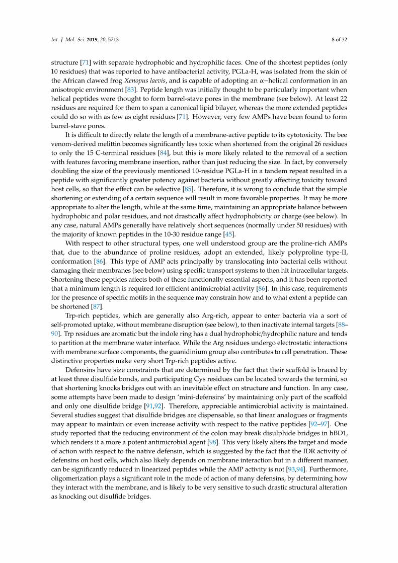
Int. J. Mol. Sci. 2019, 20, 5713 8 of 32
structure [71] with separate hydrophobic and hydrophilic faces. One of the shortest peptides (only10 residues) that was reported to have antibacterial activity, PGLa-H, was isolated from the skin ofthe African clawed frog Xenopus laevis, and is capable of adopting an α−helical conformation in ananisotropic environment [83]. Peptide length was initially thought to be particularly important whenhelical peptides were thought to form barrel-stave pores in the membrane (see below). At least 22residues are required for them to span a canonical lipid bilayer, whereas the more extended peptidescould do so with as few as eight residues [71]. However, very few AMPs have been found to formbarrel-stave pores.
It is difficult to directly relate the length of a membrane-active peptide to its cytotoxicity. The beevenom-derived melittin becomes significantly less toxic when shortened from the original 26 residuesto only the 15 C-terminal residues [84], but this is more likely related to the removal of a sectionwith features favoring membrane insertion, rather than just reducing the size. In fact, by converselydoubling the size of the previously mentioned 10-residue PGLa-H in a tandem repeat resulted in apeptide with significantly greater potency against bacteria without greatly affecting toxicity towardhost cells, so that the effect can be selective [85]. Therefore, it is wrong to conclude that the simpleshortening or extending of a certain sequence will result in more favorable properties. It may be moreappropriate to alter the length, while at the same time, maintaining an appropriate balance betweenhydrophobic and polar residues, and not drastically affect hydrophobicity or charge (see below). Inany case, natural AMPs generally have relatively short sequences (normally under 50 residues) withthe majority of known peptides in the 10-30 residue range [45].
With respect to other structural types, one well understood group are the proline-rich AMPsthat, due to the abundance of proline residues, adopt an extended, likely polyproline type-II,conformation [86]. This type of AMP acts principally by translocating into bacterial cells withoutdamaging their membranes (see below) using specific transport systems to then hit intracellular targets.Shortening these peptides affects both of these functionally essential aspects, and it has been reportedthat a minimum length is required for efficient antimicrobial activity [86]. In this case, requirementsfor the presence of specific motifs in the sequence may constrain how and to what extent a peptide canbe shortened [87].
Trp-rich peptides, which are generally also Arg-rich, appear to enter bacteria via a sort ofself-promoted uptake, without membrane disruption (see below), to then inactivate internal targets [88–90]. Trp residues are aromatic but the indole ring has a dual hydrophobic/hydrophilic nature and tendsto partition at the membrane water interface. While the Arg residues undergo electrostatic interactionswith membrane surface components, the guanidinium group also contributes to cell penetration. Thesedistinctive properties make very short Trp-rich peptides active.
Defensins have size constraints that are determined by the fact that their scaffold is braced byat least three disulfide bonds, and participating Cys residues can be located towards the termini, sothat shortening knocks bridges out with an inevitable effect on structure and function. In any case,some attempts have been made to design ‘mini-defensins’ by maintaining only part of the scaffoldand only one disulfide bridge [91,92]. Therefore, appreciable antimicrobial activity is maintained.Several studies suggest that disulfide bridges are dispensable, so that linear analogues or fragmentsmay appear to maintain or even increase activity with respect to the native peptides [92–97]. Onestudy reported that the reducing environment of the colon may break disulphide bridges in hBD1,which renders it a more a potent antimicrobial agent [98]. This very likely alters the target and modeof action with respect to the native defensin, which is suggested by the fact that the IDR activity ofdefensins on host cells, which also likely depends on membrane interaction but in a different manner,can be significantly reduced in linearized peptides while the AMP activity is not [93,94]. Furthermore,oligomerization plays a significant role in the mode of action of many defensins, by determining howthey interact with the membrane, and is likely to be very sensitive to such drastic structural alterationas knocking out disulfide bridges.
Int. J. Mol. Sci. 2019, 20, 5713 9 of 32
4.2. Charge and Hydrophobicity
The net charge of known natural AMPs varies widely from cationic (most often) to anionic (rare),which ranges from +16 to −6 [80,99–101]. The vast majority of identified peptides have an intermediatenet positive charge (centering around +6) that can be directly correlated with peptide potency andselectivity. There seems to be an optimum charge span for activity, so that higher or lower valuesoutside this range can result in reduced activity toward bacterial cells and/or increased toxicity towardhost cells. The relationship between these parameters and function has again been most extensivelyprobed in helical peptides. Dathe et al. [102] have shown that increasing the charge of magaininanalogs above +5 resulted in both increased hemolysis and loss of antimicrobial potency. Giangasperoet al. [81] came to a similar conclusion when varying the charge of helical peptides, which, otherwise,had relatively similar mean hydrophobicity, amphipathicity, and helicity. The decreased amicrobialpotency was, in part, attributed to a reduced propensity for helix formation due to the increasedcharge density (clustering of positive charges in one sector of the helix leads to repulsion). More recentfindings suggest it could also result from repulsion between highly charged peptides at the membranesurface, which leads to a lower concentration of membrane-adsorbed peptides [103]. In principle, thedistribution of positively charged residues should not correlate with peptide potency, which shouldonly depend on the overall peptide charge [81,104]. However, it can make a significant difference if itaffects the formation of helix-stabilizing salt bridges, as observed in artificial variants of the humanhelical peptide LL-37 [73].
In a similar way, there seems to be an optimal hydrophobicity window for peptides to have anoptimal balance between antimicrobial activity and host cell toxicity. On average, AMPs containapproximately 50% hydrophobic residues. The overall hydrophobicity affects the peptide’s capacityto partition into the lipid bilayer, and can be directly correlated with both potency and host celltoxicity [105]. Increasing or decreasing this property outside the optimal range can result in adecrease of antimicrobial activity and an increase in blood cell lysis, not necessarily accompanied byimproved antimicrobial activity [81,105,106]. In fact, an increased hydrophobicity can result in reducedantimicrobial activity if it promotes self-association, for the same reasons as an excessive stabilizationof the helical structure (see above). This impedes access to the bacterial membrane and, therefore,lowers the concentration of the peptide actually impacting it [104,106].
4.3. Amphipathicity and Structural Stability in Helical Peptides
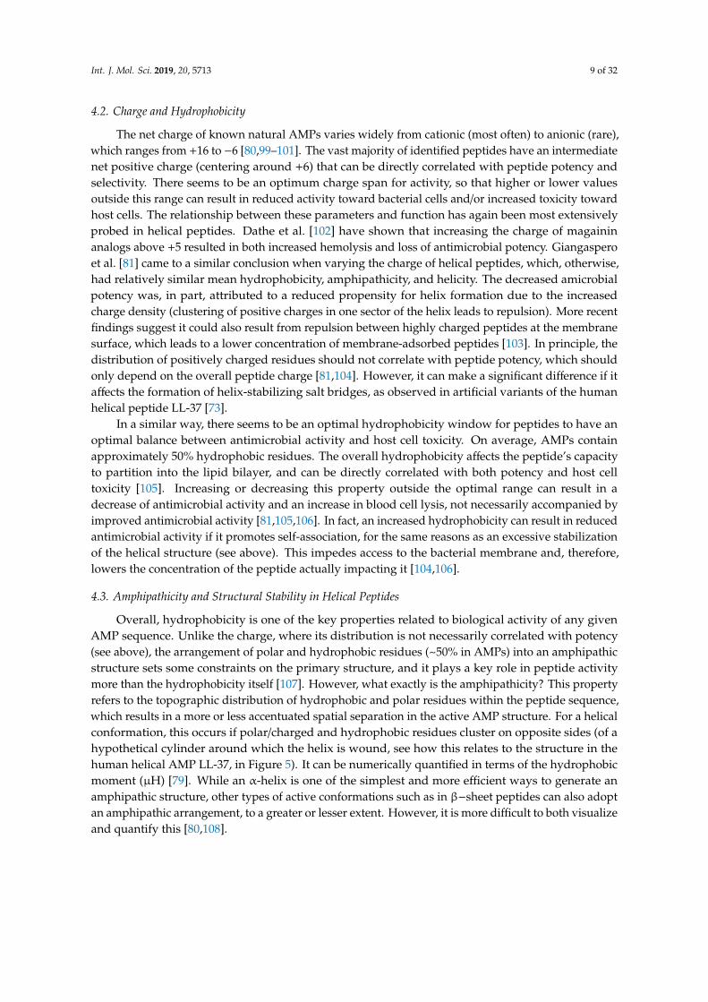
Overall, hydrophobicity is one of the key properties related to biological activity of any givenAMP sequence. Unlike the charge, where its distribution is not necessarily correlated with potency(see above), the arrangement of polar and hydrophobic residues (~50% in AMPs) into an amphipathicstructure sets some constraints on the primary structure, and it plays a key role in peptide activitymore than the hydrophobicity itself [107]. However, what exactly is the amphipathicity? This propertyrefers to the topographic distribution of hydrophobic and polar residues within the peptide sequence,which results in a more or less accentuated spatial separation in the active AMP structure. For a helicalconformation, this occurs if polar/charged and hydrophobic residues cluster on opposite sides (of ahypothetical cylinder around which the helix is wound, see how this relates to the structure in thehuman helical AMP LL-37, in Figure 5). It can be numerically quantified in terms of the hydrophobicmoment (µH) [79]. While an α-helix is one of the simplest and more efficient ways to generate anamphipathic structure, other types of active conformations such as in β−sheet peptides can also adoptan amphipathic arrangement, to a greater or lesser extent. However, it is more difficult to both visualizeand quantify this [80,108].
Int. J. Mol. Sci. 2019, 20, 5713 10 of 32Int. J. Mol. Sci. 2019, 20, x FOR PEER REVIEW 10 of 32
Figure 5. Secondary structure and helical wheel projection of human cathelicidin LL-37. The structure
and projection were, respectively, obtained from PDB [77] (ID: 2k6o) and HeliQuest [109]. The
residues were colored according to their hydrophobicity with ~40% hydrophobic and 60% polar
amino acids in an appreciable amphipathic arrangement. Hydrophobic (yellow and green), polar
charged [red (−) and blue (+)], polar uncharged (light to dark purple), and glycine (grey).
Lastly, helicity is the propensity of an AMP to adopt a helical structure. As discussed above, it
plays a significant role in the antibacterial activity, and, in general, it seems to correlate more with
the toxicity toward host cells than antimicrobial potency, in a manner that relates to its effect on
oligomerization. It can be reduced by incorporating D-amino acids into the peptide sequence,
without greatly affecting potency. However, this can narrow the activity spectrum. As reported by
Papo et al. [110], replacing up to a third of L-amino acids with their D-enantiomers resulted in
peptides devoid of haemolytic activity that maintained an appreciable antibacterial potency,
especially against Gram-negative bacteria. Furthermore, they are protected from proteolytic
degradation, which should increase the bioavailability of such synthetic peptides.
5. Mode of Action
The mechanism of action of numerous AMPs has been extensively studied. Experiments have
often been carried out with artificial membranes, typically large or giant unilamellar vesicles, and
less frequently on microbial cells, using fluorescent dyes and labeled peptides. In any case, a widely
accepted subdivision of AMPs, according to their mode of action, is i) membrane active and ii) non-
lytic [111]. Some AMPs can act upon bacteria using both of these two major mechanisms, and
sometimes switching from one to the other, depending on the peptide concentration, the membrane
characteristics of a particular bacterial species, or its growth phase [112].
5.1. Disrupting Bacterial Cytoplasmic Membrane Integrity—A Primary Inactivation Mechanism
The term “membrane permeabilizing” peptides (MPP) [75] is more general than the often, and
sometimes inappropriately used, “pore-forming” peptides [113–115]. Considering the complexity of
lytic mechanism(s) of membrane-active AMPs (which is not restricted only to “pore-forming”), it is
more appropriate (see Figure 6). An MPP must initially partition into a membrane and, therefore, be
amphipathic for at least part of its structure, i.e., it must have some form of “interfacial activity.” This,
nonetheless, allows for remarkable structural diversity, which results in functional diversity, so that
Figure 5. Secondary structure and helical wheel projection of human cathelicidin LL-37. The structureand projection were, respectively, obtained from PDB [77] (ID: 2k6o) and HeliQuest [109]. The residueswere colored according to their hydrophobicity with ~40% hydrophobic and 60% polar amino acids inan appreciable amphipathic arrangement. Hydrophobic (yellow and green), polar charged [red (−) andblue (+)], polar uncharged (light to dark purple), and glycine (grey).
Amphipathicity aids activity of helical peptides since it allows them to sink their hydrophobicfaces into the membrane bilayer, which is an essential step leading to membrane disruption. It must becorrectly tuned for an optimal balance between anti-bacterial potency and host cell toxicity. In general,the hydrophobic moment in helical AMPs is around 60% of the maximum possible value. Increasing itabove this value does not greatly increase potency but can significantly increase toxicity [81,82].
Lastly, helicity is the propensity of an AMP to adopt a helical structure. As discussed above, itplays a significant role in the antibacterial activity, and, in general, it seems to correlate more withthe toxicity toward host cells than antimicrobial potency, in a manner that relates to its effect onoligomerization. It can be reduced by incorporating D-amino acids into the peptide sequence, withoutgreatly affecting potency. However, this can narrow the activity spectrum. As reported by Papoet al. [110], replacing up to a third of L-amino acids with their D-enantiomers resulted in peptidesdevoid of haemolytic activity that maintained an appreciable antibacterial potency, especially againstGram-negative bacteria. Furthermore, they are protected from proteolytic degradation, which shouldincrease the bioavailability of such synthetic peptides.
5. Mode of Action
The mechanism of action of numerous AMPs has been extensively studied. Experiments haveoften been carried out with artificial membranes, typically large or giant unilamellar vesicles, andless frequently on microbial cells, using fluorescent dyes and labeled peptides. In any case, a widelyaccepted subdivision of AMPs, according to their mode of action, is i) membrane active and ii)non-lytic [111]. Some AMPs can act upon bacteria using both of these two major mechanisms, andsometimes switching from one to the other, depending on the peptide concentration, the membranecharacteristics of a particular bacterial species, or its growth phase [112].

Int. J. Mol. Sci. 2019, 20, 5713 11 of 32
5.1. Disrupting Bacterial Cytoplasmic Membrane Integrity—A Primary Inactivation Mechanism
The term “membrane permeabilizing” peptides (MPP) [75] is more general than the often, andsometimes inappropriately used, “pore-forming” peptides [113–115]. Considering the complexity oflytic mechanism(s) of membrane-active AMPs (which is not restricted only to “pore-forming”), it ismore appropriate (see Figure 6). An MPP must initially partition into a membrane and, therefore, beamphipathic for at least part of its structure, i.e., it must have some form of “interfacial activity.” This,nonetheless, allows for remarkable structural diversity, which results in functional diversity, so thatsome MPPs are active only against a narrow spectrum of microbial species while others have a verybroad spectrum of activity. Wimley’s group has recently pointed out that the process of membranepermeabilization should not be considered as being simply due to a series of stochastic events involvingone or more peptide molecules, nor should it be ascribed to a well-defined sequence of events. In otherwords, it requires neither discrete events nor the formation of static molecular entities. It is betterdescribed as a “mechanistic landscape” that varies depending on the experimental conditions andvariables such as peptide concentration, bilayer lipid composition, temperature, ionic strength, andpH [75].
Results from a recent molecular dynamics (MD) simulation case study on maculatin, isolated fromthe skin glands of a green-eyed tree frog Litoria genimaculata [116], are in line with the proposed scenario.This particular peptide has been reported to act by pore formation [117]. Simulation work done byWang et al. [118] showed that these pores continuously form and dissociate in the membrane. Moreover,the architecture of the pores varies, and is dominated by hexamers, heptamers, and octamers, withpeptide molecules having a strong but not absolute preference for an antiparallel peptide orientation inthe oligomers. Remarkably, the assembly of maculatin into pores seems to be driven by the successiveaddition of peptide molecules to an already existing transmembrane inserted helix to form a growingoligomer. Therefore, the translocation of the polar side chains of the incoming peptide is ‘catalyzed’ bythe polar face of the already inserted peptide(s).
It follows that, even for very well-studied AMPs, the molecular mechanisms of membranepermeabilization cannot have been completely elucidated, and many questions remain [75,119]. Yet,some common steps can be inferred to occur at the bacterial membrane, which eventually leads to itsdisruption. This includes: i) initial attraction of the AMP to the membrane surface and interaction,ii) adoption of an active conformation, iii) insertion into the bilayer and concentration dependentaccumulation, and iv) (in some cases) self-association/oligomerization [111]. The order in which thesethree steps occur will significantly affect the type of membrane lesion.
Most α−helical AMPs do not adopt this conformation in a bulk solution (see above) so that theinitial interaction at the membrane level occurs between a positively charged peptide coil and thenegatively charged phospholipid head-groups in the bacterial membrane surface [80,121]. This allowsredistribution of hydrophobic sidechains so that they can interact with the membrane acyl chainsleading to adsorption into the membrane, which, in turn, induces conformational changes in thepeptide (to a stable amphipathic helix) that maximize these interactions [122], and this allows a deeperinsertion into the lipid bilayer, which alters its structure. In other words, it follows the steps i), ii), iii),iv) in that order. By contrast, β−sheet peptides already have a stable, disulphide-braced amphipathicconformation that is maintained on membrane insertion. This is likely the case for pre-formed helicalpeptides, like LL-37, which are stabilized by internal salt-bridges [123]. In both cases, the pre-formedstructures favor oligomerization [72,124], so these peptides may follow a different order for the stepsii), iv), i), iii).

Int. J. Mol. Sci. 2019, 20, 5713 12 of 32
Int. J. Mol. Sci. 2019, 20, x FOR PEER REVIEW 11 of 32
some MPPs are active only against a narrow spectrum of microbial species while others have a very
broad spectrum of activity. Wimley’s group has recently pointed out that the process of membrane
permeabilization should not be considered as being simply due to a series of stochastic events
involving one or more peptide molecules, nor should it be ascribed to a well-defined sequence of
events. In other words, it requires neither discrete events nor the formation of static molecular
entities. It is better described as a “mechanistic landscape” that varies depending on the experimental
conditions and variables such as peptide concentration, bilayer lipid composition, temperature, ionic
strength, and pH [75].
Results from a recent molecular dynamics (MD) simulation case study on maculatin, isolated
from the skin glands of a green-eyed tree frog Litoria genimaculata [116], are in line with the proposed
scenario. This particular peptide has been reported to act by pore formation [117]. Simulation work
done by Wang et al. [118] showed that these pores continuously form and dissociate in the membrane.
Moreover, the architecture of the pores varies, and is dominated by hexamers, heptamers, and
octamers, with peptide molecules having a strong but not absolute preference for an antiparallel
peptide orientation in the oligomers. Remarkably, the assembly of maculatin into pores seems to be
driven by the successive addition of peptide molecules to an already existing transmembrane inserted
helix to form a growing oligomer. Therefore, the translocation of the polar side chains of the incoming
peptide is ‘catalyzed’ by the polar face of the already inserted peptide(s).
It follows that, even for very well-studied AMPs, the molecular mechanisms of membrane
permeabilization cannot have been completely elucidated, and many questions remain [75,119]. Yet,
some common steps can be inferred to occur at the bacterial membrane, which eventually leads to its
disruption. This includes: i) initial attraction of the AMP to the membrane surface and interaction, ii)
adoption of an active conformation, iii) insertion into the bilayer and concentration dependent
accumulation, and iv) (in some cases) self-association/oligomerization [111]. The order in which these
three steps occur will significantly affect the type of membrane lesion.
Figure 6. Proposed mechanisms of action of membrane permeabilizing peptides. Barrel-stave
mechanism (alamethicin, PDB ID: 1amt), toroidal mechanism (magainin 2, PDB ID: 2 mag), carpet
mechanism (aurein 1.2, PDB ID: 1vm5). Peptide structures were chosen from PDB [77] while taking
their specific modes of action into account. The lipid bilayer was downloaded from the CHARMM-
GUI.org website [120] (green: lipid tail, red, and blue: lipid head) and the manually created pores are
only indicative. Visualization carried out using PyMOL 1.8 [78]. Note that not all interactions include
pore formation and, for figure clarity, those are not included in this presentation (see above).
Figure 6. Proposed mechanisms of action of membrane permeabilizing peptides. Barrel-stavemechanism (alamethicin, PDB ID: 1amt), toroidal mechanism (magainin 2, PDB ID: 2 mag), carpetmechanism (aurein 1.2, PDB ID: 1vm5). Peptide structures were chosen from PDB [77] while taking theirspecific modes of action into account. The lipid bilayer was downloaded from the CHARMM-GUI.orgwebsite [120] (green: lipid tail, red, and blue: lipid head) and the manually created pores are onlyindicative. Visualization carried out using PyMOL 1.8 [78]. Note that not all interactions include poreformation and, for figure clarity, those are not included in this presentation (see above).
Once on the membrane, a critical peptide concentration is, in any case, required to induce membranelysis, which can occur by different mechanisms (see Figure 6). Several different mechanisms have beenproposed to lead to membrane lesions, which involve more or less well-defined molecular entities.
• Historically, the first proposed mechanism was for a certain number of peptide molecules toassemble and flip from a parallel to a perpendicular orientation with respect to the membranesurface, to form barrel-stave pores. The amphipathic structure would allow their hydrophobicsurface to interact with the membrane lipids and hydrophilic regions to line the core of thechannels, which promotes lateral peptide-peptide interactions. This mechanism, however, hasturned out to be rare, and seems to apply to a limited number of AMPs, such as pardaxin [125]and non-proteinogenic alamethicin [126].
• In a second, less organized model, peptides remain aligned perpendicularly to the membranesurface, with the hydrophobic region inserted in among the acyl chains. On accumulation,this causes the bilayer itself to cavitate so that the hydrophilic region of the peptides line awormhole or toroidal pore. Re-oriented phospholipid head groups also line the pore so thatprecise peptide-peptide interactions, or even a defined number of participating molecules, arenot required, which makes it much more permissive for diverse primary structures than thebarrel-stave pore. Such behavior is reported for the helical peptides magainin 2 [111] andaurein 2.2 [127]. These pores are reported to have relatively short lifetimes and can collapse,which allows the constituent AMPs to gather on the inside membrane bilayer surface, or it canextend and combine to lead to membrane micellization. For the bee toxin peptide melittin, forexample, MD simulations suggest that toroidal pores are quite disordered and follow the latterpathway [128,129].
• In a third, even less organized model, peptides concentrate on and coat the surface to leadto micellar structures involving limited areas of the lipid bilayer, which, on removal from themembrane, leave large lesions behind. This non-specific, detergent-like mechanism does not
Int. J. Mol. Sci. 2019, 20, 5713 13 of 32
necessarily require discrete pore formation but just surface accumulation, so it has been called thecarpet model. It has been proposed alternatively for magainin and aurein 1.2 [130,131].
The last two mechanisms are not necessarily mutually exclusive, but could fit into Wimley’s “mechanisticlandscape,” which occurs at different peptide concentrations, in a membrane-dependent manner. Otherless disruptive mechanisms have also been proposed for AMPs, and include membrane thinning,depolarization or fusion, electroporation, and targeting of specific phospholipids [74,80,119]. In anycase, they are all attempts to simplify mechanisms that are extremely complex and dependent on anumber of variable conditions, in an attempt to make them more comprehensible. This poses therisk of limiting the mode of action of membrane permeabilizing peptides to a few “main categories”considered separately. But how realistic are these proposed models? And how reliably do theyexplain AMP behavior? Years of research carried out mostly on very simplified membrane models(e.g., liposomes) [75], have shown that the mode of action can vary substantially with very subtlemodifications in a lipid-to-peptide ratio or membrane surface charge, even for a given AMP [132].
In summary, although we are far from a complete picture of how even the best studied peptides act,it is safe to say that AMPs, likely act in vivo using several possible membrane-disrupting mechanisms,with time frames and to extents that depend on environmental conditions. Any given permeabilizingmodel may, therefore, solve part of the puzzle but is unlikely because it does not provide the entiresolution. This lack of dependence on specific interactors or defined mechanistic pathways may havecontributed to the relatively low incidence of bacterial resistance to AMPs, despite many millions ofyears of continuous exposure to them. Bacteria can counteract them by altering the surface properties(mainly charge) in different ways, but this is a metabolically expensive, and, therefore, a transient formof induced resistance [133].
5.2. Non-Lytic Intracellular or Extracellular Mechanisms of Action
Some AMPs do not rely on a directly membranolytic mechanism, but act on extracellular orintracellular targets [74,86,112,134,135]. These act on the outside, disrupting septation or cell-wallbiogenesis to impede cell division and weaken the structural integrity of the cell, or it can passthrough the bacterial cytoplasmic membrane, without necessarily disrupting it, and inactivate specificmetabolically essential components inside the cell.
With respect to the latter type, Trp-rich AMPs have been reported to enter bacteria by directtranslocation, which is a process that has some aspects in common with pore formation, but withoutresulting in cell lysis [88–90]. Instead, the proline-rich AMPs enter susceptible bacterial cells usingspecific membrane transport proteins [86,136]. Even helical AMPs could, in principle, internalizeinto bacteria, without apparent membrane lysis, simply through the rapid formation/collapse ofpores. Regardless of the peptide uptake mechanism, and according to a slightly modified Le at al.classification [135], internally and externally acting AMPs can be classified into six groups dependingof their specific targets, which is listed below.
5.3. Nucleic Acid Biosynthesis and Metabolism Inhibitors
This group of peptides is represented by the helical buforin II and Trp-rich indolicidin [137,138].Buforin I, which is the parent peptide to buforin II (a 21 amino acid fragment), is homologues tothe N-terminal fragment of the DNA-binding protein histone H2A [139]. Some variants of buforinII have shown affinity toward double stranded nucleic acids [140], while designed analogues werefound to have a greater binding affinity for RNA [141]. Indolicidin, which is a peptide of bovine originbelonging to the cathelicidin family, has been found to act both by disrupting the bacterial membraneand by inhibiting DNA synthesis, or, more specifically, inactivating DNA topoisomerase [138,142,143].

Int. J. Mol. Sci. 2019, 20, 5713 14 of 32
5.4. Inhibitors of Protein Biosynthesis and Folding
Bovine cathelicidin Bac7, which is a 60 amino acid long peptide isolated from bovine neutrophils,interferes with complex machinery involved in protein synthesis. Its activity seems to reside at theN-terminus, so that a 35-long fragment, Bac71-35, is fully active and has been shown to inhibit proteintranslation by targeting ribosome subunits, without affecting DNA synthesis or transcription. Thisspecifically inhibits the process of protein synthesis [135,144–147]. Other proline-rich peptides witha similar mode of action include PR-39, the porcine orthologue [148], and unrelated apidaecin-typepeptides isolated in honeybees, hornets, and wasps [149]. Apart from Pro-rich peptides, CP10A,which is a synthetic indolicidin derivative in which proline has been substituted with alanine, is anexample of a short, tryptophan-rich helical peptide that, in addition to membranolytic properties, hasDNA-binding affinities, and also acts by disrupting protein metabolism [150].
Proline-rich peptides are also reported to exert antimicrobial activities by interfering withprotein-folding. Pyrrhocoricin, apidaecin, drosocin, and Bac71-35 all inhibit the major bacterial heatshock protein DnaK, and, in some cases, disrupt its ATPase activity [147,151–153]. They preventDnaK from refolding misfolded proteins, and apidaecin has been shown to also inhibit the associatedchaperonin GroEL [134,135]. Furthermore, these peptides bind stereospecifically to their bacterialtarget, which are inactive toward the human counterpart chaperone Hsp70 [151]. Other proline-richAMPs with the same mode of action are the insect abaecin, and redesigned oncocin [135].
5.5. Inhibitors of Bacterial Proteases
Some AMPs, like histatin-5, have been reported to inhibit both host-secreted and bacterium-secretedproteases [154]. Dysregulation of these enzymes is associated with oral diseases such as periodontitis.By competitively inhibiting the bacterial cysteine proteinase clostripain, produced by Clostridiumhistolyticum, whose infections cause gas gangrene [155], histatins-5 and other peptides of this kindhave been proposed as a potential therapeutic to reduce extracellular matrix degradation caused bybacterial or dysregulated host proteases. These AMPs reduce virulence and are antimicrobial.
5.6. Cell Division Inhibitors
CRAMP, the mouse helical cathelicidin orthologous to human LL-37, is a potent membranolyticexpected of a helical AMP [156]. A CRAMP fragment has also been reported to interfere with theseptation process, most likely by inhibiting bacterial cytokinesis [134]. This fragment was identifieddue to a significant sequence similarity to a bacterial peptide that regulates septation by interactingwith its machinery [157]. C18G, which is a C-terminal, α-helical fragment of platelet factor IV, was alsofound to inhibit cell division by strongly stimulating the PhoQ/PhoP signaling system. This, in turn,results in increased synthesis of QueE, which is an enzyme that inhibits septation by interacting withthe divisome [158].
Human α−defensin-5 targets different cell mechanisms, and also interferes with cell divisionprocesses, as shown by extensive elongation of peptide treated bacteria [159]. For similar reasons, AMPsof different origin such as bacterial microcin J25, insect diptericin, and the cathelicidins indolicidin andPR-39 have also been proposed to interfere with cell division processes [135]. However, the precisemechanism(s) involved have not been elucidated.
5.7. Cell Wall Biosynthesis Inhibitors
The bacterial cell wall, being essential for the cell’s structural integrity and survival, is the target fornumerous antibiotics in current use [160]. It consists of alternating β−1,4-linked N-acetylglucosamineand N-acetylmuramic acid crosslinked with peptide chains [161]. Lipid II is a crucial component ofthe cell wall synthesis process, since it is the shuttle carrier that transports disaccharide-pentapeptidebuilding blocks across the membrane to be incorporated into the existing cell wall structure [162]. Anumber of AMPs, including the bacterial lantibiotic peptides mersacidin and nisin [163,164], and the

Int. J. Mol. Sci. 2019, 20, 5713 15 of 32
fungal defensins plectasin and copsin [165,166] target this molecule in different ways, to disrupt cell-wallbiogenesis. These AMPs, therefore, act externally to disrupt peptidoglycan integrity analogously toβ−lactam antibiotics like penicillin or glycopeptides like vancomycin.
5.8. Lipopolysaccharide (LPS)-Binding Peptides
These peptides specifically act on this major structural and functional component of the outermembrane that covers the surface of Gram-negative bacteria. LPS can be released during bacterial celldivision or death and induce a variety of inflammatory effects in animals, which leads to sepsis, andthis may occur as a result of using antibiotics to treat Gram-negative infections. At the moment, thereis limited treatment for patients with septic shock, which most often results in death [167,168]. AMPsthat can bind to LPS may disrupt the outer membrane, which affects the cell’s structural integrity andreduces survival. They can make the cytoplasmic membrane more accessible to other AMPs/antibioticsthat have difficulty in passing through the outer membrane, and can also help sequester and clear LPS,which reduces its pro-inflammatory effects.
On the other hand, the LPS layer can actively neutralize the activity of AMPs by inducing theirself-association or aggregation and sequestering them [169]. This has been observed for the frogpeptides temporins A and B from Rana temporaria. However, Rosenfeld et al. [170] showed a synergiceffect between these peptides and temporin L, which prevents their LPS-mediated oligomerizationand markedly improves their activity. Another way to restore activity is by introducing a boomerangmotif (GWKRKRFG) at their C-terminus, which results in hybrid peptides no longer susceptible toLPS-induced aggregation [171]. Furthermore, melittin-cecropin hybrid peptide with two additionalpositive charges at the C-terminus proved to be effective in traversing the LPS layer [172]. In any case,amphipathicity and a high proportion of cationic residues in the AMP sequence seem to be importantproperties for the broad-spectrum LPS-binding peptides [173].
6. Strategies for Identifying or Designing New AMPs
6.1. Crude but Effective–Extraction and Assay-Guided Isolation
In the past, identification of novel AMPs involved handling of several specimens from the samespecies to obtain small amounts of active peptides. Initial tissue homogenization was followed bypeptide extraction and the crude peptide was isolated in several steps, mainly by using chromatographictechniques. In some cases, the animals were pretreated with electric shocks or noradrenaline, orwere exposed to bacterial infection, to stimulate AMP production [174,175]. Potential AMPs werethen isolated by assay-guided fractionation and the sequence determined using different techniques,including Edman degradation and mass spectrometry. Magainin, which is one of the first frog peptidesto be identified, was isolated in this manner, as were penaeidins, pleurocidin, and some molluskcysteine-rich peptides, among others [176–179]. Several human peptides were also identified in thismanner, from epithelial cells and plasma [180–183]. Some potential AMPs have been identified byanalyzing lysates from proteins, and even common food sources, in particular whey. Theolier etal. [184] have recently reported six new peptide fragments from β-lactoglobulin and one fragment fromα-lactalbumin derived by peptic cleavage of whey protein isolate, which all had antibacterial properties.
This approach is, therefore, evidently successful, but is also very time-consuming, and producesrather low yields. It can also raise ethical questions of animal protection, especially consideringrare and endangered species. Lastly, it misses AMPs that are not constitutively produced or whoseexpression cannot be stimulated.
6.2. Make the Most of the Growing Abundance of Omics Data
The rapid development and plummeting cost of sequencing techniques (next generationsequencing or NGS), combined with efficient and relatively cheap solid phase synthesis techniques,has opened the possibility of mining for valuable sequence information hidden in the genome, and

Int. J. Mol. Sci. 2019, 20, 5713 16 of 32
functional testing, without the necessity of isolating polypeptides. For example, frog peptides havebeen identified by isolating total RNA and reverse transcribing the mRNA based on the 3′ poly-A tail.A cDNA library was constructed by using appropriate vectors and the positive clones selected andanalyzed by nucleotide sequencing. This allowed the identification of several novel temporins, whichwere then either synthesized or obtained using “classical methods” including isolation and purification,before activity testing and confirmation using amino acid analyses [175]. A similar procedure led tothe discovery of several peptides in the pickerel frog, Rana palustris [185], of clavanins from tunicatehemocytes [186], of protegrins from porcine leukocytes [187] and of penaeidins from Indian whiteshrimp Fenneropenaeus indicus [188], among others. More recently, in silico analyses of cDNA data inEST databases [189] led to the discovery of trichoplaxin, which is a placozoan AMP from Trichoplaxadhaerens [190].
Improvement in NGS techniques and analysis pipelines, as well as the abundance of publiclyavailable genomic and transcriptomic data, has led to the development of high-throughput techniquesfor simultaneous identification of potential AMPs. Kim et al. [191] reported a de novo transcriptomeanalysis of the American cockroach Periplaneta americana, which leads to the discovery of 86 putativeantimicrobial peptides out of which 21 were experimentally verified for this activity. A similar approachwas used for the identification of novel AMP sequences in the grasshopper Oxya chinensis sinuosa [192].A novel method has recently been successfully developed for simultaneous identification of AMPs indifferent frog species [193]. By utilizing highly conserved signal regions of the peptide precursors (seeSection 3) to design forward degenerate primers and correlating with transcriptomic and proteomicdata available in public databases, ~130 different potential AMPs were identified, of which 29 werenovel sequences (see Figure 7). The same procedure could, in principle, be applied to other organismsthat have AMP gene families with comparable properties, i.e., a conserved signal peptide regionassociated with hypervariable mature peptide regions.
Int. J. Mol. Sci. 2019, 20, x FOR PEER REVIEW 16 of 32
techniques for simultaneous identification of potential AMPs. Kim et al. [191] reported a de novo
transcriptome analysis of the American cockroach Periplaneta americana, which leads to the discovery
of 86 putative antimicrobial peptides out of which 21 were experimentally verified for this activity.
A similar approach was used for the identification of novel AMP sequences in the grasshopper Oxya
chinensis sinuosa [192]. A novel method has recently been successfully developed for simultaneous
identification of AMPs in different frog species [193]. By utilizing highly conserved signal regions of
the peptide precursors (see Section 3) to design forward degenerate primers and correlating with
transcriptomic and proteomic data available in public databases, ~130 different potential AMPs were
identified, of which 29 were novel sequences (see Figure 7). The same procedure could, in principle,
be applied to other organisms that have AMP gene families with comparable properties, i.e., a
conserved signal peptide region associated with hypervariable mature peptide regions.
Figure 7. Schematic representation of the targeted DNA sequencing method. Figure modified from
Rončević et al. [193] and reprinted under Creative Commons Attribution 4.0 International License
(http://creativecommons.org/licenses/by/4.0/).
Brand et al. [194], on the other hand, developed a procedure for screening and identifying
“intragenic antimicrobial peptides,” which are bioactive fragments from larger proteins, based on
specific physico-chemical properties, by finding eight novel peptides with different antibacterial
potency. This experimental method was developed to identify peptide sets with membranolytic
effects in model membranes [195]. This is a complementary step to the previously developed
bioinformatic pipeline. Yi et al. [196] screened seven previously assembled genomic and
transcriptomic datasets in the amphibious mudskippers and, based on sequence similarity, identified
~500 novel peptide sequences with the correct characteristics, by opening new pathways for AMP
discovery. A similar procedure led to the analysis of gill transcriptomes from 87 ray-finned fish
species, which leads to the successful identification of some novel AMPs [197].
Figure 7. Schematic representation of the targeted DNA sequencing method. Figure modified fromRoncevic et al. [193] and reprinted under Creative Commons Attribution 4.0 International License(http://creativecommons.org/licenses/by/4.0/).
Int. J. Mol. Sci. 2019, 20, 5713 17 of 32
Brand et al. [194], on the other hand, developed a procedure for screening and identifying“intragenic antimicrobial peptides,” which are bioactive fragments from larger proteins, based onspecific physico-chemical properties, by finding eight novel peptides with different antibacterialpotency. This experimental method was developed to identify peptide sets with membranolytic effectsin model membranes [195]. This is a complementary step to the previously developed bioinformaticpipeline. Yi et al. [196] screened seven previously assembled genomic and transcriptomic datasetsin the amphibious mudskippers and, based on sequence similarity, identified ~500 novel peptidesequences with the correct characteristics, by opening new pathways for AMP discovery. A similarprocedure led to the analysis of gill transcriptomes from 87 ray-finned fish species, which leads to thesuccessful identification of some novel AMPs [197].
Another interesting approach, which combines “bioreactor” AMP synthesis and high-throughputsequencing, was reported by Tucker et al. [198]. This leads to the identification of several thousandpotential AMP sequences. To this purpose, the Surface Localized Antimicrobial displaY (SLAY)techniques were developed, inducing bacteria to express and self-test a random 20mer peptide libraryconstructed using a codons subset, which remain tethered to a protein on the bacterial membranesurface. Therefore, bacteria expressing bactericidal or bacteriostatic peptides are depleted fromthe population, so that a comparison of high-throughput DNA sequencing of plasmid librariesbefore and after induction of expression leads to the identification of potential antimicrobial hits. Asimilar technique for improving AMP potency had been proposed several years ago by researchers atNovozymes, termed the “suicide expression system” (SES), but for soluble peptides. This is a cis-actingsystem based on induced mutation of bacterially expressed, but tightly controlled peptides, that arethen secreted in increasing amounts until they result in the death of the producer strain, and wasadapted from previously reported systems [199]. A trans-acting peptide screening system (TAPS)was also developed in which peptides expressed and secreted by one bacterium are screened againstother bacterial species. This type of system was used to optimize the sequence of the fungal defensinplectasin for development as an antibiotic [165,200].
6.3. Quantitative Structure-Activity Relationship (QSAR)—From Virtual to Novel AMPs
The above-mentioned methods have proven to be effective in identifying putative AMPs,sometimes suggesting several sequences to select from, but provide no indication as to the eventualpotency of their activity toward bacteria or their toxicity toward host cells. Rational design of artificialpeptides and redesign of natural peptides, based on various physico-chemical properties associatedwith potency and/or selectivity (e.g., net charge, amphipathicity, structuring propensity, tendency forself-aggregation, etc.), has, however, provided a body of data that can be used to predict functionalcharacteristics from the sequence, especially, but not only, for linear helical AMPs [201,202].
QSAR approaches may include virtual screening studies where the biophysical properties ofknown active peptides are used to construct molecular descriptors that are associated with differentfunctional aspects. These descriptors are then used to link a novel sequence to its likely biologicalactivity [203]. The main assumption is that a mathematical function can be developed that correctlylinks physico-chemical properties (e.g., net charge and amphipathicity) with an observable outcome[e.g., minimal inhibitory concentration (MIC) values]. Typically, a number of molecular descriptors arecreated by linking physico-chemical properties with the biological effect in a training set of peptideswhere the former are measurable and the latter are experimentally determined. This is followed by astatistical analysis to determine which descriptor (combination of parameters) provides a predictedfunctional value that correlates best with the experimentally-determined values. The QSAR model isthen validated on an external (testing) set of peptides (see Figure 8) [204,205].

Int. J. Mol. Sci. 2019, 20, 5713 18 of 32Int. J. Mol. Sci. 2019, 20, x FOR PEER REVIEW 18 of 32
Figure 8. General overview of a QSAR method leading to design and validate novel AMPs. Structural
data can be collected experimentally or predicted computationally (e.g., by MD). Functional data can
be obtained from the literature or previous characterization campaigns to create specific databases.
The best correlation between molecular descriptors and activity is determined based on statistical
analysis, which allows us to propose new optimized sequences (putative AMP virtual library). These
must then be either synthesized by solid phase peptide synthesis (SPPS) for in vitro validation and/or
used for high-throughput screening (HTS) biological assays such as SLAY, SES, or TAPS (The 3D
structure of magainin 2 was downloaded from PDB database (ID: 2 mag) and prepared using PyMOL
1.8).
Recurrent neural networks have successfully been used to develop such algorithms for de novo
design of AMPs [206]. However, these models generate sequences without necessarily providing a
quantitative prediction of antimicrobial activity. Witten and Witten [207] have improved on such
models by creating a convolutional neural network, which was trained on a large set of peptides with
known MIC values. This approach proved to be successful in designing AMPs, in which two have
appreciable antimicrobial activity that has been experimentally validated.
An alternative approach is to combine QSAR with knowledge-based selection criteria for
filtering putative AMP sequences. Adepantins were designed in this way, based on descriptors
extracted from frog AMPs for which robust data on both MIC against E. coli and hemolytic activity
were available. They proved to be remarkably selective toward some Gram-negative species [208].
The D-descriptor developed in this method has been recently adapted to the Mutator tool
(http://split4.pmfst.hr/mutator/), which is a method that allows in silico re-design of peptide
sequences to potentially improve selectivity. Dadapins were designed in this manner [209], by
applying a strong filtering process on a devoted AMP database. In this case, we select for activity
against Gram-positive bacteria, which is followed by optimization using the Mutator. They were
shown to have high selectivity indices and comparable activities against Gram-positive and Gram-
negative strains [210].
QSAR predictors are normally based on 2D-models, but, more recently, 3D-descriptors have also
been developed [211,212]. This has, in part, been possible due to improved MD simulations on
AMP/model membrane systems, which are used to optimize the starting 3D structure models, since
experimentally determined 3D structures of AMPs are still rather limited [212].
Figure 8. General overview of a QSAR method leading to design and validate novel AMPs. Structuraldata can be collected experimentally or predicted computationally (e.g., by MD). Functional data canbe obtained from the literature or previous characterization campaigns to create specific databases. Thebest correlation between molecular descriptors and activity is determined based on statistical analysis,which allows us to propose new optimized sequences (putative AMP virtual library). These must thenbe either synthesized by solid phase peptide synthesis (SPPS) for in vitro validation and/or used forhigh-throughput screening (HTS) biological assays such as SLAY, SES, or TAPS (The 3D structure ofmagainin 2 was downloaded from PDB database (ID: 2 mag) and prepared using PyMOL 1.8).
Recurrent neural networks have successfully been used to develop such algorithms for de novodesign of AMPs [206]. However, these models generate sequences without necessarily providinga quantitative prediction of antimicrobial activity. Witten and Witten [207] have improved on suchmodels by creating a convolutional neural network, which was trained on a large set of peptides withknown MIC values. This approach proved to be successful in designing AMPs, in which two haveappreciable antimicrobial activity that has been experimentally validated.
An alternative approach is to combine QSAR with knowledge-based selection criteria for filteringputative AMP sequences. Adepantins were designed in this way, based on descriptors extracted fromfrog AMPs for which robust data on both MIC against E. coli and hemolytic activity were available.They proved to be remarkably selective toward some Gram-negative species [208]. The D-descriptordeveloped in this method has been recently adapted to the Mutator tool (http://split4.pmfst.hr/mutator/),which is a method that allows in silico re-design of peptide sequences to potentially improve selectivity.Dadapins were designed in this manner [209], by applying a strong filtering process on a devotedAMP database. In this case, we select for activity against Gram-positive bacteria, which is followed byoptimization using the Mutator. They were shown to have high selectivity indices and comparableactivities against Gram-positive and Gram-negative strains [210].
QSAR predictors are normally based on 2D-models, but, more recently, 3D-descriptors havealso been developed [211,212]. This has, in part, been possible due to improved MD simulations onAMP/model membrane systems, which are used to optimize the starting 3D structure models, sinceexperimentally determined 3D structures of AMPs are still rather limited [212].

Int. J. Mol. Sci. 2019, 20, 5713 19 of 32
6.4. MD Simulations—Seeing is Believing
This type of in silico approach has been used most often with helical, membrane–active AMPs [213,214], even though other types of membrane-active AMPs have also been studied [215,216]. Althoughthey must always be considered critically and subsequently verified experimentally, MD simulationshave the advantage of providing valuable information at the atomistic level, which is normally beyondexperimental determination. This was the case for maculatin (see Section 5.1) where simulation datashed new light on the mode of action of this known lytic peptide [118]. Likewise, our group hasrecently used MD simulations to elucidate the mode of action of kiadins, which is another class ofin silico designed peptides with membrane activity [217]. MD simulations can consider differenttrajectories to the membrane surface. For example, they can probe how the orientation of the peptideon its approach (e.g., how the angle of approach or peptide sector facing the membrane surface) affectsthe initial binding step, and subsequent insertion into the lipid bilayer (unpublished data). Simulationdata can provide considerable insight on the mode of action of some peptides, which improves theexisting models used for AMP prediction [218], but can also be used for a de novo design of peptideswith desirable characteristics. Recently, an all-atom, simulation-guided design process, introducedby Chen et al. [219], has proven to be successful in producing a pore-forming AMP starting from a14-residue polyleucine.
The main constraint with MD simulations is the computational time that is required to observe anygiven step in the permeabilization process [see steps i) to iv) in Section 5.1] [220,221]. Steps i) (binding)and iii) (insertion, but mostly in the specific conditions, such as at elevated temperature or when a pullingforce is applied) are observed in a relatively short time-range. Therefore, these steps are amenable toan all-atom approach, but steps ii) and iv) (structuring and aggregation/pore-formation) can requireconsiderably longer time frames and computational power to simulate. For this reason, simulationsmost often consider the AMP already in its active structure (which may not be realistic) and resorts tocoarse-grain models when dealing with processes such as pore formation or translocation [222,223].
7. Therapeutic Potential
Since their initial discovery, AMPs have often been indicated as potential leads for the developmentof novel therapeutic agents for treating microbial infections. This has been one of the driving forcesbehind AMP research. However, the transition from in vitro to in vivo and translational applicationsof molecules derived from AMPs have proven to be very difficult [224,225]. Compared to “classical”antibiotic treatment, AMPs should have several advantages, especially when they are multimodal (canhit different microbial targets simultaneously), multifunctional (can directly inactivate microbes butalso stimulate defense against them), fast acting, bactericidal, and can have accessory anti-inflammatoryand/or healing activities [226]. However, they have evolved to act in a precisely orchestrated manner andare difficult to deliver as exogenous drugs. Furthermore, bacteria can fight back and interfere with AMPactivity through proteolytic processing, active efflux, biofilm formation, and exopolymers entrapment,as well as by reducing their surface charge [227–229]. Especially worrisome is biofilm formation, sincesuch bacteria can display up to 1000-fold higher resistance compared to planktonic bacteria due to theinteraction of AMPs with specific components of the extracellular biofilm polymers [230]. Additionally,AMPs generally elicit relatively low levels of transient resistance compared to “classical” antibiotics.Several AMPs, such as the membrane-active gH625 and its analogue gH625-GCGKKKK, have beenreported to be active against biofilm-forming bacteria [231]. In addition, Berditsch et al. [232] haverecently reported a synergistic effect of two cyclic peptide antibiotics, polymyxin B and gramicidin S,against multidrug-resistant strains and biofilms of Pseudomonas aeruginosa. In this respect, AMPs cancounteract biofilms in different ways such as by preventing their formation, and/or inactivating sessilebacteria, or modulating quorum sensing or twitching motility [233].
Despite years of trials, there are still some major obstacles to overcome before clinical application.Potent antimicrobial activity in AMPs is often accompanied by toxicity toward host cells. Althoughcationic AMPs preferentially target the negatively charged bacterial membrane, it has proven to

Int. J. Mol. Sci. 2019, 20, 5713 20 of 32
be difficult to sufficiently reduce toxicity toward host cells, which can be significant, especially forhelical AMPs. Melittin, for instance, is cytotoxic at comparable concentrations to those conferringantimicrobial activity [234]. These issues can, in principle, be resolved by designing peptides with morefavorable physico-chemical properties (see above). Brevinin-1EMa analogues were less haemolyticwhen Ala residues replaced Leu, to reduce hydrophobicity [235]. A similar effect was observed whenhydrophobicity was reduced at the N-terminus of mastoparan-X peptides [236]. In general, however,there is a trade-off between reduced toxicity and reduced potency. Other approaches used to reducepeptide toxicity include nanoencapsulation, which was the case with P34 [237]. It is also importantto note that unwanted side effects may still be hidden, due to the large preponderance of in vitroover in vivo experiments involving AMPs. For example, it is well known that “classical” antibioticsimpact gut microbiota, disturbing the well-adjusted and vital symbiosis between intestinal flora andthe host [238]. It is likely that AMPs will display some toxicity toward the indigenous microflora,and partly disrupt its protective functions [239]. The host’s own AMPs likely play a significant rolein maintaining the tightly regulated homeostasis of the microbiota, which exogenous AMPs couldalter [233].
A second major concern is peptide stability under physiological conditions (i.e., in the presenceof serum, salt, pH variations, and proteolytic enzymes), or rapid clearance, which can result inunfavorable pharmacokinetics. This is especially the case with linear peptides, which are easilyattacked by host proteases and peptidases [239], and particularly problematic if the peptides areadministered systemically [240]. For this reason, AMPs are predominantly being considered fortopical applications. The stability can, however, be enhanced by peptide cyclization (linking theC- and N-terminus), which prevents the proteases interaction peptide due to steric hindrance, orintroduction of D-isomers or unnatural amino acids into the peptide sequence, which makes it unfit forenzyme degradation [241]. However, this is expensive and precludes biosynthesis. Nanoencapsulationcan improve peptide stability while, at the same time, reducing toxicity [237]. Another strategy isto PEGylate peptides (link them to polyethylene glycol), which has also been shown to increasebioavailability by reducing renal clearance [242]. Very recently, an interesting continuous subcutaneousdelivery method has been tested for another proline-rich AMP, Api137, which shows that it improvedefficacy in an in vivo model of infection [243].
Lastly, high manufacturing costs represent another major obstacle for wider use of peptideantibiotics [54,239]. Production of one gram of such a drug by means of solid phase chemical synthesiscan cost several hundred dollars. Therefore, there is a need for less expensive production platformssuch as, for instance, biosynthesis in fungal, bacterial, or even plant expression systems. In recent years,several attempts have been made in this field. However, none has hitherto proven to be commerciallyfeasible. An exception is the fungal expression system used to successfully obtain sufficient amountsof plectasin for development [200].
Despite these obstacles, approximately 20 AMPs are at various stages of clinical trials at themoment, with the majority intended for topical applications [244]. They include cyclic and linear AMPs,such as a twelve residue histatin derivative, P-113, the magainin derivative MSI-78 (pexiganan, whichfailed to gain FDA approval for its original use), the twelve residue indolicidin derivative omiganan,the arenicin-3 analog AA139, the cyclic protegrin I analog murepavadin, and others [239,244]. Thereare still years of trials before such peptides are accepted for clinical use by governing agencies. It is,however, safe to say that AMPs remain one of the more promising compounds to provide new classesof antibiotics, despite a somewhat less pronounced enthusiasm than in the past.
8. Conclusions
In the past 30 years, numerous studies have been carried out on AMPs, with most of them tryingto answer to the question posed in the title of this review. In this case, it was our intention to focus notjust on what we know, but also on some open issues surrounding the world of AMPs. It is hard to denywe are in the pre-post-antibiotic era, fully aware of the new, imminent threat to human health with no

Int. J. Mol. Sci. 2019, 20, 5713 21 of 32
adequate solutions on the horizon. Can AMPs come, at least in part, to the rescue and justify yearsof motivated work by so many researchers? We are now well acquainted with how their structuraland physico-chemical properties affect their activity but are still lacking a sufficiently deep knowledgeof their modes of actions as well as the response of bacterial and host cells among them. This seemsto be the major bottleneck in translating design and in vitro to in vivo efficacy as well as successfulclinical applications. A limiting factor may be focusing too narrowly on pieces of the puzzle instead ofadopting a wider perspective. Therefore, the question remains open—the potential is there but will webe able to tap it in a useful timeframe?
Author Contributions: Conceptualization, T.R.; funding acquisition, T.R. and J.P.; visualization, T.R.;writing—original draft, T.R., J.P. and A.T.; writing, review & editing, T.R., J.P. and A.T. All authors readand approved the final version of the manuscript.
Funding: This research received no external funding.
Acknowledgments: The authors would like to thank to Prof. Larisa Zoranic from the Faculty of Science, Universityof Split for her valuable comments regarding the MD section of the paper.
Conflicts of Interest: The authors declare no conflict of interest.
Abbreviations
AMPs antimicrobial peptidesFDA Food and Drug AdministrationHDP host defense peptidesHTS high-throughput screeningIDR innate defense regulatoryMD molecular dynamicsMPP membrane permeabilizing peptidesNGS next-generation sequencingNRPSs non-ribosomal peptide synthetasesPDB Protein Data BankQSAR quantitative structure-activity relationshipSPPS solid phase peptide synthesisSLAY Surface Localized Antimicrobial displaYSES suicide expression systemTAPS trans-acting peptide screening systemWHO World Health Organization
References
1. Gould, K. Antibiotics: From prehistory to the present day. J. Antimicrob. Chemother. 2016, 71, 572–575.[CrossRef] [PubMed]
2. Aminov, R.I. A Brief History of the Antibiotic Era: Lessons Learned and Challenges for the Future. Front.Microbiol. 2010, 1, 134. [CrossRef] [PubMed]
3. Bassett, E.J.; Keith, M.S.; Armelagos, G.J.; Martin, D.L.; Villanueva, A.R. Tetracycline-Labeled Human Bonefrom Ancient Sudanese Nubia (A.D. 350). Sci. New Ser. 1980, 209, 1532–1534. [CrossRef] [PubMed]
4. Cook, M.; Molto, E.; Anderson, C. Fluorochrome labelling in roman period skeletons from dakhleh oasis,Egypt. Am. J. Phys. Anthropol. 1989, 80, 137–143. [CrossRef]
5. Cui, L.; Su, X. Discovery, mechanisms of action and combination therapy of artemisinin. Expert Rev.Anti-Infect. Ther. 2009, 7, 999–1013. [CrossRef]
6. Zaffiri, L.; Gardner, J.; Toledo-Pereyra, L.H. History of Antibiotics. From Salvarsan to Cephalosporins. J.Investig. Surg. 2012, 25, 67–77. [CrossRef]
7. Nicolaou, K.C.; Rigol, S. A brief history of antibiotics and select advances in their synthesis. J. Antibiot. 2018,71, 153–184. [CrossRef]
8. Landecker, H. Antibiotic Resistance and the Biology of History. Body Soc. 2016, 22, 19–52. [CrossRef]
Int. J. Mol. Sci. 2019, 20, 5713 22 of 32
9. Schatz, A.; Bugle, E.; Waksman, S.A. Streptomycin, a Substance Exhibiting Antibiotic Activity AgainstGram-Positive and Gram-Negative Bacteria.∗†. Proc. Soc. Exp. Biol. Med. 1944, 55, 66–69. [CrossRef]
10. Kresge, N.; Simoni, R.D.; Hill, R.L. Selman Waksman: The Father of Antibiotics. J. Biol. Chem. 2004, 279, e7.11. Prestinaci, F.; Pezzotti, P.; Pantosti, A. Antimicrobial resistance: A global multifaceted phenomenon. Pathog.
Glob. Health 2015, 109, 309–318. [CrossRef] [PubMed]12. Draenert, R.; Seybold, U.; Grützner, E.; Bogner, J.R. Novel antibiotics: Are we still in the pre–post-antibiotic
era? Infection 2015, 43, 145–151. [CrossRef] [PubMed]13. Eliopoulos, G.M.; Meka, V.G.; Gold, H.S. Antimicrobial Resistance to Linezolid. Clin. Infect. Dis. 2004, 39,
1010–1015.14. Gleason, P.P.; Shaughnessy, A.F. Telithromycin (Ketek) for Treatment of Community-Acquired Pneumonia.
AFP 2007, 76, 1857.15. Dixit, D.; Madduri, R.P.; Sharma, R. The role of tigecycline in the treatment of infections in light of the new
black box warning. Expert Rev. Anti-Infect. Ther. 2014, 12, 397–400. [CrossRef] [PubMed]16. Organization, W.H. Executive summary: The selection and use of essential medicines 2019. In Proceedings
of the Report of the 22nd WHO Expert Committee on the Selection and Use of Essential Medicines: WHOHeadquarters, Geneva, Switzerland, 1–5 April 2019.
17. Begg, E.J.; Barclay, M.L. Aminoglycosides—50 years on. Br. J. Clin. Pharmacol. 1995, 39, 597–603. [PubMed]18. Jelic, D.; Antolovic, R. From Erythromycin to Azithromycin and New Potential Ribosome-Binding
Antimicrobials. Antibiotics 2016, 5, 29. [CrossRef]19. Levine, D.P. Vancomycin: A History. Clin. Infect. Dis. 2006, 42, S5–S12. [CrossRef]20. Nelson, M.L.; Levy, S.B. The history of the tetracyclines. Ann. N. Y. Acad. Sci. 2011, 1241, 17–32. [CrossRef]21. Kumazawa, J.; Yagisawa, M. The history of antibiotics: The Japanese story. J. Infect. Chemother. 2002, 8,
125–133. [CrossRef]22. Moellering, R.C. MRSA: The first half century. J. Antimicrob. Chemother. 2012, 67, 4–11. [CrossRef]23. Ventola, C.L. The Antibiotic Resistance Crisis. Pharm. Ther. 2015, 40, 277–283.24. Liu, Y.-Y.; Wang, Y.; Walsh, T.R.; Yi, L.-X.; Zhang, R.; Spencer, J.; Doi, Y.; Tian, G.; Dong, B.; Huang, X.; et al.
Emergence of plasmid-mediated colistin resistance mechanism MCR-1 in animals and human beings inChina: A microbiological and molecular biological study. Lancet Infect. Dis. 2016, 16, 161–168. [CrossRef]
25. Wang, X.; Wang, Y.; Zhou, Y.; Li, J.; Yin, W.; Wang, S.; Zhang, S.; Shen, J.; Shen, Z.; Wang, Y. Emergence of anovel mobile colistin resistance gene, mcr-8, in NDM-producing Klebsiella pneumoniae. Emerg. MicrobesInfect. 2018, 7, 122. [CrossRef] [PubMed]
26. Carroll, L.M.; Gaballa, A.; Guldimann, C.; Sullivan, G.; Henderson, L.O.; Wiedmann, M. Identification ofNovel Mobilized Colistin Resistance Gene mcr-9 in a Multidrug-Resistant, Colistin-Susceptible Salmonellaenterica Serotype Typhimurium Isolate. mBio 2019, 10, e00853-e19. [CrossRef] [PubMed]
27. Lesch, J.E. The First Miracle Drugs: How the Sulfa Drugs Transformed Medicine; Oxford University Press: Oxford,NK, USA, 2007; ISBN 978-0-19-518775-5.
28. Fleming, A. On the antibacterial action of cultures of a Penicillium, with special reference to their use in theisolation of B. influenzae. Br. J. Exp. Pathol. 1929, 10, 226–236. [CrossRef]
29. Emmerson, A.M. The quinolones: Decades of development and use. J. Antimicrob. Chem. 2003, 51, 13–20.[CrossRef] [PubMed]
30. Orange Book: Approved Drug Products with Therapeutic Equivalence Evaluations. Available online:https://www.accessdata.fda.gov/scripts/cder/ob/index.cfm (accessed on 27 September 2019).
31. Drugs@FDA: FDA Approved Drug Products. Available online: https://www.accessdata.fda.gov/scripts/cder/daf/index.cfm (accessed on 1 October 2019).
32. Gall, Y.M.; Konashev, M.B. The discovery of Gramicidin S: The Intellectual Transformation of G.F. Gausefrom Biologist to Researcher of Antibiotics and on its Meaning for the Fate of Russian Genetics. Hist. Philos.Life Sci. 2001, 23, 137–150.
33. Eliopoulos, G.M.; Huovinen, P. Resistance to Trimethoprim-Sulfamethoxazole. Clin. Infect. Dis. 2001, 32,1608–1614. [CrossRef]
34. De Groot, M.C.H.; van Puijenbroek, E.P. Clindamycin and taste disorders. Br. J. Clin. Pharmacol. 2007, 64,542–545. [CrossRef]
35. Papp-Wallace, K.M.; Endimiani, A.; Taracila, M.A.; Bonomo, R.A. Carbapenems: Past, Present, and Future.Antimicrob. Agents Chemother. 2011, 55, 4943–4960. [CrossRef] [PubMed]
Int. J. Mol. Sci. 2019, 20, 5713 23 of 32
36. Ordooei Javan, A.; Shokouhi, S.; Sahraei, Z. A review on colistin nephrotoxicity. Eur. J. Clin. Pharmacol. 2015,71, 801–810. [CrossRef] [PubMed]
37. Dhariwal, A.K.; Tullu, M.S. Colistin: Re-emergence of the “forgotten” antimicrobial agent. J. Postgrad. Med.2013, 59, 208. [PubMed]
38. Paterson, D.L.; Harris, P.N.A. Colistin resistance: A major breach in our last line of defence. Lancet Infect. Dis.2016, 16, 132–133. [CrossRef]
39. WHO | Global Priority List of Antibiotic-Resistant Bacteria to Guide Research, Discovery, and Developmentof New Antibiotics. Available online: http://www.who.int/medicines/publications/global-priority-list-antibiotic-resistant-bacteria/en/ (accessed on 18 December 2018).
40. Golkar, Z.; Bagasra, O.; Pace, D.G. Bacteriophage therapy: A potential solution for the antibiotic resistancecrisis. J. Infect. Dev. Cties. 2014, 8, 129–136. [CrossRef]
41. Baptista, P.V.; McCusker, M.P.; Carvalho, A.; Ferreira, D.A.; Mohan, N.M.; Martins, M.; Fernandes, A.R.Nano-Strategies to Fight Multidrug Resistant Bacteria—“A Battle of the Titans”. Front. Microbiol. 2018, 9,1441. [CrossRef]
42. Mahlapuu, M.; Håkansson, J.; Ringstad, L.; Björn, C. Antimicrobial Peptides: An Emerging Category ofTherapeutic Agents. Front. Cell. Infect. Microbiol. 2016, 6, 194. [CrossRef]
43. Nijnik, A.; Hancock, R. Host defence peptides: Antimicrobial and immunomodulatory activity and potentialapplications for tackling antibiotic-resistant infections. Emerg. Health Threats J. 2009, 2, 7078. [CrossRef]
44. Cederlund, A.; Gudmundsson, G.H.; Agerberth, B. Antimicrobial peptides important in innate immunity.FEBS J. 2011, 278, 3942–3951. [CrossRef]
45. Fan, L.; Sun, J.; Zhou, M.; Zhou, J.; Lao, X.; Zheng, H.; Xu, H. DRAMP: A comprehensive data repository ofantimicrobial peptides. Sci. Rep. 2016, 6, 24482. [CrossRef]
46. Zhang, L.; Gallo, R.L. Antimicrobial peptides. Curr. Biol. 2016, 26, R14–R19. [CrossRef] [PubMed]47. Hilchie, A.L.; Wuerth, K.; Hancock, R.E.W. Immune modulation by multifaceted cationic host defense
(antimicrobial) peptides. Nat. Chem. Biol. 2013, 9, 761–768. [CrossRef] [PubMed]48. Waghu, F.H.; Barai, R.S.; Gurung, P.; Idicula-Thomas, S. CAMPR3: A database on sequences, structures and
signatures of antimicrobial peptides. Nucleic Acids Res. 2016, 44, D1094–D1097. [CrossRef] [PubMed]49. Novkovic, M.; Simunic, J.; Bojovic, V.; Tossi, A.; Juretic, D. DADP: The database of anuran defense peptides.
Bioinformatics 2012, 28, 1406–1407. [CrossRef] [PubMed]50. Hassan, M.; Kjos, M.; Nes, I.F.; Diep, D.B.; Lotfipour, F. Natural antimicrobial peptides from bacteria:
Characteristics and potential applications to fight against antibiotic resistance. J. Appl. Microbiol. 2012, 113,723–736. [CrossRef] [PubMed]
51. Papagianni, M. Ribosomally synthesized peptides with antimicrobial properties: Biosynthesis, structure,function, and applications. Biotechnol. Adv. 2003, 21, 465–499. [CrossRef]
52. Finking, R.; Marahiel, M.A. Biosynthesis of Nonribosomal Peptides. Ann. Rev. Microbiol. 2004, 58, 453–488.[CrossRef]
53. Walsh, C.T.; O’Brien, R.V.; Khosla, C. Nonproteinogenic Amino Acid Building Blocks for NonribosomalPeptide and Hybrid Polyketide Scaffolds. Angew. Chem. Int. Ed. 2013, 52, 7098–7124. [CrossRef]
54. Hancock, R.E.W.; Sahl, H.-G. Antimicrobial and host-defense peptides as new anti-infective therapeuticstrategies. Nat. Biotechnol. 2006, 24, 1551–1557. [CrossRef]
55. Mogi, T.; Kita, K. Gramicidin S and polymyxins: The revival of cationic cyclic peptide antibiotics. Cell. Mol.Life Sci. 2009, 66, 3821–3826. [CrossRef]
56. Arnison, P.G.; Bibb, M.J.; Bierbaum, G.; Bowers, A.A.; Bugni, T.S.; Bulaj, G.; Camarero, J.A.; Campopiano, D.J.;Challis, G.L.; Clardy, J.; et al. Ribosomally synthesized and post-translationally modified peptide naturalproducts: Overview and recommendations for a universal nomenclature. Nat. Prod. Rep. 2013, 30, 108–160.[CrossRef] [PubMed]
57. Lai, Y.; Gallo, R.L. AMPed up immunity: How antimicrobial peptides have multiple roles in immune defense.Trends Immunol. 2009, 30, 131–141. [CrossRef] [PubMed]
58. Patrzykat, A.; Gallant, J.W.; Seo, J.-K.; Pytyck, J.; Douglas, S.E. Novel Antimicrobial Peptides Derived fromFlatfish Genes. Antimicrob. Agents Chemother. 2003, 47, 2464–2470. [CrossRef] [PubMed]
59. Broekaert, W.F.; Cammue, B.P.A.; De Bolle, M.F.C.; Thevissen, K.; De Samblanx, G.W.; Osborn, R.W.;Nielson, K. Antimicrobial Peptides from Plants. Crit. Rev. Plant Sci. 1997, 16, 297–323. [CrossRef]
Int. J. Mol. Sci. 2019, 20, 5713 24 of 32
60. Lu, Y.; Li, J.; Yu, H.; Xu, X.; Liang, J.; Tian, Y.; Ma, D.; Lin, G.; Huang, G.; Lai, R. Two families of antimicrobialpeptides with multiple functions from skin of rufous-spotted torrent frog, Amolops loloensis. Peptides 2006, 27,3085–3091. [CrossRef]
61. Conlon, J.M.; Sonnevend, Á.; Patel, M.; Al-Dhaheri, K.; Nielsen, P.F.; Kolodziejek, J.; Nowotny, N.; Iwamuro, S.;Pál, T. A family of brevinin-2 peptides with potent activity against Pseudomonas aeruginosa from the skin ofthe Hokkaido frog, Rana pirica. Regul. Pept. 2004, 118, 135–141. [CrossRef]
62. Ma, Y.; Liu, C.; Liu, X.; Wu, J.; Yang, H.; Wang, Y.; Li, J.; Yu, H.; Lai, R. Peptidomics and genomics analysis ofnovel antimicrobial peptides from the frog, Rana nigrovittata. Genomics 2010, 95, 66–71. [CrossRef]
63. Xu, X.; Li, J.; Han, Y.; Yang, H.; Liang, J.; Lu, Q.; Lai, R. Two antimicrobial peptides from skin secretions ofRana grahami. Toxicon 2006, 47, 459–464. [CrossRef]
64. Rholam, M.; Fahy, C. Processing of peptide and hormone precursors at the dibasic cleavage sites. Cell. Mol.Life Sci. 2009, 66, 2075–2091. [CrossRef]
65. Von Heijne, G. The signal peptide. J. Membr. Biol. 1990, 115, 195–201. [CrossRef]66. Morrison, G.M.; Semple, C.A.M.; Kilanowski, F.M.; Hill, R.E.; Dorin, J.R. Signal Sequence Conservation and
Mature Peptide Divergence Within Subgroups of the Murine β-Defensin Gene Family. Mol. Biol. Evol. 2003,20, 460–470. [CrossRef] [PubMed]
67. Amiche, M.; Seon, A.A.; Pierre, T.N.; Nicolas, P. The dermaseptin precursors: A protein family with acommon preproregion and a variable C-terminal antimicrobial domain. FEBS Lett. 1999, 456, 352–356.[CrossRef]
68. Zasloff, M. Antimicrobial peptides of multicellular organisms. Nature 2002, 415, 389–395. [CrossRef][PubMed]
69. König, E.; Bininda-Emonds, O.R.P. Evidence for convergent evolution in the antimicrobial peptide system inanuran amphibians. Peptides 2011, 32, 20–25. [CrossRef]
70. Wang, G. Post-translational Modifications of Natural Antimicrobial Peptides and Strategies for PeptideEngineering. Curr. Biotechnol. 2014, 1, 72–79.
71. Bahar, A.; Ren, D. Antimicrobial Peptides. Pharmaceuticals 2013, 6, 1543–1575. [CrossRef]72. Xhindoli, D.; Pacor, S.; Guida, F.; Antcheva, N.; Tossi, A. Native oligomerization determines the mode of
action and biological activities of human cathelicidin LL-37. Biochem. J. 2014, 457, 263–275. [CrossRef]73. Pacor, S.; Guida, F.; Xhindoli, D.; Benincasa, M.; Gennaro, R.; Tossi, A. Effect of targeted minimal sequence
variations on the structure and biological activities of the human cathelicidin LL-37. Pept. Sci. 2018, 110,e24087. [CrossRef]
74. Nguyen, L.T.; Haney, E.F.; Vogel, H.J. The expanding scope of antimicrobial peptide structures and theirmodes of action. Trends Biotechnol. 2011, 29, 464–472. [CrossRef]
75. Guha, S.; Ghimire, J.; Wu, E.; Wimley, W.C. Mechanistic Landscape of Membrane-Permeabilizing Peptides.Chem. Rev. 2019, 119, 6040–6085. [CrossRef]
76. Wang, G. Antimicrobial Peptides: Discovery, Design and Novel Therapeutic Strategies, 2nd ed.; CABI: Omaha, NE,USA, 2017; ISBN 978-1-78639-039-4.
77. Berman, H.M.; Westbrook, J.; Feng, Z.; Gilliland, G.; Bhat, T.N.; Weissig, H.; Shindyalov, I.N.; Bourne, P.E.The Protein Data Bank. Nucleic Acids Res. 2000, 28, 235–242. [CrossRef] [PubMed]
78. Delano, W. The PyMOL Molecular Graphics System. Available online: http://www.pymol.org (accessed on 9May 2017).
79. Eisenberg, D.; Schwarz, E.; Komaromy, M.; Wall, R. Analysis of membrane and surface protein sequenceswith the hydrophobic moment plot. J. Mol. Biol. 1984, 179, 125–142. [CrossRef]
80. Tossi, A.; Sandri, L.; Giangaspero, A. Amphipathic, α-helical antimicrobial peptides. Pept. Sci. 2000, 55, 4–30.[CrossRef]
81. Giangaspero, A.; Sandri, L.; Tossi, A. Amphipathic α helical antimicrobial peptides. Eur. J. Biochem. 2001,268, 5589–5600. [CrossRef] [PubMed]
82. Zelezetsky, I.; Pacor, S.; Pag, U.; Papo, N.; Shai, Y.; Sahl, H.-G.; Tossi, A. Controlled alteration of the shapeand conformational stability of α-helical cell-lytic peptides: Effect on mode of action and cell specificity.Biochem. J. 2005, 390, 177–188. [CrossRef]
83. Hou, F.; Li, J.; Pan, P.; Xu, J.; Liu, L.; Liu, W.; Song, B.; Li, N.; Wan, J.; Gao, H. Isolation and characterisation ofa new antimicrobial peptide from the skin of Xenopus laevis. Int. J. Antimicrob. Agents 2011, 38, 510–515.[CrossRef]
Int. J. Mol. Sci. 2019, 20, 5713 25 of 32
84. Subbalakshmi, C.; Nagaraj, R.; Sitaram, N. Biological activities of C-terminal 15-residue synthetic fragmentof melittin: Design of an analog with improved antibacterial activity. FEBS Lett. 1999, 448, 62–66. [CrossRef]
85. Roncevic, T.; Gajski, G.; Ilic, N.; Goic-Barišic, I.; Tonkic, M.; Zoranic, L.; Simunic, J.; Benincasa, M.;Mijakovic, M.; Tossi, A.; et al. PGLa-H tandem-repeat peptides active against multidrug resistant clinicalbacterial isolates. BBA Biomembr. 2017, 1859, 228–237. [CrossRef]
86. Scocchi, M.; Tossi, A.; Gennaro, R. Proline-rich antimicrobial peptides: Converging to a non-lytic mechanismof action. Cell. Mol. Life Sci. 2011, 68, 2317–2330. [CrossRef]
87. Guida, F.; Benincasa, M.; Zahariev, S.; Scocchi, M.; Berti, F.; Gennaro, R.; Tossi, A. Effect of Size and N-TerminalResidue Characteristics on Bacterial Cell Penetration and Antibacterial Activity of the Proline-Rich PeptideBac7. J. Med. Chem. 2015, 58, 1195–1204. [CrossRef]
88. Chan, D.I.; Prenner, E.J.; Vogel, H.J. Tryptophan- and arginine-rich antimicrobial peptides: Structures andmechanisms of action. BBA Biomembr. 2006, 1758, 1184–1202. [CrossRef]
89. Shagaghi, N.; Palombo, E.A.; Clayton, A.H.A.; Bhave, M. Archetypal tryptophan-rich antimicrobial peptides:Properties and applications. World, J. Microbiol. Biotechnol. 2016, 32, 31. [CrossRef]
90. Mishra, A.K.; Choi, J.; Moon, E.; Baek, K.-H. Tryptophan-Rich and Proline-Rich Antimicrobial Peptides.Molecules 2018, 23, 815. [CrossRef] [PubMed]
91. Scudiero, O.; Nigro, E.; Cantisani, M.; Colavita, I.; Leone, M.; Mercurio, F.A.; Galdiero, M.; Pessi, A.;Daniele, A.; Salvatore, F.; et al. Design and activity of a cyclic mini-β-defensin analog: A novel antimicrobialtool. Int. J. Nanomed. 2015, 10, 6523–6539.
92. Antcheva, N.; Morgera, F.; Creatti, L.; Vaccari, L.; Pag, U.; Pacor, S.; Shai, Y.; Sahl, H.-G.; Tossi, A. Artificialβ-defensin based on a minimal defensin template. Biochem. J. 2009, 421, 435–447. [CrossRef] [PubMed]
93. Wu, Z.; Hoover, D.M.; Yang, D.; Boulègue, C.; Santamaria, F.; Oppenheim, J.J.; Lubkowski, J.; Lu, W.Engineering disulfide bridges to dissect antimicrobial and chemotactic activities of human β-defensin 3.Proc. Natl. Acad. Sci. USA 2003, 100, 8880–8885. [CrossRef]
94. Taylor, K.; Barran, P.E.; Dorin, J.R. Structure–activity relationships in β-defensin peptides. Pept. Sci. 2008, 90,1–7. [CrossRef]
95. Krishnakumari, V.; Rangaraj, N.; Nagaraj, R. Antifungal Activities of Human Beta-Defensins HBD-1 to HBD-3and Their C-Terminal Analogs Phd1 to Phd3. Antimicrob. Agents Chemother. 2009, 53, 256–260. [CrossRef]
96. Klüver, E.; Schulz-Maronde, S.; Scheid, S.; Meyer, B.; Forssmann, W.-G.; Adermann, K. Structure–ActivityRelation of Human β-Defensin 3: Influence of Disulfide Bonds and Cysteine Substitution on AntimicrobialActivity and Cytotoxicity. Biochemistry 2005, 44, 9804–9816. [CrossRef]
97. Reynolds, N.L.; De Cecco, M.; Taylor, K.; Stanton, C.; Kilanowski, F.; Kalapothakis, J.; Seo, E.; Uhrin, D.;Campopiano, D.; Govan, J.; et al. Peptide Fragments of a β-Defensin Derivative with Potent BactericidalActivity. Antimicrob. Agents Chemother. 2010, 54, 1922–1929. [CrossRef]
98. Schroeder, B.O.; Wu, Z.; Nuding, S.; Groscurth, S.; Marcinowski, M.; Beisner, J.; Buchner, J.; Schaller, M.;Stange, E.F.; Wehkamp, J. Reduction of disulphide bonds unmasks potent antimicrobial activity of humanβ-defensin 1. Nature 2011, 469, 419–423. [CrossRef] [PubMed]
99. Tossi, A.; Sandri, L. Molecular Diversity in Gene-Encoded, Cationic Antimicrobial Polypeptides. Curr. Pharm.Des. 2002, 8, 743–761. [CrossRef] [PubMed]
100. Harris, F.; Dennison, S.; Phoenix, D. Anionic Antimicrobial Peptides from Eukaryotic Organisms. Curr.Protein Pept. Sci. 2009, 10, 585–606. [CrossRef] [PubMed]
101. Wang, Z.; Wang, G. APD: The Antimicrobial Peptide Database. Nucleic. Acids Res. 2004, 32, D590–D592.[CrossRef]
102. Dathe, M.; Nikolenko, H.; Meyer, J.; Beyermann, M.; Bienert, M. Optimization of the antimicrobial activity ofmagainin peptides by modification of charge. FEBS Lett. 2001, 501, 146–150. [CrossRef]
103. López Cascales, J.J.; Zenak, S.; García de la Torre, J.; Lezama, O.G.; Garro, A.; Enriz, R.D. Small CationicPeptides: Influence of Charge on Their Antimicrobial Activity. ACS Omega 2018, 3, 5390–5398. [CrossRef]
104. Yin, L.M.; Edwards, M.A.; Li, J.; Yip, C.M.; Deber, C.M. Roles of Hydrophobicity and Charge Distributionof Cationic Antimicrobial Peptides in Peptide-Membrane Interactions. J. Biol. Chem. 2012, 287, 7738–7745.[CrossRef]
105. Rosenfeld, Y.; Lev, N.; Shai, Y. Effect of the Hydrophobicity to Net Positive Charge Ratio on Antibacterialand Anti-Endotoxin Activities of Structurally Similar Antimicrobial Peptides. Biochemistry 2010, 49, 853–861.[CrossRef]

Int. J. Mol. Sci. 2019, 20, 5713 26 of 32
106. Chen, Y.; Guarnieri, M.T.; Vasil, A.I.; Vasil, M.L.; Mant, C.T.; Hodges, R.S. Role of Peptide Hydrophobicityin the Mechanism of Action of α-Helical Antimicrobial Peptides. Antimicrob. Agents Chemother. 2007, 51,1398–1406. [CrossRef]
107. Fernández-Vidal, M.; Jayasinghe, S.; Ladokhin, A.S.; White, S.H. Folding amphipathic helices into membranes:Amphiphilicity trumps hydrophobicity. J. Mol. Biol. 2007, 370, 459–470. [CrossRef]
108. Edwards, I.A.; Elliott, A.G.; Kavanagh, A.M.; Zuegg, J.; Blaskovich, M.A.T.; Cooper, M.A. Contribution ofAmphipathicity and Hydrophobicity to the Antimicrobial Activity and Cytotoxicity of β-Hairpin Peptides.ACS Infect. Dis. 2016, 2, 442–450. [CrossRef] [PubMed]
109. Gautier, R.; Douguet, D.; Antonny, B.; Drin, G. HELIQUEST: A web server to screen sequences with specificα-helical properties. Bioinformatics 2008, 24, 2101–2102. [CrossRef] [PubMed]
110. Papo, N.; Oren, Z.; Pag, U.; Sahl, H.-G.; Shai, Y. The Consequence of Sequence Alteration of an Amphipathicα-Helical Antimicrobial Peptide and Its Diastereomers. J. Biol. Chem. 2002, 277, 33913–33921. [CrossRef][PubMed]
111. Lee, T.H.; N Hall, K.; Aguilar, M.I. Antimicrobial Peptide Structure and Mechanism of Action: A Focus onthe Role of Membrane Structure. Curr. Top. Med. Chem. 2015, 16, 25–39. [CrossRef] [PubMed]
112. Hale, J.D.; Hancock, R.E. Alternative mechanisms of action of cationic antimicrobial peptides on bacteria.Expert Rev. Anti-Infect. Ther. 2007, 5, 951–959. [CrossRef] [PubMed]
113. Pillong, M.; Hiss, J.A.; Schneider, P.; Lin, Y.-C.; Posselt, G.; Pfeiffer, B.; Blatter, M.; Müller, A.T.; Bachler, S.;Neuhaus, C.S.; et al. Rational Design of Membrane-Pore-Forming Peptides. Small 2017, 13, 1701316.[CrossRef]
114. Wiedman, G.; Fuselier, T.; He, J.; Searson, P.; Hristova, K.; Wimley, W.C. A Novel Functional Class ofPore-Forming Peptides. Biophys. J. 2014, 106, 85a–86a. [CrossRef]
115. Brogden, K.A. Antimicrobial peptides: Pore formers or metabolic inhibitors in bacteria? Nat. Rev. Microbiol.2005, 3, 238–250. [CrossRef]
116. Rozek, T.; Waugh, R.J.; Steinborner, S.T.; Bowie, J.H.; Tyler, M.J.; Wallace, J.C. The Maculatin peptides from theskin glands of the tree frogLitoria genimaculata: A comparison of the structures and antibacterial activitiesof Maculatin 1.1 and Caerin 1.1. J. Pept. Sci. 1998, 4, 111–115. [CrossRef]
117. Sani, M.-A.; Whitwell, T.C.; Gehman, J.D.; Robins-Browne, R.M.; Pantarat, N.; Attard, T.J.; Reynolds, E.C.;O’Brien-Simpson, N.M.; Separovic, F. Maculatin 1.1 Disrupts Staphylococcus aureus Lipid Membranes via aPore Mechanism. Antimicrob. Agents Chemother. 2013, 57, 3593–3600. [CrossRef]
118. Wang, Y.; Chen, C.H.; Hu, D.; Ulmschneider, M.B.; Ulmschneider, J.P. Spontaneous formation of structurallydiverse membrane channel architectures from a single antimicrobial peptide. Nat. Commun. 2016, 7, 13535.[CrossRef] [PubMed]
119. Sani, M.-A.; Separovic, F. How Membrane-Active Peptides Get into Lipid Membranes. Acc. Chem. Res. 2016,49, 1130–1138. [CrossRef] [PubMed]
120. Jo, S.; Kim, T.; Im, W. Automated Builder and Database of Protein/Membrane Complexes for MolecularDynamics Simulations. PLoS ONE 2007, 2, e880. [CrossRef] [PubMed]
121. Melo, M.N.; Ferre, R.; Castanho, M.A.R.B. Antimicrobial peptides: Linking partition, activity and highmembrane-bound concentrations. Nat. Rev. Microbiol. 2009, 7, 245–250. [CrossRef] [PubMed]
122. Dathe, M.; Wieprecht, T. Structural features of helical antimicrobial peptides: Their potential to modulateactivity on model membranes and biological cells. BBA Biomembr. 1999, 1462, 71–87. [CrossRef]
123. Xhindoli, D.; Pacor, S.; Benincasa, M.; Scocchi, M.; Gennaro, R.; Tossi, A. The human cathelicidin LL-37—Apore-forming antibacterial peptide and host-cell modulator. BBA Biomembr. 2016, 1858, 546–566. [CrossRef]
124. Chairatana, P.; Nolan, E.M. Molecular Basis for Self-Assembly of a Human Host-Defense Peptide ThatEntraps Bacterial Pathogens. J. Am. Chem. Soc. 2014, 136, 13267–13276. [CrossRef]
125. Rapaport, D.; Shai, Y. Interaction of fluorescently labeled pardaxin and its analogues with lipid bilayers. J.Biol. Chem. 1991, 266, 23769–23775.
126. Andrew Woolley, G.; Wallace, B.A. Model ion channels: Gramicidin and alamethicin. J. Membr. Biol. 1992,129, 109–136. [CrossRef]
127. Cheng, J.T.J.; Hale, J.D.; Elliot, M.; Hancock, R.E.W.; Straus, S.K. Effect of Membrane Composition onAntimicrobial Peptides Aurein 2.2 and 2.3 From Australian Southern Bell Frogs. Biophys. J. 2009, 96, 552–565.[CrossRef]

Int. J. Mol. Sci. 2019, 20, 5713 27 of 32
128. Yang, L.; Harroun, T.A.; Weiss, T.M.; Ding, L.; Huang, H.W. Barrel-Stave Model or Toroidal Model? A CaseStudy on Melittin Pores. Biophys. J. 2001, 81, 1475–1485. [CrossRef]
129. Sengupta, D.; Leontiadou, H.; Mark, A.E.; Marrink, S.-J. Toroidal pores formed by antimicrobial peptidesshow significant disorder. BBA Biomembr. 2008, 1778, 2308–2317. [CrossRef] [PubMed]
130. Bechinger, B. Detergent-like properties of magainin antibiotic peptides: A 31P solid-state NMR spectroscopystudy. BBA Biomembr. 2005, 1712, 101–108. [CrossRef] [PubMed]
131. Fernandez, D.I.; Brun, A.P.L.; Whitwell, T.C.; Sani, M.-A.; James, M.; Separovic, F. The antimicrobial peptideaurein 1.2 disrupts model membranes via the carpet mechanism. Phys. Chem. Chem. Phys. 2012, 14,15739–15751. [CrossRef] [PubMed]
132. Manzini, M.C.; Perez, K.R.; Riske, K.A.; Bozelli, J.C.; Santos, T.L.; da Silva, M.A.; Saraiva, G.K.V.; Politi, M.J.;Valente, A.P.; Almeida, F.C.L.; et al. Peptide:lipid ratio and membrane surface charge determine themechanism of action of the antimicrobial peptide BP100. Conformational and functional studies. BBABiomembr. 2014, 1838, 1985–1999. [CrossRef] [PubMed]
133. Nuri, R.; Shprung, T.; Shai, Y. Defensive remodeling: How bacterial surface properties and biofilm formationpromote resistance to antimicrobial peptides. BBA Biomembr. 2015, 1848, 3089–3100. [CrossRef]
134. Scocchi, M.; Mardirossian, M.; Runti, G.; Benincasa, M. Non-Membrane Permeabilizing Modes of Action ofAntimicrobial Peptides on Bacteria. Curr. Top. Med. Chem. 2015, 16, 76–88. [CrossRef]
135. Le, C.-F.; Fang, C.-M.; Sekaran, S.D. Intracellular Targeting Mechanisms by Antimicrobial Peptides. Antimicrob.Agents Chemother. 2017, 61, e02340-16. [CrossRef]
136. Mattiuzzo, M.; Bandiera, A.; Gennaro, R.; Benincasa, M.; Pacor, S.; Antcheva, N.; Scocchi, M. Role of theEscherichia coli SbmA in the antimicrobial activity of proline-rich peptides. Mol. Microbiol. 2007, 66, 151–163.[CrossRef]
137. Park, C.B.; Kim, H.S.; Kim, S.C. Mechanism of Action of the Antimicrobial Peptide Buforin II: Buforin II KillsMicroorganisms by Penetrating the Cell Membrane and Inhibiting Cellular Functions. Biochem. Biophys. Res.Commun. 1998, 244, 253–257. [CrossRef]
138. Subbalakshmi, C.; Sitaram, N. Mechanism of antimicrobial action of indolicidin. FEMS Microbiol. Lett. 1998,160, 91–96. [CrossRef] [PubMed]
139. Park, C.B.; Yi, K.-S.; Matsuzaki, K.; Kim, M.S.; Kim, S.C. Structure–activity analysis of buforin II, a histoneH2A-derived antimicrobial peptide: The proline hinge is responsible for the cell-penetrating ability ofbuforin II. Proc. Natl. Acad. Sci. USA 2000, 97, 8245–8250. [CrossRef] [PubMed]
140. Uyterhoeven, E.T.; Butler, C.H.; Ko, D.; Elmore, D.E. Investigating the nucleic acid interactions andantimicrobial mechanism of buforin II. FEBS Lett. 2008, 582, 1715–1718. [CrossRef] [PubMed]
141. Hao, G.; Shi, Y.-H.; Tang, Y.-L.; Le, G.-W. The intracellular mechanism of action on Escherichia coli of BF2-A/C,two analogues of the antimicrobial peptide Buforin 2. J. Microbiol. 2013, 51, 200–206. [CrossRef] [PubMed]
142. Hsu, C.-H.; Chen, C.; Jou, M.-L.; Lee, A.Y.-L.; Lin, Y.-C.; Yu, Y.-P.; Huang, W.-T.; Wu, S.-H. Structural andDNA-binding studies on the bovine antimicrobial peptide, indolicidin: Evidence for multiple conformationsinvolved in binding to membranes and DNA. Nucleic Acids Res. 2005, 33, 4053–4064. [CrossRef]
143. Marchand, C.; Krajewski, K.; Lee, H.-F.; Antony, S.; Johnson, A.A.; Amin, R.; Roller, P.; Kvaratskhelia, M.;Pommier, Y. Covalent binding of the natural antimicrobial peptide indolicidin to DNA abasic sites. NucleicAcids Res. 2006, 34, 5157–5165. [CrossRef]
144. Mardirossian, M.; Grzela, R.; Giglione, C.; Meinnel, T.; Gennaro, R.; Mergaert, P.; Scocchi, M. The HostAntimicrobial Peptide Bac71-35 Binds to Bacterial Ribosomal Proteins and Inhibits Protein Synthesis. Chem.Biol. 2014, 21, 1639–1647. [CrossRef]
145. Graf, M.; Mardirossian, M.; Nguyen, F.; Seefeldt, A.C.; Guichard, G.; Scocchi, M.; Innis, C.A.; Wilson, D.N.Proline-rich antimicrobial peptides targeting protein synthesis. Nat. Prod. Rep. 2017, 34, 702–711. [CrossRef]
146. Krizsan, A.; Volke, D.; Weinert, S.; Sträter, N.; Knappe, D.; Hoffmann, R. Insect-Derived Proline-RichAntimicrobial Peptides Kill Bacteria by Inhibiting Bacterial Protein Translation at the 70 S Ribosome. Angew.Chem. Int. Ed. 2014, 53, 12236–12239. [CrossRef]
147. Kragol, G.; Lovas, S.; Varadi, G.; Condie, B.A.; Hoffmann, R.; Otvos, L. The Antibacterial Peptide PyrrhocoricinInhibits the ATPase Actions of DnaK and Prevents Chaperone-Assisted Protein Folding. Biochemistry 2001,40, 3016–3026. [CrossRef]
Int. J. Mol. Sci. 2019, 20, 5713 28 of 32
148. Ho, Y.-H.; Shah, P.; Chen, Y.-W.; Chen, C.-S. Systematic Analysis of Intracellular-targeting AntimicrobialPeptides, Bactenecin 7, Hybrid of Pleurocidin and Dermaseptin, Proline–Arginine-rich Peptide, andLactoferricin B, by Using Escherichia coli Proteome Microarrays. Mol. Cell. Proteom. 2016, 15, 1837–1847.[CrossRef] [PubMed]
149. Casteels, P.; Romagnolo, J.; Castle, M.; Casteels-Josson, K.; Erdjument-Bromage, H.; Tempst, P. Biodiversityof apidaecin-type peptide antibiotics. Prospects of manipulating the antibacterial spectrum and combatingacquired resistance. J. Biol. Chem. 1994, 269, 26107–26115. [PubMed]
150. Friedrich, C.L.; Rozek, A.; Patrzykat, A.; Hancock, R.E.W. Structure and Mechanism of Action of anIndolicidin Peptide Derivative with Improved Activity against Gram-positive Bacteria. J. Biol. Chem. 2001,276, 24015–24022. [CrossRef] [PubMed]
151. Otvos, L.; O, I.; Rogers, M.E.; Consolvo, P.J.; Condie, B.A.; Lovas, S.; Bulet, P.; Blaszczyk-Thurin, M. Interactionbetween Heat Shock Proteins and Antimicrobial Peptides. Biochemistry 2000, 39, 14150–14159. [CrossRef]
152. Chesnokova, L.S.; Slepenkov, S.V.; Witt, S.N. The insect antimicrobial peptide, l-pyrrhocoricin, binds to andstimulates the ATPase activity of both wild-type and lidless DnaK. FEBS Lett. 2004, 565, 65–69. [CrossRef]
153. Scocchi, M.; Lüthy, C.; Decarli, P.; Mignogna, G.; Christen, P.; Gennaro, R. The Proline-rich AntibacterialPeptide Bac7 Binds to and Inhibits in vitro the Molecular Chaperone DnaK. Int. J. Pept. Res. Ther. 2009, 15,147–155. [CrossRef]
154. Gusman, H.; Travis, J.; Helmerhorst, E.J.; Potempa, J.; Troxler, R.F.; Oppenheim, F.G. Salivary Histatin 5 Is anInhibitor of Both Host and Bacterial Enzymes Implicated in Periodontal Disease. Infect. Immun. 2001, 69,1402–1408. [CrossRef]
155. Gusman, H.; Grogan, J.; Kagan, H.M.; Troxler, R.F.; Oppenheim, F.G. Salivary histatin 5 is a potent competitiveinhibitor of the cysteine proteinase clostripain. FEBS Lett. 2001, 489, 97–100. [CrossRef]
156. Gallo, R.L.; Kim, K.J.; Bernfield, M.; Kozak, C.A.; Zanetti, M.; Merluzzi, L.; Gennaro, R. Identification ofCRAMP, a Cathelin-related Antimicrobial Peptide Expressed in the Embryonic and Adult Mouse. J. Biol.Chem. 1997, 272, 13088–13093. [CrossRef]
157. Handler, A.A.; Lim, J.E.; Losick, R. Peptide inhibitor of cytokinesis during sporulation in Bacillus subtilis.Mol. Microbiol. 2008, 68, 588–599. [CrossRef]
158. Yadavalli, S.S.; Carey, J.N.; Leibman, R.S.; Chen, A.I.; Stern, A.M.; Roggiani, M.; Lippa, A.M.; Goulian, M.Antimicrobial peptides trigger a division block in Escherichia coli through stimulation of a signalling system.Nat. Commun. 2016, 7, 12340. [CrossRef] [PubMed]
159. Chileveru, H.R.; Lim, S.A.; Chairatana, P.; Wommack, A.J.; Chiang, I.-L.; Nolan, E.M. Visualizing Attack ofEscherichia coli by the Antimicrobial Peptide Human Defensin 5. Biochemistry 2015, 54, 1767–1777. [CrossRef][PubMed]
160. Koch, A.L. Bacterial Wall as Target for Attack: Past, Present, and Future Research. Clin. Microbiol. Rev. 2003,16, 673–687. [CrossRef] [PubMed]
161. Scheffers, D.-J.; Pinho, M.G. Bacterial Cell Wall Synthesis: New Insights from Localization Studies. Microbiol.Mol. Biol. Rev. 2005, 69, 585–607. [CrossRef] [PubMed]
162. Chugunov, A.; Pyrkova, D.; Nolde, D.; Polyansky, A.; Pentkovsky, V.; Efremov, R. Lipid-II forms potential“landing terrain” for lantibiotics in simulated bacterial membrane. Sci. Rep. 2013, 3, 1678. [CrossRef]
163. Brötz, H.; Bierbaum, G.; Reynolds, P.E.; Sahl, H.-G. The Lantibiotic Mersacidin Inhibits PeptidoglycanBiosynthesis at the Level of Transglycosylation. Eur. J. Biochem. 1997, 246, 193–199. [CrossRef]
164. Hsu, S.-T.D.; Breukink, E.; Tischenko, E.; Lutters, M.A.G.; de Kruijff, B.; Kaptein, R.; Bonvin, A.M.J.J.; vanNuland, N.A.J. The nisin–lipid II complex reveals a pyrophosphate cage that provides a blueprint for novelantibiotics. Nat. Struct. Mol. Biol. 2004, 11, 963–967. [CrossRef]
165. Schneider, T.; Kruse, T.; Wimmer, R.; Wiedemann, I.; Sass, V.; Pag, U.; Jansen, A.; Nielsen, A.K.; Mygind, P.H.;Raventós, D.S.; et al. Plectasin, a fungal defensin, targets the bacterial cell wall precursor Lipid II. Science2010, 328, 1168–1172. [CrossRef]
166. Essig, A.; Hofmann, D.; Münch, D.; Gayathri, S.; Künzler, M.; Kallio, P.T.; Sahl, H.-G.; Wider, G.; Schneider, T.;Aebi, M. Copsin, a Novel Peptide-based Fungal Antibiotic Interfering with the Peptidoglycan Synthesis. J.Biol. Chem. 2014, 289, 34953–34964. [CrossRef]
167. Cohen, J. The immunopathogenesis of sepsis. Nature 2002, 420, 885–891. [CrossRef]168. Oberholzer, A.; Oberholzer, C.; Moldawer, L.L. Sepsis syndromes: Understanding the role of innate and
acquired immunity. Shock 2001, 16, 83–96. [CrossRef] [PubMed]

Int. J. Mol. Sci. 2019, 20, 5713 29 of 32
169. Papo, N.; Shai, Y. A Molecular Mechanism for Lipopolysaccharide Protection of Gram-negative Bacteriafrom Antimicrobial Peptides. J. Biol. Chem. 2005, 280, 10378–10387. [CrossRef] [PubMed]
170. Rosenfeld, Y.; Barra, D.; Simmaco, M.; Shai, Y.; Mangoni, M.L. A Synergism between Temporins towardGram-negative Bacteria Overcomes Resistance Imposed by the Lipopolysaccharide Protective Layer. J. Biol.Chem. 2006, 281, 28565–28574. [CrossRef] [PubMed]
171. Mohanram, H.; Bhattacharjya, S. Resurrecting Inactive Antimicrobial Peptides from the LipopolysaccharideTrap. Antimicrob. Agents Chemother. 2014, 58, 1987–1996. [CrossRef]
172. Piers, K.L.; Brown, M.H.; Hancock, R.E. Improvement of outer membrane-permeabilizing andlipopolysaccharide-binding activities of an antimicrobial cationic peptide by C-terminal modification.Antimicrob. Agents Chemother. 1994, 38, 2311–2316. [CrossRef]
173. Sun, Y.; Shang, D. Inhibitory Effects of Antimicrobial Peptides on Lipopolysaccharide-Induced Inflammation.Mediat. Inflamm. 2015, 2015, 167572. [CrossRef]
174. Methods in Molecular Biology. In Antimicrobial Peptides; Giuliani, A.; Rinaldi, A.C. (Eds.) Humana Press:Totowa, NJ, USA, 2010; Volume 618, ISBN 978-1-60761-593-4.
175. Simmaco, M.; Mignogna, G.; Canofeni, S.; Miele, R.; Mangoni, M.L.; Barra, D. Temporins, AntimicrobialPeptides from the European Red Frog Rana temporaria. Eur. J. Biochem. 1996, 242, 788–792. [CrossRef]
176. Zasloff, M. Magainins, a class of antimicrobial peptides from Xenopus skin: Isolation, characterization oftwo active forms, and partial cDNA sequence of a precursor. Proc. Natl. Acad. Sci. USA 1987, 84, 5449–5453.[CrossRef]
177. Cole, A.M.; Weis, P.; Diamond, G. Isolation and Characterization of Pleurocidin, an Antimicrobial Peptide inthe Skin Secretions of Winter Flounder. J. Biol. Chem. 1997, 272, 12008–12013. [CrossRef]
178. Charlet, M.; Chernysh, S.; Philippe, H.; Hetru, C.; Hoffmann, J.A.; Bulet, P. Innate Immunity: Isolation ofseveral cysteine-rich antimicrobial peptides from the blood of a mollusc, Mytilus edulis. J. Biol. Chem. 1996,271, 21808–21813. [CrossRef]
179. Destoumieux, D.; Bulet, P.; Loew, D.; Van Dorsselaer, A.; Rodriguez, J.; Bachère, E. Penaeidins, a New Familyof Antimicrobial Peptides Isolated from the Shrimp Penaeus vannamei (Decapoda). J. Biol. Chem. 1997, 272,28398–28406. [CrossRef] [PubMed]
180. Harder, J.; Schröder, J.-M. Psoriatic scales: A promising source for the isolation of human skin-derivedantimicrobial proteins. J. Leukoc. Biol. 2005, 77, 476–486. [CrossRef] [PubMed]
181. Tang, Y.-Q.; Yeaman, M.R.; Selsted, M.E. Antimicrobial Peptides from Human Platelets. Infect. Immun. 2002,70, 6524–6533. [CrossRef] [PubMed]
182. Harder, J.; Bartels, J.; Christophers, E.; Schröder, J.-M. Isolation and Characterization of Human β-Defensin-3,a Novel Human Inducible Peptide Antibiotic. J. Biol. Chem. 2001, 276, 5707–5713. [CrossRef]
183. Lande, R.; Chamilos, G.; Ganguly, D.; Demaria, O.; Frasca, L.; Durr, S.; Conrad, C.; Schröder, J.; Gilliet, M.Cationic antimicrobial peptides in psoriatic skin cooperate to break innate tolerance to self-DNA: Innateimmunity. Eur. J. Immunol. 2015, 45, 203–213. [CrossRef]
184. Théolier, J.; Hammami, R.; Labelle, P.; Fliss, I.; Jean, J. Isolation and identification of antimicrobial peptidesderived by peptic cleavage of whey protein isolate. J. Funct. Foods 2013, 5, 706–714. [CrossRef]
185. Zhou, M.; Wang, L.; Owens, D.E.; Chen, T.; Walker, B.; Shaw, C. Rapid identification of precursor cDNAsencoding five structural classes of antimicrobial peptides from pickerel frog (Rana palustris) skin secretionby single step “shotgun” cloning. Peptides 2007, 28, 1605–1610. [CrossRef]
186. Zhao, C.; Liaw, L.; Lee, I.H.; Lehrer, R.I. cDNA cloning of Clavanins: Antimicrobial peptides of tunicatehemocytes. FEBS Lett. 1997, 410, 490–492. [CrossRef]
187. Zhao, C.; Liu, L.; Lehrer, R.I. Identification of a new member of the protegrin family by cDNA cloning. FEBSLett. 1994, 346, 285–288.
188. Shanthi, S.; Vaseeharan, B. cDNA cloning, characterization and expression analysis of a novel antimicrobialpeptide gene penaeidin-3 (Fi-Pen3) from the haemocytes of Indian white shrimp Fenneropenaeus indicus.Microbiol. Res. 2012, 167, 127–134. [CrossRef]
189. DbEST—Database for “Expressed Sequence Tags” | Nature Genetics. Available online: https://www.nature.com/articles/ng0893-332 (accessed on 25 February 2019).
190. Simunic, J.; Petrov, D.; Bouceba, T.; Kamech, N.; Benincasa, M.; Juretic, D. Trichoplaxin—A newmembrane-active antimicrobial peptide from placozoan cDNA. BBA Biomembr. 2014, 1838, 1430–1438.[CrossRef] [PubMed]
Int. J. Mol. Sci. 2019, 20, 5713 30 of 32
191. Kim, I.-W.; Lee, J.H.; Subramaniyam, S.; Yun, E.-Y.; Kim, I.; Park, J.; Hwang, J.S. De Novo TranscriptomeAnalysis and Detection of Antimicrobial Peptides of the American Cockroach Periplaneta americana (Linnaeus).PLoS ONE 2016, 11, e0155304. [CrossRef] [PubMed]
192. Kim, I.-W.; Markkandan, K.; Lee, J.H.; Subramaniyam, S.; Yoo, S.; Park, J.; Hwang, J.S. Transcriptome Profilingand In Silico Analysis of the Antimicrobial Peptides of the Grasshopper Oxya chinensis sinuosa. J. Microbiol.Biotechnol. 2016, 26, 1863–1870. [CrossRef] [PubMed]
193. Roncevic, T.; Gerdol, M.; Spazzali, F.; Florian, F.; Mekinic, S.; Tossi, A.; Pallavicini, A. Parallel identification ofnovel antimicrobial peptide sequences from multiple anuran species by targeted DNA sequencing. BMCGenom. 2018, 19, 827. [CrossRef] [PubMed]
194. Brand, G.D.; Magalhães, M.T.Q.; Tinoco, M.L.P.; Aragão, F.J.L.; Nicoli, J.; Kelly, S.M.; Cooper, A.; Bloch, C., Jr.Probing Protein Sequences as Sources for Encrypted Antimicrobial Peptides. PLoS ONE 2012, 7, e45848.[CrossRef]
195. Brand, G.D.; Ramada, M.H.S.; Genaro-Mattos, T.C.; Bloch, C. Towards an experimental classification systemfor membrane active peptides. Sci. Rep. 2018, 8, 1194. [CrossRef]
196. Yi, Y.; You, X.; Bian, C.; Chen, S.; Lv, Z.; Qiu, L.; Shi, Q. High-Throughput Identification of AntimicrobialPeptides from Amphibious Mudskippers. Mar. Drugs 2017, 15, 364. [CrossRef]
197. Yi, Y.; Lv, Y.; You, X.; Chen, J.; Bian, C.; Huang, Y.; Xu, J.; Deng, L.; Shi, Q. High throughput screening ofsmall immune peptides and antimicrobial peptides from the Fish-T1K database. Genomics 2019, 111, 215–221.[CrossRef]
198. Tucker, A.T.; Leonard, S.P.; DuBois, C.D.; Knauf, G.A.; Cunningham, A.L.; Wilke, C.O.; Trent, M.S.; Davies, B.W.Discovery of Next-Generation Antimicrobials through Bacterial Self-Screening of Surface-Displayed PeptideLibraries. Cell 2018, 172, 618–628. [CrossRef]
199. Raventos, D.; Taboureau, O.; Mygind, P.; Nielsen, J.; Sonksen, C.; Kristensen, H. Improving on NaturesDefenses: Optimization & High Throughput Screening of Antimicrobial Peptides. Comb. Chem. HighThroughput Screen. 2005, 8, 219–233.
200. Mygind, P.H.; Fischer, R.L.; Schnorr, K.M.; Hansen, M.T.; Sönksen, C.P.; Ludvigsen, S.; Raventós, D.;Buskov, S.; Christensen, B.; De Maria, L.; et al. Plectasin is a peptide antibiotic with therapeutic potentialfrom a saprophytic fungus. Nature 2005, 437, 975–980. [CrossRef] [PubMed]
201. Zelezetsky, I.; Pag, U.; Sahl, H.-G.; Tossi, A. Tuning the biological properties of amphipathic α-helicalantimicrobial peptides: Rational use of minimal amino acid substitutions. Peptides 2005, 26, 2368–2376.[CrossRef] [PubMed]
202. Jiang, Z.; Vasil, A.I.; Hale, J.D.; Hancock, R.E.W.; Vasil, M.L.; Hodges, R.S. Effects of net charge and the numberof positively charged residues on the biological activity of amphipathic α-helical cationic antimicrobialpeptides. Pept. Sci. 2008, 90, 369–383. [CrossRef]
203. Wang, Y.; Ding, Y.; Wen, H.; Lin, Y.; Hu, Y.; Zhang, Y.; Xia, Q.; Lin, Z. QSAR Modeling and Design of CationicAntimicrobial Peptides Based on Structural Properties of Amino Acids. Comb. Chem. High Throughput Screen.2012, 15, 347–353. [CrossRef] [PubMed]
204. Taboureau, O. Methods for Building Quantitative Structure–Activity Relationship (QSAR) Descriptorsand Predictive Models for Computer-Aided Design of Antimicrobial Peptides. In Antimicrobial Peptides;Giuliani, A., Rinaldi, A.C., Eds.; Humana Press: Totowa, NJ, USA, 2010; Volume 618, pp. 77–86. ISBN978-1-60761-593-4.
205. Veerasamy, R.; Rajak, H.; Jain, A.; Sivadasan, S.; Varghese, C.P.; Agrawal, R.K. Validation of QSAR Models-Strategies and Importance. 9. Int. J. Drug Des. Dis. 2011, 3, 511–519.
206. Müller, A.T.; Hiss, J.A.; Schneider, G. Recurrent Neural Network Model for Constructive Peptide Design. J.Chem. Inf. Model. 2018, 58, 472–479. [CrossRef]
207. Witten, J.; Witten, Z. Deep learning regression model for antimicrobial peptide design. BioRxiv 2019.[CrossRef]
208. Ilic, N.; Novkovic, M.; Guida, F.; Xhindoli, D.; Benincasa, M.; Tossi, A.; Juretic, D. Selective antimicrobialactivity and mode of action of adepantins, glycine-rich peptide antibiotics based on anuran antimicrobialpeptide sequences. BBA Biomembr. 2013, 1828, 1004–1012. [CrossRef]
209. Kamech, N.; Vukicevic, D.; Ladram, A.; Piesse, C.; Vasseur, J.; Bojovic, V.; Simunic, J.; Juretic, D. Improving theSelectivity of Antimicrobial Peptides from Anuran Skin. J. Chem. Inf. Model. 2012, 52, 3341–3351. [CrossRef]

Int. J. Mol. Sci. 2019, 20, 5713 31 of 32
210. Roncevic, T.; Vukicevic, D.; Krce, L.; Benincasa, M.; Aviani, I.; Maravic, A.; Tossi, A. Selection and redesignfor high selectivity of membrane-active antimicrobial peptides from a dedicated sequence/function database.BBA Biomembr. 2019, 1861, 827–834. [CrossRef]
211. Haney, E.F.; Brito-Sánchez, Y.; Trimble, M.J.; Mansour, S.C.; Cherkasov, A.; Hancock, R.E.W. Computer-aidedDiscovery of Peptides that Specifically Attack Bacterial Biofilms. Sci. Rep. 2018, 8, 1871. [CrossRef] [PubMed]
212. Liu, S.; Bao, J.; Lao, X.; Zheng, H. Novel 3D Structure Based Model for Activity Prediction and Design ofAntimicrobial Peptides. Sci. Rep. 2018, 8, 11189. [CrossRef] [PubMed]
213. Leontiadou, H.; Mark, A.E.; Marrink, S.J. Antimicrobial Peptides in Action. J. Am. Chem. Soc. 2006, 128,12156–12161. [CrossRef] [PubMed]
214. Reißer, S.; Strandberg, E.; Steinbrecher, T.; Elstner, M.; Ulrich, A.S. Best of Two Worlds? How MD Simulationsof Amphiphilic Helical Peptides in Membranes Can Complement Data from Oriented Solid-State NMR. J.Chem. Theory Comput. 2018, 14, 6002–6014. [CrossRef] [PubMed]
215. Lee, J.; Jung, S.W.; Cho, A.E. Molecular Insights into the Adsorption Mechanism of Human β-Defensin-3 onBacterial Membranes. Langmuir 2016, 32, 1782–1790. [CrossRef] [PubMed]
216. Lai, P.-K.; Kaznessis, Y.N. Insights into Membrane Translocation of Protegrin Antimicrobial Peptides byMultistep Molecular Dynamics Simulations. ACS Omega 2018, 3, 6056–6065. [CrossRef] [PubMed]
217. Roncevic, T.; Vukicevic, D.; Ilic, N.; Krce, L.; Gajski, G.; Tonkic, M.; Goic-Barišic, I.; Zoranic, L.; Sonavane, Y.;Benincasa, M.; et al. Antibacterial Activity Affected by the Conformational Flexibility in Glycine–LysineBased α-Helical Antimicrobial Peptides. J. Med. Chem. 2018, 61, 2924–2936. [CrossRef]
218. Fjell, C.D.; Hiss, J.A.; Hancock, R.E.W.; Schneider, G. Designing antimicrobial peptides: Form followsfunction. Nat. Rev. Drug Discov. 2012, 11, 37–51. [CrossRef]
219. Chen, C.H.; Starr, C.G.; Troendle, E.; Wiedman, G.; Wimley, W.C.; Ulmschneider, J.P.; Ulmschneider, M.B.Simulation-Guided Rational de Novo Design of a Small Pore-Forming Antimicrobial Peptide. J. Am. Chem.Soc. 2019, 141, 4839–4848. [CrossRef]
220. Tieleman, D.P. Antimicrobial Peptides in the Cross Hairs of Computer Simulations. Biophys. J. 2017, 113, 1–3.[CrossRef]
221. Ulmschneider, J.P.; Ulmschneider, M.B. Molecular Dynamics Simulations Are Redefining Our View ofPeptides Interacting with Biological Membranes. Acc. Chem. Res. 2018, 51, 1106–1116. [CrossRef] [PubMed]
222. Marrink, S.J.; Tieleman, D.P. Perspective on the Martini model. Chem. Soc. Rev. 2013, 42, 6801–6822.[CrossRef] [PubMed]
223. Parton, D.L.; Akhmatskaya, E.V.; Sansom, M.S.P. Multiscale Simulations of the Antimicrobial PeptideMaculatin 1.1: Water Permeation through Disordered Aggregates. J. Phys. Chem. B 2012, 116, 8485–8493.[CrossRef] [PubMed]
224. Wimley, W.C.; Hristova, K. Antimicrobial Peptides: Successes, Challenges and Unanswered Questions. J.Membr. Biol. 2011, 239, 27–34. [CrossRef] [PubMed]
225. Jenssen, H.; Hamill, P.; Hancock, R.E.W. Peptide Antimicrobial Agents. Clin. Microbiol. Rev. 2006, 19, 491–511.[CrossRef]
226. Gordon, Y.J.; Romanowski, E.G.; McDermott, A.M. A Review of Antimicrobial Peptides and Their TherapeuticPotential as Anti-Infective Drugs. Curr. Eye Res. 2005, 30, 505–515. [CrossRef]
227. Joo, H.-S.; Fu, C.-I.; Otto, M. Bacterial strategies of resistance to antimicrobial peptides. Philos. Trans. R. Soc.B Biol. Sci. 2016, 371, 20150292. [CrossRef]
228. Bechinger, B.; Gorr, S.-U. Antimicrobial Peptides: Mechanisms of Action and Resistance. J. Dent. Res. 2017,96, 254–260. [CrossRef]
229. Andersson, D.I.; Hughes, D.; Kubicek-Sutherland, J.Z. Mechanisms and consequences of bacterial resistanceto antimicrobial peptides. Drug Resist. Updat. 2016, 26, 43–57. [CrossRef]
230. Otto, M. Bacterial Evasion of Antimicrobial Peptides by Biofilm Formation. In Antimicrobial Peptides andHuman Disease; Shafer, W.M., Ed.; Springer: Berlin/Heidelberg, Germany, 2006; Volume 306, pp. 251–258.ISBN 978-3-540-29915-8.
231. De Alteriis, E.; Lombardi, L.; Falanga, A.; Napolano, M.; Galdiero, S.; Siciliano, A.; Carotenuto, R.; Guida, M.;Galdiero, E. Polymicrobial antibiofilm activity of the membranotropic peptide gH625 and its analogue.Microb. Pathog. 2018, 125, 189–195. [CrossRef]
Int. J. Mol. Sci. 2019, 20, 5713 32 of 32
232. Berditsch, M.; Jäger, T.; Strempel, N.; Schwartz, T.; Overhage, J.; Ulrich, A.S. Synergistic Effect ofMembrane-Active Peptides Polymyxin B and Gramicidin S on Multidrug-Resistant Strains and Biofilms ofPseudomonas aeruginosa. Antimicrob. Agents Chemother. 2015, 59, 5288–5296. [CrossRef] [PubMed]
233. Ostaff, M.J.; Stange, E.F.; Wehkamp, J. Antimicrobial peptides and gut microbiota in homeostasis andpathology. EMBO Mol. Med. 2013, 5, 1465–1483. [CrossRef] [PubMed]
234. Maher, S.; McClean, S. Investigation of the cytotoxicity of eukaryotic and prokaryotic antimicrobial peptidesin intestinal epithelial cells in vitro. Biochem. Pharmacol. 2006, 71, 1289–1298. [CrossRef]
235. Won, H.-S.; Kang, S.-J.; Choi, W.-S.; Lee, B.-J. Activity Optimization of an Undecapeptide Analogue Derivedfrom a Frog-Skin Antimicrobial Peptide. Mol. Cells 2011, 31, 49–54. [CrossRef] [PubMed]
236. Henriksen, J.R.; Etzerodt, T.; Gjetting, T.; Andresen, T.L. Side Chain Hydrophobicity Modulates TherapeuticActivity and Membrane Selectivity of Antimicrobial Peptide Mastoparan-X. PLoS ONE 2014, 9, e91007.[CrossRef] [PubMed]
237. Brandelli, A. Nanostructures as Promising Tools for Delivery of Antimicrobial Peptides. Available online:http://www.eurekaselect.com/99018/article (accessed on 26 February 2019).
238. Yoon, M.Y.; Yoon, S.S. Disruption of the Gut Ecosystem by Antibiotics. Yonsei Med. J. 2018, 59, 4–12.[CrossRef] [PubMed]
239. Kang, S.-J.; Park, S.J.; Mishig-Ochir, T.; Lee, B.-J. Antimicrobial peptides: Therapeutic potentials. Expert Rev.Anti-Infect. Ther. 2014, 12, 1477–1486. [CrossRef] [PubMed]
240. Deslouches, B.; Islam, K.; Craigo, J.K.; Paranjape, S.M.; Montelaro, R.C.; Mietzner, T.A. Activity of the DeNovo Engineered Antimicrobial Peptide WLBU2 against Pseudomonas aeruginosa in Human Serum andWhole Blood: Implications for Systemic Applications. Antimicrob. Agents Chemother. 2005, 49, 3208–3216.[CrossRef]
241. Strömstedt, A.A.; Pasupuleti, M.; Schmidtchen, A.; Malmsten, M. Evaluation of Strategies for ImprovingProteolytic Resistance of Antimicrobial Peptides by Using Variants of EFK17, an Internal Segment of LL-37.Antimicrob. Agents Chemother. 2009, 53, 593–602. [CrossRef]
242. Benincasa, M.; Zahariev, S.; Pelillo, C.; Milan, A.; Gennaro, R.; Scocchi, M. PEGylation of the peptideBac7(1–35) reduces renal clearance while retaining antibacterial activity and bacterial cell penetration capacity.Eur. J. Med. Chem. 2015, 95, 210–219. [CrossRef]
243. Knappe, D.; Schmidt, R.; Adermann, K.; Hoffmann, R. Continuous Subcutaneous Delivery of Proline-RichAntimicrobial Peptide Api137 Provides Superior Efficacy to Intravenous Administration in a Mouse InfectionModel. Front. Microbiol. 2019, 10, 2283. [CrossRef] [PubMed]
244. Sierra, J.M.; Fusté, E.; Rabanal, F.; Vinuesa, T.; Viñas, M. An overview of antimicrobial peptides and the latestadvances in their development. Expert Opin. Biol. Ther. 2017, 17, 663–676. [CrossRef] [PubMed]
© 2019 by the authors. Licensee MDPI, Basel, Switzerland. This article is an open accessarticle distributed under the terms and conditions of the Creative Commons Attribution(CC BY) license (http://creativecommons.org/licenses/by/4.0/).