Embed Size (px)
Citation preview
Muñoz-Atienza et al. BMC Microbiology 2013, 13:15http://www.biomedcentral.com/1471-2180/13/15
RESEARCH ARTICLE Open Access
Antimicrobial activity, antibiotic susceptibility andvirulence factors of Lactic Acid Bacteria of aquaticorigin intended for use as probiotics in aquacultureEstefanía Muñoz-Atienza1, Beatriz Gómez-Sala1, Carlos Araújo1,2, Cristina Campanero1, Rosa del Campo3,Pablo E Hernández1, Carmen Herranz1 and Luis M Cintas1*
Abstract
Background: The microorganisms intended for use as probiotics in aquaculture should exert antimicrobial activityand be regarded as safe not only for the aquatic hosts but also for their surrounding environments and humans.The objective of this work was to investigate the antimicrobial/bacteriocin activity against fish pathogens, theantibiotic susceptibility, and the prevalence of virulence factors and detrimental enzymatic activities in 99 LacticAcid Bacteria (LAB) (59 enterococci and 40 non-enterococci) isolated from aquatic animals regarded as human food.
Results: These LAB displayed a broad antimicrobial/bacteriocin activity against the main Gram-positive andGram-negative fish pathogens. However, particular safety concerns based on antibiotic resistance and virulencefactors were identified in the genus Enterococcus (86%) (Enterococcus faecalis, 100%; E. faecium, 79%). Antibioticresistance was also found in the genera Weissella (60%), Pediococcus (44%), Lactobacillus (33%), but not inleuconostocs and lactococci. Antibiotic resistance genes were found in 7.5% of the non-enterococci, including thegenera Pediococcus (12.5%) and Weissella (6.7%). One strain of both Pediococcus pentosaceus and Weissella cibariacarried the erythromycin resistance gene mef(A/E), and another two P. pentosaceus strains harboured lnu(A)conferring resistance to lincosamides. Gelatinase activity was found in E. faecalis and E. faecium (71 and 11%,respectively), while a low number of E. faecalis (5%) and none E. faecium exerted hemolytic activity. Noneenterococci and non-enterococci showed bile deconjugation and mucin degradation abilities, or other detrimentalenzymatic activities.
Conclusions: To our knowledge, this is the first description of mef(A/E) in the genera Pediococcus and Weissella,and lnu(A) in the genus Pediococcus. The in vitro subtractive screening presented in this work constitutes a valuablestrategy for the large-scale preliminary selection of putatively safe LAB intended for use as probiotics in aquaculture.
Keywords: Lactic Acid Bacteria, Aquatic animals, Aquaculture probiotics, Anti-fish pathogens activity, Antibioticresistance and virulence factors, Qualified Presumption of Safety
BackgroundAquaculture has the potential to make a significant con-tribution to the increasing demand for aquatic food inmost world regions; however, in order to achieve thisgoal, the sector will have to face significant challenges,
* Correspondence: [email protected] de Seguridad y Calidad de los Alimentos por Bacterias Lácticas,Bacteriocinas y Probióticos (Grupo SEGABALBP) Departamento de Nutrición,Bromatología y Tecnología de los Alimentos, Facultad de Veterinaria,Universidad Complutense de Madrid, Madrid 28040, SpainFull list of author information is available at the end of the article
© 2013 Muñoz-Atienza et al.; licensee BioMedCreative Commons Attribution License (http:/distribution, and reproduction in any medium
including the production intensification, the disease con-trol and the prevention of the environmental deterioration[1]. In fish farming, the widespread use of antibiotics asprophylactic and therapeutic agents to control bacterialdiseases has been associated with the emergence of anti-biotic resistance in bacterial pathogens and with the alter-ation of the microbiota of the aquaculture environment[2,3]. This resulted in the ban of antibiotic usage as animalgrowth promoters in Europe and stringent worldwide reg-ulations on therapeutical antibiotic applications. This sce-nario has led to an evergrowing interest in the search and
Central Ltd. This is an Open Access article distributed under the terms of the/creativecommons.org/licenses/by/2.0), which permits unrestricted use,, provided the original work is properly cited.
Muñoz-Atienza et al. BMC Microbiology 2013, 13:15 Page 2 of 22http://www.biomedcentral.com/1471-2180/13/15
development of alternative strategies for disease control,within the frame of good husbandry practices, includingadequate hygiene conditions, vaccination programmesand the use of probiotics, prebiotics and immunostimu-lants [4-6]. Recently, novel strategies to control bacterialinfections in aquaculture have emerged, such as specifickilling of pathogenic bacteria by bacteriophages, growthinhibition of pathogen by short-chain fatty acids and poly-hydroxyalkanoates, and interference with the regulation ofvirulence genes (quorum sensing disruption), which havebeen reviewed by Defoirdt et al. [7]. With regard to pro-biotics, they are defined as live microbial adjuncts whichhave a beneficial effect on the host by: (i) modifying thehost-associated or ambient microbial community; (ii) im-proving feed use or enhancing its nutritional value; (iii)enhancing the host response towards disease; and/or (iv)improving its environment [8]. To date, most probioticsproposed as biocontrollers and bioremediation agents foraquaculture belong to the LAB group (mainly to the ge-nera Lactobacillus, Lactococcus, Leuconostoc, Enterococcusand Carnobacterium), to the genera Vibrio, Bacillus, andPseudomonas or to the species Saccharomyces cerevisiae[8,9]. Recently, a probiotic culture (BactocellW, Pediococcusacidilactici CNCM MA18/5 M) has been authorized forthe first time for use in aquaculture in the European Union.According to the FAO/WHO [10], the development of
commercial probiotics requires their unequivocal taxo-nomic identification, as well as their in vitro and in vivofunctional characterization and safety assessment. InEurope, the European Food Safety Agency (EFSA) pro-posed a system for a pre-market safety assessment ofselected groups of microorganisms used in food/feedand the production of food/feed additives leading to aQualified Presumption of Safety (QPS) status [11-13].The QPS approach propose that the safety assessmentof a defined taxonomic group could be made basedon establishing taxonomic identity, body of know-ledge, possible pathogenicity and commercial end use.According to the EFSA approach [13], most LAB spe-cies are included in the QPS list and, therefore, dem-onstration of their safety only requires confirmationof the absence of determinants of resistance to anti-biotics of human and veterinary clinical significance.However, in the case of enterococci, a more thorough,strain-specific evaluation is required to assess the riskassociated to their intentional use in the food chain.In this work, we present the antimicrobial activityagainst fish pathogens and the in vitro safety assessmentbeyond the QPS approach of a collection of 99 LABbelonging to the genera Enterococcus, Lactobacillus,Lactococcus, Leuconostoc, Pediococcus and Weissella,previously isolated from aquatic animals regarded ashuman food [14] and intended for use as probiotics inaquaculture.
ResultsDirect antimicrobial activity of the 99 LAB of aquaticoriginThe 99 LAB strains isolated from fish, seafood and fishproducts displayed direct antimicrobial activity against, atleast, four of the eight tested indicator microorganisms(Table 1). The most sensitive indicators were Listonellaanguillarum CECT4344, Ls. anguillarum CECT7199 andAeromonas hydrophila CECT5734, followed by Lactococcusgarvieae JIP29-99, Streptococcus iniae LMG14521 andStreptococcus agalactiae CF01173. On the contrary, Photo-bacterium damselae CECT626 and Vibrio alginolyticusCECT521 were the less sensitive indicator microorganisms.
Preliminary safety evaluation of enterococci: presence ofvirulence factors, production of gelatinase and hemolysinand antibiotic susceptibilityConcerning E. faecalis, most of the strains (20 strains,95%) harboured, at least, one relevant virulence factor:efaAfs (95%), gelE (71%), or agg (67%) genes (Table 2). Apositive gelatinase reaction was found in 15 E. faecalisstrains (71%) which harboured gelE, from which 12also harboured agg gene. Only one E. faecalis strain(E. faecalis SDP10) (5%), harbouring cylLL-cylLS-cylM,exerted hemolytic activity, while none of the strainsamplified hyl or esp genes. With regard to E. faecium,20 strains (53%) harboured, at least, one relevant viru-lence factor: efaAfs (45%), gelE (24%) or agg (8%), butonly 4 strains (11%) exerted gelatinase activity. Noneof the E. faecium strains exerted hemolytic activity noramplified hyl or esp genes. The results of the antibioticsusceptibility tests revealed that 39 enterococccal strains(66%) displayed acquired antibiotic resistance to antibioticsother than penicillin G, chloramphenicol and high-levelgentamicin. In this respect, 13 E. faecalis strains (62%)showed acquired resistance to (i) second generation quino-lones (ciprofloxacin and/or norfloxacin) (12 strains, 57%),(ii) rifampicin (5 strains, 24%), (iii) nitrofurantoin (5 strains,24%), (iv) glycopeptides (vancomycin and teicoplanin)(4 strains, 19%), and/or (v) erythromycin (1 strain,5%). However, 26 E. faecium strains (68%), including 17strains that encode virulence factors and nine strainswithout these traits, displayed acquired resistance to (i)erythromycin (14 strains, 37%), (ii) nitrofurantoin (11strains, 29%), (iii) second generation quinolones (cipro-floxacin and/or norfloxacin) (10 strains, 26%), (iv) ri-fampicin (4 strains, 11%), (v) tetracycline (2 strains, 5%),and/or (vi) glycopeptides (vancomycin and teicoplanin)(1 strain, 3%). Moreover, multiple antibiotic resistance(two to six antibiotics) was found in E. faecalis (10strains, 48%) and, to a lesser extent, in E. faecium (12strains, 32%) (Table 2). According to the results above,21 E. faecalis strains were discarded for further studiesbased on the presence of virulence factors (8 strains,
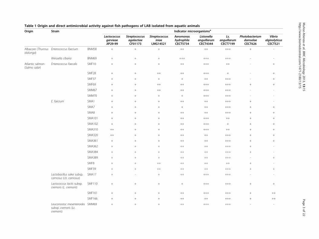
Table 1 Origin and direct antimicrobial activity against fish pathogens of LAB isolated from aquatic animals
Origin Strain Indicator microorganismsa
LactococcusgarvieaeJIP29-99
StreptococcusagalactiaeCF01173
Streptococcusiniae
LMG14521
AeromonashydrophilaCECT5734
ListonellaanguillarumCECT4344
Ls.anguillarumCECT7199
PhotobacteriumdamselaeCECT626
VibrioalginolyticusCECT521
Albacore (Thunnusalalunga)
Enterococcus faecium BNM58 + + + ++ ++ +++ + -
Weissella cibaria BNM69 + + + +++ +++ +++ - -
Atlantic salmon(Salmo salar)
Enterococcus faecalis SMF10 + + + ++ +++ ++ - +
SMF28 + + ++ ++ +++ + - +
SMF37 + + + + ++ +++ - +
SMF69 + + ++ ++ +++ +++ + +
SMM67 + + ++ ++ +++ +++ - -
SMM70 + + + + +++ +++ - -
E. faecium SMA1 + + + ++ ++ +++ + -
SMA7 + + + + ++ +++ + +
SMA8 + + + ++ ++ +++ + +
SMA101 + + + ++ +++ ++ + +
SMA102 + + + ++ +++ + + +
SMA310 ++ + + ++ +++ ++ + +
SMA320 ++ + + ++ ++ +++ + +
SMA361 + + + ++ ++ +++ + +
SMA362 + + + ++ ++ +++ + -
SMA384 + + + ++ ++ +++ + -
SMA389 + + + ++ ++ +++ - +
SMF8 + + ++ ++ ++ ++ + -
SMF39 + + ++ ++ ++ +++ + +
Lactobacillus sakei subsp.carnosus (Lb. carnosus)
SMA17 + - + ++ +++ +++ - -
Lactococcus lactis subsp.cremoris (L. cremoris)
SMF110 + + + + +++ +++ + +
SMF161 + + + ++ +++ +++ + ++
SMF166 + + + ++ ++ +++ + ++
Leuconostoc mesenteroidessubsp. cremoris (Lc.cremoris)
SMM69 + + + ++ +++ +++ - -
Muñoz-A
tienzaet
al.BMCMicrobiology
2013,13:15Page
3of
22http://w
ww.biom
edcentral.com/1471-2180/13/15

Table 1 Origin and direct antimicrobial activity against fish pathogens of LAB isolated from aquatic animals (Continued)
Pediococcus pentosaceus SMF120 ++ ++ ++ ++ +++ +++ - +
SMF130 ++ + ++ ++ +++ +++ - +
SMM73 ++ + + +++ +++ +++ + ++
W. cibaria SMA14 ++ + + ++ +++ +++ + ++
SMA25 + + + +++ +++ +++ - -
Cod (Gadusmorhua)
E. faecalis BCS27 ++ ++ ++ ++ +++ +++ - -
BCS32 + + + + ++ +++ - +
BCS53 + ++ + + +++ +++ + -
BCS67 + + - ++ +++ ++ - +
BCS72 + + + ++ +++ +++ + -
BCS92 + + + ++ +++ ++ + +
E. faecium BCS59 ++ + ++ ++ +++ +++ - +
BCS971 + + + + +++ +++ - +
BCS972 + + + + +++ +++ - +
Lactobacillus curvatussubsp. curvatus (Lb.curvatus)
BCS35 - - + ++ +++ +++ - -
Lc. cremoris BCS251 + + ++ + +++ +++ - +
BCS252 + + ++ + +++ +++ - +
P. pentosaceus BCS46 ++ + ++ +++ +++ +++ - +
W. cibaria BCS50 ++ + ++ ++ +++ +++ - +
Common cockle(Cerastodermaedule)
E. faecium B13 + + ++ ++ +++ +++ - -
B27 + + + ++ +++ ++ + +
Lb. carnosus B43 + + + ++ +++ +++ - -
P. pentosaceus B5 ++ + ++ ++ +++ +++ - -
B11 ++ + ++ +++ +++ +++ + -
B41 ++ ++ ++ +++ +++ +++ + ++
B260 ++ + ++ ++ +++ +++ - ++
W. cibaria B4620 ++ + ++ ++ +++ +++ - ++
Common ling(Molva molva)
E. faecium MV5 + + + ++ ++ +++ + +
Common octopus(Octopus vulgaris)
E. faecalis P77 ++ + ++ ++ +++ +++ - +
Muñoz-A
tienzaet
al.BMCMicrobiology
2013,13:15Page
4of
22http://w
ww.biom
edcentral.com/1471-2180/13/15
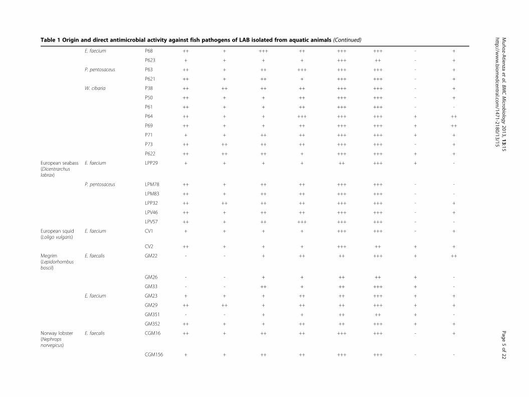
Table 1 Origin and direct antimicrobial activity against fish pathogens of LAB isolated from aquatic animals (Continued)
E. faecium P68 ++ + +++ ++ +++ +++ - +
P623 + + + + +++ ++ - +
P. pentosaceus P63 ++ + ++ +++ +++ +++ - +
P621 ++ + ++ + +++ +++ - +
W. cibaria P38 ++ ++ ++ ++ +++ +++ - +
P50 ++ + + ++ +++ +++ - +
P61 ++ + + ++ +++ +++ - -
P64 ++ + + +++ +++ +++ + ++
P69 ++ + + ++ +++ +++ + ++
P71 + + ++ ++ +++ +++ + +
P73 ++ ++ ++ ++ +++ +++ - +
P622 ++ ++ ++ + +++ +++ + +
European seabass(Dicentrarchuslabrax)
E. faecium LPP29 + + + + ++ +++ + -
P. pentosaceus LPM78 ++ + ++ ++ +++ +++ - -
LPM83 ++ + ++ ++ +++ +++ - -
LPP32 ++ ++ ++ ++ +++ +++ - +
LPV46 ++ + ++ ++ +++ +++ - +
LPV57 ++ + ++ +++ +++ +++ - -
European squid(Loligo vulgaris)
E. faecium CV1 + + + + +++ +++ - +
CV2 ++ + + + +++ ++ + +
Megrim(Lepidorhombusboscii)
E. faecalis GM22 - - + ++ ++ +++ + ++
GM26 - - + + ++ ++ + -
GM33 - - ++ + ++ +++ + -
E. faecium GM23 + + + ++ ++ +++ + +
GM29 ++ ++ + ++ ++ +++ + +
GM351 - - + + ++ ++ + -
GM352 ++ + + ++ ++ +++ + +
Norway lobster(Nephropsnorvegicus)
E. faecalis CGM16 ++ + ++ ++ +++ +++ - +
CGM156 + + ++ ++ +++ +++ - -
Muñoz-A
tienzaet
al.BMCMicrobiology
2013,13:15Page
5of
22http://w
ww.biom
edcentral.com/1471-2180/13/15

Table 1 Origin and direct antimicrobial activity against fish pathogens of LAB isolated from aquatic animals (Continued)
CGM1514 + + + ++ +++ ++ + +
CGV67 ++ + + + +++ +++ + +
E. faecium CGM171 + + + + +++ +++ + +
CGM172 + + + + +++ +++ + +
Rainbow trout(Oncorhynchusmykiss)
E. faecium TPM76 + + + + ++ +++ + +
TPP2 + + + + ++ +++ + +
P. pentosaceus TPP3 ++ + + ++ +++ +++ - ++
Sardine (Sardinapilchardus)
E. faecalis SDP10 + + + + +++ +++ - +
W. cibaria SDM381 ++ + ++ ++ +++ +++ - -
SDM389 + + ++ ++ +++ +++ - -
Swimcrab (Necorapuber)
E. faecium NV50 + + + ++ ++ ++ + -
NV51 ++ + + + ++ ++ + ++
NV52 ++ + + + ++ +++ + +
NV54 ++ + + + ++ +++ + +
NV56 ++ + + ++ ++ ++ + -aDirect antimicrobial activity was determined by a SOAT and the scores reflect different degrees of growth inhibition (diameter in mm); -, no inhibition; +, 3–5 mm inhibition zone; ++, 6–9 mm inhibition zone; +++,≥10 mm inhibition zone.
Muñoz-A
tienzaet
al.BMCMicrobiology
2013,13:15Page
6of
22http://w
ww.biom
edcentral.com/1471-2180/13/15

Table 2 Preliminary safety evaluation of enterococci
Enterococcusspp.
Strain Virulence Factors Antibiotic resistancephenotypecGenotypea Phenotypeb
E. faecalis SMF10 efaAfs+, gelE+, agg+ GelE+, Hly- CIP, NOR
SMF28 efaAfs+, gelE+ GelE+, Hly- CIP, NOR
SMF37 efaAfs+, gelE+, agg+ GelE+, Hly- -
SMF69 efaAfs+, gelE+, agg+ GelE+, Hly- CIP, RIF
SMM67 n.d. GelE-, Hly- CIP, NIT, NOR, TEC, VAN
SMM70 efaAfs+, gelE+ GelE+, Hly- ERY, NIT
BCS27 efaAfs+, gelE+, agg+ GelE+, Hly- CIP, NIT, NOR, RIF, TEC, VAN
BCS32 efaAfs+, gelE+, agg+ GelE+, Hly- NOR
BCS53 efaAfs+, gelE+, agg+ GelE+, Hly- -
BCS67 efaAfs+ GelE-, Hly- CIP
BCS72 efaAfs+, agg+ GelE-, Hly- -
BCS92 efaAfs+ GelE-, Hly- -
P77 efaAfs+, gelE+ GelE+, Hly- NIT, NOR, RIF, TEC, VAN
GM22 efaAfs+, gelE+, agg+ GelE+, Hly- CIP, NOR
GM26 efaAfs+, gelE+, agg+ GelE+, Hly- -
GM33 efaAfs+, gelE+, agg+ GelE+, Hly- -
CGM156 efaAfs+ GelE-, Hly- CIP, NIT, NOR, RIF, TEC, VAN
CGM1514 efaAfs+, agg+ GelE-, Hly- -
CGM16 efaAfs+, gelE+, agg+ GelE+, Hly- CIP, NOR, RIF
CGV16 efaAfs+, gelE+, agg+ GelE+, Hly- NOR
SDP10 efaAfs+, gelE+, agg+, cylLLLS+, cylLLLSM
+ GelE+, Hly+ -
E. faecium BNM58 n.d. GelE-, Hly- -
SMA1 n.d. GelE-, Hly- CIP
SMA7 n.d. GelE-, Hly- -
SMA8 n.d. GelE-, Hly- -
SMA101 n.d. GelE-, Hly- ERY, NIT
SMA102 efaAfs+ GelE-, Hly- ERY, NIT
SMA310 n.d. GelE-, Hly- ERY, NIT
SMA320 efaAfs+ GelE-, Hly- ERY, NIT
SMA361 efaAfs+ GelE-, Hly- ERY
SMA362 n.d. GelE-, Hly- ERY, NIT
SMA384 gelE+ GelE-, Hly- NIT
SMA389 gelE+ GelE-, Hly- CIP, NIT, NOR
SMF8 n.d. GelE-, Hly- -
SMF39 efaAfs+, gelE+ GelE-, Hly- -
BCS59 n.d. GelE-, Hly- NIT
BCS971 n.d. GelE-, Hly- ERY
BCS972 n.d. GelE-, Hly- ERY
B13 gelE+ GelE+, Hly- CIP
B27 efaAfs+, gelE+ GelE+, Hly- CIP
MV5 efaAfs+, gelE+, agg+ GelE-, Hly- CIP, NIT
P68 efaAfs+, gelE+, cylLLLS+ GelE+, Hly- CIP, NIT, NOR, RIF, TEC, VAN
P623 efaAfs+ GelE-, Hly- ERY
Muñoz-Atienza et al. BMC Microbiology 2013, 13:15 Page 7 of 22http://www.biomedcentral.com/1471-2180/13/15

Table 2 Preliminary safety evaluation of enterococci (Continued)
LPP29 n.d. GelE-, Hly- -
CV1 n.d. GelE-, Hly- -
CV2 n.d. GelE-, Hly- -
GM23 efaAfs+ GelE-, Hly- CIP, NOR, RIF, TET
GM29 efaAfs+, gelE+, cylLLLS+ GelE-, Hly- CIP, NOR, RIF
GM351 efaAfs+, gelE+, agg+ GelE+, Hly- CIP, NOR
GM352 efaAfs+ GelE-, Hly- CIP, NIT, NOR, RIF, TET
CGM171 n.d. GelE-, Hly- ERY
CGM172 efaAfs+ GelE-, Hly- ERY
TPM76 n.d. GelE-, Hly- -
TPP2 n.d. GelE-, Hly- -
NV50 efaAfs+, agg+ GelE-, Hly- -
NV51 efaAfs+ GelE-, Hly- ERY
NV52 n.d. GelE-, Hly- ERY
NV54 efaAfs+ GelE-, Hly- ERY
NV56 efaAfs+ GelE-, Hly- -an.d., not detected.bGelE and Hly refer to gelatinase and cytolysin/hemolysin activity, respectively.cAbbreviation of antibiotics: CIP, ciprofloxacin; ERY, erythromycin; NIT, nitrofurantoin; NOR, norfloxacin; RIF, rifampicin; TEC, teicoplanin; TET, tetracycline;VAN, vancomycin.
Muñoz-Atienza et al. BMC Microbiology 2013, 13:15 Page 8 of 22http://www.biomedcentral.com/1471-2180/13/15
38%), acquired antibiotic resistance (1 strain, 5%) or both(12 strains, 57%). Regarding E. faecium strains, 29 (76%)were dropped from further screening based on acquiredantibiotic resistance (9 strains, 24%), the presence of viru-lence factors (3 strains, 8%) or both (17 strains, 45%).
Extracellular antimicrobial activity of the 49 pre-selected LABThe antimicrobial activity of supernatants from the 49pre-selected LAB (9 E. faecium selected based on theirpreliminary safety assessment and 40 non-enterococcalstrains) with direct antimicrobial activity against fishpathogens was assayed against three indicator microor-ganisms by an ADT (Table 3). In this regard, 24 (49%) and10 (20%) strains displayed extracellular antimicrobial ac-tivity in their supernatants and/or 20-fold concentratedsupernatants against Pediococcus damnosus CECT4797and L. garvieae JIP 29–99, respectively, but none of thestrains inhibited the Gram-negative strain A. hydrophilaCECT5734. Interestingly, the antimicrobial activity of therespective supernatants was sensitive to proteinase K treat-ment, but was not affected by the heat treatment, revealingthe proteinaceous nature and heat stability of the secretedantimicrobial compounds (i.e., heat-stable bacteriocins).The 24 LAB strains secreting bacteriocins into the liquidgrowth medium belong to the species P. pentosaceus (15strains), E. faecium (8 strains), and Lb. curvatus (1 strain).
In vitro safety assessment of the 49 pre-selected LABThe 49 pre-selected LAB were further submitted to a com-prehensive safety assessment by different in vitro tests.
Hemolysin production, bile salts deconjugation andmucin degradationNone of the non-enterococcal strains showed hemolyticactivity, similarly as found for the 9 enterococci. More-over, bile salts deconjugation and mucin degradationabilities were not found in any of the tested strains.
Enzymatic activitiesThe results of the analysis of enzymatic activity profilesof the tested LAB are shown in Table 4. None of thestrains showed lipolytic activity, except E. faeciumLPP29, TPM76, SMA7, and SMF8 which produced es-terase (C4) and esterase lipase (C8). Moreover, none ofthe LAB strains showed protease activity (trypsinand α-chymotrypsin). Nevertheless, peptidase activity (leu-cine, valine or cystine arylamidase) was found in all thespecies. All strains showed acid phosphatase (exceptE. faecium TPM76 and Lc. cremoris) and naphthol-AS-BI-phosphohydrolase activities, but none displayed al-kaline phosphatase activity. β-Galactosidase was found inmost species (but not in all strains) except Lb. curvatusand L. cremoris. However, α-glucosidase was only found inthe three Lc. cremoris strains. β-Glucosidase and N-acetyl-β-glucosaminidase activities were observed in mostE. faecium, Lactobacillus spp., L. cremoris, and P. pentosa-ceus strains, but only in two W. cibaria strains, while thethree Lc. cremoris strains showed β-glucosidase butlacked N-acetyl-β-glucosaminidase activity. On the otherhand, α-galactosidase, β-glucuronidase, α-mannosidase, and

Table 3 Extracellular antimicrobial activity of the 49 pre-selected LABa
LAB speciesb Strain Indicator microorganisms
P. damnosus CECT4797 L. garvieae JIP29-99 A. hydrophila CECT5734
S CS S CS S CS
Enterococci
E. faecium BNM58 22.4 26.8 14.0 15.0 - -
SMA7 - - - - - -
SMA8 19.0 19.6 9.4 10.2 - -
SMF8 19.0 21.8 10.3 10.8 - -
LPP29 20.5 24.4 12.6 13.1 - -
CV1 15.0 19.2 - - - -
CV2 19.8 23.7 12.7 11.4 - -
TPM76 17.0 21.2 - 8.7 - -
TPP2 19.7 23.5 12.8 12.4 - -
Non-enterococci
Lb. curvatus BCS35 18.2 24.7 - - - -
P. pentosaceus SMF120 - - - - - -
SMF130 7.4 9.7 - - - -
SMM73 - 9.5 - - - -
BCS46 - 9.4 - - - -
B5 8.1 9.0 - - - -
B11 - 9.0 - - - -
B41 7.3 11.7 - - - -
B260 7.3 10.6 - - - -
P63 - 9.8 - - - -
P621 - 10.5 - - - -
LPM78 - 8.3 - - - -
LPM83 7.9 11.0 - - - -
LPP32 8.5 11.3 - 8.9 - -
LPV46 8.2 11.3 - 8.2 - -
LPV57 7.6 10.5 - - - -
TPP3 9.0 11.7 7.5 9.2 - -aAntimicrobial activity (mm) of supernatants (S) and 20-fold concentrated supernatants (CS) as determined by an ADT.bLb. carnosus, L. cremoris, Lc. cremoris and W. cibaria strains did not show extracellular antimicrobial activity against any of the tested indicator microorganisms.
Muñoz-Atienza et al. BMC Microbiology 2013, 13:15 Page 9 of 22http://www.biomedcentral.com/1471-2180/13/15
α-fucosidase activities were not detected in any of the testedLAB strains.
Antibiotic susceptibility determined by the brothmicrodilution testThe distribution of MICs of the tested antibiotics is sum-marized in Tables 5 and 6. Microbiological breakpoints forampicillin, vancomycin, gentamicin, kanamycin, strepto-mycin, erythromycin, clindamycin, tetracycline, and chlor-amphenicol reported by the FEEDAP document on theassessment of bacterial products used as feed additives inrelation to antibiotic resistance [15] were used to categor-ise the 49 LAB as susceptible or resistant strains. In thisdocument, the genus Weissella, which is considered a
group of heterofermentative Leuconostoc-like LAB [16], isnot included. For this reason, the respective MICs wereinterpreted by using the breakpoints given for the genusLeuconostoc. Besides, due to the lack of microbiologicalbreakpoints for penicillin and linezolid on the FEEDAPdocument, we interpreted our results on these antibioticsaccording to the cut-off levels proposed by Klare et al.[17] for pediococci, namely 1 and 2 mg/L for penicillinand linezolid, respectively. According to our results, thepercentages of strains showing antibiotic resistance inthe genera Weissella, Pediococcus, Lactobacillus andEnterococcus were 60, 44, 33 and 11%, respectively, whilenone of the leuconostocs and lactococci showed thisphenotype. In summary, 97.5% of the 40 non-enterococal
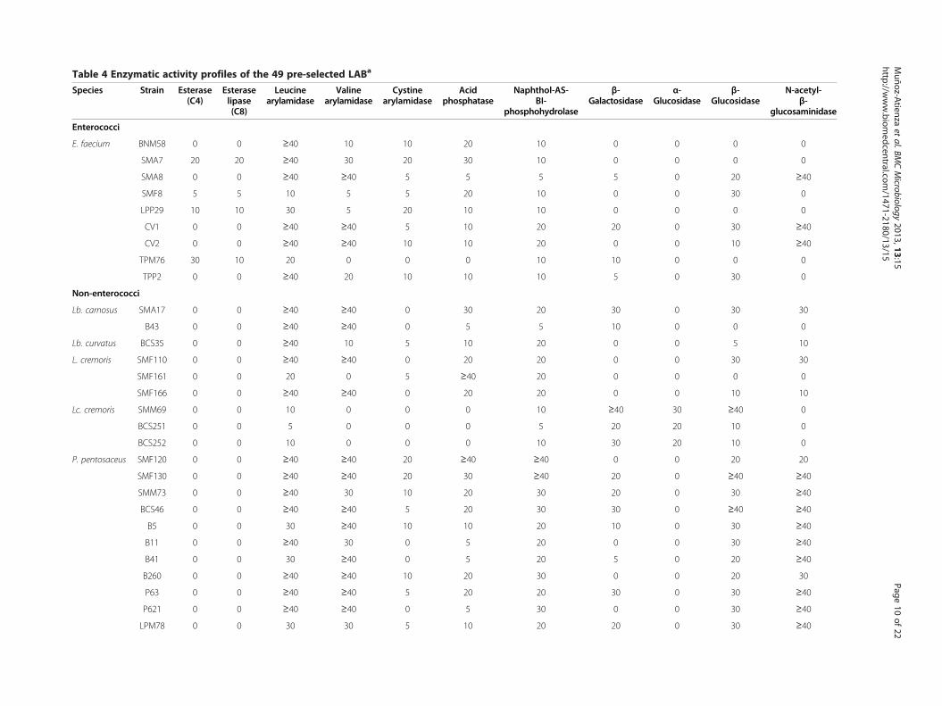
Table 4 Enzymatic activity profiles of the 49 pre-selected LABa
Species Strain Esterase(C4)
Esteraselipase(C8)
Leucinearylamidase
Valinearylamidase
Cystinearylamidase
Acidphosphatase
Naphthol-AS-BI-
phosphohydrolase
β-Galactosidas
α-Glucosidase
β-Glucosidase
N-acetyl-β-
glucosaminidase
Enterococci
E. faecium BNM58 0 0 ≥40 10 10 20 10 0 0 0 0
SMA7 20 20 ≥40 30 20 30 10 0 0 0 0
SMA8 0 0 ≥40 ≥40 5 5 5 5 0 20 ≥40
SMF8 5 5 10 5 5 20 10 0 0 30 0
LPP29 10 10 30 5 20 10 10 0 0 0 0
CV1 0 0 ≥40 ≥40 5 10 20 20 0 30 ≥40
CV2 0 0 ≥40 ≥40 10 10 20 0 0 10 ≥40
TPM76 30 10 20 0 0 0 10 10 0 0 0
TPP2 0 0 ≥40 20 10 10 10 5 0 30 0
Non-enterococci
Lb. carnosus SMA17 0 0 ≥40 ≥40 0 30 20 30 0 30 30
B43 0 0 ≥40 ≥40 0 5 5 10 0 0 0
Lb. curvatus BCS35 0 0 ≥40 10 5 10 20 0 0 5 10
L. cremoris SMF110 0 0 ≥40 ≥40 0 20 20 0 0 30 30
SMF161 0 0 20 0 5 ≥40 20 0 0 0 0
SMF166 0 0 ≥40 ≥40 0 20 20 0 0 10 10
Lc. cremoris SMM69 0 0 10 0 0 0 10 ≥40 30 ≥40 0
BCS251 0 0 5 0 0 0 5 20 20 10 0
BCS252 0 0 10 0 0 0 10 30 20 10 0
P. pentosaceus SMF120 0 0 ≥40 ≥40 20 ≥40 ≥40 0 0 20 20
SMF130 0 0 ≥40 ≥40 20 30 ≥40 20 0 ≥40 ≥40
SMM73 0 0 ≥40 30 10 20 30 20 0 30 ≥40
BCS46 0 0 ≥40 ≥40 5 20 30 30 0 ≥40 ≥40
B5 0 0 30 ≥40 10 10 20 10 0 30 ≥40
B11 0 0 ≥40 30 0 5 20 0 0 30 ≥40
B41 0 0 30 ≥40 0 5 20 5 0 20 ≥40
B260 0 0 ≥40 ≥40 10 20 30 0 0 20 30
P63 0 0 ≥40 ≥40 5 20 20 30 0 30 ≥40
P621 0 0 ≥40 ≥40 0 5 30 0 0 30 ≥40
LPM78 0 0 30 30 5 10 20 20 0 30 ≥40
Muñoz-A
tienzaet
al.BMCMicrobiology
2013,13:15Page
10of
22http://w
ww.biom
edcentral.com/1471-2180/13/15
e

Table 4 Enzymatic activity profiles of the 49 pre-selected LABa (Continued)
LPM83 0 0 30 30 5 10 20 30 0 10 ≥40
LPP32 0 0 ≥40 ≥40 5 5 20 0 0 30 ≥40
LPV46 0 0 ≥40 ≥40 5 20 30 5 0 30 30
LPV57 0 0 ≥40 ≥40 5 20 30 30 0 ≥40 ≥40
TPP3 0 0 ≥40 ≥40 5 5 5 10 0 0 0
W. cibaria BNM69 0 0 0 0 0 30 10 30 0 0 0
SMA14 0 0 0 0 0 20 5 10 0 0 0
SMA25 0 0 ≥40 ≥40 0 30 20 ≥40 0 30 30
BCS50 0 0 0 0 0 30 20 30 0 0 0
B4620 0 0 20 20 0 30 20 30 0 5 5
P38 0 0 0 0 0 ≥40 20 ≥40 0 0 0
P50 0 0 0 0 0 ≥40 20 0 0 0 0
P61 0 0 0 0 0 20 10 0 0 0 0
P64 0 0 0 0 0 30 10 0 0 0 0
P69 0 0 0 0 0 ≥40 20 ≥40 0 0 0
P71 0 0 0 0 0 ≥40 10 0 0 0 0
P73 0 0 0 0 0 30 20 30 0 0 0
P622 0 0 0 0 0 ≥40 10 0 0 0 0
SDM381 0 0 10 5 0 20 10 30 0 0 0
SDM389 0 0 0 0 0 ≥40 20 ≥40 0 0 0aEnzymatic activities determined by an APIZYM test. Relative activity between 0 and ≥ 40 nmol.
Muñoz-A
tienzaet
al.BMCMicrobiology
2013,13:15Page
11of
22http://w
ww.biom
edcentral.com/1471-2180/13/15

Muñoz-Atienza et al. BMC Microbiology 2013, 13:15 Page 12 of 22http://www.biomedcentral.com/1471-2180/13/15
strains resulted susceptible to ampicillin, 100% to gentami-cin, 72.5% to kanamycin, 100% to streptomycin, 95% toerythromycin, 87.5% to clindamycin, 95% to tetracycline,and 100% to chloramphenicol. For vancomycin, it isknown that facultative and obligate heterofermentativeLactobacillus, Pediococcus spp. and Leuconostoc spp. areintrinsically resistant. In contrast, the three lactococciwere clearly susceptible to these antibiotics, showing aMIC of 0.5 mg/L. On the other hand, according to thecut-off values proposed by Klare et al. [17], 93% of P.pentosaceus strains were susceptible to penicillin and line-zolid. With regard to E. faecium, all the tested strains weresusceptible to ampicillin, vancomycin, gentamicin, kana-mycin, streptomycin, tetracycline, chloramphenicol, anderythromycin except E. faecium BNM58 against the latterantibiotic (MIC = 8 mg/L). Moreover, multiple antibioticresistance (three antibiotics) was only detected in P.pentosaceus LPM78 (6.2%) and W. cibaria SMA25 (6.7%).
Detection of antibiotic resistance genesThe non-enterococcal strains showing antibiotic resistancesin the VetMIC assays (17 strains) were further submitted toPCR in order to identify the presence of the respective anti-biotic resistance genes. The tested strains were the follow-ing: Lb. carnosus B43 (ampicillin resistant), P. pentosaceusTPP3 and SMF120 (tetracycline resistant), P. pentosaceusLPP32, LPM83 and B5 (clindamycin resistant), P. pentosa-ceus LPV57 and W. cibaria P50, P61, P64, P73, SDM381,SDM389, SMA14 and BCS50 (kanamycin resistant), andP. pentosaceus LPM78 and W. cibaria SMA25 (kanamy-cin, erythromycin and clindamycin resistant). Acquiredantibiotic resistances likely due to added genes were onlyfound in strains within the genera Pediococcus (12.5%)and Weissella (6.7%). The genes involved in the horizontaltransfer of resistance to tetracycline [tet(K), tet(L) and tet
Table 5 MICs distribution of 10 antibiotics for the 9 enteroco
Antibiotics Number of strains with the indi
0.06 0.12 0.25 0.5 1 2 4 8 16 32
Ampicillin 5 3 1
Vancomycin 9
Gentamicin 4 5
Kanamycin 1 2
Streptomycin 1 3 5
Erythromycin 5 3 1
Tetracycline 9
Chloramphenicol 8 1
Linezolid 9
Narasin 1 8aMICs determined by a VetMIC test. The antibiotic dilution ranges were: 0.25-32 mgmg/L (kanamycin), 8-1024 mg/L (streptomycin), 0.5-64 mg/L (erythromycin, tetracycwhich exceeded the upper or lower limit of the tested range are listed in the next dbLAB with MICs higher than the EFSA breakpoints are considered as resistant strain
(M)], kanamycin [aac(6´ )-Ie-aph(2´ ´ )-Ia] and erythro-mycin [erm(A), erm(B) and erm(C)] were not detected.However, P. pentosaceus LPM78 and W. cibaria SMA25harboured the erythromycin resistance gene mef(A/E).The obtained amplicons were sequenced and found tohave 99% homology with the macrolide-efflux protein(mefE) gene described for Streptococcus pneumoniae andother Streptococcus spp. Moreover, P. pentosaceus LPM78and LPM83 harboured the lnu(A) gene encoding the linco-samide O-nucleotidyltransferase that inactivates lincomycinand clindamycin. Sequencing of both amplicons showed97% and 93% homology with lincosamide nucleotidyltrans-ferase [lnu(A)] gene described for Staphylococcus haemoly-ticus and S. aureus, respectively. Nevertheless, lnu(B) wasnot detected in any of the tested strains. With regardto E. faecium BNM58, which was phenotypically resistantto erythromycin, none of the respective genes [erm(A),erm(B), erm(C) and mef(A/E)] were detected.
DiscussionIn this work, the antimicrobial activity against fishpathogens and the in vitro safety of 99 LAB previouslyisolated from fish, seafood and fish products [14] havebeen assayed by using microbiological, biochemical andgenetic assays in order to identify and select the mostsuitable candidates to be further evaluated as probioticsfor a sustainable aquaculture. LAB are widely known fortheir ability to inhibit bacterial pathogens by the produc-tion of antimicrobial compounds such as organic acids,oxygen peroxide and ribosomally-synthesized peptidesreferred to as bacteriocins, which constitutes a desirableproperty for probiotics and a sustainable alternative toantibiotics [9,18]. In this respect, most of the LAB ofaquatic origin tested in this work displayed a broad anti-microbial spectrum against the main Gram-positive and
ccal strains
cated MIC (mg/L)a EFSA breakpoints (mg/L)b
64 128 256 512 1024 2048
2
4
32
4 2 1024
128
4
4
16
n.a.
n.a.
/L (ampicillin), 1-128 mg/L (vancomycin), 2-256 mg/L (gentamicin), 16-2048line and chloramphenicol), 0.25-16 mg/L (linezolid) and 0.12-16 (narasin). MICsilution series. MICs higher than the EFSA breakpoints are indicated in bold.s [15]. n.a., not available.
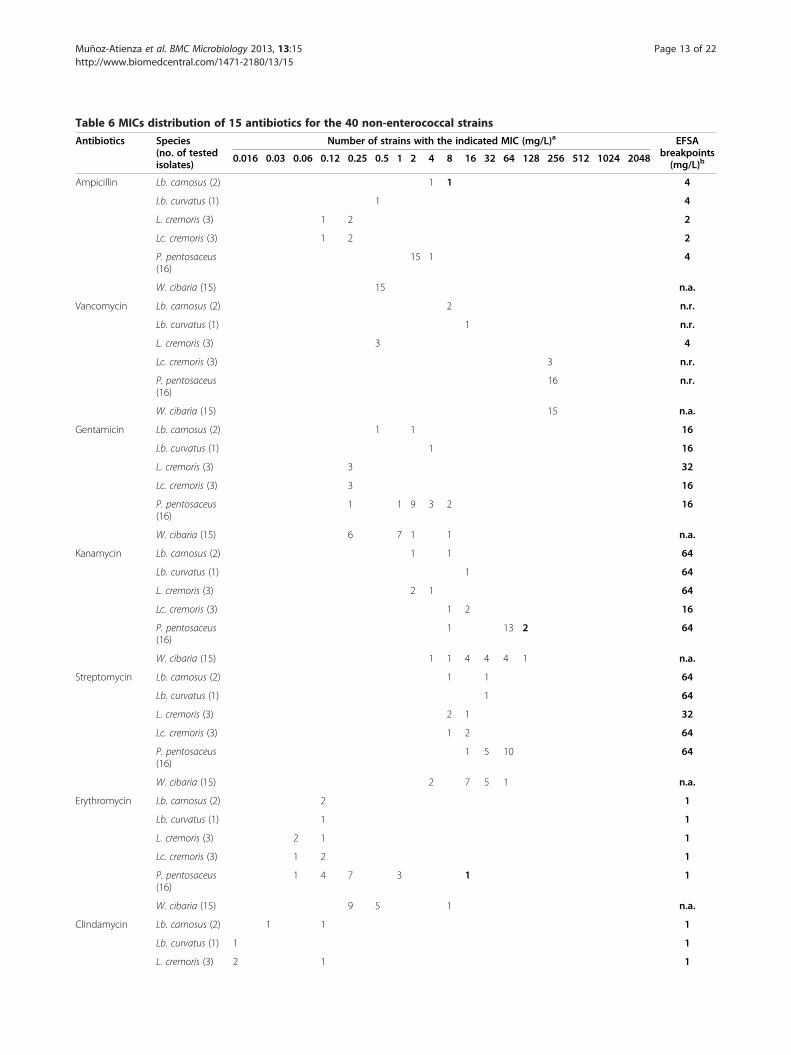
Table 6 MICs distribution of 15 antibiotics for the 40 non-enterococcal strains
Antibiotics Species(no. of testedisolates)
Number of strains with the indicated MIC (mg/L)a EFSAbreakpoints(mg/L)b
0.016 0.03 0.06 0.12 0.25 0.5 1 2 4 8 16 32 64 128 256 512 1024 2048
Ampicillin Lb. carnosus (2) 1 1 4
Lb. curvatus (1) 1 4
L. cremoris (3) 1 2 2
Lc. cremoris (3) 1 2 2
P. pentosaceus(16)
15 1 4
W. cibaria (15) 15 n.a.
Vancomycin Lb. carnosus (2) 2 n.r.
Lb. curvatus (1) 1 n.r.
L. cremoris (3) 3 4
Lc. cremoris (3) 3 n.r.
P. pentosaceus(16)
16 n.r.
W. cibaria (15) 15 n.a.
Gentamicin Lb. carnosus (2) 1 1 16
Lb. curvatus (1) 1 16
L. cremoris (3) 3 32
Lc. cremoris (3) 3 16
P. pentosaceus(16)
1 1 9 3 2 16
W. cibaria (15) 6 7 1 1 n.a.
Kanamycin Lb. carnosus (2) 1 1 64
Lb. curvatus (1) 1 64
L. cremoris (3) 2 1 64
Lc. cremoris (3) 1 2 16
P. pentosaceus(16)
1 13 2 64
W. cibaria (15) 1 1 4 4 4 1 n.a.
Streptomycin Lb. carnosus (2) 1 1 64
Lb. curvatus (1) 1 64
L. cremoris (3) 2 1 32
Lc. cremoris (3) 1 2 64
P. pentosaceus(16)
1 5 10 64
W. cibaria (15) 2 7 5 1 n.a.
Erythromycin Lb. carnosus (2) 2 1
Lb. curvatus (1) 1 1
L. cremoris (3) 2 1 1
Lc. cremoris (3) 1 2 1
P. pentosaceus(16)
1 4 7 3 1 1
W. cibaria (15) 9 5 1 n.a.
Clindamycin Lb. carnosus (2) 1 1 1
Lb. curvatus (1) 1 1
L. cremoris (3) 2 1 1
Muñoz-Atienza et al. BMC Microbiology 2013, 13:15 Page 13 of 22http://www.biomedcentral.com/1471-2180/13/15

Table 6 MICs distribution of 15 antibiotics for the 40 non-enterococcal strains (Continued)
Lc. cremoris (3) 2 1 1
P. pentosaceus(16)
3 2 7 1 3 1
W. cibaria (15) 2 6 5 1 1 n.a.
Tetracycline Lb. carnosus (2) 1 1 8
Lb. curvatus (1) 1 8
L. cremoris (3) 1 1 1 4
Lc. cremoris (3) 1 2 8
P. pentosaceus(16)
1 13 2 8
W. cibaria (15) 15 n.a.
Chloramphenicol Lb. carnosus (2) 1 1 4
Lb. curvatus (1) 1 4
L. cremoris (3) 1 2 8
Lc. cremoris (3) 3 4
P. pentosaceus(16)
1 5 10 4
W. cibaria (15) 15 n.a.
Neomycin Lb. carnosus (2) 1 1 n.a.
Lb. curvatus (1) 1 n.a.
L. cremoris (3) 2 1 n.a.
Lc. cremoris (3) 3 n.a.
P. pentosaceus(16)
1 9 4 2 n.a.
W. cibaria (15) 4 6 4 1 n.a.
Penicillin Lb. carnosus (2) 1 1 n.a.
Lb. curvatus (1) 1 n.a.
L. cremoris (3) 3 n.a.
Lc. cremoris (3) 1 2 n.a.
P. pentosaceus(16)
7 8 1 n.a.
W. cibaria (15) 7 7 1 n.a.
Linezolid Lb. carnosus (2) 2 n.a.
Lb. curvatus (1) 1 n.a.
L. cremoris (3) 1 2 n.a.
Lc. cremoris (3) 1 2 n.a.
P. pentosaceus(16)
15 1 n.a.
W. cibaria (15) 15 n.a.
Ciprofloxacin Lb. carnosus (2) 2 n.a.
Lb. curvatus (1) 1 n.a.
L. cremoris (3) 2 1 n.a.
Lc. cremoris (3) 1 2 n.a.
P. pentosaceus(16)
16 n.a.
W. cibaria (15) 5 10 n.a.
Rifampicin Lb. carnosus (2) 1 1 n.a.
Muñoz-Atienza et al. BMC Microbiology 2013, 13:15 Page 14 of 22http://www.biomedcentral.com/1471-2180/13/15
Table 6 MICs distribution of 15 antibiotics for the 40 non-enterococcal strains (Continued)
Lb. curvatus (1) 1 n.a.
L. cremoris (3) 1 2 n.a.
Lc. cremoris (3) 1 2 n.a.
P. pentosaceus(16)
2 13 1 n.a.
W. cibaria (15) 12 3 n.a.
Trimethoprim Lb. carnosus (2) 1 1 n.a.
Lb. curvatus (1) 1 n.a.
L. cremoris (3) 3 n.a.
Lc. cremoris (3) 1 2 n.a.
P. pentosaceus(16)
8 8 n.a.
W. cibaria (15) 15 n.a.aMICs determined by a VetMIC test. The antibiotic dilution ranges were: 0.03-16 mg/L (ampicillin, clindamycin, penicillin and linezolid), 0.25-128 mg/L (vancomycinand ciprofloxacin), 0.5-256 mg/L (gentamicin, streptomycin and neomycin), 2-1024 mg/L (kanamycin), 0.016-8 mg/L (erythromycin), 0.12-64 (tetracycline,chloramphenicol, rifampicin and trimethoprim). MICs which exceeded the upper or lower limit of the tested range are listed in the next dilution series. MICshigher than the EFSA breakpoints are indicated in bold.bLAB with MICs higher than the EFSA breakpoints are considered as resistant strains [15]. n.r., not required; n.a., not available.
Muñoz-Atienza et al. BMC Microbiology 2013, 13:15 Page 15 of 22http://www.biomedcentral.com/1471-2180/13/15
Gram-negative fish pathogens, being remarkable that ahigh number of strains (24 out of 49 strains, 49%) wereidentified as potential bacteriocin producers. Recently,bacteriocin production ability has been proposed as akey property for selection of probiotic LAB to be used inaquaculture as an alternative to antibiotics to fightagainst fish pathogen infections [19], similarly as pro-posed for human and farm animal probiotics [20-22]. Inaquaculture farming, lactococcosis produced by the zoo-notic agent L. garvieae, causing hemorrhagic septicaemiaand meningoencephalitis, is one of the most serious dis-eases affecting several marine and fresh water fish spe-cies [23]. With regard to this, our work shows thatputative bacteriocinogenic LAB active against this rele-vant fish pathogen are common amongst the microbiotaisolated from aquatic animals (10 strains, 20%).The application of probiotics in aquaculture may mod-
ify the microbial ecology of the aquatic hosts and theirsurrounding environment, and thus the assessment oftheir safety to the target aquatic species, the environ-ment and humans constitutes an essential issue [24]. Todate, several studies describing the screening and evalu-ation of LAB as probiotic candidates for aquaculturehave been reported [25-28]; however, the safety assess-ment of the strains is generally limited to in vivo chal-lenge tests and rearing trials in order to confirm theirlack of toxicity to the aquatic hosts [24,25,28-31]. Strik-ingly, in vitro safety assessment studies have not beengenerally addressed, despite they have lower economicand ethic costs and result very effective to evaluate thesafety of a high number of candidate probiotic strainsnot only for the host species, but also for humans andthe environment. According to EFSA [13], most of theLAB species tested in this work (P. pentosaceus, Lb.
curvatus, L. lactis, Lc. mesenteroides) are included inthe QPS list and, therefore, demonstration of their safetyonly requires confirmation of the absence of determinantsof resistance to antibiotics of human and veterinary clinicalsignificance. However, in the case of enterococci, a morethorough, strain-specific evaluation is required to assess therisk associated to their intentional use in the food chain,while no guidelines are given for the safety assessment ofthe species W. cibaria [13].Our results show that enterococcal virulence factors
were more frequently found in E. faecalis than in E.faecium, which is in concordance with previous reports[32-34]. In this respect, most of the E. faecalis (95%) and alarge percentage of the E. faecium (53%) strains evaluatedin this work showed, at least, one virulence factor, beingefaAfs, gelE and agg the most frequently detected genes.With regard to gelE, which encodes for an extracellularzinc endopeptidase that hydrolyzes gelatin, collagen,hemoglobin, and other bioactive compounds, this genewas detected at high frequency in E. faecalis, with all thegelE+ strains showing gelatinase activity. However, five outof nine E. faecium strains harbouring gelE were unable todegrade gelatin, suggesting the carriage of a non-functionalgene, as previously reported [32,33]. Likewise, in the case ofE. faecium P68 and E. faecium GM29 harbouring cylLL-cylLS, the lack of hemolytic activity may be explained bythe absence of cylM, whose product is involved in the post-translational modification of cytolysin. On the other hand,esp and hyl, which encode a cell wall-associated proteininvolved in immune evasion and an hyaluronidase enzyme,respectively, were not found in any of the tested LAB.Previous studies have reported that esp and hyl are morecommon in ampicillin-resistant/vancomycin-resistant E.faecium (VREF) than in ampicillin-susceptible/VREF

Muñoz-Atienza et al. BMC Microbiology 2013, 13:15 Page 16 of 22http://www.biomedcentral.com/1471-2180/13/15
strains [35]. In this context, the increase in the incidenceof VREF at hospital settings has been attributed mainly tothe spread of ampicillin-resistant VREF exhibiting espand/or hyl [36,37]. Therefore, the fact that the E. faeciumstrains evaluated in this work lack these genes might berelated with their non-clinical origin and absence of ampi-cillin resistance.The use and frequent overuse of antibiotics, including
those used in human medicine, in fish farming hasresulted in the emergence and spread of antibiotic-resistant bacteria in the aquaculture environment. Thispossesses a threat to human and animal health due tothe increase of acquired antibiotic resistance in fishpathogens, the transfer of their genetic determinants tobacteria of terrestrial animals and to human pathogens,and the alterations of the bacterial microbiota of theaquatic environment [11,29]. In our study, the percent-age of enterococcal strains showing acquired antibioticresistance was 68%. Interestingly, the results found in E.faecium (71%) and E. faecalis (62%) were similar, how-ever, higher percentages of resistance to ciprofloxacinand/or norfloxacin, rifampicin, and glycopeptides wereobserved in E. faecalis. Nevertheless, the occurrence oferythromycin and tetracycline resistance was frequentlydetected amongst E. faecium (45%) but only in one E.faecalis strain (5%). In spite of the high prevalence ofacquired antibiotic resistance found in enterococci ofaquatic origin, they showed low incidence or absenceof resistance to the clinically relevant antibiotics vanco-mycin (8.5%) and ampicillin, penicillin and gentamicin,respectively, which is in agreement with previous studies[33,38]. Moreover, the percentages of strains showing anti-biotic resistance in the genera Weissella, Pediococcus andLactobacillus were 60, 44 and 33%, respectively, while noneof the leuconostocs and lactococci showed this phenotype.In this regard, our results indicate that the LAB susceptibil-ity patterns of MIC values to clinically relevant antibioticsare species-dependent, similarly as previously described byother authors [39,40]. Moreover, multiple antibiotic resist-ance was commonly found in strains within thegenus Enterococcus (37%), mainly in E. faecalis, while beingvery infrequent in the non-enterococcal strains (5%).According to EFSA [29], the determination of MICs
above the established breakpoint levels, for one or moreantibiotic, requires further investigation to make the dis-tinction between added genes (genes acquired by thebacteria via gain of exogenous DNA) or to the mutationof indigenous genes. According to our results, acquiredantibiotic resistance likely due to added genes is not acommon feature amongst the non-enterococcal LAB ofaquatic origin (7.5%). In this respect, this genotype wasonly found in the genera Pediococcus (12.5%) and Weis-sella (6.7%). Although P. pentosaceus LPV57 and LPM78showed resistance to kanamycin (MIC of 128 mg/L), the
respective resistance gene [aac(6´ )-Ie-aph(2´ ´ )-Ia] wasnot found in these strains. Similarly, P. pentosaceusTPP3 and SMF120 were phenotypically resistant totetracycline (MIC of 16 mg/L), but did not contain tet(K), tet(L) or tet(M). In this respect, Ammor et al. [41]reported that pediococci are intrinsically resistant to thelatter two antibiotics, as well as to glycopeptides (vanco-mycin and teicoplanin), streptomycin, ciprofloxacin andtrimethoprim-sulphamethoxazole. Other authors proposeda MIC for tetracycline in pediococci ranging between 8 and16 mg/L [42], or of 32 mg/L for oxytetracycline in P.pentosaceus [17]. The tetracycline breakpoints suggestedfor pediococci by EFSA are lower than the MICs observedin our work and others [17,42]. On the other hand, theonly antibiotic resistance detected in Leuconostoc strainswas for vancomycin, which is an intrinsic property of thisgenus. It has been previously reported that Leuconostocstrains display poor, if any, resistance to antibiotics of clin-ical interest [38]. With regard to lactococci, the three L.cremoris strains evaluated were susceptible to all the anti-biotics; however, relatively high MICs for rifampicin (16–32 mg/L) and trimethoprim (≥ 64 mg/L) were detected. Infact, most lactococcal species are resistant to trimethoprim[41]. As expected, all strains of heterofermentative Lacto-bacillus spp. were resistant to vancomycin but susceptibleto the rest of the assayed antibiotics, except Lb. carnosusB43, which showed the highest MIC for ampicillin andpenicillin (MICs of 8 and 4 mg/L, respectively). In thiscontext, the presence of modifications in the low affinitypenicillin-binding protein (PBP) that confers resistance topenicillin and β-lactams in E. faecium and Streptococcuspneumoniae, has been reported [43,44]. Moreover, ninePBPs have been described in Lb. casei ATCC 393 [45],which leads us to suggest that a similar mechanism may bealso responsible for the ampicillin and penicillin resistancefound in Lb. carnosus B43. The resistance to vancomycindetected in Pediococcus, Leuconostoc and Lactobacillusspecies in this study might be due to the presence ofD-Ala-D-Lactate in their peptidoglycan rather thanD-Ala-D-Ala dipeptide [46]. In this context, all tested W.cibaria strains showed MICs ≥ 128 mg/L for vancomycin,suggesting that vancomycin resistance is an intrinsic prop-erty of this species. In relation to Weissella spp., studies onantibiotic resistance profiles are very limited [47] and break-points have not been defined by EFSA [15]. In our study,most W. cibaria strains showed low MIC values; howeverW. cibaria BCS50 showed relatively high MICs for penicil-lin (8 mg/L) and kanamycin (64 mg/L), and W. cibariaSMA25 showed MICs of 128 mg/L for kanamycin, 8 mg/Lfor gentamicin, erythromycin and neomycin, and 2 mg/Lfor clindamycin. Therefore, these two strains were dis-carded of this study, while W. cibaria P50, P61, P64, P73,SMA14, SDM381 and SDM389 were not included in thefinal selection due to their MICs for kanamycin (32–

Muñoz-Atienza et al. BMC Microbiology 2013, 13:15 Page 17 of 22http://www.biomedcentral.com/1471-2180/13/15
64 mg/L). According to these results, as a rule of thumb,we propose for W. cibaria the breakpoints assigned toLeuconostoc spp. by EFSA [15], until further studies estab-lish the wild-type MIC ranges within this species. In spiteof that, different MICs for rifampicin and trimethoprim forW. cibaria and Lc. cremoris were found in this study. Thereduced susceptibility of W. cibaria towards trimethoprimcould indicate an intrinsic resistance to this antibiotic [48].In our work, the only antibiotic resistance genes found weremef(A/E), which encodes a drug efflux pump conferring alow to moderate level of resistance to 14 (erythromycin andclarithromycin)- and 15 (azithromycin)-membered macro-lides but not to lincosamide or streptogramin B antibiotics[49], and lnu(A), encoding the lincosamide O-nucleotidyl-transferase that inactivates lincomycin and clindamycin[50]. In this respect, P. pentosaceus LPM78 and W. cibariaSMA25, displaying erythromycin resistance (MIC = 8and ≥ 8 mg/L, respectively), carried the gene mef(A/E),which can be found in a variety of Gram-positive bacteria,including corynebacteria, enterococci, micrococci, and sev-eral streptococcal species [51,52]. On the other hand, twopediococci (P. pentosaceus LPM78 and LPM83) thatshowed resistance to clindamycin (MIC = 4 and 2 mg/L, re-spectively) carried the gene lnu(A), which had been onlypreviously found in staphylococci, streptococci, enterococciand lactobacilli of animal origin and in staphylococci iso-lated from humans [50,53]. Strikingly, the clindamycin re-sistant strains P. pentosaceus LPP32 and B5 and W. cibariaSMA25 (MIC = 4 and 2 mg/L, respectively) did not harbourthis gene nor lnu(B). To our knowledge, this is the first de-scription of mef(A/E) in the genera Pediococcus andWeissella, and lnu(A) in the genus Pediococcus. The detec-tion of resistance genes for macrolide and lincosamide innon-enterococcal strains suggests a wider distribution ofthis group of genes than previously anticipated.The in vitro subtractive screening proposed in this
work also include the assessment of bile salts deconjuga-tion, mucin degradation, biogenic amine production andother potentially detrimental enzymatic activities such asthe β-glucuronidase activity, which should be absent inprobiotic candidates [54-56]. Excessive deconjugation ofbile salts may be unfavourable in animal productionsince unconjugated bile acids are less efficient than theirconjugated counterparts in the emulsification of dietarylipids. In addition, the formation of micelles, lipid diges-tion and absorption of fatty acids and monoglyceridescould be impaired by deconjugated bile salts [57]. Simi-larly, excessive degradation of mucin may be harmful asit may facilitate the translocation of bacteria to extrain-testinal tissues [55]. In this respect, it is worthy to notethat none of the 49 tested LAB deconjugated bile saltsnor exhibited mucinolytic activity, the latter indicatingtheir low invasive and toxigenic potential at the mucosalbarrier. These results are in accordance with previous
findings showing that LAB do not degrade mucinin vitro [58,59]. Moreover, β-glucuronidase activity hasbeen associated with the generation of potential carcino-genic metabolites [56]; however, none of the LAB testedin our study displayed this harmful enzymatic activity. Ina previous work [60], we demonstrated that none of the40 non-enterococcal strains evaluated herein producedhistamine, tyramine or putrescine. With regard to en-terococci, the nine E. faecium strains only produced tyr-amine, being E. faecium CV1 a low producer of thisbiogenic amine. Although the lack of biogenic amineproduction by probiotic strains is a desirable trait, itshould be borne in mind that tyramine production byenterococci is a very frequent trait [60,61]. Finally, sev-eral studies have suggested that probiotic microorgan-isms might exert a beneficial effect in the digestionprocess of fish due to the production of extracellularenzymes [62-65]. In our work, the LAB strains of aquaticorigin within the genera Pediococcus, Enterococcus andLactobacillus showed a higher number of enzymatic ac-tivities than Lactococcus, Leuconostoc and Weissella,being the enzymatic profiles similar amongst strainswithin the same genus. In this respect, nearly all thestrains produced phosphatases, which might be involvedin nutrient absorption [64], and peptidases and glucosi-dases that breakdown peptides and carbohydrates, re-spectively. However, the tested LAB showed weaklipolytic activity and no proteolytic activity.
ConclusionsThis work shows that antimicrobial/bacteriocin activityagainst fish pathogens is a widespread probiotic propertyamongst LAB isolated from aquatic animals regarded ashuman food. However, particular safety concerns basedon antibiotic resistances and virulence factors were dom-inant within E. faecalis (100%) and E. faecium (79%), andacquired antibiotic resistance genes were not commonlyfound (7.5%; erythromycin and clindamycin) amongstthe non-enterococcal isolates of aquatic origin. To ourknowledge, this is the first large-scale study describingthe antimicrobial activity against fish pathogens and thesafety assessment beyond the QPS approach of LAB iso-lated from aquatic animals. The in vitro subtractive screen-ing presented herein, which allowed the selection of 33strains (8 E. faecium, 11 P. pentosaceus, 1 Lb. carnosus, 1Lb. curvatus, 3 L. cremoris, 3 Lc. cremoris and 6W. cibaria)out of 99 LAB isolates of aquatic origin, constitutes a valu-able strategy for the large-scale preliminary selection of pu-tatively safe LAB intended for use as probiotics inaquaculture and to avoid the spreading of bacterial cultureswith harmful traits into the aquatic environment. Neverthe-less, a comprehensive in vivo assessment of their lack oftoxicity and undesirable effects must be also carried outusing cell lines, live food and, ultimately, aquatic animals
Muñoz-Atienza et al. BMC Microbiology 2013, 13:15 Page 18 of 22http://www.biomedcentral.com/1471-2180/13/15
before their unequivocal consideration as safe probiotics fora sustainable aquaculture.
MethodsBacterial strains and growth conditionsA total of 99 LAB (59 enterococci and 40 non-enterococci)of aquatic origin with antimicrobial activity against spoil-age and food-borne pathogenic bacteria of concern for thefish industry, previously isolated and identified by ourgroup from fish, seafood and fish products [14], were usedin this study (Table 1). The LAB strains were isolated onnon-supplemented MRS (Oxoid, Ltd., Basingstoke, UnitedKingdom) or KAA (Oxoid) agar (1,5%, w/v) at 25°C, andtaxonomically identified [14] by sequencing of the genesencoding 16S rRNA (16S rDNA) [66] and/or superoxidedismutase (sodA) [67]. Unless otherwise stated, LAB weregrown aerobically in MRS broth at 32°C.
Direct antimicrobial activity assayThe antimicrobial activity of the 99 LAB against themain Gram-positive and Gram-negative fish pathogenswas assayed by a qualitative stab-on-agar test (SOAT) aspreviously described by Cintas et al. [68]. Briefly, purecultures were stabbed onto MRS or Tryptone Soya Agar(TSA) (Oxoid) plates supplemented with glucose (2%, w/v)and incubated at 32°C for 5 h, and then 40 ml of the corre-sponding soft agar (0.8%, w/v) medium containing about1 × 105 CFU/ml of the indicator strain was poured over theplates. After incubation at 28-37°C for 16–24 h dependingon the indicator strain, the plates were checked for inhib-ition zones (absence of visible microbial growth around thestabbed cultures), and only inhibition halos with diameters>3 mm were considered positive. L. garvieae JIP29-99 wasgrown aerobically in Tryptone Soya Broth (TSB) (Oxoid) at37°C. S. agalactiae CF01173 and S. iniae LMG14521 weregrown aerobically in Brain Heart Infusion (BHI) broth(Oxoid) at 37°C. A. hydrophila CECT5734, Ls. anguillarumCECT4344, Ls. anguillarum CECT7199, and Ph. damselaeCECT626 strains were grown aerobically in TSB at 28°C. V.alginolyticus CECT521 was grown aerobically in TSB sup-plemented with NaCl (1%, w/v; Panreac Química S.A.U,Barcelona, Spain) at 28°C.
Extracellular antimicrobial activity assayThe antimicrobial activity of supernatants from LABcultures grown in MRS broth at 32°C for 16 h was deter-mined by an agar well-diffusion test (ADT) as previouslydescribed by Cintas et al. [68]. Supernatants wereobtained by centrifugation of cultures at 10,000 × g at 4°Cfor 10 min, adjusted to pH 6.2 with 1 M NaOH, filter-sterilized through 0.22 μm-pore-size filters (Millipore Corp.,Bedford, Massachussets, USA) and stored at −20°C untiluse. Fifty-μl aliquots of cell-free culture supernatants wereplaced into wells (6-mm diameter) cut in cooled MRS
or TSB agar (0.8%, wt/vol) plates previously seeded(1 × 10⁵ CFU/ml) with the indicator microorganismsPediococcus damnosus CECT4797, L. garvieae JIP29-99or A. hydrophila CECT5734. After 2 h at 4°C, the plateswere incubated under the same conditions mentionedabove to allow for the growth of the target microorganismsand then analyzed for the presence of inhibition zonesaround the wells. To determine the proteinaceous nature ofthe antimicrobial compounds, supernatants showing anti-microbial activity were subjected to proteinase K treatment(10 mg/ml) (AppliChem GmbH, Germany) at 37°C for 2 h.After proteinase K inactivation by heat treatment (100°C,10 min), samples were assayed for residual antimicrobial ac-tivity by an ADT as described above using P. damnosusCECT4797 as indicator microorganism. Supernatants withno added enzyme were treated as indicated above and usedas controls. For further characterization of the antimicrobialcompounds, 7 ml of supernatants from an overnight cul-ture of LAB were subjected to peptide concentration byammonium sulphate precipitation. Ammonium sulphatewas gradually added to the supernatants to achieve 50% sat-uration. Samples were kept at 4°C with stirring for 3 h, andthen centrifuged at 10,000 × g at 4°C for 30 min. Pellets andfloating solid material were combined and solubilized in350 μl of 20 mM sodium phosphate (pH 6.0), and anti-microbial activity of the resulting 20-fold concentratedsupernatants was determined by an ADT as describedabove.
PCR detection of potential virulence factors in enterococciDetection of genes encoding potential virulence factorsin the 59 enterococci was performed by PCR. The fol-lowing primer pairs were used: TE3/TE4 for detection ofagg (aggregation substance), TE9/TE10 for gelE (gelati-nase), TE34/TE36 for esp (enterococcal surface protein),TE5/TE6 for efaAfs (Enterococus faecalis endocarditisantigen) [32], HYLn1/HYLn2 for hyl (hyaluronidase)[35], CYLLL–R1/CYLLS–R2 for cylLL–cylLS (cytolysinprecursor) [69], and RHCT1/RHCT2 for cylLL–cylLS-cylM(cytolysin precursor and posttranslational modifier) [70].Oligonucleotide primers were obtained from Sigma-Genosys Ltd. (Cambridge, United Kingdom). The positivecontrol strains for detection of potential virulence factorswere the following: E. faecalis P4 for cylLL–cylLs, cylLL–cylLS–cylM, agg, gelE and efaAfs, E. faecalis P36 for esp[32], and E. faecium C68 for hyl [35]. PCR-amplificationswere performed from total bacterial DNA obtained usingthe Wizard DNA Purification Kit (Promega, Madrid,Spain) in 25 μl reaction mixtures with 1 μl of purifiedDNA, 0.7 μM of each primer, 0.2 mM of each dNTP, buf-fer 1×, 1.5 mM MgCl2 and 0.75 U of Platinum Taq DNApolymerase (Invitrogen, Madrid, Spain). Samples weresubjected to an initial cycle of denaturation (97°C for2 min), followed by 35 cycles of denaturation (94°C for
Muñoz-Atienza et al. BMC Microbiology 2013, 13:15 Page 19 of 22http://www.biomedcentral.com/1471-2180/13/15
45 s), annealing (48 to 64°C for 30 s) and elongation (72°Cfor 30 to 180 s), ending with a final extension step at 72°Cfor 7 min in an Eppendorf Mastercycler thermal cycler(Eppendorf, Hamburg, Germany). PCR products were ana-lyzed by electrophoresis on 1-2% (w/v) agarose (Pronadisa,Madrid, Spain) gels stained with Gel red (Biotium, California,USA), and visualized with the Gel Doc 1000 documentationsystem (Bio-Rad, Madrid, Spain). The molecular size mar-kers used were HyperLadder II (Bioline GmbH, Germany)and 1Kb Plus DNA ladder (Invitrogen).
Production of gelatinase by enterococciGelatinase production was determined using the methodpreviously described by Eaton and Gasson [32]. Briefly, en-terococci were grown in MRS broth overnight at 32°C, andstreaked onto Todd-Hewitt (Oxoid) agar plates (1.5%, w/v)containing 30 g of gelatine per litre. After incubationovernight incubation at 37°C, the plates were placedat 4°C for 5 h before examination for zones of turbidity(protein hydrolysis) around the colonies. E. faecalis P4was used as positive control.
Production of hemolysinTo investigate hemolysin production by the 99 LAB, thestrains grown in MRS broth were streaked onto layeredfresh horse blood agar plates (BioMérieux, Marcy l'Étoile,France) and grown at 37°C for 1–2 days [32]. β-hemolysiswas revealed by the formation of clear zones surroundingthe colonies on blood agar plates. E. faecalis P4 was used aspositive control.
Determination of antibiotic susceptibilityAntibiotic susceptibility of the 59 enterococci was deter-mined by overlaying antibiotic-containing disks (Oxoid)on Diagnostic Sensitivity Test Agar (Oxoid) previouslyseeded with approximately 1 × 10⁵ CFU/ml of eachenterococcal isolate. The antibiotics tested were ampicil-lin (10 μg), chloramphenicol (30 μg), ciprofloxacin (5 μg),erythromycin (15 μg), gentamicin (120 μg), nitrofurantoin(300 μg), norfloxacin (10 μg), penicillin G (10 IU), rifampi-cin (5 μg), teicoplanin (30 μg), tetracycline (30 μg), andvancomycin (30 μg). Inhibition zone diameters were mea-sured after overnight incubation of the plates at 37°C.Resistance phenotypes were recorded as recommended bythe Clinical and Laboratory Standards Institute [71]. E. fae-calis CECT795 and Staphylococcus aureus CECT435 wereused for quality control. The minimum inhibitory concen-tration for the 49 pre-selected LAB was determined by abroth microdilution test using e-cocci (for enterococci),and Lact-1 and Lact-2 (for non-enterococcal strains)VetMIC microplates (National Veterinary Institute, Upp-sala, Sweden). The antibiotics evaluated for enterococciwere ampicillin, vancomycin, gentamicin, kanamycin,streptomycin, erythromycin, tetracycline, chloramphenicol,
narasin, and linezolid, while for the non-enterococcalstrains, the tested antibiotics were ampicillin, vancomycin,gentamicin, kanamycin, streptomycin, erythromycin, clin-damycin, tetracycline, chloramphenicol, neomycin, penicil-lin, linezolid, ciprofloxacin, rifampicin, and trimethoprim.Individual colonies were suspended in a sterile glass tubecontaining 5 ml saline solution (0.85% NaCl) to a turbidityof 1 in the McFarland scale (approx. 3 × 10⁸ CFU/ml) andfurther diluted 1000-fold. Iso-sensitest (IST) broth (Oxoid)was used for enterococci, while LSM medium (IST:MRS,9:1) was used for all the non-enterococcal strains ex-cept Lactobacillus curvatus subsp. curvatus BCS35,that required LSM broth supplemented with 0.03% (w/v)L-cysteine (Merck KGaA) [72]. Fifty or 100 μl of the dilutedenterococcal and non-enterococcal suspensions, respect-ively, was added to each microplate well which was thensealed with a transparent covering tape and incubated at37°C for 18 h (in the case of Lb. curvatus BCS35, the plateswere incubated anaerobically at 32°C for 18 h). After incu-bation, MICs were established as the lowest antibioticconcentration that inhibited bacterial growth, and inter-preted according to the breakpoints identified by the FEE-DAP Panel and adopted by EFSA to distinguish betweensusceptible and resistant strains [15]. Accordingly, strainsshowing MICs higher than the respective breakpoint wereconsidered as resistant. E. faecalis CECT795 and S. aureusCECT794 were used for quality control of e-cocci, andLact-1 and Lact-2 VetMIC microplates, respectively.
Deconjugation of bile saltsThe ability of the 49 pre-selected LAB to deconjugateprimary and secondary bile salts was determined accord-ing to Noriega et al. [73]. Bile salt plates were preparedby adding 0.5% (w/v) sodium salts of taurocholate (TC)and taurodeoxycholate (TDC) (Sigma-Aldrich Corpor-ation, St. Louis, Missouri, USA) to MRS agar (1.5%, w/v)supplemented with 0.05% (w/v) L-cysteine (MerckKGaA, Darmstadt, Germany). Overnight liquid culturesof strains (10 μl) were spotted onto agar plates and incu-bated under anaerobic conditions (Anaerogen, Oxoid) at37°C for 72 h. The presence of precipitated bile acidaround the colonies (opaque halo) was considered as apositive result. A fresh fecal slurry of a healthy adulthorse was used as positive control for bile salts deconju-gating activities.
Degradation of mucinThe capacity of the 49 pre-selected LAB to degrade gas-tric mucin was determined as described by Zhou et al.[58]. Mucin from porcine stomach type III (Sigma-Aldrich Corp.) and agar were added to medium B with-out glucose at concentrations of 0.5% (w/v) and 1.5% (w/v),respectively. A volume of 10 μl of 24 h viable bacterial cul-tures was inoculated onto the surface of medium B. The

Muñoz-Atienza et al. BMC Microbiology 2013, 13:15 Page 20 of 22http://www.biomedcentral.com/1471-2180/13/15
plates were incubated anaerobically at 37°C for 72 h, subse-quently stained with 0.1% (w/v) amido black (Merck KGaA)in 3.5 M acetic acid for 30 min, and then washed with1.2 M acetic acid (Merck KGaA). A discoloured zonearound the colony was considered as a positive result. Afresh fecal slurry of a healthy adult horse was used as posi-tive control for mucin degradation ability.
Determination of enzymatic activitiesThe APIZYM test (BioMérieux, Montallieu Vercieu,France) was used for determination of enzymatic activ-ities of the 49 pre-selected LAB. Cells from cultures grownat 32°C overnight were harvested by centrifugation at12,000 g for 2 min, resuspended in 2 ml of API SuspensionMedium (BioMérieux) and adjusted to a turbidity of 5–6in the McFarland scale (approx. 1.5-1.9 × 10⁹ CFU/ml).Aliquots of 65 μl of the suspensions were added to each ofthe 20 reaction cupules in the APIZYM strip. The stripswere incubated at 37°C for 4.5 h and the reactions weredeveloped by addition of one drop each of the APIZYMreagents A and B. Enzymatic activities were graded from 0to 5, and converted to nanomoles as indicated by themanufacturer´ s instructions.
PCR detection of antibiotic resistance genesThe presence of genetic determinants conferring re-sistance to aminoglycosides except streptomycin [aac(6´)-Ie-aph(2´´)-Ia], to erythromycin [erm(A), erm(B), erm(C) and mef(A/E)], to tetracycline [tet(K), tet(L) and tet(M)], and to lincosamides [lnu(A) and lnu(B)] was deter-mined by PCR in the LAB strains showing antibioticresistance by the VetMIC assay. PCR-amplifications andPCR-product visualization and analysis were performed asdescribed above using the following primer-pairs: aacF/aacR for detection of aac(6´)-Ie-aph(2´´)-Ia [74], ermAI/ermAII for erm(A) [75,76], ermBI/ermBII for erm(B) [17],ermCI/ermCII for erm(C) [17,77], mef(A/E)I/ mef(A/E)IIfor mef(A/E) [75,76], tetKI/ tetKII for tet(K) [17], tetLI/tetLII for tet(L) [17,78], tetMI/tetMII for tet(M) [17,78],lnuA1/lnuA2 for lnu(A) [79], lnuB1/lnuB2 for lnu(B) [50].E. faecalis C1570 was used as positive control for amplifica-tion of erm(C), lnu(A) and tet(K) and E. faecalis C1231 forerm(A). E. faecium 3Er1 (clonal complex of hospital-associated strain CC9) and E. faecium RC714 were used aspositive controls for amplification of aac(6´)-Ie-aph(2´´)-Ia,tet(M) and tet(L), and for erm(B) and mef(A/E), respect-ively. The amplicons obtained with mef(A/E) and lnu(A)specific primers were purified by using the NucleoSpin Ex-tract II Kit (Macherey-Nagel GmbH & Co. KG, Düren,Germany) and both DNA strands were sequenced at theUnidad de Genómica (Parque Científico de Madrid, Facul-tad de Ciencias Biológicas, Universidad Complutense deMadrid, Spain). Analysis of DNA sequences was performed
with the BLAST program available at the National Centerfor Biotechnology Information (NCBI).
AbbreviationsLAB: Lactic Acid Bacteria; FAO: Food and Agriculture Organization of theUnited Nations; WHO: World Health Organization; EFSA: European FoodSafety Agency; QPS: Qualified Presumption of Safety; EC: EuropeanCommission; MRS: de Man, Rogosa and Sharpe; KAA: Kanamycin, AesculinAzide.
Competing interestsThe authors declare that they have no competing interests.
Authors' contributionsEMA carried out the phenotypic and genetic analyses, prepared themanuscript draft and participated in the design of the experiments. BGScarried out the isolation of the LAB strains and collaborated in the geneticstudies. CA contributed to the phenotypic analyses and to prepare themanuscript draft. CC participated in the phenotypic analyses. RC collaboratedin the antibiotic susceptibility tests. LMC conceived the study and, togetherwith CH and PEH, designed the experiments, analyzed the results andrevised the manuscript. All authors read and approved the final version ofthe manuscript.
AcknowledgementsThis work was partially supported by projects AGL2009 − 08348-ALI fromMinisterio de Ciencia y Tecnología (MCYT), Spain; GR35/10-A from BancoSantander-Central Hispano-Universidad Complutense de Madrid (UCM),Spain; S − 2009/AGR − 1489 from Dirección General de Universidades eInvestigación, Consejería de Educación, Comunidad de Madrid, Spain, andSpanish-Portuguese Integrated Action HP2008-0070 from Ministerio deCiencia e Innovación (MICINN), Spain. E. Muñoz-Atienza is recipient of apredoctoral fellowship from UCM, Spain. C. Araújo is financially supported bya predoctoral fellowship from Fundação da Ciência e Tecnologia, Portugal. C.Campanero holds a predoctoral fellowship from UCM, Spain. The authorsexpress their gratitude to Dr. C. Michel, INRA, Jouy-en-Josas, France, forproviding a number of fish pathogens strains used as indicators and to Dr. C.Torres, Universidad de la Rioja, Spain; Dr. T.J. Eaton, Institute of FoodResearch, Norwich, United Kingdom, and Dr. V. Vankerckhoven, University ofAntwerp, Belgium, for supplying strains used as PCR controls.
Author details1Grupo de Seguridad y Calidad de los Alimentos por Bacterias Lácticas,Bacteriocinas y Probióticos (Grupo SEGABALBP) Departamento de Nutrición,Bromatología y Tecnología de los Alimentos, Facultad de Veterinaria,Universidad Complutense de Madrid, Madrid 28040, Spain. 2Centro deGenética e Biotecnologia, Universidade de Trás-os-Montes e Alto Douro, VilaReal, Portugal. 3Servicio de Microbiología, Hospital Universitario Ramón yCajal, Madrid 28034, Spain.
Received: 23 July 2012 Accepted: 18 December 2012Published: 24 January 2013
References1. FAO: FAO Fisheries Department. State of world aquaculture 2006. FAO
Fish Tech Pap 2006, 500:1–134.2. FAO: Responsible use of antibiotics in aquaculture. FAO Fish Tech Pap
2005, 469:1–97.3. Cabello FC: Heavy use of prophylactic antibiotics in aquaculture: a growing
problem for human and animal health and for the environment.Environ Microbiol 2006, 8:1137–1144.
4. Austin B: The bacterial microflora of fish, revised. ScientificWorldJournal2006, 6:931–945.
5. Robertson PAW, O’Dowd C, Burrells C, Williams P, Austin B: Use ofCarnobacterium sp. as a probiotic for Atlantic salmon (Salmo salar L.)and rainbow trout (Oncorhynchus mykiss, Walbaum). Aquaculture 2000,185:235–243.
6. Wang Y-B, Li J-R, Lin J: Probiotics in aquaculture: challenges and outlook.Aquaculture 2008, 281:1–4.
7. Defoirdt T, Sorgeloos P, Bossier P: Alternatives to antibiotics for the control ofbacterial disease in aquaculture. Curr Opin Microbiol 2011, 14:251–258.

Muñoz-Atienza et al. BMC Microbiology 2013, 13:15 Page 21 of 22http://www.biomedcentral.com/1471-2180/13/15
8. Verschuere L, Rombaut G, Sorgeloos P, Verstraete W: Probiotic bacteria asbiological control agents in aquaculture. Microbiol Mol Biol Rev 2000,64:655–671.
9. Gatesoupe FJ: Updating the importance of lactic acid bacteria in fishfarming: natural occurrence and probiotic treatments. J Mol MicrobiolBiotechnol 2008, 14:107–114.
10. FAO/WHO: Probiotics in food. Health and nutritional properties andguidelines for evaluation. FAO Food Nutr Pap 2006, 85:1–50.
11. EC: On a generic approach to the safety assessment of microorganisms used infeed/food and feed/food production - A working paper open for comment;2003. http://ec.europa.eu/food/fs/sc/scf/out178_en.pdf.
12. EFSA: Introduction of a Qualified Presumption of Safety (QPS) approachfor assessment of selected microorganisms referred to EFSA. The EFSAJournal 2007, 587:1–16.
13. EFSA: Maintenance of the list of QPS biological agents intentionallyadded to food and feed (2011 update). The EFSA Journal 2011, 9:1–82.
14. Gómez-Sala B, Basanta A, Sánchez J, Martín M, Criado R, Gutiérrez J, Citti R,Herranz C, Hernández PE, Cintas LM: Antimicrobial activity of lactic acidbacteria isolated from aquatic animals and fish products. In 13émeColloque du Club des Bactéries Lactiques, p 45 Abstracts. Nantes, France:ENITIAA and French National Institute for Agricultural Research (INRA); 2004.
15. EFSA: Guidance on the assessment of bacterial susceptibility toantimicrobials of human and veterinary importance. EFSA Journal 2012,10:2740–2749.
16. Collins MD, Samelis J, Metaxopoulos J, Wallbanks S: Taxonomic studies onsome leuconostoc-like organisms from fermented sausages: descriptionof a new genus Weissella for the Leuconostoc paramesenteroides groupof species. J Appl Bacteriol 1993, 75:595–603.
17. Klare I, Konstabel C, Werner G, Huys G, Vankerckhoven V, Kahlmeter G,Hildebrandt B, Müller-Bertling S, Witte W, Goossens H: Antimicrobialsusceptibilities of Lactobacillus, Pediococcus and Lactococcus humanisolates and cultures intended for probiotic or nutritional use.J Antimicrob Chemother 2007, 59:900–912.
18. Ringø E, Gatesoupe FJ: Lactic acid bacteria in fish: a review. Aquaculture1998, 160:177–203.
19. Desriac F, Defer D, Bourgougnon N, Brillet B, Le Chevalier P, Fleury Y:Bacteriocin as weapons in the marine animal-associated bacteriawarfare: inventory and potential applications as an aquacultureprobiotic. Mar Drugs 2010, 8:1153–1177.
20. O'Shea EF, Cotter PD, Stanton C, Ross RP, Hill C: Production of bioactivesubstances by intestinal bacteria as a basis for explaining probioticmechanisms: Bacteriocins and conjugated linoleic acid. Int J FoodMicrobiol 2012, 152:189–205.
21. Gillor O, Etzion A, Riley MA: The dual role of bacteriocins as anti- andprobiotics. Appl Microbiol Biotechnol 2008, 81:591–606.
22. Corr SC, Li Y, Riedel CU, O'Toole PW, Hill C, Gahan CG: Bacteriocinproduction as a mechanism for the antiinfective activity of Lactobacillussalivarius UCC118. Proc Natl Acad Sci USA 2007, 104:7617–7621.
23. Vendrell D, Balcazar JL, Ruiz-Zarzuela I, de Blas I, Girones O, Muzquiz JL:Lactococcus garvieae in fish: a review. Comp Immunol Microbiol Infect Dis2006, 29:177–198.
24. Decamp O, Moriarty D: Aquaculture species profit from probiotics. FeedMix 2007, 15:20–23.
25. Dimitroglou A, Merrifield DL, Carnevali O, Picchietti S, Avella M, Daniels C,Güroy D, Davies SJ: Microbial manipulations to improve fish health andproduction - A Mediterranean perspective. Fish Shellfish Immunol 2011,30:1–16.
26. Nikoskelainen S, Salminen S, Bylund G, Ouwehand AC: Characterization ofthe properties of human- and dairy-derived probiotics for prevention ofinfectious diseases in fish. Appl Environ Microbiol 2001, 67:2430–2435.
27. Balcázar JL, Vendrell D, de Blas I, Ruiz-Zarzuela I, Muzquiz JL, Girones O:Characterization of probiotic properties of lactic acid bacteria isolatedfrom intestinal microbiota of fish. Aquaculture 2008, 278:188–191.
28. Merrifield DL, Dimitroglou A, Foey A, Davies SJ, Baker RTM, Bøgwald J,Castex M, Ringø E: The current status and future focus of probiotic andprebiotic applications for salmonids. Aquaculture 2010, 302:1–18.
29. Das S, Ward LR, Burke C: Screening of marine Streptomyces spp. forpotential use as probiotics in aquaculture. Aquaculture 2010, 305:32–41.
30. Wang Y-B, Tian Z-Q, Yao J-T, Li W: Effect of probiotics, Enteroccus faecium,on tilapia (Oreochromis niloticus) growth performance and immuneresponse. Aquaculture 2008, 277:203–207.
31. Olmos J, Ochoa L, Paniagua-Michel J, Contreras R: Functional feedassessment on Litopenaeus vannamei using 100% fish meal replacementby soybean meal, high levels of complex carbohydrates and Bacillusprobiotic strains. Mar Drugs 2011, 9:1119–1132.
32. Eaton TJ, Gasson MJ: Molecular screening of Enterococcus virulencedeterminants and potential for genetic exchange between food andmedical isolates. Appl Environ Microbiol 2001, 67:1628–1635.
33. Gomes BC, Esteves CT, Palazzo IC, Darini AL, Felis GE, Sechi LA, Franco BD,De Martinis EC: Prevalence and characterization of Enterococcus spp.isolated from Brazilian foods. Food Microbiol 2008, 25:668–675.
34. López M, Sáenz Y, Rojo-Bezares B, Martínez S, del Campo R, Ruiz-Larrea F,Zarazaga M, Torres C: Detection of vanA and vanB2-containingenterococci from food samples in Spain, including Enterococcus faeciumstrains of CC17 and the new singleton ST425. Int J Food Microbiol 2009,133:172–178.
35. Vankerckhoven V, Van Autgaerden T, Vael C, Lammens C, Chapelle S, RossiR, Jabes D, Goossens H: Development of a multiplex PCR for thedetection of asa1, gelE, cylA, esp, and hyl genes in enterococci andsurvey for virulence determinants among European hospital isolates ofEnterococcus faecium. J Clin Microbiol 2004, 42:4473–4479.
36. Klare I, Konstabel C, Mueller-Bertling S, Werner G, Strommenger B, Kettlitz C,Borgmann S, Schulte B, Jonas D, Serr A, et al: Spread of ampicillin/vancomycin-resistant Enterococcus faecium of the epidemic-virulentclonal complex-17 carrying the genes esp and hyl in German hospitals.Eur J Clin Microbiol Infect Dis 2005, 24:815–825.
37. Werner G, Coque TM, Hammerum AM, Hope R, Hryniewicz W, Johnson A,Klare I, Kristinsson KG, Leclercq R, Lester CH, et al: Emergence and spreadof vancomycin resistance among enterococci in Europe. Euro Surveill2008, 13:1–11.
38. Ogier JC, Serror P: Safety assessment of dairy microorganisms: theEnterococcus genus. Int J Food Microbiol 2008, 126:291–301.
39. Danielsen M, Wind A: Susceptibility of Lactobacillus spp. to antimicrobialagents. Int J Food Microbiol 2003, 82:1–11.
40. Vay C, Cittadini R, Barberis C, Hernán Rodríguez C, Perez Martínez H, GeneroF, Famiglietti A: Antimicrobial susceptibility of non-enterococcal intrinsicglycopeptide-resistant Gram-positive organisms. Diagn Microbiol Infect Dis2007, 57:183–188.
41. Ammor MS, Flórez AB, Mayo B: Antibiotic resistance in non-enterococcallactic acid bacteria and bifidobacteria. Food Microbiol 2007, 24:559–570.
42. Danielsen M, Simpson PJ, O'Connor EB, Ross RP, Stanton C: Susceptibility ofPediococcus spp. to antimicrobial agents. J Appl Microbiol 2007,102:384–389.
43. Klare I, Konstabel C, Badstübner D, Werner G, Witte W: Occurrence andspread of antibiotic resistances in Enterococcus faecium. Int J FoodMicrobiol 2003, 88:269–290.
44. Albarracín Orio AG, Piñas GE, Cortes PR, Cian MB, Echenique J:Compensatory evolution of pbp mutations restores the fitness costimposed by beta-lactam resistance in Streptococcus pneumoniae. PLoSPathog 2011, 7:e1002000.
45. Piuri M, Sanchez-Rivas C, Ruzal SM: Cell wall modifications during osmoticstress in Lactobacillus casei. J Appl Microbiol 2005, 98:84–95.
46. Klein G, Hallmann C, Casas IA, Abad J, Louwers J, Reuter G: Exclusion ofvanA, vanB and vanC type glycopeptide resistance in strains ofLactobacillus reuteri and Lactobacillus rhamnosus used as probiotics bypolymerase chain reaction and hybridization methods. J Appl Microbiol2000, 89:815–824.
47. Ayeni FA, Sánchez B, Adeniyi BA, de Los Reyes-Gavilán CG, Margolles A,Ruas-Madiedo P: Evaluation of the functional potential of Weissella andLactobacillus isolates obtained from Nigerian traditional fermented foodsand cow's intestine. Int J Food Microbiol 2011, 147:97–104.
48. Ayeni FA, Adeniyi BA, Ogunbanwo ST, Tabasco R, Paarup T, Peláez C,Requena T: Inhibition of uropathogens by lactic acid bacteria isolatedfrom dairy foods and cow's intestine in western Nigeria. Arch Microbiol2009, 191:639–648.
49. Del Grosso M, Iannelli F, Messina C, Santagati M, Petrosillo N, Stefani S, Pozzi G,Pantosti A:Macrolide efflux genesmef(A) andmef(E) are carried by differentgenetic elements in Streptococcus pneumoniae. J Clin Microbiol 2002, 40:774–778.
50. Bozdogan B, Berrezouga L, Kuo MS, Yurek DA, Farley KA, Stockman BJ,Leclercq R: A new resistance gene, linB, conferring resistance tolincosamides by nucleotidylation in Enterococcus faecium HM1025.Antimicrob Agents Chemother 1999, 43:925–929.
Muñoz-Atienza et al. BMC Microbiology 2013, 13:15 Page 22 of 22http://www.biomedcentral.com/1471-2180/13/15
51. Roberts MC, Sutcliffe J, Courvalin P, Jensen LB, Rood J, Seppala H:Nomenclature for macrolide and macrolide-lincosamide-streptogramin Bresistance determinants. Antimicrob Agents Chemother 1999, 43:2823–2830.
52. Leclercq R: Mechanisms of resistance to macrolides and lincosamides:nature of the resistance elements and their clinical implications.Clin Infect Dis 2002, 34:482–492.
53. Achard A, Villers C, Pichereau V, Leclercq R: New lnu(C) gene conferringresistance to lincomycin by nucleotidylation in Streptococcus agalactiaeUCN36. Antimicrob Agents Chemother 2005, 49:2716–2719.
54. Marteau P, Gerhardt MF, Myara A, Bouvier E, Trivin F, Rambaud JC:Metabolism of bile salts by alimentary bacteria during transit in thehuman small intestine. Microb Ecol Health D 1995, 8:151–157.
55. Ruseler-van Embden JG, van Lieshout LM, Gosselink MJ, Marteau P: Inabilityof Lactobacillus casei strain GG, L. acidophilus, and Bifidobacteriumbifidum to degrade intestinal mucus glycoproteins. Scand J Gastroenterol1995, 30:675–680.
56. Heavey PM, Rowland IR: Microbial-gut interactions in health and disease,Gastrointestinal cancer. Best Pract Res Clin Gastroenterol 2004, 18:323–336.
57. Begley M, Gahan CG, Hill C: The interaction between bacteria and bile.FEMS Microbiol Rev 2005, 29:625–651.
58. Zhou JS, Gopal PK, Gill HS: Potential probiotic lactic acid bacteriaLactobacillus rhamnosus (HN001), Lactobacillus acidophilus (HN017) andBifidobacterium lactis (HN019) do not degrade gastric mucin in vitro. Int JFood Microbiol 2001, 63:81–90.
59. Delgado S, O'Sullivan E, Fitzgerald G, Mayo B: Subtractive screening forprobiotic properties of Lactobacillus species from the humangastrointestinal tract in the search for new probiotics. J Food Sci 2007,72:M310–M315.
60. Muñoz-Atienza E, Landeta G, De Las Rivas B, Gómez-Sala B, Muñoz R,Hernández PE, Cintas LM, Herranz C: Phenotypic and genetic evaluationsof biogenic amine production by lactic acid bacteria isolated from fishand fish products. Int J Food Microbiol 2011, 146:212–216.
61. Ladero V, Fernández M, Calles-Enríquez M, Sánchez-Llana E, Canedo E,Martín MC, Alvarez MA: Is the production of the biogenic aminestyramine and putrescine a species-level trait in enterococci? FoodMicrobiol 2012, 30:132–138.
62. Ringø E, Strom E, Tabachek JA: Intestinal microflora of salmonids: areview. Aquac Res 1995, 26:773–789.
63. Bairagi A, Sarkar Ghosh K, Sen SK, Ray AK: Enzyme producing bacterialflora isolated from fish digestive tracts. Aquacult Int 2002, 10:109–121.
64. Ramirez RF, Dixon BA: Enzyme production by obligate intestinalanaerobic bacteria isolated from oscars (Astronotus ocellatus), angelfish(Pterophyllum scalare) and southern flounder (Paralichthys lethostigma).Aquaculture 2003, 227:417–426.
65. Fjellheim AJ, Klinkenberg G, Skjermo J, Aasen IM, Vadstein O: Selection ofcandidate probionts by two different screening strategies from Atlanticcod (Gadus morhua L.) larvae. Vet Microbiol 2010, 144:153–159.
66. Kullen MJ, Sanozky-Dawes RB, Crowell DC, Klaenhammer TR: Use of theDNA sequence of variable regions of the 16S rRNA gene for rapid andaccurate identification of bacteria in the Lactobacillus acidophiluscomplex. J Appl Microbiol 2000, 89:511–516.
67. Poyart C, Quesnes G, Trieu-Cuot P: Sequencing the gene encodingmanganese-dependent superoxide dismutase for rapid speciesidentification of enterococci. J Clin Microbiol 2000, 38:415–418.
68. Cintas LM, Rodríguez JM, Fernández MF, Sletten K, Nes IF, Hernández PE,Holo H: Isolation and characterization of pediocin L50, a new bacteriocinfrom Pediococcus acidilactici with a broad inhibitory spectrum.Appl Environ Microbiol 1995, 61:2643–2648.
69. Gilmore MS, Segarra RA, Booth MC, Bogie CP, Hall LR, Clewell DB: Geneticstructure of the Enterococcus faecalis plasmid pAD1-encoded cytolytictoxin system and its relationship to lantibiotic determinants. J Bacteriol1994, 176:7335–7344.
70. Hickey RM, Twomey DP, Ross RP, Hill C: Production of enterolysin A by araw milk enterococcal isolate exhibiting multiple virulence factors.Microbiology 2003, 149:655–664.
71. CLSI: Performance Standards for Antimicrobial Susceptibility Testing: Twenty–firstInformational Supplement M100–S21. Wayne, PA, USA: CLSI; 2011.
72. Klare I, Konstabel C, Müller-Bertling S, Reissbrodt R, Huys G, Vancanneyt M,Swings J, Goossens H, Witte W: Evaluation of new broth media formicrodilution antibiotic susceptibility testing of Lactobacilli, Pediococci,Lactococci, and Bifidobacteria. Appl Environ Microbiol 2005, 71:8982–8986.
73. Noriega L, Cuevas I, Margolles A, Reyes-Gavilán CG Dl: Deconjugation andbile salts hydrolase activity by Bifidobacterium strains with acquiredresistance to bile. Int Dairy J 2006, 16:850–855.
74. Donabedian SM, Thal LA, Hershberger E, Perri MB, Chow JW, Bartlett P,Jones R, Joyce K, Rossiter S, Gay K, et al: Molecular characterization ofgentamicin-resistant Enterococci in the United States: evidence ofspread from animals to humans through food. J Clin Microbiol 2003,41:1109–1113.
75. Sutcliffe J, Grebe T, Tait-Kamradt A, Wondrack L: Detection oferythromycin-resistant determinants by PCR. Antimicrob Agents Chemother1996, 40:2562–2566.
76. Sutcliffe J, Tait-Kamradt A, Wondrack L: Streptococcus pneumoniae andStreptococcus pyogenes resistant to macrolides but sensitive toclindamycin: a common resistance pattern mediated by an effluxsystem. Antimicrob Agents Chemother 1996, 40:1817–1824.
77. Strommenger B, Kettlitz C, Werner G, Witte W: Multiplex PCR assay forsimultaneous detection of nine clinically relevant antibiotic resistancegenes in Staphylococcus aureus. J Clin Microbiol 2003, 41:4089–4094.
78. Werner G, AWillems RJ, Hildebrandt B, Klare I, Witte W: Influence oftransferable genetic determinants on the outcome of typing methodscommonly used for Enterococcus faecium. J Clin Microbiol 2003,41:1499–1506.
79. Lina G, Quaglia A, Reverdy ME, Leclercq R, Vandenesch F, Etienne J:Distribution of genes encoding resistance to macrolides, lincosamides,and streptogramins among staphylococci. Antimicrob Agents Chemother1999, 43:1062–1066.
doi:10.1186/1471-2180-13-15Cite this article as: Muñoz-Atienza et al.: Antimicrobial activity, antibioticsusceptibility and virulence factors of Lactic Acid Bacteria of aquaticorigin intended for use as probiotics in aquaculture. BMC Microbiology2013 13:15.
Submit your next manuscript to BioMed Centraland take full advantage of:
• Convenient online submission
• Thorough peer review
• No space constraints or color figure charges
• Immediate publication on acceptance
• Inclusion in PubMed, CAS, Scopus and Google Scholar
• Research which is freely available for redistribution
Submit your manuscript at www.biomedcentral.com/submit