Embed Size (px)
Citation preview
Iran.J.Immunol. VOL.12 NO.1 March 2015
Antigenic Profile of Muscularis Mucosae and Muscularis Propria of the Urinary
Bladder
Sławomir Poletajew1*, Ewa Wilczek1, Aleksander Wasiutyński1, Barbara Górnicka1
1Department of Pathology, Medical University of Warsaw, Warsaw, Poland
ABSTRACT Background: Differentiation between the muscularis mucosae (MM) and muscularispropria (MP) of the bladder remains challenging. Objective: To identify MM- and MP-specific antigens that could be of potential value for staging of urothelialcarcinomain a pilot study. Method: The expression of 12 protein antigens in 11 human bladder specimens was examined. There were 5 post radical cystectomy specimens and 6 normal bladder autopsy specimens. Antibodies against actin, caldesmon, type IV collagen, cytokeratin, desmin, elastin, fibronectin, filamin, laminin, miotilin, smoothelin, and vimentin were used. Slides were stained with immunohistochemical reagents and assessed using light microscopy. The intensity of the immune reaction within MM and MP was evaluated in a four-level scale as negative, weakly, moderately, or strongly positive. Results: The presence of MM was noticed in 63.6% of the specimens.The expression of desmin, filamin, and smoothelin was stronger within MP compared to MM in all cases. Stronger reaction with anti-type IV collagen antibodies was noticed within MP in 80% of the cases. In the whole study group, the expression of vimentin was stronger within MM than MP. Conclusions: MM and MP cells are of different antigenic characteristics. This can be used in the microscopic diagnostics of selected cases. The results need to be validated in a series of specimens from transurethral resection. Poletajew S,et al. Iran J Immunol. 2015; 12(1): 50-63 Keywords: Bladder Cancer, Immunohistochemistry, Microscopic Diagnostics, Muscularis Mucosae, Staging --------------------------------------------------------------------------------------------------------------------------------------------------------------- *Corresponding author: Dr. Sławomir Poletajew, Department of Pathology, Medical University of Warsaw, Poland, e-mail: [email protected]

Poletajew S, et al.
Iran.J.Immunol. VOL.12 NO.1 March 2015 51
INTRODUCTION Pathological staging is the most important tool of treatment selectionin bladder cancer patients (1,2). Simultaneously, microscopic examination of specimens obtained at the transurethral resection of the bladder tumor (TURBT) is one the most challenging aspects of uropathology (3). In patients with T1 tumors, microscopic examination after the second resection reveals the presence of muscle-invasive bladder cancer in 4-50% of the cases (3-6). When comparing the final stage in cystectomized patients with the initial stage diagnosed after TURBT, upstaging is observed in 32-76,2% of cases and down staging in 0-46,2% (7-11). Apart from the technical issues of TURBT and potentially possible progression of the cancer, we can suspect a staging error in a portion of the cases. The presence of muscularis mucosae (MM) is probably the most significant issue, complicating the microscopic image of TURBT specimen (7-9,12). Due to similar morphology, muscularispropria (MP) cantheoretically be misdiagnosed with hyperplastic MM. In such case, a patient would not be qualified for radical cystectomy, while surgery is still needed. There have been few attempts to develop a tool capable of a reliable identification of MM and MP. However, the conclusions from previously published studies are not unequivocal enough to be adopted into every day practice. Despite the fact that MM has been described in bladder specimens over 30 years ago, it is still problematic to differentiate it from MP in select cases. The aim of this pilot study was to identify MM and MP antigens, the use of which could facilitate the differentiation of these two bladder histological structures. MATERIALS AND METHODS Specimens. 11 bladder specimens were examined in this study:
• 5 bladders, which were removed due to the muscle-invasive bladder cancer in patients with the mean age of 71.2 years (group 1),
• 6 bladders, which were removed during the autopsy in patients deceased from non-urological conditions with the mean age of 67.0 years (group 2).
The samples were taken from 6 males and 5 females. Immunohistochemistry. All specimens were fixed in formalin and embedded in paraffin blocks. After dehydration, blocks were serially cut into 3-μm slices by a microtome. Finally, the slides were stained with immunohistochemical reagents. 12 antigens, potentially specific for lamina propria or muscularispropria, were selected such as actin, caldesmon, type IV collagen, pancytokeratin, desmin, elastin, fibronectin, filamin, laminin, myotilin, smoothelin, and vimentin. Staining was performed manually in a two-day long procedure. The antigen retrieval methods involved thermal incubation in Target Retrieval Solution (Dako, Denmark) in all cases, as well as a 3-minute or 30-second enzymatic exposition to proteinase K (Dako, Denmark) in all slides stained with type IV collagen or laminin, respectively. Peroxidase was blocked with hydrogen peroxide, while non-specific binding sites were blocked with normal donkey serum. Mouse anti-human and donkey anti-mouse antibodies were used as the primary and secondary antibodies, respectively. Details regarding the antibodies are presented in table 1. As for the color reaction, horseradish peroxidase and diaminobenzidine, as the

Histological markers of the Bladder
Iran.J.Immunol. VOL.12 NO.1 March 2015 52
chromogen, were applied. The slides were assessed in light microscopy, with special attention paid to the presence of muscularis mucosae and the intensity of the immune response within MM and MP. The intensity of the reaction was evaluated on a four-level scale as negative (0), weakly (1), moderately (2), and strongly positive (3). Table 1. Primary antibodies used in the study.
Antigen Manufacturer Clone Antigen Retrieval
Method Concentration Dilution
Actin Dako, Denmark HHF35 Thermal 93μg/ml 1:1
Caldesmon Thermo Fisher Scientific, USA
h-CALD Thermal 200μg/ml 1:250
Cytokeratin Dako, Denmark AE1 and
AE3 Thermal 150μg/ml 1:50
Desmin Dako, Denmark D33 Thermal 115μg/ml 1:100
Elastin Abcamplc, UK BA-4 Thermal 125μg/ml 1:50
Fibronectin Thermo Fisher Scientific, USA
A32 Thermal 1 mg/ml 1:100
Filamin Leica Biosystems
Newcastle Ltd, UK PM6/317 Thermal 50 μmol/ml 1:100
Collagen IV Dako, Denmark CIV22 Thermal and Enzymatic
182μg/ml 1:200
Laminin Leica Biosystems
Newcastle Ltd, UK LAM-89
Thermal and Enzymatic
50 μmol/ml 1:100
Miotilin Leica Biosystems
Newcastle Ltd, UK RSO34 Thermal 50 μmol/ml 1:20
Smoothelin Abcamplc, UK R4A Thermal 1 mg/ml 1:50
Vimentin Dako, Denmark V9 Thermal 50μg/ml 1:100
Statistical Analysis. Results are presented as modal values, which present the most commonly observed intensity of the reaction, and as repeatability of modal values, which indicates the percentage of cases in which modal value was observed. The differences in the intensity of the reaction between MM and MP were evaluated for clinical and statistical significance. Arbitrarily, it had been assumed that clinical significance was achieved when the intensity of the reaction differed between MM and MP in at least 80% of the cases. Statistical significance was tested with the Pearson formula of the χ2 test.

Poletajew S, et al.
Iran.J.Immunol. VOL.12 NO.1 March 2015 53
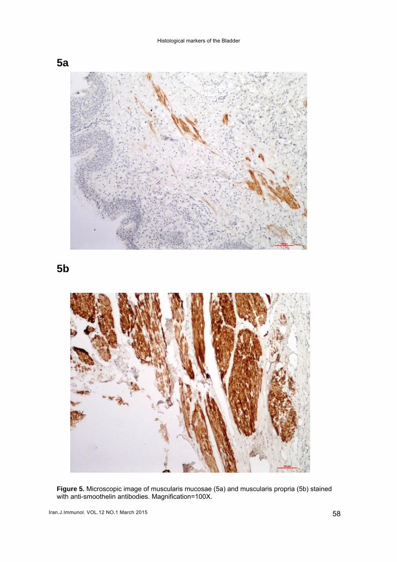
RESULTS The presence of MM was noticed in 63.6% of the specimens, including 4 cases in group one and 3 cases in group two. In each and every case it was found to be close to the basal membrane, as presented in Figure 1. Figure 1. The appearance of muscularis mucosae in a bladder specimen. H&E stain, magnification 200X. The volume of MM varied and was not identifiable in all slides. Hence, the expressions of the antigens were not assessed in all cases. Results of the immune reaction are presented in Table 2 as modal values and repeatability of modal values among patients, separately for MM and MP. Clinically significant differences were noticed for type IV collagen, desmin, filamin, smoothelin, and vimentin. Detailed results of these 5 antigens are presented in Table 3. No statistically significant difference was observed, however, it might be that the results are rather under emphasized due to the small sample size. The incidence of the same level of reaction power was 20% for type IV collagen and 0% for desmin, filamin, smoothelin, and vimentin. Microscopic images of MM and MP stained with these antibodies are presented in Figures 2-6. Comparison of normal and cancer samples did not reveal significant differences. While the general pattern of the intensity of reactions was similar, exact comparative analysis was not performed due to the small sample size.

Histological markers of the Bladder
Iran.J.Immunol. VOL.12 NO.1 March 2015 54
Table 2. Results of immune reaction in both groups. Modal value presents the most commonly observed intensity of the reaction. Repeatability indicates the percentage of cases in which modal value was observed.
MM MP
Modal Value Repeatability Modal Value Repeatability
Actin 1 66.7% 2 100%
Caldesmon 2 60.0% 2 85.6%
Collagen IV 2 80% 3 71.4%
Cytokeratin 0 100% 0 100%
Desmin 2 50% 3 60%
Elastin 0 100% 0 100%
Fibronectin 1 100% 1 50%
Filamin 0 100% 2 80%
Laminin 1 100% 1 57.4%
Myotilin 1 100% 2 50%
Smoothelin 1 100% 2 80%
Vimentin 2 50% 0 83.3%
Table 3. Detailed results of immune reactions of 5 antibodies with diagnostic potential. P value is expressed where calculation was possible.
Power of Reaction MM MP P Value
Collagen IV
0 20% 0%
0.14 1 0% 0% 2 80% 28.6% 3 0% 71.4%
Desmin
0 25% 0%
0.06 1 25% 0% 2 50% 40% 3 0% 60%
Filamin
0 100% 0% 1 0% 20% 2 0% 80% 3 0% 0%
Smoothelin
0 0% 0% 1 100% 0% 2 0% 80% 3 0% 20%
Vimentin
0 50% 83.3% 1 0% 16.7% 2 50% 0% 3 0% 0%

Poletajew S, et al.
Iran.J.Immunol. VOL.12 NO.1 March 2015 55
2a 2b Figure 2. Microscopic image of muscularis mucosae (2a) and muscularispropria (2b) stained with anti-type IV collagen antibodies. Magnification=100X.

Histological markers of the Bladder
Iran.J.Immunol. VOL.12 NO.1 March 2015 56
3a 3b Figure 3. Microscopic image of muscularis mucosae (3a) and muscularispropria (3b) stained with anti-desmin antibodies. Magnification=100X.

Poletajew S, et al.
Iran.J.Immunol. VOL.12 NO.1 March 2015 57
4a 4b Figure 4. Microscopic image of muscularis mucosae (4a) and muscularis propria (4b) stained with anti-filamin antibodies. Magnification=100X.
Histological markers of the Bladder
Iran.J.Immunol. VOL.12 NO.1 March 2015 58
5a 5b Figure 5. Microscopic image of muscularis mucosae (5a) and muscularis propria (5b) stained with anti-smoothelin antibodies. Magnification=100X.

Poletajew S, et al.
Iran.J.Immunol. VOL.12 NO.1 March 2015 59
6a 6b Figure 6. Microscopic image of muscularis mucosae (6a) and muscularis propria (6b) stained with anti-vimentin antibodies. Magnification=100X.

Histological markers of the Bladder
Iran.J.Immunol. VOL.12 NO.1 March 2015 60
DISCUSSION The presence of MM complicates the microscopic image of bladder cancer specimens. In low-quality and low-quantity surgical specimens obtained at TURBT, it can be misdiagnosed as MP. The differentiation of these two histological structures is a key point in proper bladder cancer staging and hence, treatment selection. However, it still remains challenging, especially in cases of hyperplastic MM. We performed a study aimed at practical antigenic characterisation of bladder MM and MP. Due to the problematic identification of MM and MP in TURBT specimens, cystectomy specimens were used in this pilot study. Differentiation of MM and MP can be made based on histological form, histological relations or antigenic properties. Some authors outline the different morphology of these structures. MM is usually dispersed and has an interrupted layer, whereas MP forms regular bundles (8,10). However, in as many as 17-53% of patients, pathologists observe hyperplastic MM (10,12). In these cases a criterion of histological form would fail. Close relation of MM to vascular plexus has also been postulated (11,13-14). Despite the fact that vascular plexus is absent in MP, it can also be absent in MM, especially in TURBT specimens. This criterion would also fail in many patients. Immunological profile seems to be the most potentially reliable tool for the differentiation of MM and MP. However, until today, data is very limited and no widely accepted antigen has yet been identified as a sensitive marker. In the present study, clinically important differences between MM and MP in the expression of type IV collagen, desmin, filamin, smoothelin, and vimentin were noticed. Few studies on the role of immunohistochemistry in the staging of bladder cancer have been published in the past. As they cover mainly TURBT specimens, a direct comparison of results was not possible. Type IV collagen remains the most specific characteristic for lamina propria. It has been postulated that the elasticity of bladder basal membrane depends mainly on the structure of its collagen (15). In previously published studies, type IV collagen has been used mainly to identify cancer invasion into lamina propria, recently especially in the context of microinvasion(16-19). Simultaneously, type IV collagen has never been tested as a marker of bladder layers. Hence, we propose a new role of this antigen in bladder cancer diagnostics. We found stronger expression of type IV collagen in MP compared to MM. However, this finding has no statistical significance. Additionally, the expression level was comparable in 20% of cases. In this study, the expression of desmin in MP was found to be clinically, but not statistically, stronger than MM in all cases. Surprisingly, in the study published by Council et al. no significant difference was observed (20). The numbers of patients in both studies are equal; however, Council et al. had only examined bladder cancer specimens. 'This can be crucial, while expression of desmin can be different depending on cell differentiation, as it was shown in colorectal cancer (21,22). What is more, Council et al. used a clone DE-R-11 of anti-desmin antibody, while in the recent study D33 clone was involved. Due to the lack of studies on desmin expression in human bladder, more data is needed for final conclusions. Filamin has never been used in bladder cancer diagnostics. In the recent study, MM, in all cases, showed no reaction with filamin. Malmqvistet al. suggested that filamin expression in MP is related to structural changes in detrusor, occurring in patients with bladder outlet obstruction (23). This phenomenon can partially explain the differences

Poletajew S, et al.
Iran.J.Immunol. VOL.12 NO.1 March 2015 61
in the expression of filamin in MP among patients in the recent study. However, with such limited published data, it is difficult to discuss the role of filamin expression in the differentiation of bladder layers. This antigen needs to be further studied. Weakly positive expression of smoothelin in MM, and moderately or strongly positive expression in MP was noticed in this study, which is fully consistent with the results obtained independently by Council et al. and Paneret al. There were 59 patients involved in these studies in total. Smoothelin proved to be capable of inducing differentiation of MM, myofibroblasts, and MP (20,24). More recently, these findings were confirmed by Paner et al. and Bovioet al. in TURBT specimens (25,26). However, caution should be taken, because even in 25% of cases the expression of smoothelin is comparable in MM and MP (27). This means we cannot differentiate MM and MP based on smoothelin based on expression only. Expression of vimentin, inversely to results for antigens discussed above, was found to be stronger within MM than MP in all cases. In the paper published by Council et al. a similar relation was identified in 14 of the 15 examined bladders (27). Because of different patterns of expression, vimentin seems to be a very promising antigen for differentiation between MM and MP. However, the diagnostic role of vimentin expression in bladder cancer may not be limited to identifying bladder layers. Recently the expression of vimentin was reported in 24.8% of bladder cancer cells and was found to be predictive of the progression-free survival of patients after surgical treatment (28). Also an in vitro study performed by Le et al. confirmed the relationship between vimentin expression and the invasiveness of bladder cancer cells (29). The present study has a few considerable limitations. The study group is relatively small. However, the clinical problem is mainly meaningful to TURBT specimens. Thus, it is not imperativeto perform large scale studies on cystectomy specimens. This study is a pilot and the authors will implement its results into future projects. The use of an auto stainer would bring more definitive results in comparison to recently adopted methodology. Moreover, grading of the immunohistochemical reaction intensity is subjective and additional parameters or a second opinion would be valuable. However, the authors have a deep understanding of manual experimental immunohistochemical staining and are aware of its limitations and influences on microscopic imaging. In conclusion, muscularis mucosae and muscularis propria differ in their level of expression of several protein antigens. By proper identification of bladder layers, immunohistochemistry can be useful in the histological staging of bladder cancer. It seems important to develop the optimal combination of antibodies for microscopic assessment of TURBT specimens. ACKNOWLEDGEMENTS This study was performed as 1M11/PM11D/12 research project, funded with the statutory subsidy of the First Faculty of Medicine, Medical University of Warsaw. Project co-financed by the European Union under the European Social Fund.

Histological markers of the Bladder
Iran.J.Immunol. VOL.12 NO.1 March 2015 62
REFERENCES
1. Babjuk M, Burger M, Zigeuner R, Shariat SF, van Rhijn BW, Comperat E, et al. EAU guidelines on non-muscle-invasive urothelial carcinoma of the bladder: update 2013. Eur Urol. 2013; 64:639-53.
2. Witjes JA, Comperat E, Cowan NC, De Santis M, Gakis G, Lebret T, et al. EAU Guidelines on Muscle-invasive and Metastatic Bladder Cancer: Summary of the 2013 Guidelines. Eur Urol. 2014; 65:778-92.
3. Fritsche HM, Burger M, Svatek RS, Jeldres C, Karakiewicz PI, Novara G, et al. Characteristics and outcomes of patients with clinical T1 grade 3 urothelial carcinoma treated with radical cystectomy: results from an international cohort. Eur Urol. 2010; 57:300-9.
4. Kulkarni GS, Hakenberg OW, Gschwend JE, Thalmann G, Kassouf W, Kamat A, et al. An updated critical analysis of the treatment strategy for newly diagnosed high-grade T1 (previously T1G3) bladder cancer. Eur Urol. 2010; 57:60-70.
5. Dalbagni G, Vora K, Kaag M, Cronin A, Bochner B, Donat SM, et al. Clinical outcome in a contemporary series of restaged patients with clinical T1 bladder cancer. Eur Urol. 2009; 56:903-10.
6. Brauers A, Buettner R, Jakse G. Second resection and prognosis of primary high risk superficial bladder cancer: is cystectomy often too early? J Urol. 2001; 165:808-10.
7. Holmang S, Hedelin H, Anderstrom C, Holmberg E, Johansson SL. The importance of the depth of invasion in stage T1 bladder carcinoma: a prospective cohort study. J Urol. 1997; 157:800-3.
8. Ro JY, Ayala AG, el-Naggar A. Muscularis mucosa of urinary bladder. Importance for staging and treatment. Am J Surg Pathol. 1987; 11:668-73.
9. Dixon JS, Gosling JA. Histology and fine structure of the muscularis mucosae of the human urinary bladder. J Anat. 1983; 136:265-71.
10. Paner GP, Ro JY, Wojcik EM, Venkataraman G, Datta MW, Amin MB. Further characterization of the muscle layers and lamina propria of the urinary bladder by systematic histologic mapping: implications for pathologic staging of invasive urothelial carcinoma. Am J Surg Pathol. 2007; 31:1420-9.
11. Aydin A, Ucak R, Karakok M, Guldur ME, Kocer NE. Vascular plexus is a differentation criterion for muscularis mucosa from muscularis propria in small biopsies and transurethral resection materials of urinary bladder? Int Urol Nephrol. 2002; 34:315-9.
12. Vakar-Lopez F, Shen SS, Zhang S, Tamboli P, Ayala AG, Ro JY. Muscularis mucosae of the urinary bladder revisited with emphasis on its hyperplastic patterns: a study of a large series of cystectomy specimens. Ann Diagn Pathol. 2007; 11:395-401.
13. Smits G, Schaafsma E, Kiemeney L, Caris C, Debruyne F, Witjes JA. Microstaging of pT1 transitional cell carcinoma of the bladder: identification of subgroups with distinct risks of progression. Urology. 1998; 52:1009-13.
14. Younes M, Sussman J, True LD. The usefulness of the level of the muscularis mucosae in the staging of invasive transitional cell carcinoma of the urinary bladder. Cancer. 1990; 66:543-8.
15. Kiyofuji MA, Iyama K, Kitaoka M, Sado Y, Ninomiya Y, Ueda S. Quantitative analysis of type IV collagen alpha chains in the basement membrane of human urogenital epithelium. Histochem J. 2002; 34:479-86.
16. Schapers RF, Pauwels RP, Havenith MG, Smeets AW, van den Brandt PA, Bosman FT. Prognostic significance of type IV collagen and laminin immunoreactivity in urothelial carcinomas of the bladder. Cancer. 1990; 66:2583-8.
17. Alampi G, Gelli C, Mestichelli M, Brizio R, Piccaluga A. Distribution of basement membrane antigens in bladder carcinomas: an additional prognostic parameter. Immunohistochemical study. Arch Anat Cytol Pathol. 1989; 37:224-30.
18. Conn IG, Crocker J, Wallace DM, Hughes MA, Hilton CJ. Basement membranes in urothelial carcinoma. Br J Urol. 1987; 60:536-42.
19. Daher N, Abourachid H, Bove N, Petit J, Burtin P. Collagen IV staining pattern in bladder carcinomas: relationship to prognosis. Br J Cancer. 1987; 55:665-71.
20. Council L, Hameed O. Differential expression of immunohistochemical markers in bladder smooth muscle and myofibroblasts, and the potential utility of desmin, smoothelin, and vimentin in staging of bladder carcinoma. Mod Pathol. 2009; 22:639-50.

Poletajew S, et al.
Iran.J.Immunol. VOL.12 NO.1 March 2015 63
21. Arentz G, Chataway T, Price TJ, Izwan Z, Hardi G, Cummins AG, et al. Desmin expression in colorectal cancer stroma correlates with advanced stage disease and marks angiogenic microvessels. Clin proteomics. 2011; 8:16.
22. Ma Y, Peng J, Liu W, Zhang P, Huang L, Gao B, et al. Proteomics identification of desmin as a potential oncofetal diagnostic and prognostic biomarker in colorectal cancer. Mol Cell Proteomics. 2009; 8:1878-90.
23. Malmqvist U, Arner A, Uvelius B. Cytoskeletal and contractile proteins in detrusor smooth muscle from bladders with outlet obstruction--a comparative study in rat and man. Scand J Urol Nephrol. 1991; 25:261-7.
24. Paner GP, Shen SS, Lapetino S, Venkataraman G, Barkan GA, Quek ML, et al. Diagnostic utility of antibody to smoothelin in the distinction of muscularis propria from muscularis mucosae of the urinary bladder: a potential ancillary tool in the pathologic staging of invasive urothelial carcinoma. Am J Surg Pathol. 2009; 33:91-8.
25. Bovio IM, Al-Quran SZ, Rosser CJ, Algood CB, Drew PA, Allan RW. Smoothelin immunohistochemistry is a useful adjunct for assessing muscularis propria invasion in bladder carcinoma. Histopathology. 2010; 56:951-6.
26. Paner GP, Brown JG, Lapetino S, Nese N, Gupta R, Shen SS, et al. Diagnostic use of antibody to smoothelin in the recognition of muscularis propria in transurethral resection of urinary bladder tumor (TURBT) specimens. Am J Surg Pathol. 2010; 34:792-9.
27. Miyamoto H, Sharma RB, Illei PB, Epstein JI. Pitfalls in the use of smoothelin to identify muscularis propria invasion by urothelial carcinoma. Am J Surg Pathol. 2010; 34:418-22.
28. Zhao J, Dong D, Sun L, Zhang G, Sun L. Prognostic significance of the epithelial-tomesenchymal transition markers e-cadherin, vimentin and twist in bladder cancer. Int Braz J Urol. 2014; 40:179-89
29. Lu G, Ding J, Wang Z. Vimentin gene transfection promotes the invasion of bladder cancer SW780 cells. Xi Bao Yu Fen Zi Mian Yi Xue Za Zhi. 2014; 30:713-6
30.