Embed Size (px)
Citation preview

International Research Journal of Engineering and Technology (IRJET) e-ISSN: 2395 -0056
Volume: 04 Issue: 01 | Jan-2017 www.irjet.net p-ISSN: 2395-0072
© 2017, IRJET | Impact Factor value: 5.181 | ISO 9001:2008 Certified Journal | Page 1310
Antibacterial and UV- Protective Cotton fabric made by Herbal
Synthesized Silver Nanoparticles
Mangesh D. Teli1*, Malaya Ranjan Sahoo2 and Pintu Pandit3
1Professor, Dept. of Fibres and Textile Processing Technology, Institute of chemical Technology, Mumbai, India. 2M.Tech, Dept. of Fibres and Textile Processing Technology, Institute of Chemical Technology, Mumbai, India.
3Ph. D. Tech., Dept. of Fibres and Textile Processing Technology, Institute of Chemical Technology, Mumbai, India. Email: [email protected]
---------------------------------------------------------------------***----------------------------------------------------------------
Abstract - Ecofriendly Herbal synthesis of silver nanoparticle (H-AgNPs) was carried out using Narikel Leaf, Panasa Leaf, and Amalaki Fruit. Their application on the cotton fabric was studied to create protective fabric in terms of Antibacterial and UV-Protective properties. The resultant metal particles were characterized by VU-vis spectra, Particle size analyzer and FTIR. The H-AgNPs deposition on the cotton fabric was characterized FTIR, EDX, SEM, TGA and XRD analysis. The antibacterial activity of H-AgNPs loaded cotton show 99% bacterial reduction against S. Aureus and E. Coli bacteria. UV protection was assessed by AS-NZS 4399 standard and excellent UPF of the order 32 was achieved. Key Words: Herbal synthesis, silver nanoparticle, cotton fabric, polyelectrolyte multilayer, antibacterial, UV-Protective.
1. INTRODUCTION Nanoparticles exhibit unique properties due to their size, distribution, and morphology. Silver nanoparticles have many applications because of its well-known antimicrobial activity which can be used in microbial protective textiles, wound dressing and drug delivery, coating for medical implants and dental material, food storage container and packaging, water filters, dyeing, finishing and cosmetics [1-5]. The emerging infectious diseases all around is a matter of serious concern for the development of textiles with resistance to the pathogenic bacteria. Although AgNPs has potential antimicrobial efficacy against a broad spectrum of bacteria, still they have negligible toxicity to human cells and hence their applicability becomes quite promising [6]. Several mechanisms have been proposed to explain the inhibitory effect of silver nanoparticles on bacteria. It is assumed that the high affinity of silver towards sulfur and phosphorus is the key element of the antimicrobial effect. Due to the abundance of sulfur-containing proteins on the bacterial cell membrane, silver nanoparticles can react with sulfur-containing amino acids inside or outside the cell membrane, which in turn affects bacterial cell viability [7-8] It was also suggested that silver ions (particularly Ag+) released from silver nanoparticles can interact with
phosphorus moieties in DNA, resulting in inactivation of DNA replication, or can react with sulfur-containing proteins, leading to the inhibition of enzyme functions [8- 10]. Again it is suggested that the bacterial DNA molecules are in relaxed state replicates DNA effectively. If DNA molecules are in condensed form they lose their replicability. AgNPs goes inside the microbial cell, and DNA molecules turn into condensed form by losing their replicability leading to bacterial death [11- 12]. Similarly, it is also becoming now a days necessary to build up a UV protective textile once, as in these days UV radiation is found to be reaching earth surface. The ultraviolet radiation (UVR) ranges from 40 nm to 400nm with further classified into UV-A (320 to 400 nm), UV-B (290 to 320 nm), and UV-C (200 to 290 nm). UV-C is totally absorbed by the upper atmospheric ozone layer and does not reach the earth [13]. UV-A weaken the immunological response of skin cells and UV-B creates dangerous skin cancer. UV protective textile can be made by colouration and natural polyphenols deposition in textile [14-15], silver nanoparticles are found to give colouration [16], if it is herbal synthesised.
Having a huge area of applications, its path of synthesis by chemical root is creating a bottleneck situation, as it is toxic one where NaBH4 [17-18], Aniline [19], Sodium citrate [20],etc. and a polymeric compound such as poly (vinylpyrrolidone) (PVP) [21], poly (ethylene glycol) (PEG)[22], and some surfactants are used as stabilizers to prevent nanoparticles agglomeration and precipitation. These chemicals are toxic and hazardous to the environment. There of stems a desire for developing an economic, eco-friendly and easily scalable process for large scale synthesis of AgNPs. The researchers have been thus focusing on the green synthesis of nanoparticles using microorganisms, enzymes, and herbal extracts which could offer several benefits over conventional physical and chemical methods. Herbal synthesis pathways don't need to use toxic chemicals, high pressure, temperature, and energy like microwave-assisted method [16, 23] with enhanced applicability. Among these eco-friendly methods, the usage of plant materials i.e. the herbs is more advantageous than other biological processes as it
International Research Journal of Engineering and Technology (IRJET) e-ISSN: 2395 -0056
Volume: 04 Issue: 01 | Jan-2017 www.irjet.net p-ISSN: 2395-0072
© 2017, IRJET | Impact Factor value: 5.181 | ISO 9001:2008 Certified Journal | Page 1311
eliminates the risk as well as the elaborate process preparation.
Since a few years, green synthesis of AgNPs has been reported using Olive leaf (Mostafa M.H. Khalil et.al., 2013)[24], Aloe Vera (Chandran, S.P et.al., 2006) [25], Averrhoa carambola fruit (Ipsita Hazra Chowdhury et.al., 2014) [26], Marigold flower (Hemali Padalia et.al, [24, 19, 27], Lantana camara leaf (B. Ajitha et.al., 2015)[28], Catharanthus roseus leaf (Ponarulselvam S et.al., 2012)[29], Eucalyptus chapmaniana leaves (Ghassan Mohammad Sulaiman et.al., 2013)[30], etc. The important plant metabolites such as terpenoids, polyphenols, sugars, alkaloids and phenolic acids are responsible for the bio reduction of metal ions into nanoparticles [31- 34]. Cocos nucifera, Artocarpus heterophyllus and Phyllanthus Emblica are commonly known as Coconut, Jackfruit and Indian gooseberry in English language and in India known as Narikel, Panasa, and Amalika in the Sanskrit language. These plants well known for their medicinal application as their body parts like leaves and fruits are rich in polyphenols, flavonoid and ascorbic acid [35-36] also contain amino acids and antioxidant. Therefore, their aqueous extract can be used as both reducing and capping agents for the preparation of AgNPs. The present paper deals with the herbal synthesis of silver nanoparticle (H-AgNPs) using mentioned herbal extract (Narikel leaf, Panasa leaf, and Amalika fruit). And the performance properties of the fabric carrying such nanoparticles have been reported.
2. MATERIALS AND METHODS
AgNO3 99% purity supplied from Sigma Aldrich¬, Cocos nucifera Leaf (Narikel), Artocarpus heterophyllus Leaf (Panasa) are collected from the Institute of Chemical Technology campus and Phyllanthus emblica Fruit (Amalaki) is purchased from local market, Distilled water. 120 GSM plain weave 100% cotton fabric, Cationic Polyacrylamide polyelectrolytes (CPAPE) purchase from Rishabh Metals & Chemicals Pvt.Ltd. and Escherichia coli Gram-negative bacteria, Staphylococcus aureus Gram-negative bacteria.
2.1 Preparation herbal extracts (HE)
Fresh leaves of Narikel, Panasa, and Fruits of Amalaki were cleaned two times with distilled water and chopped into small pieces. Then 20 g of leaves and 10 g of fruit were taken in 500 ml round bottom flask containing 100 ml distilled water under reflux condition for 1 hr. at 100°C, The herbal extract (HE) was filtrate obtained after filtering this contents through nylon mesh followed 2 times through Whatman No.42 filter paper and kept at 4° C for repeated (2 to 3 times) use.
2.2 Synthesis of H-AgNPs
Three different H-AgNPs were synthesized using Narikel
leaf extract (N-LE), Panasa leaf extract (P-LE) and Amalaki
fruit extract (A-FE) by two different processes. In the first
process standard solution of AgNO3 (0.01 M), Water and
HE were mixed together in a stoichiometric ratio to form 1
mM, 2 mM, 3 mM solution of AgNO3 of 100 ml, where HE
added drop by drop under magnetic stirring and kept for
24 hrs to complete the reaction. In the case of the second
process the solution mixture was prepared in a similar
manner but under a high-temperature condition. The
temperature of the AgNO3 solution was raised to 100° C
after addition of HE and kept under magnetic stirring for a
different period of time to study its effect on particle size.
2.3. Deposition of H-Ag-NPs onto cotton fabric
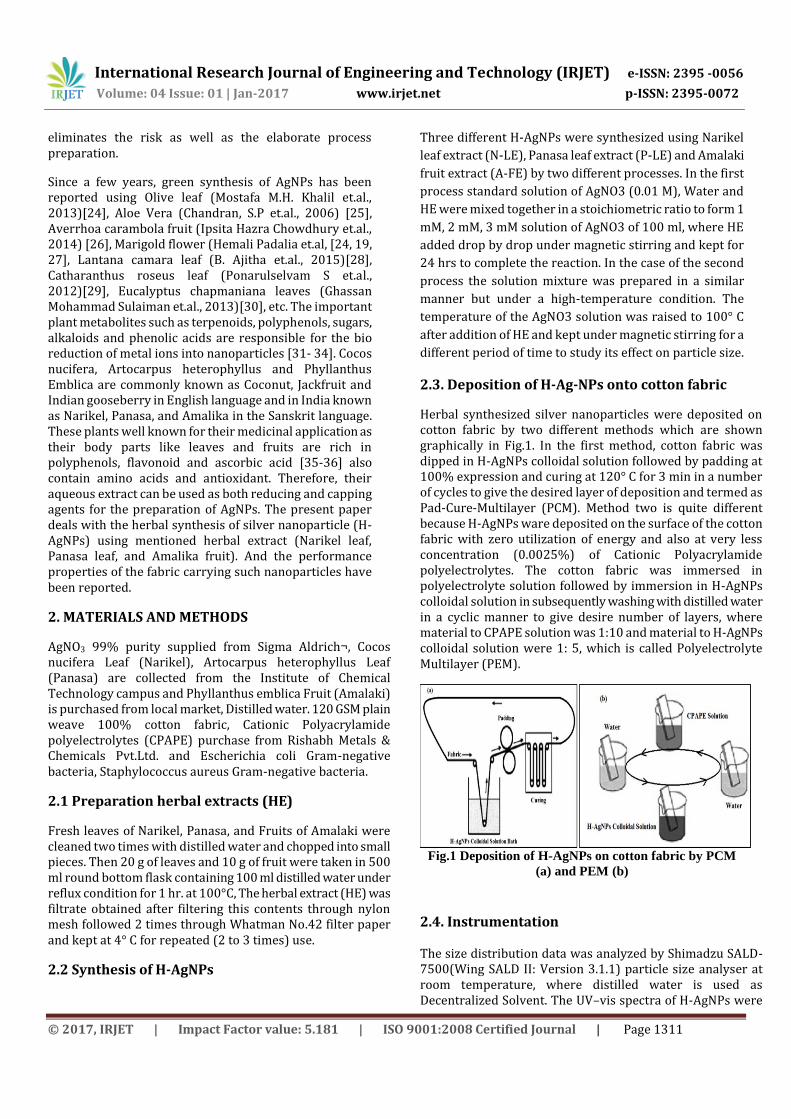
Herbal synthesized silver nanoparticles were deposited on cotton fabric by two different methods which are shown graphically in Fig.1. In the first method, cotton fabric was dipped in H-AgNPs colloidal solution followed by padding at 100% expression and curing at 120° C for 3 min in a number of cycles to give the desired layer of deposition and termed as Pad-Cure-Multilayer (PCM). Method two is quite different because H-AgNPs ware deposited on the surface of the cotton fabric with zero utilization of energy and also at very less concentration (0.0025%) of Cationic Polyacrylamide polyelectrolytes. The cotton fabric was immersed in polyelectrolyte solution followed by immersion in H-AgNPs colloidal solution in subsequently washing with distilled water in a cyclic manner to give desire number of layers, where material to CPAPE solution was 1:10 and material to H-AgNPs colloidal solution were 1: 5, which is called Polyelectrolyte Multilayer (PEM).
Fig.1 Deposition of H-AgNPs on cotton fabric by PCM
(a) and PEM (b)
2.4. Instrumentation
The size distribution data was analyzed by Shimadzu SALD-7500(Wing SALD II: Version 3.1.1) particle size analyser at room temperature, where distilled water is used as Decentralized Solvent. The UV–vis spectra of H-AgNPs were

International Research Journal of Engineering and Technology (IRJET) e-ISSN: 2395 -0056
Volume: 04 Issue: 01 | Jan-2017 www.irjet.net p-ISSN: 2395-0072
© 2017, IRJET | Impact Factor value: 5.181 | ISO 9001:2008 Certified Journal | Page 1312
recorded at room temperature using Shimadzu UV-1800 Spectrophotometer. Fourier transforms infrared (FTIR) spectra were recorded at room temperature on a Shimadzu FTIR-8400S spectrometer. For the FTIR measurements of herbal synthesized silver nanoparticles, a few silver nanoparticles (20 ml colloidal solution) dried at 60° C for 24 h was mixed with KBr to form a round disk suitable for FTIR measurements. FTIR of deposited cotton and pristine fabric were carried at normal condition. Surface characterisation of treated cotton fabric was done by Field Emission Gun-Scanning Electron Microscope (FEG-SEM) MIRA3 TESCAN at 7kV electron voltage. Energy dispersive X-ray (EDX) of treated fabric sample was studied using a JEOL JED 2300 Analysis Station for chemical characterisation. Thermogravimetric analysis was carried out with a heating rate of 10° C /min using a Shimadzu DTG-60H Simultaneous DTA-TG Apparatus. X-ray diffraction (XRD) pattern was obtained using LabX XRD-6100 made by Shimadzu X-Ray Diffractometer with Cu Ka (k= 1.54056 A˚).
2.5. Colour co-ordinates analysis
Colorimetric data for H-AgNPs deposited Cotton fabric was recorded with a colorimeter connected to RAYSCAN Spectra Scan 5100+. The equipment was adjusted as follows: CIE LAB colour space, 10° observers with D65 illuminant, d/2 viewing geometry and measurement area of 2 mm. Colours were expressed by measuring parameters like lightness (L*) from black (0) to white (100), a* is red (+) / green (−) ratio, b* is yellow (+) / blue (−) ratio and colour difference (ΔE), by taking untreated cotton fabric as reference. On the basis of these parameter colour difference (ΔE) by was determined as:
where ΔL is the colour lightness difference between treated (Cotton fabric loaded with H-AgNPs) and (Untreated Cotton fabric) samples; Δa red/green difference between treated and untreated samples; Δb yellow/blue difference between treated and untreated samples. Also, the colour strength was measured in terms of K/S as followed equation.
Where R is reflectance
2.6. UV protection Test Protection against ultra-violate rays was accessed by UV Transmittance Analyzer of Shimadzu UV-2600 UV-vis spectrophotometer equipped with AS_NZS 4399 standard. The Ultra-violate protection factor (UPF) is the ratio of the average effective ultraviolet radiation (UVR) irradiance calculated for unprotected skin (ED) to the average effective UVR irradiance calculated for skin protected by the test fabric (Edm) 34 shown in Eqn (3).
Transmission percentage (T %) against UVA and UVB was obtained from transmittance spectra of fabric samples which were to calculate UPF and T% calculated as:
Where, Eλ = erythemal spectral effectiveness, Sλ = solar spectral irradiance in W/m2/nm, Tλ =spectral transmittance of fabric, Δλ = the bandwidth in nm, and λ = the wavelength of nm.
2.7. Antibacterial activity Test
The antibacterial efficiencies of treated samples were quantitatively estimated using Gram-positive bacteria S. aureus and Gram-negative bacteria E. coli according to the American Association of Textile Chemists and Colorists (AATCC) 100-2004 standard. The percentage of microbe reduction (R %) was calculated using the following equation:
Where, B (CFUs) is the number of microbial colonies of the untreated fabrics after inoculation for 24 hr., and A (CFUs) is the number of microbial colonies of the treated fabrics after inoculation for 24 hr.
3. RESULTS AND DISCUSSIONS
3.1. Particle size analysis Results in Table 1 indicate that particle size decreased with a decrease in molar concentration of AgNO3 in extract solution. Further, it decreased with increase in extract amount in room temperature for 24 hr. the process of N-AgNPs. But in higher temperature process, it decreased with a decrease in time. A similar trend was found in the case of synthesis of P-AgNPs. In the case of A-AgNPs synthesis at room temperature particle size decreased with a decrease in extract amount and time. For higher temperature process also the particle size decreased with decreasing time. By comparing the three herbal reductants and two different temperature process it was found that the least mean particle size was obtained at higher temperature process with Panasa leaf extract (18 nm) followed by Narikel leaf (30 nm) and Amalaki fruit extract (35 nm). The stability of the synthesized H-AgNP was confirmed by measuring particle size distribution after storage for several months, up to six months. The measurement of particle size distribution data did not show any appreciable change in the size distribution graph as well as in mean size. This clearly rules out the possibility of aggregation of the nanoparticles upon storage and hence, it indicates the stability of the silver nanoparticles obtained by herbal synthesis.

International Research Journal of Engineering and Technology (IRJET) e-ISSN: 2395 -0056
Volume: 04 Issue: 01 | Jan-2017 www.irjet.net p-ISSN: 2395-0072
© 2017, IRJET | Impact Factor value: 5.181 | ISO 9001:2008 Certified Journal | Page 1313
In particle size distribution Fig. 2 (a) of P-AgNPs shows that 90% of normalized particle amount is less than 25 nm with 50% of normalized particle amount is less than 18 nm. Fig. 2 (b) of N-AgNPs shows that 90% of normalized particle amount is less than 42 nm with 50% of normalized Particle amount are less than 29 nm. Fig. 2 (c) of A-AgNPs shows that 90% of normalized particle amount are less than 55 nm with 50% of normalized Particle amount are less than 35 nm. This certifies that the particles were of uniform and even in size.
Table1. Particle size data of H-AgNPs at different parameters
Fig. 2 Particle size distribution of P-AgNPs (a), N-AgNPs (b) and A-AgNPs (c)
3.2. Ultraviolet–visible (UV–vis) spectral analysis
Ultraviolet–visible (UV–vis) spectral analysis is a sensitive technique which proves the formation of metal nanoparticles as it exhibits an intense absorption peak due to the surface plasmon excitation of conduction electrons in metal. The absorption spectra of herbal synthesized AgNPs were recorded in 290 nm to 800 nm range. As from the various study, it is well known that silver nanoparticles show an absorption band in the range of 350–450 nm due to surface plasmon vibrations of conducting electrons of silver [37-38]. In the herbal synthesis of nanoparticles, reddish black, dark brown and gray colour was noticed with respective to P-AgNPs, N-AgNPs, and A-AgNps. Those coloured colloidal silver nanoparticle solutions were observed under UV-vis spectrophotometer. In Fig. 3 herbal synthesised nanoparticles were observed at the absorption band of 406.50 nm, 425.00

International Research Journal of Engineering and Technology (IRJET) e-ISSN: 2395 -0056
Volume: 04 Issue: 01 | Jan-2017 www.irjet.net p-ISSN: 2395-0072
© 2017, IRJET | Impact Factor value: 5.181 | ISO 9001:2008 Certified Journal | Page 1314
nm and 429.00 nm for P-AgNPs, N-AgNPs, and A-AgNPs respectively, which conforms the reduction of Ag+ ions into Ag° nanoparticles. The absorption curves show single sharp intense band suggesting the formation of monodispersed nanoparticles.
Fig. 3 UV–vis absorption spectroscopy of silver nanoparticles.
3.3 Deposition of H-AgNps on Cotton fabric
The deposition of the herbal synthesized silver nanoparticle is a simultaneous coloration process of cotton as the colloidal silver nanoparticles were showing intense colour. The colouration effect was monitored by two different processes i.e. pad cure multilayer (PCM) and polyelectrolyte multilayer (PEM) divided into five different cycles. Polyelectrolyte multilayer (PEM) process exhaustion of nanoparticle was observed, causing a decrease in colour intensity of colloidal H-AgNPs solution in the bath in each cycle as seen in Fig. 4.
Fig. 4 P-AgNPs was deposited onto cotton fabric by PEM process, (a) pure colloidal silver nanoparticle solution, (b) after 1st cycle, (c) after 2nd cycle, (d) after 4th cycle (e) after 6th cycle, (f) after 8th cycle, (g) after 10th cycle and (h) 12th after cycle.
Whereas significant colour bleeding was noticed in wash beaker in subsequent washing even after completion of end
cycle. This is possible due to the decrease in zeta potential of cotton fabric on dipping in CPAPE solution and silver in Ag° having nanoparticle goes onto +ve fibre surface. Such effect was not noticed in the care of pad cure multilayer process, where in first adsorption of silver nanoparticle on fiber surface takes place followed by diffusion into the fibre and finally, fixation is aided by the application of a temperature in the curing process.
3.4 FTIR analysis
In the Fig. 5(a) FTIR spectra of all the three herbal nanoparticles (P-AgNPs, N-AgNPs, and A-AgNPs) in dried form were observed and the peaks at 3298 cm-1, 3272 cm-1, and 3242 cm-1 can be assigned to O-H stretch of phenols. The peak at 1604 cm-1, 1608 cm-1 and 1610 cm-1 (P-AgNPs, N-AgNPs, and A-AgNPs) which could be C=C stretching correspond to flavonoids and probably are overlapped by much intense amide peaks along with 1436 cm-1 and 1446 cm-1 (P-AgNPs and A-AgNPs) aromatic ring stretching. The peak at 1708 cm-1 of A-AgNPs is the characteristic peak of C=O stretching vibration of free galic acid together with 1027 cm-1,1039 cm-1 and 1045 cm-1 aryl phenolic ester C-O-C symmetric stretching and 871 cm-1 (C-C stretching). Groups possibly C=C skeletal ring stretching or skeletal vibration of the aromatic ring and C-O stretching found in phenolic compounds could be assigned to 1519 cm-1 in P-AgNPs. The intense peak at 1317 cm-1, 1350 cm-1 and 1359 cm-1 and the brand at 763 cm-1 (sugar ring, breathing vibration) are consistent with the presence of hydrolysable tannins. The peak at 1211 cm-1, 1197 cm-1 and 1203 cm-1 (C–O bond stretching vibration) in P-AgNPs, N-AgNPs and A-AgNPs must be responsible for the presence of tannins.

International Research Journal of Engineering and Technology (IRJET) e-ISSN: 2395 -0056
Volume: 04 Issue: 01 | Jan-2017 www.irjet.net p-ISSN: 2395-0072
© 2017, IRJET | Impact Factor value: 5.181 | ISO 9001:2008 Certified Journal | Page 1315
Fig. 5 FTIR graph of H-AgNPs (a) and untreated and treated cotton fabric sample (b)
The presence of AgNPs on the cotton fabric was further confirmed by the FTIR measurement. The FTIR spectra of untreated cotton fabric and cotton containing AgNPs is shown in Fig. 5(b). The characteristic peaks of cotton due to cellulose macromolecule, which appear at 3,334 cm-1 (O–H stretching), 2,900 cm-1 (C–H stretching), 1,430 cm-1 (C–H wagging), 1,368 cm-1 (C–H bending) and 1,029 cm-1 (C–O stretching) have become broad, red shifted and with strong intensity for silver nanocoated cotton fabric. These results indicate binding of AgNPs with O in cellulose macromolecule. On the other hand, the spectra of nanosilver treated cotton fabric don't reveal new peaks different from the case of untreated sample, which means that no chemical reaction with cellulose takes place during the deposition of AgNPs.
3.5 EDX analysis
Fig. 6 EDX spectrum of H-AgNPs deposited by PCM (a) and PEM (b)
EDX spectrum of cotton fabric deposited with H-AgNPs on the surface of a particular scan area confirms the existence of silver element. In the Fig. 6 peaks for silver is seen at 2.983 keV with mass % of 34.60 and 37. 35 in 4 layers MPC and MPE deposited cotton respectively. The peak at 0.392 keV in 4 layer MPE H-AgNPs deposited cotton shows 27.02 % nitrogen mass % which may be due to the layering of cationic polyacrylamide polyelectrolytes where as in 4 layer MPC H-AgNPs deposited cotton, nitrogen mass % remains nil.
3.6 SEM

International Research Journal of Engineering and Technology (IRJET) e-ISSN: 2395 -0056
Volume: 04 Issue: 01 | Jan-2017 www.irjet.net p-ISSN: 2395-0072
© 2017, IRJET | Impact Factor value: 5.181 | ISO 9001:2008 Certified Journal | Page 1316
Fig. 7 SEM image of Untreated Cotton (a), PCM H-AgNPs Cotton (b) and PEM H-AgNPs Cotton (c)
The surface topography of cotton fabric was changed due to the deposition of H-AgNPs by PCM and PEM processes as seen in Fig.7. It was noticed that the deposition of silver nanoparticle was much higher in PEM deposited cotton (Fig. 7(c)) than PCM deposited cotton (Fig. 7(b)) whereas untreated (Fig. 7(a)) cotton fabric remained clear of any deposits. This is due to the decrease in zeta-potential of cotton fabric causing the improved affinity of cotton towards silver nanoparticles. Although the CPAPE layers were not found but the dense deposition of silver nanoparticle was due to the MPE process. Furthermore, the morphology seen in SEM image reveal that the data of Particle Size analysis is complimenting this observation.
3.7 TGA Analysis
Fig. 8 TGA curve of cotton samples treated with AgNPs and untreated
Thermal behaviour of untreated cotton and H-AgNPs deposited cotton was studied using TGA and the thermograms shown in Fig. 8 indicate that deposition of AgNPs changed the thermal behaviour of cotton fabric. Initially, weight loss was more on silver deposited cotton, this was may be due to the increased moisture content of nanoparticle deposited cotton fabric by PEM process. But overall weight loss % was decreased in comparison to that of untreated cotton fabric. The weight loss value in PEM H-AgNPs cotton, PCM H-AgNPs cotton, and untreated one was found to be 90.437 %, 92.208 %, and 93.694 %, respectively.
3.8 XRD Analysis
The X-ray diffraction spectra of the cotton fabric treated and untreated are presented in Fig. 9. The characteristic peaks of the cellulose structure appeared in all samples, near to the 2θ angles of 14.94°, 16.42°, 22.41°, 43.86° and 77.43° which are associated with (101), (101), (002), (200) and (311)[39] respectively. Their respective intensity (counts) was given in Table 2.
Fig. 9 XRD graph of Cotton fabric samples
Table 2. Intensity at 2θ of treated and untreated
fabric sample for XRD curve
Peak
(2θ)
Intensity (counts)
Untreated
Cotton
PCM H-
AgNPs
Cotton
PEM H-
AgNPs
Cotton
14.94 1506 1342 1315
16.42 1261 1136 1104
22.7 4861 4644 4267
43.86 524 533 590
77.34 495 525 588

International Research Journal of Engineering and Technology (IRJET) e-ISSN: 2395 -0056
Volume: 04 Issue: 01 | Jan-2017 www.irjet.net p-ISSN: 2395-0072
© 2017, IRJET | Impact Factor value: 5.181 | ISO 9001:2008 Certified Journal | Page 1317
It was found that the characteristic peak 14.94, 16.42 and 22.41 associated with cellulose their intensity (counts) decrease whereas the peaks, 43.86 and 77.43 are nearer to the characteristic picks of pure silver nanoparticle (i.e 44.38 and 77.64)[40] were increase in counts which conforms the deposition of silver nanoparticle on cotton fabric. Also, it was found that the crystallinity slightly decreases (30. 29 and 29. In untreated PCM and PEM-treated cotton respectively).
3.9 Colour Measurement
On the deposition of three H-AgNPs, three different colourations of cotton fabric were noticed. These are pink, saffron-brown and gray colour obtained by C-AgNPs, P-AgNPs, and A-AgNPs respectively. CIE Lab Parameter L*, a*, and b* was measured to get an appropriate idea of colouration through herbal synthesised silver nanoparticles as function layers. It is known that, if the colour difference (ΔE) is greater than one, then can colour change detected visually. It is obviously from Table 3 colour changes were obtained considerable good in number and all of them could be visually detected as ΔE exceeded one. The higher the concentration of applied colloid, the larger the color change (ΔE = 39.87, 36.82 and 31.38 for 12L MPE C-AgNPs, P-AgNPs, and A-AgNPs cotton fabrics, respectively). These results are in good correlation with the number of layers of H-AgNPs cotton fabric. In other way, it can be said that the higher the concentration of applied herbal synthesised AgNPs colloid, the larger the amount of deposited Ag NPs.
Table 3. Colour coordinates data (CIE lab) for H-AgNPs deposited cotton fabric as a function of layers
A single trend was found in Table 3 for both nanoparticle deposition process with an increase in layers, that is a decrease in L*, increase in a (+) sign of a* and a little increase in a (+) sign of b*. There was an increase in increasing order of colour strength in terms of K/s for individual H-AgNPs cotton in Fig. 10 the colour strength increases step wise from 2L to 12L due to the increase in silver concentration on each layer of deposition by indicating a direct relation towards K/S.
Fig. 10 K/S of cotton fabric deposited by different layers of herbal synthesized nanoparticle
3.9 UV Resistivity Analysis
The significance of UV protection in our research due to the simultaneous colouration of cotton fabric on the process of deposition of H-AgNPs. Although it is known that the colouring compound and pigments are responsible elements of against the protection from UV rays. In other hand, since our synthesised silver colloidal solution contains herbal extract, those are non-other than phytochemical also interfere in the protection from UV radiation.
Fig. 12 UPF Curve of H-AgNPs Cotton fabric as a function of layers

International Research Journal of Engineering and Technology (IRJET) e-ISSN: 2395 -0056
Volume: 04 Issue: 01 | Jan-2017 www.irjet.net p-ISSN: 2395-0072
© 2017, IRJET | Impact Factor value: 5.181 | ISO 9001:2008 Certified Journal | Page 1318
In Table 4 it was noticed that the 12 layer of PEM deposited N-AgNPs, A-AgNPs and P-AgNPs cotton fabric give excellent, very good and good UV protection respectively and the transmittance percentage (T%) decreases for both UVA and UVB in comparison to the untreated cotton fabric. the observation in Fig. that the UPF increases with increase in layers for all herbal synthesized silver nanoparticles.
Table 4. UPF of cotton samples treated with H-AgNPs
3.10 Antibacterial Efficacy
Effect of H-AgNPs on the bacterial resistivity of cotton quantitatively mentioned in Table 5, also the after wash effect on antibacterial activity was reported using the washing standard AATCC-64. The bacterial reduction percentage (R%) was calculated taking untreated as a reference, more than 99% efficiency was achieved for both 4L process H-AgNps deposited cotton against S. Aureus and E. Coli bacteria before wash. After 10th wash PEM Process shows more than 97 % reduction activity where 95% reduction was found in PCM process against S. Aureus Bacteria. Similarly, against E. Coli bacteria PEM Process shows more than 98 % reduction
activity where 96% reduction was achieved in PCM. This means PEM process gives higher antibacterial activity than PCM, it may be due to the presence of more silver nanoparticle because of CPAPE in PEM process even after 10th washes than PCM process. Fig. 13 was given as a proof of the quantitative result.
Fig. 13 Quantitative analysis of antibacterial activity against S.A of untreated (a), treated unwashed (b) and 10th washed (c), against E.C of untreated (d), treated unwashed (e) and 10th washed (f)
Table 5. Antibacterial activity of Cotton samples deposited with H-AgNPs
4 Conclusions

International Research Journal of Engineering and Technology (IRJET) e-ISSN: 2395 -0056
Volume: 04 Issue: 01 | Jan-2017 www.irjet.net p-ISSN: 2395-0072
© 2017, IRJET | Impact Factor value: 5.181 | ISO 9001:2008 Certified Journal | Page 1319
Exploitation three herbal reducing and stabilizing agents such as Narikel leaf, Panasa leaf and of Amalaki fruit for the synthesis of Ag nanoparticles and Characteristic functional groups of these herbs were proved by FTIR analysis of herbal synthesized silver nanoparticle. Reduction of Ag+ ion into Ag° was confirmed by Surface Plasmon Resonance study. The further size of particles was measured by particle size analyzer testing which yielding 18 nm in P-AgNPs. Synthesized silver nanoparticles were deposited on cotton fabric by two different process pad cure multilayer and polyelectrolyte multilayer to fabricate durable protective textile in terms of Antibacterial and UV-Protective. Deposition of silver nanoparticle was proven by XRD, EDX and SEM analysis. SEM analysis ravels the same with the data of particle size analyzer. Change in thermal behavior due to deposition of silver nanoparticles was noticed in thermogravimetric analysis. Also, herbal synthesized silver nanoparticle gives colouration effect to the cotton fabric. The colour difference was accessed by CIE Lab. Coloured Cotton fabric, because of silver nanoparticle gives good to excellent UV protection and antibacterial activity was outstand even after 10th wash against both Gram Positive S. aureus and Gram-Negative E. coli bacteria.
Acknowledgment: The authors gratefully acknowledge the Fellowship from All India Council for Technical Education (AICTE), India, for carrying out this research work.
REFERENCES
[1] Boris lldusovich Kharisov, Oxana Vasilievna Kharissova, Ubaldo Oritz Mendez; CRC Concise Encyclopedia of Nanotechnology, 2016 pg 1-8
[2] Teli, M. D., Samanta, K. K., Pandit, P., Basak, S., & Chattopadhyay, S. K. (2014). Low Temperature Dyeing of Silk using Atmospheric Plasma Treatment. Indian Journal of Natural Fibres: v, 1(1), 1-7.
[3] Teli, M. D., Samanta, K. K., Pandit, P., Basak, S., & Gayatri, T. N. (2015). Hydrophobic silk fabric using atmospheric pressure plasma. International Journal of Bioresource Science, 2(1), 15-19.
[4] Teli, M. D., Samanta, K. K., Pandit, P., Basak, S., & Chattopadhyay, S. K. (2015). Low-temperature dyeing of silk fabric using atmospheric pressure helium/nitrogen plasma. Fibers and Polymers, 16(11), 2375.
[5] Das, D., Pandit, P., Maiti, S., & Dalapati, K. K. (2016). Development of Protective Clothing for Pesticide Operation. 3(8), 851-855.
[6] a tia Ornelas-Megiatto, Parth N. Shah, Peter . Wich, essica L. ohen, asur A. Tagaev, ustin A. Smolen, rian . Wright, Matthew . Panzner, Wiley . oungs,
ean M. . re chet and arolyn L. annon; Aerosolized Antimicrobial Agents Based on Degradable Dextran Nanoparticles Loaded with Silver Carbene omplexes; Mol. Pharmaceutics 2012, 9, 3012−3022
[7] Sukumaran Prabhu and Eldho K Poulose; Silver nanoparticles: mechanism of antimicrobial action, synthesis, medical applications, and toxicity effects, International Nano Letters 2012, 2:32
[8] Woo Kyung Jung, Hye Cheong Koo, Ki Woo Kim, Sook Shin, So Hyun Kim, and Yong Ho Park; Antibacterial Activity and Mechanism of Action of the Silver Ion in Staphylococcus aureus and Escherichia coli, Applied And Environmental Microbiology, Apr. 2008, P. 2171–2178
[9] Gupta A, Silver S.; Silver as a biocide: will resistance become a problem? Nat Biotechnol. 1998; 16:888-890.
[10] Matsumura Y, Yoshikata K, Kunisaki S, Tsuchido T. Mode of bactericidal action of silver zeolite and its comparison with that of silver nitrate. Appl Environ Microbiol. 2003; 69:4278-4281.
[11] Padma S. Vankar, Dhara Shukla; Biosynthesis of silver nanoparticles using lemon leaves extract and its application for antimicrobial finish on fabric; Appl Nanosci (2012) 2:163–168
[12] Meiwan Chen, Zhiwen Yang, Hongmei Wu, Xin Pan, Xiaobao Xie, Chuanbin Wu; Antimicrobial activity and the mechanism of silver nanoparticle thermosensitive gel, International Journal of Nanomedicine 2011:6 2873–2877
[13] D. Saravanan; UV Protection Textile Materials, AUTEX Research Journal, Vol. 7, No 1
[14] Ajoy K Sarkar; An evaluation of UV protection imparted by cotton fabrics dyed with natural colorants, BMC Dermatology 2004, 4:15
[15] Daniele Grifoni, Laura Bacci, Gaetano Zipoli, Lorenzo Albanese, Francesco Sabatini; The role of natural dyes in the UV protection of fabrics made of vegetable fibres, Dyes and Pigments 91 (2011) 279-285
[16] Sladana Davidovic, Miona Miljkovic, Vesna Lazic, Danica Jovic, Bojan Jokic, Suzana Dimitrijevic, Maja Radetic; Impregnation of cotton fabric with silver nanoparticles synthesized by dextran isolated from bacterial species Leuconostoc mesenteroides T3, Carbohydrate Polymers 131 (2015) 331–336

International Research Journal of Engineering and Technology (IRJET) e-ISSN: 2395 -0056
Volume: 04 Issue: 01 | Jan-2017 www.irjet.net p-ISSN: 2395-0072
© 2017, IRJET | Impact Factor value: 5.181 | ISO 9001:2008 Certified Journal | Page 1320
[17] Anna Zielinska, Ewa Skwarek, Adriana Zaleska, Maria Gazda, Jan Hupka; Preparation of silver nanoparticles with controlled particle size, Procedia Chemistry 1 (2009) 1560–1566
[18] Lunhong Ai, Xiaomin Liu, Jing Jiang; Synthesis of loofah sponge carbon supported bimetallic silver–cobalt nanoparticles with enhanced catalytic activity towards hydrogen generation from sodium borohydride hydrolysis, Journal of Alloys and Compounds 625 (2015) 164–170
[19] Javed Ijaz Hussain, Sunil Kumar, Athar Adil Hashmi , Zaheer Khan: Silver nanoparticles: preparation, characterization, and kinetics; Adv. Mat. Lett. 2011, 2(3), 188-194
[20] Shenava Aashritha; synthesis of silver nanoparticles by chemical reduction method and their antifungal activity; Int. Res. J. Pharm. 2013, 4(10)
[21] A. Slistan-Grijalva, R. Herrera-Urbina, J.F. Rivas-Silva, M. A´valos-Borja, F.F. Castillo´n-Barraza, A. Posada-Amarillas; Synthesis of silver nanoparticles in a polyvinylpyrrolidone (PVP) paste, and their optical properties in a film and in ethylene glycol, Materials Research Bulletin 43 (2008) 90–96
[22] Andriy Shkilnyy,a Martin Souce,a Pierre Dubois,a Fabienne Warmont,b Marie-Louise Saboungib and Igor Chourpa; Poly(ethylene glycol)-stabilized silver nanoparticles for bioanalytical applications of SERS spectroscopy, Analyst, 2009, 134, 1868–1872
[23] Xihui Zhaoa, Yanzhi Xia, Qun Li, Xiaomei Maa, Fengyu Quana, Cunzhen Genga, Zhenyu Hana; Microwave-assisted synthesis of silver nanoparticles using sodium alginate and their antibacterial activity, Colloids and Surfaces A: Physicochem. Eng. Aspects 444 (2014) 180–188
[24] Khalil, M.M.H. et al., Green synthesis of silver nanoparticles using olive leaf extract and its antibacterial activity. Arabian Journal of Chemistry (2013) doi.org/10.1016/j.arabjc.2013.04.007
[25] Chandran, S.P., Chaudhary, M., Pasricha, R., Ahmad, A., Sastry, M. Synthesis of gold nanotriangles and silver nanoparticles using Aloe Vera plant extract. Biotechnol. 2006 Prog. 22, 577–588.
[26] Ipsita Hazra Chowdhury, Sourav Ghosh, Mouni Roy, Milan Kanti Naskar, Green synthesis of water-dispersible silver nanoparticles at room temperature using green carambola (star fruit) extract, J Sol-Gel Sci Technol (2014) DOI 10.1007/s10971-014-3515-1
[27] Hemali Padalia, Pooja Moteriya, Sumitra Chanda; Green synthesis of silver nanoparticles from marigold flower and its synergistic antimicrobial potential, Arabian Journal of Chemistry (2015) 8, 732–741
[28] B. Ajitha, Y. Ashok Kumar Reddy, P. Sreedhara Reddy; Green synthesis and characterization of silver nanoparticles using Lantana camara leaf extract, Materials Science and Engineering C 49 (2015) 373–381
[29] Ponarulselvam S, Panneerselvam C, Murugan K, Aarthi N, Kalimuthu K, Thangamani S; Synthesis of silver nanoparticles using leaves of Catharanthus roseus Linn. G. Don and their antiplasmodial activities, Asian Pac J Trop Biomed 2012; 2(7): 574-580
[30] Ghassan Mohammad Sulaiman, Wasnaa Hatif Mohammed1, Thorria Radam Marzoog, Ahmed Abdul Amir Al-Amiery, Abdul Amir H. Kadhum, Abu Bakar Mohamad; Green synthesis, antimicrobial and cytotoxic effects of silver nanoparticles using Eucalyptus chapmaniana leaves extract, Asian Pac J Trop Biomed 2013; 3(1): 58-63
[31] V. V. Makarov, A. J. Love, O. V. Sinitsyna, S. S. Makarova, I. V. Yaminsky, M. E. Taliansky, N. O. Kalinina; “Green” Nanotechnologies: Synthesis of Metal Nanoparticles Using Plants, ACTA NATURAE 2014 VOL. 6 № 1 (20) 35-44.
[32] Teli, M.D. and Pandit, P. (2016). A Novel Natural Source Sterculia Foetida Fruit Shell Waste as Colourant and Antibacterial Agent for otton” paper published in Journal of the Textile Association, 76(5), 293-297.
[33] Teli, M. D., Pandit, P., & Basak, S. (2017). Coconut shell extract imparting multifunction properties to ligno-cellulosic material. Journal of Industrial Textiles, 1528083716686937.
[34] Basak, S., Samanta, K. K., Chattopadhyay, S. K., Pandit, P., & Maiti, S. (2016). Green fire retardant finishing and combined dyeing of proteinous wool fabric. Coloration Technology, 132(2), 135-143.
[35] Singh MK, Yadav SS, Yadav RS, Singh US, Shukla Y, Pant KK, Khattri S. Efficacy of crude extract of Emblica officinalis (amla) in arsenic-induced oxidative damage and apoptosis in splenocytes of mice. Toxicol Int 2014;21:8-17
[36] Ekta Singh, Sheel Sharma, Ashutosh Pareek, Jaya Dwivedi, Sachdev Yadav and Swapnil Sharma; Phytochemistry, traditional uses and cancer chemopreventive activity of Amla (Phyllanthus

International Research Journal of Engineering and Technology (IRJET) e-ISSN: 2395 -0056
Volume: 04 Issue: 01 | Jan-2017 www.irjet.net p-ISSN: 2395-0072
© 2017, IRJET | Impact Factor value: 5.181 | ISO 9001:2008 Certified Journal | Page 1321
emblica): The Sustainer. Journal of Applied Pharmaceutical Science 02 (01); 2011: 176-183
[37] A. Hebeishb, M.E. El-Naggara,b, Moustafa M.G. Foudaa,b,∗, M.A. Ramadanb, Salem S. Al-Deyabc, M.H. El-Rafieb; Highly effective antibacterial textiles containing green synthesized silverNanoparticles; Carbohydrate Polymers 86 (2011) 936– 940
[38] Siby Josepha, Beena Mathewb; Facile synthesis of silver nanoparticles and their application in dye degradation, Materials Science and Engineering B 195 (2015) 90–97.
[39] Hossein Barani; Surface activation of cotton fiber by seeding silver nanoparticles and in situ synthesizing ZnO nanoparticles, New J. Chem., 2014, 38, 4365
[40] Vanmathi, S. K. and Sivakumar,T. Isolation and characterization of silver nanoparticles from Fusarium oxysporum, Int. J. Curr. Microbiol. App.Sci, 2012, 1(1):56-62.
BIOGRAPHIES
Prof. (Dr.) M. D. Teli, a Fellow of CSIR-CNRS (France), Maharashtra Academy of Science, Honorary F.T.A and Shiksha Ratna, has guided 120 Master's and Doctoral students with over 370 publications/conference presentations and edited 25 books. He is an Independent Director of Siyaram Silk Mills, Chairman of Editorial Board of JTA and a Managing Trustee of Baha'i Lotus Temple, Delhi. (Email: [email protected]) Mr. Malaya Ranjan Sahoo has completed his B. Tech in Textile Technology. Currently, he is pursuing M. Tech at Institute of Chemical Technology, Mumbai. His research area includes herbal extract nanoparticles and application on the textile material. (Email: [email protected])
Mr. Pintu Pandit is presently pursuing his Ph. D (Tech.) after completed his M. Tech. in Fibres and Textile Processing Technology at Institute of Chemical Technology, Mumbai. He has completed his B. Tech from Calcutta University. His research area include works on Atmospheric Plasma Technology, Nanotechnology, natural dyeing and multifunctional finishing to textile materials. (Email: [email protected])