Embed Size (px)
Citation preview

Purdue UniversityPurdue e-Pubs
Open Access Theses Theses and Dissertations
12-2016
Antibacterial activity of essential oil encapsulatedsodium iota-carrageenan fibersCarlos D. CarterPurdue University
Follow this and additional works at: https://docs.lib.purdue.edu/open_access_theses
Part of the Biochemistry Commons, Food Science Commons, and the Microbiology Commons
This document has been made available through Purdue e-Pubs, a service of the Purdue University Libraries. Please contact [email protected] foradditional information.
Recommended CitationCarter, Carlos D., "Antibacterial activity of essential oil encapsulated sodium iota-carrageenan fibers" (2016). Open Access Theses. 838.https://docs.lib.purdue.edu/open_access_theses/838

Graduate School Form30 Updated
PURDUE UNIVERSITYGRADUATE SCHOOL
Thesis/Dissertation Acceptance
This is to certify that the thesis/dissertation prepared
By
Entitled
For the degree of
Is approved by the final examining committee:
To the best of my knowledge and as understood by the student in the Thesis/Dissertation Agreement, Publication Delay, and Certification Disclaimer (Graduate School Form 32), this thesis/dissertation adheres to the provisions of Purdue University’s “Policy of Integrity in Research” and the use of copyright material.
Approved by Major Professor(s):
Approved by:Head of the Departmental Graduate Program Date
Carlos D. Carter
ANTIBACTERIAL ACTIVITY OF ESSENTIAL OIL ENCAPSULATED SODIUM IOTA-CARRAGEENAN FIBERS
Master of Science
Dr. Srinivas JanaswamyCo-chair
Dr. Arun K. Bhunia Co-chair
Dr. Jairus R.D. David
Dr. Srinivas Janaswamy & Dr. Arun K. Bhunia
Dr. Carlos Corvalan 9/20/2016


i
ANTIBACTERIAL ACTIVITY OF ESSENTIAL OIL ENCAPSULATED SODIUM IOTA-
CARRAGEENAN FIBERS
A Thesis
Submitted to the Faculty
of
Purdue University
by
Carlos D. Carter
In Partial Fulfillment of the
Requirements for the Degree
of
Master of Science
December 2016
Purdue University
West Lafayette, Indiana

ii
For my family.

iii
ACKNOWLEDGEMENTS
I would like to acknowledge the Janaswamy and Bhunia Lab groups in the
department of Food Science at Purdue University. I acknowledge the Food Science
department at Purdue University and Cargill, Inc. for financial support. I would also like
to acknowledge my committee Dr. Jairus R.D. David, Dr. Arun K. Bhunia, and Dr. Srinivas
Janaswamy.

iv
TABLE OF CONTENTS
Page
LIST OF TABLES ................................................................................................................... vi
LIST OF FIGURES ................................................................................................................ vii
ABSTRACT ........................................................................................................................... ix
CHAPTER 1. REVIEW OF ESSENTIAL OILS: COMPOSITION, CURRENT APPLICATIONS,
AND POTENTIAL DELIVERY SYSTEMS FOR FOOD ................................................................ 1
1.1 Introduction ............................................................................................................... 1
1.2 History of Essential Oils (EOs) ................................................................................... 3
1.3 Current applications of EOs ....................................................................................... 5
1.4 Composition of EOs ................................................................................................... 6
1.5 Encapsulation and Carrier Systems for EOs ............................................................ 14
1.6 Conclusion ............................................................................................................... 21
CHAPTER 2. INTORDCUTION & METHODS ................................................................. 22
2.1 Introduction ............................................................................................................. 22
2.2 Materials and Methods .......................................................................................... 24
CHAPTER 3. RESULTS & DISCUSSION ........................................................................... 31
3.2 Disc Diffusion Assay ................................................................................................. 32
3.3 Antibacterial Activity Assay ..................................................................................... 33
3.4 Scanning Electron Microscopy ................................................................................ 41
3.5 Discussion ................................................................................................................ 44
REFERENCES ..................................................................................................................... 47
APPENDICES
Appendix A Additional Results of Antibacterial Activity Assays ................................. 53

v
Page
Appendix B Raw Data .................................................................................................. 56
Appendix C Media Composition .................................................................................. 68
Appendix D Microorganism Growth Conditions ......................................................... 69
VITA ................................................................................................................................... 70

vi
LIST OF TABLES
Table ...............................................................................................................................Page
1. Bacterial Cultures tested in the study ........................................................................... 25
2. Types of solutions and concentrations examined in this study .................................... 27
Appendix Table
B. 1. Eugenol and carvacrol release from IC fibers .......................................................... 56
B. 2. IC: Eugenol Fibers on the growth Salmonella Heidelberg 57
B. 3. IC: Eugenol Fibers on the growth Listeria innocua .................................................. 58
B. 4. Comparison of eugenol and carvacrol in an antibacterial assay .............................. 59
B. 5. IC: Carvacrol fibers on the growth of Salmonella Heidelberg in 1.5% TSB .............. 60
B. 6. IC: Carvacrol fibers on the growth of Salmonella Heidelberg in 0.3% TSB .............. 61
B. 7. Effect of antibacterial fibers on Salmonella Enteritidis ........................................... 62
B. 8. Effect of antibacterial fibers on Salmonella Heidelberg .......................................... 63
B. 9 Effect of antibacterial fibers on E. coli O157: H7 ...................................................... 64
B. 10. Effect of antibacterial fibers on Listeria innocua ................................................... 65
B. 11. Effect of antibacterial fibers on Listeria monocytogenes ...................................... 66
B. 12. Effect of antibacterial fibers on Staphylococcus aureus ........................................ 67
C. 1. Brain Heart Infusion Broth ....................................................................................... 68
C. 2. Brain Heart Infusion Agar ......................................................................................... 68
C. 3. Tryptic Soy Broth with Yeast Extract ........................................................................ 68
C. 4. Tryptic Soy Agar with Yeast Extract ......................................................................... .68
C. 5. 1.5% Tryptic Soy Broth ............................................................................................. 68
C. 6.Table C. 6. 0.3% Tryptic Soy Broth ............................................................................ 68

vii
Appendix Table Page
D. Microorganism Growth Conditions ............................................................................. 69
viii
LIST OF FIGURES
Figure .............................................................................................................................Page
1. Release Profiles ............................................................................................................. 31
2. Zone of Inhbition Assay ................................................................................................. 32
3. Effect of IC: Eugenol fibers on the growth of bacteria ................................................. 34
4. Comparison of eugenol and carvacrol in an antibacterial assay .................................. 35
5. Effect of IC: Carvacrol fibers on the growth of bacteria in reduced nutrient broth ..... 36
6. Comparison of various IC fibers in an antibacterial assay ............................................ 37
7. Effect of antibacterial fibers on gram-negative bacteria .............................................. 39
8. Effect of antibacterial fibers on gram-positive bacteria ............................................... 40
9. Scanning Electron Microscopy ...................................................................................... 42
Appendix Figure
A.1. Effect of Tween 80 on the growth of Listeria innocua .............................................. 54
A.2. Listeria innocua treated with IC: Eugenol fibers at different time intervals ............. 54
A. 3. Comparison of IC fibers encapsulated with carvacrol at various concentrations .... 55
A. 4. IC fibers encapsulated with essential oil on the growth of Listeria innocua ............ 55

ix
ABSTRACT
Carter, Carlos, Devon M.S., Purdue University, December 2016. Antibacterial Activity of Essential Oil Encapsulated Sodium Iota-Carrageenan Fibers. Major Professors: Dr. Arun K. Bhunia & Dr. Srinivas Janaswamy
Spoilage microorganisms cause food waste and loss of quality. While the
foodborne pathogen outbreaks lead to thousands of hospitalizations and deaths.
Essential oils (EOs), plant extracts, possess the required antimicrobial activities and thus
their usage stands out as a feasible approach for controlling the undesirable bacterial
growth in food systems. However, EOs are highly volatile and lose their activity upon
exposure to environmental conditions. In this regard, their encapsulation in Generally
Recognized As Safe (GRAS) matrices such as food grade polysaccharides especially iota-
carrageenan could be one of the viable alternatives. Iota-carrageenan, sulfated
polysaccharide from marine algae, is being used in food, pharmaceutical and medical
application as a gelling and thickening agent. Ordered networks composed of water
pockets of the dimensions of EOs could be created by stretching the oriented fibers of
iota-carrageenan. These water pockets readily encapsulate the EOs, protect from
external stresses, e.g. heat, light, moisture, and release in a controlled manner. Herein,
two EOs, carvacrol and eugenol mixed in ethanol and Tween 80 were encapsulated in
sodium salt form of iota-carrageenan fibers. The antimicrobial activity was tested
x
against Listeria innocua F4248, Listeria monocytogenes F4244, Salmonella Enteritidis
ENT 1344, Salmonella enterica serovar Heidelberg 513, Escherichia coli O157: H7 ATCC
43295, and Staphylococcus aureus ATCC 25923 using the disc diffusion and macro broth
dilution assay. Results reveal that the complexes dissolve in the deionized water and EO
release in about 40 mins. The complexing, indeed, have inhibitory effect on the growth
of the microorganisms, especially in significantly reducing the proliferation of L.
monocytogenes and L. innocua. Overall, the intrinsic functionality of essential oils could
be preserved by encapsulating them in the ordered polysaccharide matrices for
inhibiting the growth of spoilage microorganisms in food systems.

1
CHAPTER 1. REVIEW OF ESSENTIAL OILS: COMPOSITION, CURRENT APPLICATIONS, AND POTENTIAL DELIVERY SYSTEMS FOR FOOD
1.1 Introduction
Foodborne illnesses are major issues in the world today. In year 2013, the Center for
Disease Control (CDC) reported 19,162 laboratory-confirmed cases of infection, 4,276
hospitalizations and 88 deaths [FoodNet, 2016]. The CDC also estimates every 1 in 6
Americans are infected with a foodborne illness. The top contributing pathogens are
Salmonella, Listeria monocytogenes, Campylobacter spp., Toxoplasma gondii, and the
Norovirus. Food is generally contaminated during processing and handling - person with
poor hygiene - cross contamination and poor sanitation in facilities. Bacterial
persistence and resistance has also been a growing challenge in the agricultural and
food industry. Fortunately, the government, food industry, and universities have made
great strides in reducing foodborne illness outbreaks through research.
In 2011, the Food Safety Modernization Act (FSMA) was signed and it shifted the
focus from responding to contamination to preventing it. The FSMA further strengthens
the food system by allowing the FDA to mandate new legislation on contamination
prevention, inspection & compliance, response, imported products, and enhanced
partnerships with state and local agencies (U.S. Food and Drug Admin).

2
According to the CDC, in 2012, laboratory-confirmed infections for Listeria,
Shigella, Shiga toxin-producing E. coli O157:H7, and Yersinia decreased by about 6, 13,
10, and 6%, respectively. However, confirmed infections for Campylobacter, Salmonella,
and Virbrio increased by about 14, 3, and 43%, respectively. Microbial contamination
and spoilage continue to be issues to be addressed in the agricultural and food sectors
around the world. Thus, there is further scope for improving the current traits so as to
decrease the food borne illness outbreaks.
Many research groups have explored the option of incorporating the
antimicrobial compounds (AMCs) into food matrix towards limiting the pathogenic
growth. Among the several available AMCs, essential oils (EOs) have found an
indispensable niche in the food safety and preservation. EOs are aromatic liquids
obtained from plants. They can be obtained by expression, fermentation, or steam
distillation (Burt, 2004). EOs have been recognized for their antimicrobial activity for
centuries. They were primarily used for medicinal and pharmaceutical purposes.
However, stability has been an issue in introducing EOs in food systems as they are
highly volatile and can be easily oxidized by light. Additionally, high levels of fat and/or
protein in food could shield bacteria from the action of EOs or interact with them and
reduce the antimicrobial effect. Consequently, increased amounts of EOs have to be
used to achieve the same antimicrobial activity effect. However, the organoleptic
impact of pungent EOs should be considered, as they tend to alter the taste and smell or
as well exceed the acceptable flavor thresholds. In this regard, various encapsulation
methods have been employed to alleviate the challenges of EOs addition to food

3
systems. Rest of the section will focus on the functional components, current
applications and delivery systems of EOs
1.2 History of Essential Oils (EOs)
The production of oils from distilled plant material dates all the way back to
more than 2,000 years. Greek historian, Herodotus (484-425 B.C.), Roman historian Pliny
(23-79) and Dioscorides mention oil of turpentine in their literature, but with limited
information on how it was produced. During the Middle Ages, rose oil was regarded as
an undesirable byproduct from producing distilled rose water. Odoriferous oils and
ointments were traded in ancient Greece and Rome; however, they were not real EOs.
The oils traded were produced by placing flowers, roots and other plant material into a
fatty oil of high quality, exposing the glass bottles to the sun and then separating the oil
from the solid constituents (Guenther, 1948). The first description of real EOs from
distillation has generally been attributed to Catalan physician, Arnald de Villanova who
may have also introduced distillation into European therapy. It should be noted the
term “distilled” does not have the same meaning now as it did in ancient and medieval
writings, and it is possible that modern distilled EOs might possess different contents
than the ancient ones.
Significant development of oils began in the thirteenth century, when
pharmacies started to prepare “remedy oils’ and studied the properties and
physiological effects (Surburg, 2006). The actual use of essential oils does not become
common until and after the scientific discoveries made during the 21st century.
Hieronymus Brunshwig’s mentions oils of turpentine, juniper wood and rosemary in his

4
book “Liber De Arte” (Guenther, 1948). Loncier addresses the medicinal value of EOs
and further expresses that distillation is rather a recent invention but not as an ancient
invention (Guenther, 1948). The definition of distillation indeed changed over time and
scientific advancement. German physician Valerius Cordus brought more knowledge on
the nature and preparation of essential oils in the “De Artificiosis Extractionibus”
(Guenther, 1948). Neapolitan, Giovanni Battista della Porta also wrote a very important
publication on essential oils, ‘De Destillatione libri IX,”. Porta differentiates between
fatty and distilled oils, their preparation, the ways of separating the volatile oils
(Guenther, 1948).
The systematic study of EOs began towards the end of the 18th century.
Lavoisier and Houton de la Billarddiè analyzed oil of turpentine and found that the ratio
of carbon to hydrogen to be five to eight and the same ratio was also found in all
hermiterpenes, terpenes, sequiterpenes and polyterpenes (Guenther, 1948). French
Chemist Dumas also analyzed various stearoptenes (Guenther, 1948). Berthelot focused
on the hydrocarbons in EOs (Guenther, 1948). Further advancement led to more
discoveries and production of newly identified constituents. Some researchers refer to
this period of advancement as the “Elizabethan Age” of EOs. The industrialization of EOs
was indeed an important step in the history of natural fragrance materials.
The first EO produced on a large scale in the US was oil of turpentine. This was
due to many southern states being covered with pine forest like North and South
Carolina and Georgia as well as the national and global demand for EOs (Guenther,
1948). Additionally, oils from three American plants of sassafras, wormseed and

5
wintergreen were also produced in large scale. Oils of wintergreen and wormseed were
highly used in North America. Williams Proctor, Jr. also known as “The Father of
American Pharmacy” identified the main components of the oils of wintergreen
(Guenther, 1948). The wintergreen oils gained medical, cosmetic and flavor applications.
There is no evidence of large-scale production of wintergreen oil before the nineteenth
century apart from the US (Guenther, 1948). James Thatcher noted in 1821 that oil from
wormseed is one of the most efficacious vermifuge medicines ever employed
(Guenther, 1948). Additionally, the oil of peppermint was also produced in bulk in North
America. It is believed that the American distillation of peppermint oil on a commercial
scale had its origin in New York during 1816 (Guenther, 1948). In 1850, single organic
compounds were employed as fragrance materials. This resulted from the isolation of
cinnamaldehyde from cinnamon oil (Dumas and Pèligot, 1834) and benzaldehyde from
almond oil (Surburg and Panten, 2006). Certainly, the vast production of EOs took place
during the twentieth century.
1.3 Current applications of EOs
EOs have been employed for various applications in food, pharmaceutical and
perfumes. Single components of EOs have been isolated and used in flavoring or
perfumery. Geraniol is isolated from cintronella oils and in small quantities to emphasize
citrus notes. Menthol is isolated from cornmint oil and is used for its refreshing effect in
cigarettes, toothpastes, chewing gum, snacks, and medicine (Surburg and Panten, 2006).
EOs are also popular in the aromatherapy market holding over 2% of the total market.
EOs and their components, either plant extracts or synthetically produced, have also

6
been used as food flavorings (Burt, 2004). Their antimicrobial properties and
components have been adopted for various applications.
Many EOs are used as flavoring agents in food categories like meat, alcoholic
beverages, soft drinks and savory foods. The pharmaceutical industry added EOs and
their derivatives in cough medicines to mask undesirable odors and flavors. EOs have
also been used in pharmaceutical preparations of ointments and lotions for
dermatological disease and skin healing effects. EOs of elemi have been used as a
diagnostic reagent in blood test. Interestingly, oils of calamintha have been used to lure
wild cats (Surburg and Panten, 2006). In the veterinary field, EOs have been used as
preventatives of ticks and fleas. Additionally, they have been employed as a
prophylactic, in incipient paralysis, for rheumatism and arthritis and to get rid of lice.
Domca, in Alhendin, Granada, Spain produces DMC Base, which comprises of 50% EOs
like rosemary, sage and citrus. Bavaria Corp. in Apopka, FL produces Protecta One and
Proteca Two which are blended herb extracts and are Generally Recognized As Safe
(GRAS) as food additives in the US (Burt, 2004).
1.4 Composition of EOs
EOs are synthesized by all the plant organs (flowers, buds, seeds, leaves, twigs,
bark, herbs, wood, fruits and roots) and are stored in secretory cells, cavities, canals, or
glandular trichomes. The proportions of the components present vary greatly. Factors,
like the plant type, geographic conditions, climate, harvest period and processing
technique could have an effect on the chemical composition. In general, they have a
complex composition and contain several hundred different components. The majority

7
includes terpenes, monoterpenes, and sequiterpenes. Additionally, allyl- and
propenylphenols are also important components. Their antioxidant activity correlates
well with the content of oxygenated phenolic monoterpenes (Bozin et al., 2006). The
individual contents play specific roles in the properties such as antimicrobial,
antioxidant, and anti-inflammatory. Although numerous EOs have been chemically
characterized, as of today, the composition-functionality relationship is not well
understood yet.
1.4.1 Antibacterial Components
The antibacterial activity of many EOs or EO components has already been
demonstrated by many research groups. There is some evidence that minor
components play a vital role in the antimicrobial activity, producing a synergistic effect
with major components. It has been reported that the phenolic components of essential
oils are responsible for the antibacterial properties (Burt, 2004). In general,
antimicrobial activity can be attributed to more than one component of an essential oil.
For example, oil from Thymus vulgaris (Thyme) contains thymol, carvacrol, γ-Terpiene,
and p-Cymeme, and all have been proven to be antibacterial. Origanum vulgare
(oregano) also contains these same components in different amounts. Rosmarinus
officinalis (rosemary) also contains four antimicrobial compounds, α-pinene, Bornyl
acetate, Camphor, and 1,8-cineole (Burt 2004).
The EOs and EO components are lethal to bacteria at various concentrations.
Baydar et al., 2004 showed oil from oregano and black thyme created larger zones of
inhibition than wild oregano at various concentrations. These oils contained high

8
amounts of carvacrol. Oils from medicinal plants from the Democratic Republic of Congo
were screened against several bacterial species. The most antibacterial EOs were those
isolated from Eucalyptus camadulensis and Eucalyptus terticoris (12-30 mm zone of
inhibition diameter) (Cimanga, 2010). Other EOs show exceptional antibacterial activity
as well (≤ 15mm zone of inhibition diameter). Eucalyptus propinqua, Eucalyptus
urophylla and Ocimum gratissimum oils weren’t as effective as others in this study. They
reported that no correlation was observed between the amount of major constituents
such as 1,8-cineol, α-pinene, p-cymeme, cryptone, or thymol and the antibacterial
activity (Cimanga, 2010).
Three EOs obtained from different species of Thymus plants growing wild in
Sardinia was analyzed by GC/MS and their antimicrobial activity was determined against
multiple strains of bacteria (Cosentino, 1999). The results showed the species had
comparable antimicrobial activity to the commercial reference, Thymus capitatus. The
major components of the oils were α-pinene, thymol, and carvacrol. Interestingly, the
reference EO contained higher amounts of α-pinene (25.2%) than the wild EOs (0.8-
1.9%) (Cosentino, 1999).
Eleven EOs (Cananga odorta, Cupressus sempervirens, Curuma longa,
Cymbopogon citratus, Eculyptus globulus, Pinus radiate, Piper crassinervium, Psidium
quayava, Rosmarinus officinalis, Thymus citriodorus, Zinger officinale) were screened to
test for antioxidant, antiradical, and antimicrobial activity. The minimum inhibitory
concentration of the oils was determined by the antimicrobial disc diffusion assay
against 5-food spoilage yeast. Most EOs tested showed moderate antimicrobial activity

9
against the yeast. Piper crassinervium, which was never analyzed before, contained high
amounts of limonene (26.6%), α- and β-pinene (10.0% and 15.2%, respectively). There
weren’t many differences observed in composition by C. citratus, C. sempervirens, E.
globulus, C. odorata. The composition of other oils was unique (Sacchetti, 2005).
In a study, sodium nitrate was used to induce the inhibition of Lactobacillus
growth by monolaurin and eugenol. Combinations of 100 to 250 ppm monolaurin with
500 and 100 ppm eugenol, and 0.2% were more effective at preventing detectable
growth of 5 meat spoilage and 2 pathogenic organisms (Blaszyk, 1998). Microemulsions
composed of eugenol, poly (vinyl alcohol), and Surfynol ® 465, were electrospun to
create nanofiber carrier systems. The eugenol nanofibers were successful at suppressing
the growth of Salmonella enterica serovar Typhimurium (2476 and 2576) and Listeria
monocytogenes (Scott A and 101) (Kriegel, 2010).
EOs from lemon, sweet orange and bergamot and their components, linalool and
citral had antibacterial effects as direct oil and vapor form against Campylobacter jejuni,
E. coli O157, L. monocytogenes, Bacillus cereus and S. aureus. Only linalool, bergamot,
and citral had MICs acceptable for food application (0.06-0.125% v/v, 1-0.125% v/v, and
0.03-0.06% v/v, respectively) (Fisher and Philips,2006).
1.4.2 Antioxidant Components
The antioxidant activity of EOs has been examined by many using various
methods. Yang et al. (2010) found that Lanvandula angustifolia Mill oil from Australia
was effective against lipid peroxidation (Yang, 2010.) The main contents of this oil are
linalool and lynalyl acetate. Wei and Shibamoto (2010) evaluated the antioxidant

10
activity of oils from thyme, cloves, and basil using the conjugated diene assay. All oils
had comparable antioxidant activity levels comparable to α-tocopherol, a common
antioxidant. By conducting the β-carotene test, Joshi et al. (2010) showed that oils of
Dodecadenia grandiflora, Lindera pulcherrima, and Persea gamblei were able to inhibit
the oxidizing activity of the radical linoleic acid peroxide. Sesquiterpenoids were the
major contents in this oil. Furanodienone and germacrene D were main components of
Dodecadenia grandiflora, while furanodiene and curzerenone were the main
components of Lindera pulcherrima oil. Oils from Persea gamblei were constituted by β-
caryophyllene, γ-gurjunene and β-cubenee. Mighri et al. (2010) studied the oils of
Artemisa herbalba also using the bleaching test. The results suggest four oil types β-
thujone, α-thujone, thujones (α + β), and 1,8 cineole/ camphor/ thujones (α + β) that
exhibited weak antioxidant activity that was attributed to the absence of nonphenolic
compounds.
Using thiobarbituric acid reactive substances, Patil et al, (2010) showed oils from
Ageratum conyzoides prevented lipid peroxidation better than the reference Butylated
hydroxyanisole (BHA) (Patil, 2010). These oils were dominated by precocene I and
precocene II. The antioxidant activity of five spice plants in the Mediterranean diet was
also studied using the TBARS test (Viuda-Martos, 2010). All essential oils tested (Thymus
vulgaris., [Eugenia caryophyllus (C. Spreng) Bell et Hare], Origanum vulgare L., Salvia
officinalis L. and Rosmarinus officinalis L.) had antioxidant activity, but Thymus vulgaris
was the most comparable to the reference chemical, butylated hydroxytoluene. The
spices were predominated by Terpinen-4-ol, γ-terpinene, cis-sabinene hydrate, linalool
11
and p-cymeme. Oils from Capparis spinosa L. and Crithmum maritimum were evaluated
by the TBARS method. They found that at the highest concentration, the oils showed
lower ability to inhibit lipid peroxidation than butylated hydroxyanisole, but close to
that of butylated hydroxytoluene. The differences among the antioxidant activity were
not significant as compared to the chemical compositions. The major component in
Capparis spinose oil was methyl isothicynate, while Crithmum maritimum oil was
dominated by sabiene and limonene (Kulisic-Bilusic, 2010). On contrary, Miguel et al.
(2010) showed oils from various aerial parts and seeds from Foeniculum vulgare Mill
were pro-oxidant at higher concentrations (750 mg/ml for the areal parts and 1,000
mg/L for seeds). Aerial parts of the plant were dominated by trans-anethole and seeds
by methyl chavicol.
The aldehyde/carboxylic acid assay is convenient for evaluating the effects of
antioxidants against slow oxidation that occur over longer periods of time in foods
(Miguel, 2010). Using this method, Thymus vulgaris, Eugenia caryophyllus,
Cinnamomum zeylanicum Blume. Ocimum basilicum L and Illicum verum Hook f.
prevented oxidation of hexanal to hexanoic acid after 40 days of storage. Interestingly,
the results showed the low importance of phenolic compounds to prevent hexanal
oxidation, as Illicium verum oil was primarily dominated by anethole (Wei and
Shibamoto, 2010).
The formic acid measurement method is an automated test that measures the
conductivity of low molecular weight fatty acids (formic acid) produced during oxidation
of lipids (Miguel, 2010). The antioxidant activity of five spice plants found in the

12
Mediterranean diet was evaluated by the formic acid measurement. The results showed
the oils of Thymus vulgaris and Origanum vulagre L. showed the best antioxidant
activity, but less than the reference, BHT. Carvacrol was the main component of
Origanum vulagare L. showed the greatest antioxidant activity (Viuda-Martos, 2010).
1.4.3 Anti-inflammatory Components
EOs are known to possess anti-inflammatory properties. Archidonis acid is
released from cell membranes in inflammatory response. It is metabolized by
Lipoxygenase pathways (Miguel, 2010). Essential oils from Citrus aurantiuum subsp.
bergamia, Cinnamomun zeylanicum Blume, Eucalyptus globus Labill, Juniperus
communis L. and Thymus vulgaris L., showed strong lipoxygenase inhibitory effects.
These oils were dominated by limonene, linalyl acetate, β-trans-caryophyllene, 1, 8-
cineole, p-cymeme, thymol and eugenol. The authors attributed the anti-inflammatory
activity to 1, 8-cineole, β-trans-caryophyllene, and α-pinene. Chamazulene and α-
bisabolol were major components of oils from the four plants of the Helichrysum species
in South Africa in the inhibition of 5-lipoxygenase. Those same components were also
present in chamomile essential oil. EOs of Alpinia murdocii Ridl., Alpinia scarbra, and
Alpinia pahangeneis Ridl have also been shown to be good 5-lipoxygease inhibitors. Oils
from the leaves were dominated by β-Pinene, α-pinene and sabiene, while the rhizome
oils were dominated by γ-selinene, α-selinene and α-panasinsen (Kamatou, 2006).
Anti-inflammatory agents can inhibit the secretion of pro-inflammatory
cytokines (Miguel, 2010). Interlukin-1β (IL-1β) and tumor necrosis factor-α (TNF- α) are
cytokines that play major role in inflammatory response. Monocytes/ macrophages are

13
the major sources of TNF- α in inflammatory responses. IL-1β can be produced by a
various cell lines like monocytes, macrophages, fibroblasts, and endothelial cells.
Researchers found that oil from Melaleuca alternifolia Cheel was able to
suppress the production of TNF-α and IL-1β in vitro. The oil was predominated by
terpinen-4-ol (Hart, 2006). EO of Cheistocalx operculatus inhibited lipopolysaccharide
(LPS)-induced secretion of TNF- α and IL-1β (Dung, 2006). The same species reportedly
also suppressed pro-inflammatory cytokine and stimulated the secretion of the anti-
inflammatory IL-4 and IL-10. EO from Taxandrua fragans decreased the production of
TNF-α and IL-6. It was mainly composed of 1,8-cineole and α-linalool (Hammer, 2008).
EO extracted from the leaves of Cinnamomum osmophleom had the ability to
inhibit the production of IL-1β and IL-6 but not TNF- α. This was attributed to the
content of 1,8-cineole, santoline, spathulenol and caryophyllene oxide. The anti-
inflammatory activity of this species could also be due to cinnamaldehyde (Chao, 2005).
Cinnamaldehyde isolated from leaves of Cinnamomum osmophloeum inhibited the
secretion of IL-1β and TNF- α in multiple cell lines (Chao, 2005). Interestingly, oil from
Cordia verbenacea reduced TNF- α levels, but not IL-1β (Passos, 2007). Other
researchers showed that α-humelene isolated from the same species significantly
reduced TNF- α and IL-1β levels in the tissue of the rat paw (Medeiros, 2007). Some
single components of EOs could be more effective for specific functions (antimicrobial,
antioxidant, anti-inflammatory).
Using an immunoassay, some researchers reported that EO from Cryptomeria
japonica inhibited IL-1β, TNF- α, and IL-6 remarkably. This oil mainly contained kaurene,
14
elemol, γ-eudesmol and sabiene (Yoon, 2009). These same cytokines were also inhibited
by oil from Artemisia fukdo, mainly dominated by α-Thujone, β-thujone, camphor and
caryophyllene (Yoon, 2010). Main constituents of Cymbopogon citratus oil, geranial and
neral, inhibited the secretion of IL-1β and IL-6 (Sforin, 2009). They later reported
eugenol isolated from Syzgium aromaticum inhibited the release of PGE2, TNF-α, and IL-
1β (Rodrigues, 2009). Lin et al. reported, citral, the main component in oil from
Cinnamomum isularimontanum Hayata, inhibits TNF-α. IL-1 and TNF-α levels were
reduced in mice treated with oil from Pterodon emarginatus. The main components
were germacrene D, β-elemene and caryophyllen. Combinations of thyme and oregano
oils were capable of reducing levels of IL-1β and IL-6 (Dutra, 2009).
1.5 Encapsulation and Carrier Systems for EOs
As stated previously, incorporation of EOs and EO components has been a
challenge for the food industry. High concentrations of EOs have to be used in food to
retain their functional properties. EOs are very pungent and could change the
organoleptic properties of foods. Additionally, food ingredients can protect the
pathogenic and food spoilage microorganisms from the action of EOs (Gutierrez, 2008).
Consequently, encapsulation and delivery systems for EOs has intrigued the interest of
many researchers. Encapsulation, microemulsions, electrospraying, and electrospinning
has been used to advance the application of EO and EO components in food. These
methods could be useful in incorporating EOs in food to extend the shelf life and making
food more functional.

15
1.5.1 Encapsulation of EOs
Oils isolated from oregano red thyme, and cassio have been encapsulated into
zein nanospheres. Scanning Electron Microscopy (SEM) images indicated that the
powders were made up of irregularly shaped particles, approximately (50 µm)
containing closed-packed nanospheres (Parris, 2005). Oils extracted from eucalyptus
and lemon peel were encapsulated as a mean to control their release. The SiO2 were
prepared by employing a sol-gel method to oil-in-water-in-oil multiple emulsions. The
release profiles indicated depended on the chemical properties of each component
(Sousa, 2014).
Essential oils distilled from Artemisia afra, Eucalyptus globulus and Melaleuca
alternifolia were encapsulated into diasteroyl phosphatidycholine and
phosphatidylethanolamine liposomes employing a reverse phase evaporation
technique. The MIC assay results showed that E. globulus and M. alternifolia
encapsulated liposomes were able to inhibit the microbial growth comparable to non-
encapsulated oils. The addition of the chitosan coating did enhance the antimicrobial
activity of the liposomes and had positive effect on the membrane stability (van Vuuren,
2010). Thyme essential oils were encapsulated into chitosan-benzoic acid nanogel (CS-
BA) to enhance the antimicrobial activity against Aspergillus flavus. Under sealed
conditions, the MIC of the CS-BA encapsulated essential oils was recorded at 300 mg/l
while the nonencapsulated Thyme oil MIC 400 mg/l. Under non-sealed condition, the
MIC of encapsulated thyme oils and non-encapsulated thyme oils were 500 mg/L and

16
1000 mg/l, respectively. Interestingly, in vivo test revealed significant anti-fungal
properties of CS-BA encapsulated EOs at concentrations above 700 mg/l (Khalili, 2015).
Chitosan-Alginate nanocapsules were formulated to encapsulate turmeric and
lemongrass oil. Results showed that 0.3 mg/mL alginate and 0.6 mg/mL chitosan
produced minimum sized particles (<300 nm) with great stability. It was also reported,
the nanocapsules were hemocompatible, suggesting potential for biomedical and
pharmaceutical applications. Antiproliferative activity was also shown using the 3-(4,5-
dimethy;thiazol-2-yl)-2,5,diphenyltetrzolium bromide assay. The nanocapsules were
significantly more antiproliferative than the raw oils.
1.5.2 Essential Oils in Microemulsions
Formulation of microemulsions has been an avenue in applying EOs to foods.
The Merriam Webster dictionary defines microemulsions as, “an emulsion in which the
dispersed phase in the form of very small droplets usually produced and maintained
with the aid of surfactants and having diameters of 50 to 500 angstroms. Sottmann and
Stubenruach (2009) defines microemulsions as macroscopically isotropic mixtures of at
least a hydrophilic, a hydrophobic and an amphiphilic component. Microemulsions could
be vital in stabilizing EOs in food systems in liquid and solid phases.
Zhang et al. (2014) formulated microemulsions for potential washing solutions
for organic fresh produce production. The oils from clove bud, cinnamon bark, and
thyme were mixed with sucrose octanoate ester (SOE) and soy lecithin at various mass
ratios before dilution with water to 40% (w/w.) The EOs were then mixed with the
surfactant solution by hand shaking. The formation of microemulsions was favored at

17
lecithin: SOE mass ratios at 4% clove bud oil 2:8 and 3:7, 4% cinnamon bark oil and 3%
thyme oil at 2:8 and 1:9, respectively. Microemulsions comprised of polysorbate 80
(Tween 80 TM 80) as a surfactant, water, and propylene glycol as the polar phase have
also been prepared. The oil phase contained EOs from cinnamon bark, eugenol, or
thymol and Soybean oil at mass ratios of 1:0, 2:1, or 4:1. Results showed that SBO added
to EOs expands the regimes of microemulsions and reduced the droplet dimensions that
were stable over 90 days (Qiumin and Zhong, 2016).
In a study on the fatty liver and dyslipidemia in rats, clove essential oil (CO) and
eugenol were formulated in water-based microemulsions. Results showed that CO
microemulsions (COM) and eugenol microemulsions had particles sizes 8.0 nm and 8.9
nm, respectively. Excess dilution and incubation of these microemulsions in 1.2 N HCl, to
mimic stomach juice, for 5 hours, lead to an establishment of a sub population with
larger particle sizes of diameters less 100.0 nm. Rats who were administered daily doses
of COM and EM, produced improvement in all biological evaluations. The controlled
group exhibited dyslipidemia, high plasma tumor necrosis factor-α, and liver
dysfunction. There was no correlation in the biochemical parameters when the
experimental groups were given the different formulations (Al-Okbi, 2014).
1.5.3 Electrospraying Essential Oils
Electrospraying is a method of liquid atomization by means of electrical forces. In
this methodology, a liquid flows out of a capillary nozzle, which is maintained at high
electrical potential, and forced by an electrical field to be dispersed into fine droplets. It
is currently being researched for food and biomedical applications. The size of

18
electrospray droplets could range from hundreds of micrometers to several tens of
nanometers. Droplet generation and size could be controlled via the flow rate of the
liquid and the voltage being applied to the capillary nozzle (Jawrek & Sobczykl, 2008).
No heat is used during the electrospraying process. More recently, this technique is
being used to encapsulate health promoting compounds including EOs.
Ghayempour and Mortazavi (2014) created antibacterial micro-nanocapsules by
electrospraying oils from peppermint. In this study coaxial jet electrospray technique
was developed to prepare the capsules. Encapsulation was achieved by preparing an
emulsion of peppermint oil with polyoxyethylene sorbitan monolaurate emulsifier as
the core material with 2% (w/v) sodium alginate as the wall material. SEM images
showed that small spherical macro-nanocapsules were created. In various samples, 72%
to 96.4% of the oil was successfully encapsulated and non-degraded. Using the shake
flask method, the encapsulated macro-nano capsules reduced bacterial growth by
100%. Sunflower oil based material was electrosprayed on to complex surfaces as
coatings. Selected model surfaces were nickel membranes with large rectangular pores
(13 µm width), polyether sulfone (PES) membranes with small interconnected pores (0.2
µm) and dense cellulose membranes. The EO coating material penetrated the pores of
PES and nickel membranes, filling them up and thereby significantly decreasing water
vapor permeation flux. However, the materials accumulate on the cellulose membrane
and the resulting reduction in water vapor permeation rate was much lower (Khan,
2012).
19
1.5.4 Electrospinning of Essential Oils
Electrospinning is very similar to electrospraying in that an electrical current is applied
to capillary nozzle. A direct electrical current (10-30kV) is applied to the polymer
solution creating a charged and continuous jet. The electrified jet is stretched due to the
electrostatic repulsions between the surface charges and evaporation of the solvent.
Fibrous mats are collected on a collector plate. Electrospraying creates random spherical
macro or nano-capsules while electrospinning creates well-organized fibrous mats.
Indeed, electrospinning is a simple and rapid technique. Thus fabricated fibers display
small diameters (10-1000 nm) and high surface-area-to-volume ratio.
In a two-part study, antimicrobial nanofibers were fabricated by solubilizing
eugenol (0.75-1.5 wt%) in surfactant micelles (Surfynol 465; 5-10% wt%) to form
eugenol microemulsions (Kriegel, 2009). The microemulsions were then mixed with
nonionic synthetic polymer poly (vinly alcohol) (PVA) to induce fiber formation. The
mean diameter of the obtained fibers ranged from 57 - 126 nm. The surface
conductivity and viscosity of the polymer solutions increased, while surface tension
decreased. The mean diameter of the nanofibers decreased with increasing surfactant
concentration and decreasing eugenol concentration. Subsequent, transmission
electron microscopy showed that microemulsion droplets were homogenously
dispersed throughout the nanofibers (Kriegel, 2009). Later, the antimicrobial activity and
release characteristics of the electrospun eugenol nanofibers were tested. A burst
release of the dispersed eugenol was noticed and the release rate was dependent on
the eugenol and surfactant concentration. The antimicrobial activity of nanofibers was

20
evaluated against Salmonella Typhimurium (2476 and 2576) and Listeria monocytogenes
(Scott A and 101). The presence of nanofibers in bacterial suspensions suppressed the
test pathogens and in some cases decreased initial cell numbers (Kriegel, 2010).
Rieger and Schiffman (2014) electrospun essential oil component,
cinnamaldehyde into chitosan (CS) /poly(ethylene oxide) (PEO). CA (0.5 and 5%.0) was
incorporated into CS-PEO into mats with diameters of approximately 50 nm. Release
studies showed 5.0 CA fiber mats released higher amounts of CA-liquid (545% more)
and CA-vapor (279% more) than the 0.5% fiber mats. The quick release of CA enabled
high inhibition rates of Escherichia coli and Pseudomonas aeruginosa. Reiger et al.
(2016) later successfully electrospun CA and hydrocinnamic alcohol (H-CIN) in CS-PEO.
Viscosity stress sweeps determined how the oils affected solution viscosity and chain
entanglement concentration. The maximum polymer: oil mass ratio electrospun was 1:3
and 1:6 for CS/PEO; CA and H-CIN, respectively; a higher chitosan degree of acetylation
increased the incorporation of H-CIN.
Vanillin/ cyclodextrin inclusion complex nanofibers (vanillin/CD-IC NFs) were
fabricated from the modified CD types (HPβCD, HPγCD and MβCD) in three different
solvent systems (water, DMFand DMAc). The polymer-free vanillin/CD-IC NFs allowed a
much higher loading of vanillin (~12% w/w) compared to polymeric nanofibers
(~5%W/W). Maximum preservation of vanillin was observed for vanillin/MβCD-IC NFs,
approximately ~85% w/w, while vanillin/HPβCD-IC NS and vanillin/HPγCD-IN NFs
preserved ~75% vanillin (Celebioglu, 2016).

21
1.6 Conclusion
Natural preservatives are in high demand for food. Today’s consumers are more
conscious about what they consume and prefer things that come from the natural
sources. Food spoilage and foodborne illness is still a global issue. Pathogenic
microorganisms cause thousands of hospitalizations and deaths annually, food loss, and
huge economic losses. Essential oils and EO components possess antimicrobial,
antioxidant, and anti-inflammatory properties and thus they stand out as viable option
for addressing the pathogenic issues. However, incorporation of EOs is a challenge as
they are unstable, volatile, pungent and not water soluble. High amounts of EOs in foods
could change the sensory attributes. Several methodologies such as encapsulation,
microemulsions, eletrospraying, and electrospinning are being pursued to apply EOs to
food. Though there has been tremendous success there are still some critical issues to
be addressed and the future is set to formulate more stable and hydrophilic delivery
systems that are human compatible, wide spread, inexpensive as well as simple to
handle.

22
CHAPTER 2. INTORDCUTION & METHODS
2.1 Introduction
Carrageenans are hydrocolloids consisting mainly of D-galactose and anhydro-
galatose units. They are used as gelling, thickening and stabilizing agents in food
products. They are also used in medicine, pharmaceutical formulations, and cosmetics
(Necas, 2013). They are extracted from the seaweed for example, from the coast of
North America and Europe. They occur within the cell wall of red seaweeds and are
usually extracted from species of Chondrus, Eucheuma, Polyides, Irideae, and Hypnea.
Since 1962, carrageenans have been suggested to be suitable for use in food by the
Food Additives and Contaminants Committee (Fd. Cosmet. Toxical, 1971). The FAO/
WHO first allocated an acceptable daily intake (ADI) of 0 – 50 mg/kg. Data from the
expert Committee showed that carrageenans are not absorbed when ingested by
animals nor did they cause an increase in mortality (Fd. Cosmet. Toxical, 1971). Thus,
carrageenan is classified as safe and used for commercial applications.
There are carbohydrate residues (e.g., xylose, glucose) and substituents (e.g.,
methyl ethers and pyruvate groups) present in carrageenan. Carrageenans are classified
into to various types such as λ, κ, ι, ε, µ and as of today, there are 15 known varieties
23
Higher levels of ester sulfate mean lower solubility temperature and lower gel strength
(Necas, 2013).
The most common types of carrageenans used in the food industry are kappa-,
lambda-, and iota-carrageenan. They dissolve in water to form highly viscous solutions.
Potassium, calcium, and sodium ions together produce high gel strengths. Gels made
with kappa(κ)-carrageenan are the strongest of all carrageenan gels but they tend to
synerese. Iota(ι)- carrageenan yields gelation best with calcium ions. The resulting gel is
soft, resilient and has a good freeze-thaw stability. It does not synerese, presumably
fewer junction zones are created compared to κ-carrageenan (Damodaran, 2008). The λ-
carrageenan is also water-soluble but does not gel.
It has been shown that carrageenans can be encapsulated with health promoting
molecules. Polysaccharides like carrageenan cannot grow as large crystals, but can be
stretched into well-oriented fibers. When carrageenans are in fiber form they take on a
helical structure. Interestingly, carrageenan fibers held at a relative humidity (RH) of 66-
75% creates a network stabilized by a series of ordered water molecules and amorphous
water pockets. In these water pockets are where health promoting substances can be
deposited. These studies have also shown that carrageenan fibers can be paired with
balancing cations like sodium, potassium, and calcium (Janaswamy and Youngren,
2012). When molecules enter the water pockets of the carrageenan lattice it changes
the normal packaging arrangement of the carrageenan. X-Ray Fiber Diffraction has been
used to examine the atomic structural variations among the various forms of
carrageenans, especially in the fiber forms. Carrageenan- cation combinations that have

24
high gel strength are likely to be capable of being grown into fiber form (Janawamy and
Youngren, 2012). Weak gels tend to break before being stretched into fibers.
In this study, we hypothesize that fibers can be fabricated using sodium-ι-
carrageenan solutions and encapsulate with EO components, eugenol and carvacrol.
The EO components will be loaded into the water pockets of the fibers and the fibers
will adopt the antibacterial functionality of eugenol and carvacrol. We suggest the
antimicrobial compounds can quickly release from fibers and inhibit the growth of
pathogenic bacteria. The objectives of this study are to demonstrate that well-oriented
carrageenan fibers encapsulated with essential oils can inhibit bacterial growth and
determine the antibacterial activity against Gram-positive and Gram-negative bacterial
strains.
2.2 Materials and Methods
2.2.1 Chemicals
All solutions were prepared with deionized water. Research grade ι-carrageenan
sample (RE-PR-4018) was provided by FMC Corporation, USA. Sodium Chloride (NaCl)
(X190-1Kg, Lot #; 1502C144), and eugenol (A14332, Lot #: 10172314) were purchased
from VWR International (Batavia, IL) and Alfa Aesar, respectively. Carvacrol (Lot #:
MKBP5684V) was purchased from Sigma-Aldrich (Milwaukee, WI). Agar, Bacteriological
(Lot: 107530A) and Brain Heart Infusion (BHI) Broth (Lot #: 106517A) were purchased
from Neogen Corporation (Lansing, Michigan). Bacto ™ Tryptic Soy Broth (TSB) (Lot #:
5215822) was purchased from Fischer Scientific (Hanover Park, IL). Tween 80, ethanol,
and acetone were used to prepare encapsulation solutions.

25
Bacterial Test Cultures
To test the antibacterial activity of the fibers, Salmonella enterica serovar
Heidelberg 513, Salmonella Enteritidis ENT 1344, Escherichia coli O157: H7 ATCC 43295,
Staphylococcus aureus ATCC 25923, Listeria monocytogenes F4244, and Listeria innocua
F4248 were used. The cultures were obtained from the Bhunia Lab culture collection,
Department of Food Science, Purdue University.
Table 1. Bacterial Cultures tested in the study
2.2.2 Preparation of Sodium Iota-Carrageenan (IC) Fibers
The experimental approach to engineer IC fibers and encapsulate EO
components in the organized network was derived from Polowsky and Janaswamy,
(2015). This, in part, requires well-organized fiber formation, orientation and
crystallinity. Briefly, 100 mg of IC and 36 mg of NaCl were dissolved in 10 mL of
deionized water. The homogeneous solution was heated at 90 °C for 45 minutes with
intermediate vortexing then cooled to ambient temperature to obtain a viscous gel. In
# Bacterial Strain
1 Salmonella enterica serovar Heidelberg 513
2 Salmonella Enteritidis ENT 1344
3 Escherichia coli O157: H7 ATCC 43295
4 Staphylococcus aureus ATCC 25923
5 Listeria monocytogenes F4244
6 Listeria innocua F4248

26
actual fiber preparation, a 20 µl droplet of the IC gel was placed in between two glass
rods in a fiber puller at 66% relative humidity (RH). Once the droplet dried partially
(after around 3 – 4 hours), it was stretched to approximately 2 – 3 mm in length at
regular intervals, and was allowed to dry further for 12 hours in the fiber puller. Then
the fibers were cut loose from the glass rods and stored in a desiccator at 66% RH for
further experimentation.
2.2.4 Preparation of Antibacterial Fibers
Several encapsulation solutions were used in the study to determine the most
effective against the selected test organisms. Table 2 displays the various solutions and
concentrations. All solutions were mixed until a homogenous mixture was obtained. The
solutions were stored at ambient temperatures. Stretched fibers were immersed in the
selected solution for 7 days at an ambient temperature. The complex fibers were then
removed from the solution, dried, and equilibrated at 66% RH for further analysis.

27
Table 2. Types of solutions and concentrations examined in this study
2.2.5 Eugenol and carvacrol release from the complexes and encapsulated amount
Concentration testing was performed using a Beckman Coulter DU 730 UV/Vis
spectrophotometer with disposable UV cuvettes. Initially, wavelength scan was
performed in the range 200 to 600 nm for determining the optimum wavelength of
absorbance (276 nm) for eugenol and carvacrol (270 nm). Subsequently, calibration
curve was generated by dissolving known amounts of eugenol and carvacrol in isopropyl
alcohol. Distilled deionized water was used as solvent for estimating the eugenol or
carvacrol content in the complex. The fibers weighing 1.0 mg were placed at the bottom
of the cuvette containing 600 µL of distilled deionized water. The spectrometer was
zeroed before the measurements with cuvette containing only water. The absorbance of
the sample was taken every 2 min for 40 mins. Eugenol and carvacrol amounts in the
complexes were estimated using the calibration curve with the known absorbance.
Results are presented with means of duplicate experiments.
# Solution and concentration combination
1 3% water, 47% Eugenol, 50% Ethanol
2 3% water, 47% Carvacrol, 50% Ethanol
3 25% Carvacrol, 25% Ethanol, 50% Tween 80
4 50% Carvacrol, 50% Tween 80
5 30% Carvacrol, 70% Tween 80

28
2.2.6 Antibacterial Activity Assay
Disc diffusion assay was the first test chosen to examine the antibacterial activity
of the fibers. Listeria innocua F4248 and Brain Heart Infusion Broth/Agar were chosen as
the initial test microorganism and media, respectively. L. innocua was grown for
approximately 18 h. Soft BHI agar (0.75% agar, Bacteriological) was microwaved until
melting and allowed to cool slightly. L. innocua was inoculated at a concentration 1 x 10⁶
cfu/ml into 5 ml of soft agar. The inoculated soft agar was then poured onto the surface
of a BHI agar petri dish and allowed to solidify. The control experimental groups were
placed in the center of soft agar plates. The plates were incubated at 37oC for 24 h. After
several trials, the zone of inhibition assay was shown to not be a viable method to
display the antibacterial activity of carrageen-based fibers encapsulated with eugenol or
carvacrol. Carrageenan fibers form a gel-like mass agar and possibly did not release the
antibacterial compound onto the inoculated soft agar plate. Herein, a different method
was assessed.
The Beckman Coulter DU 730 UV/Vis spectrophotometer was used to conduct
further antibacterial test. Listeria innocua F4248 and Salmonella Heidelberg 513 were
chosen as the model test microorganisms and TSB with 0.5%yeast extract (TSBYE) was
the media used in all further studies. The test microorganisms were grown for
approximately 18 h then inoculated at a concentration of 1 x 10⁶ cfu/ml. The controls
and experimental groups were added to the appropriate 4 ml glass test tubes of
inoculated TSBYE after 2 h of incubation for 37oC. The absorbance of all samples was
taken every 2 hours for 12 hours at a wavelength of 600 nm. Some experiments
29
required longer analysis up to 24 hours. The UV/Vis Spectrophotometer was also used
to rapidly screen encapsulation solutions, effect of tween 80 on EO-bacterial
interactions, and to monitor the effect of carrageenan-based antibacterial fibers grown
in 10% and 50% TSB. Minimum antibacterial activity was shown using the UV/Vis
Spectrophotometer. Results are presented with means of duplicate experiments. Cell
number calculations were derived to determine the antibacterial activity of the fibers.
The antibacterial activity of the carrageenan-based antibacterial fibers was also
evaluated by treating test microorganism with the fibers and monitoring growth by
calculating cell numbers over a 12 or 16 hour period. Initially, the test microorganisms
were grown for approximately 18 hours then inoculated at a concentration of 1 x 10⁶
cfu/ml. Results in later experiments showed an initial inoculating concentration of 1 x
104 cfu/ml is sufficient for antibacterial test. Controls and experimental groups were
added to appropriate tubes containing 4 ml glass test tubes of TSB after 2 hours.
Bacterial growth was monitored by enumerating cell numbers after 0, 4, 6, 8, 10, and 12
h of incubation at 37oC. Enumeration was carried out by preparing 10-fold serial
dilutions in phosphate buffer saline (PBS) for plating on tryptic soy agar with yeast
extract (TSAYE). Plates were incubated for 24 h at 37oC. Results are means of duplicate
experiments with four replicates.
2.2.7 Scanning Electron Microscopy
S. Enteritidis 1344 and L. monocytogenes F4244 cells were treated with
antibacterial fibers for 8 h. The treated cells were then centrifuged for 4 min at 8000
rpm. The 0.3% TSB was decanted to leave a visible pellet of treated cells. The cells were

30
fixed in 2.5% glutaraldehyde fixative solution. The cells were then treated with 1%
osmium tetroxide for one hour followed by three washes of deionized water. Next, the
cells were washed with 50% ethanol, washed twice with 70% ethanol, washed with 85%
ethanol, three times with 95% ethanol, and three times with 100% ethanol. Lastly, cells
were dried using hexamethyldisilazane. The cells were observed under a Nova NanoSEM
™ 200 series microscope using an accelerating voltage of 50 kV. Images were captured
at magnifications of 15,000 and 50, 0000.

31
CHAPTER 3. RESULTS & DISCUSSION
3.1 Release Profile
In the IC: eugenol fiber release was conducted in deionized water and there was
an initial burst of eugenol release during the first 10 mins (Figure 1). The fibers
completely released the encapsulated eugenol around 40 mins. IC: carvacrol fibers
release studies were conducted in PBS to determine if the antibacterial component is
released during the lag phase of bacteria growth. Results indicate the fibers also dissolve
in PBS, and gradually release carvacrol in approximately, 40 minutes. It was concluded
that the antibacterial molecules could be released in broth-based assays.
0 1 0 2 0 3 0 4 0 5 0
0
1
2
3
4
tim e (m in )
EO
co
nte
nt
(g
/mg
)
E u g e n o l
C a r v a c r o l
Figure 1. Release Profiles of eugenol and carvacrol encapsulated Iota-carrageenan fibers

32
3.2 Disc Diffusion Assay
In the disc diffusion assay analysis, antibacterial fibers did not create a
measureable zone when BHI soft agar was inoculated with L. innocua 1x10⁷ cfu/ml,
1x10⁶ cfu/ml, or 1x10⁵ cfu/ml (Figure 2). Fibers seem to absorb water from the soft agar,
gel and do not release the eugenol. In order for the maximum antibacterial activity to be
demonstrated, the encapsulated eugenol must be released completely. However, when
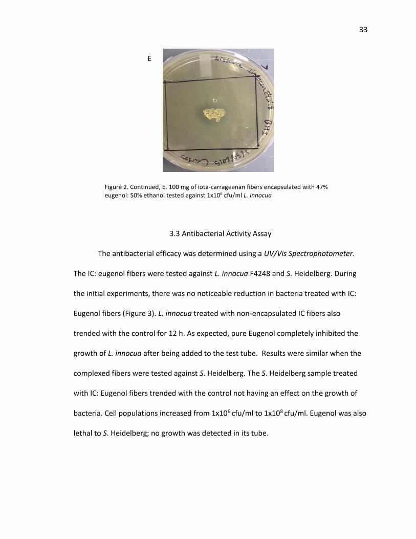
100 mg of complexed fibers were tested against 1x10⁶ cfu/ml of L. innocua, an inhibition
of approximately 24.53 mm has been observed.
B
C
A
D
Figure 2. Zone of Inhibition Assay, A. T (iota-carrageenan fibers encapsulated with 47% eugenol: 50% ethanol) and U (iota-carrageenan fibers) tested against 1x10⁷ cfu/ml L. innocua. B. T and U tested against 1x10⁶ cfu/ml C. T and U tested against 1x10⁵ cfu/ml D. 5 µl of eugenol tested against 1x10⁷ cfu/ml.
33
3.3 Antibacterial Activity Assay
The antibacterial efficacy was determined using a UV/Vis Spectrophotometer.
The IC: eugenol fibers were tested against L. innocua F4248 and S. Heidelberg. During
the initial experiments, there was no noticeable reduction in bacteria treated with IC:
Eugenol fibers (Figure 3). L. innocua treated with non-encapsulated IC fibers also
trended with the control for 12 h. As expected, pure Eugenol completely inhibited the
growth of L. innocua after being added to the test tube. Results were similar when the
complexed fibers were tested against S. Heidelberg. The S. Heidelberg sample treated
with IC: Eugenol fibers trended with the control not having an effect on the growth of
bacteria. Cell populations increased from 1x10⁶ cfu/ml to 1x10⁸ cfu/ml. Eugenol was also
lethal to S. Heidelberg; no growth was detected in its tube.
Figure 2. Continued, E. 100 mg of iota-carrageenan fibers encapsulated with 47% eugenol: 50% ethanol tested against 1x10⁶ cfu/ml L. innocua
E

34
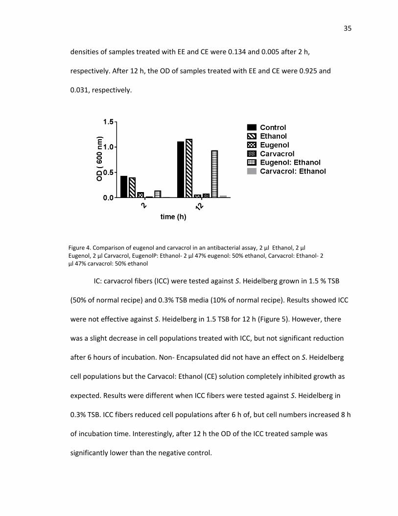
Carvacrol and Eugenol were mixed with ethanol to determine their efficacy
against S. Heidelberg. Ethanol did not have an effect on growth of the bacteria (Figure
4). After two hours of incubation time the optical densities (OD) of the negative control
and ethanol sample were 0.423 and 0.391, respectively. After 12 hours of incubation
time their optical densities were 1.099 and 1.149. No growth was detected in S.
Heidelberg samples treated with eugenol and carvarol. After 12 hours of incubation
time, their OD was 0.055 and 0.075. The 47% carvacrol: 50% ethanol solution was more
effective against S. Heidelberg than the 47% eugenol: 50% ethanol solution. Optical
Figure 3. Effect of IC: Eugenol fibers on the growth of bacteria, 1 µl eugenol, Non-encapsulated Fibers- 1 mg iota-carrageenan fibers, IC- 1 mg iota-carrageenan fibers encapsulated with 47% eugenol: 50% ethanol
35
densities of samples treated with EE and CE were 0.134 and 0.005 after 2 h,
respectively. After 12 h, the OD of samples treated with EE and CE were 0.925 and
0.031, respectively.
IC: carvacrol fibers (ICC) were tested against S. Heidelberg grown in 1.5 % TSB
(50% of normal recipe) and 0.3% TSB media (10% of normal recipe). Results showed ICC
were not effective against S. Heidelberg in 1.5 TSB for 12 h (Figure 5). However, there
was a slight decrease in cell populations treated with ICC, but not significant reduction
after 6 hours of incubation. Non- Encapsulated did not have an effect on S. Heidelberg
cell populations but the Carvacol: Ethanol (CE) solution completely inhibited growth as
expected. Results were different when ICC fibers were tested against S. Heidelberg in
0.3% TSB. ICC fibers reduced cell populations after 6 h of, but cell numbers increased 8 h
of incubation time. Interestingly, after 12 h the OD of the ICC treated sample was
significantly lower than the negative control.
Figure 4. Comparison of eugenol and carvacrol in an antibacterial assay, 2 µl Ethanol, 2 µl Eugenol, 2 µl Carvacrol, EugenolP: Ethanol- 2 µl 47% eugenol: 50% ethanol, Carvacrol: Ethanol- 2 µl 47% carvacrol: 50% ethanol

36
The effect of fibers encapsulated with carvacrol: Tween 80 at various concentrations
was compared to fibers encapsulated with 49% carvacrol: 50% ethanol (Figure 6). All
fiber samples were effective at reducing cell populations S. Heidelberg in 0.3% TSB.
Fibers encapsulated with 50% carvacrol: 50% Tween 80 was the most effective against S.
Hiedelberg and was comparable to positive control, Carvacrol: Ethanol After 16 h, the
OD of the samples treated with CE and C 50%: T80 50%-F were 0.1508 and 0.2298
respectively. However, fibers encapsulated with CE were the least effective. After 16 h
the OD of the samples was 0.3752.
Figure 5. Effect of IC: Carvacrol fibers on the growth of bacteria in reduced nutrient broth, Carvacrol: Ethanol- 2 µl 47% carvacrol: 50% ethanol, Non-encapsulated Fibers- 5 mg iota-carrageenan fibers, IC: Carvacrol- 5 mg iota-carrageenan fibers encapsulated with 47% carvacrol: 50% ethanol
Treatment added
Treatment added
37
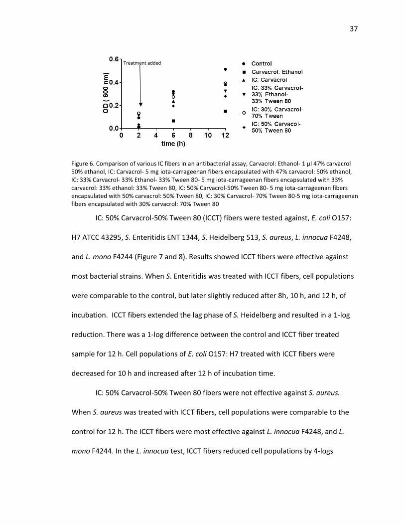
IC: 50% Carvacrol-50% Tween 80 (ICCT) fibers were tested against, E. coli O157:
H7 ATCC 43295, S. Enteritidis ENT 1344, S. Heidelberg 513, S. aureus, L. innocua F4248,
and L. mono F4244 (Figure 7 and 8). Results showed ICCT fibers were effective against
most bacterial strains. When S. Enteritidis was treated with ICCT fibers, cell populations
were comparable to the control, but later slightly reduced after 8h, 10 h, and 12 h, of
incubation. ICCT fibers extended the lag phase of S. Heidelberg and resulted in a 1-log
reduction. There was a 1-log difference between the control and ICCT fiber treated
sample for 12 h. Cell populations of E. coli O157: H7 treated with ICCT fibers were
decreased for 10 h and increased after 12 h of incubation time.
IC: 50% Carvacrol-50% Tween 80 fibers were not effective against S. aureus.
When S. aureus was treated with ICCT fibers, cell populations were comparable to the
control for 12 h. The ICCT fibers were most effective against L. innocua F4248, and L.
mono F4244. In the L. innocua test, ICCT fibers reduced cell populations by 4-logs
Figure 6. Comparison of various IC fibers in an antibacterial assay, Carvacrol: Ethanol- 1 µl 47% carvacrol 50% ethanol, IC: Carvacrol- 5 mg iota-carrageenan fibers encapsulated with 47% carvacrol: 50% ethanol, IC: 33% Carvacrol- 33% Ethanol- 33% Tween 80- 5 mg iota-carrageenan fibers encapsulated with 33% carvacrol: 33% ethanol: 33% Tween 80, IC: 50% Carvacrol-50% Tween 80- 5 mg iota-carrageenan fibers encapsulated with 50% carvacrol: 50% Tween 80, IC: 30% Carvacrol- 70% Tween 80-5 mg iota-carrageenan fibers encapsulated with 30% carvacrol: 70% Tween 80
Treatment added

38
compared to the control after 12 h. Similarly, in the L. monocytogenes test, ICCT fibers
reduced cell populations by 3-logs after 12 h of incubation time. Interestingly, the L.
monocytogenes treated with ICCT fibers increased to 1 x 10⁵ cfu/ml after 4 h of
incubation, but later decreased back to 1 x 10⁴ cfu/ml for the remaining time intervals of
the assay.

39
Figure 7. Effect of antibacterial fibers on gram-negative bacteria, IC: 50% Carvacrol-50% Tween 80- 10 mg iota-carrageenan fibers encapsulated with 50% carvacrol: 50% Tween 80
Fibers added
Fibers added
Fibers added

40
Figure 8. Effect of antibacterial fibers on gram-positive bacteria, IC: 50% Carvacrol-50% Tween 80- 10 mg iota-carrageenan fibers encapsulated with 50% carvacrol: 50% Tween 80
Fibers added
Fibers added
Fibers added

41
3.4 Scanning Electron Microscopy (SEM)
SEM was carried out using a Nova NanoSEM ™ 200 series microscope. Images of
IC fibers, and IC fibers encapsulated with 50% Carvacrol: 50 % Tween 80 were captured
(Figure 9). IC fibers appeared light and were composed of flaky crystals on the surface.
The average diameters of non-encapsulated fibers were 188.35 µm ± 59.85. However, IC
fibers encapsulated with 50% Carvacrol: 50 % Tween 80 had smooth surfaces and
average diameter increased to 204.70 µm ± 58. 05.
SEM images were also captured for the test microorganisms Salmonella
Heidelberg and L. monocytogenes, grown in the presence of IC 50%C: 50%T80 fibers.
The images revealed the S. Heidelberg and L. monocytogenes growth controls had
general characteristics of the respective bacteria and appeared healthy. S. Heidelberg
treated with IC 50%C: 50%T80 fibers were not affected by the antibacterial compound.
The Salmonella aggregated into large clusters. L. monocytogenes treated with IC 50%C:
50%T80 fibers were damaged. While some cells appeared healthy and unaffected,
others were transparent and dense (Figure 9, E&F).

42
A
D
B
C
Figure 9. Scanning Electron Microscopy images of fibers, Salmonella Enteritidis and Listeria monocytogenes treated with antibacterial fibers A. iota-carrageenan fibers, B. iota-carrageenan fibers encapsulated with 50% carvacrol: 50% Tween 80, C & D. S. Enteritidis growth control captured at a magnification of 15, 000 (low magnification) and 50, 000 (high magnification) respectively, E & F. S. Enteritidis treated with antibacterial fibers captured at a low and high magnification,
F
E

43
H
J
G
I
Figure 9 Continued. G & H. L. monocytogenes growth control captured at a low and high magnification, I & J. L. monocytogenes captured at a low and high magnification
44
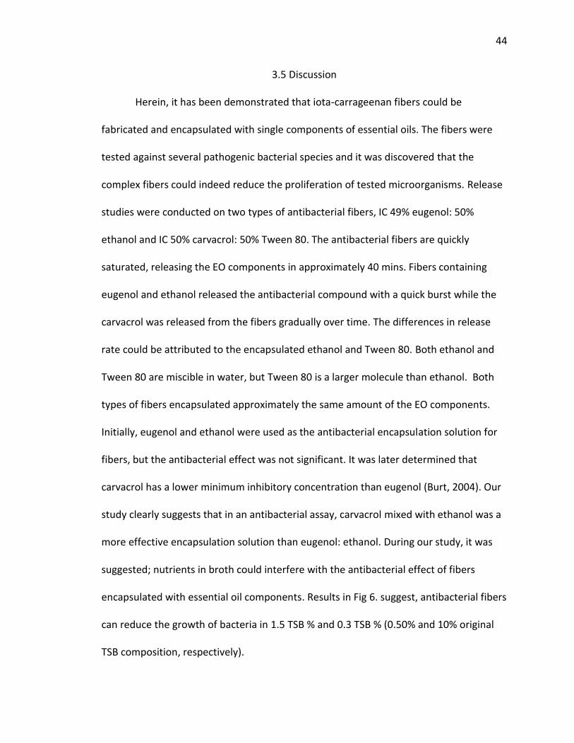
3.5 Discussion
Herein, it has been demonstrated that iota-carrageenan fibers could be
fabricated and encapsulated with single components of essential oils. The fibers were
tested against several pathogenic bacterial species and it was discovered that the
complex fibers could indeed reduce the proliferation of tested microorganisms. Release
studies were conducted on two types of antibacterial fibers, IC 49% eugenol: 50%
ethanol and IC 50% carvacrol: 50% Tween 80. The antibacterial fibers are quickly
saturated, releasing the EO components in approximately 40 mins. Fibers containing
eugenol and ethanol released the antibacterial compound with a quick burst while the
carvacrol was released from the fibers gradually over time. The differences in release
rate could be attributed to the encapsulated ethanol and Tween 80. Both ethanol and
Tween 80 are miscible in water, but Tween 80 is a larger molecule than ethanol. Both
types of fibers encapsulated approximately the same amount of the EO components.
Initially, eugenol and ethanol were used as the antibacterial encapsulation solution for
fibers, but the antibacterial effect was not significant. It was later determined that
carvacrol has a lower minimum inhibitory concentration than eugenol (Burt, 2004). Our
study clearly suggests that in an antibacterial assay, carvacrol mixed with ethanol was a
more effective encapsulation solution than eugenol: ethanol. During our study, it was
suggested; nutrients in broth could interfere with the antibacterial effect of fibers
encapsulated with essential oil components. Results in Fig 6. suggest, antibacterial fibers
can reduce the growth of bacteria in 1.5 TSB % and 0.3 TSB % (0.50% and 10% original
TSB composition, respectively).

45
Initially, fibers were dried of the encapsulation solution overnight and treated to
the test microorganisms in the beginning of the assay the following day (Fig 3 & 4). Due
to the volatility of essential oils, some of the antibacterial effect was lost. For the
purpose of optimization, fibers were dried for two hours and treated to the test
microorganisms two hours after inoculation of bacteria (Fig 5-8). We also showed
Tween 80 serves as a viable surfactant in Iota-carrageenan fibers. Herein, IC 50%
carvacrol: 50% Tween 80 fibers were used to be tested against pathogenic bacteria (Fig
6).
Our antibacterial compounds reduced the growth of L. monocytogenes and L.
innocua, but were not effective when tested against Salmonella and E. coli. Similar
results were reported by Krigel et al (2009, 2010) when they electrospun fibers
containing eugenol, surfactant solution Surfynol 465®, and poly (vinyl alcohol). These
fibers were tested against two stains of L. monocytogenes and Salmonella Typhimurium.
Cell populations in both bacteria species were reduced for 12 hours. However, cell
populations increased Salmonella Typhimurium 2486 increased comparable to the
growth control after 30 hours. Interestingly, cell populations were decreased
tremendously after 30 hours in assay with Salmonella Typhimurium 2576. Some
researchers have reported Gram-positives to be more susceptible to the action of EOs
and EO components. The claim cannot be made in the case of our iota-carrageenan-EO
complexes as Salmonella and E. coli (Gram-negative) were more susceptible than
Staphylococcus aureus (Gram-positive). Gram-negative cells possess an outer membrane
surrounding their cell wall, which could restrict diffusion of hydrophilic compounds

46
through its lipopolysaccharide layer. However, EOs are capable of disintegrating through
the outer membrane layer and increasing permeability in the cytoplasmic membrane.
Perhaps the microorganism’s response to the mode of action of antibacterial
compounds is genetic or they could be resistant. Organisms may acquire genes encoding
enzymes, such as β-lactamases, that destroy the antibacterial agent before it takes
effect. Bacteria may acquire efflux pumps that extrude the antibacterial agent from the
cell before it can reach its target site. Bacteria can also acquire several genes for a
metabolic pathway that alters the cell wall that no longer contains the binding sites of
the antimicrobial agents (Tenover, 2006). However, there is no scientific evidence of EO
resistance due to genetics. This may be of scientific interest in the future.
Tween 80 was a more effective surfactant than ethanol in terms of the
antibacterial activity of the fibers. It should be investigated how other surfactants could
affect the loading and release nature of essential oils from the polysaccharide fibers.
Various balancing ions could also be paired with iota-carrageenan, namely calcium,
magnesium and potassium, to name a few and have an effect on the loading amount
and mechanism. Combinations of EO components have reportedly had synergistic
effects when tested against bacteria. EO components have also had synergetic effects
with food preservatives (Burt, 2004). This should be considered in further studies with
polysaccharide fibers. Encapsulation of essential oils into antibacterial polysaccharides
should also be considered in further studies.

REFERENCES
47
REFERENCES
Al-Okbi, S. Y., Mohamed, D. A., Hamed, T. E., and Edris, A. E. "Protective Effect of Clove Oil and Eugenol Microemulsions on Fatty Liver and Dyslipidemia as Components of Metabolic Syndrome." Journal of Medicinal Food 17.7 (2014): 764-71. Web.
Baydar, H., Sagdic, O., Ozkan, G., and Karadogan, T. "Antibacterial Activity and Composition of Essential Oils from Origanum, Thymbra and Satureja Species with Commercial Importance in Turkey." Food Control 15 (2004): 169-72. Web. 27 July 2016.
Blaszyk, M., and Holley, R. A. “Interaction of Monolaurin, Eugenol and Sodium Citrate on Growth of Common Meat Spoilage and Pathogenic Organisms." International Journal of Food Microbiology 39.3 (1998): 175-83. Web.
Bozin, B., Mimica-Dukic, N., Simin, N., and Anackov, G., "Characterization of the Volatile Composition of Essential Oils of Some Lamiaceae Spices and the Antimicrobial and Antioxidant Activities of the Entire Oils." Journal of Agricultural and Food Chemistry 54 (n.d.): 1822-828. 14 Feb. 2006. Web. 27 July 2016.
Burt, S. "Essential Oils: Their Antibacterial Properties and Potential Applications in Foods—a Review." International Journal of Food Microbiology 94.3 (2004): 223-53. Web.
Carrageenan. Food and Cosmetics Toxicology 8 (1971): 561-80. Web. 4 Mar. 2015 <http://www.sciencedirect.com/science/article/pii/0015626471900940>.
Celebioglu, A., Kayaci-Senirmak, F., Ipek, S., Durgun, E., and Uyar, T., "Polymer-free Nanofibers from Vanillin/cyclodextrin Inclusion Complexes: High Thermal Stability, Enhanced Solubility and Antioxidant Property." Food Funct. 7.7 (2016): 3141-153. Web.
Cimanga, K., Kambu, K., Tona, L., Apers, S., De Bruyne, T., Hermans, N., Totté, J., Pieters, L., and Vlietinck, A.j., "Correlation between Chemical Composition and Antibacterial Activity of Essential Oils of Some Aromatic Medicinal Plants Growing in the Democratic Republic of Congo." Journal of Ethnopharmacology 79.2 (2002): 213-220.Web.
48
Chao, L. K., Hua, K.F., Hsu, H.Y., Cheng, S.S., Lin, I.F., Chen, C.J., Chen, S.T., and Chang. S.T, "Cinnamaldehyde Inhibits Pro-inflammatory Cytokines Secretion from Monocytes/macrophages through Suppression of Intracellular Signaling." Food and Chemical Toxicology 46.1 (2008): 220-31. Web. Chao, L.K., Hua K.F., Hsu, H.Y., Cheng, S.S., Liu, J.Y., and Chang, S.T., "Study on the Anti- inflammatory Activity of Essential Oil from Leaves of Cinnamomum Osmophloeum." J. Agric. Food Chem. Journal of Agricultural and Food Chemistry 53.18 (2005): 7274-278. Web. Cosentino, S., Tuberoso, C.I.G., Pisano, B., Satta, M., Mascia, V., Arzedi, E., and Palmas.
F.,"Result Filters." Letters in Applied Microbiology 29 (1999): 130-35. National Center for Biotechnology Information. U.S. National Library of Medicine. Web. 27 July 2016.
David, J. R.D., L. R. Steenson, and P. Michael Davidson. "Expectations and Applications of Natural Antimicrobials to Foods: A Guidance Document for Users, Suppliers, Research and Development, and Regulatory Agencies." Food Protection Trends 33.4 (n.d.): 238-47. Print.
Dung, N.T., Bajpai, V.K, Yoon, J.I., and Kang, S.C., "Anti-inflammatory Effects of Essential Oil Isolated from the Buds of Cleistocalyx Operculatus (Roxb.) Merr and Perry." Food and Chemical Toxicology 47.2 (2009): 449-53. Web.
Dutra, R. C., Marcelo B.F., Alves, C.C., Ferreira, A.P., Barbosa. N.R., "Antiulcerogenic and Anti-inflammatory Activities of the Essential Oil from Pterodon Emarginatus Seeds." Journal of Pharmacy and Pharmacology 61.2 (2009): 243-50. Web.
Guenther, E., The Essential Oils -: History - Origin in Plants - Production - Analysis, Volume 1. Vol. 1. N.p.: Read, 2008. Google Books. Web. 27 July 2016.
Fisher, K., and Phillips, C., "The Effect of Lemon, Orange and Bergamot Essential Oils and Their Components on the Survival of Campylobacter Jejuni, Escherichia Coli O157, Listeria Monocytogenes, Bacillus Cereus and Staphylococcus Aureus in Vitro and in Food Systems." Journal of Applied Microbiology 101.6 (2006): 1232-240. Web.
"FoodNet Data and Reports." Centers for Disease Control and Prevention. Centers for Disease Control and Prevention, 14 Apr. 2016. Web. 27 July 2016.
Ghayempour, S., and Mortazavi, S.M., "Antibacterial Activity of Peppermint Fragrance Micro–nanocapsules Prepared with a New Electrospraying Method." Journal of Essential Oil Research 26.6 (2014): 492-98. Web.
Gutierrez, J., Barry-Ryan, C., and Bourke, P., "The Antimicrobial Efficacy of Plant Essential Oil Combinations and Interactions with Food Ingredients." International Journal of Food Microbiology 124.1 (2008): 91-97. Web.
Hammer, K. A., Carson, C. F., Dunstan, J. A., Hale, J., Lehmann, H., Robinson, C.J., Prescott, S.L., and Riley, T.V., "Antimicrobial and Anti-inflammatory Activity of Five Taxandria Fragrans Oils in Vitro." Microbiology and Immunology 52.11 (2008): 522-30. Web.

49
Hart, P.H., Brand, C., Carson, C.F., Riley, T.V., Prager, R.H., and Finlay-Jones, J.J., "Terpinen-4-ol, the Main Component of the Essential Oil of Melaleuca Alternifolia (tea Tree Oil), Suppresses Inflammatory Mediator Production by Activated Human Monocytes." Inflammation Research 49.11 (2000): 619-26. Web.
Janaswamy, S., and Youngren, S., "Hydrocolloid-based Nutraceutical Delivery Systems." Food & Function10 (2012): 503-07. Hydrocolloid-based Nutraceutical Delivery Systems - (RSC Publishing). 15 Mar. 2012. Web. 03 Mar. 2015.
Jaworek, A., and Sobczyk, A.T., “Electrospraying Route to Nanotechnology: An Overview." Journal of Electrostatics 66.3-4 (2008): 197-219. Web.
Joshi, S. C., Verma, A. R., and Mathela, C.S., “Antioxidant and Antibacterial Activities of the Leaf Essential Oils of Himalayan Lauraceae Species." Food and Chemical Toxicology 48.1 (2010): 37-40. Web.
Kamatou, G.P.P., Van Zyl, R.L., Van Vuuren, S.F., Viljoen, A.M., Figueiredo, A.C., Barroso, J.G., Pedro, L.G., and Tilney, P.M., "Chemical Composition, Leaf Trichome Types and Biological Activities of the Essential Oils of Four Related Salvia Species Indigenous to Southern Africa." J. Essent. Oil Res 18 (2006): 72-79. Web.
Khalili, S., Mohsenifar, A., Beyki, M., Zhaveh, S., Rahmani-Cherati, T., Abdollahi, A., Bayat, M., and Tabatabaei. M., "Encapsulation of Thyme Essential Oils in Chitosan-benzoic Acid Nanogel with Enhanced Antimicrobial Activity against Aspergillus Flavus." LWT - Food Science and Technology 60.1 (2015): 502-08. Web.
Khan, M.K.I., Mujawar, L.H., Schutyser, M.A. I., Schroën, K., and Boom, R., "Deposition of Thin Lipid Films Prepared by Electrospraying." Food Bioprocess Technol Food and Bioprocess Technology 6.11 (2012): 3047-055. Web.
Kriegel, C., Kit, K. M., Mcclements, D. J., and Weiss, J., “Nanofibers as Carrier Systems for Antimicrobial Microemulsions. II. Release Characteristics and Antimicrobial Activity." Journal of Applied Polymer Science J. Appl. Polym. Sci. 118.5 (2010): 2859-868. Web.
Kriegel, C., Kit, K. M., Mcclements, D. J., and Weiss, J., "Nanofibers as Carrier Systems for Antimicrobial Microemulsions. Part I: Fabrication and Characterization." Langmuir 25.2 (2009): 1154-161. Web.
Kulisic-Bilusic, T., Blažević, I., Dejanović, B., Miloš, M., and Pifat. G., "Evaluation Of The Antioxidant Activity Of Essential Oils From Caper (Capparis Spinosa) And Sea Fennel (Crithmum Maritimum) By Different Methods." Journal of Food Biochemistry 34 (2010): 286-302. Web.
Lin, C.T., Chen, C.J., Lin, T.Y., Tung, J.C., and Wang, S.Y., "Anti-inflammation Activity of Fruit Essential Oil from Cinnamomum Insularimontanum Hayata." Bioresource Technology 99.18 (2008): 8783-787. Web.
Ma, Q., and Zhong, Q., "Incorporation of Soybean Oil Improves the Dilutability of Essential Oil Microemulsions." Food Research International 71 (2015): 118-25. Web.

50
Medeiros, R., Passos, G. F., Vitor, C. E., Koepp, J., Mazzuco, T. L., Pianowski, L. F., Campos, M. M., and Calixto, J. B., "Effect of Two Active Compounds Obtained from the Essential Oil of Cordia Verbenacea on the Acute Inflammatory Responses Elicited by LPS in the Rat Paw." British Journal of Pharmacology 151.5 (2007): 618-27. Web. Mighri, H., Hajlaoui, H., Akrout, A., Najjaa, H., and Neffati, M., "Antimicrobial and Antioxidant Activities of Artemisia Herba-alba Essential Oil Cultivated in Tunisian Arid Zone." Comptes Rendus Chimie 13.3 (2010): 380-86. Web. Miguel, M.G.,"Antioxidant and Anti-Inflammatory Activities of Essential Oils: A Short
Review." Molecules 15.12 (2010): 9252-287. Web. Natrajan, D., Srinivasan, S., Sundar, K., and Ravindran, A., "Formulation of Essential Oil-
loaded Chitosan–alginate Nanocapsules." Journal of Food and Drug Analysis 23.3 (2015): 560-68. Web.
Parris, N., Cooke, P. H., and Hicks, K. B., "Encapsulation of Essential Oils in Zein Nanospherical Particles." Journal of Agricultural and Food Chemistry 53.12 (2005): 4788-792. Web.
Necas, L., and Bartosikova, L.,"Carrageenan: A Review." Veterinarni Medicina 59 (2013): 187 205. Http://vri.cz/docs/vetmed/58-4-187.pdf. 09 Apr. 2013. Web. 3 Mar. 2015. <http://vri.cz/docs/vetmed/58-4-187.pdf>.
Passos, G.F., Fernandes, E.S., Da Cunha, F.M., Ferreira, J., Pianowski, L.F., Campos, M.M., and Calixto, J.B., "Anti-inflammatory and Anti-allergic Properties of the Essential Oil and Active Compounds from Cordia Verbenacea." Journal of Ethnopharmacology 110.2 (2007): 323-33. Web.
Patil, R.P., Nimbalkar, M.S., Jadhav, U.U., Dawkar, V.V., and Govindwar, S. P., "Antiaflatoxigenic and Antioxidant Activity of an Essential Oil from Ageratum Conyzoides L." Journal of the Science of Food and Agriculture J. Sci. Food Agric. 90 (2010): 608-14. Web.
Polowsky, P.J., and Janaswamy, S., "Hydrocolloid-based Nutraceutical Delivery Systems: Effect of Counter-ions on the Encapsulation and Release." Food Hydrocolloids 43 (2015): 658-63. Web
Rieger, K. A., and Schiffman, J.D., "Electrospinning an Essential Oil: Cinnamaldehyde Enhances the Antimicrobial Efficacy of Chitosan/poly(ethylene Oxide) Nanofibers." Carbohydrate Polymers 113 (2014): 561-68. Web.
Rieger, K. A., Birch, N.P., and Schiffman, J.D., "Electrospinning Chitosan/poly(ethylene Oxide) Solutions with Essential Oils: Correlating Solution Rheology to Nanofiber Formation." Carbohydrate Polymers 139 (2016): 131-38. Web.
Rodrigues, T.G., Fernandes, A., Sousa, J.P.B., Bastos, J.K., and Sforcin, J.M., “In Vitro and in Vivo Effects of Clove on Pro-inflammatory Cytokines Production by Macrophages." Natural Product Research 23.4 (2009): 319-26. Web.
Sacchetti, G., Maietti, S., Muzzoli, M., Scaglianti, M., Manfredini, S., Radice, M., and Bruni, R., "Comparative Evaluation of 11 Essential Oils of Different Origin as Functional Antioxidants, Antiradicals and Antimicrobials in Foods." Food Chemistry 91.4 (2005): 621-32. Web.

51
Sforcin, J.M., Amaral, J.T., Fernandes, Sousa, J.P.B., and Bastos, J.K., "Lemongrass Effects on IL-1β and IL-6 Production by Macrophages." Natural Product Research 23.12 (2009): 1151-159. Web.
Sousa, F.L., Santos, M., Rocha, S.M., and Trinade, T., "Encapsulation of Essential Oils in SiO2 Microcapsules and Release Behaviour of Volatile Compounds." Journal of Microencapsulation 31.7 (2014): 627-35. Web.
Stubenrauch, C., and Scottmann, T., "Phase Behaviour, Interfacial Tension and Microstructure of Microemulsions." Microemulsions: Background, New Concepts, Applications, Perspectives. Chichester, West Sussex, U.K.: Wiley, 2009. 1-47. Print.
Surburg, H., and Panten, J., Common Fragrance and Flavor Materials: Preparation, Properties and Uses. 5th ed. N.p.: n.p., n.d. Wiley. Mar. 2006. Web. 27 July 2016.
Tenover, F. C., "Mechanisms of Antimicrobial Resistance in Bacteria."The American Journal of Medicine 119 (206): S3-S10. Web.
"U.S. Food and Drug Administration." FDA Food Safety Modernization Act (FSMA). N.p., n.d. Web. 27 July 2016.
Van Vuuren, S.F., Du Toit, L.C., Pillay, V., and Choonara, Y.E., "Encapsulation of Essential Oils within a Polymeric Liposomal Formulation for Enhancement of Antimicrobial Efficacy." Natural Product Commun 5 (2010): 1401-408. Web.
Viuda-Martos, M., Navajas, Y.R., Zapata, E.S., Fernández-López, J., and José A. Pérez-Álvarez. "Antioxidant Activity of Essential Oils of Five Spice Plants Widely Used in a Mediterranean Diet." Flavour and Fragrance Journal Flavour Fragr. J. 25.1 (2010): 13-19. Web.
Wei, A., and Shibamoto,T., "Antioxidant/Lipoxygenase Inhibitory Activities and Chemical Compositions of Selected Essential Oils." J. Agric. Food Chem. Journal of Agricultural and Food Chemistry 58.12 (2010): 7218-225. Web.
Yang, S.A., Jeon, S.K., Eun-Jung Lee, Chang-Hyun Shim, and In-Seon Lee. "Comparative Study of the Chemical Composition and Antioxidant Activity of Six Essential Oils and Their Components." Natural Product Research 24.2 (2010): 140-51. Web.
Yoon, W.J. "Cryptomeria Japonica Essential Oil Inhibits the Growth of Drug-resistant Skin Pathogens and LPS-induced NO and Pro-inflammatory Cytokine Productio." Polish J. Microbiol 58 (2009): 61-68. Web.
Yoon, W.j., J.y. Moon, G. Song, Y.k. Lee, M.s. Han, J.s. Lee, B.s. Ihm, W.j. Lee, N.h. Lee, and C.g. Hyun. "Artemisia Fukudo Essential Oil Attenuates LPS-induced Inflammation by Suppressing NF-κB and MAPK Activation in RAW 264.7 Macrophages." Food and Chemical Toxicology 48.5 (2010): 1222-229. Web.
Yoon, W.J., Kim, S.S., Oh, T.H., Lee, N.H., and Hyun, C.G., "Cryptomeria Japonica Essential Oil Inhibits the Growth of Drug-resistant Skin Pathogens and LPS-induced NO and Pro-inflammatory Cytokine Production." (n.d.): n. pag. Web.
Zhang, L., Critzer, F., Davidson, P.M., and Zhong, Q., "Formulating Essential Oil Microemulsions as Washing Solutions for Organic Fresh Produce Production." Food Chemistry 165 (2014): 113-18. Web.

52
Zhaveh, S., Mohsenifar, A., Beiki, M., Khalili, S.T., Abdollahi, A., Rahmani-Cherati, T., and Tabatabaei, M., "Encapsulation of Cuminum Cyminum Essential Oils in Chitosan caffeic Acid Nanogel with Enhanced Antimicrobial Activity against Aspergillus flavus." Industrial Crops and Products 69 (2015): 251-56. Web.

APPENDICES

53
Appendix A Additional Results of Antibacterial Activity Assays
Appendix A contains results from trial antibacterial assays that contributed to the
foundation of our methodology to demonstrate the antibacterial activity of well-
organized iota-carrageenan fibers encapsulated with essential oil components. Figure
A.1. displays results from an assay conducted to determine if 0.01% Tween 80 had an
effect on the growth of bacteria when it is added to tryptic soy broth with yeast extract
(TSBYE) at various times intervals or premixed in TSBYE. Results showed, when 0.01%
Tween 80 is added to TSBYE after 0, 2, and 4 h of inoculation, it does not have an effect
on the growth of bacteria. When 0.01% Tween is premixed with TSBYE, it slightly
reduced the proliferation of Listeria innocua F4248 after 6 hours. Figure A.2. contains
results from an assay conducted to determine if the most viable time treat bacteria with
IC: eugenol fibers. Eugenol (1μl) was a positive control. Results showed that IC: eugenol
fibers did not have an effect on the growth of Listeria innocua F4248 when treated after
0, 2, or 4 h after inoculation. Figure A.3. displays results for an assay conducted to
determine the efficacy of various essential oils encapsulated in iota-carrageenan fibers
and tested against Listeria innocua F4248. IC fibers encapsulated with 47% eugenol,
carvacrol, or trans-cinnamaldehyde did not show major differences in levels of efficacy.
Carvacrol (1μl) inhibited the growth of L. innocua F4248 significantly. Figure A.4. dsiplays
results from an experiment conducted to compare IC fibers containing 0.1% Tween 80,

54
encapsulated with various concentrations of carvacrol, and tested against Salmonella
enterica serovar Heidelberg 513. Results showed fibers at effective at several
concentrations (10-100%). IC fibers containing 0.1% Tween 80 and encapsulated with
75% and 100% carvacrol were most effective against Salmonella Heidelberg.
Figure A.2. Listeria innocua F4248 treated with IC: Eugenol fibers at different time intervals
(h)
Figure A.1. Effect of Tween 80 on the growth of Listeria innocua F4248
(h)

55
Figure A.3. Comparison of IC fibers encapsulated with various essential oil components on the growth of Listeria innocua F4248
Figure A.4. Comparison of IC fibers encapsulated with carvacrol at various concentrations and treated to Salmonella Heidelberg

56
Appendix B Raw Data
Appendix B contains raw data from results displayed in Chapter 3 (Table B.1-12).
Table B. 1. Eugenol and carvacrol release from IC fibers and encapsulated amount
Time
(min)
Eugenol
(ug/mg)
Carvacrol
(ug/mg)
0 0 0
2 2.301673 0.539125
4 2.671729 0.915995
6 2.691003 1.476985
8 2.770025 1.764672
10 3.438825 1.957422
12 2.814355 2.216341
14 3.128517 2.377445
16 2.823992 2.506904
18 3.030221 2.647871
20 2.881813 2.691024
22 2.787372 2.705409
24 2.929998 2.826237
26 2.829774 2.875144
28 2.949271 2.912543
30 2.785444 2.901036
32 2.976255 2.938435
34 2.89145 2.978711
36 2.625472 3.027618
38 3.246087 3.050633
40 2.978182
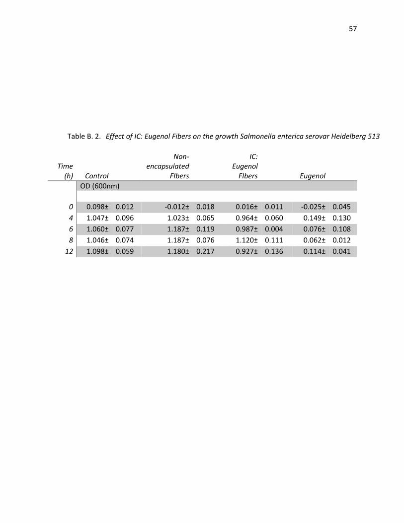
57
Time (h) Control
Non-encapsulated
FIbers
IC: Eugenol
FIbers
Eugenol
OD (600nm)
0 0.098± 0.012 -0.012± 0.018 0.016± 0.011 -0.025± 0.045
4 1.047± 0.096 1.023± 0.065 0.964± 0.060 0.149± 0.130
6 1.060± 0.077 1.187± 0.119 0.987± 0.004 0.076± 0.108
8 1.046± 0.074 1.187± 0.076 1.120± 0.111 0.062± 0.012
12 1.098± 0.059 1.180± 0.217 0.927± 0.136 0.114± 0.041
Table B. 2. Effect of IC: Eugenol Fibers on the growth Salmonella enterica serovar Heidelberg 513

58
Time
(h)
Control
Non-
encapsulated
FIbers
IC-
Eugenol
FIbers
Eugenol
OD (600nm)
0 0.030± 0.034 -0.076± 0.026 -0.004± 0.041 -0.040 0.029
2 0.081± 0.054 0.071± 0.018 0.107± 0.014 0.104 0.002
4 0.068± 0.096 0.104± 0.072 0.087± 0.103 0.046 0.007
6 0.517± 0.508 0.503± 0.326 0.547± 0.475 0.110 0.062
8 1.125± 0.112 1.158± 0.192 1.170± 0.099 0.146 0.041
12 1.160± 0.072 1.119± 0.241 1.167± 0.097 0.096 0.141
Table B. 3. Effect of IC: Eugenol Fibers on the growth Listeria innocua F4248

59
Table B. 4 Comparison of eugenol and carvacrol in an antibacterial assay against
Salmonella Heidelberg
Treatment 2 h 12 h
OD (600 nm)
Control 0.423 1.100
Ethanol 0.391 1.150
Eugenol 0.0977 0.0548
47% Eugenol: 50% Ethanol 0.134 0.926
47% Carvacrol: Ethanol 0.0045 0.0316

60
Table B. 5 Effect of IC: Carvacrol fibers on the growth of Salmonella Heidelberg in 1.5% tryptic soy broth
Time (h) Control
47% Carvacrol: 50% Ethanol
Non-
encapsulated Fibers
IC:
Carvacrol
(600nm)
0 0 0 0 0
2 0.253± 0.126 0.140± 0.089 0.333± 0.191 0.271± 0.164
4 0.747± 0.055 0.214± 0.286 0.756± 0.039 0.627± 0.223
6 0.753± 0.065 0.223± 0.322 0.749± 0.028 0.729± 0.038
8 0.778± 0.070 0.240± 0.306 0.739± 0.082 0.686± 0.073
10 0.775± 0.076 0.204± 0.294 0.755± 0.061 0.680± 0.119
12 0.780± 0.125 0.274± 0.358 0.722 0.629± 0.011

61
Table B. 6 Effect of IC: Carvacrol fibers on the growth of Salmonella Heidelberg in 0.3% tryptic soy broth
Time (h) Control
47% Carvacrol: 50% Ethanol
Non-
encapsulated Fibers
IC:
Carvacrol
(600nm)
0 0 0 0 0
2 0.087± 0.077 0.089± 0.001 0.169± 0.071 0.163± 0.0001
4 0.163± 0.035 0.286± 0.022 0.196± 0.067 0.163± 0.052
6 0.230± 0.047 0.322± 0.001 0.219± 0.063 0.163± 0.004
8 0.301± 0.042 0.306± 0.001 0.261± 0.050 0.163± 0.039
10 0.312± 0.020 0.294± 0.028 0.354± 0.023 0.163± 0.005
12 0.42 0.35 0.32 0.21

62
Table B. 7. Effect of antibacterial fibers on Salmonella Enteritidis ENT 1344
Time (h)
Control
IC: 50% Carvacrol: 50% Tween 80
cfu/ml
0 1.943E+04± 1.034E+04 1.886E+04± 1.195E+04
4 2.985E+06± 2.350E+06 6.167E+05± 2.881E+05
6 3.871E+07± 3.005E+07 6.385E+07± 1.043E+08
8 1.943E+08± 6.680E+07 6.850E+07± 3.548E+07
10 3.800E+08± 1.310E+08 8.900E+07± 3.252E+07
12 3.929E+08± 1.186E+08 8.000E+07± 3.763E+07

63
Table B. 8. Effect of antibacterial fibers on Salmonella enterica serovar Heidelberg 513
Time (h) Control
IC: 50% Carvacrol: 50% Tween 80
cfu/ml
0 1.500E+04± 6.904E+03 1.771E+04± 1.242E+04
4 1.186E+06± 8.295E+05 2.475E+05± 2.473E+05
6 3.338E+07± 2.087E+07 1.208E+06± 5.665E+05
8 1.829E+08± 7.111E+07 8.350E+06± 2.333E+06
10 2.457E+08± 7.300E+07 3.100E+07± 1.273E+07
12 2.825E+08± 8.049E+07 1.900E+07± 8.544E+06

64
Table B. 9. Effect of antibacterial fibers on Escherichia coli O157: H7 ATCC 43295
Time (h) Control IC: 50% Carvacrol: 50% Tween 80
cfu/ml
0 1.414E+04± 7.734E+03 1.313E+04± 7.080E+03
4 5.800E+04± 3.033E+04 2.500E+04± 7.071E+03
6 2.117E+05± 1.745E+05 8.500E+04± 1.732E+04
8 3.620E+05± 1.795E+05 1.800E+05± 9.274E+04
10 1.349E+06± 1.306E+06 5.950E+05± 5.049E+05
12 5.300E+06± 1.901E+06 7.450E+06± 1.909E+06

65
Time (h) Control
IC: 50% Carvacrol: 50% Tween 80
cfu/ml
0 1.650E+04± 6.714E+03 1.188E+04± 5.083E+03
4 2.430E+05± 5.962E+04 1.617E+04± 1.070E+04
6 2.115E+06± 6.063E+05 2.450E+04± 1.168E+04
8 2.000E+0 2.850E+04± 7.550E+03
10 7.720E+07± 2.687E+07 2.500E+04± 2.121E+04
12 2.175E+08± 5.679E+07 1.188E+04± 5.083E+03
Table B. 10. Effect of antibacterial fibers on Listeria innocua F4248

66
Table B. 11. Effect of antibacterial fibers on Listeria monocytogenes F4244
Time (h) Control IC: 50% Carvacrol: 50% Tween 80
cfu/ml
0 2.620E+04± 1.566E+04 2.286E+04± 1.842E+04
4 6.675E+05± 5.000E+05 2.000E+05± 1.358E+05
6 4.606E+06± 4.130E+06 1.000E+05± 1.131E+05
8 1.338E+07± 4.106E+06 1.300E+05± 7.550E+04
10 2.617E+07± 1.063E+07 9.667E+04± 9.292E+04
12 2.100E+07± 5.944E+06 1.500E+04± 7.071E+03
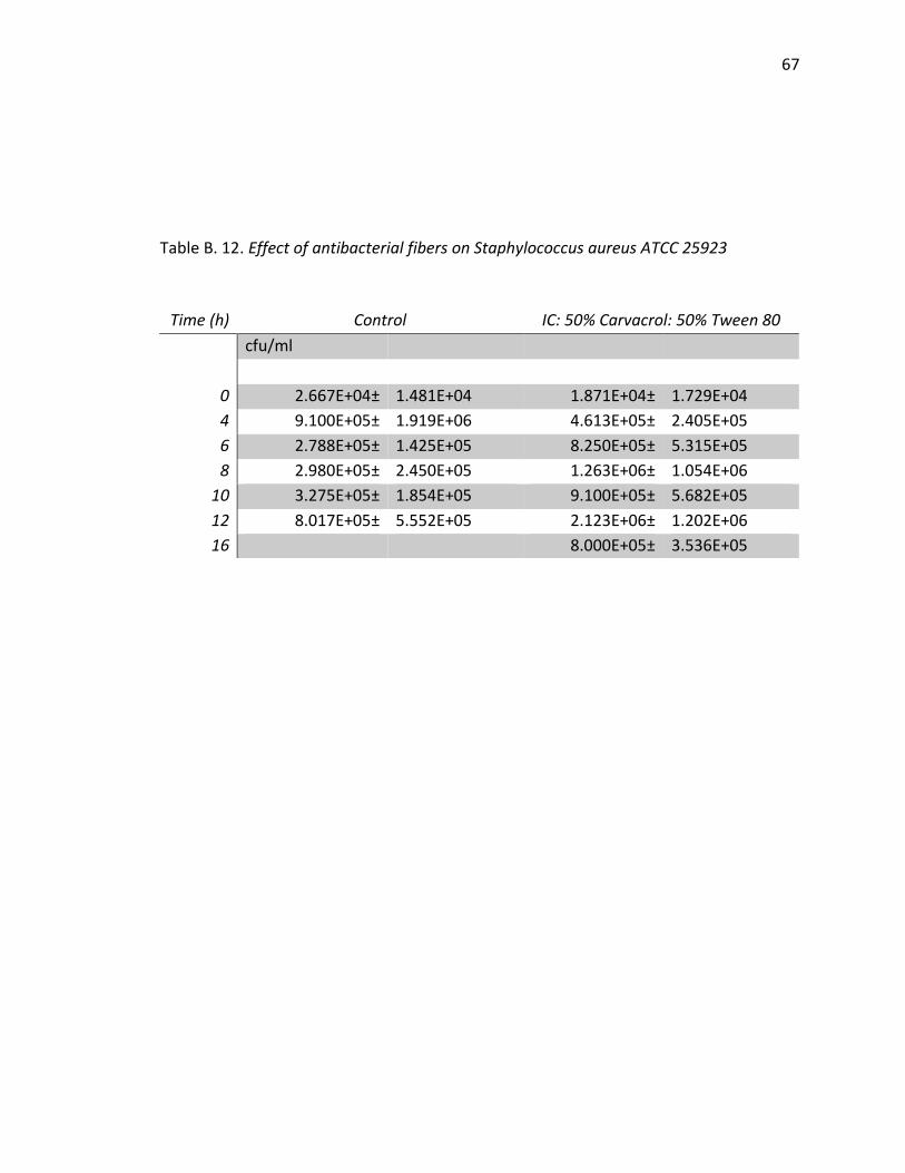
67
Table B. 12. Effect of antibacterial fibers on Staphylococcus aureus ATCC 25923
Time (h) Control IC: 50% Carvacrol: 50% Tween 80
cfu/ml
0 2.667E+04± 1.481E+04 1.871E+04± 1.729E+04
4 9.100E+05± 1.919E+06 4.613E+05± 2.405E+05
6 2.788E+05± 1.425E+05 8.250E+05± 5.315E+05
8 2.980E+05± 2.450E+05 1.263E+06± 1.054E+06
10 3.275E+05± 1.854E+05 9.100E+05± 5.682E+05
12 8.017E+05± 5.552E+05 2.123E+06± 1.202E+06
16 8.000E+05± 3.536E+05
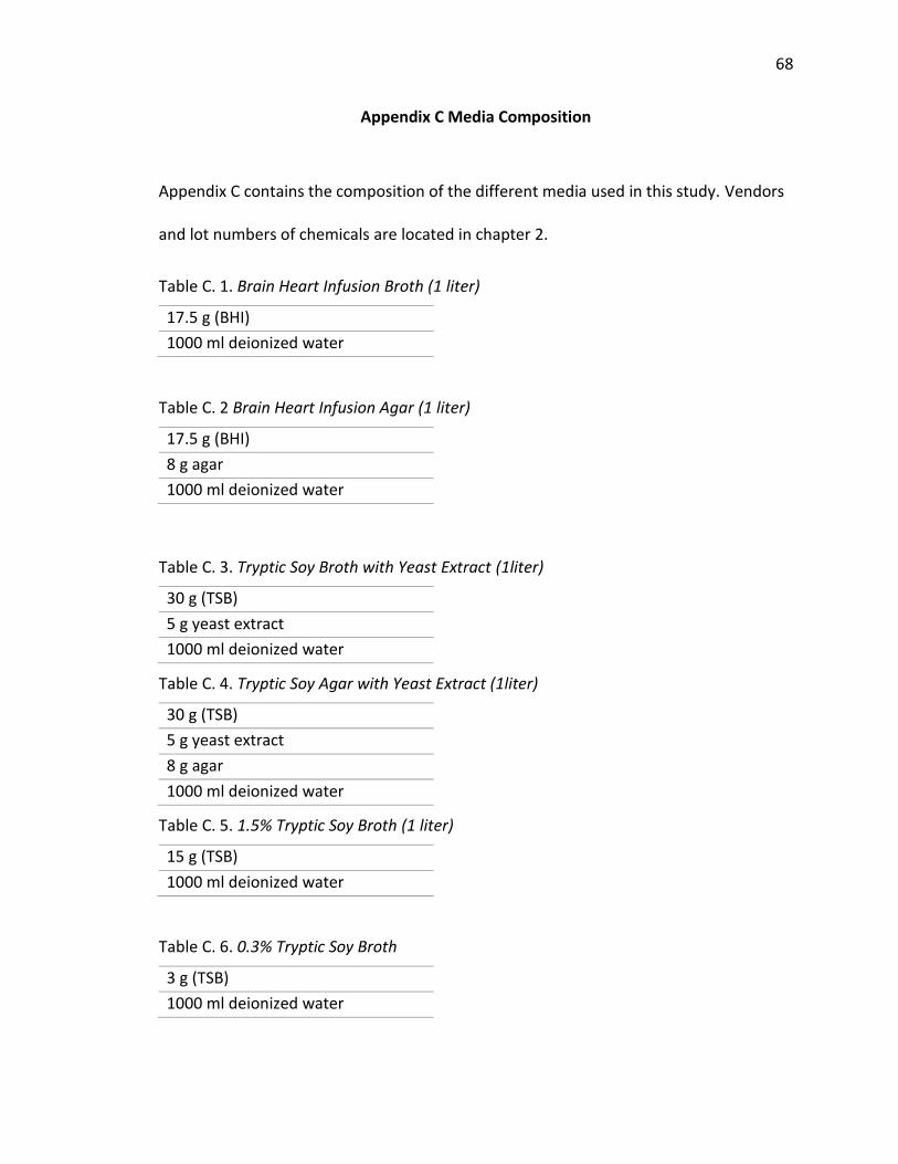
68
Appendix C Media Composition
Appendix C contains the composition of the different media used in this study. Vendors
and lot numbers of chemicals are located in chapter 2.
Table C. 1. Brain Heart Infusion Broth (1 liter)
17.5 g (BHI)
1000 ml deionized water
Table C. 2 Brain Heart Infusion Agar (1 liter)
17.5 g (BHI)
8 g agar
1000 ml deionized water
Table C. 3. Tryptic Soy Broth with Yeast Extract (1liter)
30 g (TSB)
5 g yeast extract
1000 ml deionized water
Table C. 4. Tryptic Soy Agar with Yeast Extract (1liter)
30 g (TSB)
5 g yeast extract
8 g agar
1000 ml deionized water
Table C. 5. 1.5% Tryptic Soy Broth (1 liter)
15 g (TSB)
1000 ml deionized water
Table C. 6. 0.3% Tryptic Soy Broth
3 g (TSB)
1000 ml deionized water

69
Appendix D Microorganism Growth Conditions
Brain heart infusion (BHI) media was used to grow L. innocua F4248 for disc
diffusion assay. One loop of L. innocua was taken from a frozen stock and inoculated
into 4 ml of BHI. The inoculated test tube was incubated at 37oC for approximately 18 h.
Tryptic soy broth with yeast extract (TSBYE) was used to cultivate all microorganisms in
antibacterial activity assays in the study. Subcultures of the test microorganism were
prepared by streaking one loop onto tryptic soy agar plates with yeast extract. The
streaked plates were incubated at 37oC for 24 h. One single colony from the subculture
plate was inoculated into 4 ml of TSBYE. The inoculated tube was incubated at 37oC for
approximately.

VITA
70
VITA
Carlos Carter was born in Washington D.C., on December 19, 1991. His family
moved to Warrenton, NC when he was eleven years old. After graduating from Warren
County High School in 2010, he went on to pursue a B.S. in Food & Nutritional Sciences
at North Carolina Agricultural and Technical State University in Greensboro, NC. After
receiving his B.S., in 2014, he furthered his education at Purdue University in achieving a
M.S., in Food Science in West Lafayette, IN. He started his professional career as a Food
Scientist with Cargill, Inc. working with the Snacks & Cereal, Food Applications Teams.