Embed Size (px)
Citation preview
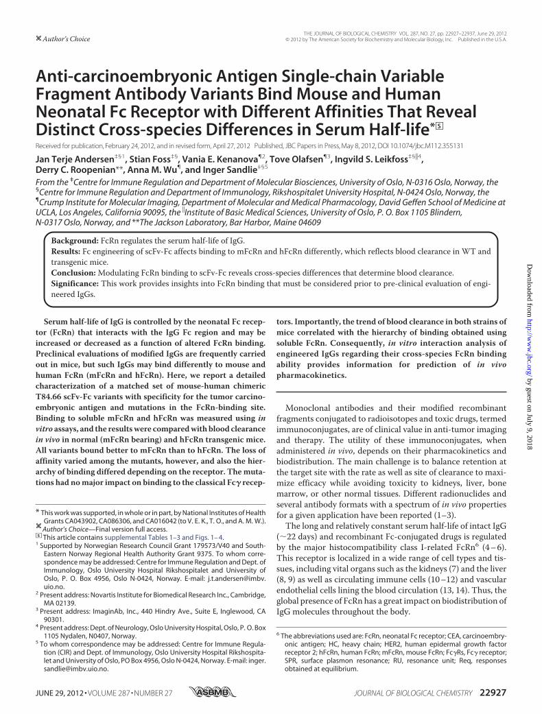
Anti-carcinoembryonic Antigen Single-chain VariableFragment Antibody Variants Bind Mouse and HumanNeonatal Fc Receptor with Different Affinities That RevealDistinct Cross-species Differences in Serum Half-life*□S
Received for publication, February 24, 2012, and in revised form, April 27, 2012 Published, JBC Papers in Press, May 8, 2012, DOI 10.1074/jbc.M112.355131
Jan Terje Andersen‡§1, Stian Foss‡§, Vania E. Kenanova¶2, Tove Olafsen¶3, Ingvild S. Leikfoss‡§�4,Derry C. Roopenian**, Anna M. Wu¶, and Inger Sandlie‡§5
From the ‡Centre for Immune Regulation and Department of Molecular Biosciences, University of Oslo, N-0316 Oslo, Norway, the§Centre for Immune Regulation and Department of Immunology, Rikshospitalet University Hospital, N-0424 Oslo, Norway, the¶Crump Institute for Molecular Imaging, Department of Molecular and Medical Pharmacology, David Geffen School of Medicine atUCLA, Los Angeles, California 90095, the �Institute of Basic Medical Sciences, University of Oslo, P. O. Box 1105 Blindern,N-0317 Oslo, Norway, and **The Jackson Laboratory, Bar Harbor, Maine 04609
Background: FcRn regulates the serum half-life of IgG.Results: Fc engineering of scFv-Fc affects binding to mFcRn and hFcRn differently, which reflects blood clearance in WT andtransgenic mice.Conclusion:Modulating FcRn binding to scFv-Fc reveals cross-species differences that determine blood clearance.Significance: This work provides insights into FcRn binding that must be considered prior to pre-clinical evaluation of engi-neered IgGs.
Serum half-life of IgG is controlled by the neonatal Fc recep-tor (FcRn) that interacts with the IgG Fc region and may beincreased or decreased as a function of altered FcRn binding.Preclinical evaluations of modified IgGs are frequently carriedout in mice, but such IgGs may bind differently to mouse andhuman FcRn (mFcRn and hFcRn). Here, we report a detailedcharacterization of a matched set of mouse-human chimericT84.66 scFv-Fc variants with specificity for the tumor carcino-embryonic antigen and mutations in the FcRn-binding site.Binding to soluble mFcRn and hFcRn was measured using invitro assays, and the resultswere comparedwith blood clearancein vivo in normal (mFcRn bearing) and hFcRn transgenic mice.All variants bound better to mFcRn than to hFcRn. The loss ofaffinity varied among the mutants, however, and also the hier-archy of binding differed depending on the receptor. Themuta-tions had nomajor impact on binding to the classical Fc� recep-
tors. Importantly, the trend of blood clearance in both strains ofmice correlated with the hierarchy of binding obtained usingsoluble FcRn. Consequently, in vitro interaction analysis ofengineered IgGs regarding their cross-species FcRn bindingability provides information for prediction of in vivopharmacokinetics.
Monoclonal antibodies and their modified recombinantfragments conjugated to radioisotopes and toxic drugs, termedimmunoconjugates, are of clinical value in anti-tumor imagingand therapy. The utility of these immunoconjugates, whenadministered in vivo, depends on their pharmacokinetics andbiodistribution. The main challenge is to balance retention atthe target site with the rate as well as site of clearance to maxi-mize efficacy while avoiding toxicity to kidneys, liver, bonemarrow, or other normal tissues. Different radionuclides andseveral antibody formats with a spectrum of in vivo propertiesfor a given application have been reported (1–3).The long and relatively constant serum half-life of intact IgG
(�22 days) and recombinant Fc-conjugated drugs is regulatedby the major histocompatibility class I-related FcRn6 (4–6).This receptor is localized in a wide range of cell types and tis-sues, including vital organs such as the kidneys (7) and the liver(8, 9) as well as circulating immune cells (10–12) and vascularendothelial cells lining the blood circulation (13, 14). Thus, theglobal presence of FcRn has a great impact on biodistribution ofIgG molecules throughout the body.
* This work was supported, in whole or in part, by National Institutes of HealthGrants CA043902, CA086306, and CA016042 (to V. E. K., T. O., and A. M. W.).Author’s Choice—Final version full access.
□S This article contains supplemental Tables 1–3 and Figs. 1– 4.1 Supported by Norwegian Research Council Grant 179573/V40 and South-
Eastern Norway Regional Health Authority Grant 9375. To whom corre-spondence may be addressed: Centre for Immune Regulation and Dept. ofImmunology, Oslo University Hospital Rikshospitalet and University ofOslo, P. O. Box 4956, Oslo N-0424, Norway. E-mail: [email protected].
2 Present address: Novartis Institute for Biomedical Research Inc., Cambridge,MA 02139.
3 Present address: ImaginAb, Inc., 440 Hindry Ave., Suite E, Inglewood, CA90301.
4 Present address: Dept. of Neurology, Oslo University Hospital, Oslo, P. O. Box1105 Nydalen, N0407, Norway.
5 To whom correspondence may be addressed: Centre for Immune Regula-tion (CIR) and Dept. of Immunology, Oslo University Hospital Rikshospita-let and University of Oslo, PO Box 4956, Oslo N-0424, Norway. E-mail: [email protected].
6 The abbreviations used are: FcRn, neonatal Fc receptor; CEA, carcinoembry-onic antigen; HC, heavy chain; HER2, human epidermal growth factorreceptor 2; hFcRn, human FcRn; mFcRn, mouse FcRn; Fc�Rs, Fc� receptor;SPR, surface plasmon resonance; RU, resonance unit; Req, responsesobtained at equilibrium.
THE JOURNAL OF BIOLOGICAL CHEMISTRY VOL. 287, NO. 27, pp. 22927–22937, June 29, 2012Author’s Choice © 2012 by The American Society for Biochemistry and Molecular Biology, Inc. Published in the U.S.A.
JUNE 29, 2012 • VOLUME 287 • NUMBER 27 JOURNAL OF BIOLOGICAL CHEMISTRY 22927
by guest on July 9, 2018http://w
ww
.jbc.org/D
ownloaded from
The fundamental importance of FcRn in IgG homeostasishas been demonstrated using an engineered mouse strain inwhich FcRn can be conditionally deleted in both endothelialand hematopoietic cells. Lack of FcRn expression in these cellsresulted in a 4-fold lower serum level of IgG than what wasfound in wild type (WT) mice, whereas the half-life of an exog-enous injected human IgG1 (hIgG1) decreased by 21-fold (13).The cellular mechanism by which IgGs are rescued has been
revealed using advanced microscopy technologies (15, 16),where IgG, continually taken up by fluid phase endocytosis, isdelivered to early endosomes, where FcRn predominantlyresides. The acidified endosomal environment favors pH-de-pendent binding of the Fc part of IgG to FcRn. After binding,the complex is recycled to the cell surface, where the physiolog-ical pH of the blood triggers release of IgG. Thus, IgG Fc con-taining molecules are rescued from lysosomal degradation viaan efficient FcRn-mediated recycling pathway.The interaction site for FcRn on IgG (human and rodents)
has been mapped using site-directed mutagenesis as well asx-ray crystallography and shown to involve negatively chargedresidues on the �2-domain of the FcRn heavy chain (HC) (Glu-115 and Glu-116) and conserved amino acid residues localizedto the CH2-CH3 IgG Fc interface that include three highly con-served key residues, namely Ile-253, His-310, and His-435 (17–19). The central role of the histidine residues reflects the strictlypH-dependent mode of binding that is explained by the imid-azole side chain that is neutral at physiological pH and posi-tively charged at acidic pH.Despite conservation of the key residues across species,
hFcRn discriminates between IgG from several species, includ-ing mouse IgGs (mIgG), that do not interact, except from weakbinding of mIgG2b (20–22). This fact largely explains the dis-appointing results obtained fromclinical trials during the 1980susing murine monoclonal IgGs and also why mouse immuno-conjugates, such as 131I-tositumomab (Bexxar, Cortixa Corp.)and 90Y-ibritumomab-tiuxetan (Zevalin, IDEC Pharmaceuti-cals Corp.), are cleared very rapidly from the circulation.Engineered hIgG1 and hIgG2 with improved affinity for
hFcRn at acidic pH show increased serumhalf-lives in primates(21, 23, 24). However, negligible binding at physiological pH isnecessary (4, 23–26), and an increase has the opposite effect.This has been exemplified for a new class of engineered anti-bodies, termed Abdegs (enhancing IgG degradation), withshort serumhalf-life that furthermore accelerates the clearanceof circulating IgGs due to saturation of binding to FcRn thatblocks further IgG binding (27, 28). However, favorable bindingto hFcRn does not necessarily imply similar binding kineticstowardmFcRn, as for instance demonstrated by the hIgG1 var-iant with two Fc point mutations (H433K/N434F) that resultsin a 4-fold reduced serum half-life in WT mice but enhancedtransport in an ex vivo human placenta model system (29).One may alter the half-life of immunoconjugates by
reducing the size or by introducing site-directed mutations,as exemplified by antibody derivatives with specificity for thetumor antigen carcinoembryonic antigen (CEA) (30–33).CEA is found in colorectal, breast, and lung cancers but alsoin low amounts in noncancerous tissue. The concentrationof the antigen in tumors is �60 times higher than that in
healthy tissue (34, 35), and thus, CEA is an attractive candi-date for antibody-based tumor targeting in diagnostic imag-ing and radioimmunotherapy.Several different recombinant anti-CEA T84.66 antibody
derivatives have been generated, and their in vivo tumor target-ing properties and biodistributionwere investigated inmice (3).For instance, the minibody (scFv-CH3) format demonstratesfast clearance, excellent tumor uptake, and high contrastimages in both LS174T xenografted mice and in a clinical pilotstudy (30–33). A set of anti-CEA T84.66 chimeric mouse-hu-man scFv-Fc mutants showed a spectrum of in vivo half-livesand positron emission tomography imaging properties inBALB/c mice and tumor-bearing LS174T xenografted athymicmice (36, 37). Five scFv-Fc mutants were produced, termedI253A, H310A, H435Q, H435R, and H310A/H435Q (EU num-bering system).In this study, we thoroughly investigate the cross-species
binding properties of these scFv-Fc variants toward the mouseand human forms of FcRn and the classical Fc�Rs usingenzyme-linked immunosorbent assays (ELISA) and surfaceplasmon resonance (SPR) measurements. Furthermore, wemeasure the serum half-life in mice transgenic for hFcRn.We find that the interaction properties of the scFv-Fc vari-
ants toward mFcRn correlated well with the in vivo serum half-lives and tissue biodistribution obtained in WTmice. Interest-ingly, binding to hFcRn was distinct from that of mFcRn andcorrelatedwith serumhalf-lives obtained from in vivo clearancestudies performed in mice transgenic for hFcRn. In such mice,the half-life values were considerably shorter than thoseobtained in WT mice, and the clearance rate hierarchy amongthe variants was changed. Such variations in FcRn bindingacross species as well as differences in half-life obtained usingdifferent preclinicalmodelsmust be taken into account prior topredicting pharmacokinetics in humans.
EXPERIMENTAL PROCEDURES
Construction, Production, and Purification of anti-CEAscFv-Fc Antibody Fragments—The anti-CEA cT84.66 scFv-FcWT (nonmutated) and mutants (H435Q, H435R, H310A,I253A, and H310A/H435Q) used in these studies have beenpreviously described (36, 37). The scFv-Fc proteins wereexpressed in NS0 murine myeloma cells following transfectionof the mammalian expression vector pEE12 containing thescFv-Fc cDNAs, driven by the human cytomegalovirus pro-moter and a glutamine synthetase selection marker. Collectedsupernatantswere centrifuged to remove cell debris and treatedwith AG1-X8 resin (Bio-Rad) in phosphate buffered saline(PBS) to remove phenol red. Then the fractions were filtratedand dialyzed against 50 mM Tris-HCl (pH 7.4). RecombinantscFv-Fc variants were purified using protein A affinity chroma-tography (Poros 20A, Applied Biosystems) using the GE Phar-maciaAKTApurifier FPLC system (GEHealthcare), where PBSwas used to equilibrate the column prior to loading the sample.Bound protein was eluted with a linear pH gradient (pH 7.0 topH 2.1) using 0.2 M citrate buffer. Eluted protein was immedi-ately neutralized with 80% v/v of 1 M Tris-HCl (pH 8.2), pooled,and dialyzed in 1� PBS. Then anion exchange chromatographyusing a Source HQ50 column (Amersham Biosciences) fol-
Binding of Anti-CEA scFv-Fc Variants to FcRn
22928 JOURNAL OF BIOLOGICAL CHEMISTRY VOLUME 287 • NUMBER 27 • JUNE 29, 2012
by guest on July 9, 2018http://w
ww
.jbc.org/D
ownloaded from
lowed by a ceramic hydroxyapatite column (Bio-Rad) and anionexchange chromatography using a Source 15Q column (Amer-sham Biosciences) were performed, all as described previously(36). The final fractions were pooled, dialyzed against PBS, andconcentrated byCentriprep 30 (Millipore Corp., Bedford,MA).Absorbance was monitored at 280 nm, and final protein con-centration was determined using a NanoDrop 2000 spectro-photometer (Thermo Fisher Scientific). The A280 value wasdivided by an extinction coefficient of 1.4.Construction, Production, and Purification of Recombinant
Soluble Forms of Mouse and Human Fc� Receptors and FcRnVariants—Vectors containing truncated versions of mFcRnand hFcRn HC cDNAs encoding their three ectodomains (�1–�3) genetically fused to a cDNA encoding the Schistosomajaponicum glutathione S-transferase (GST) have been de-scribed (22, 38). The vectors denoted pcDNA3-hFcRn-GST-h�2m-oriP and pcDNA3-mFcRn-GST-h�2m-oriP also con-tain a cDNA encoding human �2-microglobulin and theEpstein-Barr virus origin of replication (oriP). Soluble versionsof FcRn (mFcRn and hFcRn) were produced in HEK 293E cells,and secreted receptors were purified using a GSTrap column asdescribed (25). An mFcRn variant expressed in infected High-Five cells was a generous gift fromDr. SallyWard (University ofTexas Southwestern Medical Center, Dallas) (39).Recombinant soluble forms consisting of the ectodomains of
hFc�RI, hFc�RIIa, and hFc�RIIb, fused to GST, were producedfrom pcDNA3-GST-oriP vectors as described previously (40).Truncated cDNA segments encoding the extracellular domainsof the human forms of Fc�RIIIa and Fc�RIIIb as well as themouse forms of Fc�RI Fc�RIIb, Fc�RIV, and Fc�RIII were syn-thesized by Genscript and subcloned into the pcDNA3-GST-oriP using the restriction sites XhoI and HindIII and sub-sequently produced in HEK 293E cells as above.ELISA—A 96-well plate (Nunc) was coated with 100 �l of
recombinantCEA (0.5�g/ml; Abcam) and incubated overnightat 4 °C followed by washing three times with 1� PBS/Tween(pH 7.4). The wells were blocked with 4% skimmed milk (Neo-gen Europe Ltd.) for 1 h at room temperature (RT) and washedin PBS/Tween (pH 6.0). The anti-CEA T84.66 scFv-Fc variantswere diluted in 4% skimmed milk, 1� PBS/Tween (pH 6.0) asserial concentrations and added to the wells. After incubationfor 1 h at RT, purified hFcRn-GST or mFcRn-GST (1 �g/ml)was diluted in 4% skimmed milk, 1� PBS/Tween (pH 6.0) andpreincubated with goat HRP-conjugated anti-GST IgG (GEHealthcare) diluted 1:5000 and added to the wells. The plateswere incubated for 1 h at RT and washed with 1� PBS/Tween(pH 6.0). Bound receptor was detected by adding 100 �l ofthe substrate 2,2�-azino-bis(3-ethylbenzothiazoline-6-sulfonicacid/H2O2 (Sigma) or 3,3�,5,5�-tetramethylbenzidine substrate(Calbiochem). The absorbance was measured at 405 or 450 nmusing a Sunrise TECAN spectrophotometer. The assaydescribed above was also performed using 1� PBS/Tween (pH7.4) in all steps. In addition, the same setup was used with GST-fused versions of hFc�RI, hFc�RIIa, hFc�RIIb, hFc�RIIIa,hFc�RIIIb, mFc�RI, mFc�RIIb, mFc�RIV, and mFc�RIII. Allreceptorswere added at 1�g/ml except from the Fc�RI variantsthat were added at 0.50 �g/ml. Receptor binding was visualizedusing a goat HRP-conjugated anti-GST antibody.
Dialysis of IgG and scFv-Fc Preparations—Prior to all SPRbinding analyses, IgG variants and scFv-Fc fragments wereextensively dialyzed overnight using Slide-A-Lyzer DialysisCassettes (cutoff at 10 kDa; 0.5-ml capacity; Pierce) at 4 °C tothe same buffer used as running buffer (67 mM phosphatebuffer) during the experiments. Anti-hydroxy-5-iodo-3-nitro-phenylacetyl mouse IgG1 (mIgG1) was a gift from Dr. GregoryWinter (Centre for Protein Engineering, Medical ResearchCouncil Centre, UK).Surface Plasmon Resonance—SPR experiments were carried
out using a semiautomatic Biacore 3000 instrument (GEHealthcare). Flow cells of research grade CM5 sensor chipswere directly coupled with GST-tagged hFcRn or mFcRn usingamine coupling chemistry as described in the standard aminecoupling kit (GE Healthcare). For coupling, the proteins wereinjected at a concentration of 10 to 1 �g/ml in 10 mM sodiumacetate (pH 5.0), with contact times of 5 min at a flow rate of 5�l/min. The approximate amounts of immobilized receptorsare indicated in each experiment in resonance units (RU). Ref-erence flow cells were prepared in an analogous manner withbuffer only during the coupling cycle followed by deactivationof the dextran surface with 1 M ethanolamine. Several experi-ments were run with different analyte concentrations(0.0004–2�M) of theT84.66 anti-CEAH435R,H435Q,H310A,I253A, H310A/H435Q, nonmutated WT scFv-Fc, intactT84.66 IgG1, or anti-hydroxy-5-iodo-3-nitrophenylacetylmIgG1 over hFcRn-GST as above. All experiments were per-formed with 67 mM phosphate buffer containing 0.05% Tween20 (pH 6.0 or 7.4), both as running and dilution buffers at a flowrate of 50 �l/ml at 25 °C. The running buffer at pH 7.4 was usedto regenerate the flow cells at the end of each dissociationphase. To correct for nonspecific binding and bulk buffereffects, the responses obtained from the control surfaces andblank injections were subtracted from each interaction curve.Affinity constants were calculated from the resulting sensor-grams using the heterogeneous ligand binding model providedby the BIAevaluation 4.1 software. The model assumes thatthere are two noninteracting ligand-binding sites on the immo-bilized ligand.Radioiodination—Purified scFv-Fc fragments were each
radioiodinated with carrier-free 123I (sodium iodide in 0.1 N
NaOH from MDS Nordion) using the IODO-GEN method.Briefly, reaction volumes of 0.1ml containing 150�g of protein(414–520 �Ci) of 123I and carrier iodide at a ratio of 0.5 perantibody molecule in Pierce pre-coated iodination tubes(Thermo Fisher Scientific) were incubated for 10 min at RT.The labeling reactions were stopped by transferring the reac-tion mixtures into 1.6-ml microcentrifuge tubes. Labeling effi-ciencies were measured by instant thin layer chromatographyusing the monoclonal antibody instant TLC strips from BiodexMedical Systems and 0.9%NaCl as running buffer, as described(41).Pharmacokinetic Studies in hFcRn TransgenicMice—All ani-
mal studies were conducted under protocols approved by theChancellor’s Animal Research Committee at UCLA. Twentyfour 15–16-week-old transgenic mice with a homozygousmFcRn gene deletion and a hFcRn transgene knock-in(mFcRn�/�,�hFcRn) on aC57BL/6J backgroundwere used for
Binding of Anti-CEA scFv-Fc Variants to FcRn
JUNE 29, 2012 • VOLUME 287 • NUMBER 27 JOURNAL OF BIOLOGICAL CHEMISTRY 22929
by guest on July 9, 2018http://w
ww
.jbc.org/D
ownloaded from

these studies (42). Mice were divided into six groups of fourmice, each group consisting of two female and two male micewith the exception of the group injected with 123I-labeledscFv-Fc H310A/H435Q that had one female and three malemice. Each animal was injected intravenously with 96–130 �Ciof 123I-labeled scFv-Fc proteins (30 �g of labeled protein permouse) under anesthesia. Immediately after injection (0-h timepoint), the tip of each mouse tail was nicked with a scalpel, and�10 �l of blood was collected in a pre-weighed capillary tube.Time points of blood collection were 0, 2, 4, 6, 12, 24, 48, and72 h. Blood activity was determined using a Wallac WIZARDautomatic gamma counter (PerkinElmer Life Sciences Inc.).Results were decay corrected and calculated as percentage ofinjected dose/g (% ID/g). To calculate the blood clearancetimes, GraphPad Prism 5 for Windows, Version 5.03(GraphPad Software Inc.), was used. Two rate constants (k1 andk2) characteristic of each engineered fragment were deter-mined. Biexponential functions were fitted to each blood clear-ance curve (% ID/g) and the distribution (t1⁄2�) and elimination(t1⁄2�) half-life of each scFv-Fc molecule in the hFcRn mice wasdetermined.Statistical Analysis—Significant differences in values were
examined by comparing the 95% confidence intervals for thevariable estimates. A two-way analysis of variance test(GraphPad Prism 5 forWindows, Version 5.03; GraphPad Soft-ware Inc.) was used to compare the following: 1) the bloodactivity curves of different scFv-Fc proteins in hFcRnmice, and2) the same scFv-Fc fragment in hFcRn and BALB/c mice (36).Standard error was used to measure variability.
RESULTS
Dramatic Differences in Binding Specificity for mIgG1 andscFv-Fc toward hFcRn andmFcRn—The hFcRn HC shares 66%amino acid sequence identity with the mouse receptor ortho-logue (supplemental Fig. 1), where several critical key residuesinvolved in the interaction between IgG and FcRn are con-served, such as Glu-115, Glu-116, and Trp-131 localized to the�2-domain (human numbering). However, despite highsequence homology, the human andmouse forms of FcRn showdramatic differences in binding specificity for IgG from a range
of animals (20, 22). This finding is confirmed by the SPR anal-ysis where a full-length chimeric mouse-human anti-CEAT84.66 IgG1 consisting ofmouse� light chains and humanHCsbinds hFcRn reversibly at pH 6.0. In contrast, a fully WTmIgG1, also with � light chain and with specificity for thehapten hydroxy-5-iodo-3-nitrophenylacetyl, does not bind(Fig. 1A).Regarding the Fc part of the four mouse and human IgG
subclasses, a high degree of conservation exists. Amino acidresidues central in pH-dependent binding to FcRn are fullyconserved, except mIgG2b and hIgG3 that have a Tyr or Arg atposition 435, respectively. In addition, distinct differences existin flanking amino acids that may affect binding (supplementalFig. 2). The three conserved residues, Ile-253, His-310, andHis-435, found at the Fc elbow region were studied in this report,and their location is highlighted in the crystallographic illustra-tion of hIgG1 Fc shown in Fig. 1B.ELISA Measurements of FcRn Binding to the Anti-CEA
scFv-Fc Variants—Several anti-CEA scFv-Fc variants have pre-viously been designed based on the monoclonal T84.66 IgG1(36, 37), schematically illustrated in Fig. 1B. Five mutant vari-ants were constructed by site-directed mutagenesis; Ile-253 inthe CH2-domain was mutated to alanine (I253A), His-310 andHis-435 within the CH3 domain were mutated to alanine, glu-tamine, or arginine (H310A, H435Q, and H435R), respectively.In addition, a double mutant was produced by combiningH310A andH435Q (H310A/H435Q). The rationale behind thetwo mutations of His-435 was to mimic the two protonationstates of the histidine residue under acidic (optimal FcRn bind-ing) and neutral pH (no binding or release from the FcRn).To explore their FcRn binding properties, the mutant vari-
ants were initially compared with theWT scFv-Fc counterpartfor pH-dependent binding to mFcRn and hFcRn by ELISA.Wells were directly coated with recombinant soluble humanCEA followed by capture of titrated amounts of the anti-CEAscFv-Fc variants. The mouse or human forms of GST-taggedFcRnwere added, and bound receptorswere visualized using ananti-GST-HRP antibody. The experiment was performed atboth acidic (pH 6.0) and neutral pH. The WT variant showed
FIGURE 1. Schematic illustration of the scFv-Fc format and binding of T84.66 to hFcRn. A, representative SPR sensorgrams of anti-hydroxy-5-iodo-3-nitrophenylacetyl mIgG1 and chimeric mouse-human anti-CEA T84.66 IgG1 injected over immobilized hFcRn at pH 6.0. B, illustration of the scFv-Fc format. Theanti-CEA-binding sites are indicated by arrows, and the amino acid residues of the Fc targeted by site-directed mutagenesis (H310A, I253A, H435R, and H435Q)are highlighted as spheres (lime) in the close-up of the human IgG1-derived Fc crystal structure. The glycans attached to residue Asn-297 within the CH2domains are highlighted in blue. The CH2 and CH3 domains are highlighted in gray and pink, respectively. The figure was designed using PyMOL with thecrystallographic data of the human IgG1 Fc (57). Fv, fragment variable; Fc, fragment crystallizable; CH, constant heavy.
Binding of Anti-CEA scFv-Fc Variants to FcRn
22930 JOURNAL OF BIOLOGICAL CHEMISTRY VOLUME 287 • NUMBER 27 • JUNE 29, 2012
by guest on July 9, 2018http://w
ww
.jbc.org/D
ownloaded from

pH-dependent binding to bothmFcRn and hFcRn (Fig. 2,A–D)with considerably stronger binding to mFcRn than hFcRn atacidic pH. The binding responses obtained at neutral pH werevery low compared with that at acidic pH, and again, a strongerbinding was detected for mFcRn (Fig. 2, B and D).The positively charged state of H435R at both acidic and
neutral pHmight promote FcRn binding at both pHconditions.However, in the ELISA, this molecule showed reduced bindingtoward both receptor species at pH 6.0, andmore so for mFcRnthan for hFcRn (Fig. 2, A and C). At neutral pH, no detectablebinding was observed for hFcRn, and the strong bindingdetected for theWT construct towardmFcRnwas lost (Fig. 2,Band D). The single mutants, I253A and H310A, as well as thedouble mutant, H310A/H435Q, showed no binding to eitherreceptor (Fig. 2, A–D).Impact of the scFv-Fc Mutant Variants on Binding to the
Classical Fc�Rs—Besides FcRn, the classical Fc�Rs bind to anonoverlapping binding site in the lower hinge and CH2-do-main (43). To investigate whether the scFv-Fc format and theintroduced mutations had any effect on binding to this class ofreceptors, we screenedT84.66 and some of the scFv-Fc variantsfor binding to all mouse and human Fc�Rs using ELISA. Themouse and human high affinity Fc�RI and low affinity Fc�RIIbwere added to titrated amounts of the antibody variants. Onlyminor binding differences were detected (Fig. 3, A–D). Simi-larly, minor differences in binding capacity were seen for bind-ing to hFc�RIIa, hFc�RIIIa, hFc�RIIIb, mFc�RIII, andmFc�RIV (supplemental Fig. 3).
Differential Binding of the Anti-CEA scFv-Fc Variants toFcRn Revealed by SPR Analysis—The more sensitive SPRassay was employed to complete a detailed characterizationof the interaction between the mouse and human forms ofFcRn and the anti-CEA scFv-Fc variants. In contrast toELISA, the SPR technology allows direct visualization of realtime macromolecular interactions independent of captureon antigen or “tagging” of the interacting partners usingdetection antibodies.In all SPR experiments, soluble forms of FcRn were cova-
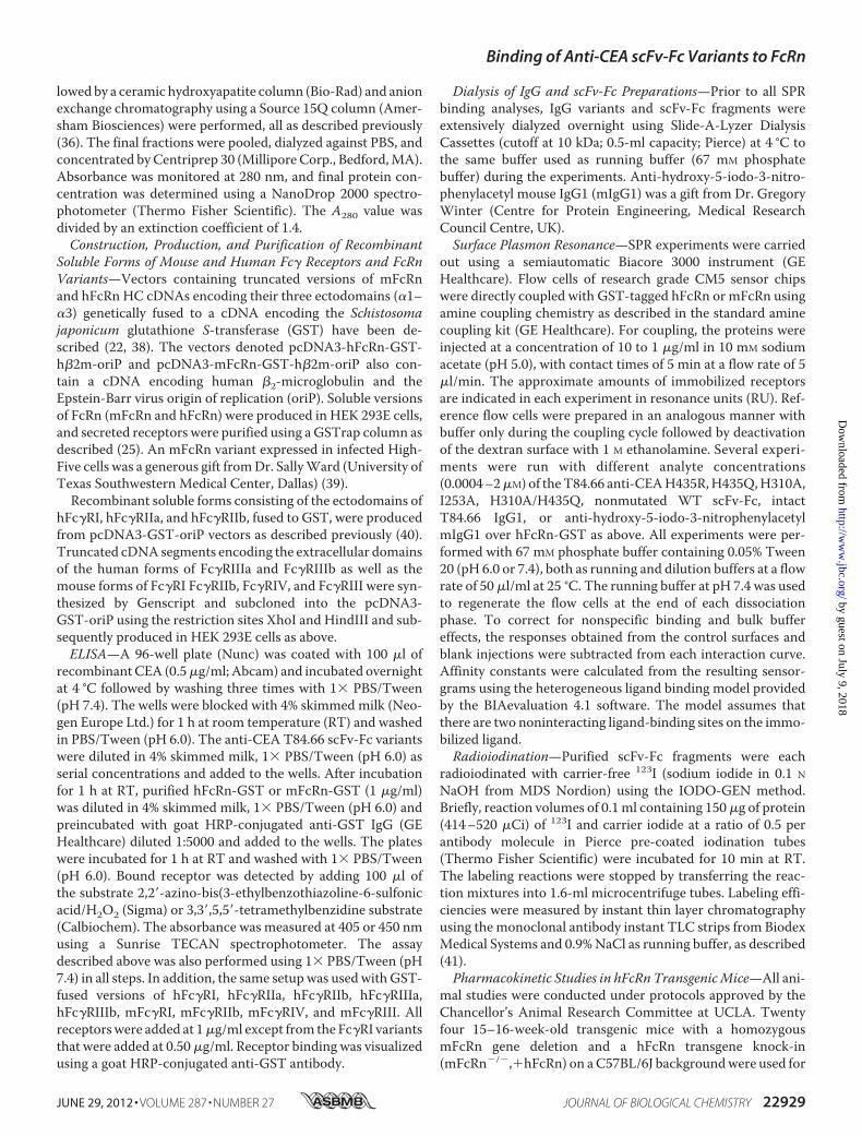
lently immobilized on aCM5biosensor chip tomimic the phys-iological situation where FcRn is a transmembrane receptorthat binds circulating soluble IgGmolecules. Initially, we inves-tigated the binding property of the intact anti-CEA T84.66IgG1 by injected serial dilutions over immobilized mFcRn orhFcRn at pH 6.0. Representative sensorgrams (Fig. 4, A and B)clearly demonstrate concentration-dependent and reversiblebinding, although the SPR profiles revealed considerably stron-ger binding tomFcRn than to the human form. To compare therelative binding properties of the scFv-Fc antibody fragments,samples of 0.5 or 1.0 �M of each variant were injected at pH 6.0or 7.4 overmFcRn immobilized at twodifferent densities (�110and �900 RU). The nonmutated scFv-Fc variant boundstrongly to mFcRn at acidic pH, and some binding was alsodetected at neutral pH (Fig. 5A). The same result was obtainedfor H435R (Fig. 5D). Although the binding kinetics at acidic pHwere characterized by slow dissociation, interacting moleculesdissociated rapidly at neutral pH. All single mutants showeddetectable binding to mFcRn at acidic pH, although the
FIGURE 2. ELISA measurements of pH-dependent binding of mFcRn andhFcRn to the scFv-Fc variants. Binding responses of mFcRn to titratedamounts of WT, I253A, H310A, H435R, H435Q, and H310A/H435Q at pH 6.0 (A)and pH 7.4 (B), respectively. Binding of hFcRn to titrated amounts of WT,I253A, H310A, H435R, H435Q, and H310A/H435Q at pH 6.0 (C) and pH 7.4 (D).The numbers given represent the mean of triplicates.
FIGURE 3. ELISA measurements of Fc�R binding to scFv-Fc variants. Bind-ing of mFc�RI (A), mFc�RIIb (B), hFc�RI (C), and hFc�RIIb (D) to titratedamounts of T84.66 and the scFv-Fc variants (WT, I253A, H310A, and H435Q).The numbers given represent the mean of triplicates.
Binding of Anti-CEA scFv-Fc Variants to FcRn
JUNE 29, 2012 • VOLUME 287 • NUMBER 27 JOURNAL OF BIOLOGICAL CHEMISTRY 22931
by guest on July 9, 2018http://w
ww
.jbc.org/D
ownloaded from
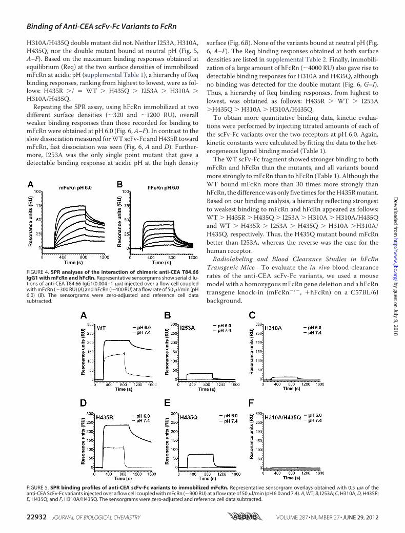
H310A/H435Q double mutant did not. Neither I253A, H310A,H435Q, nor the double mutant bound at neutral pH (Fig. 5,A–F). Based on the maximum binding responses obtained atequilibrium (Req) at the two surface densities of immobilizedmFcRn at acidic pH (supplemental Table 1), a hierarchy of Reqbinding responses, ranking from highest to lowest, were as fol-lows: H435R �/ � WT � H435Q � I253A � H310A �H310A/H435Q.Repeating the SPR assay, using hFcRn immobilized at two
different surface densities (�320 and �1200 RU), overallweaker binding responses than those recorded for binding tomFcRn were obtained at pH 6.0 (Fig. 6, A–F). In contrast to theslow dissociationmeasured forWT scFv-Fc andH435R towardmFcRn, fast dissociation was seen (Fig. 6, A and D). Further-more, I253A was the only single point mutant that gave adetectable binding response at acidic pH at the high density
surface (Fig. 6B). None of the variants bound at neutral pH (Fig.6, A–F). The Req binding responses obtained at both surfacedensities are listed in supplemental Table 2. Finally, immobili-zation of a large amount of hFcRn (�4000 RU) also gave rise todetectable binding responses for H310A and H435Q, althoughno binding was detected for the double mutant (Fig. 6, G–I).Thus, a hierarchy of Req binding responses, from highest tolowest, was obtained as follows: H435R � WT � I253A�H435Q � H310A � H310A/H435Q.
To obtain more quantitative binding data, kinetic evalua-tions were performed by injecting titrated amounts of each ofthe scFv-Fc variants over the two receptors at pH 6.0. Again,kinetic constants were calculated by fitting the data to the het-erogeneous ligand binding model (Table 1).The WT scFv-Fc fragment showed stronger binding to both
mFcRn and hFcRn than the mutants, and all variants boundmore strongly tomFcRn than to hFcRn (Table 1). Although theWT bound mFcRn more than 30 times more strongly thanhFcRn, the differencewas only five times for theH435Rmutant.Based on our binding analysis, a hierarchy reflecting strongestto weakest binding to mFcRn and hFcRn appeared as follows:WT � H435R � H435Q � I253A � H310A � H310A/H435Qand WT � H435R � I253A � H435Q � H310A �H310A/H435Q, respectively. Thus, the H435Q mutant bound mFcRnbetter than I253A, whereas the reverse was the case for thehuman receptor.Radiolabeling and Blood Clearance Studies in hFcRn
Transgenic Mice—To evaluate the in vivo blood clearancerates of the anti-CEA scFv-Fc variants, we used a mousemodel with a homozygous mFcRn gene deletion and a hFcRntransgene knock-in (mFcRn�/�, �hFcRn) on a C57BL/6Jbackground.
FIGURE 4. SPR analyses of the interaction of chimeric anti-CEA T84.66IgG1 with mFcRn and hFcRn. Representative sensorgrams show serial dilu-tions of anti-CEA T84.66 IgG1(0.004 –1 �M) injected over a flow cell coupledwith mFcRn (�300 RU) (A) and hFcRn (�400 RU) at a flow rate of 50 �l/min (pH6.0) (B). The sensorgrams were zero-adjusted and reference cell datasubtracted.
FIGURE 5. SPR binding profiles of anti-CEA scFv-Fc variants to immobilized mFcRn. Representative sensorgram overlays obtained with 0.5 �M of theanti-CEA ScFv-Fc variants injected over a flow cell coupled with mFcRn (�900 RU) at a flow rate of 50 �l/min (pH 6.0 and 7.4). A, WT; B, I253A; C, H310A; D, H435R;E, H435Q; and F, H310A/H435Q. The sensorgrams were zero-adjusted and reference cell data subtracted.
Binding of Anti-CEA scFv-Fc Variants to FcRn
22932 JOURNAL OF BIOLOGICAL CHEMISTRY VOLUME 287 • NUMBER 27 • JUNE 29, 2012
by guest on July 9, 2018http://w
ww
.jbc.org/D
ownloaded from

The scFv-Fc variantswere radiolabeled, and the efficiency forthe 123I radiolabeling reaction averaged 82.9%, ranging from69.7% (WT) to 88.4% (I253A), although themean specific activ-ity was 2.59 � 0.131 �Ci/�g, ranging from 2.23 to 3.12 �Ci/�g.The blood activity data at 0.5, 2, 4, 6, 12, 24, 48, and 72 h post-injection were measured. Analysis of variance between the bloodactivity curves (Fig. 7) was done using analysis of variance andrevealed that all six scFv-Fc fragments had significantly differentblood clearance profile with p values ranging from 0.0001 to0.049. The rate constants (k1 and k2), the area under the curvederived from the fitted curves, and the calculated distribution(t1⁄2�) and elimination (t1⁄2�) half-lives of each scFv-Fc protein in
blood are summarized in Table 2. The order of blood clearancebased on the area under the curve, from slow to fast, is as follows:WT � H435R � I253A � H435Q � H310A/H435Q � H310A.
FIGURE 6. SPR binding profiles of anti-CEA scFv-Fc variants to immobilized hFcRn. Representative sensorgram overlays obtained with 0.5 �M of each anti-CEAscFv-Fc variant injected over a flow cell coupled with hFcRn (�1200 RU) at a flow rate of 50 �l/min (pH 6.0 and 7.4). A, WT; B, I253A; C, H310A; D, H435R; E, H435Q; andF, H310A/H435Q. Representative sensorgram overlays were obtained with 2.0 �M WT and H310A/H435Q (G), H310A (H), and H435Q (I) and injected over a flow cellcoupled with large amounts of hFcRn (�4000 RU) at a flow rate of 50 �l/min (pH 6.0). All sensorgrams were zero-adjusted and reference cell data subtracted.
TABLE 1SPR-derived affinities of the interaction of scFv-Fc variants with FcRn
scFc-Fcvariant
mFcRnKD1 mFcRn KD2
hFcRnKD1
hFcRnKD2
nM nM nM nMWT 0.2 � 0.0 43.8 � 2.7 6.4 � 2.6 105.3 � 64H435R 1.8 � 0.1 122.1 � 55.8 9.4 � 0.7 196.6 � 19.3H435Q 80.9 � 18.1 400.1 � 112.8 NDa NDI253A 443.5 � 4.9 1054 � 1.9 ND NDH310A ND ND ND NDH310A/H435Q ND ND ND ND
a NDmean not determined because of no binding or very weak binding.
FIGURE 7. Blood activity curves of 123I-labeled scFv-Fc fragments in hFcRntransgenic mice. Mice were injected with 96 –130 �Ci of 123I-labeled scFv-Fcproteins (30 �g of labeled protein/mouse) under anesthesia. Blood sampleswere collected from the tail immediately after injection (0-h time point) and attime points 2, 4, 6, 12, 24, 48, and 72 h post-injection. Blood activity wasdetermined using a Wallac WIZARD automatic gamma. The percentage ofinjected dose/g (% ID/g) was calculated. Each group represents four mice.Logarithmic scale, S.E. was used to measure variability.
Binding of Anti-CEA scFv-Fc Variants to FcRn
JUNE 29, 2012 • VOLUME 287 • NUMBER 27 JOURNAL OF BIOLOGICAL CHEMISTRY 22933
by guest on July 9, 2018http://w
ww
.jbc.org/D
ownloaded from
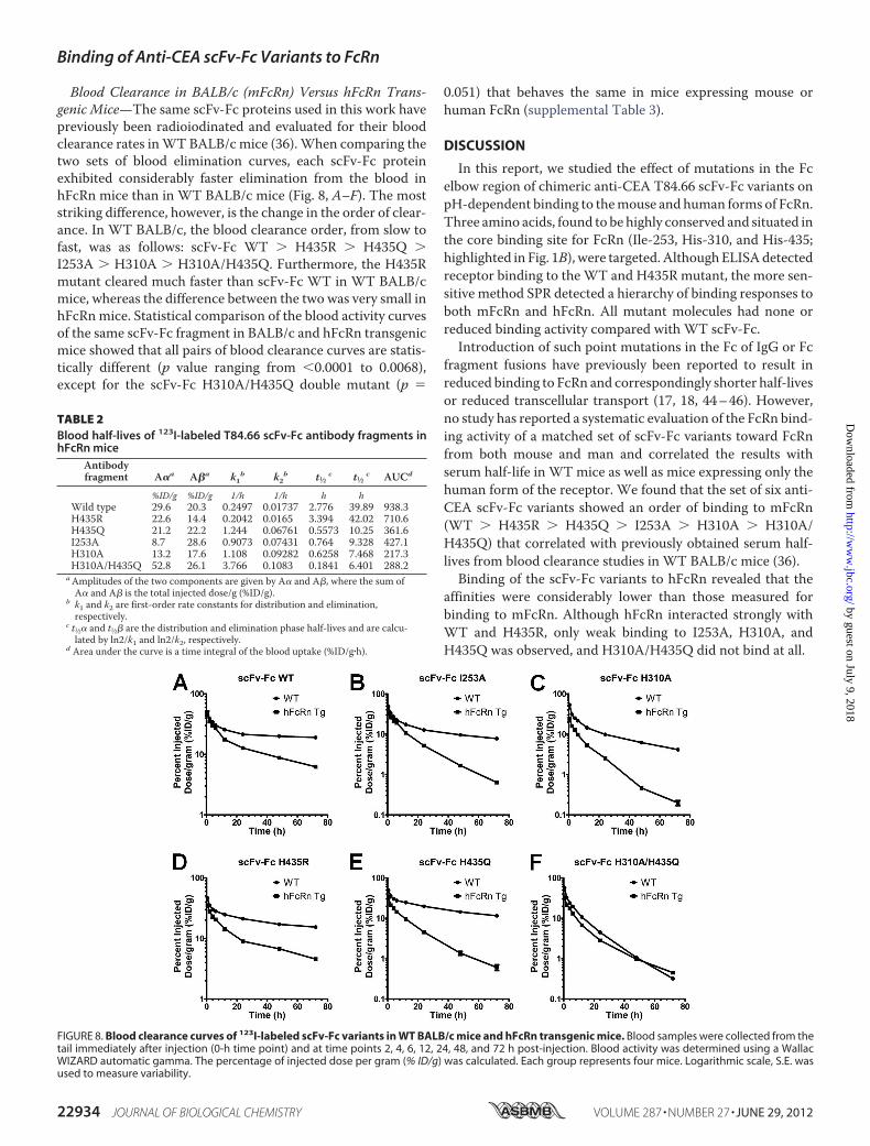
Blood Clearance in BALB/c (mFcRn) Versus hFcRn Trans-genic Mice—The same scFv-Fc proteins used in this work havepreviously been radioiodinated and evaluated for their bloodclearance rates inWT BALB/c mice (36).When comparing thetwo sets of blood elimination curves, each scFv-Fc proteinexhibited considerably faster elimination from the blood inhFcRn mice than in WT BALB/c mice (Fig. 8, A–F). The moststriking difference, however, is the change in the order of clear-ance. In WT BALB/c, the blood clearance order, from slow tofast, was as follows: scFv-Fc WT � H435R � H435Q �I253A � H310A � H310A/H435Q. Furthermore, the H435Rmutant cleared much faster than scFv-Fc WT in WT BALB/cmice, whereas the difference between the two was very small inhFcRnmice. Statistical comparison of the blood activity curvesof the same scFv-Fc fragment in BALB/c and hFcRn transgenicmice showed that all pairs of blood clearance curves are statis-tically different (p value ranging from 0.0001 to 0.0068),except for the scFv-Fc H310A/H435Q double mutant (p �
0.051) that behaves the same in mice expressing mouse orhuman FcRn (supplemental Table 3).
DISCUSSION
In this report, we studied the effect of mutations in the Fcelbow region of chimeric anti-CEA T84.66 scFv-Fc variants onpH-dependent binding to themouse and human forms of FcRn.Three amino acids, found to be highly conserved and situated inthe core binding site for FcRn (Ile-253, His-310, and His-435;highlighted in Fig. 1B), were targeted.AlthoughELISAdetectedreceptor binding to theWT and H435R mutant, the more sen-sitive method SPR detected a hierarchy of binding responses toboth mFcRn and hFcRn. All mutant molecules had none orreduced binding activity compared with WT scFv-Fc.Introduction of such point mutations in the Fc of IgG or Fc
fragment fusions have previously been reported to result inreduced binding to FcRn and correspondingly shorter half-livesor reduced transcellular transport (17, 18, 44–46). However,no study has reported a systematic evaluation of the FcRn bind-ing activity of a matched set of scFv-Fc variants toward FcRnfrom both mouse and man and correlated the results withserum half-life in WT mice as well as mice expressing only thehuman form of the receptor. We found that the set of six anti-CEA scFv-Fc variants showed an order of binding to mFcRn(WT � H435R � H435Q � I253A � H310A � H310A/H435Q) that correlated with previously obtained serum half-lives from blood clearance studies in WT BALB/c mice (36).Binding of the scFv-Fc variants to hFcRn revealed that the
affinities were considerably lower than those measured forbinding to mFcRn. Although hFcRn interacted strongly withWT and H435R, only weak binding to I253A, H310A, andH435Q was observed, and H310A/H435Q did not bind at all.
FIGURE 8. Blood clearance curves of 123I-labeled scFv-Fc variants in WT BALB/c mice and hFcRn transgenic mice. Blood samples were collected from thetail immediately after injection (0-h time point) and at time points 2, 4, 6, 12, 24, 48, and 72 h post-injection. Blood activity was determined using a WallacWIZARD automatic gamma. The percentage of injected dose per gram (% ID/g) was calculated. Each group represents four mice. Logarithmic scale, S.E. wasused to measure variability.
TABLE 2Blood half-lives of 123I-labeled T84.66 scFv-Fc antibody fragments inhFcRn mice
Antibodyfragment A�a A�a k1b k2b t1⁄2 c t1⁄2 c AUCd
%ID/g %ID/g 1/h 1/h h hWild type 29.6 20.3 0.2497 0.01737 2.776 39.89 938.3H435R 22.6 14.4 0.2042 0.0165 3.394 42.02 710.6H435Q 21.2 22.2 1.244 0.06761 0.5573 10.25 361.6I253A 8.7 28.6 0.9073 0.07431 0.764 9.328 427.1H310A 13.2 17.6 1.108 0.09282 0.6258 7.468 217.3H310A/H435Q 52.8 26.1 3.766 0.1083 0.1841 6.401 288.2
aAmplitudes of the two components are given by A� and A�, where the sum ofA� and A� is the total injected dose/g (%ID/g).
b k1 and k2 are first-order rate constants for distribution and elimination,respectively.
c t1⁄2� and t1⁄2� are the distribution and elimination phase half-lives and are calcu-lated by ln2/k1 and ln2/k2, respectively.
d Area under the curve is a time integral of the blood uptake (%ID/g�h).
Binding of Anti-CEA scFv-Fc Variants to FcRn
22934 JOURNAL OF BIOLOGICAL CHEMISTRY VOLUME 287 • NUMBER 27 • JUNE 29, 2012
by guest on July 9, 2018http://w
ww
.jbc.org/D
ownloaded from

The H435R mutant showed slightly higher Req bindingresponses for both mFcRn and hFcRn compared with the WT,although KD1 was lower for the WT. Another difference wasthat H435Q bound more strongly than I253A to the mousereceptor, whereas I253A bound more strongly to shFcRn thanH435Q. Thus, differences in the order of binding of the scFv-Fcmutants to the two receptor species were detected.Mice are not necessarily ideal models from which half-life in
humans may be predicted. Major differences in binding ofhIgG1 variants to FcRn frommouse andman have already beendemonstrated (20, 21, 25, 28), and the phenomenon clearlyunderscores the importance of considering cross-species FcRnbinding properties of engineered IgG variants and Fc fusionmolecules. Rather, direct measurement of the binding affinityof novel engineered IgG variants toward both hFcRn and theFcRn species used for in vivo evaluations should be tested.In this report, we evaluated the blood clearance activity in a
mouse strain that lacks expression of its endogenous FcRn HCand is transgenic for the hFcRn HC (42). The rate of bloodclearance correlated with binding to recombinant solublehFcRn, and low binding corresponded to fast clearance. Thehierarchy of binding from high to low corresponded to theclearance rates from slow to fast. However, the order of clear-ance differed from that obtained inWTmice. Thus, the order ofclearance depends on the FcRn variant expressed by the miceused.The histidine residues situated at the Fc elbow are key players
in the interaction with FcRn. The H435R and H435Q mutantswere generated to mimic the two protonation states of the His-435 residue during the transition from the physiological pH ofthe bloodstream to the acidic environment in endosomal com-partments. At neutral pH, the histidine imidazole side chain isuncharged, although at acidic pH it is positive. Arginine is pos-itively charged at either pH, whereas glutamine is neutral.Both the H435R and H435Q mutations had an impact on
FcRn binding, where H435R showed a somewhat reduced butstill considerable binding at acidic pH, compared with H435Qthat only interacted weakly with the receptors. The H435Rmutant did not show stronger binding at physiological pH,whereH435Qdid not bind. These results are in agreementwitha shorter half-life in WT BALB/c mice where H435R showed a3.5-fold drop in half-life followed by H435Q (5.5-fold) (36).The biodistribution data obtained from WT mouse studies
are informative to understand how a single amino acid Fc sub-stitution affects half-life in mice. Interestingly, the dramaticeffect on clearance of H435R in WT mice may reflect the dif-ference in serum half-life observed between hIgG1 (His-435)and hIgG3 (Arg-435) in humans, where hIgG1 has a three timeslonger half-life than hIgG3 (21 versus 7 days) (47–49). Werecently reported that the short half-life of hIgG3 is due to pref-erential binding of hIgG1 to FcRn, which is present in a 10-foldhigher concentration than hIgG3 in blood. The effect wasreversed when Arg-435 of hIgG3 was replaced with a histidine(R435H), and theHis-435 of IgG1was replacedwith an arginine(50). In contrast, the in vivo data presented here from hFcRntransgenic mice, show only minor differences in blood clear-ance between H435R and the WT. This may well be due to the
fact that thesemice have very low endogenous IgG serum levels,as endogenous IgG binds hFcRn very poorly (20, 22).The hydrophobic Ile-253 is located centrally at the interac-
tion interface and is important for proper packing of the com-plex (51). Its importance is reflected in the great effect on half-life when mutated to alanine, as shown in this study, where theI253A variant showed a 5.5- and 4.3-fold decreased half-life inWT and hFcRn transgenic mice, respectively (36). The H310Amutant interacted only weakly with FcRn from both species, asmirrored by its short half-life in both mouse strains. Further-more, the importance of the conserved His-310 and His-435was also demonstrated for the doublemutant (H310A/H435Q),which cleared most rapidly from the blood circulation, asexpected.Several of the scFv-Fcmutants have been evaluated in differ-
ent mouse tumor models. Initially, the fastest clearing doublemutant was shown to be optimal for imaging in CEA-positiveLS174T xenografted athymic mice examined by positron emis-sion tomography, followed by H310A, although the I253A,H435Q, H435R, and WT variants were eliminated at muchslower rates and thus gave poor tissue to tumor ratios (36, 37).Furthermore, anti-CEA scFv-Fc H310A has been shown to bean attractive candidate for targeting of pancreatic cancer inCEA-positive pancreas cancer xenografted mice (52). In addi-tion, the double mutant with specificity for either human epi-dermal growth factor receptor 2 (HER2) or CD20 has demon-strated favorable positron emission tomography imaging innude mice with MCF7/HER2 breast cancer or mice carryinghuman CD20 expressing lymphomas, respectively (53, 54).H310A/H435Q degraded in the liver of both tumor-free and
mice xenografted with CEA-positive tumor much faster thanthe single mutant I253A (supplemental Fig. 4) (37). In thisstudy, radiometal labeling of the scFv-Fc variants via a bifunc-tional chelator was used, as this conjugation is more stable invivo than radioiodination (55). Thus, liver accumulation ofradiometal-labeled scFv-Fc mutants correlated with the rate ofblood clearance (37).FcRn has been shown to be expressed in the mouse liver (9),
and we have detected FcRn in human hepatocyte cell lines andliver tissue biopsies7 These findings indicate that FcRnexpressed by the liver may function to rescue endogenous IgGand injected radioconjugated IgG Fc fragments with retainedFcRn binding activity from intracellular degradation, althoughnoninteracting mutants accumulate and degrade. This mecha-nism should be considered in further studies as liver accumu-lation and toxicity may become dose-limiting in antibody-based radiometal therapy. Hence, our findings suggest that IgGor Fc fragments such as the scFv-Fc mutants with substantiallyreduced FcRn binding capacity may be trapped within this vitalorgan, although single mutants such as I253A and H310A maybe better choices for radioimmunotherapy because of lowerliver accumulation and potential toxicity.Regarding expanding these studies to a more humanized in
vivo system, immunodeficient mice (Rag1�/�) transgenic forhFcRn have recently been described (56). Such mice may be
7 M. B. Daba, J. T. Andersen, and I. Sandlie, unpublished data.
Binding of Anti-CEA scFv-Fc Variants to FcRn
JUNE 29, 2012 • VOLUME 287 • NUMBER 27 JOURNAL OF BIOLOGICAL CHEMISTRY 22935
by guest on July 9, 2018http://w
ww
.jbc.org/D
ownloaded from

more ideal in the next phase of in vivo evaluations of tumortargeting and imaging using the Fc-engineered scFv-Fcvariants.
Acknowledgments—We are grateful to Sathiaruby Sivaganesh forexcellent technical assistance.We thank Dr. DerekW. Bartlett (Imag-inab Inc., Los Angeles) for assistance with pharmacokinetic analysis.
REFERENCES1. Holliger, P., and Hudson, P. J. (2005) Engineered antibody fragments and
the rise of single domains. Nat. Biotechnol. 23, 1126–11362. Kenanova, V., andWu, A.M. (2006) Tailoring antibodies for radionuclide
delivery. Expert Opin. Drug Deliv. 3, 53–703. Wu, A. M., and Senter, P. D. (2005) Arming antibodies. Prospects and
challenges for immunoconjugates. Nat. Biotechnol. 23, 1137–11464. Ghetie, V., Popov, S., Borvak, J., Radu, C., Matesoi, D., Medesan, C., Ober,
R. J., and Ward, E. S. (1997) Increasing the serum persistence of an IgGfragment by random mutagenesis. Nat. Biotechnol. 15, 637–640
5. Kamei, D. T., Lao, B. J., Ricci, M. S., Deshpande, R., Xu, H., Tidor, B., andLauffenburger, D. A. (2005) Quantitative methods for developing Fc mu-tants with extended half-lives. Biotechnol. Bioeng. 92, 748–760
6. Roopenian, D. C., Christianson, G. J., Sproule, T. J., Brown, A. C., Akilesh,S., Jung, N., Petkova, S., Avanessian, L., Choi, E. Y., Shaffer, D. J., Eden,P. A., and Anderson, C. L. (2003) The MHC class I-like IgG receptorcontrols perinatal IgG transport, IgG homeostasis, and fate of IgG-Fc-coupled drugs. J. Immunol. 170, 3528–3533
7. Akilesh, S., Huber, T. B., Wu, H., Wang, G., Hartleben, B., Kopp, J. B.,Miner, J. H., Roopenian, D. C., Unanue, E. R., and Shaw, A. S. (2008)Podocytes use FcRn to clear IgG from the glomerular basement mem-brane. Proc. Natl. Acad. Sci. U.S.A. 105, 967–972
8. Blumberg, R. S., Koss, T., Story, C. M., Barisani, D., Polischuk, J., Lipin, A.,Pablo, L., Green, R., and Simister, N. E. (1995) A major histocompatibilitycomplex class I-related Fc receptor for IgG on rat hepatocytes. J. Clin.Invest. 95, 2397–2402
9. Akilesh, S., Christianson, G. J., Roopenian, D. C., and Shaw, A. S. (2007)Neonatal FcR expression in bone marrow-derived cells functions to pro-tect serum IgG from catabolism. J. Immunol. 179, 4580–4588
10. Zhu, X., Meng, G., Dickinson, B. L., Li, X., Mizoguchi, E., Miao, L., Wang,Y., Robert, C., Wu, B., Smith, P. D., Lencer, W. I., and Blumberg, R. S.(2001) MHC class I-related neonatal Fc receptor for IgG is functionallyexpressed inmonocytes, intestinalmacrophages, and dendritic cells. J. Im-munol. 166, 3266–3276
11. Qiao, S. W., Kobayashi, K., Johansen, F. E., Sollid, L. M., Andersen, J. T.,Milford, E., Roopenian, D. C., Lencer, W. I., and Blumberg, R. S. (2008)Dependence of antibody-mediated presentation of antigen on FcRn. Proc.Natl. Acad. Sci. U.S.A. 105, 9337–9342
12. Baker, K., Qiao, S.W., Kuo, T. T., Aveson, V.G., Platzer, B., Andersen, J. T.,Sandlie, I., Chen, Z., de Haar, C., Lencer,W. I., Fiebiger, E., and Blumberg,R. S. (2011) Neonatal Fc receptor for IgG (FcRn) regulates cross-presen-tation of IgG immune complexes by CD8-CD11b� dendritic cells. Proc.Natl. Acad. Sci. U.S.A. 108, 9927–9932
13. Montoyo, H. P., Vaccaro, C., Hafner, M., Ober, R. J., Mueller, W., andWard, E. S. (2009) Conditional deletion of theMHC class I-related recep-tor FcRn reveals the sites of IgG homeostasis inmice. Proc. Natl. Acad. Sci.U.S.A. 106, 2788–2793
14. Ward, E. S., Zhou, J., Ghetie, V., andOber, R. J. (2003) Evidence to supportthe cellular mechanism involved in serum IgG homeostasis in humans.Int. Immunol. 15, 187–195
15. Ober, R. J.,Martinez, C., Lai, X., Zhou, J., andWard, E. S. (2004) Exocytosisof IgG as mediated by the receptor, FcRn. An analysis at the single-mole-cule level. Proc. Natl. Acad. Sci. U.S.A. 101, 11076–11081
16. Prabhat, P., Gan, Z., Chao, J., Ram, S., Vaccaro, C., Gibbons, S., Ober, R. J.,and Ward, E. S. (2007) Elucidation of intracellular recycling pathwaysleading to exocytosis of the Fc receptor, FcRn, by using multifocal planemicroscopy. Proc. Natl. Acad. Sci. U.S.A. 104, 5889–5894
17. Kim, J. K., Firan, M., Radu, C. G., Kim, C. H., Ghetie, V., and Ward, E. S.
(1999) Mapping the site on human IgG for binding of the MHC classI-related receptor, FcRn. Eur. J. Immunol. 29, 2819–2825
18. Medesan, C., Matesoi, D., Radu, C., Ghetie, V., and Ward, E. S. (1997)Delineation of the amino acid residues involved in transcytosis and catab-olism of mouse IgG1. J. Immunol. 158, 2211–2217
19. Raghavan, M., Chen, M. Y., Gastinel, L. N., and Bjorkman, P. J. (1994)Investigation of the interaction between the class I MHC-related Fc re-ceptor and its immunoglobulin G ligand. Immunity 1, 303–315
20. Ober, R. J., Radu, C. G., Ghetie, V., and Ward, E. S. (2001) Differences inpromiscuity for antibody-FcRn interactions across species. Implicationsfor therapeutic antibodies. Int. Immunol. 13, 1551–1559
21. Zhou, J., Mateos, F., Ober, R. J., and Ward, E. S. (2005) Conferring thebinding properties of themouseMHC class I-related receptor, FcRn, ontothe human ortholog by sequential rounds of site-directed mutagenesis. J.Mol. Biol. 345, 1071–1081
22. Andersen, J. T., Daba, M. B., Berntzen, G., Michaelsen, T. E., and Sandlie,I. (2010) Cross-species binding analyses of mouse and human neonatal Fcreceptor show dramatic differences in immunoglobulin G and albuminbinding. J. Biol. Chem. 285, 4826–4836
23. Hinton, P. R., Johlfs, M. G., Xiong, J. M., Hanestad, K., Ong, K. C., Bullock,C., Keller, S., Tang, M. T., Tso, J. Y., Vásquez, M., and Tsurushita, N.(2004) Engineered human IgG antibodies with longer serum half-lives inprimates. J. Biol. Chem. 279, 6213–6216
24. Hinton, P. R., Xiong, J. M., Johlfs, M. G., Tang, M. T., Keller, S., andTsurushita, N. (2006) An engineered human IgG1 antibody with longerserum half-life. J. Immunol. 176, 346–356
25. Petkova, S. B., Akilesh, S., Sproule, T. J., Christianson,G. J., Al Khabbaz,H.,Brown, A. C., Presta, L. G., Meng, Y. G., and Roopenian, D. C. (2006)Enhanced half-life of genetically engineered human IgG1 antibodies in ahumanized FcRn mouse model. Potential application in humorally medi-ated autoimmune disease. Int. Immunol. 18, 1759–1769
26. Dall’Acqua, W. F., Kiener, P. A., and Wu, H. (2006) Properties of humanIgG1s engineered for enhanced binding to the neonatal Fc receptor(FcRn). J. Biol. Chem. 281, 23514–23524
27. Blumberg, R. S., and Lencer, W. I. (2005) Antibodies in the breakdownlane. Nat. Biotechnol. 23, 1232–1234
28. Vaccaro, C., Zhou, J., Ober, R. J., andWard, E. S. (2005) Engineering the Fcregion of immunoglobulin G to modulate in vivo antibody levels. Nat.Biotechnol. 23, 1283–1288
29. Vaccaro, C., Bawdon, R., Wanjie, S., Ober, R. J., and Ward, E. S. (2006)Divergent activities of an engineered antibody in murine and human sys-tems have implications for therapeutic antibodies. Proc. Natl. Acad. Sci.U.S.A. 103, 18709–18714
30. Wu, A. M., Yazaki, P. J., Tsai, S., Nguyen, K., Anderson, A. L., McCarthy,D. W., Welch, M. J., Shively, J. E., Williams, L. E., Raubitschek, A. A.,Wong, J. Y., Toyokuni, T., Phelps, M. E., and Gambhir, S. S. (2000) Highresolution microPET imaging of carcinoembryonic antigen-positive xe-nografts by using a copper-64-labeled engineered antibody fragment.Proc. Natl. Acad. Sci. U.S.A. 97, 8495–8500
31. Sundaresan, G., Yazaki, P. J., Shively, J. E., Finn, R. D., Larson, S. M.,Raubitschek, A. A., Williams, L. E., Chatziioannou, A. F., Gambhir, S. S.,and Wu, A. M. (2003) 124I-Labeled engineered anti-CEA minibodies anddiabodies allow high contrast, antigen-specific small animal PET imagingof xenografts in athymic mice. J. Nucl. Med. 44, 1962–1969
32. Wong, J. Y., Chu, D. Z.,Williams, L. E., Yamauchi, D.M., Ikle, D.N., Kwok,C. S., Liu, A., Wilczynski, S., Colcher, D., Yazaki, P. J., Shively, J. E., Wu,A. M., and Raubitschek, A. A. (2004) Pilot trial evaluating a 123I-labeled80-kilodalton engineered anticarcinoembryonic antigen antibody frag-ment (cT84.66 minibody) in patients with colorectal cancer. Clin. CancerRes. 10, 5014–5021
33. Hu, S., Shively, L., Raubitschek, A., Sherman, M., Williams, L. E., Wong,J. Y., Shively, J. E., and Wu, A. M. (1996) Minibody. A novel engineeredanti-carcinoembryonic antigen antibody fragment (single-chain Fv-CH3)that exhibits rapid, high level targeting of xenografts. Cancer Res. 56,3055–3061
34. Boucher, D., Cournoyer, D., Stanners, C. P., and Fuks, A. (1989) Studies onthe control of gene expression of the carcinoembryonic antigen family inhuman tissue. Cancer Res. 49, 847–852
Binding of Anti-CEA scFv-Fc Variants to FcRn
22936 JOURNAL OF BIOLOGICAL CHEMISTRY VOLUME 287 • NUMBER 27 • JUNE 29, 2012
by guest on July 9, 2018http://w
ww
.jbc.org/D
ownloaded from
35. Esteban, J. M., Paxton, R., Mehta, P., Battifora, H., and Shively, J. E. (1993)Sensitivity and specificity of Gold types 1–5 anti-carcinoembryonic anti-gen monoclonal antibodies. Immunohistologic characterization in colo-rectal cancer and normal tissues. Hum. Pathol. 24, 322–328
36. Kenanova, V., Olafsen, T., Crow, D. M., Sundaresan, G., Subbarayan, M.,Carter, N. H., Ikle, D. N., Yazaki, P. J., Chatziioannou, A. F., Gambhir, S. S.,Williams, L. E., Shively, J. E., Colcher, D., Raubitschek, A. A., and Wu,A. M. (2005) Tailoring the pharmacokinetics and positron emission to-mography imaging properties of anti-carcinoembryonic antigen single-chain Fv-Fc antibody fragments. Cancer Res. 65, 622–631
37. Kenanova, V., Olafsen, T., Williams, L. E., Ruel, N. H., Longmate, J.,Yazaki, P. J., Shively, J. E., Colcher, D., Raubitschek, A. A., and Wu, A. M.(2007) Radioiodinated versus radiometal-labeled anti-carcinoembryonicantigen single-chain Fv-Fc antibody fragments. Optimal pharmacokinet-ics for therapy. Cancer Res. 67, 718–726
38. Andersen, J. T., Justesen, S., Fleckenstein, B., Michaelsen, T. E., Berntzen,G., Kenanova, V. E., Daba, M. B., Lauvrak, V., Buus, S., and Sandlie, I.(2008) Ligand binding and antigenic properties of a human neonatal Fcreceptor with mutation of two unpaired cysteine residues. FEBS J. 275,4097–4110
39. Popov, S., Hubbard, J. G., Kim, J., Ober, B., Ghetie, V., and Ward, E. S.(1996) The stoichiometry and affinity of the interaction of murine Fcfragments with the MHC class I-related receptor, FcRn. Mol. Immunol.33, 521–530
40. Berntzen, G., Lunde, E., Flobakk, M., Andersen, J. T., Lauvrak, V., andSandlie, I. (2005) Prolonged and increased expression of soluble Fc recep-tors, IgG and a TCR-Ig fusion protein by transiently transfected adherent293E cells. J. Immunol. Methods 298, 93–104
41. Blumberg, D., Hochwald, S., Brennan,M. F., and Burt, M. (1995) Interleu-kin-6 stimulates gluconeogenesis in primary cultures of rat hepatocytes.Metabolism 44, 145–146
42. Stein, C., Kling, L., Proetzel, G., Roopenian, D. C., de Angelis, M. H.,Wolf,E., and Rathkolb, B. (2012) Clinical chemistry of human FcRn transgenicmice.Mamm. Genome 23, 259–269
43. Nimmerjahn, F., and Ravetch, J. V. (2008) Fc� receptors as regulators ofimmune responses. Nat. Rev. Immunol. 8, 34–47
44. Firan, M., Bawdon, R., Radu, C., Ober, R. J., Eaken, D., Antohe, F., Ghetie,V., and Ward, E. S. (2001) The MHC class I-related receptor, FcRn, playsan essential role in the maternofetal transfer of �-globulin in humans. Int.Immunol. 13, 993–1002
45. Bitonti, A. J., Dumont, J. A., Low, S. C., Peters, R. T., Kropp, K. E., Palom-bella, V. J., Stattel, J. M., Lu, Y., Tan, C. A., Song, J. J., Garcia, A. M.,Simister, N. E., Spiekermann, G. M., Lencer, W. I., and Blumberg, R. S.(2004) Pulmonary delivery of an erythropoietin Fc fusion protein in non-
human primates through an immunoglobulin transport pathway. Proc.Natl. Acad. Sci. U.S.A. 101, 9763–9768
46. Spiekermann, G.M., Finn, P.W.,Ward, E. S., Dumont, J., Dickinson, B. L.,Blumberg, R. S., and Lencer, W. I. (2002) Receptor-mediated immuno-globulin G transport across mucosal barriers in adult life: functional ex-pression of FcRn in the mammalian lung. J. Exp. Med. 196, 303–310
47. Spiegelberg, H. L., Fishkin, B. G., and Grey, H. M. (1968) Catabolism ofhuman �G-immunoglobulins of different heavy chain subclasses. I. Ca-tabolism of �G-myeloma proteins in man. J. Clin. Invest. 47, 2323–2330
48. Morell, A., Terry, W. D., and Waldmann, T. A. (1970) Metabolic proper-ties of IgG subclasses in man. J. Clin. Invest. 49, 673–680
49. Spiegelberg, H. L. (1974) Biological activities of immunoglobulins of dif-ferent classes and subclasses. Adv. Immunol. 19, 259–294
50. Stapleton, N. M., Andersen, J. T., Stemerding, A. M., Bjarnarson, S. P.,Verheul, R. C., Gerritsen, J., Zhao, Y., Kleijer, M., Sandlie, I., de Haas, M.,Jonsdottir, I., van der Schoot, C. E., and Vidarsson, G. (2011) Competitionfor FcRn-mediated transport gives rise to short half-life of human IgG3and offers therapeutic potential. Nat. Commun. 2, 599
51. Martin, W. L., West, A. P., Jr., Gan, L., and Bjorkman, P. J. (2001) Crystalstructure at 2.8 Å of an FcRn/heterodimeric Fc complex. Mechanism ofpH-dependent binding.Mol. Cell 7, 867–877
52. Girgis, M. D., Olafsen, T., Kenanova, V., McCabe, K. E., Wu, A. M., andTomlinson, J. S. (2011) Targeting CEA in pancreas cancer xenografts witha mutated scFv-Fc antibody fragment. EJNMMI Res. 1, 24
53. Olafsen, T., Kenanova, V. E., Sundaresan, G., Anderson, A. L., Crow, D.,Yazaki, P. J., Li, L., Press, M. F., Gambhir, S. S., Williams, L. E., Wong, J. Y.,Raubitschek, A. A., Shively, J. E., andWu, A. M. (2005) Optimizing radio-labeled engineered anti-p185HER2 antibody fragments for in vivo imag-ing. Cancer Res. 65, 5907–5916
54. Olafsen, T., Sirk, S. J., Betting, D. J., Kenanova, V. E., Bauer, K. B., Ladno,W., Raubitschek, A. A., Timmerman, J. M., and Wu, A. M. (2010) Immu-noPET imaging of B-cell lymphoma using 124I-anti-CD20 scFv dimers(diabodies). Protein Eng. Des. Sel. 23, 243–249
55. Milenic, D. E., Brady, E. D., and Brechbiel, M. W. (2004) Antibody-tar-geted radiation cancer therapy. Nat. Rev. Drug Discov. 3, 488–499
56. Zalevsky, J., Chamberlain, A. K., Horton, H. M., Karki, S., Leung, I. W.,Sproule, T. J., Lazar, G. A., Roopenian, D. C., and Desjarlais, J. R. (2010)Enhanced antibody half-life improves in vivo activity.Nat. Biotechnol. 28,157–159
57. Deisenhofer, J. (1981) Crystallographic refinement and atomicmodels of ahuman Fc fragment and its complex with fragment B of protein A fromStaphylococcus aureus at 2.9- and 2.8-Å resolution. Biochemistry 20,2361–2370
Binding of Anti-CEA scFv-Fc Variants to FcRn
JUNE 29, 2012 • VOLUME 287 • NUMBER 27 JOURNAL OF BIOLOGICAL CHEMISTRY 22937
by guest on July 9, 2018http://w
ww
.jbc.org/D
ownloaded from
Derry C. Roopenian, Anna M. Wu and Inger SandlieJan Terje Andersen, Stian Foss, Vania E. Kenanova, Tove Olafsen, Ingvild S. Leikfoss,
That Reveal Distinct Cross-species Differences in Serum Half-lifeVariants Bind Mouse and Human Neonatal Fc Receptor with Different Affinities
Anti-carcinoembryonic Antigen Single-chain Variable Fragment Antibody
doi: 10.1074/jbc.M112.355131 originally published online May 8, 20122012, 287:22927-22937.J. Biol. Chem.
10.1074/jbc.M112.355131Access the most updated version of this article at doi:
Alerts:
When a correction for this article is posted•
When this article is cited•
to choose from all of JBC's e-mail alertsClick here
Supplemental material:
http://www.jbc.org/content/suppl/2012/05/08/M112.355131.DC1
http://www.jbc.org/content/287/27/22927.full.html#ref-list-1
This article cites 57 references, 25 of which can be accessed free at
by guest on July 9, 2018http://w
ww
.jbc.org/D
ownloaded from