Embed Size (px)
Citation preview

ORIGINAL PAPER
Anti-cancer studies of noble metal nanoparticles synthesizedusing different plant extracts
Deshpande Raghunandan & Bhat Ravishankar & Ganachari Sharanbasava &
D. Bedre Mahesh & Vasanth Harsoor & Manjunath S. Yalagatti & M. Bhagawanraju &
A. Venkataraman
Received: 10 February 2011 /Accepted: 26 April 2011 /Published online: 10 May 2011# Springer-Verlag 2011
Abstract Biofunctionalized gold and silver nanoparticlessynthesized using different plant extracts of guava andclove in vitro anti-cancer efficacy against four differentcancer cell lines human colorectal adenocarcinoma, humankidney, human chronic myelogenous, leukemia, bonemarrow, and human cervix have been studied and reported.The present experimental study suggests that flavonoidsfunctionalized gold nanoparticles synthesized using aque-ous clove buds extract are more potential than guava leafextract towards anti-cancer activities. The microscopic and2,3-bis (2-methoxy-4-nitro-5-sulfophenyl)-5-[(phenylamino)carbonyl]-2H-tetrazolium hydroxide (XTT) assay infer thatthe functionalized irregular shaped gold nanoparticles synthe-sized with aqueous clove bud extract showed a satisfactory
anti-cancer effect on all the cell lines. The silver nanoparticlessynthesized using same extracts are devoid of anti-canceractivity. The XTT assay revealed dose-dependent cytotoxicityto cancer cell lines. The study revealed that the free radicalsgenerated by gold nanoparticles are responsible for anti-cancer effect. To confirm the free-radical scavenging efficacyof gold nanoparticle, nitric oxide assay is followed. Weobserved that the gold nanoparticles swabbed the free radicalsin dose-dependent manner. With continued improvements,these nanoparticles may prove to be potential anti-canceragents.
Keywords Flavonoid functionalized gold and silvernanoparticles . Cell lines . In vitro activity . XTT assay .
Anti-cancer agent
1 Introduction
Cancer is observed as the most dangerous class of diseasecategorized by uncontrolled cell growth (Chow 2010;Suriamoorthy et al. 2010). There is a marginal increase incancer cases in the last few years, and most of the time, itends up with taking life (Dite et al. 2010; Parveen andSahoo 2010; Smith et al. 2010). In many types of cancer,we are yet to find a satisfactory medicine or carrier ofmedicine as in case of drug delivery to be used as asatisfactory chemotherapeutic agent. Nanotechnology, aninterdisciplinary research field comprising chemistry, engi-neering, biology, and medicine, has great potential for earlydetection, accurate diagnosis, and tailored treatment ofcancer (Sakamoto et al. 2010). Nanoparticles are usuallysmaller than several hundred nanometers in size, compara-ble to large biological molecules such as enzymes,receptors, of a size about 100 to 10,000 times smaller than
D. RaghunandanHKES Matoshree Taradevi RampureInstitute of Pharmaceutical Sciences,Sedam Road, Gulbarga-585105 Karnataka, India
B. Ravishankar :G. Sharanbasava :D. B. Mahesh :A. Venkataraman (*)Materials Chemistry Laboratory, Department of Material Science,Gulbarga University,Gulbarga 585106, Karnataka, Indiae-mail: [email protected]
V. HarsoorPeriferal Cancer Institute,Sedam Road, Gulbarga-585105 Karnataka, India
M. S. YalagattiSri Krupa institute of Pharmaceutical Sciences,Village Velkatta, Siddipet-502277, Medak, Andhra Pradesh, India
M. BhagawanrajuCM College of Pharmacy,Maisammaguda, Dulapally, Hyderabad-500014,Andhra Pradesh, India
Cancer Nano (2011) 2:57–65DOI 10.1007/s12645-011-0014-8

human cells. These nanoparticles can offer unprecedentedinteractions with biomolecules both on the surface and insidethe body cells, whichmay bring revolution in cancer diagnosisand treatment (Seigneuric et al. 2010; Liu et al. 2010).
Nanotechnology is a burgeoning arena which takesalong with it a myriad of prospects and possibilities foradvancing disease treatment in pharmaceutical and medicalfield. At nanometric scale, the physico-chemical andbiological properties of materials differ fundamentally fromtheir corresponding bulk counterpart because of the size-dependent quantum effect. The noble metal nanoparticleslike gold nanoparticles (AuNP), especially surface func-tionalized represent smart and promising candidates in thedrug delivery applications due to their unique dimensions,tunable functionalities on the surface, and controlled drugrelease (Datar and Richard 2010). Another essential aspectwhile working with AuNP in bio-applications is safety andbiocompatibility (AuNP is already approved by the USFood and Drug Administration.) Biologically synthesizedand functionalized, AuNP provide many desirable attributesfor use as carriers in drug delivery systems as thefunctionalized AuNP core is essentially inert and nontoxic1 reported in recent studies (Han et al. 2007; Kim et al.2009). Monodispersed nanoparticles can be formed with acore size from <30 nm and also with metal–core–organic–shell morphology; the mono-metal layer can be tailoredwith a range of biological ligands, help in effective cellularuptake, controlled drug release, and targeted drug delivery(Ghosha et al. 2008; Salmaso et al. 2010). These function-alized nanodelivery systems can be used directly aspromising lead molecules in the detection of cancer cells.In this applied research work, we have deduced anddetailed the use of synthesized bio-functional noble metalnanoparticles application as an anti-cancer drug anddemonstrate the anti-cancer effect of these functionalizedAuNP using nitric oxide method. It gives a strongspeculation, for the influence of free electrons generatedby the surface of the functionalized AuNP has a lethaleffect on the electronegative surface membrane of thecancer cells. The free-radical scavenging effect of AuNP iscompared with the well-known anti-oxidant butyl hydroxyanisole (BHA), which is determined by nitric oxide method.The end results confirm that functionalizing AuNP with thewater-soluble organic moieties can show the synergic anti-proliferative effect in various cancer cell lines, and thus proveto be useful in various types of anti-cancer control systems.
We have already worked for the synthesis of differentfunctionalized Au and Ag nanoparticles using differentplant extracts of clove and guava (Raghunandan et al. 2009;Raghunandan et al. 2010a; Raghunandan et al. 2010b). Thecharacterization is elaborately discussed with respect tosize, shape stability, and functionalization using differentspectroscopic and microscopic techniques. The shape of
silver nanoparticles (AgNP) found to be roughly sphericaland in 30–60 nm and 20–30 nm range in case of guava leafand clove buds-mediated synthesis, respectively. The clove-mediated AuNP yielded highly unpredictable irregular-shaped particles in the range of 5–100 nm, whereas guavaleaf-mediated synthesis produced poly-shaped nanopar-ticles in the narrow range of 25–30 nm. In all the cases ofnoble nanoparticle synthesis, the preliminary studies usingFTIR (Fourier transformed infrared spectroscopy) confirmsthat the different polyphenols of the flavonoids of therespective plants are responsible for the biosynthesis, andthe same are capped on the surface of the nanoparticles.Though the further studies on the exact chemical moietyresponsible for bioreduction pathway is still underway, inthis paper, we are making an effort to understand the anti-proliferative effect of these functionalized “lead” indifferent cancer cell lines. The encouraging results offunctionalized AuNP as an anti-cancer agent using differentplant extract has a promising lead for further exploitation.
2 Experimental
2.1 Biosynthesis of nanoparticles
The AuNP and AgNP were synthesized separately treating the10−3 N HAuCl4 and 10−3 AgNO3 solutions with guava leaf(Psidium guajava) extract and clove bud (Syzygium aroma-ticum) extract, respectively, as reported in our earlier researchpapers (Raghunandan et al. 2009; Raghunandan et al. 2010a;Raghunandan et al. 2010b) The flavonoid-conjugated AuNPusing guava and clove are represented as AuNP-Gua andAuNP-Clo, respectively, and is shown in Fig. 1.
2.2 Anti-cancer activity study
Four different cells lines namely HT-29 (human colorectaladenocarcinoma), HEK-293 (human kidney), K-562 (humanchronic myelogenous, leukemia, bone marrow), and HeLa(human cervix) cell lines were obtained from the AmericanType Culture Collection, USA. The growth media: RPMI-1640, sufficient minimum essential medium (MEM), and thephosphate buffer solution (PBS) tablets were obtained fromSigma Chemical Co., St Louis, USA. Standard quality fetalcalf serum, penicillin–streptomycin, and trypsin wereobtained from Sigma labs. The XTT kit, which consists ofthe 2,3-bis(2-methoxy-4-nitro-5-sulfophenyl)-5-[(phenyla-mino)carbonyl]-2H-tetrazolium hydroxide (XTT) labelingreagent and the solubilisation solution was procured fromSigma labs, USA. Trypsin (0.25%+EDTA, 1 mM in PBSA),XTT dye–50 mg/mL, sterilized Sorensen's glycine buffer(0.1 M glycine, 0.1 M NaCl adjusted to pH 10.5 with 1 MNaOH), DMSO, growth medium is procured from Aldrich
58 D. Raghunandan et al.

labs. XTT labeling reagent and electron-coupling reagentwere thawed, respectively, in a water bath at 37°C. Each vialwas mixed thoroughly to obtain a clear solution. To performa cell proliferation assay (XTT) with one microplate (96wells), 5 mL XTT labeling reagent was mixed with 0.1 mLelectron-coupling reagent.
2.3 Free-radical scavenging activity of AuNP
Aqueous solution of AuNP–clo of different concentrations(2.5 μg, 5 μg, 10 μg/μL) were taken in different test tubesand made up to 3 ml with 0.1 M phosphate buffer (pH 7.2).Sodium nitroprusside (5 mM) prepared in buffered saline(pH 7.2) and 1 mL was added to each tube. The reactionmixture was incubated for 30 min at RT. A control withoutthe test compound, but with an equivalent amount ofmethanol was prepared for comparison purpose. After30 min, 1.5 mL of above solution was mixed with 1.5 mLof Griess reagent (1% sulphanilamide, 2% phosphoric acidand 0.1% N-1-naphthylethylenediamine dihydrochloride).The absorbance of the samples was measured at 546 nm.Nitric oxide radical scavenging activity was calculatedusing the following formula:
%radical scavenging activity
¼ control OD� aq:AuNP ODð Þ = control OD½ � � 100
2.4 Cell culture
A 96-well micrometer plate equipment (Nunc, Denmark)is used for this experiment. Two hundred microliters of
MEM is added in the required amount of wells; in eachcell, 50 μL of different cell lines are added. Biofunction-alized AgNP and AuNP of different concentrations (10, 20,30 μg/L) were added into the wells separately. The cells werecultured in the appropriate medium supplemented with 5–10% fetal calf serum and 1% penicillin–streptomycin, using25 cm2 flasks in a 37°C incubator with 5% CO2. Tosubculture the cells, the cells were separated, and the freshculture medium was used with fresh medium as follows: inthe first step, the old medium was removed, and then thecells were rinsed briefly with PBS. After adding 1–2 μL oftrypsin, the flask was incubated at 37°C using 5% CO2 for5 min. The upper part of the liquid is discarded, and thedetached cells, which are present in the lower part of theflask, are taken in another flask, and after adding 20 μL ofmedium to it, the culture was divided in two parts. One partwas then transferred to a new flask. The cells were grown tillthey become turbid and confluent. Then, trypsin is added tothese cells to prepare a cell suspension, and the numbers ofviable cells were counted with a hemocytometer. Onehundred microliters of suspension containing 1×10⁵ cellswas seeded in each well of a 96-well microtiter plate. Theplate was then incubated overnight at 37°C with 5% CO2.Periodically, the medium was replaced.
2.5 Cell proliferation assay
Each cancer cell line was grown in a 96-well microtiterplate of a final volume of 100 μL culture medium per well.The cells were then treated with functionalized AgNP andAuNP developed with different plant extract and bio-
Fig. 1 Microscopic image showing the anti-proliferative effect of AuNP and AgNP synthesized using aqueous extracts of guava leaf and buds ofthe clove, respectively, at different concentrations. [X] indicates the absence of the activity
Anti-cancer studies of bio-functionalized noble metal nanoparticles 59

excretories at a dose of 10, 20, and 30 μg/mL and maintainedthem in incubator at 37°Cwith CO2 for 48 h. Fifty microlitersof the saturating solution (Roche Diagnostics, USA) wasthen added into each well. The plate was kept overnight inthe incubator. Viability of the cells was counted using anELISA reader (ELx 800) at 550 nm. The experiment wascarried out in six replicates.
2.6 Measurement of growth-inhibitory effect
Cells were grown in 96-well plates in a final volume of100 μL of culture medium per well. Each well contained1×105 cells/mL and was incubated for 24 h in a 5% CO2
incubator at 37°C. As detailed above, the same procedurewas adopted for cell culture. The HeLa cell line was used asit has the lowest IC50 (the concentration of test compoundthat can inhibit 50% of the cancer cells from proliferating)for functionalized AuNP and the same was observed usingmicroscope. The cells were treated with different nano-particles at a concentration that was similar to the IC50
value. The plate was then incubated again in the 5% CO2
incubator at 37°C for 48 h. The untreated cells (control)were also incubated for 48 h. The growth of the cells wasphotographed using a phase contrast microscope (Olympus,USA). Cells, grown in a 96-well tissue culture plate, areincubated with the yellow XTT solution (0.3 mg/mL) foradditional 4 h.
The cells were grown till they become turbid andconfluent. Trypsin is then added to the cells to prepare acell suspension, and the number of viable cells was countedwith a hemocytometer. Fifty microliters of suspensioncontaining 1×10⁵ cells was seeded in each well of a 96-well microtiter plate. The plate was then incubatedovernight at 37°C with 5% CO2. Periodically, the mediumwas replaced. A 96-well micrometer plates are used for thisexperiment. Two hundred microliters of MEM is added inthe required number of wells; to this, 50 μL of different celllines are added in each well. Biofunctionalized AuNP ofdifferent concentrations (10, 20, 30 μL) were administeredinto the wells separately. The cells were cultured in theappropriate medium, supplemented with 5–10% fetal calfserum and 1% penicillin–streptomycin, using 25 cm2 flasksin a 37°C incubator with 5% CO2. To subculture the cells,the cells were separated, and the fresh culture medium wasused with fresh medium as follows: in the first step, the oldmedium was removed, and then the cells were rinsedbriefly with PBS to wash the cells. After this, 1–2 μL oftrypsin was added, and the flask was incubated at 37°C and5% CO2 for 5 min. The upper part of the liquid isdiscarded, and the remaining detached cells, present in thelower part of the flask, are taken in a separate flask; 20 μLof medium was added, and the culture was divided in twoparts. One part of this was transferred to a new flask.
3 Result and discussion
Functionalized AuNP developed with clove buds extract(AuNP-Clo) inhibit 50% of the proliferation of HeLa,HEK-293, and HT-29 cancer cells at 20 μg/mL concentra-tion. The same nanoparticles inhibited the K-562 cancercell line at 30 μg/mL concentration. This colorimetric assayis based on the capacity of mitochondria succinatedehydrogenase enzymes in living cells to reduce the yellowwater-soluble substrate XTT into an insoluble, coloredformazan product, and therefore, this conversion onlyoccurs in viable cells (Berridge et al. 1996). Additionalincubation period for 4 h after the addition of XTT labelingreagent, the change in the color of the solution in differentwells is observed. A solution of orange formazan is formed,which is quantified spectrophotometrically using an ELISAplate reader at 490 nm (Sacconi et al. 2009). For this, agraph of absorbance against drug (AuNP) concentrationwas plotted and the inhibitory effect (IC50) was calculated.The reduction of XTT can only occur in metabolicallyactive cells; the level of activity is a measure of viability ofcells. Absorbance values that are lower than the control celllines reveals decline in the rate of cell proliferation.Conversely, a higher absorbance indicates an increase inthe cell proliferation. Untreated microtiter plates of celllines with only vehicle (0.3% v/v DMSO in water) isconsidered as proliferative control.
An increase in number of living viable cells results in anincrease in the total activity of mitochondrial dehydrogenasesin the anti-malignant cells. As observed by the absorbance, theincrease is directly proportional to the orange formazanformed quantitatively, (Leviar et al. 2003). This process issafe, unlike available other alternatives, as no radioactiveisotopes are used. The results found are precise and can becorrelated to the number of viable cell number. The methodis also sensitive and can be used for large number ofsamples. The inhibition of cell proliferation percentage bythe AuNP with different plant extracts was calculated basedon difference in inhibitory effect between treated cell linesand their respective controls, where 100% cell proliferationwas taken from corresponding controls. Table 1 showsgrowth of inhibitory effect of functionalized AuNP ondifferent cell lines. From the table, it is evident that cloveflavonoids-conjugated AuNP can be used effectively at avery low concentration against different types of cancer.
3.1 Statistical analysis
All the results are expressed as mean±SD of triplicate.The difference in inhibitory effect at different dosesbetween treated and corresponding controls was analyzedfor statistical significance by performing Student's t test.P<0.05 implies significance. Nil inhibitory effect on
60 D. Raghunandan et al.

cancer cell lines was observed by both extracts at dilutionsless than 10 μg/mL. Anti-proliferative activity of ethanolextract on Vero cell line was constantly less at experi-mented dilutions as compared to cancer cell lines.
3.2 Growth-inhibitory effect
The ability of AuNP developed from clove extractinhibit proliferation of HEK-293, HeLa, and HT-29 cellline at 20 μg/mL was estimated by its observed effecton the growth of the cells. The growth of the untreated(control) and treated HEK-293, HeLa, and HT-29 cellline after incubation for 48 h was photographed using aphase-contrast microscope. The observation of HEK-293, HeLa, and HT-29 cells showed that most of thecells were attached in the culture containing 20 μg/mLof AuNP functionalized with clove moieties. The cellswere viable and adhered to the bottom of the well after48 h of treatment, although a large proportion of thecells were found in isolated condition. Cells of allcancer cell lines are shrunken and isolated conditionafter 48 h of incubation. The effect of functionalizedgold and AgNP on the proliferation of human cancercells was determined. The percentage of cell viabilitywas measured by comparing the optical density (OD) ofabovementioned cancer cells culture treated with differ-ent functionalized AuNP against the control. AuNPsynthesized from clove, causes 50% cell death in allhuman cancer cell lines tested. In this study, cloveextract-mediated AuNP has been proven to inhibit theproliferation of three cancer cell lines out of four thatwere tested at a strong IC50 value (<20 μg/mL). Thesenanoparticles was not active (IC50>30 μg/mL) againstK-562. Figure 2 shows the microscopic images of wellscontaining the different cancer cell lines treated withdifferent flavonoid-conjugated AuNP and AgNP. TheXTT assay as discussed above is done on onlyflavonoid-conjugated AuNP based on the positive resultsobtained by this microscopic analysis.
The freely water-soluble flavonoids present in the guavaleaf and clove buds solution that could have been adsorbedon the surface can stimulate or suppress the immune system
due to the presence of −OH groups. Presence of suchphenolic moieties may be assumed to have synergic effectfor the anti-proliferative activities of these bio-adsorbedmetal nanoparticles (Kawaii et al. 1999; Salucci et al. 2002;Lee and park 2010). The anti-proliferative activity is alsoattributed to the irregular shape of the nanoparticles (Chenet al. 2008). These shape-dependent properties of AuNPhave different behaviors and make them suitable fortherapeutic utilization (Greenfield 2005; Gobin et al.2007; Kevin 2008; Sperling et al. 2008; Bertussi et al.2005). AuNP of certain non-regular shapes can readily beadsorbed to the surfaces of the biomolecules which showhigher surface plasmon resonance and will have a greatercontrast effect than those of photothermal dyes that are usedregularly in detection of cancer cells (Giljohann et al. 2010;Katti et al. 2006). Here, the bio-detection sensitivity andbiocompatibility parameters become very important. Thebio-detection sensitivity of nanoparticles often is associatedwith their physical and chemical properties, which in turndepend on the shape of the particles (Rinaldo et al. 2006;Hu et al. 2007). Nanoparticles with different dimensionshave been applied widely to detect biological molecules.Colloidal AuNP is used to detect specific DNA sequencesand single-base mutations in a homogeneous format. AuNPsynthesized with biological base are interesting, predomi-nantly because they exhibit the best compatibility withbiomolecules. But, bio-detection sensitivity derived fromspherical nanoparticles is not strong enough to trace theinteraction of biomolecules (Maxwell et al. 2002; Orendorffet al. 2006). Looking into all these aspects, it is reasonableto infer that the biosynthesis of irregular-shaped nano-particles hopefully might reach this aim because theydisplay novel properties and may improve biologicaldetection sensitivity greatly. The shape of the noble nano-particle will also play important role as an anti-canceragent. On the basis of our results, we can speculate on themechanisms that govern shape-dependent extracellulareffect of nanoparticles.
The microscopic image in Fig. 2 shows that the sameflavonoid-conjugated AgNP are devoid of anti-cancer effecton all the cell lines. This study infers that the flavonoidfunctionalization helps for reducing the toxicity in the host
Table 1 Growth-inhibitory effect of functionalized AuNP on different cell lines
Treatment Inhibitory effect on cell lines (IC50)
HEK-293 HeLa HT-29 HEK-293 Vero
AuNP-Clo 19.88±0.006* (62.89%) 20.05±0.002* (58.77%) 20.12±0.007* (56.78%) 28.56±0.010** (51.55%) 56.78±0.001 (32.56%)
AuNP-Gua 31.22±0.011 (48.56%) 36.11±0.001 (42.45%) 39.44±0.010 (38.54%) 45.67±0.002 (35.66%) 61.23±0.005 (28.44%)
Each value represents mean±SD. Data were analyzed by Student's t test. Number in the parenthesis denotes percent inhibition of cell proliferation
*P<0.01, **P<0.05 vs. corresponding control
Anti-cancer studies of bio-functionalized noble metal nanoparticles 61

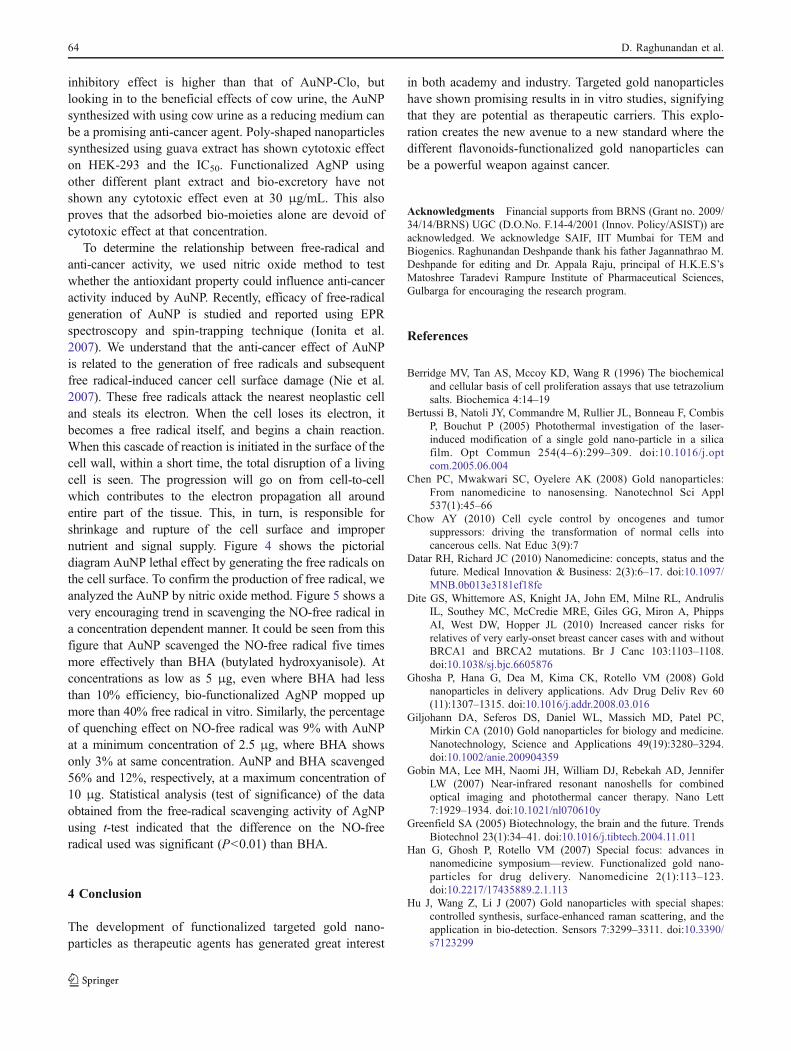
and offers only synergic effect in case of the functionalizedAuNP. The activity might be because of the size, shape, orother intrinsic properties of the gold nanoparticles. Manystudies prove that the gold nanoparticles of 30–50 nm rangeshow good anti-cancer effect. At this range, our researchshows that the irregular-shaped AuNP offers maximumanti-cancer effect on all the cell line compared to poly-shaped, nearly spherical, or spherical noble nanoparticles.The most possible reason that because of their unpredict-able shape, the moment in between the cells, and also thenon-specific adsorption to the cell surface is high comparedto particles of other shapes. Since these flavonoids areimportant in the internal stabilization of gold nanoparticles,a further detailed study is needed. This will provide a betterunderstanding of how this flavonoid-conjugated AuNPdisturbs and destroys the cell externally. Figure 3 showsthe pictorial representation of shape-dependent anti-cancereffect of gold nanoparticles. The irregularly shaped nano-
particles can move easily in between the abnormally highand irregularly spread neoplastic cells compared to poly-shaped or roughly spherical ones; it is reasonable to inferthat these special unpredictable amoebic shaped is havingbetter anti-proliferative effect.
As such, the clove extract itself has reported anti-malignant effect at higher concentration in different typesof cancer. The morphology of HeLa, HEK-293, and HT-29cell line that were treated with AuNP-Clo at 20 μg/mL was
Fig. 3 Shape-dependent anti-proliferative activity of AuNP
Fig. 2 TEM images of AuNPand AgNP synthesized usingaqueous extracts of guava leafand buds of the clove,respectively (reproduced withpermission from A) Elsevier, B)& C) Springer)
62 D. Raghunandan et al.

studied to prove the effect of these AuNP on the cellgrowth. Our observations showed that the growth of thecells was inhibited. However, further study is needed tounderstand the exact mechanism of anti-cancer activity ofthese nanoparticles. AuNP-Clo proved to possess anti-proliferative properties against all the cancer cell linestested. The prominent anti-proliferative effect of functional-ized AuNP on HEK-293, HeLa, and HT-29 cancer cell lines,as revealed by its IC50 based on XTT assay was found to be19.88±0.006, 20.05±0.002, 20.12±0.007, and 28.56±0.010,respectively. IC50 of AuNP-Clo was specifically lesssignificant on Vero cell line, i.e., 55.3±2.74. Therefore, itcan be said that AuNP-Clo is a promising anti-cancer “lead”.However, the exact mechanism behind the anti-proliferativeeffects of AuNP-Clo needs to be studied to determinewhether the effect is due to an increase in apoptosis. Theplasma membrane of the cancer cell defines the separationbetween the internal constituents of a cell and the outsideenvironment. This semi-permeable membrane allows freediffusion of small and non-polar molecules. However, biggerones like nanomaterials are incapable of crossing the plasmamembrane which requires uptake mechanisms such asendocytosis. Most of bio-functionalized AuNP are easilytaken up by the cells through endocytotic mechanisms, butthey remain in endosome vesicles, become incapable ofreaching the cytosol system. Although endocytotic uptake is
the normal phenomenon for a variety of nanomaterials, theefficiency predominantly depends on shape, size, thedispersivity, and the other physico-chemical parameters. Thisis important, because there may be many factors that affectthe anti-proliferative activity in the in vivo study. Furtherstudy is underway.
AuNP-Gua has shown activity against HEK-293 but theIC50>30 μg/mL. These are devoid of anti-proliferativeactivity against other three cell lines. AuNP developed withcow urine as a reducing agent has shown activity againstHeLa, HEK-293, and HT-29 at 30 μg/mL. Though the
Fig. 5 Graph showing free-radical scavenging activity of AuNP
Fig. 4 Probable anti-proliferativemechanism of AuNP
Anti-cancer studies of bio-functionalized noble metal nanoparticles 63
inhibitory effect is higher than that of AuNP-Clo, butlooking in to the beneficial effects of cow urine, the AuNPsynthesized with using cow urine as a reducing medium canbe a promising anti-cancer agent. Poly-shaped nanoparticlessynthesized using guava extract has shown cytotoxic effecton HEK-293 and the IC50. Functionalized AgNP usingother different plant extract and bio-excretory have notshown any cytotoxic effect even at 30 μg/mL. This alsoproves that the adsorbed bio-moieties alone are devoid ofcytotoxic effect at that concentration.
To determine the relationship between free-radical andanti-cancer activity, we used nitric oxide method to testwhether the antioxidant property could influence anti-canceractivity induced by AuNP. Recently, efficacy of free-radicalgeneration of AuNP is studied and reported using EPRspectroscopy and spin-trapping technique (Ionita et al.2007). We understand that the anti-cancer effect of AuNPis related to the generation of free radicals and subsequentfree radical-induced cancer cell surface damage (Nie et al.2007). These free radicals attack the nearest neoplastic celland steals its electron. When the cell loses its electron, itbecomes a free radical itself, and begins a chain reaction.When this cascade of reaction is initiated in the surface of thecell wall, within a short time, the total disruption of a livingcell is seen. The progression will go on from cell-to-cellwhich contributes to the electron propagation all aroundentire part of the tissue. This, in turn, is responsible forshrinkage and rupture of the cell surface and impropernutrient and signal supply. Figure 4 shows the pictorialdiagram AuNP lethal effect by generating the free radicals onthe cell surface. To confirm the production of free radical, weanalyzed the AuNP by nitric oxide method. Figure 5 shows avery encouraging trend in scavenging the NO-free radical ina concentration dependent manner. It could be seen from thisfigure that AuNP scavenged the NO-free radical five timesmore effectively than BHA (butylated hydroxyanisole). Atconcentrations as low as 5 μg, even where BHA had lessthan 10% efficiency, bio-functionalized AgNP mopped upmore than 40% free radical in vitro. Similarly, the percentageof quenching effect on NO-free radical was 9% with AuNPat a minimum concentration of 2.5 μg, where BHA showsonly 3% at same concentration. AuNP and BHA scavenged56% and 12%, respectively, at a maximum concentration of10 μg. Statistical analysis (test of significance) of the dataobtained from the free-radical scavenging activity of AgNPusing t-test indicated that the difference on the NO-freeradical used was significant (P<0.01) than BHA.
4 Conclusion
The development of functionalized targeted gold nano-particles as therapeutic agents has generated great interest
in both academy and industry. Targeted gold nanoparticleshave shown promising results in in vitro studies, signifyingthat they are potential as therapeutic carriers. This explo-ration creates the new avenue to a new standard where thedifferent flavonoids-functionalized gold nanoparticles canbe a powerful weapon against cancer.
Acknowledgments Financial supports from BRNS (Grant no. 2009/34/14/BRNS) UGC (D.O.No. F.14-4/2001 (Innov. Policy/ASIST)) areacknowledged. We acknowledge SAIF, IIT Mumbai for TEM andBiogenics. Raghunandan Deshpande thank his father Jagannathrao M.Deshpande for editing and Dr. Appala Raju, principal of H.K.E.S’sMatoshree Taradevi Rampure Institute of Pharmaceutical Sciences,Gulbarga for encouraging the research program.
References
Berridge MV, Tan AS, Mccoy KD, Wang R (1996) The biochemicaland cellular basis of cell proliferation assays that use tetrazoliumsalts. Biochemica 4:14–19
Bertussi B, Natoli JY, Commandre M, Rullier JL, Bonneau F, CombisP, Bouchut P (2005) Photothermal investigation of the laser-induced modification of a single gold nano-particle in a silicafilm. Opt Commun 254(4–6):299–309. doi:10.1016/j.optcom.2005.06.004
Chen PC, Mwakwari SC, Oyelere AK (2008) Gold nanoparticles:From nanomedicine to nanosensing. Nanotechnol Sci Appl537(1):45–66
Chow AY (2010) Cell cycle control by oncogenes and tumorsuppressors: driving the transformation of normal cells intocancerous cells. Nat Educ 3(9):7
Datar RH, Richard JC (2010) Nanomedicine: concepts, status and thefuture. Medical Innovation & Business: 2(3):6–17. doi:10.1097/MNB.0b013e3181ef18fe
Dite GS, Whittemore AS, Knight JA, John EM, Milne RL, AndrulisIL, Southey MC, McCredie MRE, Giles GG, Miron A, PhippsAI, West DW, Hopper JL (2010) Increased cancer risks forrelatives of very early-onset breast cancer cases with and withoutBRCA1 and BRCA2 mutations. Br J Canc 103:1103–1108.doi:10.1038/sj.bjc.6605876
Ghosha P, Hana G, Dea M, Kima CK, Rotello VM (2008) Goldnanoparticles in delivery applications. Adv Drug Deliv Rev 60(11):1307–1315. doi:10.1016/j.addr.2008.03.016
Giljohann DA, Seferos DS, Daniel WL, Massich MD, Patel PC,Mirkin CA (2010) Gold nanoparticles for biology and medicine.Nanotechnology, Science and Applications 49(19):3280–3294.doi:10.1002/anie.200904359
Gobin MA, Lee MH, Naomi JH, William DJ, Rebekah AD, JenniferLW (2007) Near-infrared resonant nanoshells for combinedoptical imaging and photothermal cancer therapy. Nano Lett7:1929–1934. doi:10.1021/nl070610y
Greenfield SA (2005) Biotechnology, the brain and the future. TrendsBiotechnol 23(1):34–41. doi:10.1016/j.tibtech.2004.11.011
Han G, Ghosh P, Rotello VM (2007) Special focus: advances innanomedicine symposium—review. Functionalized gold nano-particles for drug delivery. Nanomedicine 2(1):113–123.doi:10.2217/17435889.2.1.113
Hu J, Wang Z, Li J (2007) Gold nanoparticles with special shapes:controlled synthesis, surface-enhanced raman scattering, and theapplication in bio-detection. Sensors 7:3299–3311. doi:10.3390/s7123299
64 D. Raghunandan et al.

Ionita P, Conte M, Gilbert BC, Chechik V (2007) Gold nanoparticle-initiated free radical oxidations and halogen abstractions. OrgBiomol Chem 5:3504. doi:10.1039/B711573C
Katti KV et al (2006) Nanocompatible chemistry toward fabricationof target-specific gold nanoparticles. J Am Chem Soc128:11342–11343. doi:10.1021/ja063280c
Kawaii S, Tomono Y, Katase E, Ogawa K, Yano M (1999) Antiprolifer-ative activity of flavonoids on several cancer cell lines. BiosciBiotechnol Biochem 63(5):896–899. doi:10.1271/bbb.63.896
Kevin B (2008) Shape matters for nanoparticles, technology publishedby MIT review.
Kim C, Ghosh P, Rotello VM (2009) Multimodal drug delivery usinggold nanoparticles. Nanoscale 1:61–67. doi:10.1039/b9nr00112c
Lee S, Park H (2010) Anticancer activity of guava (Psidium guajavaL.) branch extracts against HT-29 human colon cancer cells.Journal of Medicinal Plants Research 4(10):891–896
Leviar N, Dewey RA, Daley E, Bates TE, Davies D, Kos J, PilkingtonGJ, Lah TT (2003) Selective suppression of cathepsin L byantisense cDNA impairs human brain tumor cell invasion in vitroand promotes apoptosis. Canc Gene Ther 10:141–151.doi:10.1038/sj.cgt.7700546
Liu Z, Kiessling F, Gatjens J (2010) Advanced nanomaterials inmultimodal imaging: design, functionalization, and biomed-ical applications. Journal of Nanomaterials 2010:894303.doi:10.1155/2010/894303
Maxwell DJ, Taylor JR, Nie S (2002) Self-assembled nanoparticleprobes for recognition and detection of biomolecules. Am ChemSoc 124(32):9606–9612. doi:10.1021/ja025814p
Nie Z, Liu KJ, Zhong C, Wang L, Yang Y, Tian Q, Liu Y (2007)Enhanced radical scavenging activity by antioxidant-functionalizedgold nanoparticles: a novel inspiration for development of newartificial antioxidants. Free Radic Biol Med 43:1243–1254.pmid:17893037
Orendorff CJ, Sau TK, Murphy C (2006) Shape-dependent plasmon-resonant gold nanoparticles. Small 2(5):636–639. doi:10.1002/smll.200500299
Parveen S, Sahoo SK (2010) Evaluation of cytotoxicity andmechanism of apoptosis of doxorubicin using folate-decoratedchitosan nanoparticles for targeted delivery to retinoblastoma.Cancer Nanotechnology. doi:10.1007/s12645-010-0006-0
Raghunandan D, Basavaraja S, Mahesh B, Balaji S, Manjunath SY,Venkataraman A (2009) Biosynthesis of stable polyshaped goldnanoparticles from microwave-exposed aqueous extracellularanti-malignant guava (Psidium guajava) leaf extract. Nano-Biotechnology 5(1–4):34–41. doi:10.1007/s12030-009-9030-8
Raghunandan D, Mahesh BD, Basavaraja S, Balaji SD, ManjunathSYA, Venkataraman A (2010a) Microwave-assisted rapid extra-cellular synthesis of stable bio-functionalized silver nanoparticlesfrom guava (Psidium guajava) leaf extract. Journal of Nano-particle Research. doi:10.1007/s11051-010-9956-8
Raghunandan D, Basavaraja S, Mahesh B, Balaji S, Manjunath SY,Venkataraman A (2010b) Rapid biosynthesis of irregularshaped gold nanoparticles from macerated aqueous extracel-lular dried clove buds (Syzygium aromaticum) solution.Colloids and Surfaces B: Biointerfaces 79:235–240.doi:10.1016/j.colsurfb.2010.04.003
Rinaldo P, Matteo G, Maila S (2006) Nanosystems. Inorganic and bio-inorganic chemistry. In: Bertini I (ed) Encyclopedia of LifeSupport Systems (EOLSS), developed under the auspices of theUNESCO. Eolss Publishers, Oxford, UK
Sacconi S, Simkin D, Arrighi N, Chapon F, Larroque MM, Vicart S,Sternberg D, Fontaine B, Barhanin J, Desnuelle C, Bendahhou S(2009) Mechanisms underlying Andersen's syndrome pathologyin skeletal muscle are revealed in human myotubes. Am J PhysiolCell Physiol 297:C876–C885. doi:10.1152/ajpcell.00519.2008
Sakamoto JH et al (2010) Enabling individualized therapy throughnanotechnology. Pharmacol Res 62(2):57–89. doi:10.1016/j.phrs.2009.12.011
Salmaso S, Bersani S, Scomparin A, Mastrotto F, Caliceti P (2010)Supramolecular bioconjugates for protein and small drugdelivery. Isr J Chem 50(2):160–174. doi:10.1002/ijch.201000022
Salucci M, Stivala LA, Maiani G, Bugianesi R, Vannini V (2002)Flavonoids uptake and their effect on cell cycle of human colonadenocarcinoma cells (Caco2). Br J Canc 86:1645–1651.doi:10.1038/sj.bjc.6600295
Seigneuric R, Markey L, Nuyten DSA, Dubernet C, Evelo CTA, FinotE, Garrido C (2010) From nanotechnology to nanomedicine:applications to cancer research. Curr Mol Med 10:640–652.doi:10.2174/156652410792630634
Smith RA, Cokkinides V, Brooks D, Saslow D, Brawley OW (2011)Cancer screening in the United States, 2011: a review of currentAmerican Cancer Society guidelines and issues in cancerscreening. CA Cancer J Clin 60:99–119. doi:10.3322/caac.20096
Sperling RA, Gil PR, Zhang F, Zanella M, Parak WJ (2008)Biological applications of gold nanoparticles. Chem Soc Rev37:189–1908. doi:10.1039/B712170A
Suriamoorthy P, Zhang X, Hao G, Joly AG, Singh S, Hossu M, SunX, Chen W (2010) Folic acid-CdTe quantum dot conjugates andtheir applications for cancer cell targeting. Cancer Nanotechnology.doi:10.1007/s12645-010-0003-3
Anti-cancer studies of bio-functionalized noble metal nanoparticles 65