Embed Size (px)
Citation preview

3/16/2018
1
Anterior Segment OCTWhat does it have to offer?
Engy M Mostafa MD, PHD
Assistant Professor of Ophthalmology
Sohag University
EOS 2018
No financial interest to disclose
3/16/2018
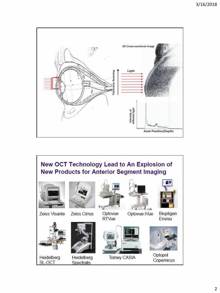
2

3/16/2018
3
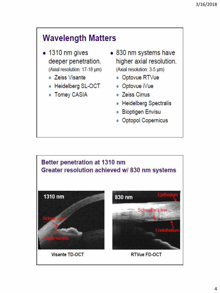
3/16/2018
4

3/16/2018
5

3/16/2018
6
UBMSL-OCTAS-OCTScheimflugTopography
USOpticalOpticalOpticalImage Source
SupineSittingSittingSittingPosition
YesNoNoNoContact
HighMediumLowLowOperator Skill

3/16/2018
7
UBMSL-OCTAS-OCTScheimpflugTpography
Features
NoNoNoYesTopography
NoNoNoYesIOP correction
NoNoNoYesLens densimetry
NoNoNoYesWavefrontanalysis
Imaging capabilitiesUBMSL-OCTAS-OCTScheimpflug
topography
25m<25m18mN/AOptical axial resolution
YesYesYesYesPachymetry
YesYesYesNoAngle Visualization
YesYesYesYesAngle estimation
YesNoNoNoCiliry sulcusvisible
YesYesYesNoOpaquemedia

3/16/2018
8
Why do we need AS-OCT?

3/16/2018
9
Quantitative
Qualitative QuantitativeQualitativeDiagnosticProperative
Postoperative
Image through an opaque cornea
Cannot Visualize deeply pigmented lesions.The posterior boundary of heavily-pigmentedlesions greater then 2.5 mm becomes blurred.

3/16/2018
10
Why Do I Image the Cornea?Deeper look
Cornea Layers
Air-tear interface Tear film EpitheliumEndotheliumStroma

3/16/2018
11
Measure Corneal Thickness
OCT measurements of Central corneal
thickness have shown good correlation with
ultrasound pachymetry.

3/16/2018
12
A thickness of 104 µm which the ultrasound
pachymeter did not measure, as it was below
its lower measurement limit.
Reis-Bucklers Dystrophy

3/16/2018
13
Granular Dystrophy
Is it Terrien’s marginal degeneration???
The absence of peripheral thinning typical of
this entity, and a diagnosis of primary lipid
degeneration

3/16/2018
14
Peter’s Anomaly

3/16/2018
15

3/16/2018
16
Pellucid Mardinal Degeneration
3/16/2018
17
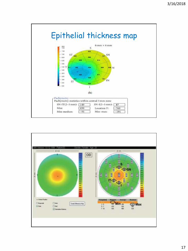
Epithelial thickness map

3/16/2018
18
(1) I-S (the difference between the average
thickness of the inferior octant and that of
the superior octant) >31 μm
(2) IT-SN (the difference between the
inferotemporal octant and the superonasal
octant) >48 μm
(3) Minimum <492 μm
(4) Minimum-maximum <−63 μm
(5) The thinnest region of the cornea is located
outside the central 2 mm area.
LASIK Flap Thickness

3/16/2018
19
3/16/2018
20
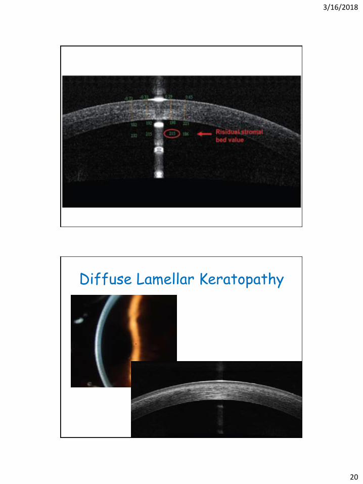
Diffuse Lamellar Keratopathy
3/16/2018
21
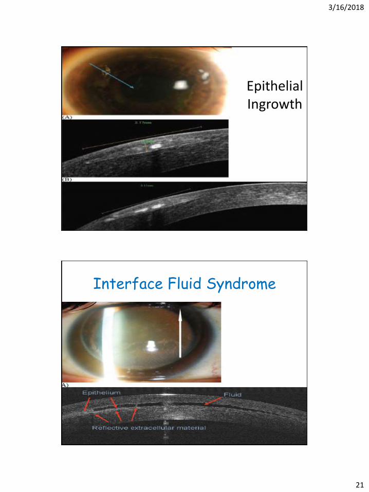
Epithelial Ingrowth
Interface Fluid Syndrome

3/16/2018
22
3/16/2018
23

3/16/2018
24
Intracorneal Rings
After Corneal Crosslinking
3/16/2018
25
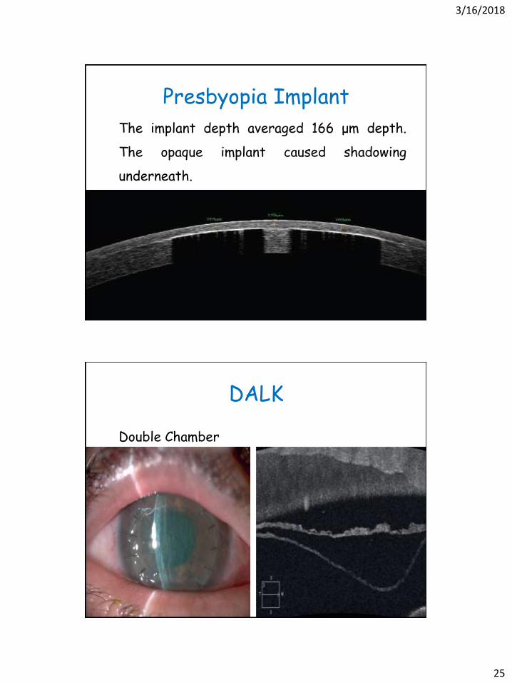
Presbyopia Implant
The implant depth averaged 166 μm depth.
The opaque implant caused shadowing
underneath.
DALK
Double Chamber

3/16/2018
26

3/16/2018
27

3/16/2018
28

3/16/2018
29
Corneal Trauma
3/16/2018
30
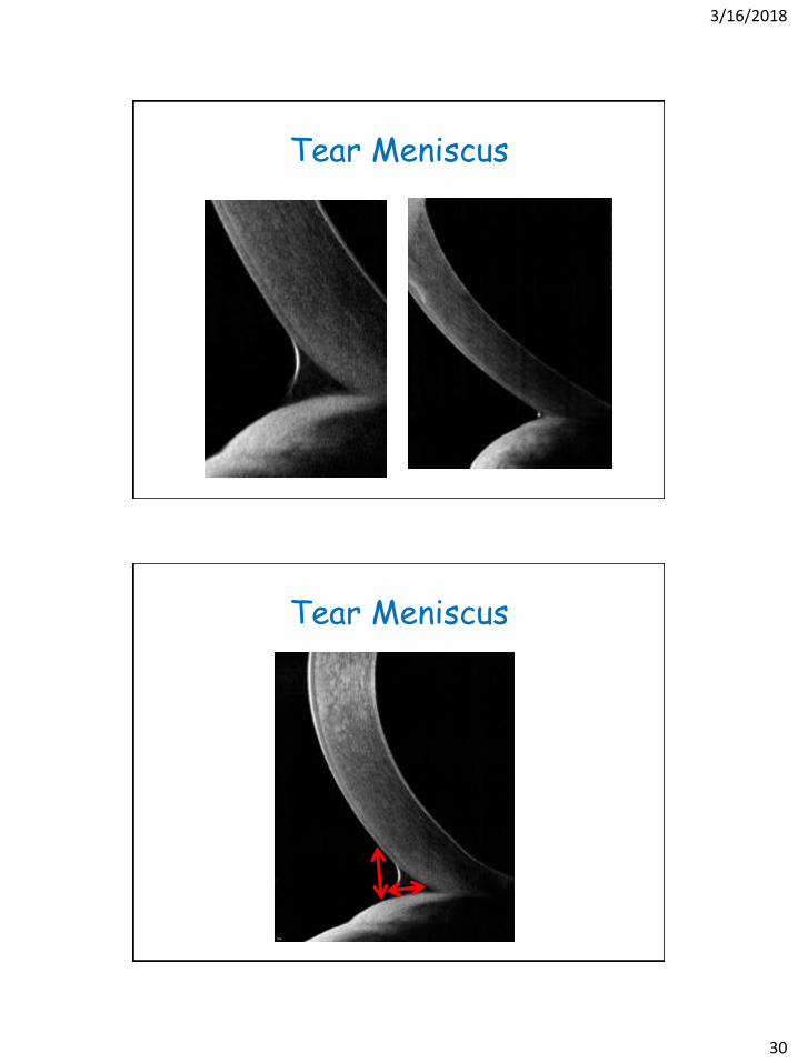
Tear Meniscus
Tear Meniscus

3/16/2018
31
Anterior Chamber Depth
Why measure it?
• Phakic IOL implantation.
• Detect occludable angles
• Important for IOL calculation as in Theoretical
prediction formula: Haigis
• 0.05 mm ACD error = 0.03 diopter IOL power
error
Phakic IOL
Before phakic IOL surgery it is mandatory
1. AC depth : endothelium safety
2. Crytalline lens rise & IOL vault :to respect
the lens

3/16/2018
32
Crystalline Lens Rise
A CLR of a 600 or more is a contraindication
for doing a Phakic implantation

3/16/2018
33
Phakic IOL
Good clearance of the sulcus-based over the
natural crystalline lens.
3/16/2018
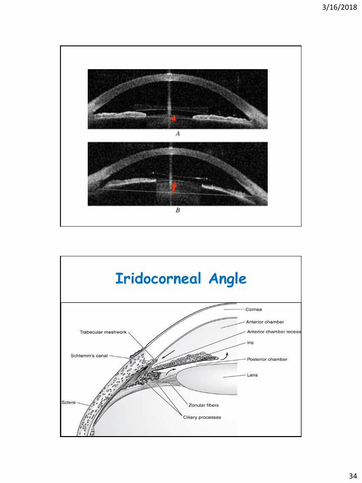
34
Iridocorneal Angle
3/16/2018
35
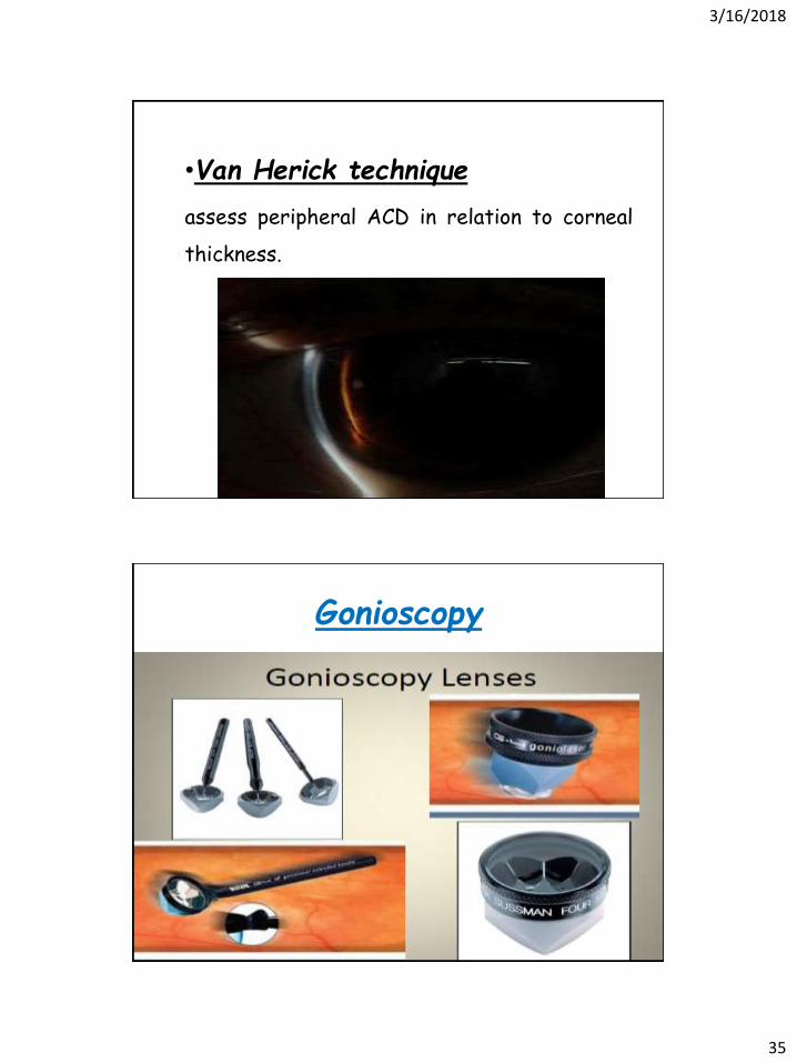
•Van Herick technique
assess peripheral ACD in relation to corneal
thickness.
Gonioscopy

3/16/2018
36
Grading of the angle is subjective.
Gonioscopy has low sensitivity (68.3%)
and high specificity (96.6%).

3/16/2018
37

3/16/2018
38
Accurate localization of the scleral spur
(reference point) for all the other
quantitative measurements.
3/16/2018
39
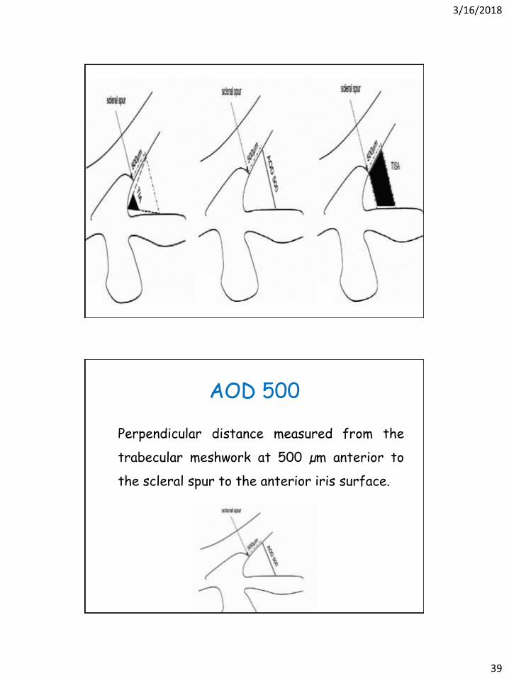
AOD 500
Perpendicular distance measured from the
trabecular meshwork at 500 µm anterior to
the scleral spur to the anterior iris surface.

3/16/2018
40
TISA 500
Anterior: AOD 500
Posterior: line drawn from
the scleral spur
perpendicular to the plane of
the inner scleral wall to the
opposing iris
Superior: inner corneoscleral
wall
Inferior: by the iris surface.
TIA 500
An angle measured with the
apex in the iris recess and
the arms of the angle
passing through a point on
the trabecular meshwork
500 µm from the scleral
spur and the point on the
iris perpendicularly

3/16/2018
41

3/16/2018
42
Need multiple scans all around limbus for full
analysis
Angle measurement is influenced by patient’s
age and gender, direction of gaze,
accommodation, room illumination, meridians
scanned.

3/16/2018
43
Light affects angle configuration

3/16/2018
44
Complete Angle Closure Glaucoma
Angle closure glaucoma
cornea is noticeably edematous

3/16/2018
45
Pigmentary Glaucoma
3/16/2018
46
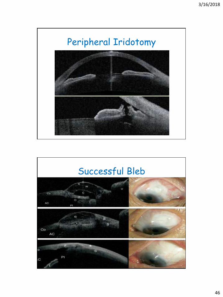
Peripheral Iridotomy
Successful Bleb

3/16/2018
47
Failed Bleb
Cystic Bleb
Singh et al. Imaging of Trabeculectomy Blebs Using Anterior Segment Optical Coherence
TomographyOphthalmology 2007;114:47–53

3/16/2018
48

3/16/2018
49
A cyclodialysis cleft.Disinsertion of the ciliary body from the scleralspur and an associated ciliary body detachment.

3/16/2018
50
Posterior polar cataract demonstrating an
intact posterior capsule .

3/16/2018
51
Pre and post Cataract Extraction

3/16/2018
52
Tilted posterior chamber intraocular lens pushing iris forward

3/16/2018
53

3/16/2018
54
Corneal-conjunctivalintraepithelial squamous
neoplasia

3/16/2018
55
Pterygium with
corneal opacity
beneath
Pterygium with
clear cornea

3/16/2018
56
Pseudopterygium Pinguecula
Conjunctival nevus
cysts and the well-defined posterior limit of
the lesion which are signs of its benign nature

3/16/2018
57
Iris Cyst
the cyst is anterior to the pigment epithelium
of the iris, it can be visualized.

3/16/2018
58
Iridoschesis
Accomodationchanges ACD depth by 30 µm for every
1 D

3/16/2018
59
What is real and what is artifact?
• Corneal Reflex
• Inverted Image (in Spectral Domain)
• Shadowing
• Image Averaging
• Algorithm Failure
–Pachymetry: Corneal surface lines
–Pachymetry: Lids

3/16/2018
60

3/16/2018
61

3/16/2018
62
Averaging

3/16/2018
63
Thank You
Seeing is Believing