Embed Size (px)
Citation preview

Anorectal Abscessand Fistula-in-Ano:Evidence-BasedManagement
Julie A. Rizzo, MDa, Anna L. Naig, MDa,Eric K. Johnson, MD, FACS, FASCRSb,c,*
KEYWORDS
� Anorectal abscess � Anal fistula � Incontinence
Anorectal abscess, and the fistula that may result, are long established processes thatwere originally described at the beginning of recorded medical history as part of the‘‘Corpus Hippocraticum’’ in a treatise termed ‘‘On Fistulae.’’1 The basic principlesregarding the treatment of this disease have remained the same: resolution of perianalsepsis, and treatment of the resulting fistula without leading to impairment incontinence.This second principle remains a challenge, and there are continued efforts to achieve anoptimal form of therapy. A recent surge of interest in this disease process occurred afterthe release of the collagen anal fistula plug. Hopefully this will lead to an improvement inthe care of patients with this inconvenient and embarrassing condition.
PATHOPHYSIOLOGY
Anorectal abscess occurs commonly in normal, healthy individuals. The most widelyrecognized cause is described in the cryptoglandular theory, which suggests thatan anal crypt gland becomes obstructed with inspissated debris and leads to infec-tion. These glands penetrate the anal sphincter complex to varying degrees, andthe suppuration tends to follow the path of least resistance. The abscess collects inwhichever anatomic space the gland terminates, or wherever the path of least resis-tance leads. A basic understanding of anorectal anatomy and the perianal and perirec-tal spaces is critical for grasping this concept.
a Department of Surgery, Dwight David Eisenhower Army Medical Center, 300 Hospital Road,Fort Gordon, GA, USAb Colorectal Surgery and Surgical Endoscopy, Dwight David Eisenhower Army Medical Center,300 Hospital Road, Fort Gordon, GA, USAc Uniform Services University of Health Sciences, F. Edward Hebert School of Medicine,Bethesda, MD, USA* Corresponding author. Colorectal Surgery and Surgical Endoscopy, Dwight David EisenhowerArmy Medical Center, 300 Hospital Road, Fort Gordon, GA.E-mail address: [email protected] (E.K. Johnson).
Surg Clin N Am 90 (2010) 45–68doi:10.1016/j.suc.2009.10.001 surgical.theclinics.com0039-6109/09/$ – see front matter. Published by Elsevier Inc.

Rizzo et al46
Anal fistulas develop in approximately one-third of patients who undergo drainageof an anorectal abscess. In a series2 of 170 patients without previous fistulas whowere followed for an average of 99 months after abscess drainage, a fistula occurredin 37% and recurrent abscess was reported in an additional 10%. A retrospectivecohort study3 of 148 patients with anorectal abscesses showed a 37% rate of fistulaformation. Patients younger than 40 years and nondiabetic patients had a higher likeli-hood of developing a fistula-in-ano over the mean follow-up of 38 months. Any recur-rent abscess that occurs at the same site as a previous abscess can be considereda fistula and treated as such. There are other notable causes of atypical/complicatedabscess and fistula, including inflammatory bowel disease, fungal infection, mycobac-terial infection, neoplasm, and trauma. Fistulas that are secondary to these processesare classified as complex and require the use of nonstandard methods ofmanagement.
CLASSIFICATION
Anorectal abscesses are classified based on their location. Four types of anorectalabscesses are commonly described: perianal (superficial), ischiorectal (perirectal), in-tersphincteric, and supralevator. Perianal is the most common type and is the simplestto treat. The collections are located in the superficial perianal tissues and are typicallylocated close to the anal verge. Ischiorectal abscesses are located more deeply in theischiorectal fossa and may communicate to the contralateral side via the deep posta-nal space; this would be a classic example of a horseshoe abscess. Intersphinctericabscesses are often difficult to diagnose as they may reside completely within theanal canal. They are located in the intersphincteric space between the internal andexternal sphincter muscles. Patients affected by this process complain of severeanal pain and often cannot tolerate an examination without anesthesia. The fluctuantcollection may be found only by performing a digital rectal examination or anoscopy.Supralevator abscesses are rare and are typically diagnosed through computed tomo-graphic scanning. A patient presenting with this condition might complain of pelvicand rectal pain with tenesmus. The abscess can sometimes be palpated througha digital rectal examination performed by an experienced examiner. These abscessesare often related to perforated diverticular disease, inflammatory bowel disease, orrarely neoplastic disease in the pelvis. Sometimes an abscess occurs in the suprale-vator location because cryptoglandular suppuration followed the path of least resis-tance. Simple internal drainage often ameliorates this problem. The management ofthe processes already mentioned outside drainage is complex and is beyond thescope of this discussion. A study of more than 1000 patients who presented with ano-rectal abscess revealed that perianal abscess occurred in 42.7%, ischiorectal in22.7%, intersphincteric in 21.4%, and supralevator in 7.33%.4
A question that often arises is whether or not to treat a fistula that is noted duringa procedure performed to drain perianal sepsis. A randomized clinical trial comparingsimple drainage alone to drainage plus fistula tract treatment was published in 2002.5
The investigators randomized 200 patients to one of the two treatment arms,excluding any patient who had incontinence or a history of inflammatory boweldisease. Internal openings were found in 83% of the patients and they were treatedwith simple fistulotomy or seton fistulotomy if they had been randomized to thetract-treatment arm. Recurrence was noted in 29% of the group who receiveddrainage only compared with 5% of the group who received tract treatment. In lowfistulas treated by fistulotomy, incontinence was seen in only 2.8%. Patients whohad high fistulas that were managed by seton (delayed) fistulotomy developed

Anorectal Abscess and Fistula 47
incontinence 37% of the time. This result illustrates a major concern in the treatment ofhigh fistula tracts. A later meta-analysis addressing this same issue evaluated 5 trialscontaining 405 patients and found an 83% reduction in recurrence in those who hadtheir fistula tracts addressed at the initial procedure with no significant increase in therate of postoperative incontinence.6
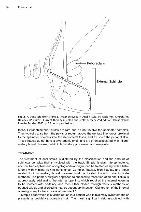
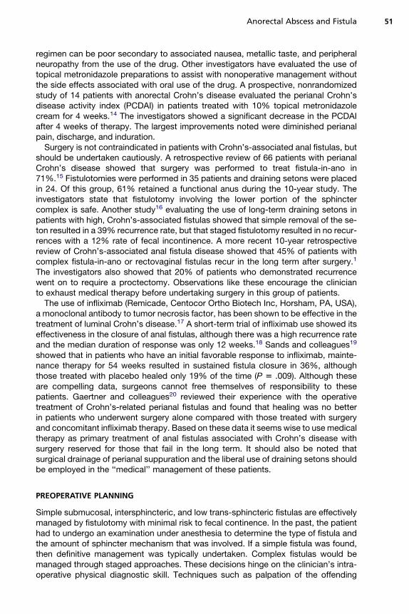
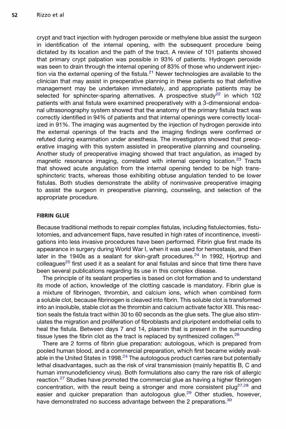
Fistulas are classified based on their relation to the anal sphincter complex. They aretypically divided into 5 common classifications: submucosal, intersphincteric (Fig. 1),trans-sphincteric (divided into high and low) (Fig. 2), suprasphincteric (Fig. 3), and ex-trasphincteric (Fig. 4). Trans-sphincteric fistulas cross through the internal andexternal sphincter muscles to varying degrees. Low fistulas involve only the outer ordistal one-third of the external sphincter muscle, whereas high fistulas involve greaterdegrees of the external sphincter. This characteristic is clinically significant becausedivision of greater amounts of the external sphincter leads to higher rates of fecalincontinence. Intersphincteric fistulas cross through the internal sphincter and exitthrough the intersphincteric plane. They do not involve the external sphincter muscleand can therefore be opened without high risk of incontinence. Submucosal fistulastypically originate at an offending crypt at the level of the dentate line, but track onlyjust beneath the submucosa and do not involve the sphincter complex. These fistulasmay be opened without fear of compromising continence. Suprasphincteric fistulastypically originate at the dentate line internally, cross above the external sphincterbut below the puborectalis, and exit onto the perianal skin through the ischiorectal
Levator
Puborectalis
External Sphincter
internal Sphincter
Dental Line
Fig. 1. An intersphincteric fistula. (From Belliveau P. Anal fistula. In: Fazio VM, Church JM,Delaney CP, editors. Current therapy in colon and rectal surgery. 2nd edition. Philadelphia:Elsevier Mosby; 2005. p. 28; with permission.)
Puborectalis
External Sphincter
Fig. 2. A trans-sphincteric fistula. (From Belliveau P. Anal fistula. In: Fazio VM, Church JM,Delaney CP, editors. Current therapy in colon and rectal surgery. 2nd edition. Philadelphia:Elsevier Mosby; 2005. p. 28; with permission.)
Rizzo et al48
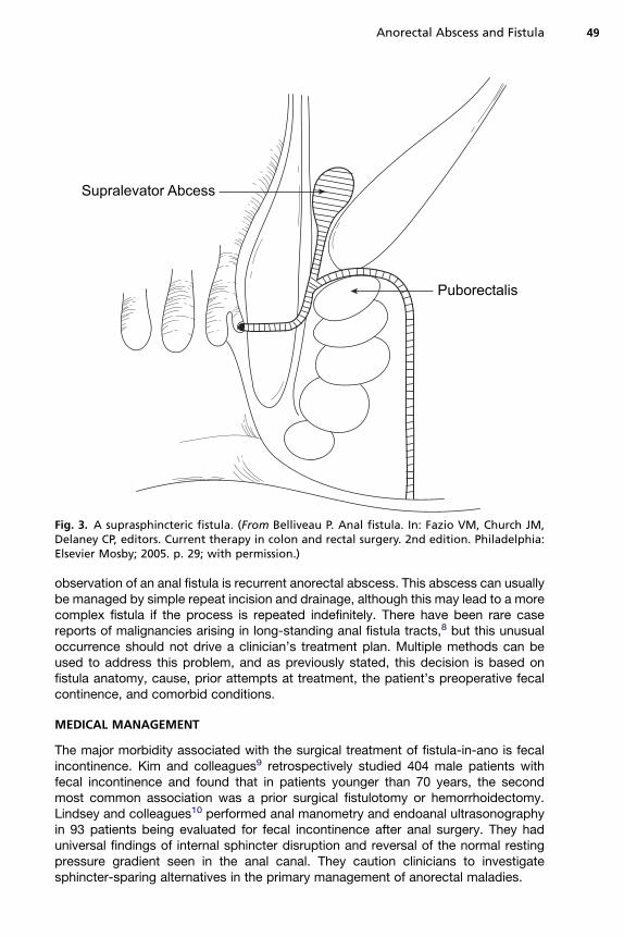
fossa. Extrasphincteric fistulas are rare and do not involve the sphincter complex.They typically arise from the pelvis or rectum above the dentate line, cross proximalto the sphincter complex into the ischiorectal fossa, and exit onto the perianal skin.These fistulas do not have a cryptogenic origin and are often associated with inflam-matory bowel disease, pelvic inflammatory processes, and neoplasia.
TREATMENT
The treatment of anal fistula is dictated by the classification and the amount ofsphincter complex that is involved with the tract. Simple fistulas, intersphincteric,and low trans-sphincteric of cryptoglandular origin, can be treated easily with a fistu-lotomy with minimal risk to continence. Complex fistulas, high fistulas, and thoserelated to inflammatory bowel disease must be treated through more intricatemethods. The primary surgical approach to successful resolution of an anal fistula isappropriately addressing the internal opening, which requires the internal openingto be located with certainty, and then either closed through various methods oropened widely and allowed to heal by secondary intention. Obliteration of the internalopening is key to the success of treatment.7
Simple observation is a viable option in a patient who is minimally symptomatic orpresents a prohibitive operative risk. The most significant risk associated with
Supralevator Abcess
Puborectalis
Fig. 3. A suprasphincteric fistula. (From Belliveau P. Anal fistula. In: Fazio VM, Church JM,Delaney CP, editors. Current therapy in colon and rectal surgery. 2nd edition. Philadelphia:Elsevier Mosby; 2005. p. 29; with permission.)
Anorectal Abscess and Fistula 49
observation of an anal fistula is recurrent anorectal abscess. This abscess can usuallybe managed by simple repeat incision and drainage, although this may lead to a morecomplex fistula if the process is repeated indefinitely. There have been rare casereports of malignancies arising in long-standing anal fistula tracts,8 but this unusualoccurrence should not drive a clinician’s treatment plan. Multiple methods can beused to address this problem, and as previously stated, this decision is based onfistula anatomy, cause, prior attempts at treatment, the patient’s preoperative fecalcontinence, and comorbid conditions.
MEDICAL MANAGEMENT
The major morbidity associated with the surgical treatment of fistula-in-ano is fecalincontinence. Kim and colleagues9 retrospectively studied 404 male patients withfecal incontinence and found that in patients younger than 70 years, the secondmost common association was a prior surgical fistulotomy or hemorrhoidectomy.Lindsey and colleagues10 performed anal manometry and endoanal ultrasonographyin 93 patients being evaluated for fecal incontinence after anal surgery. They haduniversal findings of internal sphincter disruption and reversal of the normal restingpressure gradient seen in the anal canal. They caution clinicians to investigatesphincter-sparing alternatives in the primary management of anorectal maladies.
Pelvic Abcess
Levator
Fig. 4. An extrasphincteric fistula. (From Belliveau P. Anal fistula. In: Fazio VM, Church JM,Delaney CP, editors. Current therapy in colon and rectal surgery. 2nd edition. Philadelphia:Elsevier Mosby; 2005. p. 29; with permission.)
Rizzo et al50
Medical management of fistula-in-ano is most often associated with patientssuffering from inflammatory bowel disease. As mentioned previously, there is littleharm in observing a fistula that is minimally symptomatic. After first draining the perianalsuppuration, simple observation and medical treatment of associated Crohn’s diseaseled to healing in 8 of 15 patients followed for 10 years.11 Halme and Sainio12 noteda similar healing rate of 50% in patients with Crohn’s disease with anal fistulas whowere only observed. Oral metronidazole is a useful agent in the medical managementof fistula-in-ano in the population with Crohn’s disease. A dosage of 20 mg/kg/ddivided into 3 or 4 doses has been shown to eliminate drainage, erythema, and indura-tion in as many as 80% of patients treated within 8 weeks.13 Compliance with this
Anorectal Abscess and Fistula 51
regimen can be poor secondary to associated nausea, metallic taste, and peripheralneuropathy from the use of the drug. Other investigators have evaluated the use oftopical metronidazole preparations to assist with nonoperative management withoutthe side effects associated with oral use of the drug. A prospective, nonrandomizedstudy of 14 patients with anorectal Crohn’s disease evaluated the perianal Crohn’sdisease activity index (PCDAI) in patients treated with 10% topical metronidazolecream for 4 weeks.14 The investigators showed a significant decrease in the PCDAIafter 4 weeks of therapy. The largest improvements noted were diminished perianalpain, discharge, and induration.
Surgery is not contraindicated in patients with Crohn’s-associated anal fistulas, butshould be undertaken cautiously. A retrospective review of 66 patients with perianalCrohn’s disease showed that surgery was performed to treat fistula-in-ano in71%.15 Fistulotomies were performed in 35 patients and draining setons were placedin 24. Of this group, 61% retained a functional anus during the 10-year study. Theinvestigators state that fistulotomy involving the lower portion of the sphinctercomplex is safe. Another study16 evaluating the use of long-term draining setons inpatients with high, Crohn’s-associated fistulas showed that simple removal of the se-ton resulted in a 39% recurrence rate, but that staged fistulotomy resulted in no recur-rences with a 12% rate of fecal incontinence. A more recent 10-year retrospectivereview of Crohn’s-associated anal fistula disease showed that 45% of patients withcomplex fistula-in-ano or rectovaginal fistulas recur in the long term after surgery.1
The investigators also showed that 20% of patients who demonstrated recurrencewent on to require a proctectomy. Observations like these encourage the clinicianto exhaust medical therapy before undertaking surgery in this group of patients.
The use of infliximab (Remicade, Centocor Ortho Biotech Inc, Horsham, PA, USA),a monoclonal antibody to tumor necrosis factor, has been shown to be effective in thetreatment of luminal Crohn’s disease.17 A short-term trial of infliximab use showed itseffectiveness in the closure of anal fistulas, although there was a high recurrence rateand the median duration of response was only 12 weeks.18 Sands and colleagues19
showed that in patients who have an initial favorable response to infliximab, mainte-nance therapy for 54 weeks resulted in sustained fistula closure in 36%, althoughthose treated with placebo healed only 19% of the time (P 5 .009). Although theseare compelling data, surgeons cannot free themselves of responsibility to thesepatients. Gaertner and colleagues20 reviewed their experience with the operativetreatment of Crohn’s-related perianal fistulas and found that healing was no betterin patients who underwent surgery alone compared with those treated with surgeryand concomitant infliximab therapy. Based on these data it seems wise to use medicaltherapy as primary treatment of anal fistulas associated with Crohn’s disease withsurgery reserved for those that fail in the long term. It should also be noted thatsurgical drainage of perianal suppuration and the liberal use of draining setons shouldbe employed in the ‘‘medical’’ management of these patients.
PREOPERATIVE PLANNING
Simple submucosal, intersphincteric, and low trans-sphincteric fistulas are effectivelymanaged by fistulotomy with minimal risk to fecal continence. In the past, the patienthad to undergo an examination under anesthesia to determine the type of fistula andthe amount of sphincter mechanism that was involved. If a simple fistula was found,then definitive management was typically undertaken. Complex fistulas would bemanaged through staged approaches. These decisions hinge on the clinician’s intra-operative physical diagnostic skill. Techniques such as palpation of the offending
Rizzo et al52
crypt and tract injection with hydrogen peroxide or methylene blue assist the surgeonin identification of the internal opening, with the subsequent procedure beingdictated by its location and the path of the tract. A review of 101 patients showedthat primary crypt palpation was possible in 93% of patients. Hydrogen peroxidewas seen to drain through the internal opening of 83% of those who underwent injec-tion via the external opening of the fistula.21 Newer technologies are available to theclinician that may assist in preoperative planning in these patients so that definitivemanagement may be undertaken immediately, and appropriate patients may beselected for sphincter-sparing alternatives. A prospective study22 in which 102patients with anal fistula were examined preoperatively with a 3-dimensional endoa-nal ultrasonography system showed that the anatomy of the primary fistula tract wascorrectly identified in 94% of patients and that internal openings were correctly local-ized in 91%. The imaging was augmented by the injection of hydrogen peroxide intothe external openings of the tracts and the imaging findings were confirmed orrefuted during examination under anesthesia. The investigators showed that preop-erative imaging with this system assisted in preoperative planning and counseling.Another study of preoperative imaging showed that tract angulation, as imaged bymagnetic resonance imaging, correlated with internal opening location.23 Tractsthat showed acute angulation from the internal opening tended to be high trans-sphincteric tracts, whereas those exhibiting obtuse angulation tended to be lowerfistulas. Both studies demonstrate the ability of noninvasive preoperative imagingto assist the surgeon in preoperative planning, counseling, and selection of theappropriate procedure.
FIBRIN GLUE
Because traditional methods to repair complex fistulas, including fistulectomies, fistu-lotomies, and advancement flaps, have resulted in high rates of incontinence, investi-gations into less invasive procedures have been performed. Fibrin glue first made itsappearance in surgery during World War I, when it was used for hemostasis, and thenlater in the 1940s as a sealant for skin-graft procedures.24 In 1992, Hjortrup andcolleagues25 first used it as a sealant for anal fistulas and since that time there havebeen several publications regarding its use in this complex disease.
The principle of its sealant properties is based on clot formation and to understandits mode of action, knowledge of the clotting cascade is mandatory. Fibrin glue isa mixture of fibrinogen, thrombin, and calcium ions, which when combined forma soluble clot, because fibrinogen is cleaved into fibrin. This soluble clot is transformedinto an insoluble, stable clot as the thrombin and calcium activate factor XIII. This reac-tion seals the fistula tract within 30 to 60 seconds as the glue sets. The glue also stim-ulates the migration and proliferation of fibroblasts and pluripotent endothelial cells toheal the fistula. Between days 7 and 14, plasmin that is present in the surroundingtissue lyses the fibrin clot as the tract is replaced by synthesized collagen.26
There are 2 forms of fibrin glue preparation: autologous, which is prepared frompooled human blood, and a commercial preparation, which first became widely avail-able in the United States in 1998.24 The autologous product carries rare but potentiallylethal disadvantages, such as the risk of viral transmission (mainly hepatitis B, C andhuman immunodeficiency virus). Both formulations also carry the rare risk of allergicreaction.27 Studies have promoted the commercial glue as having a higher fibrinogenconcentration, with the result being a stronger and more consistent plug27,28 andeasier and quicker preparation than autologous glue.29 Other studies, however,have demonstrated no success advantage between the 2 preparations.30
Anorectal Abscess and Fistula 53
The procedure is promoted as simple and repeatable with no significant learningcurves, in direct contrast to other procedures, such as the advancement flap. Bothopenings of the tract are identified and the tract is mechanically curetted and irrigatedwith normal saline or hydrogen peroxide. The double-barreled syringe, containing the2 components of the glue, is inserted through the external opening until the tip is seenat the internal opening. The syringe is depressed, which mixes the 2 components asthey are injected into the canal. The tract is filled completely until a blob of glue is seenat the external opening. The glue is allowed to set for 30 to 60 seconds to form itsstable clot. Different investigators have advocated suturing the internal or externalopening around the bead of glue,31 although others have demonstrated no statisticallysignificant benefit.27,29 Postoperatively, the use of antibiotics and diet restrictions wasvariable, but sitz baths, excessive straining, or vigorous exercise were prohibiteduniformly to prevent dislodgement of the plug.32 Preoperative bowel preparations,the use of perioperative antibiotics, and the use of setons is widely variable withnone shown to confer a benefit.27,33
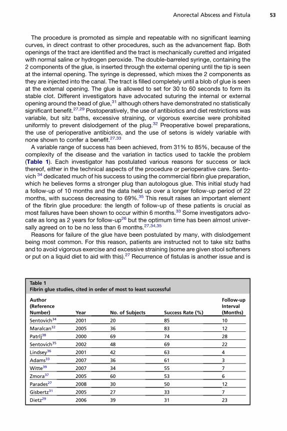
A variable range of success has been achieved, from 31% to 85%, because of thecomplexity of the disease and the variation in tactics used to tackle the problem(Table 1). Each investigator has postulated various reasons for success or lackthereof, either in the technical aspects of the procedure or perioperative care. Sento-vich 34 dedicated much of his success to using the commercial fibrin glue preparation,which he believes forms a stronger plug than autologous glue. This initial study hada follow-up of 10 months and the data held up over a longer follow-up period of 22months, with success decreasing to 69%.35 This result raises an important elementof the fibrin glue procedure: the length of follow-up of these patients is crucial asmost failures have been shown to occur within 6 months.33 Some investigators advo-cate as long as 2 years for follow-up26 but the optimum time has been almost univer-sally agreed on to be no less than 6 months.27,34,35
Reasons for failure of the glue have been postulated by many, with dislodgementbeing most common. For this reason, patients are instructed not to take sitz bathsand to avoid vigorous exercise and excessive straining (some are given stool softenersor put on a liquid diet to aid with this).27 Recurrence of fistulas is another issue and is
Table 1Fibrin glue studies, cited in order of most to least successful
Author(ReferenceNumber) Year No. of Subjects Success Rate (%)
Follow-upInterval(Months)
Sentovich34 2001 20 85 10
Maralcan32 2005 36 83 12
Patrlj38 2000 69 74 28
Sentovich35 2002 48 69 22
Lindsey36 2001 42 63 4
Adams33 2007 36 61 3
Witte39 2007 34 55 7
Zmora37 2005 60 53 6
Parades27 2008 30 50 12
Gisbertz31 2005 27 33 7
Dietz29 2006 39 31 23
Rizzo et al54
believed to be caused by inadequate removal of granulation tissue during tract prep-aration with mechanical curetting and irrigation26 and the natural course of fistuladisease. Another cause of failure is abscess formation, which has been quoted ashigh as 5% in some studies.33 This is believed to be caused by a lack of complete tractfilling with glue, representing a technical error,36 or a lack of proper tract cleansingbefore glue instillation.35,36 Some investigators have sought to prevent this by makinga glue/antibiotic mixture,37 whereas others have irrigated the tract before glue instilla-tion with an antibiotic irrigant.38
A controversy continues to exist regarding the length of the tract and its impact onglue success. Patrlj and colleagues38 and Lindsey and colleagues36 demonstratedgreater success with longer tracts, attributing this to the ability of glue to leak fromshorter tracts (<3.5 cm) more easily. On the contrary, Sentovich,34,35 Cintron andcolleagues30 and Maralcan and colleagues32 had greater success with shorter tracts.To date, there is no consensus on the tract length most amenable to success with glueand therefore no patient should be excluded by tract length.
Two main advantages of glue that should be remembered are that no patient experi-enced a decrease in level of continence from the procedure,29 and that treatment withfibrin glue does not preclude the patient from receiving other treatments, such as repeatfibrin glue instillation, or conventional fistula treatments.39 Despite its varying success todate, fibrin glue offers the patient a less invasive option for first-line fistula treatment.
FISTULA PLUG
Fibrin glue studies failed to achieve results that were reproducible, but did showpromise in muscle-sparing, noninvasive operative techniques for anal fistulas. Thisresult led to the development of additional sphincter-sparing therapies. The conceptof a plug was first introduced in 2006 by Robb and colleagues40 and Johnson andcolleagues41 with the idea that securing the plug into the primary opening of a fistulatract could close the tract more reliably than previous procedures, without compro-mising continence because the sphincters were not incised or divided. The biologicplug (Surgisis Anal Fistula Plug, Cook Surgical, Bloomington, IN) is made of lyophilizedporcine small intestinal submucosa, which has an inherent resistance of infection,generates no foreign body or giant cell reaction, and is repopulated by host cell tissuewithin 3 months.42 Its conical shape allows for added mechanical stability as highpressures within the anal canal maintain the plug in its proper position, avoidingdislodgement during straining.
Regarding the procedure, the critical points for correct plug insertion are as follows:the plug must be rehydrated first, usually in a 0.9% normal saline solution for 3 to 5minutes, before insertion; it must be inserted in the internal (also known as primary)opening and then pulled through the tract until light resistance is met; and it mustbe sutured securely in the primary opening. Various suture types have been usedfor securing the plug. Champagne and colleagues43 noticed plug dislodgement asthe primary cause of failure in their study, prompting them to use a 2–0 Vicryl (EthiconInc, Sommerville, NJ) to provide a stronger securing suture. This choice resulted ina lower incidence of dislodgement and consequently a lower failure rate. Trimmingexcess plug from the external (secondary) opening at the skin level and irrigatingthe tract with hydrogen peroxide before insertion are options during the procedure.The external (secondary) opening must be partially open at the end of the procedureas this is the path that allows drainage and prevents a closed-space infection.44 Theuse of a bowel preparation, choice of preoperative antibiotics, patient position(lithotomy verses prone jackknife), and the concurrent use of setons are dictated by
Anorectal Abscess and Fistula 55
surgeon preference. A recent consensus statement of 15 colorectal surgeons certifiedby the American Board of Medical Specialties stated that a seton should always betemporally employed until there is no evidence of acute inflammation or drainage.There was no consensus on the use of bowel preparation or best patient position.45
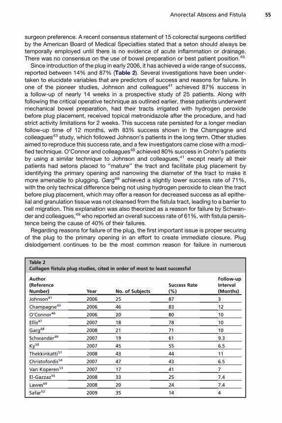
Since introduction of the plug in early 2006, it has achieved a wide range of success,reported between 14% and 87% (Table 2). Several investigations have been under-taken to elucidate variables that are predictors of success and reasons for failure. Inone of the pioneer studies, Johnson and colleagues41 achieved 87% success ina follow-up of nearly 14 weeks in a prospective study of 25 patients. Along withfollowing the critical operative technique as outlined earlier, these patients underwentmechanical bowel preparation, had their tracts irrigated with hydrogen peroxidebefore plug placement, received topical metronidazole after the procedure, and hadstrict activity limitations for 2 weeks. This success rate persisted for a longer medianfollow-up time of 12 months, with 83% success shown in the Champagne andcolleagues43 study, which followed Johnson’s patients in the long term. Other studiesaimed to reproduce this success rate, and a few investigators came close with a modi-fied technique. O’Connor and colleagues46 achieved 80% success in Crohn’s patientsby using a similar technique to Johnson and colleagues,41 except nearly all theirpatients had setons placed to ‘‘mature’’ the tract and facilitate plug placement byidentifying the primary opening and narrowing the diameter of the tract to make itmore amenable to plugging. Garg48 achieved a slightly lower success rate of 71%,with the only technical difference being not using hydrogen peroxide to clean the tractbefore plug placement, which may offer a reason for decreased success as all epithe-lial and granulation tissue was not cleansed from the fistula tract, leading to a barrier tocell migration. This explanation was also theorized as a reason for failure by Schwan-der and colleagues,49 who reported an overall success rate of 61%, with fistula persis-tence being the cause of 40% of their failures.
Regarding reasons for failure of the plug, the first important issue is proper securingof the plug to the primary opening in an effort to create immediate closure. Plugdislodgement continues to be the most common reason for failure in numerous
Table 2Collagen fistula plug studies, cited in order of most to least successful
Author(ReferenceNumber) Year No. of Subjects
Success Rate(%)
Follow-upInterval(Months)
Johnson41 2006 25 87 3
Champagne43 2006 46 83 12
O’Connor46 2006 20 80 10
Ellis47 2007 18 78 10
Garg48 2008 21 71 10
Schwander49 2007 19 61 9.3
Ky50 2007 45 55 6.5
Thekkinkatti51 2008 43 44 11
Christofordis54 2007 47 43 6.5
Van Koperen53 2007 17 41 7
El-Gazzaz55 2008 33 25 7.4
Lawes69 2008 20 24 7.4
Safar52 2009 35 14 4

Rizzo et al56
studies. Steps to prevent dislodgement include adequately securing the plug to theprimary opening, ensuring it is not dangling,44 and instructing the patient to avoidstrenuous activity for at least 2 weeks.43,45,46 Avoidance of securing the plug at thesecondary opening has been advocated because it provides countertraction to thesuture at the primary opening, leading to dislodgement. Another cause of dislodge-ment is enlarging the fistula tract, which has been done by curetting or overdebride-ment of the tract. Multiple fistula tracts are often associated with a higher failurerate caused by the persistence of 1 or more tracts, usually those not treated by theplug at the time of the initial procedure.46 These tracts are candidates for plug insertionat a later date. The importance of ensuring that the secondary opening remains openas a site for drainage cannot be overemphasized because this prevents the formationof abscesses, which is not only a cause of failure but also a cause of mortality in thesepatients because of perineal sepsis.52
There are a few controversies about the various modifications of the procedurethat can be performed and their influence on the success rate. The most publishedmodification is the concurrent use of the seton. The concept of the seton maturingthe tract, making the wall more fibrotic, which results in increased healing, hasbeen proposed by several investigators54,55 and is recommended by a recentconsensus.45 It has also been shown to minimize sepsis and facilitate fistula closurewhen used in conjunction with other procedures, such as an advancement flap.56
O’Connor and colleagues46 and Champagne and colleagues43 found no correlationbetween seton placement and increasing healing rates. They stated that the pres-ence of the seton resulted in a technically easier insertion of the plug because ithelped define the anatomy of the primary and secondary opening and helped‘‘pull’’ the plug through the tract.
There is little doubt that the anal fistula plug is a promising new method of treatingthis complex problem but with the variable success rates, more studies need to beperformed to elucidate the best procedure and postoperative care to ensure the high-est chance of success.
ADVANCEMENT FLAP
Before the advent of the collagen anal fistula plug or the use of fibrin glue, surgeonsdevised the endorectal/endoanal advancement flap as a sphincter-sparing methodto treat complex anal fistulas. It was believed that this would preserve continencebecause there is no surgical division of the anal sphincter complex. There are severalmethods, but the technical aspects common to all methods are cleaning/debridementof the fistula tract, mobilization of a well-vascularized rectal mucosal or anodermalflap, and coverage of the internal opening of the tract with or without closure of thetract before coverage. Healing rates have been reported to be from 77% to 100%in various studies.1,2,4,57–64 Length of follow-up is important when evaluating thesuccess of various methods of the surgical treatment of fistula-in-ano. Ortiz andcolleagues4 performed a retrospective study of 91 patients who underwent flap repairof complex fistulas. The median follow-up was 42 months and there was a recurrencerate of 19%. These investigators noted that the median time to relapse was 5 monthsand that no recurrences were noted beyond 1 year of follow-up. Van Koperen andcolleagues62 evaluated their long-term outcomes from flap repair of high anal fistulasand noted that after 76 months of follow-up, recurrence was seen in 21% of patientsand fecal soiling was reported in 40%.59 This addresses an important issue anddispels the myth that incontinence is not a potential risk of flap repair.

Anorectal Abscess and Fistula 57
A recent study2 reported a transient minor incontinence rate of 8% withcomplete resolution by 2 months of follow-up after advancement flap repair. Addi-tional studies have reported minor incontinence rates of 0% to 23% associatedwith advancement flap procedures.57,58,63,65 Uribe and colleagues58 performeda prospective study of 56 patients with complex fistulas who underwent advance-ment flap repair. Preoperative and postoperative anal manometry were performedin all patients. A significant reduction was demonstrated in mean resting pressureand maximal squeeze pressure in the study subjects 3 months postoperatively. A21% rate of incontinence was reported, with 9% of patients reporting major distur-bances in fecal continence. Perez and colleagues60 performed a manometric studyof patients who were randomized to undergo either advancement flap repair ofcomplex fistulas or fistulotomy with concomitant sphincter repair. Their data re-vealed that mean resting pressure was significantly diminished postoperatively inboth groups, but that maximal squeeze pressure was reduced only in the groupundergoing flap repair. This finding did not equate to any difference in continencebetween the groups. The study results showed equivalent rates of healingbetween the 2 methods.
Various methods have been espoused to improve the success rates of flaprepairs. Van der Hagen and colleagues65 evaluated their experience with the treat-ment of complex anal fistula disease and included patients with Crohn’s disease intheir study group. They propose that the initial placement of a loose seton allowsfor resolution of sepsis and improves subsequent outcomes with advancementflap repair. They reported only 1 flap failure in 26 patients who underwent theprocedure. Others have suggested that combining the use of fibrin glue oblitera-tion of the fistula tract with endorectal advancement flap repair would potentiallyimprove the rates of healing. Ellis and Clark61 and van Koperen and colleagues62
settled this argument with 2 studies that showed higher failure rates in patientswho had fibrin glue instilled into their fistula tracts as an adjunct to flap repair.Perhaps the glue instillation prevents adequate drainage of fluid trapped underthe flap through the external opening. It is easy to imagine how this might leadto flap failure.
The question of whether to use a partial or full thickness advancement flap wasaddressed in a study in 2008 by Dubsky and colleagues.66 These investigatorsdemonstrated a higher flap failure rate in the partial thickness group (35% vs5%). They also demonstrated a higher incontinence score in the patients treatedwith partial thickness flaps, although the statistical significance of this result wasnot reported. There were no differences in continence that correlated to flapfailure. It is difficult to draw any definitive conclusion regarding incontinencefrom this small data set. The likely causes of incontinence after flap repair arepartial division of the internal sphincter during flap mobilization, and sphinctertrauma from unintentional anal dilation during retraction for operative exposure.At present these technical aspects of the procedure seem to be unavoidableand must be taken into consideration when counseling a patient preoperatively.Tyler and colleagues63 reported their success in treating anal fistulas usinga ‘‘sphincter-sparing only’’ algorithm. They performed fistulotomies only whensimple submucosal fistulas were present. All other fistulas were treated with loosesetons followed by fibrin glue injection or advancement flaps. Glue failures wenton to repeat therapy with fibrin glue or advancement flap repair. These investiga-tors reported a 100% success rate using this algorithm with no resulting fecalincontinence. These data support the recommendation of sphincter-sparingsurgery for all anal fistulas.
Rizzo et al58
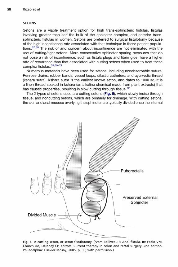
SETONS
Setons are a viable treatment option for high trans-sphincteric fistulas, fistulasinvolving greater than half the bulk of the sphincter complex, and anterior trans-sphincteric fistulas in women. Setons are preferred to surgical fistulotomy becauseof the high incontinence rate associated with that technique in these patient popula-tions.67,68 The risk of and concern about incontinence are not eliminated with theuse of cutting/tight setons. More conservative sphincter-sparing measures that donot pose a risk of incontinence, such as fistula plugs and fibrin glue, have a higherrate of recurrence than that associated with cutting setons when used to treat thesecomplex fistulas.35,69–71
Numerous materials have been used for setons, including nonabsorbable suture,Penrose drains, rubber bands, vessel loops, silastic catheters, and ayurvedic thread(kshara sutra). Kshara sutra is the earliest known seton, and dates to 1000 BC. It isa linen thread soaked in kshara (an alkaline chemical made from plant extracts) thathas caustic properties, resulting in slow cutting through tissue.72
The 2 types of setons used are cutting setons (Fig. 5), which slowly incise throughtissue, and noncutting setons, which are primarily for drainage. With cutting setons,the skin and anal mucosa overlying the sphincter are typically divided once the internal
Puborectalis
Preserved ExternalSphincter
Divided Muscle
Fig. 5. A cutting seton, or seton fistulotomy. (From Belliveau P. Anal fistula. In: Fazio VM,Church JM, Delaney CP, editors. Current therapy in colon and rectal surgery. 2nd edition.Philadelphia: Elsevier Mosby; 2005. p. 30; with permission.)
Anorectal Abscess and Fistula 59
and external openings of the fistula are identified. The seton is then placed through thefistula tract and tightened at varying intervals (from a few days to every 2 weeks). Theremay be no need for further tightening if elastic materials are used and are securedtightly at the time of surgery.73 The time it takes to heal can range from 1 month tomore than a year.70,71 These patients require numerous follow-up visits, which requiretightening of the seton, examination of the fistula tract, and potentially performance ofa second procedure. For this reason Mentes and colleagues73 used pieces of surgicalgloves as cutting setons in 20 patients. These setons were secured tightly at the initialprocedure and slowly cut through the sphincter mechanism without additional tight-ening. The average time for the seton to cut through the sphincter completely was19 days, and a 20% rate of minor incontinence was reported. This seton has theadvantages of avoiding numerous postoperative visits for adjustment, the pain asso-ciated with tightening, and the need for secondary procedures.
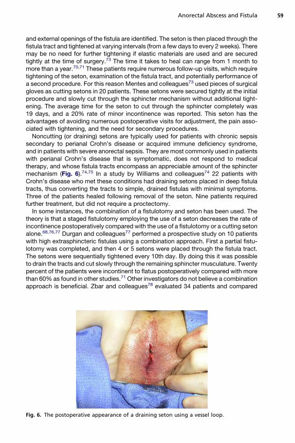
Noncutting (or draining) setons are typically used for patients with chronic sepsissecondary to perianal Crohn’s disease or acquired immune deficiency syndrome,and in patients with severe anorectal sepsis. They are most commonly used in patientswith perianal Crohn’s disease that is symptomatic, does not respond to medicaltherapy, and whose fistula tracts encompass an appreciable amount of the sphinctermechanism (Fig. 6).74,75 In a study by Williams and colleagues74 22 patients withCrohn’s disease who met these conditions had draining setons placed in deep fistulatracts, thus converting the tracts to simple, drained fistulas with minimal symptoms.Three of the patients healed following removal of the seton. Nine patients requiredfurther treatment, but did not require a proctectomy.
In some instances, the combination of a fistulotomy and seton has been used. Thetheory is that a staged fistulotomy employing the use of a seton decreases the rate ofincontinence postoperatively compared with the use of a fistulotomy or a cutting setonalone.68,76,77 Durgan and colleagues77 performed a prospective study on 10 patientswith high extrasphincteric fistulas using a combination approach. First a partial fistu-lotomy was completed, and then 4 or 5 setons were placed through the fistula tract.The setons were sequentially tightened every 10th day. By doing this it was possibleto drain the tracts and cut slowly through the remaining sphincter musculature. Twentypercent of the patients were incontinent to flatus postoperatively compared with morethan 60% as found in other studies.71 Other investigators do not believe a combinationapproach is beneficial. Zbar and colleagues78 evaluated 34 patients and compared
Fig. 6. The postoperative appearance of a draining seton using a vessel loop.
Rizzo et al60
the combination approach with the use of a cutting seton alone. Eighteen patientswere treated with a combined technique and 16 were treated with a cutting seton.The internal anal sphincter muscle was repaired after it was incised, and this was fol-lowed by placement of a seton through the intersphincteric space. No difference wasfound between the 2 groups in healing time, recurrence rates, or incontinence rates.
As mentioned earlier, incontinence is still common with the use of setons. It is rarelyseen with noncutting setons because their function is primarily to drain, not to incisethe anal sphincters.16 Incontinence has been attributed to several factors, includinghard and gutter-shaped scars in the anal canal, loss of sphincter function, and lossof anal canal sensation.71 Patients may experience incontinence to flatus, liquid stool,or solid stool at varying levels, with different effects on their quality of life. Not allstudies identify specifically the type of incontinence patients are experiencing, nordo they differentiate transient postoperative incontinence from long-term dysfunction.In addition, many studies refer simply to major and minor incontinence withoutdefining them. Minor incontinence is defined as persistent incontinence to gas oroccasionally liquid stool. Major incontinence refers to inability to control the passageof formed stool or frequently liquid stool. Isbister and Sanea70 performed a retrospec-tive review of 47 patients who had cutting setons placed for complex fistulas. Theirpostoperative incontinence rate was 36.2% for gas, 8.5% for liquid stool, and 2.3%for solid stool. Before seton placement 14.9% of their study group were incontinentto gas or liquid stool. One fistula recurrence was reported. An additional study re-ported a minor incontinence rate of 63% with a 6% recurrence rate.71
Although the risk of an impairment in continence is high with seton fistulotomy, thistechnique is used for complex, hard-to-treat fistulas that recur more frequently withsphincter-sparing methods including fibrin glue and fistula plugs.
FISTULOTOMY
Fistulotomy is still considered the standard by many surgeons for low, simple analfistulas, such as submucosal, intersphincteric, and low trans-sphincteric fistulas(Fig. 7).67,79 According to ‘The practice parameters for the treatment of perianalabscess and fistula-in-ano,’ completed by Whiteford and colleagues79 in 2005, fistu-lotomies may be used to treat simple anal fistulas in cryptoglandular disease andsimple, low Crohn’s fistulas that are symptomatic. Their definition of simple includesa fistula tract that crosses less than 30% to 50% of the external sphincter, is not ante-rior in women, has only 1 tract, is not recurrent, and is present in a patient with perfectcontinence. In addition, the patient should not carry a diagnosis of Crohn’s disease orhave received pelvic radiation for the fistula to be considered simple. Whiteford andcolleagues79 also recommended the use of tract debridement and fibrin glue injectionfor simple fistulas, because it is a benign treatment with no detrimental effects onfurther treatment, although the recurrence rates are higher than with fistulotomy. Fis-tulotomy can be used as a staged procedure for complex fistulas in conjunction witha seton according to the practice parameters.
Fistulotomy is the standard treatment for submucosal fistulas because there is noconcern for incontinence and recurrence is low.63 Controversy arises in the treatmentof fistulas that involve the sphincter mechanism because of the potential for inconti-nence with nonsphincter-sparing methods. Most surgeons use fistulotomies forsimple intersphincteric fistulas; however, some groups such as Tyler and colleagues63
use fistulotomies only for submucosal fistulas. In all of their patients with sphincterinvolvement a seton is placed, followed by fibrin glue or a rectal advancement flapas a staged procedure.

ProximalExternal Sphincter
Divided Muscle
Levator
Fig. 7. Fistulotomy of a low trans-sphincteric fistula. (From Belliveau P. Anal fistula. In: FazioVM, Church JM, Delaney CP, editors. Current therapy in colon and rectal surgery. 2ndedition. Philadelphia: Elsevier Mosby; 2005. p. 29; with permission.)
Anorectal Abscess and Fistula 61
Other surgeons use fistulotomies more frequently and apply them to complexfistulas also. Perez and colleagues80 have completed multiple studies looking at fistu-lotomies with primary sphincter reconstruction for complex fistulas. They conducteda prospective study with 16 patients who had recurrent high trans-sphincteric, supra-sphincteric, or extrasphincteric fistulas. Each patient underwent a fistulotomy in whichthe internal and external sphincters were divided. All accessory tracts were excised.The internal and external anal sphincters were repaired using an overlapping tech-nique. All patients who were incontinent preoperatively improved their continencescore postoperatively (the Wexner grading scale was used) except for 1 female patientwhose incontinence score was unchanged. Two patients (25%) developed new minorincontinence. The results were similar in a larger prospective study of 35 patients, inwhich 45% had recurrent disease preoperatively.81 Perez and colleagues60 alsocompared their method with advancement flaps. They completed a prospectiverandomized study of 55 patients with complex fistulas (high trans-sphincteric andsuprasphincteric) and found no difference in the recurrence or incontinence ratebetween the 2 methods.
As mentioned earlier, the incontinence rate with fistulotomy is high and can varygreatly, with rates from 0% to 40% in low intersphincteric fistulas.59,82 As a result,not only have new methods been developed to treat fistulas, such as fibrin glue and
Rizzo et al62
fistula plugs, but numerous studies have been undertaken to determine predisposingfactors for incontinence. Toyonaga and colleagues83 performed a prospectiverandomized study with 148 patients with intersphincteric fistulas and found thatage, sex, previous surgery, duration of fistula, and location and level of the internalopening did not significantly influence incontinence postoperatively in patients under-going a fistulotomy for treatment. In addition, low anal sphincter resting pressure andshort anal canal length did not predispose patients to postoperative incontinence.These investigators found that low voluntary contraction pressure and multipleprevious drainage procedures predisposed patients to postoperative incontinence.They recommend preoperative manometry for all patients, and avoidance of fistulot-omy if the risk factors already mentioned were identified. Koperen and colleagues59
performed a study of 179 patients in which gender, age, tertiary referral, prior fistulasurgery, and smoking were not found to be risk factors. Other studies show conflictingresults. Cavanaugh and colleagues82 in a study of 110 patients found trans-sphinc-teric tracts and the extent of external sphincter involvement to be risk factors for post-operative incontinence after fistulotomy. In addition, Garcia-Aguilar and colleagues84
determined female gender, previous surgery, high internal opening, and type of fistulasurgery (procedures performed for high fistulas) were all risk factors for postoperativeincontinence following a fistulotomy.
Gupta85,86 conducted fistulotomies using radiofrequency ablation in an effort todecrease complications associated with conventional fistulotomy. In a study of 100patients with low anal fistulas he compared conventional fistulotomy to radiofre-quency fistulotomy and found those in the radiofrequency group had less gas incon-tinence (4 vs 12%). It is speculated that the reason for decreased incontinence is thatthere is less damage to the surrounding tissue because radiofrequency does not heatsurrounding tissue. Similar results could be expected with the use of an ultrasonicdissector, although this has not been studied.
Recurrence has been reported to be lower with fistulotomy for low intersphinc-teric fistulas when compared with other methods, such as fibrin glue36,59;however, a long-term study conducted by Van der Hagen and colleagues87
suggests that the recurrence rate is higher if follow-up of the patients is performedlong enough. These investigators looked at the long-term (72 months) recurrencerates of fistulotomy for low perianal fistulas. Many studies follow patients up for 12to 24 months, which for this study had a recurrence rate of 7% and 16%, respec-tively. These investigators noted at 72 months of follow-up the recurrence ratewas 39%. Fifty-four percent of recurrences occurred in a new location, andpatients with Crohn’s disease were included in this trial. At 48 months, fistulahad recurred in 60% of the patients with Crohn’s disease included in the trial.Any study that includes inflammatory bowel disease patients in long-term follow-up will have a higher rate of recurrence compared with studies that do not.Although these data are useful, the elevated long-term recurrence rate seen inthis study should be considered when selecting a method of treatment.
Although incontinence may be seen with low intersphincteric fistulotomy, mostcases are minor (involving gas and occasionally liquid incontinence), but may stillsignificantly affect the patient’s lifestyle. Cavanaugh and colleagues82 conducteda study to examination how postoperative incontinence affects the quality of life forthese patients. They conducted a retrospective study of 110 patients that revealedthat 64% of patients had at least occasional incontinence, with 14% reporting mildlifestyle restriction, 10% reporting moderate restriction, 9% reporting mild depression,and 4% reporting moderate depression. Moderate and severe embarrassment werereported by 5% and 1%, respectively.
Anorectal Abscess and Fistula 63
Fistulotomy remains a major part of fistula treatment, despite the high rate of incon-tinence that may be seen postoperatively. It is an effective method of dealing with thedisease, with the recurrence rates being much lower than those for sphincter-sparingmethods. Although many patients experience minor incontinence that does not affecttheir lifestyle, it can have a severe effect on others.
NEWER METHODS OF TREATMENT
The search for the optimal treatment of anal fistula continues because of disappointingsuccess rates with sphincter-sparing options and high incontinence rates associatedwith sphincter dividing procedures. The ligation of the intersphincteric fistula tract(LIFT) procedure was first described in Thailand by Rojanasakul.88 This is a procedurein which a small incision is made in the intersphincteric groove (much like an openinternal sphincterotomy) just over where the fistula tract crosses from the internal tothe external sphincter. The intersphincteric space is opened and the fistula tract isclearly defined and ligated with suture. Short-term results in 18 patients treated bythe LIFT procedure and observed prospectively showed healing in all but 1 bya mean time of 4 weeks.88 These results prompted the University of Minnesota Colo-rectal Surgery Group to adopt the technique and study it in the long term. Beals89 pre-sented results with the use of the LIFT technique at the 94th annual American Collegeof Surgeons Clinical Congress and reported a 58% success rate in 31 patients fol-lowed for a mean time of 35 weeks. Time until failure ranged from 4 to 63 weekswith a median of 19 weeks. This result has prompted further investigation of this prom-ising technique, and a trial comparing the LIFT technique with the collagen fistula plugis currently accruing patients.
Garcia-Olmo and colleagues90 investigated the use of injected adipose-derivedstem cells in the treatment of complex anal fistulas. Their idea was based on experi-ence from plastic surgery of the use of these cells in tissue repair.91 These investiga-tors compared the use of injected fibrin glue with glue that contained 20 million units ofadipose-derived stem cells in 49 patients with complex fistulas that were either cryp-toglandular or related to Crohn’s disease. Fistula healing was noted in 71% of thegroup treated with stem cells as opposed to only 16% healing in the group whoreceived fibrin glue. There were no differences in adverse reactions between thegroups and none appeared to be related to treatment with stem cells. A 17% recur-rence rate was noted after 1 year of follow-up in the group treated with stem cells. Gar-cia-Olmo and colleagues concluded that fistula tract injection of adipose-derived stemcells was safe and effective in the treatment of this complex disease. Both of thesetechniques show promise and warrant further long-term, randomized investigation.
SUMMARY
The surgical management of fistula-in-ano is driven by the amount of sphinctercomplex that is involved with the tract, and the potential coexistence of Crohn’sdisease. The preferred method of management is dictated by these factors.Sphincter-sparing methods have lower success rates than nonsphincter-sparingtechniques, but come with little to no risk of fecal continence. The first line of treatmentof this disease should focus on methods that do not require any sphincter division.These techniques do not prevent a more aggressive surgical approach if they fail.Submucosal fistulas can be treated by fistulotomy with little risk. Intersphinctericand low trans-sphincteric fistulas may be treated with fistulotomy as first-line manage-ment if the patient has perfect continence preoperatively, and the patient has noprevious history of sphincter injury. Anterior fistulas in women must be approached

Rizzo et al64
with caution. It is to be hoped continued research will lead to improved success ratesin sphincter-sparing options.
REFERENCES
1. Loffler T, Welsch T, Muhl S, et al. Long-term success rate after surgical treatmentof anorectal and rectovaginal fistulas in Crohn’s disease. Int J Colorectal Dis2009. [Epub ahead of print].
2. Abbas MA, Lemus-Rangel R, Hamadani A. Long-term outcome of endorectaladvancement flap for complex anorectal fistulae. Am Surg 2008;74(10):921–4.
3. Hamadani A, Haigh PI, Liu IL, et al. Who is at risk for developing chronic analfistula or recurrent anal sepsis after initial perianal abscess? Dis Colon Rectum2009;52:217–21.
4. Ortiz H, Marzo M, de Miguel M, et al. Length of follow-up after fistulotomy andfistulectomy associated with endorectal advancement flap repair for fistula inano. Br J Surg 2008;95(4):484–7.
5. Oliver I, Lavueva FJ, Piorez VF, et al. Randomized clinical trial comparing simpledrainage of anorectal abscess with and without fistula tract treatment. Int JColorectal Dis 2003;18(2):107–10.
6. Quah HM, Tang CL, Eu KW, et al. Meta-analysis of randomized clinical trialscomparing drainage alone vs primary sphincter-cutting procedures for anorectalabscess-fistula. Int J Colorectal Dis 2006;21(6):602–9.
7. Aluwihar A. Finding the source of a fistula. Colorectal Dis 2005;7(5):528–9.8. Erhan Y, Sakarya A, Aydede H, et al. A case of large mucinous adenocarcinoma
arising in a long-standing fistula-in-ano. Dig Surg 2003;20(1):69–71.9. Kim T, Chae G, Chung SS, et al. Faecal incontinence in male patients. Colorectal
Dis 2008;10(2):124–30.10. Lindsey I, Jones OM, Smilgin-Humphreys MM, et al. Patterns of fecal inconti-
nence after anal surgery. Dis Colon Rectum 2004;47(10):1643–9.11. Buchmann P, Keighley MR, Allan RN, et al. Natural history of perianal Crohn’s
disease. Ten-year follow up: a plea for conservatism. Am J Surg 1980;140(5):642–4.
12. Halme L, Sainio AP. Factors related to frequency, type and outcome of analfistulas in Crohn’s disease. Dis Colon Rectum 1995;35:55–9.
13. Jakobovits J, Schuster MM. Metronidazole therapy for Crohn’s disease and asso-ciated fistulae. Am J Gastroenterol 1984;79:533–40.
14. Stringer EE, Nicholson TJ, Armstrong D. Efficacy of topical metronidazole (10percent) in the treatment of anorectal Crohn’s disease. Dis Colon Rectum 2005;48:970–4.
15. Sangwan YP, Schoetz DJ, Murray JJ, et al. Perianal Crohn’s disease: results oflocal surgical treatment. Dis Colon Rectum 1996;39(5):529–35.
16. Faucheron J, Saint-Marc O, Guibert L, et al. Long-term seton drainage for highanal fistulas in Crohn’s disease – a sphincter-saving operation? Dis Colon Rectum1996;39:208–11.
17. Hanaver SB, Feagan BG, Lichtenstein GR, et al. Maintenance infliximab forCrohn’s disease: the ACCENT I randomized trial. Lancet 2002;359:1541–9.
18. Present DH, Rutgerts P, Targan S, et al. Infliximab for the treatment of fistula inpatients with Crohn’s disease. N Engl J Med 1999;340:1398–405.
19. Sands BE, Anderson FH, Bernstein CN, et al. Infliximab maintenance for fistuliz-ing Crohn’s disease. N Engl J Med 2004;350:876–85.

Anorectal Abscess and Fistula 65
20. Gaertner WB, Decanini A, Mellgren A, et al. Does infliximab infusion impactresults of operative treatment for Crohn’s perianal fistulas? Dis Colon Rectum2007;50(11):1754–60.
21. Gonzalez-Ruiz C, Kaiser AM, Vukasin P, et al. Intraoperative physical diagnosis inthe management of anal fistula. Am Surg 2006;72(1):11–5.
22. Ratto C, Grillo E, Parello A, et al. Endoanal ultrasound-guided surgery for analfistula. Endoscopy 2005;37(8):722–8.
23. Buchanan GN, Williams AM, Bartram CI, et al. Potential clinical implications ofdirections of a trans-sphincteric anal fistula track. Br J Surg 2003;90(10):1250–5.
24. Swinscoe MT, Ventakasubramanium AK, Jayne DG. Fibrin glue for fistula-in-ano:the evidence reviewed. Tech Coloproctol 2005;9(2):89–94.
25. Hjortrup A, Moesgaard F, Kjaergard J. Fibrin adhesive in the treatment of perinealfistulas. Dis Colon Rectum 1991;34:752–4.
26. Hammond TM, Grahn MF, Lunniss PJ. Fibrin glue in the management of analfistulae. Colorectal Dis 2004;6(5):308–19.
27. de Parades V, Far HS, Etienney I, et al. Seton drainage and fibrin glue injection forcomplex anal fistulas. Colorectal dis 2008. [Epub ahead of print].
28. Ritchie RD, Sackier JM, Hodde JP. Incontinence rates after cutting seton treat-ment for anal fistula. Colorectal Dis 2008. [Epub ahead of print].
29. Dietz DW. Role of fibrin glue in the management of simple and complex fistula inano. J Gastrointest Surg 2006;10(5):631–2.
30. Cintron JR, Park JJ, Orsay CP, et al. Repair of fistula-in-ano using fibrin adhesive.Long-term follow up. Dis Colon Rectum 2000;43:944–50.
31. Gisbertz SS, Sosef MN, Festen S, et al. Treatment of fistulas in ano with fibrin glue.Dig Surg 2005;22(1–2):91–4.
32. Maralcan G, Bakukonu I, Aybasti N, et al. The use of fibrin glue in the treatment offistula-in-ano: a prospective study. Surg Today 2006;36(2):166–70.
33. Adams T, Yang J, Kondylis LA, et al. Long-term outlook after successful fibringlue ablation of cryptoglandular transsphincteric fistula-in-ano. Dis Colon Rectum2008;51(10):1488–90.
34. Sentovich SM. Fibrin glue for all anal fistulas. J Gastrointest Surg 2001;5(2):158–61.
35. Sentovich SM. Fibrin glue for anal fistulas: long-term results. Dis Colon Rectum2003;46(4):498–502.
36. Lindsey I, Smilgin-Humphreys MM, Cunningham C, et al. A randomized,controlled trial of fibrin glue vs conventional treatment for anal fistula. Dis ColonRectum 2002;45(12):1608–15.
37. Zmora O, Neufeld D, Ziv Y, et al. Prospective, multicenter evaluation of highlyconcentrated fibrin glue in the treatment of complex cryptogenic perianal fistulas.Dis Colon Rectum 2005;7(5):528–9.
38. Patrlj L, Kocman B, Martinac M, et al. Fibrin glue-antibiotic mixture in thetreatment of anal fistulae: experience with 69 cases. Dig Surg 2000;17(1):77–80.
39. Witte ME, Klaase JM, Gerritsen JJ, et al. Fibrin glue treatment for simple andcomplex anal fistulas. Hepatogastroenterology 2007;54(76):1071–3.
40. Robb BW, Vogler SA, Nussbaum MN, et al. Early experience using porcine smallintestinal submucosa to repair fistulas-in-ano. Dis Colon Rectum 2004;47:565–660.
41. Johnson EK, Gaw JU, Armstrong D. Efficacy of anal fistula plug vs fibrin glue inclosure of anorectal fistulas. Dis Colon Rectum 2006;49(3):371–6.
Rizzo et al66
42. Ueno T, Pickett LC, de la Fuente SG, et al. Clinical application of porcine smallintestinal submucosa in the management of infected or potentially contaminatedabdominal defects. J Gastrointest Surg 2004;8:109–12.
43. Champagne BJ, O’Connor LM, Ferguson M, et al. Efficacy of anal fistula plug inclosure of cryptoglandular fistulas: long-term follow-up. Dis Colon Rectum 2006;49(12):1817–21.
44. Hammond TM, Lunniss PJ. Novel biomaterials in the management of anal fistulas.Dis Colon Rectum 2006;49(9):1463–4.
45. The Surgisis AFP anal fistula plug: report of a consensus conference. ColorectalDis 2008;10(1):17–20.
46. O’Connor L, Champagne BJ, Ferguson MA, et al. Efficacy of anal fistula plug inclosure of Crohn’s anorectal fistulas. Dis Colon Rectum 2006;49(10):1569–73.
47. Ellis CN. Bioprosthetic plugs for complex anal fistulas: an early experience.J Surg Educ 2007;64(1):36–40.
48. Garg P. To determine the efficacy of anal fistula plug in the treatment of high fistula-in-ano: an initial experience. Colorectal Dis 2008. [Epub ahead of print].
49. Schwandner O, Stadler F, Dietl O, et al. Initial experience on efficacy in closure ofcryptoglandular and Crohn’s transsphincteric fistulas by the use of the anal fistulaplug. Int J Colorectal Dis 2008;23(3):319–24.
50. Ky AJ, Sylla P, Steinhagen R, et al. Collagen fistula plug for the treatment of analfistulas. Dis Colon Rectum 2008;51(6):838–43.
51. Thekkinkattil D, Botterill I, Ambrose S, et al. Efficacy of the anal fistula plug incomplex anorectal fistulae. Colorectal Dis 2008. [Epub ahead of print].
52. Safar B, Jobanputra S, Sands D, et al. Anal Fistula Plug: intial experience andoutcomes. Dis Colon Rectum 2009;52:248–52.
53. van Koperen PJ, D’Hoore A, Wolthuis AM, et al. Anal fistula plug for closure ofdifficult anorectal fistula: a prospective study. Dis Colon Rectum 2007;50(12):2168–72.
54. Christoforidis D, Etzioni DA, Goldberg SM, et al. Treatment of complex analfistulas with the collagen fistula plug. Dis Colon Rectum 2008;51(10):1482–7.
55. El-Gazzaz G, Zutshi M, Hull T. A retrospective review of chronic anal fistulaetreated by anal fistulae plug. Colorectal Dis 2009. [Epub ahead of print].
56. Sonoda T, Hull T, Piedmonte MR, et al. Outcomes of primary repair of anorectaland rectovaginal fistulae using endorectal advancement flap. Dis Colon Rectum2002;45:1622–8.
57. Golub RW, Wise WE, Kerner BA, et al. Endorectal mucosal advancement flap: thepreferred method for complex cryptoglandular fistula-in-ano. J Gastrointest Surg1997;1(5):487–91.
58. Uribe N, Million M, Minguez M, et al. Clinical and manometric results of endorectaladvancement flaps for complex anal fistula. Int Colorectal Dis 2007;22(3):259–64.
59. Koperen P, Wind J, Bemelman W, et al. Long-term functional outcome and riskfactors for recurrence after surgical treatment for low and high perianal fistulasof cryptoglandular origin. Dis Colon Rectum 2008;51:1475–81.
60. Perez F, Arroyo A, Serrano P, et al. Randomized clinical and manometric study ofadvancement flap versus fistulotomy with sphincter reconstruction in themanagement of complex fistula-in-ano. Am J Surg 2006;192:34–40.
61. Ellis CN, Clark S. Fibrin glue as an adjuvant to flap repair of anal fistulas:a randomized, controlled study. Dis Colon Rectum 2006;49(11):1736–40.
62. van Koperen PJ, Wind J, Bemelman WA, et al. Fibrin glue and transanal rectaladvancement flap for high transsphincteric perianal fistulas; is there any advan-tage? Int J Colorectal Dis 2008;23(7):697–701.

Anorectal Abscess and Fistula 67
63. Tyler K, Aarons C, Sentovich S. Successful sphincter-sparing surgery for all analfistulas. Dis Colon Rectum 2007;50:1535–9.
64. Ho KS, Ho YH. Controlled, randomized trial of island flap anoplasty for treatment oftrans-sphincteric fistula-in-ano: early results. Tech Coloproctol 2005;9(2):166–8.
65. van der Hagen SJ, Baeten CG, Soeters PB, et al. Staged mucosal advancementflap for the treatment of complex anal fistulas: pretreatment with noncuttingsetons and in case of recurrent multiple abscesses a diverting stoma. ColorectalDis 2005;7(5):513–8.
66. Dubsky PC, Stift A, Friedl J, et al. Endorectal advancement flaps in the treatmentof high anal fistula of cryptoglandular origin: full-thickness vs. mucosal-rectumflaps. Dis Colon Rectum 2008;51(6):852–7.
67. Towsend C, Beauchamp R, Evers B, editors. Sabiston textbook of surgery: thebiological basis of modern surgical practice. 18th edition. Philadelphia: SaundersElsevier; 2008. p. 1447–9.
68. Vasilevsky C, Gordon P. Results of treatment of fistula-in-ano. Dis Colon Rectum1985;28(4):225–31.
69. Lawes DA, Efron JE, Abbas M, et al. Early experience with the bioabsorbableanal fistula plug. World J Surg 2008;32(6):1157–9.
70. Isbister WH, Sanea NA. The cutting seton: an experience at King Faisal SpecialistHospital. Dis Colon Rectum 2001;44:722–7.
71. Hamalainen KJ, Sainio AP. Cutting seton for anal fistulas: high risk of minor controldefects. Dis Colon Rectum 1997;40:1443–7.
72. Ho KS, Tsang C, Seow-Choen F, et al. Prospective randomized trial comparingayurvedic cutting seton and fistulotomy for low fistula-in-ano. Tech Coloproctol2001;5:137–41.
73. Mentes BB, Oktemer S, Tezcaner T, et al. Elastic one-stage cutting seton for thetreatment of high anal fistulas: preliminary results. Tech Coloproctol 2004;8:159–62.
74. Williams JG, Rothenberger DA. Fistula-in-ano in Crohn’s disease: results ofaggressive surgical treatment. Dis Colon Rectum 1991;34:378–84.
75. Person B, Wexner S. Management of perianal Crohn’s disease. Current treatmentoptions. Gastroenterology 2005;8:197–209.
76. McCourtney JS, Finlay IG. Cutting seton without preliminary internal sphincteroto-my in management of complex high fistula-in-ano. Dis Colon Rectum 1996;39:55–8.
77. Durgun V, Perek A, Kapan M, et al. Partial fistulotomy and modified cutting setonprocedure in the treatment of high extrasphincteric perianal fistula. Dig Surg2002;19:56–8.
78. Zbar AP, Ramesh J, Beer-Gabel M, et al. Conventional cutting vs. internal analsphincter-preserving seton for high trans-sphincteric fistula: a prospectiverandomized manometric and clinic trial. Tech Coloproctol 2003;7:89–94.
79. Whiteford M, Kilkenny J, Hyman N, et al. Practice parameters for the treatment ofperianal abscess and fistula-in-ano (revised). Dis Colon Rectum 2005;48:1337–42.
80. Perez F, Arroyo A, Serrano P, et al. Prospective clinical and manometric study offistulotomy with primary sphincter reconstruction in the management of recurrentcomplex fistula-in-ano. Int J Colorectal Dis 2006;21:522–6.
81. Perez F, Arroyo A, Serrano P, et al. Fistulotomy with primary sphincter reconstruc-tion in the management of complex fistula-in-ano: prospective study of clinicaland manometric results. J Am Coll Surg 2005;200:897–903.
82. Cavanaugh M, Hyman N, Osler T. Fecal incontinence severity index after fistulot-omy: a predictor of quality of life. Dis Colon Rectum 2002;45:349–53.

Rizzo et al68
83. Toyonaga T, Matsushima M, Kiriu T, et al. Factors affecting continence for fistulot-omy for intersphincteric fistula-in-ano. Int J Colorectal Dis 2007;22:1071–5.
84. Garcia-Aguilar J, Belmone C, Wong D, et al. Anal fistula surgery: factors associ-ated with recurrence and incontinence. Dis Colon Rectum 1996;39:723–9.
85. Gupta P. Anal fistulotomy with radiofrequency. Dig Surg 2004;21:72–3.86. Gupta P. Radiosurgical fistulotomy: an alternative to conventional procedure in
fistula in ano. Curr Surg 2003;60:524–8.87. van der Hagen S, Baeten C, Soeters P, et al. Long-term outcome following
mucosal advancement flap for high perianal fistulas and fistulotomy for low peria-nal fistulas. Recurrent perianal fistulas: failure of treatment or recurrent patientdisease? Int J Colorectal Dis 2006;21:784–90.
88. Rojanasakul A. Total anal sphincter saving technique for fistula-in-ano: the ligationof intersphincteric fistula tract. J Med Assoc Thai 2007;90(3):581–6.
89. Beals JK. Novel surgical correction of intersphincteric perianal fistulas preservesanal sphincter. Presented at American College of Surgeons 94th Annual ClinicalCongress. October 15, 2008.
90. Garcia-Olmo D, Herreros D, Pascual I, et al. Expanded adipose-derived stemcells for the treatment of complex perianal fistula: a Phase II clinical trial. DisColon Rectum 2009;52:79–86.
91. De Ugarte DA, Ashjian PH, Elbarbary A, et al. Future of fat as raw material fortissue regeneration. Ann Plast Surg 2003;50(2):215–9.


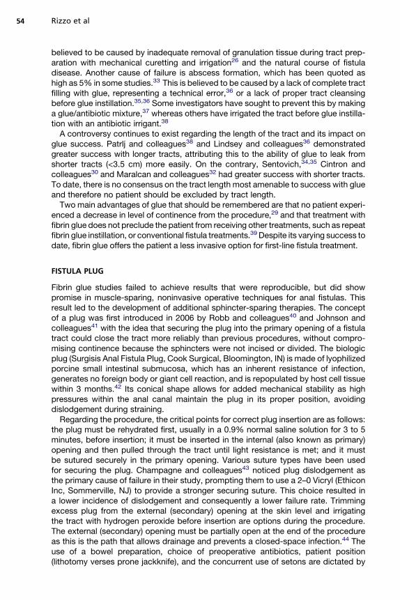
![A Rare Case of Anorectal Abscess due to Foreign Ingested ...A].pdf · the patient’s perianal abscess was drained. Due to the presence of gaseous, purulent drainage from the perianal](https://img.dokumen.tips/doc/110x75/60858a3928e9e201eb61b9d8/a-rare-case-of-anorectal-abscess-due-to-foreign-ingested-apdf-the-patientas.jpg)