Embed Size (px)
Citation preview

Annals of the National Academy of Medical Sciences (India)
JUN BE ID LL EO E
GVolume 53, No. 1Jan-March, 2017
ISSN 0379 - 038X (Print)ISSN 2454-5635 (Online)

Members of the Advisory Board
Emeritus Editor: Prof. J.S. Bajaj
Honorary SecretaryNational Academy of Medical Sciences (India)
NAMS House, Ansari Nagar, Mahatma Gandhi Marg, New Delhi-110029Tel.: 011-26589289 Email: [email protected]
Website: www.nams-india.in
Assistant EditorDr. Mohan Kameswaran
Associate EditorsDr. V Mohan KumarDr. Kuldeep Singh
Editor
Dr. Sanjeev Misra
Annals of the National Academy of Medical Sciences (India)-: A quarterly Journal :-
Editorial Board Editorial AssociatesDr. Snehalata Deshmukh Dr. W Selvamurthy Dr. J N Pande Dr. Prema Ramachandran Dr. H S Sandhu Dr. Lalita S Kothari Dr. Vinod Paul Dr. Sanjay Wadhwa
Dr. M V Padma SrivastavaDr. R K ChaddaDr. Deep N SrivastavaDr. Promila BajajDr. N R JagannathanDr. Subrata SinhaDr. Ravinder GoswamiDr. (Brig) Velu Nair
Dr. P K MisraDr. M Berry Air Marshal Dr. M S Boparai Dr. Y K Chawla Dr. P K Dave Dr. Amod Gupta Dr. Ravi Kant Dr. Balram AiranDr. Saroj Chooramani Gopal
Dr. Rajeshwar DayalDr. C S SaimbiDr. R MadanDr. Raj KumarDr. Mukund S JoshiDr. Kamal BucksheeDr. Haribhai L PatelDr. I C VermaDr. Geeta K Vemuganti
Annual Subscription Rates
InlandForeign
Single Copy
Rs. 500.00$ 30.00£ 15.00
Rs. 150.00
CorrespondenceAll correspondence concerning the Journal should be addressed to:
Ann Natl Acad Med Sci (India), 53(1): 1-65, 2017
ANAMS 53(1): 1-65, 2017
CONTENTS
Genetic Basis of Diabetic NephropathyO P Kalra
01
Essential Skills in Postgraduate Medical Curriculum of Community MedicineJugal Kishore, Tanu Anand, Sneha Kumari
21
Neurodevelopmental Disorders: The Journey, the Dreams and their RealizationSheffali Gulati
30
Biomedical Applications of Nanomaterials: Diagnosis and Therapy of Thrombotic DisordersD Dash
36
Editorial
Sanjeev Misra
i
Skindex-29 to Determine Quality of Life and Emotional Factorsin Dermatological ConditionsDesiree Saimbi, MSVK Raju, Vaibhav Dubey, Vivek Kumar Dey
41
Restorative Therapies after Stroke: Drugs, Devices and RoboticsMV Padma Srivastava
51

Editorial
The National Academy of Medical Sciences (India) completed its 50 glorious years in 2011. Established as a unique institution which fosters and utilizes academic excellence as its resource to meet medical and social goals, the Academy has been recognised by the Government of India as a Nodal Agency for Continuing Education for Medical and Allied Health Professionals and is advising the Government of India in matters of National Health Policy and Planning.
The Academy encourages and sponsors nation-wide CME programmes, Symposia, Workshops and Conferences. Over the years the Academy has recognized the outstanding achievements made by the Indian scientists in the field of medicine and allied sciences and conferred Fellowship as well as Membership to the selected persons through a structured peer reviewed process and voting by all the Fellows.
One of the key role of Academy is dissemination of peer reviewed scientific material contributed by biomedical scientists. Efforts of our esteemed fellows have kept the literary and scientific contents of the Journal “Annals of the National Academy of Medical Sciences (India)” to the highest level. With unrelenting efforts of our dynamic community we have been able to maintain the continuity of the publication also. The Academy is trying to get the Annals indexed in PubMed and are leaving no stone unturned to get the job done. The major criteria for the same are the quality of articles being submitted and published, peer-review process, and regularity of publication. In its endeavour to do so, we are changing the size of the journal at par with our global counterparts. The Annals Vol 53, first issue is in your hands with improved size. Secondly, our Annals is now online also at annals-nams.in and all articles are available with open access. Not only the site provides access to reading, one can also submit their research and other contribution online without any charges. Thirdly, we have already started our indexing with various agencies to get wider coverage of our contents. The Annals is indexed with Index Medicus of South-East Asia region (IMSEAR) of WHO and is extracted by Directory of Open Access Journal (DOAJ), Google Scholar, Harvard Database, BASE, Citefactor and other indexing agencies.
The family of our Academy has grown enormously in size in numbers, variety and multidisciplinary talent. We have more than 900 Fellows and 6300 members as of December 2016. Whenever we wish to share our scientific achievements, we tend to become very selective in choosing the journal and our criteria are restricted on indexing status, suitability for the journal and type of research work focused on particular speciality and journal impact factor. With the support given by all fellows and members, Annals is now getting manuscripts which are aligned with our mandate to uplift the status of public health, medical education with emphasis on competency and skill development, and innovations in healthcare. One of the unique quality of Annals is its diversity and assortment rarely seen in speciality journals. The current issue exemplify this diversity. Readers gets an opportunity to peep into domain of other scientists who are doing path breaking work in their areas of expertise.Many epigenetic and genetic factors may determine diabetic patients' susceptibility to renal disease
Ann Natl Acad Med Sci (India), 53(1): i-iii, 2017
(ii)
development. Diabetic Nephropathy (DN) is a result of multifactorial mechanisms and may ultimately lead to Chronic Kidney Disease (CKD). Dr O P Kalra has shared his work on "Genetic Basis of Diabetic Nephropathy" based on his extensive experience in this issue of the Annals.
For a long time Medical Educationists in India are struggling to implement reforms in curriculum both for under-graduates and post-graduates education. Many radical changes have been implemented in curriculum in Western world and but are still being evaluated for their learning outcomes. No better time exists for medical faculty in India to just carry out smaller Quality Improvement (QI) changes in existing system and study the impact in existing settings where Indian system of education is still producing physician who are being recognized and appreciated for their skills globally as the world is closely watching the developments in medical education in India. Post-graduate students' self-assessment regarding their competencies and skills showed adequacy as far as Communication and health education is concerned but lacked confidence in epidemiology and occupational health. This was the conclusion drawn in a study by Kishore et al. Authors felt a need for reforms in existing curriculum.
Innovations do help mankind in preserving their health has been immensely proved by work by Dr. Gulati and Dr. Dash. Despite improvement in technology and advances in healthcare, we are still not able to achieve a single digit score in Infant Mortality Rate except in state of Kerala. To a large extent perinatal factors and genetic conditions are responsible among survivors having adverse developmental outcome and disability. The children need an early detection of neurodevelopmental conditions and early intervention coupled with augmentation of skills among the healthcare workers at grass root level. This can be achieved by developing pediatric sub-specialities with innovative approaches towards diagnosis, management and research. Autism was once a rare diagnosis. Observant paediatricians are now picking more cases from their busy practices coupled with the help of informed public. Innovative tools have been widely researched in India and are making life of the families having child suffering from Autistic Spectrum Disorder (ASD) much easier. All credit goes to Indian biomedical scientists who selflessly get involved with these children and their families. Dr. Gulati from AIIMS Delhi has shared her dream of developing Pediatric Neurology and how the vision got realized through offbeat journey less travelled by others is being highlighted in her article “Neurodevelopmental disorders: The Journey, the dreams and their realization”. On the other hand Dr. Dash from BHU, Varanasi has described innovative applications of Nanomaterials. Graphene based biosensor can detect individuals with high risk for thrombosis while near-infrared laser-irradiated gold Nano rods can ablate pathologic thrombus in-situ. Academy provides a platform for biomedical scientists for sharing, networking and integrating all medical specialities since the collaboration is the buzzword for all round development of health of mankind.
Public awareness with the help of mass media, mobile technology and education has definitely brought down diseases related to infections, poverty and nutritional disorders to great extent if not completely. However, India is facing problems with non-communicable diseases and emerging infections due to changes in socio-environmental milieu and changing lifestyles. The lurking danger of cancer, metabolic disorders and silent diseases like hypertension and diabetes mellitus on one hand and continuing threats of infectious diseases on the other hand is creating heavy stress on our healthcare system. Unless we take our vision to a futuristic horizon and start harnessing technology coupled with clinical research we will be at great disadvantage in the healthcare we are going to provide. Neuro-

(iii)
restoration in stroke patients using multimodality approaches incorporating stem cells, robotics and drugs do help patients regain their functional capabilities has been highlighted by Dr. Padma Srivastava. She has delved deeper into newer cell based therapies, appliances, drugs and devices which have worked advantageously both in experimental and clinical studies in stroke patients.
Editorial board hopes that this present issue will instil new life in our journey towards academic excellence in showcasing the admirable work of Indian biomedical scientists to our global community.
Dr. Sanjeev Misra

Genetic Basis of Diabetic Nephropathy
It is well known that all patients with Type 2 Diabetes Mellitus (T2DM) do not develop chronic kidney disease (CKD). Several metabolic, hemodynamic and intracellular mechanisms have been proposed to play role in the pathogenesis of Diabetic Nephropathy (DN). Clustering of patients with DN in certain ethnic groups and families suggests the role of genetic factors. We have studied various facets about genetic determinants which may influence the development of kidney disease in patients with T2DM.
We have found that Angiotensin Converting Enzyme (ACE) DD genotype conferred the maximum risk, whereas ACE II genotype seemed to confer protective role against development of diabetic and nondiabetic CKD. Further, we found that oxidative stress (OS) plays a significant role in the development of DN and that Glutathione S-transferase theta-1and/or Glutathione S-transferase Mu-1 null genotypes are associated with higher OS in patients with DN. In addition, we also found that increased levels of inflammatory mediators, i.e. Tumor necrosis factor-α (TNF-α), high-sensitivity C-reactive protein (hsCRP) and Urinary Monocyte Chemoattractant Protein-1 (uMCP-1) play a significant role in contributing to OS. We have shown that genetic polymorphism of NF-kB gene and TNF-α gene plays a role in determining serum level of various inflammatory markers and oxidant stress parameters. We found significant association of -429T/C and Gly82Ser Receptors for Advanced Glycation End-products (RAGE) polymorphisms with the development of macrovascular and microvascular complications, respectively in T2DM subjects. Further, we have observed that AGE-mediated exacerbation of RAGE expression may play a role in pathogenesis of various vascular complications in T2DM.
To conclude, polymorphisms of various genes involved in renin-angiotensin aldosterone system (RAAS), inflammatory, oxidant stress, cytoprotective and nitrous oxide pathways and enhanced RAGE mRNA expression may adversely influence final common pathway through oxidant stress mechanisms, and influence the levels of various cytokines and intracellular signaling mechanisms, thereby influencing the susceptibility of patients with diabetes mellitus for development of kidney disease and vascular complications.
Keywords : Diabetic nephropathy, genetic factor, chronic disease.
O.P. KalraDepartment of Nephrology
Pt. BD Sharma University of Health Sciences, Rohtak.
Ann Natl Acad Med Sci (India), 53(1): 1-20, 2017
Correspondence : Prof. O. P. Kalra, Vice-Chancellor, Pt. B.D. Sharma University of Health Sciences, Rohtak (Haryana), India, Tel : +91-1262-272812, Fax : +91-1262-272811, E-mail : [email protected], [email protected].
GEN. AMIR CHAND ORATION delivered during NAMSCON 2016 at the All India Institute of Medical Sciences, Raipur.
ABSTRACT

Introduction
Diabetes Mellitus (DM) has become a leading cause of morbidity and mortality and is considered a major public health problem that places a significant burden on global healthcare resources. It is, however, much less appreciated that the diabetes epidemic would also be accompanied by an epidemic of Chronic Kidney Disease (CKD), which brings with it a huge burden of Cardiovascular Disease (CVD) and End-stage Renal Disease (ESRD), leading to premature death. It is estimated that the number of people with diabetes will rise from 171 million in 2000 to 366 million in 2030, resulting in millions of new cases of CKD, most of them b e i n g i n t h e d e v e l o p i n g w o r l d ( 1 ) . Approximately one-fourth to one-third of all diabetics go on to develop Diabetic Nephropathy (DN), making it one of the leading cause of CKD and ESRD requiring renal replacement therapy (2,3). Recent data from CKD Registry of India shows that DN accounts for the single largest group of patients with CKD (4). Since treating ESRD is simply unaffordable for most developing countries, the emphasis has to be on prevention, early detection and slowing of progression from early stages of CKD to ESRD.
Despite relentless research, the complex etiopathogenesis of kidney disease in DM has not been fully understood. The pathogenesis of DN is clearly multifactorial and several metabolic, hemodynamic and intracellular mechanisms or factors have been proposed to play a role in the onset and progression of disease (5), and are currently under active investigation at various centres all over the world. In addition, certain hereditary and environmental factors have been implicated in the etiopathogenesis of DN. During the last few years, we have focused our research into various genetic factors which may be potentially associated with the development of nephropathy in patients with Type 2 diabetes mellitus (T2DM).
Predictors for Development of Nephropathy in DM
There are various factors which play a role in the development of nephropathy in DM. The most well-known amongst these include poor glycemic control, family history of diabetes or hypertension, increased activity of sodium-lithium counter-transport mechanism in red blood cells, etc. Of these, most of the studies have focused on the role of glycemic control in the development of various complications. Studies done in Type1diabetes mellitus (T1DM), such as Diabetes Control and Complications Trial (DCCT) have shown that tight glycemic control using multiple insulin i n j e c t i o n s r e d u c e s t h e i n c i d e n c e o f microalbuminuria by 39% (6). Similarly, in United Kingdom Prospective Diabetes Study (UKPDS), a 30% risk reduction for the development of microalbuminuria was observed i n t h e g r o u p i n t e n s i v e l y t r e a t e d f o r hyperglycemia (7). In the Kumamoto Study, intensive glycemic control reduced the rate of micro- and macroalbuminuria (8). However, it is pertinent to note that in these studies several patients developed DN despite tight glycemic control and vice versa. In a cross-sectional survey of 507 patients with T2DM, we found that self-efficacy was the single most important determinant of current diabetes control (p<0.01) and self-efficacy was influenced by various factors, such as educational status, employment, family support and mental attitude (9). Previously, i t was bel ieved that once albuminuria had become persistent, glycemic control lost its beneficial effect on the kidney, but several recent studies have documented the importance of glycemic control on the progression of nephropathy in patients with T1DM. Among the most important putative promoters of progression of kidney disease, blood pressure has been documented to have a close relation with rate of decline of glomerular filtration rate in both T1DM and T2DM. Serum cholesterol concentration has been shown to be another predictor of progression of nephropathy in both types of diabetes. Dietary protein
02 O.P. Kalra
restriction retards the progression of renal disease in diabetes while smoking has been suggested to play a role in the progression of nephropathy in both types of diabetes.
Hereditary and Ethnic Factors
Present day knowledge states that specific genetic backgrounds might influence the development of DN. Indeed, only 30% patients with T1DM and 40% patients with T2DM develop DN irrespective of treatment for diabetes (10), and DN often shows a familial clustering in siblings with diabetes. It has been noted that the prevalence rates of DN in subjects with T2DM show a marked ethnic variation. Higher rates of diabetic renal disease are seen in Indo-Asians in the UK, in African-Americans (11), in Nauruans (12) and Pima Indians (13). The reason for this inter-racial difference in the incidence of DN is unclear, but ethnic variation in genetic susceptibility to nephropathy is a possibility. It is noteworthy that these ethnic groups not only have a very high incidence of T2DM, bu t a l so a h igh inc idence o f hypertension. This suggests that differences in genetic predisposition to hypertension may contribute to the higher prevalence of nephropathy in certain racial groups; although an alternative explanation may be that the presence of hypertension may accelerate an already present renal disease and lead to the condition becoming clinically manifest more quickly.
Rationale for Genetic Studies in DN
The fact that, a fairly large number of patients with DM goes on to develop nephropathy even in the absence of various factors mentioned above, has led scientists to postulate and investigate various genetic factors leading to this dreadful complication. There is enough evidence supporting the concept of genetic susceptibility to nephropathy in patients with diabetes (14,15). Discovery of genetic variants that underpin susceptibility to nephropathy could yield important insights into
this condition. Firstly, it would permit identification of patients at risk of nephropathy shortly after diagnosis of diabetes rather than much later when persistent microalbuminuria develops, by which time there is already histological evidence of renal injury. This would facilitate targeted therapeutic interventions aimed at primary prevention rather than secondary treatment of established nephropathy. Secondly, and perhaps more importantly, if the susceptibility variants are located in genes that have not previously been implicated in DN, this may lead to improved understanding of its pathophysiology and development of targeted novel therapies.
Strategies for Identifying Susceptibility Genes
The etiology of DN is multifactorial, yet there is clear evidence of genetic basis. Strong association of familial aggregation and the heritability of DN in patients with T2DM provide compelling evidence that DN and its related traits are influenced by genetic factors and suggest a complex, multifactorial mode of i n h e r i t a n c e w i th o n e o r mo r e ma j o r susceptibility genes. Familial clustering of renal disease in T2DM supports the hypothesis that the increased risk of DN in T2DM is partly due to a shared gene or set of genes among affected f a m i l y m e m b e r s a n d h a s m o t i v a t e d investigations aimed at identifying the specific chromosomal regions that harbor genes contributing to its susceptibility. The major approaches that are currently being used to identify DN susceptibility genes are: (i) Candidate gene approach, (ii) Linkage analysis, and (iii) Genome-wide association studies.
The Candidate Gene Approach
The candidate gene approach involves assessment of genetic variation, typically Single Nucleotide Polymorphisms (SNPs) in one or more genes with plausible physiological roles in DN. These SNPs lie within a candidate gene or
03Genetic Basis of Diabetic Nephropathy

region and are selected from the literature or from the Hap Map database (www.hapmap.org). The goal is to demonstrate a significant difference in allele frequencies between cases with DN and control subjects. In various studies done in our laboratory at University College of Medical Sciences, Delhi, in the field of DN, we have followed the candidate gene approach. A large number of candidate genes involved in several pathways have been tested for association with the development and progression of DN based on the possible physiological role of the genes in patients with DM and kidney disease, such as, renin-angiotensin aldosterone system (RAAS), glucose metabolism, growth factors, oxidative stress (OS), inflammation, lipid metabolism, etc.
Several candidate gene studies involving the above-mentioned genes to study the association with DN have been reported; however, the results have largely been inconsistent. Limitations of this approach include that candidate gene studies are frequently based on small number of cases and controls resulting in underpowered analyses. Various meta-analyses are being carried out to overcome the limitations of individual candidate gene study. Various candidate genes which we have studied belonging to different classes are discussed below:
A. Genes Involved in RAAS
RAAS has been shown to play a central role in the pathogenesis of most forms of CKD. Prorenin, renin, Angiotensin-Converting Enzyme (ACE) and angiotensin II levels are all noted to be elevated in DN (16). Furthermore, genes of the RAAS have been suggested as being genetic determinants for both hypertension and CVD, both of which are common in patients with DN.
Polymorphism of ACE gene has been implicated in determining the blood level of ACE and thereby may play an important role in the pathogenesis of DN. Initially we did a pilot
study to investigate the prevalence of polymorphisms of ACE genotype in 100 subjects including patients with T2DM with/without DN, patients with non-diabetic CKD and healthy controls (17). We found that D allele of ACE gene acts as a risk factor for the development of nephropathy in patients with T2DM as well as for nondiabetic CKD, while I allele of ACE gene was protective in nature.
Few other investigators have studied ACE gene insertion-deletion polymorphism in DN patients with T2DM (18). A large meta-analysis found the association of ACE D allele with DN risk with an odds ratio (OR) in the range of 1.25–1.57 in the Asian subgroup (19); however, no significant effects were detected for the Caucasian subgroup. In DN, two small studies have suggested an association between the D allele of the ACE gene and nephropathy (20,21); however, other subsequent large studies with and without nephropathy have shown no association between nephropathy and the D allele (22). Overall, the cumulative results from a large number of studies suggest that if the ACE gene has any effect, it is likely to be small, and it is not useful as a screening marker for nephropathy.
In an ongoing study on 'Role of genetic polymorphisms of RAAS on the reno-protective efficacy of ACE inhibitors in patients with DN', we are studying various genes involved in RAAS, viz., ACE (I/D), angiotensinogen (AGT M235T) and angiotensin type I receptor genes in 255 patients of T2DM with nephropathy. Genotype frequency for ACE I/D polymorphism was II-36.7%, ID-50.0%, DD-13.3% and allele frequency was found to be I-61.7% and D-38.3%. We found that ACE inhibitor treatment in patients with DN resulted in significant reduction in urinary protein excretion which was found to be independent of ACE I/D and AGT M235T polymorphism (23).
All the patients have been put on ACE inhibitor therapy and are being followed-up at three-month intervals for a period of minimum
04 O.P. Kalra

two years alongwith monitoring of albumin: creatinine ratio and estimated GFR to assess whether the gene polymorphism of RAAS can modulate the degree of beneficial response seen following ACE inhibitor therapy in preventing the progression of DN.
B. Genes Involved in Inflammatory Pathways
Association of biomarkers of inflammation with the risk of chronic kidney disease in T2DM
Traditionally, DN has been considered a nonimmune, degenerative disease; however, in 1991, Bohle et al (24) described the presence of monocytes, macrophages, T-cells , and fibroblasts associated with the tubulo-interstitial changes seen in DN. More recent reports (25, 26) have suggested that inflammation may underlie disease progression in DN. The activation of Nuclear Factor kappa B (NF-kB) - linked regulatory pathway generally underlies inflammatory processes, and an increase in the nuclear translocation of NF-kB has been demonstra ted in human DN (27, 28) . Polymorphism of NF-kB1 gene may influence activation / inactivation of NF-kB1 in renal cells which may influence urinary monocyte chemo-attractant protein-1 (uMCP-1) levels in patients with DM (29, 30).
Recent evidence has highlighted the role of uMCP-1 in DN and showed it as a major factor influencing macrophage accumulation in renal disease. MCP-1 is a member of the CC chemokine family which is produced by endothelial cells, vascular smooth cells, keratinocytes, fibroblasts, mesangial cells, tubular epithelial cells, lymphocytes and monocytes/macrophages in response to a variety of pro-inflammatory stimuli. It is the strongest known chemotactic factor for monocytes and is upregulated in DN. Its expression has been identified in kidney diseases which involve significant inflammation (31-34). We recruited 150 subjects which were divided into 3 groups having 50 subjects in each group, viz; Group: I- Healthy Controls (HC), Group: II- Patients with T2DM without nephropathy (DM), Group: III- patients with T2DM with nephropathy (DM–CKD) in pre-dialysis stage (35) (Table 1). We have observed that increased level of inflammatory mediators such as TNF- α, hsCRP and MCP-1 may play independent as well as interdependent roles by influencing intracellular s i g n a l l i n g w h i c h m a y c o n t r i b u t e t o h y p e rg l y c e m i a - m e d i a t e d i n c r e a s e i n inflammation and lead to development and progression of DN (35-39).
05
Table 1: Plasma levels of inflammatory markers in various study groups
Parameters Group I ( HC)
(n = 50) Group II (DM)
(n = 50) Group III (DM -CKD) (n = 50)
TNF-α (pg/mL) 14.5 ± 5.2 (13.1–16.1)
15.3 ± 3.7 (14.3–16.4)
20.6 ± 3.9 (19.5–21.8)
hsCRP (mg/L) 0.74 ± 0.46 (0.61–0.88)
3.6 ± 1.5a (3.3–4.1)
8.5 ± 1.7 (8.0–9.0)
uMCP-1 (pg/mg creatinine)
124.1 ± 46.6 (76.3–171.7)
278.5 ± 125.0 (153.1–400.8)
5632.7 ± 2275.8(3351.5–8001.2)
Abbre: Group I- Healthy controls (HC), Group II- Diabetes mellitus (DM), Group III- Diabetes mellitus with CKD (DM-CKD). Tumor necrosis factor-alpha (TNF- α), High sensitive C-reactive protein (hsCRP),Urinary monocyte chemoattractant protein-1 (uMCP-1)
Genetic Basis of Diabetic Nephropathy

06
NF-kB1, which encodes for p105 subunit, that is ultimately processed to p50 subunit. TNF-α is a pro-inflammatory cytokine and both TNF-α and p85/p50 heterodimer (NF-kB) have been implicated in the pathogenesis of DN. We studied various genotypes (ins/ins, ins/del, del/del) of -94ins/del NF-kB1 gene to find their association in influencing the susceptibility of patients with DM to develop kidney disease. We studied NF-kB1 gene polymorphism in a total of 200 subjects which were divided in four groups of 50 subjects each: Group I-Healthy controls (HC), Group II- Diabetes mellitus (DM), Group III – DM with CKD (DM-CKD) and Group IV– Non-diabetic CKD (Non-DM-CKD). We observed that ins/del NF-kB1 genotype was
present in highest number of subjects among all study groups except in patients with DM-CKD, where highest prevalence was of ins/ins genotype (Fig.1).
We also measured uMCP levels in these patients and found that these levels were significantly higher in patients of DM-CKD group as compared to HC and DM (p<0.001). These were also significantly higher than non-DM-CKD group (p<0.001). Further the patients with ins/ins NF-kB1 genotype had the highest level of uMCP suggesting the role of inflammatory pathway in pathogenesis of DN (Fig. 2).
Group I-Healthy controls (HC), Group II- Diabetes mellitus (DM), Group III – DM with CKD (DM-CKD) and Group IV – Nondiabetic CKD (Non DM-CKD)
Fig.1 :Distribution of various genotypes of-94 insertion / deletion ATTG polymorphism of NF-kB1 gene in study groups
60
50
40
30
20
10
0
Gp 1
(HC)
Gp 2
(DM)
Gp 3
(DM-CKD)
Gp 4
(Non DM-CKD)
Study groups
Nu
mb
er
of
pa
tie
nts
15
28
7 7 7 6
2418
27
1925
17
deletion/deletion genotype insertion/deletion genotype insertion/insertion genotype
O.P. Kalra

-031T/C (rs1799964) polymorphisms in the promoter region of TNF-α gene with plasma TNF-α levels among patients with T2DM with and without nephropathy.
We found that the allele frequencies of -863C/A were 0.86/0.14 in HC group, 0.72/0.23 in patients with T2DM and 0.84/0.16 in DN, and that of -1031T/C were 0.89/0.11 in HC,
07
10000
9000
8000
7000
6000
5000
4000
3000
2000
1000
0
del/del genotype ins/del genotype ins/ins genotype
6405.1
4880
4809.1
1746.41796.3
1339.1
Gp 1
(HC)
Gp 2
(DM)
Gp 3
(DM-CKD)
Gp 4
(Non DM-CKD)
Study groups
Uri
nary
MC
P (
pg
/mg
of
cre
ati
nin
e)
278.9
212.6 302.2
111
85.1 166.8
Group I-Healthy controls (HC), Group II- Diabetes mellitus (DM), Group III- DM with CKD (DM-CKD) and Group IV- Non-diabetic CKD (Non DM-CKD).
Fig. 2 : Urinary MCP-1 levels in relation to different NF-kB1 genotypes in various study groups
A s s o c i a t i o n o f T N F - α p r o m o t e r polymorphisms with plasma TNF-α levels and susceptibility to DN
TNF-α is a pro-inflammatory cytokine which plays an important role in the pathogenesis of various inflammatory diseases including DN. Therefore, we evaluated the association of -863C/A (rs1800630) and
Table 2: Plasma TNF- levels (pg/mL) in subjects with different TNF- genotypesα α
Groups Gp I(HC)
(n=100) Gp II(DM)
(n=100) Gp III(DM -
CKD) (n=100) p-value
Total 14.57+5.23 15.34+3.78 20.67+3.98a,b -863 CàA
C/C C/A A/A
15.38+5.45 13.75+4.85 9.75+1.06
16.06+4.40 14.75+2.88 13.66+3.32
21.08+3.70 20.19+5.28 18.62+0.17
<0.010F-
ratio=13.97df=2
-1031TàC
T/T Non T/T
14.31+5.16
15.37+5.62
15.18+3.91
16.76+1.94
19.44+3.60
23.29+3.52 0.104
F-ratio=1df=4.71
Values are given as mean ± SDNon T/T= T/C+C/C
a bData are expressed as mean±SD. p< 0.001 vs HC; p<0.001 vs DMAbbre: Healthy Controls – HC, Diabetes Mellitus – DM, Diabetes mellitus with chronic kidney disease –DM-CKD.
Genetic Basis of Diabetic Nephropathy

08
Role of glutathione S-transferase M1 and T1 gene polymorphism in patients with DN
The glutathione S-transferases (GSTs) (EC 2.5.1.18) belong to a family of ubiquitous and multifunctional enzymes that work as one of the endogenous antioxidants through their ability to catalyze the conjugation of reduced GSH with electrophilic compounds and through their GSH peroxidase activity. Hence, reduced GST expression may result in diminished capacity of defense against OS. Interestingly, an
earlier study has documented over-expression of GSTs in erythrocytes of CKD patients pointing to the fact that this group of enzymes might be involved in the pathogenesis of CKD (40). The mechanism by which GST polymorphism leads to CKD is not well-understood. In a previous study, Hayek et al (41) have shown that GSTT1 null genotype is associated with increase in markers of lipid peroxidation among diabetics.
In the last few years, there has been a surge of reports studying the association of DN with
Table 3: Plasma levels of oxidative stress markers in various study groups
Parameters Group I (H C)
(n = 50) Group II (DM)
(n = 50) Group III (DM -CKD) (n = 50)
GSH (mg/g Hb) 3.37 ± 0.35 (3.20–3.50)
1.89 ± 0.06 (1.70–1.80)
0.90 ± 0.01 (0.97–0.98)
FRAP (μmol/L) 549.6 ± 49.1 (535.6–563.6)
409.0 ± 55.1 (399.5–451.8)
170.7 ± 142.3 (166.6–231.4)
MDA (nmol/mL) 1.48 ± 0.20 (1.40–1.50)
2.60 ± 0.35 (2.50–2.70)
5.14 ± 0.39 (5.00–5.20)
Abbre: Glutathione (GSH), Ferric Reducing Ability of Plasma (FRAP), Malondialdehyde (MDA)
0.95/0.05 in T2DM and 0.80/0.20 in DN. We found that total TNF-α levels were significantly higher in patients with DM-CKD as compared to patients with DM without nephropathy and HC. The carriers of -863A allele had significantly lower plasma TNF-α levels (p<0.05); however, no significant association was observed between -1031T/C polymorphism and TNF-α levels (Table 2) . We concluded that -63C/A polymorphism was found to be protective; whereas -1031T/C allele may be associated with increased risk for DN in T2DM patients from North India.
C. Genes Involved in OS Pathway
Few studies have shown that OS might play an important role in the pathogenesis of CKD. The OS is also considered to be the final common pathway for the development of diabetic complications including nephropathy. Several factors are responsible for the regulation
of the balance between pro-oxidants and antioxidants in the body. We measured OS parameters in patients of T2DM with and without nephropathy and compared these with HC. We enrolled 50 patients in each group. We found that antioxidant parameters like reduced Glutathione (GSH) and Ferric Reducing Ability of Plasma (FRAP) were lower in patients with DM and DM-CKD as compared to HC (Table 3). The decrease of these parameters was more severe in patients with DM-CKD. Further, oxidant stress parameters like Malondialdehyde (MDA) were raised in DM-CKD.
Further, we found that there was significant correlation between markers of OS and inflammation in patients of T2DM with/without nephropathy in all subjects (35). Similarly, parameters of antioxidant activity such as reduced GSH and FRAP showed negative correlation with inflammatory markers (p < 0.001 for both).
O.P. Kalra
the genetic polymorphisms of GST. Although no association of GSTM1 deletion has been found with DN in Japanese T2DM patients (42), GSTT1 null genotype has been shown to be a risk factor for development of DN in the Chinese (43). In the Korean population (44), GSTM1 null genotype is found to be associated with nephropathy in T2DM patients. Ours is the first study regarding the association of GST polymorphism with DN in Indian population.
We investigated the role of the GST polymorphisms in determining variation in susceptibility of individuals to CKD and to pinpoint the probable underlying mechanism (45, 46). A total of 200 subjects were enrolled under four study groups, viz., (i) Healthy subjects, n = 50; (ii) Patients of T2DM for at least 10 years without any microalbuminuria or overt proteinuria, n = 50; (iii) Patients of T2DM with nephropathy (DM-CKD), characterized by the presence of microalbuminuria or overt proteinuria, n = 50; (iv) Patients with nondiabetic CKD (NDM-CKD) having evidence of overt proteinuria and/or deranged renal function for more than 3 months in the absence of DM and any systemic or local infection, n = 50. We found that GSTM1 and GSTT1 deletions singly or together were associated with lower GST levels and higher OS in both diabetic and nondiabetic CKD. Interestingly GSTT1 deletion appears to be
associated with both diabetic and nondiabetic CKD irrespective of the GSTM1 status.
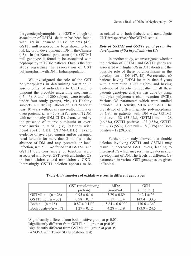
Role of GSTM1 and GSTT1 genotypes in the development of OS in patients with DN
In another study, we investigated whether the deletion of GSTM1 and GSTT1 genes are associated with higher OS in DN patients and the possible role of these polymorphisms in the development of DN (47, 48). We recruited 60 patients having T2DM for more than 5 years with albuminuria >300 mg/day and having evidence of diabetic retinopathy. In all these patients genotypic analysis was done by using multiplex polymerase chain reaction (PCR). Various OS parameters which were studied included GST activity, MDA and GSH. The prevalence of different genetic polymorphisms of GST in patients with DN was: GSTM1 positive - 32 (53.4%), GSTM1 null – 28 (46.6%), GSTT1 positive – 27 (45%), GSTT1 null – 33 (55%), Both null – 18 (30%) and Both positive – 17 (28.3%).
Further, our study showed that double deletion involving GSTT1 and GSTM1 may result in decreased GST levels, leading to increased OS which may result in greater risk for development of DN. The levels of different OS parameters in various GST genotypes are given in Table 4.
09
Table 4: Parameters of oxidative stress in different genotypes
GST (nmol/min/mg
protein) MDA
(nmol/mL) GSH
(μmol/dL)
GSTM1 null(n = 28) 0.95 ± 0.16 5.29 ± 0.89 142.1 ± 26 GSTT1 null(n = 33) 0.98 ± 0.17 5.17 ± 1.14 143.4 ± 33.3 Both null(n = 18) 0.87 ± 0.11a,b
5.84 ± 0.6 a,b,c 130.6 ± 34a
Both positive(n = 17) 1.27 ± 0.12 4.28 ± 1.19 173.9 ± 24.5
aSignificantly different from both positive group at p<0.05,
bsignificantly different from GSTT1 null group at p<0.05,csignificantly different from GSTM1 null group at p<0.05(ANOVA with Tukey SD as post-hoc test)
Genetic Basis of Diabetic Nephropathy

10
Table 5: Plasma levels of oxidant-antioxidant markers in various study groups
Parameters Group I
(HC)
(n=100)
Group II
(DM)
(n=100)
Group III
(DM-CKD)
(n=100)
GSH (mg/g Hb)
GST (nmol/min )
3.2±0.2 (3.0-3.9) 6.3±0.6 (4.1-7.8)
1.7±0.2a,c
(1.0-1.9) 8.6±0.5a,c (7.6-10.2)
0.85±0.1a,b
(0.12-1.1) 7.5±0.3a,b (6.25-8.1)
MDA (nmol/mL) 1.5±0.3 (0.9-2.2)
2.5±0.3a,c
(2.0-3.1)
5.2±0.5a,b
(4.3-6.8) FRAP (µmol/L) 542.9±54.0
(425-660)
404.8±48.4a,c
(300.8-494.9)
163±40.2a,b
(95.5-335)
Data are expressed as mean±SD.Group I- Healthy control (HC); Group II- Type 2 diabetes mellitus without nephropathy (DM) Group III- Diabetes mellitus with CKD (DM-CKD)asignificantly different from healthy control at p<0.05,
bsignificantly different from diabetic patients without nephropathy at p<0.05, csignificantly different from diabetic patients with nephropathy at p<0.05.
D. Genes Involved in Cytoprotective Pathway
NAD(P)H quinone oxidoreductase 1 (NQO1) plays a prominent role in maintaining cellular homeostasis and is an essential component of the antioxidant defense system. It
catalyzes metabolic detoxification of quinines and protects cells against chemical induced OS. Expression of NQO1 increases in response to oxidant and electrophilic radicals to counteract OS, and in fact, it is a kind of cytoprotective defense mechanism for cells.
GST levels in patients with T2DM with and without nephropathy
Hyperglycemia induced OS is implicated as a contributor to the onset and progression of T2DM and its complications like DN. GST is primarily involved in the neutralization of Reactive Oxygen Species (ROS) by enzymatic conjugation with the scavenger peptide GSH (49-51). In another study, we evaluated the role of GST along with OS markers and their correlation in patients with T2DM with and without nephropathy. We have recruited 300 study subjects divided into three groups of 100 each: HC, DM and DM-CKD. Plasma GST, MDA, reduced GSH levels and FRAP were estimated spectrophotometrically.
GST levels were found to be raised in the patient groups in comparison to HC, however,
the highest significant levels were seen in T2DM as compared to DM-CKD (p<0.05). Other oxidant and antioxidant markers are shown in Table 5. The antioxidant parameters including GSH and FRAP were significantly lower in DN as compared to T2DM and HC (p<0.05). Higher lipid peroxidation was seen in patients of DM-CKD as MDA levels were significantly raised when compared to T2DM and HC (p<0.05). We found negative correlation between HbA1c and GSH (r= -0.942, p<0.01) and FRAP (r= -0.854, p<0.01). A positive association was observed between HbA1c and GST (r= 0.606, p<0.01) and MDA (r= 0.839, p<0.01); however, a significant negative correlation between GST activity and GSH levels (r= -0.530, p<0.01) and FRAP (r= -0.294, p<0.01) was observed. GST activity was positively correlated with MDA levels (r= 0.253, p<0.01).
O.P. Kalra

11
Table 6. NQO1 levels in plasma (ng/mL) in context of different NQO1 genotypes
Groups Group I
(HC) (n=200)
Group II
(DM) (n=200)
Group III
(DN) (n=200)
p-value
NQO1*1/*1 NQO1*1/*2 NQO1*2/*2
4.16±0.47 1.9±0.46 a
0.62±0.06a,b
31.36±1.19 25.99±0.81a
20.32±1.28a,b
15.51±0.78 11.06±0.67a
7.43±0.85a,b
<0.05
Plasma NQO1 levels are given as mean±SD. Group I- Healthy Control (HC), Group II- Type 2 Diabetes Mellitus without complications (DM), Group III- Diabetic Nephropathy (DN)asignificantly different from homozygous NQO1*1/*1 in study groups at p<0.05bsignificantly different from heterozygous NQO1*1/*2 in study groups at p<0.05
Among DN and T2DM patients, the OR for the development of DN was 1.72-fold higher in T2DM patients carrying the NQO1*2/*2 genotype than in those carrying NQO1*1/*1 or NQO1*1/*2 genotypes (95% CI=1.133 to 2.600). A significant association was observed for NQO1*2 polymorphism in patients of T2DM when compared to HC (OR=6.638, 95% CI=1.427–30.876, p=0.016) . NQO1*2 polymorphism was shown to be associated with an increased risk of DN in comparison to HC (OR=22.00, 95% CI=5.075-95.376, p=0.000). Therefore, it may be concluded that NQO1*2 allele may increase the risk for developing DN in T2DM patients as well as HC.
E. Genes Involved in Nitric Oxide (NO) Pathway
NO is a major regulator of renal hemodynamics, its production being catalysed by endothelial nitric oxide synthase (eNOS). Reduction in the generation of NO acts as a deteriorating factor for progressive renal disease. Polymorphisms in the eNOS gene may alter its expression, thus affecting the production of NO. Familial clustering DN points to a role of genetic factors in the pathogenesis of renal disease.
Genetic association of NAD(P)H Quinone Oxidoreductase (NQO1*2) polymorphism with NQO1 levels and risk of DN
NQO1 catalyzes reactions having cyto-protective effect against redox cycling and OS. A single base polymorphism (C/T) at nucleotide 609 of NQO1 gene impairs the stability and function of its protein (52-54). Its role in the development of DN has not been studied earlier. We evaluated the association of NQO1*2 (rs1800566) polymorphism with plasma NQO1 activity and DN. We have screened 600 study subjects including healthy controls (HC), Type 2 diabetes mellitus without complications (T2DM) and diabetic nephropathy (DN): (200 subjects in each group) for studying NQO1*2 gene polymorphism using the PCR-RFLP.
Plasma NQO1 activity was measured by ELISA.
NQO1 activity was significantly increased in both the diseased groups, however, it was highest in T2DM patients (8 times) followed by DN (3.5 times) vs HC group. Correlation between NQO1*2 polymorphism and NQO1 activity in plasma was studied. It was found that in all the genotypes, NQO1 activity was highest in T2DM vs DN as compared to HC where it was the lowest; however, NQO1*1 allele was associated with higher NQO1 activity and NQO1*2 allele with lower activity of NQO1. SNP NQO1*2 is a functional polymorphism since it was seen to influence plasma NQO1 activity and NQO1*2 allele was associated with decreased plasma NQO1 activity (p<0.01) in T2DM, DN and HC (Table 6).
Genetic Basis of Diabetic Nephropathy

bind to specific RAGE which is expressed in many of the cell types such as endothelial cells, monocytes and lymphocytes, including β cells of pancreas (60-62). The gene for RAGE is located on the chromosome 6p21.3 near the HLA locus, and at least 30 polymorphisms have been identified of which 9 are in promoter region, 11 in exon region and 10 in intron region.
Three polymorphisms of RAGE gene namely -374T/A, -429T/C and Gly82Ser have been widely studied with regard to the development of diabetic complications in different populations all over the world. We investigated the association of -374T/A, - 4 2 9 T / C a n d G l y 8 2 S e r R A G E g e n e polymorphisms and their haplotypes with vascular complications in T2DM patients which may help in identifying DM patients predisposed to possible micro- and macrovascular complications as a result of their genetic makeup (63). A total of 427 patients of T2DM with disease duration > 5 years were enrolled in this study. These patients were divided into three groups. The first group referred to as 'DM' comprised of 140 T2DM subjects without any vascular complications. The second group referred to as 'DM-micro' consisted of 152 T 2 D M s u b j e c t s w i t h m i c r o v a s c u l a r complications (retinopathy and nephropathy). The third group referred to as 'DM-macro' consisted of 135 T2DM subjects with macrovascular complications. In addition, 176 HCs were enrolled in the study. We found that -429T/C and Gly82Ser RAGE polymorphisms were significantly a s s o c i a t e d w i t h t h e d e v e l o p m e n t o f macrovascular and microvascular complications respectively in T2DM subjects while -374A allele showed reduced risk towards the development of macrovascular complications. Further, -429T/C, -374T/A and Gly82Ser haplotype analysis revealed association of CTG haplotype with development of macrovascular complications while haplotype TAG was observed to be significantly protective towards development of macrovascular complications in
Hey et al published a meta-analysis of 24 studies and analysed the polymorphisms of eNOS genes (4b/a,G894T and T786C) associated with DN (55). It was found that 4b/a and G894T polymorphisms in the eNOS gene were associated with susceptibility to DN in Asian populations, but not in Caucasian populations. A meta-analysis of 8 studies performed by Zhou et al evaluated the association of G894T gene polymorphism alone with DN susceptibility (56). These studies included 850 cases and 1254 controls. In the Asian population, the average frequency of T-allele was 19.19% in DN patients and 8.68% in controls. A significant association was observed between the presence of T-allele and DN risk in the overall population.
We s t u d i e d t h e e N O S G 8 9 4 T polymorphism in patients of diabetes with and without nephropathy and measured the serum NO levels in these patients and compared them in the HCs. It was found that patients with DN add lowest levels of NO (22.02±16.18 µM) as compared to diabetics without nephropathy (63.86 ± 29.49 µM) and HC (38.42 ± 13.71 µM) (p< 0.001). Further a positive association was observed between eNOS G894T polymorphism as the frequency of TT genotype as well as that of mutant T allele was increased in patients with DN as compared to diabetics without nephropathy and HCs (57).
F. Genes Involved in Advanced Glycation End-products (AGE) and its Receptor (RAGE) Interaction
Association of RAGE gene polymorphism with vascular complications in patients with T2DM
Hyperglycemia associated with DM stimulates non-enzymatic glycation and oxidation of proteins and lipids leading to enhanced formation of AGEs. There is growing evidence that production and accumulation of AGEs is involved in the initiation and development of micro- and macrovascular complications observed in DM (58, 59). AGEs
12 O.P. Kalra

T2DM subjects (OR = 0.617, p = 0.0202).
G. Gene Expression Studies
Role of AGE-RAGE expression in diabetic vascular complications
Interaction of AGE with its receptor RAGE transduces multiple signals such as NAD(P)H oxidase, Mitogen-Activated Protein (MAP) kinases, extracellular signal regulated kinases, GTPase, etc. (64, 65). Activation of NAD(P)H oxidase causes enhanced Reactive Oxygen Species (ROS) generation which may lead to peroxidation and glycoxidation reactions that results in Protein Carbonyl (PCO) formation, Advanced Oxidation Protein Products (AOPP) generation and lipid peroxidation. These OS markers have been shown to be enhanced significantly in diabetic patients (66, 67). On the other hand, activation of kinases and GTPases causes activation of nuclear transcription factor including NF-kB which transcribes its target genes such as Vascular Cell Adhesion Molecule – 1 (VCAM-1), E-selectin and pro-inflammatory cytokines.
AGE-RAGE interaction is one of the mediators of vascular complications in DM; however, factors that possibly induce exaggerated AGE-RAGE interaction are not well known. RAGE is usually expressed at low levels in adults. Enhanced AGE-RAGE interaction possibly requires increased expression of RAGE. In various diseased states such as CVD, diabetes, inflammation, etc., there is higher expression of RAGE; however, conditions and factors that may induce RAGE expression particularly in T2DM have not been elucidated.
Since AGE formation is an integral phenomenon in T2DM, we investigated the dependence of RAGE expression on circulating AGE level and have examined the outcome of AGE-RAGE interaction by measurement of OS status in those patients (68). We recruited 75 patients of T2DM with disease duration > 5
years for this study. These patients were divided into three groups. The first group referred to as 'DM' comprised of 25 T2DM subjects without any vascular complications. The second group referred to as 'DM-micro' consisted of 25 T2DM subjects with microvascular complications (retinopathy and nephropathy). The third group referred to as 'DM-macro' consisted of 25 T2DM subjects with macrovascular complications. In addition, 25 HCs were also enrolled in the study by voluntary participation.
We observed that serum AGEs levels were significantly higher in diabetic patients having vascular complications as compared to T2DM without complications (p < 0.01) (Table 7). RAGE m-RNA expression level in PBMCs assayed by quantitative real time PCR was four times higher in diabetic subjects without vascular complications while DM patients having microvascular or macrovascular complications showed 12 fold and 8 fold higher RAGE m-RNA expression, respectively, compared to HCs. Further, circulating AGEs levels showed significant positive correlation with RAGE m-RNA expression and OS markers (Fig.3).
H. Epigenetic Studies
Epigenetics is the study of inherital changes in phenotype or gene expression caused by mechanisms other than changes in the underlying DNA sequence; therefore, non-genetic factors cause the organism's genes to behave differently. There is some evidence to suggest the role of epigenetic factors in the complex interplay between genes and environment. Epigenetic mechanisms include DNA methylation, histone modification and microRNAs. Few studies have suggested that hyperglycemia may induce epigenetic changes of pro-inflammatory genes, which subsequently regulate gene expression and thereby the development of vascular complications (69, 70); however, improved glycemic control for 3 – 5 years at a later stage in diabetic patients did not reduce the risk of macrovascular complications
13Genetic Basis of Diabetic Nephropathy

14
Fig. 3: Circulating AGEs shows significant positive correlation with RAGE m-RNA expression among diabetic subjects. (A) Relationship between circulating AGEs and
RAGE m-RNA expression among diabetic subjects. (B) Relationship between fluorescent AGEs and RAGE m-RNA expression among diabetic
subjects. Correlation analysis was performed using Pearson's coefficient.
Table 7. Serum AGE levels and oxidative stress markers in diabetic patients and healthy subjects
Parameters
DM
DM-micro
DM-macro
Healthy subjects
AGE ELISA (µg/mL)
1.4±0.54
3.4±0.95a
2.32±0.88a 1.12±0.38
AGE-F (AU)
1.97±0.43
2.68±0.39a
2.50±0.32a
1.87±0.29MDA (nmol/mL)
0.43±0.15
0.76±0.22a
0.81±0.37a
0.26±0.06PCO (nmol/mg protein)
1.6±0.43
2.8±1.0a
3.0±0.72a
1.4±0.43AOPP (µmol/L of chloramines T-
equivalent)
103±27.2
163±42.2a 206±54.2a
85±21.9
Data are presented as mean ± SD. Comparison between the groups was performed with one-way ANOVA and followed by post hoc
aTukey's analysis. p< 0.05 compared with controls and DM group.Advanced glycation end-product (AGE), Malonyldialdehyde (MDA), Protein carbonyl (PCO),Advanced oxidation protein product (AOPP), Diabetes mellitus (DM), Diabetes mellituswith microvascular complications (DM-micro), Diabetes mellitus with macrovascular complications (DM-macro).
25.0
20.0
15.0
10.0
5.0
0.0
0 1 2 3 4 5
r=0.835p<0.01
RA
GE
exp
ressio
n (
Nu
mb
er
of
tota
l)
r=0.557p<0.01
Blood Level of AGEs (µg/ml)
25.0
20.0
15.0
10.0
5.0
0.0
1 2 3 4
RA
GE
exp
ressio
n (
Nu
mb
er
of
tota
l)
0
Fluorescent AGEs (AU)
A
B
O.P. Kalra

significant association of -429T/C and Gly82Ser RAGE polymorphisms with the development of m a c r o v a s c u l a r a n d m i c r o v a s c u l a r complications, respectively in T2DM subjects while -374A allele showed reduced risk towards t h e d e v e l o p m e n t o f m a c r o v a s c u l a r complications. Further, we have observed that AGE-mediated exacerbation of RAGE expression may contribute to OS generation that plays a key role in pathogenesis of various vascular complications in DM.
To conclude, polymorphisms of various genes involved in RAAS, inflammatory, oxidant stress, cytoprotective and nitrous oxide pathways may influence the levels of various cy tokines and in t race l lu la r s igna l ing mechan i sms , the reby influenc ing the susceptibility of patients with DM for development of kidney disease. In addition, r a i s e d A G E a n d A O P P l e v e l s a n d polymorphisms of RAGE and enhanced RAGE mRNA expression may adversely influence final common pathway through oxidant stress mechanisms, and thereby determine the development of microvascular and/or macrovascular complications in patients with DM.
Acknowledgements
The Author would like to gratefully acknowledge the financial support provided by Department of Biotechnology, Government of India, Indian Council of Medical Research and University Grants Commission, in addition to Intramural Research Grant provided by University College of Medical Sciences, University of Delhi. In addition, immense contribution by the Faculty, PhD students, Postgraduate students and Research Scholars from the Departments of Medicine and Biochemistry (Divisions of Nephrology and Endocrinology and Gene Environment Tox ico logy Labo ra to ry ) i s s i nce re ly acknowledged. These include: Prof. SV Madhu, Prof. JK Gambhir, Prof. BD Banerjee, Prof. AK Tripathi, Prof. S Agarwal, Dr. RS Ahmed, Dr.
(71, 72). It is possible that the effects of hyperglycemia may be long term and that epigenetic changes induced by hyperglycemia may persist for more than 5 years. Based on the outcome of DCCT trial, it was hypothesized that transient exposure to hyperglycemia may induce sustained epigenetic changes and thereby increased risk of vascular complications over a longer period of time. In fact, a transient exposure to hyperglycemia induces epigenetic changes in the promoter region of NF-kB subunit of p65 and subsequently p65 expression and NF-kB activity is increased and these changes persist even after normal glucose is attained.
Key Findings and Summary
There is enough evidence from our studies to support the hypothesis that genetic factors play a crucial role in determining susceptibility of pat ients wi th DM for development of nephropathy. We have found that D allele of ACE gene acts as risk factor for the development of nephropathy in patients with T2DM as well as for nondiabetic CKD; whereas I allele of ACE gene was protective in nature. Further, whether ACE gene polymorphism can modulate the degree of beneficial reno-protective effect following ACE inhibitor therapy is currently under investigation in our laboratory. We have found that OS plays a significant role in the development of DN, and that GSTT1 and/or GSTM1 null genotypes are associated with higher OS in patients with DN. In addition, we also found that increased levels of inflammatory mediators i.e. TNF-α, hsCRP and uMCP-1 play an independent as well as interdependent roles, via several signaling pathways contributing to hyperglycemia-mediated increase in OS. We have shown that genetic polymorphism of NF-kB gene and TNF-α gene plays a significant role in determining serum level of various inflammatory markers and OS parameters. An increase in OS may further amplify inflammation, thus setting up a vicious cycle. Therefore, inflammation interlinked with OS may be major mechanisms in the pathogenesis and progression of nephropathy in susceptible diabetic patients. We found
15Genetic Basis of Diabetic Nephropathy

16
AK Yadav, Dr. A Raizada, Prof. J Rohatgi, Dr. SK Datta, Dr. A Mehndiratta, Ms. M Siddarth, Ms. S Gupta, Dr. M Sharma, Mr. D. Chawla, Dr. A Gautam, Dr. S Kumar, Dr. R Pathak, Dr. V Kumar, Neerja Aggarwal, P Kare and R Ghosh.
References
1. Wild S, Roglic G, Green A, Sicree R, King H (2004). Global prevalence of diabetes: estimates for the year 2000 and projections for 2030. Diabetes Care 27: 1047-1053.
2. Kalra OP (2008). Preventive strategies for diabetic nephropathy. In: Renal Disease -Prevent ion and Management – A Physician's Perspective, 1st edn., Chapter 10. Kalra OP, ed. New Delhi : Jaypee Brothers Medical Publishers (Pvt.) Ltd., 66-67.
3. Kalra OP (2007). Preventive strategies for diabetic nephropathy. In: Medicine Update. The Association of Physicians of India. Singal RK, ed. New Delhi : Jaypee Brothers Pvt. Ltd., 17 : 261-272.
4. Rajapurkar MM, John GT, Kirpalani AL, et al (2012). What do we know about chronic kidney disease in India: first report of the Indian CKD Registry. BMC Nephrology 13: 1-8.
5. Schrijvers BF, De Vriese AS, Flyvbjerg A (2004). From hyperglycemia to diabetic kidney disease: the role of metabolic, hemodynamic, intracellular factors and growth factors/cytokines. Endocr Rev 25: 971–1010.
6. The Diabetes Control and Complications Trial Research Group (1993). The effect of intensive treatment of diabetes on the development and progression of long-term complications in insulin-dependent diabetes mellitus. N Engl J Med 329: 977-986.
7. UK Prospective Diabetes Study (UKPDS)
Group (1998). Intensive blood-glucose control with sulphonylureas or insulin compared with conventional treatment and risk of complications in patients with type 2 diabetes (UKPDS 33). Lancet 352: 837-853.
8. Shichiri M, Kishikawa H, Ohkubo Y, Wake N (2000). Long-term results of the Kumamoto Study on optimal diabetes control in type 2 diabetic patients. Diabetes Care 23: B21-B29.
9. Venkataraman K, Kannan AT, Kalra OP, et al (2012). Diabetes self-efficacy strongly influences actual control of diabetes in patients attending a tertiary hospital in India. J Community Health 37: 653-662.
10. Fogarty DG, Hanna LS, Wantman M, et al (2000). Segregation analysis of urinary albumin excretion in families with type 2 diabetes. Diabetes 49: 1057–1063.
11. Cowie CC, Port FK, Wolfe RA, Savage PJ, Moll PP, Hawthorne VM (1989) . Disparities in incidence of diabetic end-stage renal disease according to race and type of diabetes. N Engl J Med 321: 1074-1079.
12. Collins VR, Dowse GK, Finch CF, Zimmet PZ, Linnanae AW (1989). Prevalence and risk factors for micro- and macroalbuminuria in diabetic subjects and entire population of Nauru. Diabetes 38: 1602-1610.
13. Pettitt DJ, Saad MF, Bennett PH, Nelson RG, Knowler WC (1990). Familial predisposition to renal disease in two generations of Pima Indians with type 2 (non- insul in-dependent) d iabetes mellitus. Diabetologia 33: 438-443.
14. Kalra OP, Tripathi AK, Chawla D (2014). Genet ic determinants of diabet ic nephropathy. In: Medicine Update. The
O.P. Kalra
Association of Physicians of India. Joshi SR, ed. New Delhi : Jaypee Brothers Pvt. Ltd., 24.1 : 663-674.
15. Kalra OP, Datta SK, Kumar S (2010). Genetic basis of diabetic nephropathy. In: Medicine Update. The Association of Physicians of India. Muralidhar S Rao, ed. New Delhi : Jaypee Brothers Pvt. Ltd., 20 : 695-701.
16. Hallab M, Bled F, Ebran JM (1992). Elevated serum angiotensin converting enzyme activity in type 1, insulin dependent diabet ic subjects with persistent microalbuminuria. Acta Diabetol 29: 82-85.
17. Kumar S, Kalra OP, Datta SK, Agarwal S, R o h a t g i J , Tr i p a t h i A K ( 2 0 1 0 ) . Association of angiotensin converting enzyme gene polymorphism with diabetic nephropathy and non-diabetic chronic kidney disease. Indian J Nephrol 20(Suppl): S30.
18. Yu ZY, Chen LS, Zhang LC, Zhou TB (2012). Meta-analysis of the relationship between ACE I/D gene polymorphism and end-stage renal disease in patients with diabetic nephropathy. Nephrology (Carlton)17:480-487.
19. Wang F, Fang Q, Yu N, et al (2012). A s s o c i a t i o n b e t w e e n g e n e t i c polymorphism of the angiotensin-conver t ing enzyme and d iabe t i c nephropathy: a meta-analysis comprising 26,580 subjects. J Renin Angiotensin Aldosterone Syst 13:161-174.
20. Doria A, Warram JH, Krolewski AS (1994). Genetic predisposition to diabetic nephropathy: Evidence for a role of the angiotensin I-converting enzyme gene. Diabetes 43: 690-695.
21. Marre M, Bernadet P, Gallois Y, et al
( 1 9 9 4 ) . R e l a t i o n s h i p s b e t w e e n angiotensin I-converting enzyme gene polymorphism, plasma levels, and diabetic retinal and renal complications. Diabetes 43: 384-388.
22. Schmidt S, Schone N, Ritz E (1995). Association of ACE gene polymorphism and diabetic nephropathy? The Diabetic Nephropathy Study Group. Kidney Int 47: 1176-1181.
23. Aggarwal N, Kare PK, Varshney P, et al (2017). Role of angiotensin converting enzyme and angiotensinogen gene p o l y m o r p h i s m s o n a n g i o t e n s i n converting enzyme inhibitor-mediated antiproteinuric action in type 2 diabetic nephropathy patients. World J Diab 8(3):112-119.
24. Bohle A, Wehrmann M, Bogenschutz O, et al (1991). The pathogenesis of chronic renal failure in diabetic nephropathy: investigation of 488 cases of diabetic glomerulosclerosis. Pathol Res Pract 187: 251 –259.
25. Galkina E, Ley K (2006). Leukocyte recruitment and vascular injury in diabetic nephropathy. J Am Soc Nephrol 17: 368 –377.
26. Navarro JF, Mora C (2005). Role of inflammation in diabetic complications. Nephrol Dial Transplant 20: 2601 –2604.
27. Mezzano S, Aros C, Droguett A, et al (2004). NF-kappaB activation and over-expression of regulated genes in human diabetic nephropathy. Nephrol Dial Transplant 19: 2505 –2512.
28. Sakai N, Wada T, Furuichi K, et al (2005). Involvement of extracellular signal-regulated kinase and p38 in human diabetic nephropathy. Am J Kidney Dis 45: 54 –65.
29. Gautam A, Kalra OP, Agarwal S, Gambhir
17Genetic Basis of Diabetic Nephropathy

18
JK, Gupta S, Mehndiratta M (2012). Association of nuclear factor kappa B1 gene polymorphism in relation to the risk of developing nephropathy in type 2 diabetes mellitus. Proc. of 43rd Annual Conference Indian Society of Nephrology. Dec. 6-9, 28-29.
30. Gambhir JK, Sharma M, Gupta S, Mehndiratta M, Shukla R, Kalra OP (2013). Evaluation of nuclear factor-ķB levels in diabetic patients with and without nephropathy. Proc. of 40th Annual Conference of Association of Clinical Biochemists of India. Dec. 3-6, 131-132.
31. Parving HH, Osterby R, Ritz E (2000). Diabetic nephropathy. In: The Kidney. Brenner BM, ed. Philadelphia: WB Saunders Company, 1731–1773.
32. Rovin BH, Rumancik M, Tan L, D ickerson J (1994) . Glomeru la r expression of monocyte chemoattractant protein-1 in experimental and human g l o m e r u l o n e p h r i t i s . L a b I n v e s t 71:536–542.
33. Oppenheim JJ, Zachariae C, Mukaida N, Matsushima K (1991). Properties of the novel pro-inflammatory super gene “intercrine” cytokine family. Annu Rev Immunol 12:503–633.
34. Banba N, Nakamura T, Matsumura M, Kuroda H, Hattori Y, Kasai K (2000). Possible relat ionshipof monocyte chemoattractant protein-1 with diabetic nephropathy. Kidney Int 58:684–690.
35. Gupta S, Gambhir JK, Kalra OP, et al (2013). Association of biomarkers of inflammation and oxidative stress with the risk of chronic kidney disease in Type 2 diabetes mellitus in North Indian population. J Diab Compl 27: 548-552.
36. Gupta S, Gambhir JK, Mehndiratta M, et
al (2012). Association of tumor necrosis factor-alpha and oxidative stress in diabetic nephropathy. AACC Annual Meeting Abstracts, E-174: p A-288.
37. Gambhir JK, Gupta S, Gautam A, et al (2012). A comparative study of monocyte chemoattractant protein-1 and oxidative stress in diabetic nephropathy. AACC Annual Meeting Abstracts, E-174: p A-288-289.
38. Gupta S, Gambhir JK, Kalra OP, et al (2011). A comparative study of oxidative stress parameters in diabetic and non-diabetic chronic kidney disease. Indian J Clin Biochem 26: 56-57.
39. Gupta S, Mehndiratta M, Kalra S, Kalra OP, Shukla R, Gambhir JK (2015). Association of tumor necrosis factor (TNF) promoter polymorphisms with plasma TNF-α levels and susceptibility to diabetic nephropathy in North Indian population. J Diab Compl 29:338-342.
40. Galli F, Rovidati S, Benedetti S, et al (1999). Over-expression of erythrocyte glutathione S-transferase in uremia and dialysis. Clin Chem 45:1781–1788.
41. Hayek T, Stephens JW, Hubbart CS, et al (2006). A common variant in the glutathione S-transferase gene is associated with elevated markers of inflammation and lipid peroxidation in sub jec t s wi th d iabe tes me l l i tu s . Atherosclerosis 184:404–412.
42. Fujita H, Narita T, Meguro H, et al (2000). No associat ion of glutathione S-transferase M1 genetic polymorphism with diabetic nephropathy in Japanese Type 2 diabetes patients. Ren Fail 22: 479-486.
43. Yang Y, Kao M, Chang C, et al (2004).
O.P. Kalra

Glutathione S-transferase T1 deletion is a risk factor for developing end-stage renal disease in diabetic patients. Int J Mol Med 14: 855-859.
44. Kim JH, Moon MK, Kim SW, et al (2005). Glutathione S-Transferase M1 gene polymorphism is associated with Type 2 diabetic nephropathy. J Korean Diab Assoc 29: 315-321.
45. Datta SK, Kumar V, Pathak R, et al (2010). Association of glutathione S-transferase M1 and T1 gene polymorphism with ox ida t ive s t r e s s in d i abe t i c and nondiabetic chronic kidney disease. Ren Fail 32: 1189-1195.
46. Datta SK, Kumar V, Pathak R, et al (2009). Association of GSTM1 and GSTT1 genetic polymorphism with diabetic and non-diabetic chronic kidney disease and its role in causing renal injury. Indian J Nephrol 19: S1.
47. Datta SK, Kumar V, Ahmed RS, Tripathi AK, Kalra OP, Banerjee BD (2010). Effect of GSTM1 and GSTT1 double deletions in the development of oxidative stress in diabetic nephropathy patients. Indian J Biochem Biophysics 47: 100-103.
48. Datta SK, Kumar V, Ahmed RS, Tripathi AK, Kalra OP, Banerjee BD (2009). Role of concomitant deletion of glutathione S-transferase M1 and T1 genes in the development of oxidative stress in diabetic nephropathy. JAPI 57: 838.
49. Maritim AC, Sanders RA, Watkins JB 3rd (2003). Diabetes, oxidative stress, and antioxidants: a review. J Biochem Mol Toxicol 17: 24-38.
50. Hayes JD, Flanagan JU, Jowsey IR (2005). Glutathione transferases. Annu Rev Pharmacol Toxicol 45: 51-88.
51. Sharma M, Gupta S, Singh K, et al (2016).
Association of Glutathione-S-transferase with patients of type 2 diabetes mellitus with and without nephropathy. Diab Metabol Synd: 10 : 194-197.
52. Dinkova-Kostova AT, Talalay P (2010). N A D ( P ) H : q u i n o n e a c c e p t o r o x i d o r e d u c t a s e 1 ( N Q O 1 ) , a multifunctional antioxidant enzyme and exceptionally versatile cytoprotector. Arch Biochem Biophys 501:116-123.
53. Eickelmann P, Schulz WA, Rohde D, Schmitz-Dräger B, Sies H (1994). Loss of heterozygosity at the NAD(P)H: quinone oxidoreductase locus associated with increased resistance against mitomycin C in a human bladder carcinoma cell line. Biol Chem Hoppe Seyler 375:439-445.
54. Sharma M, Mehndiratta M, Gupta S, Kalra OP, Shukla R, Gambhir JK (2016). Genetic association of NAD(P)H Quinone O x i d o r e d u c t a s e ( N Q O 1 * 2 ) polymorphism with NQO1 levels and risk of diabetic nephropathy. Biol Chem 397(8):725-730.
55. Hey Y, Fan Z, Zhang J, et al (2011). Polymorphisms of eNOS gene are associated with diabetic nephrology: a meta-analysis. Mutagenesis 26: 339-349.
56. Zholu TB, Xu HL, Yin SS (2013). Association between endothelial nitric o x i d e s y n t h a s e G l u 2 9 8 A g e n e polymorphism and diabetic nephropathy susceptibility. Renal Failure 35 (1): 173-178.
57. Bhardwaj N, Sharma S, Kalra OP, Yadav AK, Sikka M, Sharma S (2016) . Endothelial nitric oxide synthase gene polymorphism in diabetic patients with and without nephropathy. J Am Soc Nephrol 27 : 429A.
58. Nin JW, Jorsal A, Ferreira I, et al (2011).
19Genetic Basis of Diabetic Nephropathy
20
Higher plasma levels of advanced glycation end products are associated with incident cardiovascular disease and all-cause mortality in type 1 diabetes. Diabetes Care 34:442–447.
59. Goh SY, Cooper ME (2008). The role of advanced glycation end products in progression and complications of diabetes. J Clin Endocrinol Metab 93:1143-1152.
60. Yan SF, Ramasamy R, Schmidt AM (2008). Mechanisms of disease: advanced glycation end-products and their receptor i n i n fl a m m a t i o n a n d d i a b e t e s complications. Nat Clin Pract Endocrinol Metab 4:285-293.
61. Herold K, Moser B, Chen Y, et al (2007). Receptor for advanced glycation end products (RAGE) in a dash to the rescue: inflammatory signals gone awry in the primal response to stress. J Leukoc Biol 82:204-212.
62. Clynes R, Moser B, Yan SF, Ramasamy R, Herold K, Schmidt AM (2007). Receptor for AGE (RAGE): weaving tangled webs within the inflammatory response. Curr Mol Med 7:743-751.
63. Tripathi AK, Chawla D, Bansal S, Banerjee BD, Madhu SV, Kalra OP (2014). Association of RAGE gene p o l y m o r p h i s m w i t h v a s c u l a r complications in Indian type 2 diabetes mellitus patients. Diab Res Clin Prac 103: 474-481.
64. Higashi Y, Noma K, Yoshizumi M, et al (2009). Endothelial function and oxidative stress in cardiovascular diseases. Circ J 73: 411-418.
65. Wautier MP, Chappey O, Corda S, et al (2001). Activation of NADPH oxidase by AGE links oxidant stress to altered gene expression via RAGE. Am J Physiol
Endocrinol Metabol 280: 685–694.
66. Kaneda H, Taguchi J, Ogasawara K, et al (2002). Increased level of advanced oxidation protein products in patients with coronary artery disease. Atherosclerosis 162: 221-225.
67. Pan HZ, Zhang H, Chang D, et al (2008). The change of oxidative stress products in diabetes mellitus and diabetic retinopathy. Br J Ophthalmol 92: 548–551.
68. Chawla D, Bansal S, Banerjee BD, Madhu SV, Kalra OP, Tripathi AK (2014). The role of advanced glycation end products (AGEs)-induced receptor (RAGE) e x p r e s s i o n i n d i a b e t i c v a s c u l a r complications. Microvasc Res 95:1-6.
69. Villeneuve LM, Reddy MA, Lanting LL, Wang M, Meng L, Natarajan R (2008). E p i g e n e t i c h i s t o n e H 3 l y s i n e 9 methylation in metabolic memory and inflammatory phenotype of vascular smooth muscle cells in diabetes. Proc Natl Acad Sci USA 105: 9047– 9052.
70. Miao F, Smith DD, Zhang L, Min A, Feng W, Natarajan R (2008). Lymphocytes from patients with type 1 diabetes display a distinct profile of chromatin histone H3 lysine 9 dimethylation: an epigenetic study in diabetes. Diabetes 57: 3189– 3198.
71. Action to Control Cardiovascular Risk in Diabetes Study Group (2008). Gerstein HC, Miller ME, Byington RP, Goff DC Jr, et al. Effects of intensive glucose lowering in type 2 diabetes. N Engl J Med ; 358: 2545– 2559.
72. ADVANCE Collaborative Group, Patel A, MacMahon S, Chalmers J, et al (2008). Intensive blood glucose control and vascular outcomes in patients with type 2 diabetes. N Engl J Med 358: 2560– 2572.
O.P. Kalra

Essential Skills in Postgraduate Medical Curriculum of Community Medicine
1 2 1Jugal Kishore , Tanu Anand , Sneha Kumari
1Vardhman Mahavir Medical College & Safdarjng Hospital, New Delhi ,2
North Delhi Municipal Corporation Medical College & Hindu Rao Hospital, Delhi .
Ann Natl Acad Med Sci (India), 53(1): 21-29, 2017
Correspondence : Dr. Jugal Kishore, Director Professor & Head, Department of Community Medicine, Vardhman Mahavir Medical College & Safdarjung Hospital, New Delhi.
GOLDEN JUBILEE COMMEMORATION AWARD LECTURE delivered during NAMSCON 2016 at the All India Institute of Medical Sciences, Raipur.
Introduction: Community-based education has been considered a suitable approach for health promotion and for requisite skill development regarding primary health care. In the current perspective, public health training and research, being two important aspects require immediate attention.
Objective: To assess the skills of Postgraduate Students in the Department of Community Medicine in four Medical Colleges of Delhi.
Materials and Methods: It was a cross-sectional study conducted among 70 Postgraduate Medical Students of 4 Medical Colleges in Delhi. The data were collected through a self administered, pre-tested questionnaire containing items assessing socio-demographic profile and skills essential for Postgraduate Students of Community Medicine.
Results: There were 58.6% male and 29% female students. A large proportion of participants were having age range between 25-29 years. Ability 'to resolve conflict among the nurse at Primary Health Centre (PHC)', 'generate community participation', 'making thick and thin smear in case of fever', 'making a chart showing month-wise distribution of CuT', and 'calculating Chi-square of data', were found to significantly higher in 2nd and 3rd year PG students than first year PG students (p<0.01). Only 27.1% of students felt that they could test water sample for microbiological aspects while only 47.1% said that they could examine an industrial worker for pre-placement examination.
Conclusions: PG students assessed themselves to possess necessary skills on communication, counselling and health education. However, many students lacked skills pertaining to occupational health and epidemiology.
Keywords: Competency in community medicine, public health, epidemiological skills, communication skills.
ABSTRACT

22
Introduction
Based on the principles outlined at Alma-Ata in 1978, there is an urgent call for revitalizing primary health care. India is not only committed to strengthening primary care but is already well aware of the means to achieving this goal (1). The National Rural Health Mission (NRHM) i s a g rea t t e s t amen t t o t he determination of the Indian government to deliver universal primary health care and it has had impact also (2). However, there are a number of operational issues that need to be addressed to ensure that primary care is delivered effectively. And, some of the major problems in implementation and practice of primary health care relate to training and capacity building of health service providers in foreseeable future (3). It is suggested that human resources with requisite knowledge, skills, attitudes, values and responsiveness to people's health needs and health promotion are needed.
Community-based education has been considered a suitable approach for health promotion and for requisite skill development regarding primary health care (4, 5). In the current perspective, public health training and public health research, being two important aspects require immediate attention. The Task Force on Medical Education for the NRHM and National Health Policy 2002 have recommended increase in postgraduate seats in the discipline of Community Health/Public Health/Preventive, Social Medicine and Family Medicine (3). Thus, it is evident that in current scenario, public health specialists are the need of the hour in community health care.
The objectives and goals of Postgraduate (PG) Medical Education in Community Medicine are to produce competent specialists to manage the teaching departments in the Medical Colleges, or to manage health services and national health programs at various levels or to conduct biomedical research in the discipline of
Community Medicine (6). During training, PG are expected to acquire a substantial knowledge and necessary skills in: concepts of health and illness and their determinants, methods in c o m m u n i t y h e a l t h , h e a l t h s e r v i c e s organizations, community health programs and communication and advocacy (7).
There are regional variations in the way PG training in India is imparted by various institutions. There is no well defined curriculum worked out, if at all it is there, the methods of acquiring the desired competencies are vague or not at all stated (6). Though Medical Council of India has reiterated competency-based PG training in community health care, yet, detailed structured curriculum in PG training in Community Medicine is being followed in handful institutions only (8).
Considering the fact acquiring relevant public health skills during PG training is pertinent, the present study was undertaken to assess the skills of PG students in department of Community Medicine in four Medical Colleges of Delhi. This would also enable us to know the variations in PG training and evaluate whether the medical pedagogic approach is in line with objectives of such training. Materials and Methods
Study Setting and Study Participants
It was a cross-sectional study conducted among PG Medical Students (Doctor of Medicine: MD) of 4 Medical Colleges in Delhi. Every year, 8-9 students are admitted in each of the Medical Colleges; therefore, there are around 24-27 students in each of the Department of Community Medicine of a Medical College, studying at any given time. The entire course of MD is of 3 years. The PG Students are posted for practical training in primary health care in Department of Community Medicine from first year onwards.
Jugal Kishore

Description of Curriculum
As per MCI 2000 regulations, the major components of PG Curriculum in Community Medicine constitutes:1. Theoretical knowledge2. Practical and clinical skills (Managerial
skills)3. Thesis skills4. Attitudes including communication skills5. Training in research methodology.
In order to achieve this practical training in postgraduation in Medical Colleges of Delhi is divided as follows:
The PG students are posted on rotational basis in the field practice areas of their respective Departments in different Medical Colleges for their practical training. The field areas usually cover the urban slums or resettlement colonies and rural areas include villages. The posting is of 3-6 months duration in each area. During this posting, PG students are expected to know the community, establish rapport with the community and provide essential primary care to the community. They study the community and learn to make community diagnosis. Further, they learn the implementation of national health programs at field level. They are also expected to learn management of human resources. Sometimes, their research work is also carried out in these areas.
PG students are also posted in Family H e a l t h C a r e p r o g r a m ( F H C P ) f o r undergraduates which is covered in 4th and 5th semester and epidemiology and biostatistics which are covered in 6th and 7th semester. Besides, some extra-mural visits are arranged during this posting that includes visit to a health cen t re , Anganwadi , Medica l Records Department, Nursing College for injection techniques, MCH centre, Institute of Physically H a n d i c a p p e d ( I P H ) , a n d C e n t r e f o r Environmental and Occupational Health (CEOH) for Biomedical Waste Management
t r a i n i n g . I n a d d i t i o n t o t h i s , h e a l t h communication classes are also held where in students learn the skills of imparting health education in a community.
PG students are also scheduled to present journal articles and take seminars on topics of public health importance. It is mandatory to do research work towards a thesis for award of the PG degree in their third year of postgraduation, they are also posted in various public health institutes and agencies like National Centre for Disease Control (NCDC), National Institute of Health and Family Welfare (NIHFW), Municipal Corporation of Delhi (MCD), New Delhi Municipal Corporation (NDMC), etc. They are also posted in the Department of Microbiology of their respective colleges for learning organisms of public health importance. Additionally, the students participate in seminars, symposia, conferences and workshops for enhancing their knowledge about the advances in public health. They are encouraged to present their research work in these fora.
The students are finally evaluated at the end of third year. There are 4 theory papers followed by 2-day practical exam that includes long case, two short cases, epidemiological and statistical exercises, spots and viva voce. Some colleges also hold microbiology practicals.
Sample Size and Sampling
There are about 8-9 PG seats for the subject in the studied Medical Colleges of Delhi. All the PG Students during the study period were included in the study. There were total of 96 PG seats in three years in the four Medical Colleges (24 in each college). Out of the 96, only 90 were filled and only 70 returned the completed questionnaires. Hence, they were included for analysis.
Study Tool
The study tool was developed after e x t e n s i v e r e v i e w o f c u r r i c u l u m o f
23Essential Skills in Postgraduate Medical Curriculum of Community Medicine

24
Postgraduation in Community Medicine in various institutes. The questionnaire so developed was self-administered, pre-tested and consisted of two parts. The first part contained items to gather socio-demographic information of the participants. The second part consisted of various questions to assess public health skills. To identify skills needed for PG students, emails to 50 faculty of Community Medicine were sent and their responses were listed. Questions were framed covering all skills suggested by faculty members. The skills that were assessed covered following areas: communication with the community, school health, human resource management, social mobilization, laboratory services, management of public health problem, fami ly p lanning counse l l ing , graphic representation of data, immunization, nutrition, water testing and disinfection, occupational health, health education, investigation of an ep idemic , s t a t i s t i ca l t e s t s , inven tory management, evaluation of national health program, qualitative research, and surveillance.
Methodology
All the PG students of four medical colleges were contacted and the objective of study was explained to them. Informed written consent was obtained from them and then the questionnaires were given to them. The students were asked to return the filled questionnaire in 3 days. Those who failed to return were contacted again. Those students who failed to submit the filled questionnaire even after 2 remainders were excluded from the study.
Ethical Considerations
The privacy and confidentiality of data was ensured. The ethical approval of the research was obtained from the Institutional Ethics Committee (IEC) of the research institute.
Statistical Analysis
The obtained data were collected, entered and analyzed in SPSS (version 17.0). Data were
expressed in terms of percentages. Differences between the proportions were observed by Chi-square test and p < 0.05 was considered as significant. Difference between independent groups for continuous variables was assessed using student t test (2 groups) and One-way ANOVA (for more than 2 groups).
Results
Out of the total 70 respondents, 58.6% were male and 29% were females. Large proportion of participants was between the age of 25-29 years. More than half of the participants had their undergraduation from a private college (51.4%). Majority of the students belonged to general category (n=49; 70%). There were 29 s t u d e n t s w h o w e r e i n fi r s t y e a r o f postgraduation, 18 were in 2nd year while 23 students were studying in 3rd year. While 34.3% students had graduate mothers, half of the PG students had Fathers with professional education. Majority of students said that their mothers were housewives (87.1%) while half had fathers engaged in semi-professional occupation (Table 1).
There were 22 competencies that were self-assessed by the students. Most of the competencies did not show year of PG wise difference. However, ability 'to resolve conflict among the nurse at Primary Health Centre (PHC)', 'generate community participation', 'making thick and thin smear in case of fever', 'making a char t showing month-wise distribution of CuT', and 'calculating Chi-square of data', was found to be significantly higher in 2nd and 3rd year PG students than first year PG students (p<0.01). Only 27.1% of students felt that they could test water sample for microbiological aspects while only 47.1% said that they could examine an industrial worker for pre-placement examination. Abil i ty to evaluative DOTS or any national health program and manage inventory of health centre was possessed by 48.6% and 54.3%, respectively (Table 2).
Jugal Kishore

25
Table 1: Socio-demographic Characteristics of Students (n=70)
Variables Frequency (%)Gender
-
Male
-
Female
41 (58.6)
29 (41.4)
Age
-
20-24 years
-
25-29 years
-
30-34 years
4 (5.7)
61 (87.2)
5 (7.1)
Type of medical college
(where MBBS done)
-
Government
-
Private
34 (48.6)
36 (51.4)
Category
-
General
-
Scheduled Caste (SC)
-
Scheduled Tribe (ST)
-
Other Backward Class (OBC)
49 (70.0)
9 (12.9)
5 (7.1)
7 (10.0)
Year of Postgraduation
-
1st
year
-
2nd
year
-
3rd
year
29 (41.4)
18 (25.7)
23 (32.9)
Mother’s education
-
Professional
-
Graduate
-
12th standard
-
10th standard
-
Middle school
-
Primary school
-
Illiterate
16 (22.8)
24 (34.3)
11 (15.8)
11 (15.8)
2 (2.8)
1 (1.4)
5 (7.1)
Father’s education
-
Professional
-
Graduate
-
12th standard
-
10th standard
-
Primary school
- Illiterate
35 (50)
26 (37.1)
4 (5.7)
3 (4.3)
1 (1.4)
1 (1.4)
Mother’s occupation
- Professional
- Semi-professional
- Housewives
2 (2.9)
7 (10.0)
61 (87.1)
Father’s occupation
- Professional - Semi-professional - Clerical, shop owners, etc. - Skilled worker - Unskilled worker- Unemployed
11 (15.8) 35 (50) 12 (17.1) 2 (2.9) 1 (1.4)9 (12.9)
Essential Skills in Postgraduate Medical Curriculum of Community Medicine

26
Table 2: Skill Assessment of Postgraduate Students
Skill Year of Postgraduation (n, %) Total1st
Year 2nd Year 3rd
Year Communicate with a senior family member at home and in field to get the information about socio-demography
26 (89.7) 15 (83.3) 20 (87) 61 (87.1);p=0.82
Plan and carrying out school health program in a school
16 (55.2)
11 (61.1)
15 (65.2)
42 (60);p=0.76
Resolve conflict between two nurses working under you at Primary Health Centre (PHC)
14 (48.3)
16 (88.9)
18 (78.3)
48 (68.6); p=0.007
Generate community participation for clean surrounding of their houses
15 (51.7)
15 (83.3)
23 (100)
53 (75.7);p<0.001
Negotiate with local NGO’s for decreasing female feticide/ANC/Institutional delivery
16 (55.2)
11 (61.1)
15 (65.2)
42 (60);p=0.76
Make thick and thin smear
at home of patient with fever
10 (34.5)
13 (72.2)
18 (78.3)
41 (58.6);p=0.003
Categorize TB patients based on the sputum exam and history at PHC
26 (89.7)
17 (94.4)
22 (95.6)
65 (92.8);p=0.67
Motivate a couple to use contraceptives of their choice
27 (93.1)
18 (100)
23 (100)
68 (97.1);p=0.23
Make chart showing month-wise distribution of Copper T users
5 (17.2)
16 (88.9)
21(91.3)
42 (60);p<0.001
Plot weight of a <5 child on “Road to health” card
25 (86.2)
17 (94.4)
22 (95.6)
64 (91.4);p=0.42
Give BCG
vaccine to an infant
28 (96.6)
17 (94.4)
23 (100)
68 (97.1); p=0.22
Plan a diet for a child suffering from grade 2 PEM
17 (58.6)
14 (77.8)
19 (82.6)
50 (71.4);p=0.14
Chlorinate one bucket of water at home
17 (58.6)
10 (55.6)
17 (73.9)
44 (62.9);p=0.40
Examine thoroughly an
industrial worker for pre-placement examination
11 (37.9)
9 (50)
13 (56.5)
33 (47.1);p=0.40
Deliver a health talk on diarrhoea prevention and control at health centre
27 (93.1)
17 (94.4)
23 (100)
67 (95.7);p=0.45
Investigate an epidemic of food poisoning in a village/hostel
14 (48.3)
12 (66.7)
15 (65.2)
41 (58.6);p=0.34
Calculate Chi- square value for a given data
13 (44.8)
13 (72.2)
21 (91.3)
47 (67.1);p=0.002
Manage the inventory/drug store at the health centre
15 (51.7)
7 (38.9)
16 (69.6)
38 (54.3);p=0.14
Evaluate a DOTS/Any other program in your district/state
14 (48.3)
6 (33.3)
14 (60.9)
34 (48.6);p=0.22
Hold a focus group discussion for evaluating barriers in early initiation of breastfeeding
24 (82.8) 16 (88.9) 22 (95.7) 62 (88.6);p=0.35
Test the water sample for micro-biological aspects
8 (27.6) 4 (22.2) 7 (30.4) 19 (27.1);p=0.84
Carry out dengue/vector borne surveillance in a village or college
14 (48.3) 13 (72.2) 17 (73.9) 44 (62.9);p=0.10
Jugal Kishore

Univariate analysis of public health skills with socio-demographic factors showed that students who are females, having graduation from Government College, being in 3rd year of PG, aged younger, mother's and father's professional education and occupation, have higher skill score than others, though the difference was not significant (Table 3).
Discussion
The medical education regulatory authority Medical Council of India (MCI) has defined essential competencies to be acquired at the end of PG education in Community Medicine. These include managerial and epidemiological skills in health care delivery system and national health programs, skills to
identify community health needs, research methodology and abilities to impart training to medical and paramedical personnels (9). The present study was undertaken to assess some of these skills among the PG students of four Medical Colleges in Delhi.
Self assessment of communication and counselling skills by the PG students revealed that majority of them considered themselves to be competent to communicate with a family member of a patient and motivate eligible couple for contraceptive use. This finding is in contrast to the findings of study by Avan et al in 2005 in Pakistan (10). These high proportions give some credence to recognition by PG students of the importance of effective communication with the community and their patients. Further, interaction with people and their organization in
27
Variables Score (mean±s.d.) p valueGender
-
Male (n=41)
-
Female (n=29)
15.71±4.11
17.00±3.79
0.185
Type of Medical College (from where MBBS done)
-
Government
(n=34)
-
Private
(n=36)
16.68±3.63
15.83±4.44
0.383
Year of Postgraduation
-
1st year
(n=29)
-
2nd year
(n=18)
- 3rd year (n=23)
15.34±4.05
16.28±3.01
17.35±4.49
0.203
Age
- 20-24 years (n=4) - 25-29 years (n=61) - 30-34 years (n=5)
17.00±2.16 16.44±3.88 13.20±5.85
0.206
Mother’s occupation - Professional & Semi-professional (n=9) -
Others (n=61)
20.33±2.08 16.06±3.98
0.070
Father’s occupation -
Professional & Semi-professional (n=44)
-
Others (n=26)
16.39±3.51 15.96±4.89
0.671
Mother’s education -
Professional (n=16)
-
Others (n=59)
16.44±3.46
16.19±4.18
0.829
Father’s education
- Professional (n=35)- Others (n=35)
16.94±3.2415.54±4.58
0.145
Table 3: Univariate Analysis of Total Skills Scores with Socio-demographic Variables
Essential Skills in Postgraduate Medical Curriculum of Community Medicine

28
urban and rural settings offers a wonderful opportunity for learning from people (9). Epidemiology is an important part of C o m m u n i t y M e d i c i n e a n d a c q u i r i n g epidemiological competencies is essential for community physician (11). In the current study, only 58.6% considered themselves skilled enough to investigate an epidemic. Outbreak investigation is an opportunity to contain the epidemic and prevent future occurrence and is the best example for participatory and active learning of epidemiology. The finding highlights the need to focus on this skill among PG s tuden t s dur ing the i r t r a in ing in epidemiology. Similarly planning evaluation of national health program or carrying out surveillance of a vector borne disease are essential components of epidemiology that offer opportunity to learn multiple things.
It is a ritual to establish public health laboratory to meet the requirements of MCI. However, at many places the laboratories have not been developed. Due to lack of infrastructure in the field areas, the skills in laboratory organization for organisms of public health importance is mostly lacking among PG students as was evident in the current study as well. Even though, the students were posted in public heal th laboratory of MCD and Microbiology Department of their respective colleges, many students found it hard to make smears for malaria diagnosis (41.4%) or test water sample for quality (72.9%). This finding points to the fact even though knowledge can be gained, learning is best experienced by doing, skills need to be practiced repeatedly to develop perfection (9).
In India, the requirement of occupational health manpower is accomplished by medical graduates, community medicine specialists, and public health specialists working as industrial medical officers (12). Thus, it is evident that PG student of Community Medicine should be trained in occupational health. However, in the present study, little less than half felt that they
can do pre-placement examination of an industrial worker. This is probably is due to the fact occupational health training in India is most neglected in medical curriculum.
Limitations of Study
Since our study relied on self-reporting by the PG students, this may not correspond to their performance in actual clinical and community settings. The study also could not include reporting from community people and patients themselves, which might have strengthened the results and reflected their actual behavior. Also this was a cross-sectional study involving Medical Colleges in Delhi only. The variability in PG curriculum that exists in different medical colleges and universities of the country has not been investigated in the present study.
Nevertheless, the study highlights the skills which should be possessed by a PG student of Community Medicine. In the present study, though PG students assessed themselves to possess necessary skills on communication, counseling and health education, some skills related to occupational health, public health laboratory and epidemiology need to be inculcated among them with greater stress and indicate that these needed to be included in the PG Curriculum of the MD Program in Community Medicine. Further, there is a need to reform the curr iculum in terms of competencies in several areas.
References
1. Rao M, Mant D (2012). Strengthening primary healthcare in India: white paper on opportunities for partnership. Br Med J 344: e3151.
2. Bhutta ZA, Chopra M, Axelson H, et al (2010). Countdown to 2015 decade report (2000-10): taking stock of maternal, newborn, and child survival. Lancet 375: 2032-2044.
Jugal Kishore
3. MoHFW. Report: Task Force on Medical Education for the National Rural Health Mission. New Delhi : MoHFW: GoI.
4. The World Summit on Medical Education (1993). Med Educ 27:467-469.
5. Mash B, Villiers M (1999). Community –based training in Family Medicine a different paradigm. Med Educ 33:725-729.
6. Lal S (2004). Scenario of postgraduate medical educat ion in community medicine in India. Indian J Com Med 29(2):56-61.
7. Community Medicine. [Internet] (2013). Available from: http://www.oiucm.org/ community_medicinepublic_health.htm [cited on 2013 Dec 8].
8. Anonymous (2011), MCI. Vision 2015. New Delhi: Medical Council of India.
9. Lal S, Kumar R, Prinja S, Singh GPI (2011). Postgraduate teaching and evaluation in community medicine: Challenges ahead. Indian J Prev Soc Med 42(3):220-225.
10. Avan BI, Raza SA, Afridi HR (2005). Resident's perception of communication skills in postgraduate medical training programs of Pakistan. J Postgrad Med 51:85-91.
11. WHO (2011). Improving the teaching of public health at undergraduate level in medical schools-suggested guidelines. New Delhi: SEARO, WHO. Available from: http: //www.searo.who.int/entity/ primary_health_care/documents/pch_sea_hsd_344.pdf.
12. Tiwari RR, Sharma A, Zodpey SP (2016). Occupational health training in India: Need for a competency-driven approach. Indian J Occup Environ Med 20:39-43.
29Essential Skills in Postgraduate Medical Curriculum of Community Medicine

Neurodevelopmental Disorders: The Journey, the Dreams and their Realization
Sheffali GulatiChild Neurology Division, Department of Pediatrics, All India Institute of Medical Sciences, New Delhi.
Ann Natl Acad Med Sci (India), 53(1): 30-35, 2017
Correspondence : Prof. Sheffali Gulati, Chief, Child Neurology Division, Department of Pediatrics, All India Institute of Medical Sciences, New Delhi-110029.
DR. S. JANAKI MEMORIAL ORATION delivered during NAMSCON 2016 at the All India Institute of Medical Sciences, Raipur.
Neuro Developmental Disorders (NDDs) are associated with significant morbidity. This involves early identification of the disorder, the correct management of the disorder and associated disabilities. In India, the paucity of trained personnel and lack of knowledge about these disorders has been instrumental in inadequate management and recognition of these NDDs. The Child Neurology Division, Department of Pediatrics at All India Institute of Medical Sciences has made few noteworthy and meaningful contributions in these aspects: devising a DM curriculum for pediatric neurology, developing indigenous tools for diagnosing these NDDs and performing relevant research. These endeavors would go a long way in serving the children with NDDs.
Keywords: Neurodevelopmental disorders, autism, cerebral palsy, attention deficit hyperactivity disorder.
ABSTRACT
Introduction
Neuro Developmental Disorders (NDDs) constitute a significant proportion of morbidity handled by pediatric health care services. These include cerebral palsy, autism, attention deficit hyperactivity disorder (ADHD) and intellectual disability, epilepsy, autoimmune disorders including autoimmune encephalitis and neuromuscular disorders (including muscular dystrophy). A recent study estimated prevalence of 'any NDD' in 2–9 year old children in India to be 12·0% (95% CI: 10·9–13·2%), and 21·8% of these had more than one NDD (unpublished data). Thus, neurological disorders represent a significant cause of morbidity among children.
In addition, there has been a paucity of physicians trained in pediatric neurology and also, inadequate research focusing on disorders specific to our nation, which has further compromised care of these children. Hence, to overcome these lacunas, the Child Neurology Division, All India Institute of Medical Sciences has taken enormous steps over the past several years. These have been highlighted in subsequent sections.
DM Pediatric Neurology
In order to overcome the scarcity of physicians trained in Pediatric Neurology in our country, Pediatric Neurology program was

initiated in 2004, at All India Institute of Medical Sciences, under the Child Neurology Division as a part of Department of Pediatrics. Over the past 12 years, twenty-five residents have qualified as DM Pediatric Neurology. These residents are subjected to rigorous training under all aspects of clinical care as well as neuroradiology, neuropathology and other clinical and rehabilitative aspects of child neurology. The DM program at AIIMS has contributed to trained manpower not only in our country, but has also trained fellows from other SAARC nations, United States of America, France, and United Kingdom.
Research
There has been scarcity of research in pediatric neurological disorders from our country. Numerous national and international research projects have been undertaken at the Child Neurology Division, AIIMS, which contributed to the arena of epilepsy, autism, other NDD (l ike cerebral palsy) , and autoimmune disorders. The division has been a pioneer in the country in developing dietary therapies for drug resistant epilepsy (1) . The children whose epilepsy fails to be adequately controlled despite two antiepileptic drugs are defined to have drug resistant epilepsy (2) . The
therapeutic options for these children include epilepsy surgery or dietary therapy. The division has demonstrated the efficacy of these dietary therapies in drug resistant epilepsies using ketogenic diet, modified Atkins diet, and low glycemic index dietary therapy (3-7). In addition, it has contributed to the development of 2.5:1 ketogenic diet and proved its efficacy in institutional research (3) . The division also, has been the frontrunner in diagnosing, managing and optimizing the treatment for autoimmune encephalitis (8) , leukodystrophies (9) , and acquired demyelinating disorders of CNS (10) . Also, the research focusing on domiciliary management of int ranasal and buccal midazolam has helped to standardize the management protocols for seizures at home (11). These researches have been instrumental in developing various therapeutic guidelines, and have also paved the way for various national programs including Rashtriya Bal Swasthya Karyakram (RBSK) (12).
Public Health Initiatives
Educating the parents and children and the society at large is an important aspect about management of any disease. The Child Neurology Division has played a pivotal role in educating the society about NDDs (Fig. 1),
31
Fig. 2 : The building of AIIMS, New Delhi being lit blue on 30 Apr 2015 to
commemorate Autism Day.
Fig. 1: Neurodevelopmental Disorders workshop was organized in December 2014.
The picture shows the different events during the workshop.
Neurodevelopmental Disorders: The Journey, The Dreams and Their Realization

32
autism (Fig. 2 to 4), and epilepsy by organizing regular public health lectures, television and radio shows/ interviews, newspaper interviews and information materials. In addition, the division has published patient educational booklets on al l the common pediatr ic neurological disorders. These are freely d o w n l o a d a b l e f r o m http://www.aiims.edu/en/2014-12-24-07-16-28/neurology_educatio.html (Fig. 3) (13). Also the division has launched a helpline (with mobile n u m b e r 9 8 6 8 3 9 9 0 3 7 a n d e m a i l : a u t i s m h e l p . p e d s a i i m s @ g m a i l . c o m , [email protected]) to facilitate help to parents of sick children.
Assisting Government Initiatives
The division was part of the INCLEN Collaborative study (with me being the Network Coordinator and Site PI), which estimated the prevalence of various neurological disorders ac ross the coun t ry (14-17) . Th i s was instrumental in developing the convergence framework of RBSK (12). Also, the division has worked in close liaison with the Government of India for the certification of autism as a disability
and issue of certificate for this disorder. The division in close collaboration with Ministry of Social Justice & Empowerment, Government of India, organized the workshop for training the master trainers for diagnosis of autism across the country in August- September 2016. This was aimed at formulating a uniform instrument for diagnosis of autism, and training health care professionals to use that tool and spreading awareness about the certification process.
Innovation
The division has been significantly contributed to several decisive innovations. These include use of skin biopsy to diagnose muscular dystrophy (especially collagen 6 related disorders) (18-20). Also, the division has revolutionized telephonic follow-up for disorders like neurocysticercosis, Lennox-Gastaut Syndrome, and Duchenne Muscular Dystrophy.
The division has also been pivotal in developmental of tools for diagnosis of various NDDs. Initially these tools were developed in collaboration with INCLEN (14-17). The tools
Fig. 3 : Autism-related patient information booklets launched by Child Neurology Division, Department of Pediatrics, AIIMS
Sheffali Gulati

were later updated as per the latest guidelines. These AIIMS modified INDT tools for diagnosis of epilepsy, neuromuscular impairment, ADHD, and autism have been validated in institutional research. The tools for Autism Spectrum D i s o r d e r ( h t t p s : / / p l a y. g o o g l e . c o m / store/apps/details?id=app.autism.sanyakhurana.diagnostictoolforautismspectrumdisorder and https:// i tunes.apple.com/us/app/autism-
s p e c t r u m - d i s o r d e r - d i a g n o s t i c / i d 11 5 1 5 2 4 6 9 7 ? m t = 8 ) a n d e p i l e p s y (https://play.google.com/store/apps/details?id=com.weboutsourcing.childhoodepilepsy.childhoodepi lepsydiagnost ic tool&hl=en and https://itunes.apple.com/in/app/childhood-epilepsy-diagnostic/id1078173463?mt=8) have been converted into a mobile application (Fig. 4) which is downloadable free of cost, while applications for other tools are under development.
Microsite
The Child Neurology Division of Department of Pediatrics at AIIMS launched a m o b i l e - b a s e d w e b s i t e (m.nddworkshop2014.org) on 03-December-2014. This microsite was renamed as m.pedneuroaiims.org, in Dec 2015. This microsite deals in NDD including ADHD, Autism Spectrum Disorders, Intellectual Disability, Epilepsy, Learning Disability, Neuromuscular Disorders, Cerebral Palsy, Speech and Language Disorders, Hearing, and Vision Impairment. The microsite has various educational materials including videos, for doctors for clinical applications and for parents for home-based intervention (Fig. 5).
Future
The various initiatives mentioned above have laid the foundation for a healthier child neurology environment, where we can expect evidence-based management of neurological disorders among children. To build on these foundations, it is planned to perform high quality research covering all domains of neurology in collaboration with international leaders.
Conclusion
Neurological disorders are an enormous burden in terms of associated disabilities. In association most of these disorders are chronic and require patience and expertise for management. Various measures including
33
Fig. 4 : Snapshot of mobile app on Autism launched by Child Neurology Division,
Department of Pediatrics, AIIMS on 28 Apr 2016
Fig. 5 : Snapshot of microsite, with autism related information, launched by
Child Neurology Division, Department of Pediatrics, AIIMS
Neurodevelopmental Disorders: The Journey, The Dreams and Their Realization
34
training physicians, research, innovations and public health initiatives will immensely contribute in improving the management of these children individually and also educate the society about the rights of these disabled children.
References
1. Sharma S, Gulati S, Kalra V, Agarwala A, Kabra M (2009). Seizure control and biochemical profile on the ketogenic diet in young children with refractory e p i l e p s y - I n d i a n e x p e r i e n c e . Seizure18(6):446-449.
2. Kim JA, Yoon JR, Lee EJ, et al (2016). Efficacy of the classic ketogenic and the modified Atkins diets in refractory childhood epilepsy. Epilepsia 57(1):51-58.
3. Raju KN, Gulati S, Kabra M,, et al (2011). Efficacy of 4:1 (classic) versus 2.5:1 ketogenic ratio diets in refractory epilepsy in young children: a randomized open labeled study. Epilepsy Res 96(1-2):96-100.
4. Sharma S, Gulati S (2012). The ketogenic diet and the QT interval. J Clin Neurosci 19(1):181-182.
5. Sharma S, Sankhyan N, Gulati S, Agarwala A (2012). Use of the modified Atkins diet in infantile spasms refractory to first-line treatment. Seizure 21(1):45-48.
6. Sharma S, Sankhyan N, Gulati S, Agarwala A (2013). Use of the modified Atkins diet for treatment of refractory childhood epilepsy: a randomized controlled trial. Epilepsia 54(3):481-486.
7. Sharma S, Jain P, Gulati S, Sankhyan N, Agarwala A (2015). Use of the modified Atkins diet in Lennox Gastaut syndrome. J Child Neurol 30(5):576-579.
8. Pandit AK, Ihtisham K, Garg A, Gulati S, P a d m a M V, Tr i p a t h i M ( 2 0 1 3 ) . Autoimmune encephalitis: A potentially reversible cause of status epilepticus, epilepsy, and cognitive decline. Ann Indian Acad Neurol 16(4):577-584.
9. Gulati S, Jain P, Chakrabarty B, Kumar A, Gupta N, Kabra M (2016). The spectrum of leukodyst rophies in chi ldren: Experience at a tertiary care centre from North India. Ann Indian Acad Neurol 19(3):332-338.
10. Gulati S, Chakrabarty B, Kumar A, et al (2015). Acquired demyelinating disorders of central nervous system: A pediatric cohort. Ann Indian Acad Neurol 18(Suppl 1):S48-S55.
11. Bhattacharyya M, Kalra V, Gulati S (2006). Intranasal midazolam vs rectal diazepam in acute childhood seizures. Pediatr Neurol 34(5):355-359.
12. Anonymous. (2014). Setting up district early intervention centres: Operational Guidelines. Rashtriya Bal Swasthya Karyakam, Child Health Screening and Early Intervention Services under NHM. New Delhi: Ministry of Health & Family Welfare, GoI.
13. Anonymous (2014). Patient Education Booklet. New Delhi : All India Institute of Medical Sciences.
14. Gulati S, Aneja S, Juneja M, et al (2014). INCLEN Diagnostic Tool for Neuromotor Impairments (INDT-NMI) for primary care physician: development and validation. Indian Pediatr 51(8):613-619.
Sheffali Gulati

15. Juneja M, Mishra D, Russell PS, et al (2014). INCLEN Diagnostic Tool for Autism Spectrum Disorder (INDT-ASD): development and validation. Indian Pediatr 51(5):359-365.
16. Konanki R, Mishra D, Gulati S, et al (2014). INCLEN Diagnostic Tool for Epilepsy (INDT-EPI) for primary care physicians: development and validation. Indian Pediatr 51(7):539-543.
17. Mukherjee S, Aneja S, Russell PS, et al (2014). INCLEN diagnostic tool for attention deficit hyperactivity disorder (INDT-ADHD): development and validation. Indian Pediatr 51(6):457-462.
18. Tanveer NO, Sharma MC, Sarkar C, et al (2009). Diagnostic utility of skin biopsy in dys t roph inopa th ies . Cl in Neuro l Neurosurg111(6):496-502.
19. Chakrabarty B, Sharma MC, Gulati S, et al (2014). Skin biopsy: a new tool to diagnose sarcoglycanopathy. J Child Neurol 29(8):NP5-8.
20. Chakrabarty B, Sharma MC, Gulati S, et al (2014). Dystrophinopathy diagnosis made easy: skin biopsy, an emerging novel tool. J Child Neurol 29(4):469-474.
35Neurodevelopmental Disorders: The Journey, The Dreams and Their Realization

Biomedical Applications of Nanomaterials: Diagnosis and Therapy of Thrombotic Disorders
D. DashDepartment of Biochemistry, Institute of Medical Sciences
Banaras Hindu University,Varanasi.
Ann Natl Acad Med Sci (India), 53(1): 36-40, 2017
Correspondence : Dr. D. Dash, Professor & Head, Department of Biochemistry, Institute of Medical Sciences, Banaras Hindu University, Varanasi – 221005.
DR.V.R. KHANOLKAR ORATION delivered during NAMSCON 2016 at the All India Institute of Medical Sciences, Raipur.
We have employed unique properties of carbon-based as well as metallic nanomaterials to develop diagnostic / therapeutic devices targeted against thrombotic disorders. We have designed a novel graphene-based biosensor that can detect individuals with high coronary risk. Further, we describe an innovative strategy to ablate pathological thrombus in situ employing near-infrared laser-irradiated gold nanorods (photothermal therapy).
Keywords: Thrombus, biosensor, photothermal therapy, platelet-derived microparticles.
ABSTRACT
Introduction
Global status report of World Health Organization has categorized cardiovascular diseases, comprising of coronary artery disease, stroke, atherosclerosis and heart failure, as leading causes of death resulting in millions of casualties worldwide, followed by deaths due to diabetes. According to the WHO projection, the spectrum of cardiovascular diseases will be the largest cause of disability and death in India by 2020, thus replacing infectious diseases from that position. Although cardiovascular diseases affect people with advanced age, risk factors may appear relatively early. One such risk factor is circulating Platelet-derived Micro Particles (PMPs) in blood of patients suffering from myocardial infarction or peripheral arterial diseases (1). Elevated plasma levels of PMPs have been associated with enhanced risk of coronary heart disease in healthy individuals (2).
Platelet activation is central to the pathogenesis of arterial thrombosis. PMPs are membrane vesicles of less than 1 μm diameter released from stimulated platelets (3). They play significant role in haemostatic response and their presence in circulation represents serious procoagulantrisk. Detection of PMPs at early stage of disease would aid in diagnosis, prevention and management of the pathology. Flow cytometry is the available technique for PMP detection but is associated with major drawbacks that include underestimation of PMP count, lack of universal standardization, time consuming experiments, high cost of the equipment and need for skilled operator. In contrast electrochemical biosensing stands far superior chance as an efficient and affordable tool for PMP detection.
'Biosensing' is an emerging concept based on amperometry, potentiometry and impedance

analysis, which detects transduction of biological events to electrochemical signals with high sensitivity (4). Nanoscale particles of gold, iron and silicon, as well as graphene and carbon n a n o t u b e s h a v e b e e n e m p l o y e d a s immobilization matrices during fabrication of electrodes. Material immobilized on exposed surface of electrodes determines its specificity, which include enzymes, antibodies, proteins, aptamers or nucleotides depending upon the nature of the target. Although there has been attempt to detect microparticles using electrochemistry, the method does not differentiate between their cells of origin and
types (5) and thus has limited medical application. Here we describe a simple, quick, sensitive and cost-effective method to detect PMPs circulating in blood of individuals with potential for point-of-care diagnostic at peripheral health care system. The novelty of this product lies in its ease of fabrication and detection method, as no electrochemical sensor has yet been reported which could facilitate quick screening and diagnosis of individuals at 'high-risk' for developing cardiovascular diseases, eliminating requirement of high end lab facilities and experienced technicians as needed in current PMP detection procedures.
37
Fig. 1. Schematic design of nano-biosensor for detection of PMPs depicting stepwise immobilization of GO and PAC1 antibody on electrode surface. Subsequent incubation of
coated electrode with sample resulted in binding of platelet-derived microparticles, bearing active conformation of integrins α β , to the sensor surface, which can be detected IIb 3
by impedance analysis.
Biomedical Applications of Nanomaterials: Diagnosis and Therapy of Thrombotic Disorders

38
We have designed a graphene oxide-based electrochemical biosensor for detection of PMPs, a major risk factor for arterial pro-thrombotic pathologies like acute myocardial infarction and ischemic stroke. Electrodes were fabricated with immobilized layers of graphene oxide and a specific antibody targeted against active conformation of integrin α β on PMP IIb 3
surface. Results showed progressive rise in impedance in Nyquist plots with increasing number of PMPs in analyte. The sensor was highly specific for PMPs and did not identify microparticles originating from other cells. Blood obtained from patients diagnosed with acute myocardial infarct ion exhibi ted significantly higher values of impedance, consistent with larger number of circulating PMPs in these patients, as compared to samples from healthy individuals, thus validating biosensor as a specific, sensitive, label-free and cost-effective tool for rapid point-of-care detection of PMPs at bedside. Our biosensor is most ideal for mass population screening programs at periphery-level healthcare units with limited resources. It is aimed at early detection of individuals having higher imminent cardiovascular risk, as well as for routine analysis, which in turn would contribute to better management and survival of screened 'high-risk' subjects (6, 7).
Despite major scientific advances, non-communicable diseases like acute myocardial infarction, cerebral ischemic stroke, and deep vein thrombosis, underpinned with vascular blockage mediated by thrombus, remain major causes of death, and morbidity across the globe, incidence of which far exceeds that of cancer. Medication with fibrinolytic agents (tissue plasminogen activator, or tPA, streptokinase, and urokinase) are the widely used clinical practice that involves activation of plasminogen leading to proteolyt ic degradat ion of polymerized fibrin clot. However, fibrinolytic therapy is frequently associated with serious life-threatening complications like severe haemorrhage, embolism, haemorrhagic stroke, and reperfusion arrhythmias, and requires
consis tent physician supervis ion, and monitoring. Parenteral, or oral therapy would lead to generalized effect as it is not directed at localized clot, which, therefore, is another major limitation of this therapy. In this study, we have exploited biocompatibility, and photothermal attributes of NIR-active materials like gold nanorods (8) to dissolve fibrin clots at site of lesion, and restore lumen patency. The so-called photothermal therapy has been widely suggested as an anti-cancer measure to ablate solid tumours. This is the first report on application of photothermal therapy as an anti-thrombotic measure. The serine protease thrombin proteolytically degrades fibrinogen, the soluble blood protein component, to fibrin. Fibrin monomers are held together by non-covalent bonds constituting the nascent insoluble thrombus, which are further cross-linked by transglutaminase action of clotting factor XIII. Subjecting thrombus to photothermal heating would lyse non-covalent interactions, resulting in loosening, and downsizing of clot. Force of arterial, or venous fluid shear would disperse the remnants of loosened clot leading to drop in local concentration of polymerized fibrin, and relief from occlusion. Clot rarefication would also facilitate permeation of thrombolytics inside the thrombus mass.
Fibrinolytic therapy for arterial or venous thrombotic disorders warrants systemic admin i s t r a t ion o f th romboly t i c s l ike streptokinase, which is associated with serious bleeding complications. In this study, we have provided proof-of-concept of photothermal ablation of thrombus Thrombi were generated in vitro either from purified fibrinogen or from plasma, or in vivo in murine blood vessels. Gold nanorods were added on fibrin-rich clots in vitro or targeted towards thrombi in situ in mice, followed by irradiation with a 808 nm near-infrared laser source at power density of 1·05
2W/cm . Local rise in temperature (up to 55–65°C) was detected with an infrared thermal camera that leads to nearly 15% lysis of clot. This is the first report on application of photothermal therapy as an anti-thrombotic
D. Dash

measure. Remarkably, addition of streptokinase has a multimodal additive effect in accelerating the photothermal lysis of thrombi (up to 40%) even at a dose significantly lower (by 30 to 50 times) than therapeutic concentration, thus minimizing life-threatening side effects and adverse complications. This combinatorial approach has great potential in bringing about lysis of pathological clots that can effectively overcome the drawbacks of existing therapies (9, 10). Thus, the synergism between photothermal therapy and chemotherapy with reduced bleeding risk opens possibility of a smart and safe multimodal thrombolytic regimen for future.
Acknowledgements
This research was supported by grants received from the Department of Science and Te c h n o l o g y ( D S T ) , D e p a r t m e n t o f Biotechnology (DBT), Indian Council of Medical Research (ICMR), and the Council of Scientific and Industrial Research (CSIR), Government of India. Author thankfully acknowledges DST-FIST program and Tata Innovation Fellowship grant received from DBT.
39
Fig. 2: Targeted photothermal therapy, when synergized with chemotherapy/thrombolytics at sub-therapeutic doses, exhibits great potential for the lysis of pathological clots
with effective minimization of the life-threatening side effects and off-target adverse complications associated with existing therapy.
This introduces the possibility of smart and safe multi modal thrombolytic regimen in the future.
Biomedical Applications of Nanomaterials: Diagnosis and Therapy of Thrombotic Disorders

40
References
1. van der Zee PM, Biro E, Ko Y, et al (2006). P-selectin- and CD63-exposing platelet microparticles reflect platelet activation in peripheral arterial disease and myocardial infarction. Clin Chem 52: 657-664.
2. Ueba T, Nomura S, Inami N, et al (2010). P lasma leve l of p la te le t -der ived microparticles is associated with coronary heart disease risk score in healthy men. J Atheroscler Thromb 17: 342-349.
3. Siljander PR (2011). Platelet-derived microparticles - an updated perspective. Thromb Res 127 (Suppl 2):S30-S33.
4. Higgins IJ, Lowe CR (1987). Introduction to the principles and applications of biosensors. Philos Trans R Soc Lond B Biol Sci 316: 3-11.
5. Lvovich V, Srikanthan S, Silverstein RL (2010). A novel broadband impedance method for detection of cell-derived microparticles. Biosens Bioelectron 26: 444-451.
6. Kailashiya J, Singh N, Singh SK, Agrawal V, Dash D (2015). Graphene oxide-based biosensor for detection of platelet-derived microparticles: a potential tool for thrombus risk identification. Biosens Bioelectron 65: 274-280.
7. Patent No. 1959/DEL/2013, dated 02.07.2013.
8. Tsai MF, Chang SH Gilbert, Cheng FY, et al (2013). Au nanorod design as light-absorber in the first and second biological near-infrared windows for in vivo photothermal therapy. ACS Nano 7: 5330–5342.
9. Singh N, Varma A, Verma A, Maurya BN, Dash D (2016). Relief from vascular occlusion using photothermal ablation of thrombus with a multimodal perspective. Nano Res 9: 2327-2337.
10. Patent No. 3168/DEL/2014, dated 03.11.2014.
D. Dash

Skindex-29 to Determine Quality of Life and Emotional Factors in Dermatological Conditions
1 1 1 2Desiree Saimbi , MSVK Raju , Vaibhav Dubey , Vivek Kumar Dey
1 2Department of Psychiatry , Dermatology , Venereology and LeprologyPeople's College of Medical Science, Bhopal.
Ann Natl Acad Med Sci (India), 53(1): 41-50, 2017
Correspondence : Dr. Desiree Saimbi, Department of Psychiatry, People's College of Medical Sciences, Bhopal (M.P). Mob : 7354547713. E-mail: [email protected].
DR. S. S. MISRA MEMORIAL AWARD delivered during NAMSCON 2014 at the All India Institute of Medical Sciences, Rishikesh.
Though rarely fatal, skin diseases are known to be associated with increased psychiatric morbidity and considerable impairment of quality of life (QoL). Health Related Quality of Life (HRQoL) in skin diseases can be assessed by generic or skin specific instruments. One hundred sixty patients with a range of dermatological diagnoses were studied on a cross sectional observational paradigm. Skindex-29, a skin specific instrument, was used to assess QoL, while anxiety and depression were assessed by Anxiety Status Inventory (ASI) and Depression Status Inventory (DSI), respectively. Sixty two patients (39%) had impaired QoL out of which 37(60%) had severe impairment. 11 patients (7%) had anxiety and 22(14%) had depression in the mild to moderate range. Gender, anxiety, depression and effect on appearance seem to be not related to impaired QoL. Further large scale studies are needed to ascertain the factors impinging on the QoL of dermatologically ill patients.
Keywords: Quality of life, anxiety, depression, skindex-29, skin diseases.
ABSTRACT

Introduction
Skin gets frequently damaged because it is directly in the 'firing line'. The 3000 and odd known diseases of skin form the most common health problems afflicting people worldwide (1). Some of these conditions are among the top ten leading causes of non-fatal disease burden (2). Though rarely life threatening, the effects of cutaneous disorders on patients' lives can be profound as in addition to the distress brought about by symptoms and lesions, they affect interpersonal interactions as the lesions as well as the treatment thereof affect the appearance of individuals (3). Prevalence of psychiatric disorders in dermatological patients is higher than that of certain medical conditions like cancer, cardiac and neurological disorders (4) but remain unrecognized and underestimated (5, 6). The interrelationships between skin and psychiatric disorders can be quite complex: Social isolation and stigma associated with skin conditions may result in psychiatric illnesses; poor hygiene in chronic psychiatric disorders can cause skin problems; psychiatric disorders may present as dermatological problems, ( D e l u s i o n a l p a r a s i t o s i s , b o d y dysmorphophobia) certain drugs used in dermatology may have secondary psychiatric problems and vice versa; psychiatric disorders may aggravate skin conditions and also may lead to poor help seeking behavior (7-9). Psychiatric morbidity in skin diseases therefore may adversely affect quality of life (QoL) in many ways.
Health-related quality of life (HRQoL), defined as the patients subjective evaluation of the influences of their current health status on their ability to achieve and maintain a level of overall functioning that allow them to achieve valued life goals and that are reflected in their general wellbeing (10), is considered as an essential outcome measure in addition to clinical parameters in skin diseases (11, 12). Upto a third of persons with skin diseases are known to suffer from psychopathology mainly in the form of anxiety and depression (13-16) but due to
shortfall of required skills dermatologist's assessment of mental status of patients is known to be sensitive at 30% only (5) which underscores the importance of collaborative management of patients by dermatologists as well as psychiatrists. However, it would be wrong to assume that QoL is totally linked to psychiatric morbidity (17).
Psychiatric morbidity and HRQoL are f a i r l y w e l l i n v e s t i g a t e d i n s p e c i fi c dermatological conditions like Psoriasis (18, 19), Vitiligo (20, 21), Acne (22, 23), Alopecia (24, 25), Eczema (26) and Hansen's disease (27). There are just a few studies which investigated psychia t r ic morbid i ty and HRQoL in dermatological patients in general (8, 28-30). Somehow this field did not appear to have attracted investigators from India; two recent reviews from India hardly had any references of Indian studies (4, 17).
Material and Method
The study was conducted at a hospital attached to a medical college in central India on a cross sectional observational design. One hundred and sixty patients were selected at random from in-patient and outpatient population of the dermatology department. All the patients who were above 18 years of age and those who were willing to participate in the study only were accepted. Patients suffering from major medical illnesses, psychoses and significant cognitive impairments were excluded from the study. All the patients were furnished with written information about the nature of the study. The study was approved by the research and ethics committee of the institution.
All the skin diagnoses were made by dermatologists. Every patient was subjected to standard mental status examination. The following instruments were used to measure anxiety, depression and QoL :
42 Desiree Saimbi

Anxiety Status Inventory (ASI): This is a structured interview version of Zung's self-rating anxiety scale (31). 20 affective and somatic symptoms associated with anxiety are rated on a 4 point Likert scale (none, mild, moderate and severe). Maximum score is 80. Obtained score is converted into an index and graded into mild, moderate and severe. The test was translated into vernacular Hindi by translation and back translation by different experts and administered to 50 bilinguals. The correlation between English and Hindi version was found to good (r=0.76, p<0.001). The Hindi version was utilized for the study.
Depression Status Inventory (DSI): This is a structured interview version of the Zung Self Rating Depression Scale (32). There are 20 items in this scale that explore the affective, psychological and somatic symptoms associated with depression. Each question is scored on a scale of 1 to 4. Maximum possible score is 80. Procedure similar to anxiety inventory was adapted to obtain Hindi version and correlation was found to be good (r=0.79, p < 0.01). Obtained score in a given individual is converted into an index and graded into mild, moderate and severe grades. Skindex-29: This is a skin specific QoL instrument devised by Chren (33). It has 30 items distributed into domains (symptoms, function and emotion). Each of the items is scaled on a 5 point Likert scale. All the responses are transferred to a linear scale (Never=0, rarely=25, sometimes=50, often=75 and all the time =100). Domain and overall scores can be computed. Overall score is the mean of the responses. Item number 18 is excluded from scoring as per the guidelines of the originators of the test. This test was also subjected to translation and back translation. Hindi version was found to be well correlated to the original English version ( r=0.86, p< 0.001). QoL scores were graded into mild, moderate and severe grades as per Prinsen et al (34).
Statistical Analysis: Depression and anxiety scores were correlated with overall Skindex-29 scores by means of Pearson's product moment correlation test. Mean anxiety and depression scores of each grade of Skindex-29 were compared by means of one-way ANOVA followed by post hoc Tukey's test. One-way ANOVA was also used to compare the anxiety, depression and QoL scores in relation to symptom and diagnostic categories. Student's t-test was used to compare the anxiety, depression and Skindex-29 scores between genders. Domain scores were not subjected to analysis in this study.
Results
A total of 62 patients (39%) had impaired QoL out of which 15 had mild, 10 had moderate and 37 had severe grades impairments. Ninty eight patients reported no impairment in their QoL. Twenty two patients (14%) had depression while 11 (7%) had anxiety. Mean overall Skindex score was 25.27±20.21 (Mean±SD) while that of anxiety and depression was 32.3±8.43 and 39± 8.4, respectively. There was no significant difference (t 0.39, p<0.05) between Skindex scores of males (26.1±20.38) and females (24.51±20.22). No significant difference was also found in the anxiety (t 0.37, p<0.05), depression (t 0.12, p<0.05) between genders. Overall Skindex-29 scores were found to be highly correlated to ASI (r= 0.29, p< 0.001) and DSI scores (r=0.37, p<0.001). We categorized patients into seven groups based on the predominant symptoms that they had, and compared their anxiety, depression and Skindex-29 scores. No significant differences in anxiety (F 1.09, p>0.05) depression (F 0.71, p>0.05) and Skindex scores ( F 1.22, p>0.05) were found between patients with different symptoms (Table 1) . ANOVA revealed uniform distribution of anxiety (F 1.69, p>0.05) and depression (F 1.02, p>0.05) scores across diagnostic categories. However, Skindex-29 scores were found to be differentially distributed ( F 2.69, p< 0.007) in various diagnoses: scabies patients had the lowest score (mean 13.8, SD
43Skindex-29 to Determine Quality of Life and Emotional Factors in Dermatological Conditions

44 Desiree Saimbi
ANOVA Anxiety F=1.09 Df = 6,153 P=0.366ANOVA Depression F= 0.71 Df = 6,153 P=0.642ANOVA QOL F=1.22 Df = 6,153 P=0.298
Table 1: Anxiety, Depression, Skindex-29 QoL scores in-relation to Symptoms
Symptoms
Anxiety
Depression
Skindex-29
Eruptions
(n=45) 26.6
(7.4) 31.6
(6.2) 24.9
(18.5)
Erythema
(n=37) 27.1
(9.1) 33.4
(9.5) 27
(17.1)
Hyperpigmentation
(n=32) 26.7
(8.2) 31.8
(6.5) 24.3
(21.1)
Hypopigmentation (n=20)
25.3 (7.2)
30.6 (5.9)
22.1 (18)
Itching (n=13)
21.8 (2.1)
29.3 (1.8)
22 (17)
Impaired Sensation
(n=5)
30.4
(9.4)
31.8
(3.3)
45.2
(22) Alopecia
(n=8)
23 (4.85)
30 (20)
29.8 (1.8)
Diagnosis
N
Anxiety
Depression
Skindex-29
Acne
38
(23.7) 29.9
(8.2) 32.2
(7.1) 26
(20.2)
Eczema
29
(18.1) 25.8
(6.2) 32.3
(5.6) 33
(19.8)
Tinea 27
(16.8) 28.4
(7.3)
32.6
(3.5) 27.6
(20.1)
Hansendisease
4 (2.5)
31.2 (10.9)
32 (3.8)
45.8 (25)
Psoriasis 17 (10.6)
30.7 (13.9)
34.6 (13)
38 (21.1)
Scabies 20 (12.5)
23 (3.2)
30 (2.3)
13.8 (9.9)
Vitiligo 6 (3.75)
22.1 (3.4)
26.1 (4.3)
16.1 (4.2)
Alopecia
8
(5)
23
(4.8)
30
(2)
29.8
(1.8)
Urticaria
11 (6.8)
22.3 (3.4)
30.4 (4.1)
21.7 (16)
Table 2 : Anxiety, Depression, Skindex-29 QoL scores in-relation to Diagnosis
ANOVA Anxiety F=1.69 Df = 8,151 P=0.099 NS ANOVA Depression F= 1.02 Df =8,151 P=0.429 NSANOVA QOL F= 2.69 Df =8,151 P=0.007 (Sig.)

45Skindex-29 to Determine Quality of Life and Emotional Factors in Dermatological Conditions
Normal
(N=98) Mild
(N=15) Moderate
(N=10) Severe
(N=37)
Mean 28.1 31.5 32.8 38.4
SD 7.9 5.7 8.7 14.3
Table 3 : Skindex vs ASI
F:4.69 P<0.05 Df: 3,156
Tukeys TestNormal vs mild t=1.11 P>0.05 (NS)Mild vs moderate t=0.15 P>0.05 (NS)Moderate vs severe t=1.53 P>0.05 (NS)
9.8) while people with Hansen's disease had the highest impairment (mean 45.8, SD 25.0) (Table 2). Anxiety scores of patients with various grades of impairment of QoL though found to be non- homogenous ( F 4.69, p <0.05) but multiple comparisons by Tukey's test revealed no significant differences (Table 3). Depression scores of patients with different grades of QoL impairments were also found to be not uniform (F 4.37, p <0.05 ) but post hoc analysis by Tukey's test revealed no significant differences (Table 4).
Discussion
The interaction of dermatological conditions on the psychosocial aspects of existence can be quite complex with matters impinging on such issues as satisfaction with appearance, body image, identity, socialization and sexuality with potential for restricted social interactions, reduced opportunities for employment (35, 29) and the obvious threat of, and sometimes actual, diminished opportunities for interpersonal attraction, friendship and mate selection. Clinicians routinely interpret measurements of physical functioning clinically and by investigations. Interpretations of the
findings, in most medical illnesses, is fairly simple as they are compared against norms but in cases of skin disease due to intervening emotional factors, patient's experience with illnesses becomes a crucial factor in determining effectiveness of treatment (11). Suffering could be unique in many ways in skin conditions.
We attempted to assess QoL in a large sample of dermatologically ill by means of Skindex-29 which is considered to be an ideal tool for such an investigation (36). We analyzed only the overall scores in the present paper. We also attempted to find if the grades of severity of HRQoL correspond in any way to the grades of anxiety and depression measured by standard instruments. We believe this is the first time that such an investigation was carried out in India.
In the present study, we found more than a third of patients had reported impaired QoL, majority (60%) of which to a severe degree. HRQoL is a subjective estimate. Wide variations in scores are therefore possible as disease perceptions may vary from individual to individual and culture to culture. Skindex-29 scores ranging from 20 to 78 have been reported in literature (18, 29, 37-39).

46 Desiree Saimbi
Normal
(N=98) Mild
(N=15) Moderate
(N=10) Severe
(N=37)
Mean 36.5 39.0 39.5 43.9
SD 5.7 5.8 12.3 12.2
Table 4 : Skindex vs DSI
F: 4.37 P<0.05 Df: 3,156
Tukeys TestNormal vs mild t=1.27 P>0.05 (NS)Mild vs moderate t=0.33 P>0.05 (NS)Moderate vs severe t=1.62 P>0.05 (NS)
The widely dispersed scores indicate differential effects but were not related to s y m p t o m s w h i c h w e fi n d s o m e w h a t counterintuitive as we imagined symptoms like pain, lack of pain, hyperpigmentation and hypopigmentation eruptions and so on will have different implications for the patients. Patients with different symptoms appear to be uniformly distressed (Table 1). The diagnostic label of the disease and chronicity seem to be significant determining factors as we found conditions like Hansens and psoriasis are associated with poor QoL in contrast to scabies, findings are in agreement with Kosaraju et al (40), Sanclemente et al (41). Effect of the disease lesions on appearance do not seem to be affecting the QoL much as we find obviously visible conditions like acne, vitiligo, tinea and urticaria are associated with fairly good QoL (Table 2). This finding, once again, is contrary to what is expected; perhaps analysis of domain scores will throw some light on this phenomenon.
In the present study, the prevalence of anxiety was found to be much less than that reported by Seyhan et al (8) (13.4%), Kumar et al (19) (52%), Jindal et al (42) (11.28%) and Aslam et al (43) (20%), Abebe et al (44) (37.4%). It is to be noted that they investigated individual skin conditions. Our study sample
included all major and minor conditions hence it is possible the prevalence figures are getting averaged. We found significant correlation between anxiety scores and Skindex scores (r=0.29, p <0.001). Other investigators reported similar results (45-47). Though significant, anxiety is found to be contributing to only 8% of the variance of HRQoL. Further, we tried to assess if different grades of anxiety correspond to similar grades of impairment of HRQoL as reflected in Skindex grades. On the face of it A N O VA g i v e s f a l s e i m p r e s s i o n o f correspondence between anxiety and HRQoL scores. This is expected because the scores are correlated. But multiple comparison with Tukey's test of the anxiety scores between normal and mild, mild and moderate and between moderate and severe grades of Skindex revealed no significant difference (Table 3).
The prevalence of depression (14%) in the present study also is less than that reported by Schmit and Ford (48) (31%), Basher et al (49) (34%), Sharma et al (50) (46.25%), Ponarovsky et al (30) (23.1%), Seyhan et al (8) (32%), Solgajova et al (51) (37.5%). Our finding of significant correlation (r 0.34, p<0.001) between depression scores and Skindex scores are similar to other studies (45-48). However, depression is seen to be

47Skindex-29 to Determine Quality of Life and Emotional Factors in Dermatological Conditions
contributing to only 11% of the variance in Skindex-29 scores. ANOVA and posthoc Tukey's test analysis of depression scores of different grades of Skindex scores yielded results similar to anxiety scores (Table 4). It appears depression and anxiety scores cannot serve as anchors to determine grades of severity of impairment of QoL.
Conclusion
The present study raises the familiar difficulty in interpreting HRQoL scores in patients with skin diseases (34, 52). The anchor based categories of Prinsen et al (34) may not serve the purpose in our settings. It also appears that skin diseases have different meanings to Indian patients than what is generally perceived. An anchor-based approach with appropriately worded anchor questions as well as a distribution based method Nijsten et al (53) with an additional normal sample may help in determining the grades of impairment of QoL. The cross sectional nature of the present study does not allow any inferences to be drawn about the responsivity of the scores to treatment. All categories of skin diseases are also not represented in adequate numbers in the present study. More importantly, what are the factors that have unique influence in determining QoL of dermatologically ill in our setting need to be explored fully to enable clinicians to chalk out appropriate strategies collaboratively to deliver comprehensive and holistic care to the clientele.
References
1. Basra KAM, Shahrukh M (2009). Burden o f s k i n d i s e a s e s . E x p e r t R e v Pharmacoeconomics Outcomse Res 9(3):271-283.
2. Hay RJ, Johns NE, Williams HC, et al (2014). The global burden of skin diseases in 2010: An analysis of the prevalence and impact of skin conditions. J Invest Dermatol 134:1527-1534.
3. Chren MM, Rebecca LJ, Linda QM, et al (1996). Skindex, a quality-of-life measure for patients with skin diseases: Reliability, validity and responsiveness. J Invest Dermatol 107:707-713.
4. Ghosh S, Behere V Rishikesh, Sharma PVSN, Sreejayan K (2013). Psychiatric evaluation in dermatology: An overview. Indian J Dermatol 58:39-43.
5. Picardi A, Amerio P, Baliva G, et al (2004). Recognition of depressive and anxiety disorders in dermatological out patients. Acta Derm Venereol 84(3): 213-217.
6. Schmid-Ott G, Steen T (2010). Skin Disorders and Quality of Life. In: I n t e r n a t i o n a l E n c y c l o p e d i a o f Rehabilitation. Stone JH and Blovin M e d s . h t t p : / / c i r r i e . b u f f a l o . e d u / encyclopedia/en/article/152/2010.
7. S i b e l M , A l t u n a y I K ( 2 0 0 6 ) . Psychodermatology : A collaborative subject of psychiatry and dermatology. Turkish J Psychiatry 17(4): 305-313.
8. Seyhan M, Aki T, Karincaoglu Y, Ozcan H (2006). Psychiatric morbidity in dermatology patients: Frequency and results of consultations. Indian J Dermatol 51(1):18-22.
9. Kiec-Swierczynska M, Dudek B, Krecirz B, et al (2006). The role of psychological factors and psychiatric disorders in skin diseases. Med Pr 57(6): 551-555.
10. Shumaker SA, Naughton MJ (1995). The international assessment of health related quality of life. In: The International Assessment of Health Related Quality of Life: Theory, Translation, Measurement and Analysis. Shumaker SA, Berzon RA eds. New York : Rapid Communications, 3-10.

11. Higaki Y, Kawamoto K, Kamo T, et al (2002). The Japanese version of Skindex-16: A brief quality-of-life measure for patients with skin diseases. J Dermatol 29:693-698.
12. Chren MM (2010). Interpretation of quality-of-life scores. J Invest Dermatol 130:1207-1209.
13. Hughes JE, Barraclough BM, Hamblin LG, White JE (1983). Psychiatric symptoms in dermatology patients. Br J Psychiatry 143:1-54.
14. Picardi A, Abeni D, Melchi CF, et al (2000) . Psychia t r ic morbidi ty in dermatological outpatients: An issue to be recognized. Br J Dermatol 143: 983-991.
15. Zachariae R, Zachariae C (2004). Psychiatric symptoms and quality of life of dermatological outpatients and hospitalized dermatological patients. Acta Derm Veneriol 84:205-212.
16. Gupta MA, Gupta AK, Ell is CN, Koblenzer CS (2005). Psychiatric evaluation in dermatology patient's. Dermatologic clinics 23(4) : 591-599.
17. Basavaraj KH, Navya MA, Rashmi R (2010). Relevance of psychiatry in dermatology. Indian J Psychiatry 52(3) : 270-275.
18. Lee YW, Park EJ, Kwon IH, et al (2010). Impact of psoriasis on quality of life. Ann Dermatol 22(4):389-396.
19. Kumar S, Kachhawha D, Das Koolwal GD, et al (2011). Psychiatric morbidity in psoriasis patients : a pilot study. Indian J Dermatol Venereol Leprol 77:625.
20. Mattoo SK, Hande S, Kaur I, et al (2001). Psychiatric morbidity in vitiligo and psoriasis. J Dermatol 28(8): 424-432.
21. Marina RM, Desire DM, Miguel SR, et al (2012).Assessment of psychiatr ic disorders in vitiligo. Considerations for our daily practice. Our Dermatol online 3(1):61-62.
22. Klassen AF, Newton JN, Mallon E (2000). Measuring quality of life in people referred for specialist care of acne: Comparing generic and disease specific measures. J Am Acad Dermatol 43(2):229-233.
23. Pruthi GK, Babu N (2012). Physical and psychosocial impact of acne in adult females. Indian J Dermatol 57(1):26-29.
24. Choudhary S, Raju MSVK, Dubey BL (2001). Alopecia Areata : Evaluation using psychological tests and somatic ink blot test. J Pr Psy and Mental Health 8:143-148.
25. Lamieux J, Maunsel E, Provencher (2008). Chemotherapy-induced alopecia and effects on quality of life among woman with breast cancer. Psycho-oncology 17:317-328.
26. Ujwala PC, Dincy Peter CV, Pulimood SA (2013). Impact of hand eczema severity on quality of life. Indian Dermatol online 4(2):102-105.
27. Joseph GA, Sunder Rao PSS (1999). Impact of leprosy on the quality of life. Bulletin of WHO 77(6):515-517.
28. Sampogna F, Picardi A, Chren MM, et al (2004). Association between poorer quality of life and psychiatric morbidity in patients with different dermatological conditions. Psychosomatic Medicine 66:620-624.
48 Desiree Saimbi

29. Abolfotouh MA, Al-khowailed MS, Suliman WE, et al (2012). Quality of life in patients with skin diseases in central Saudi Arabia. Int J Gen Medicine 5: 633-642.
30. Ponarovsky B, Amital D, Lazarov A, et al (2011). Anxiety and depression in patients with allergic and non-allergic cutaneous disorders. Int J Dermatol 50(10) : 1217-1222.
31. Zung WW (1971). A rating instrument for anxiety disorders. Psychosomatics 12:371-379.
32. Zung WW (1972). The depression status inventory : an adjunct to the self-rating depression scale. J Clin Psychol 28:539-543.
33. Chren MM, Lasek RJ, Flocke SA, Z y z a n s k i S J ( 1 9 9 7 ) . I m p r o v e d discriminative and evaluative capability of a refined version of Skindex, a quality-of-life instrument for patients with skin disorders. Arch Dermatol 133(11):1433-1440.
34. Prinsen Cecilea AC, Lindeboom R, De corti J (2011). Interpretation of skindex -29 scores-cutoffs for mild moderate and severe impairment of health related quality of life. J Invest Dermatol 131:1935-1947.
35. Tan JK, Vassey K, Furg KY (2001). Beliefs and perceptions of patients with acne. J Am Acad Derm 44(3):439-445.
36. Both H, Essink-Bot ML, Busschback J, Nijsten T (2007). Critical review of generic and dermatology-specific health-related quality of life instruments. J Invest Dermatol 127:2726-1239.
37. Bae JM, Beoman H, Hong Surlee (2012). Prevalence of common skin diseases and their associated factors among military personnel in Korea-A cross sectional study. J Korean Med Sci 27:1248-1254.
38. Peyri J, Lleonart M (2007). Clinical and therapeutic profile of quality of life of patients with seborrheic dermatitis. Acats Dermosifiliogr 98:476-482.
39. Kopel E, Levi A, Harari M, et al (2013). Effects of dead sea climatotherapy for psoriasis on quality of life. IMAJ 15(2) : 99-102.
40. Kosaraju SKM, Reddy KS, Naresh V, et al (2015). Psychological morbidity among dermatological patients in rural setting. Indian J Dermatol 60(6): 635.
41. Sanclemente G, Burgos C, Nova J, et al (2017). The impact of skin diseases on quality of life : A multicenter study – original article. Actas Dermosifiliogr 108(3): 244-252.
42. Jindal KC, Singh GP, Mohan V (2013). Psychiatric morbidity among inmates of leprosy homes. Indian J Psychol Med 35:335-340.
43. Aslam R, Qadir A, Asad F (2007). Psychiatric morbidity in dermatological outpatients : an issue to be recognized. J Pakistan Assoc Dermatologists 17 : 235-239.
44. Abebe G, Ayano G, Andargie G, et al (2016). Prevalence and factors associated with anxiety among patients with common skin disease on follow up at Alert Referral Hospital, Addis Ababa, Ethiopia. J Psychiatry 19(3) : 367.
49Skindex-29 to Determine Quality of Life and Emotional Factors in Dermatological Conditions

45. Yazici K, Baz K, Yazici AE, et al (2004). Disease specific quality of life is associated with anxiety and depression in patients with acne. JEADV 18(4) : 435-439.
46. Staubach P, Eckhardt-Henn, Dechene M, et al (2006). Quality of life in patients with chronic urticaria in differentially impaired patients and determined by psychiatric morbidity. Br J Dermatology 154(2) : 294-298.
47. Deaver DM (2013). Predictors of quality of life in patients with cutaneous l y m p h o m a . T h e s i s . http://[email protected].
48. Schmit JM, Ford DE (2007). Role of depression in quality of life for patients with psoriasis. Dermatology 215(1) : 17-27.
49. Basher K, Nasser RD, Rao SU (2010). Depression in adult dermatology patients. JCPSP 20(12) : 811-813.
50. Sharma S, Bassi R, Singh A (2011). A comparative study of depression and anxiety in psoriasis and other skin diseases. J Pakistan Assoc Dermatologists 24(4):235-240.
51. Solgajova A, Sollar T, Vorosova G, et al (2016). The incidence of anxiety, depression and quality of life in patients with dermatological diseases. Cent Eur Nurs Midw 7(3): 467-483.
52. Chren MM (2012) . The sk index instruments to measure the effects of skin diseases on quality of life. Dermatol Clin 30(2) : 231-236.
53. Nijsten T, Sampogna F, Abeni D (2009). Categorization of Skindex -29 scores using mixture analysis. Dermatology 218:151-154.
50 Desiree Saimbi
Restorative Therapies after Stroke: Drugs, Devices and Robotics
MV Padma SrivastavaDepartment of Neurology
All India Institute of Medical Sciences, New Delhi.
Ann Natl Acad Med Sci (India), 53(1): 51-65, 2017
Correspondence : Dr. MV Padma Srivastava, Professor, Head Unit II Neurology, All India Institute of M e d i c a l S c i e n c e s , A n s a r i N a g a r, R i n g R o a d , N e w D e l h i – 11 0 0 2 9 . E - m a i l : [email protected]. Mob : 9868398261.
Restorative therapies aim to improve outcome and function by promoting plasticity within a therapeutic time window between days to weeks to years. In this article, the mechanisms by which cell-based, pharmacological and robotic treatments stimulate endogenous brain remodelling after stroke, particularly neurogenesis, axonal plasticity and white-matter integrity are described with a brief outline of the potential of neuroimaging (fMRI) techniques. Stem cells aid stroke recovery via mechanisms depending on the type of cells used. Transplanted embryonic stem cells (ESCs), induced pluripotent stem cells (iPSCs), and neural stem cells (NSCs) can replace the missing brain cells in the Infarcted area, while adult stem cells, such as mesenchymal stem cells or multipotent stromal cells (MSCs) and MNCs, provide trophic support to enhance self-repair systems such as endogenous neurogenesis. Most preclinical studies of stem cell therapy for stroke have emphasized the need to enhance self-repair systems rather than to replace lost cells, regardless of the type of cells used. Noninvasive brain stimulation (NIBS) provides a valuable tool for interventional neurophysiology by modulating brain activity in a specific distributed, cortico-subcortical network. The two most commonly used techniques for noninvasive brain stimulation are transcranial magnetic stimulation (TMS) and transcranial direct current stimulation (tDCS). The article also discusses the potential role and current evidence for the use of pharmacological therapy, robotics and specific forms of physiotherapy regimes in optimizing stroke recovery. Neurorestoration is a concept that has been proven emphatically in several experimental models and clinical studies of stroke. Elucidating the underlying mechanisms of cell-based, pharmacological and rehabilitative therapies is of primary interest and crucial for translation of treatments to clinical use. The knowledge must provide an impetus for the development of superior, advanced and cost effective neuro restorative interventions that will enhance stroke recovery.
Keywords : Cerebral stroke, stroke therapy, functional neuroimaging.
ABSTRACT

Introduction
Stroke is the second leading cause of mortality and morbidity with approximately half of them dependent on care givers resulting in s i g n i fi c a n t h e a l t h c a r e c o s t s ( 1 ) . Neuroprotection in the form of thrombolytic therapy is an inherent treatment for ischemia with only 5.2% receiving treatment in the window period (2). Acute stroke interventions for hemorrhagic stroke are limited. After much research focussed on acute neuroprotection, the National Institute of Neurological Disease and Stroke (NINDS) progress review group in 2006 and 2011 identified neurorestoration after stroke as a major priority for stroke research (3).
Restorative therapies aim to improve outcome and function by promoting plasticity within a therapeutic time window between days to weeks to years vis-a-vis reperfusion or neuroprotective drugs which act on salvageable brain (4). The aforementioned armamentarium includes growth factors, cell-based therapies, drugs and devices. Pharmacological treatment includes drugs that increase cGMP (e.g. phosphodiesterase 5 inhibitors, such as sildenafil and tadalafil) , s ta t ins , erythropoiet in , granulocyte-colony stimulating factor and minocycline (5).
There is an unmet need to design and improvise newer interventions to help patients repair and return to normal function. In this article, the mechanisms by which cell-based, pharmacological and robotic treatments stimulate endogenous brain remodelling after stroke, particularly neurogenesis, axonal plasticity and white-matter integrity are described with a brief outline of the potential of neuroimaging (fMRI) techniques (6).
Brain Insult: Repair and Recovery
The injury and recovery mechanisms after stroke have been extensively studied. The first epoch is related to acute injury and takes place in the first initial hours after stroke when changes in
blood flow, edema, metabolism rate and diaschisis occur. A second epoch is related to repair, which starts days after stroke and lasts for several weeks and is referred to as endogenous repair suggesting a golden period for initiating restorative therapies. A third epoch occurs weeks to months after stroke when spontaneous recovery gains have plateaued with residual deficits which may or may not be modifiable (7, 8).
Cell-based Interventions :Stem Cells
Stem cells can be defined as clonogenic cells that have the capacity to self-renew and differentiate into multiple cell lineages. They are divided according to the body's development process and their ability to form other cells (9). Totipotent stem cells are capable of giving rise to an entire organism and can be derived from fertilized oocytes and cells of the developing zygote up to the eighth cell stage. They have the potential to differentiate into derivatives of all germ layers (ectoderm, endoderm and mesoderm). Pluripotent stem cells can give rise to all tissue types, from any of the three embryonic germ layers, but unlike totipotent cells cannot give rise to an entire organism (10). These cells can give rise to different types of cells representing derivatives of two different germ layers, e.g. skin (ectoderm) and muscle (mesoderm). Multipotent stem cells are able to differentiate into multiple types of cells, but within one organ system (e.g. blood) (11). Progenitor cells differentiate into mature cells (e.g. endothelial progenitor cells) and can only divide a limited number of times, are laid between stem cells and fully differentiated cells (12-14).
The omnipresent nature of these cells, their clear role in neural tissue development, their presumed participation in repair and regeneration and the irrefutable success of bone marrow stem cell therapy have raised high expectations to cure diseases that have thus far proven resistant to conventional therapy such as
52 MV Padma Srivastava
stroke (15,16). The success of “bench to bedside” of cell transplantation in the last decade has seen a spurt of stem cell research in various pathological disorders, albeit all such clinical studies are/were phase 1/2 which aimed at safety and feasibility of cells.
Human Umbilical Cord Blood Cells (UCB)
These cells are derived from umbilical cord blood with a wonderous potential of differentiation into neural lineages (17). When exposed to nerve growth factor and retinoic acid, the derived umbilical cord blood cells produce progeny that shows positivity of neural and glial cells markers. A better understanding of these cells is needed before clinical transplantation studies ensue, although experimental data in animal models of stroke have shown functional benefits (18,19). However, biology of these cells is poorly understood, and it is likely that positive effects of these cells are related to their neurotrophic action, rather than actual neuronal circuitry formation.
Immortalised Cell Lines
These cell lines are derived by infecting neuroepithelial precursor cells from predefined CNS regions before terminal mitosis, with a retrovirus encoding an immortalizing oncogene view of the ethical difficulties in transplanting embryonic cells and technical problems in xenotransplantation, alternative sources of graft cells have been devised (20).
Fetal Neural Stem Cells
These cells maintain a normal karyotype for a significant number of passages in culture and can produce a large number of neurons and astrocytes and are harvested from the post-mortem human fetal brain. These possess a relatively high proliferative capacity without any evidence of tumorogenesis following transplantation (21).
Adult Neural Stem Cells (NSC)
Adult stem cells are multipotent stem cells found in developed organisms, which are used to replace cells that have died or lost function (22). NSCs are defined as undifferentiated cells that are able to self - renew as well as generate three major cell types of CNS: neurons, astrocytes and oligodendrocytes, signifying their pluripotent nature (23). They have been identified within many different organ systems, including bone marrow, brain, heart, skin and bone. Adult stem cells make up 1-2% of the total cell population within a particular tissue type. They are usually quiescent and held in an undifferentiated state until they receive a stimulus to differentiate (24).
Bone Marrow Derived Cells
These have hematopoietic and non-hematopoietic component, the former being abundant in bone marrow (25). Mobilized Peripheral Blood (MPB) is also a clinical source of heme cells, containing a mixture of hematopoietic stem and progenitor cells enriched with CD34 (26,27). These cells have the potential to regenerate the brain tissue by release of neurotrophic growth hormones. The other component of bone marrow contains mesenchymal stem cells or multipotent stromal cells (MSCs) described as colony-forming units (CFUs) that adhere to cell culture surfaces and can be differentiated into osteoblasts, adipocytes and chondrocytes (28-30). MSCs secrete inter leukin-6 (IL-6), IL-7, IL-11, IL-12, leukemia inhibitory factor (LIF), macrophage-colony stimulating factor (M-CSF), stem cell factor (SCF) and flt-3ligand (31).
Induced Pluripotent Stem Cells (iPSCs)
These cells are similar to human embryonic stem cells (ESCs) in morphology, proliferation, surface antigens, gene expression, epigenetic status of pluripotent cell-specific genes and telomerase activity (32). Adult human cells from skin were transformed to a pluripotent state using genetic engineering
53Restorative Therapies after Stroke: Drugs, Devices and Robotics

54 MV Padma Srivastava
Study design control cell group
Characteristics of stroke
Manipulation(cell done)
Route Efficacy Adverse effects
Autologous bone marrow mononuclear cells
1. None : 5 patients 1-year f/u
Chronic Ischemic or ICH
Isolation using normal saline
IC
N/A
None
2. None
:
6 patients 6-month f/u
Subacute MCA infarct
Isolation using human albumin-containing normal saline (0.6-5x10²)
IA
N/A
Seizure after 200 days
3. None : 10 patients 6-month f/u
Acute Large MCA infarct
Isolation using human albumin-containing normal saline (0.6-5x10²)
IV
Limited study design
None
4. None : 20 patients 6-month f/u
Acute Nonlacunar infarct
Isolation using human albumin-containing normal saline (0.6-5x10²)
IA
Limited study design
None
5. 40 : 60 patients 6-month f/u
Acute ICH
Isolation using normal saline (1.33x10²)
IC
NIHSS and BI improved
None
6. 60 : 60 patients Subacute MC/ACA infarct
Isolation using normal saline (2.8x10²)
IV B1 and mRS at day 180
Similar in the two groups
Autologous bone marrow-derived mesenchymal stem cells 7. 25 : 5 patients
1-year f/u Subacute Large MCA infarct
Ex vivo culture expansion using fetal bovine serum (1x10²)
IV B1 improved at 3 months
None
8. 36 : 16 patients 5-year f/u
Subacute Large MCA infarct
Ex vivo culture expansion using fetal bovine serum (1x10²)
IV mRS 0-3, increased in MSC group
None
9. None : 12 patients 1-year f/u
Subacute to chronic Variable
Ex vivo culture
expansion using autologous serum (1x10²)
IV
Limited study design
None
10.
6:6 patients 24-week f/u
Chronic Ischemic or ICH
Ex vivo culture
expansion using serum-free media (5-6x10²)
IV
Modest increase in FM and mBI
None
Allogeneic neural stem/progenitor cells
11.
None:5 patients Terminated early
Chronic MCA infarct affecting striatum
Ex vivo
culture expansion of NSCs obtained from primordial porcine striatum
IC
Limited study design
Seizure, aggravation of hemiplegia
12.
None:8 patients 2-year f/u
Subacute
to chronic MCA/ACA infarct
Ex vivo
culture expansion of NSCs obtained from fetal brain
IC
Limited study design
Transient low-grade fever only
Sr. No.
Table : Clinical Trials of Stem Cells in Patient with Stroke
ACA : anterior cerebral artery, B1 : Barthel index, FM : Meyer score, f/u : follow-up, IA : intra-arterial, IC : intra-cerebral, ICH : intra-cerebral hemorrhage, IV : intravenous, mBI : modified Barthel index, MCA : middle cerebral artery, mRS : modified Rankin Score, MSC : mesenchymal stem cell, N/A : not available, NIHSS : National Institutes of Health Stroke Scale, NSCs : neural stem/progenitor cells
techniques which could help generate patient and disease specific cells.
Stem cells aid stroke recovery via mechanisms depending on the type of cells used. Transplanted ESCs, iPSCs, and NSCs can replace the missing brain cells in the infarcted area, while adult stem cells, such as MSCs and MNCs, provide trophic support to enhance self-r e p a i r s y s t e m s s u c h a s e n d o g e n o u s neurogenesis. Most preclinical studies of stem cell therapy for stroke have emphasized the need to enhance self-repair systems rather than to replace lost cells, regardless of the type of cells used.
Clinical Trials of Stem Cell Research in Stroke
Among all types of cells, bone marrow- derived stem cells are used frequently in clinical trials with stroke (Table 1). We have successfully transplanted bone marrow derived mononuclear and mesenchymal stem cells in chronic stroke (33-35).
The functional benefits after neural transplantation are likely to be mediated by one o f t h e f o l l o w i n g m e c h a n i s m s , i . e . , neurotransmitters released from the graft tissue act on the afferent deprived limb of the post synaptic receptors, release of the neurotrophic / growth factors, Brain-derived Neurotrophic Factor (BDNF), Glial-derived Neurotrophic Factor (GDNF), Nerve Growth Factor (NGF) acting as local pumps to support cell function and to prevent cascade of apoptosis , regenerating neuronal population further prevents subsequent cell death, reestablishment of local interneuronal connections and synaptic connectivity between the host and graft, cell differentiation and integration, improvement of regional oxygen tension (36,37).
Our current stem cell trial investigates the paracrine mechanisms of mononuclear stem cells in chronic ischemic stroke, no serious adverse events were observed during the study.
There was no statistically significant clinical improvement between the groups (FM: 95% CI 15.2-5.35, p=0.25; mBI: 95% CI 14.3-4.5, p=0.31). Vascular Endothelial Growth Factor (VEGF) and BDNF expression was found to be greater in one group compared to other (VEGF: 442.1 vs. 400.3 pg/ml, p=0.67; BDNF: 21.3 vs. 19.5 ng/ml) without any statistically significant difference (38).
Role of Noninvasive Brain Stimulation
Noninvasive Brain Stimulation (NIBS) provides a valuable tool for interventional neurophysiology by modulating brain activity in a specific distributed, cortico-subcortical network (39, 40). Therapeutic utility of NIBS has been claimed in the literature for psychiatric disorders, such as depression, acutemania, bipolar disorders, schizophrenia, catatonia, post-traumatic stress disorder and neurologic diseases, such as stroke and parkinson's disease (41). The two most commonly used techniques for NIBS are Transcranial Magnetic Stimulation (TMS) and Transcranial Direct Current Stimulation (tDCS). TMS is a neurostimulation and neuromodulation application, whereas tDCS is a purely neuromodulatory intervention, both having their effect on the parameters of stimulation (42,43).
Hsu et al conducted a meta-analysis on eighteen studies involving 392 patients on efficacy of TMS post-stroke(44). A significant effect size of 0.55 was found for motor outcome (95% CI, 0.37-0.72) with subgroup analysis demonstrating more prominent effects on subcortical stroke (mean effect size~0.73; 95% CI, 0.44-1.02). Only 4 patients of the 18 articles included in this analysis reported adverse effects from rTMS (44). Low-frequency rTMS over the unaffected hemisphere may be more beneficial than high-frequency rTMS. Recent data suggest that intermittent theta-burst stimulation over the affected hemisphere might be a useful intervention (45).
55Restorative Therapies after Stroke: Drugs, Devices and Robotics

Cicinelli et al (46,47) used focal TMS to map cortical representation via abductor digiti minimi (ADM) of the damaged hemisphere. It was observed that cortical excitability changed and there was an improvement in hand function. An ongoing research by our group which evaluates upregulation VEGF after acute ischemic stroke and its correlation with clinical recovery. It also examines the effects of rTMS (1Hz) and correlates the expression of VEGF in stroke patients. The results of this study are awaited.
Plautz (48) and Kleim et al (49) showed that cortical stimulation can reorganize movement representations to peri-infarct areas in primates and rats after ischemic lesions to their motor cortices. The importance of contralesional activation during motor tasks involving the recovering hand or arm is not clear. The effects seem to range from neutral or positive consequence such as adaptive neuroplastic process to negative maladaption that may interfere with recovery.
In Transcranial Direct Current Stimulation (tDCS), low-amplitude direct currents are applied via scalp electrodes and penetrate the skull to enter the brain. Although the currents applied do not usually elicit action potentials, they modify the transmembrane neuronal potential and thus influence the level of excitability modulating the firing rate of individual neurons (50,51).
Lindenberg et al (52) conducted a study on twenty chronic stroke patients which were randomly assigned to receive 5 consecutive sessions of either a) bihemispheric tDCS (anodal tDCS to upregulate excitability of ipsilesional motor cortex and cathodal tDCS to down-regulate excitability of contralesional motor cortex) with physiotherapy or b) sham st imulat ion with physiotherapy. The improvement o f moto r func t ion was significantly greater in the real stimulation group (20.7% in Fugl-Meyer and 19.1% in Wolf Motor Function Test scores) when compared to
the sham group (3.2% in Fugl-Meyer and 6.0% in Wolf Motor Function Test scores) lasting for 1 week.
Anodal s t imulat ion increases the spontaneous firing rate and the excitability of cortical neurons by depolarizing the membranes where as cathodal stimulation leads to neuronal hyperpolarization resulting in a decrease of the neuronal firing rate and excitability. It was found that anodal (facil i tatory) tDCS stimulation of the ipsilesional hemisphere was associated with greater behavioral gains (better response time) as compared to cathodal (inhibitory) tDCS of the contralesional hemisphere and was associated with increased activation within ipsilesional M1 and PMd (53, 54). This pattern of activity was first shown in animals receiving stimulation via epidural or intracerebral electrodes. Therefore, facilitatory stimulation of the ipsilesional hemisphere may lead to gains in motor function by increasing activation within ipsilesional motor areas (55).
Our group, in a randomized placebo controlled trial is evaluating the efficacy of tDCS and fluoxetine on chronic stroke on balance and gait. The patients are randomized to four groups; real and sham tDCS and fluoxetine groups with dual task exercise regime administered to all patients. This is an ongoing study with results awaited.
Role of Mirror Therapy/ Virtual Reality
Mirror therapy was first described by Ramachandran in 1996 reporting its efficacy on pain reduction in armamputees and later was claimed to alleviate hemiparesis after stroke. A pilot study confirmed the positive effects of mirror therapy on facilitation in upper limb hemiparesis after stroke (56). Mirror therapy is defined as an intervention that uses a mirror to create a reflection of the non-paretic upper or lower limb, thus giving the patient visual feedback of normal movement of the paretic limb. In a systematic review, fourteen studies were included with 9-121 participants. It was
56 MV Padma Srivastava
found that mirror therapy had a significant effect on motor function (SMD 0.61;95%; CI~0.22
2to1;p=0.002; I =75%) (57). We also conducted a study with mirror therapy in chronic stroke patients using a web cam that captured the normal hand which was seen as affected in the laptop screen. Bilateral hand training were administered to patients and it was observed that MT improved hand function in FM and mBI scores along with an increased in laterality index (LI) in ipsilesional BA 4 and 6 (58).
Role of Constraint Induced Movement Therapy (CIMT) and Electrical Stimulation
CIMT has been investigated in 51 RCTs including 1784 patients with adult stroke, only 15 trials included patients within the first three months post-stroke. From systematic review (59), it is evident that original and modified versions of CIMT have a robust, clinically meaningful impact on patient's outcomes for arm-hand activities, self-reported hand use in daily life and basic ADL, making it one of the most effective interventions for the paretic limb post-stroke. It has also been reported that patients with poorer baseline behavior showed largest improvement on the wolf motor function test with increases in activation within the ipsilesional sensorimotor cortex during finger flexion/extension (60). Increased movement of the paretic upper extremity and decreased reliance on the non-paretic upper extremity are core features of CIMT (61).
Neuromuscular Electrical Stimulation (NMES) induces depolarization of peripheral neurons and subsequently elicits muscle contractions (62). It causes physiological changes after the stimulation, facilitating plastic changes during recovery and leading to improvement of voluntary functions. Functional Electrical Stimulation (FES) is one of the methods that uses electrical currents in stimulating the nerves connected to the paralyzed muscles in precise sequence and magnitude so that the outcome resembles functional tasks. FES aims to generate
movements or functions, which mimic normal voluntary movements and therefore restore the functions (63). FES has some specific characteristics that makes it distinct from other forms of electrical stimulation. The frequency range of FES falls between 10 and 50 Hz and it directly stimulates the nerves or their motor points, not the muscle fibers. In 2002, Ada and Foongchomcheay (64) conducted a meta-analysis on the effect of electrical stimulation on shoulder outcomes after stroke. They showed that FES was superior to conventional therapy alone in the treatment of shoulder subluxation and arm motor function but was not effective in the treatment of pain early after stroke.
Robotic Technology in Stroke
The role of robotics in post-stroke rehabilitation has been investigated intensively. The robot-assisted rehabilitation of the upper limb in the acute and subacute post-stroke phase is successfully used an alternative to conventional mobilization, resulting in effective conventional therapy. Masiero et al (65) hypothesized that an optimal robotic training protocol for acute and subacute stroke patients should be divided in two stages: initial additional robotic training (first stage) followed by substitution of part of the conventional therapy with the robotic exercise (second stage) (66). The introduction of robotic systems into clinical practice is useful in promoting a cost-effective use of human resources and the standardization of rehabilitation treatments. Hornby et al (67) performed a randomized controlled study comparing the effects of robot-assisted gait training that uses exoskeleton devices and manual facilitation that uses an assist on gait function in patients with chronic stroke. Another investigator also studied the usefulness of robot-assisted therapy in patients with subacute stroke in a multicenter randomized trial. They concluded that the diversity of conventional gait training interventions appeared to be more effective than robot-assisted gait training for improving walking ability (68). A study by Lo et al
57Restorative Therapies after Stroke: Drugs, Devices and Robotics

recruited 127 chronic stroke patients reported that robot-assisted therapy and conventional therapy produced s imilar amounts of improvement after 12 weeks of treatment and after 36 weeks of therapy, the robot-assisted therapy achieved greater motor improvement than did conventional therapy (69).
Pharmacological Agents
Pharmacological therapy post-stroke may seem merely a chimera, translation of drugs from the laboratory to the clinic should be performed with caution, failure of which from the bench to bed side transition will be devastating. For example erythropoietin (EPO) was demonstrated in multiple clinical studies to provide therapeutic benefit. Phase II clinical trial was unsuccessful and had to be terminated because of high mortality and adverse events.
Nitric Oxide (NO)
NO is an “endothelial-derived relaxing factor” which is involved in maintaining endothelial cell integrity, as well as participating in hemodynamic homeostasis (70). NO is also a potent activator of soluble guanylatecyclase, the enzyme that converts GTP to cGMP, the delivery of NO donor increases cGMP levels within both ischemic and non-ischemic rat brains, suggesting a permissive role for NO in neurogenesis. The increased expression of neuronal NO synthase within the Subvenricular Zone (SVZ) during embryogenesis suggests an important role for the NO pathway in neurogenesis (71,72). In addition to enhancing cGMP levels by augmenting NO availability, cGMP levels may also be increased by i n h i b i t i n g i t s m e t a b o l i s m b y t h e Phosphodiesterase-5 (PDE5) enzyme (73). Animals treated with sildenafil, a PDE5 inhibitor post-stroke achieved significant and substantial increase in neurological functional recovery (74). It demonstrated improved cerebral blood flow (CBF), neurogenesis and synaptogenesis following experimental stroke, even when therapy was delayed for up to 1 week.
Gamma Amino Butyric Acid (GABA)
Recovery after stroke involves remapping of the neuronal circuitry in the regions adjacent to the site of injury or the peri infarct zone (75). A pharmacological approach to re-establish functional neuronal connections that are lost during stroke could enhance current physical rehabilitation therapies. It is also proven that inhibiting GABAergic signaling pathways after stroke can improve locomotor function, suggesting a therapeutic approach that is less time sensitive than acute reperfusion therapies.
Selective Serotonin Reuptake Inhibitors (SSRIs)
Animal studies suggest that SSRIs may be involved in neurogenesis and activation of cortical motor areas modulating neuronal plasticity (76). These drugs are essential in m a i n t a i n i n g s l e e p r h y t h m , a n d neurotransmitters levels within the brain and have been tried in stroke rehabilitation trials. A single dose of citalopram can normalize the balance in cortical excitability, as measured by transmagnetic stimulation. Patients more than 6 months after stroke, in a single dose cross over exper iment wi th c i t a lopram, showed improvement in hand dexterity as measured by the nine-hole peg test (77). A single dose of fluoxetine given 2-3 weeks after stroke showed improved motor skills on the none-hole peg test, and increased activation of the affected side on functional resonance imaging. A meta-analysis of randomized controlled trials on stroke patients treated with SSRI compared to usual care or sham; identified 52 trials for analysis and it was found that these drugs are associated with an improvement in functionality, neurological impairment, disability and depression (78).
Minocycline
Minocycline is the second generation tetracycline derivative known to have anti-inflammatory effects independent of its antimicrobial action (79). Studies have shown
58 MV Padma Srivastava

that minocycline prevents microglial activation, and has notable beneficial effects in animal models of global and transient focal cerebral ischemia. The proposed mechanisms of minocycline include anti-inflammatory effects, reduction of microglial activation, MMP reduction, NO production and inhibition of apoptotic cell death (80). In a randomized single blinded study, we studied the effects of oral minocycline (200 mg/day for 5 days) post-stroke versus placebo. Of 50 patients included in the trial, patients who received minocycline had better recovery in stroke outcome as noted on NIHSS, mBI and mRS scores (81).
Cerebrolysin
This is a peptide-based drug with potential to be used as a restorative agent. Multiple laboratories have demonstrated the safety and efficacy of this drug in the treatment of experimental stroke. It has been known to induce neurogenesis and angiogenesis in animal models of stroke and concomitantly enhances brain plasticity and recovery from stroke.
Niaspan
It is an extended release formulation of Niacin, proposed to be effective in reducing neurological deficits post-stroke by promoting axonal remodel ing , angiogenes is and arteriogenesis (82). Niacin-induced increase in synaptic plasticity and axon growth may be mediated by the up-regula t ion in the BDNF–TrkB axis (Cui et al, 2010). In the mature nervous system, BDNF/TrkB plays an important role in regulating neuronal migration, differentiation, synaptic remodeling, and survival. Niacin treatment after stroke significantly increases BDNF/TrkB expression both in the ischemic brain and in primary cortical neuron (PCN) cultures (82). It has also been proven that combination of Niaspan with Simvastatin helped improve overall functional outcome significantly and decreased axonal damage and density (83).
Neuroimaging Provides Insight into Neuroplasticity
A wide number of neuroimaging methods exist for evaluating the state of Central Nervous System (CNS) function and structure after stroke. Brain function can be measured using functional Magnetic Resonance Imaging (fMRI), Positron Emission Tomography (PET), e l e c t r o e n c e p h a l o g r a p h y ( E E G ) , magnetoencephalography (MEG), Transcranial Magnetic Stimulation (TMS) and near infrared spectroscopy (84). These techniques measure the volume of regional brain activation, the magnitude of activation and the balance of activation across hemispheres, often reported as a laterality index, during a task or at rest. Each technique has its merits and limits, MRI involves no isotopes and can also measure Cerebral Blood Flow (CBF) and angiography; PET can be used to measure CBF, metabolism, neurochemistry, and receptor kinetics; and TMS and MEG have temporal resolution at the millisecond level. Diffusion Tensor Imaging (DTI) is an MRI method for examining white matter integrity via measures such as Fractional Anisotropy (FA) enabling delineation of the anatomical connectivity of white-matter pathways (85).
An increasing number of studies have examined the mechanisms of spontaneous recovery after stroke. Studies have elucidated many of the cellular and molecular events, both near and remote from the lesion, that underlie spontaneous post-stroke improvements. These results are concordant with many of the findings from noninvasive neuroimaging methods in human subjects mentioned above .
Conclusion
Neurorestoration is a concept that has been proven emphatically in several experimental models and clinical studies of stroke. Elucidating the underlying mechanisms of cell-based, pharmacological and rehabilitative therapies is of primary interest and crucial for
59Restorative Therapies after Stroke: Drugs, Devices and Robotics

translation of treatments to clinical use. The knowledge must provide an impetus for the development of superior, advanced & cost- effective neurorestorative interventions that will enhance stroke recovery.
References
1. Z h a n g Z G , C h o p p M ( 2 0 0 9 ) . Neurorestorative therapies for stroke: underlying mechanisms and translation to the clinic. Lancet Neurol 8(5):491-500.
2. Hacke W, Kaste M, Bluhmki E, et al (2008). Thrombolysis with alteplase 3 to 4.5 hours after acute ischemic stroke. NEJM 359(13):1317-1329.
3. Grotta JC, Jacobs TP, Koroshetz WJ, Moskowitz MA (2008). Stroke program review group: an interim report. Stroke 39:1364-1370.
4. Burke E, Cramer SC (2013). Biomarkers and predictors of restorative therapy effects after stroke. Curr Neurol Neurosci Rep13: 329.
5. Kawamata T, Speliotes EK, Finklestein SP (1997). The role of polypeptide growth factors in recovery from stroke. In: Brain Plasticity. Freund HJ, Sabel BA, Witte OW, eds. Philadelphia : Lippincott-Raven, 377-382.
6. Kononen M, Tarkka IM, Niskanen E, et al (2012). Functional MRI and motor behavioral changes obtained with constraint-induced movement therapy in c h r o n i c s t r o k e . E u r J N e u r o l 19(4):578–586.
7. Cramer SC (2008). Repairing the human brain after stroke. II Restorative therapies. Annal Neurol 63(5):549-560.
8. Nudo RJ (2013) . Recovery af te r braininjury : mechanisms and principles. Front Hum Neurosci 7:1-9.
9. Young HE, Black AC Jr (2004). Adult stem cells. Anat Rec A Discov Mol Cell Evol Biol 276: 75-102.
10. Seaberg RM, van der Kooy D (2003). Stem and progenitor cells: the premature desertion of rigorous definitions. Trends Neurosci 26: 125-131.
11. Jaenisch R, Young R (2008). Stem cells, the molecular circuitry of pluripotency and nuclear reprogramming. Cell 132: 567-582.
12. Tandon PN (2007). Brain cells--recently u n v e i l e d s e c r e t s : t h e i r c l i n i c a l significance. Neurol India 55: 322-327.
13. McKay R (1997). Stem cells in the central nervous system. Science 276: 66-71.
14. Takahashi K, Okita K, Nakagawa M, Yamanaka S (2007). Induction of pluripotent stem cells from fibroblast cultures. Nat Protoc 2: 3081-3089.
15. Yamanaka S (2007). Strategies, new developments in the generation of patient-specific pluripotent stem cells. Cell Stem Cell 1: 39-49.
16. Hess DC, Borlongan CV (2008). Stem cells and neurological diseases. Cell Prolif 41 Suppl 1: 94-114.
17. Yu G, Borlongan CV, Stahl CE, et al (2009). Systemic delivery of umbilical cord blood cells for stroke therapy: a review. Restor Neurol Neurosci 27:41-54.
18. Park CH, Minn YK, Lee JY, et al (2005). In vitro and in vivo analyses of human embryonic stem cell-derived dopamine neurons. J Neurochem 92: 1265-1276.
60 MV Padma Srivastava
19. Perrier AL, Tabar V, Barberi T, et al (2004). Derivation of midbrain dopamine neurons from human embryonic stem cells. Proc Natl Acad Sci USA 101:2543-2548.
20. Patkar S, Tate R, Modo M, Plevin R, Carswell HV (2012). Conditionally immortalised neural stem cells promote functional recovery and brain plasticity after transient focal cerebral ischemia in mice. Stem Cell Res 8:14-25.
21. Tabar V, Panagiotakos G, Greenberg ED, et al (2005). Migration and differentiation of neural precursors derived from human embryonic stem cells in the rat brain. Nat Biotechnol 23:601-606.
22. Vroemen M, Aigner L, Winkler J, Weidner N (2003). Adult neural progenitor cell grafts survive after acute spinal cord injury and integrate along axonal pathways. Eur J Neurosci 18: 743-751.
23. Evans MJ, Kaufman MH (1981) . Establishment in culture of pluripotential cells from mouse embryos. Nature 292:154-156.
24. Lie DC, Dziewczapolski G, Willhoite AR, et al (2002). The adult substantianigra contains progenitor cells with neurogenic potential. J Neurosci 22:6639-6649.
25. Morrison SJ, Uchida N, Weissman IL (1995). The biology of hematopoietic stem cells. Annu Rev Cell DevBiol 11: 35-71.
26. Domen J, Weissman IL (1999). Self-renewal, differentiation or death: r e g u l a t i o n a n d m a n i p u l a t i o n o f hematopoietic stem cell fate. Mol Med Today 5: 201-208.
27. Ploemacher RE (1997). Stem cells: characterization and measurement. Bailliere's Clin Haematol 10: 429-444.
28. Bianco P, Riminucci M, Gronthos S, Robey PG (2001). Bone marrow stromal stem cells: nature, biology, and potential applications. Stem Cells 19: 180-192.
29. Kolf CM, Cho E, Tuan RS (2007). Mesenchymal stromal cells. Biology of adult mesenchymal stem cells: regulation of niche, self-renewal and differentiation. Arthritis Res Ther 9:204.
30. Jiang Y, Jahagirdar BN, Reinhardt RL, e t a l ( 2 0 0 2 ) . P l u r i p o t e n c y o f mesenchymal stem cells derived from adult marrow. Nature 418:41-49.
31. Prockop DJ (1997). Marrow stromal cells as stem cells for nonhematopoietic tissues. Science 276: 71-74.
32. Yu F, Li Y, Morshead CM (2013). Induced pluripotent stem cells for the treatment of stroke: the potential and the pitfalls. Current Stem Cell Res & Therapy 8(5):407-414.
33. Bhasin A, Srivastava MV, Mohanty S, Bhatia R, Kumaran SS (2013). Stem cell therapy: a clinical trial of stroke. Clin Neurol Neurosurg 115:1003- 1008.
34. Bhasin A, Srivastava MV, Bhatia R, et al ( 2 0 1 2 ) . A u t o l o g o u s i n t r a v e n o u s mononuclear stem cell therapy in chronic ischemic stroke. JSRM 8:181-189.
35. Bhasin A, Srivastava MV, Kumaran SS, et al (2011). Autologous mesenchymal stem cells in chronic stroke. Cerebrovasc Dis Extra 1: 93-104.
36. Meamar R, Dehghani L, Ghasemi L, Khorvash F, Shaygannejad V (2013). Stem cell therapy in stroke: a review of literature. Int J Prev Med 4(2):S139.
61Restorative Therapies after Stroke: Drugs, Devices and Robotics

37. Bang YO (2016). Clinical trials of adult stem cell therapy in patients with ischemic stroke. J Clin Neurol 12(1):14-20.
38. Bhasin A, Srivastava MV, Mohanty S, et al (2016). Paracrine mechanisms of intravenous bone marrow-derived mononuclear stem cells in chronic ischemic stroke. Cerebrovasc Dis Extra 6:107-119
39. Auriat AM, Neva JL, Peters S, Ferris JK, Boyd LA (2015). A review of transcranial magnetic stimulation and multimodal neuroimaging to characterize post-stroke neuroplasticity. Front Neurol 6:226-231.
40. Bindman LJ, Lippold OC, Redfearn JW (1964). Relation between the size and form of potentials evoked by sensory. J Physiol 171:1-25.
41. H o y e r E H , C e l h i k PA ( 2 0 1 1 ) . Understanding and enhancing motor recovery after stroke using transcranial magnetic stimulation. Restor Neurol Neurosc 29(6):395-409.
42. Reato D, Rahman A, Bikson M, Parra LC (2010) . Low- in tens i ty e lec t r i ca l stimulation affects network dynamics by modulating population rate and spike timing. J Neurosci 30(45) :15067–15079.
43. Corti M, Pattern C, Triggs W (2012). Repet i t ive t ranscrania l magnet ic stimulation of motor cortex after stroke. Arch Phy Med Rehabil 91(3):254-270.
44. Hsu WY, Cheng CH, Liao KK, Lee IH, Lin YY (2012). Effects of repetitive transcranial magnetic stimulation on motor functions in patients with stroke: a meta-analysis. Stroke 43 :1849–1857
45. Kubis N (2016). Non-Invasive Brain Stimulation to Enhance Post-Stroke Recovery. Front Neural Circuits10:56-62.
46. Cicinelli P, Pasqualetti P, Zaccagnini M (2006). Imagery-induced cort ical exci tabi l i ty changes in s t roke: a transcranial magnetic stimulation study. Cereb Cortex 16(2):247-253.
47. Cicinelli P, Pasqualetti P, Zaccagnini M (2003). Interhemispheric asymmetries of motor cortex excitability in the post acute stroke stage: a paired pulse transcranial stimulation study. Stroke 34(11):2653-2658.
48. Plautz EJ, Barbay S, Frost SB, et al (2003). Post-infarct cortical plasticity and behavioral recovery using concurrent cortical stimulation and rehabilitative training: a feasibility study in primates. Neurological Research 25: 801–810.
49. Kleim JA, Kleim ED, Cramer SC (2007). Systematic assessment of training-induced changes in corticospinal output to hand using frameless s tereotaxic transcranial magnetic stimulation. Nature Protocols 2:1675–1684.
50. Gomez A, Schjetnan P, Faraji J, Gerlinde A, Tatsuno MM, Luczak A (2013). Transcranial Direct Current Stimulation in Stroke Rehabilitation: A Review of Recent Advancements. Review. Stroke Research and Treatment ; article ID170456.
51. Nitsche MA, Liebetanz D, Lang N, et al (2003). Safety criteria for transcranial direct current stimulation (tDCS) in h u m a n s . C l i n N e u r o p h y s i o l 11:2220–2223.
52. Leindenberg R, Rengha V, Zhu LL, Nair D, Schlaug G (2010). Bihemispheric brain stimulation facilitates motor recovery in chronic stroke patients. Neurology 75:2176-2184.
62 MV Padma Srivastava
53. Kim DY, Ohn SH, Yang EJ, Park CI, Jung KJ (2009). Enhancing motor performance by anodal transcranial direct current stimulation in subacute stroke patients. A J Phy Med Rehabil 88:829-836.
54. Fregni S, Boggio PS, Mansur CG, et al (2005). Transcranial direct current stimulation of the unaffected hemisphere in stroke patients. Neuro Report 16:1551-1555.
55. Webster BR, Celnik PA, Cohen LG (2006). Noninvasive brain stimulation in stroke rehabilitation. NeuroRx 3:474-481.
56. Ramachandran VS, Altschuler EL, Stone L, Al-Aboudi M, Schwartz E, Siva N (1999). Can mirrors alleviate visual hemineglect? Medical Hypotheses 52(4):303-305.
57. Dohle C, Pullen J, Nakaten A, Kust J, Rietz C, Karbe H (2009). Mirror therapy p r o m o t e s r e c o v e r y f r o m s e v e r e hemiparesis: a randomized controlled trial. Neurorehabil Neural Repair 23(3):209-217.
58. Bhasin A, Srivastava MV, Kumaran SS, Bhatia R, Mohanty S (2012). Neural interface of mirror therapy in chronic stroke. A functional magnetic resonance imaging study. Neurology India 60(6): 570-576.
59. Kwakkel G, Veerbeek JM, Wegen EEM, Wolf SL (2015). Constraint-Induced Movement Therapy after Stroke. Lancet Neurol 14(2):224-234.
60. Reiss A, Wolf S, Hammel E, McLeod E, Williams E (2012). Constraint-Induced Movement Therapy (CIMT): Current Perspectives and Future Directions. Stroke Research and Treatment 8:159391-159396.
61. Nijland R, Kwakkel G, Bakers J, van Wegen E (2011). Constraint-induced movement therapy for the upper paretic limb in acute or sub-acute stroke: a s y s t e m a t i c r e v i e w. I n t J S t ro k e 6(5):425–433.
62. S h e f fl e r L R , C h a e J ( 2 0 0 7 ) . Neuromuscular electrical stimulation in neurorehabilitation. Muscle & Nerve 35:562–590.
63. Quandt F, Hummel FC (2014). The influence of funct ional electr ical stimulation on hand motor recovery in stroke patients: a review. Exp Transl Stroke Med 6:9-15.
64. Ada L, Foongchomcheay A (2002). Efficacy of electrical stimulation in preventing or reducing subluxation of the shoulder after stroke: a meta-analysis. Aust J Physiother 48(4) : 257–267.
65. Masiero S, Celia A, Rosati G, Armani M (2007). Robotic-assisted rehabilitation of the upper limb after acute stroke. Arch Phys Med Rehabil 88(2):142-147.
66. Masiero S, Celia A, Armani M, Rosati G (2006) . A nove l robot dev ice in rehabilitation of post-stroke hemiplegic upper limbs. Aging Clin Exp Res 18(6):531-535.
67. Hornby TG, Campbell DD, Kahn JH, Demott T, Moore JL, Roth HR (2008). Enhanced gait-related improvements after therapist- versus robotic-assisted locomotor training in subjects with chronic stroke: a randomized controlled study. Stroke 39(6):1786-1792.
63Restorative Therapies after Stroke: Drugs, Devices and Robotics
68. Mayr A, Kofler M, Quirbach E, Matzak H,
F r ö h l i c h K , S a l t u a r i L ( 2 0 0 7 ) .
Prospective, blinded, randomized
crossover study of gait rehabilitation in
stroke patients using the Lokomat gait
orthosis. Neurorehabil Neural Repair
21(4):307-314.
69. Lo AC, Guarino PD, Richards LG, et al
(2010). Robot-assisted therapy for long-
term upper-limb impairment after stroke.
N Engl J Med 362:1772-1783.
70. Palmer RM, Ferrige AG, Moncada S
(1987). Nitric oxide release accounts for
the biological activity of endothelium-
derived relaxing factor. Nature 327: 524-
526.
71. Bredt DS, Snyder SH (1994). Nitric oxide:
a physiologic messenger molecule. Annu
Rev Biochem 63: 175-195.
72. Zhang RL, Zhang Z, Zhang L, et al (2006).
Delayed treatment with sildenafil
enhances neurogenesis and improves
functional recovery in aged rats after focal
cerebral ischemia. J Neurosci Res 83:
1213-1219.
73. Berends HI, Nijlant JM, Movig KL, et al
(2009). The clinical use of drugs
influencing neurotransmitters in the brain
to promote motor recovery after stroke; a
Cochrane systematic review. Eur J Phys
Rehabil Med 45: 621-630.
74. Clarkson AN, Huang BS, Macisaac SE,
Mody I, Carmichael ST (2010). Reducing
excess ive GABA-media ted ton ic
inhibition promotes functional recovery
after stroke. Nature 468: 305-309.
75. Acler M, Robol E, Fiaschi A, Manganotti P (2009). A double blind placebo RCT to investigate the effects of serotonergic modulation on brain excitability and motor recovery in stroke patients. J Neurol 256(7): 1152-1158.
76. Pariente J, Loubinoux I, Carel C, et al (2001). Fluoxetine modulates motor performance and cerebral activation of patients recovering from stroke. Ann Neurol 50(6): 718-729.
77. Chollet F, Tardy J, Albucher JF, et al (2011). Fluoxetine for motor recovery after acute ischemic stroke (FLAME): a randomized placebo-controlled trial. Lancet Neurol 10(2): 123-130.
78. Mead GE, Hsieh CF, Lee R, et al (2010). Selective serotonin reuptake inhibitors (SSRIs) for stroke recovery. Cochrane Database Syst Rev 310(10):1066-1067.
79. Fagan SC, Waller JL, Nichols FT, et al (2010) . Minocyc l ine to improve neurologic outcome in stroke (minos): A dose-finding study. Stroke 41(10): 2283-2287.
80. Murata Y, Rosell A, Scannevin RH, et al (2008). Extension of the thrombolytic time window with minocycline in experimental stroke. Stroke 39(12): 3372-3377.
81. Padma MV, Bhasin A, Bhatia R, et al (2012). Efficacy of minocycline in acute ischemic stroke: A single-blinded, placebocontrolled trial. Neurol India 60(1): 23-28
82. Yan T, Chopp M, Ye X, et al (2012). Niaspan increases axonal remodeling after stroke in type 1 diabetes rats. Neurobiol Dis 46:157-164.
64 MV Padma Srivastava

83. Shehadah A, Chen J, Cui X, Roberts C, Lu M, Chopp M (2010). Combination treatment of experimental stroke with Niaspan and Simvastatin, reduces axonal damage and improves funct ional outcome. J Neurol Sci 94: 107–111.
84. Hosp JA, Luft AR (2011). Cortical plasticity during motor learning and recovery after ischemic stroke. Neural Plast 2011(2):871296.
85. Westlake KP, Nagarajan SS (2011). Functional connectivity in relation to motor performance and recovery after stroke. Front Syst Neurosci 5(2):8-13.
65Restorative Therapies after Stroke: Drugs, Devices and Robotics

The Annals of the National Academy of Medical Sciences (India), appearing quarterly welcomes the submiss ion of or ig ina l contributions in all topics of biomedical sciences. Submission of a manuscript for publication in this journal implies that it has not been published and is not under consideration for publication elsewhere.
Review articles will be featured only by invitation. In the case of a multi-author submission, the contribution of each author must be clearly stated. The authors must declare conflict of interest, if any.
Three copies of the manuscript and a CD containing the manuscript complete with tables and figures should be submitted to: The Editor, Annals of the National Academy of Medical Sciences (India), NAMS House, Ansari Nagar, Mahatma Gandhi Marg, New Delhi-110 029.
Preparation of Manuscript
Type the manuscript on one side of bond paper of standard A4 size with 2.5 cm margin all around in double spacing throughout, including the title page, text, acknowledgement, references, tables and legends for illustrations.
Title
The title page should carry (1) the title of the article; (2) a short running title of not more than 10-12 words or 40 characters; (3) name of each author: first name, middle initial and surname; (4) name of the department(s) and institution(s) to which the work is attributed; (5) name and address of the author responsible for correspondence.
Text
The second page should carry an abstract
of not more than 150 words and should state the
purpose of study, basic procedures, main
findings and the principal conclusions. Below
the Abstract three to ten keywords or short
phrases that will assist indexers should be
provided. The third page should begin with the
main text which should usually, but not
necessarily, be divided into sections with
headings: Introduction, Methods, Results and
Discussion. In Discussion, emphasis should be
given to the new and important aspects of the
study and conclusions. The data given in the
Results should not be repeated. The Discussion
should include the implications of the findings
and their limitation and observations should be
re la ted to the o ther re levant s tudies .
Conclusion(s) should be linked with the goals of
the study but unqualified statements and
conclusions not completely supported by the
data should be avoided. At the end of the text
under Acknowledgement(s), person(s) who have
made substantial contribution(s) to the study
may be acknowledged.
References
References to literature cited should be
numbered by arabic numerals in parenthesis in
the text. At the end of the text on a new page the
list of References by numbers as cited in the text
should be provided. The style of the examples as
given below should be used. The title of the
journal(s) should be abbreviated according to the
style used in Index Medicus and printed in its
January issue each year. Some examples are
given below:
Instructions to Authors
Ann Natl Acad Med Sci (India)

Journals
Standard journal article
List all authors when six or less; when seven or more, list only first three and add et al after a comma, e.g.
Yan T, Chopp M, Ye X, et al (2012). Niaspan increases axonal remodeling after stroke in type 1 diabetes rats. Neurobiol Dis 46:157-164.
You CH, Lee KY, Chey WY, Menguy R (1980). Electrogastrographic study of patients with unexplained nausea, bloating and vomiting. Gastroenterology 78: 311-314.
Corporate author
The Royal Marsden Hospital Bone-Marrow Transplantation Team (1977). Failure of syngeneic bone-marrow graft without preconditioning in post hepatitis marrow aplasia. Lancet 2: 242-244.
No author given
Anonymous (1981). Coffee drinking and cancer of the pancreas (Editorial). Br Med J 283: 628.
Books and Monographs
Personal author(s)
E i sen HN (1974) . Immuno logy : An Introduction to Molecular and Cellular
thPrinciples of the Immune Response. 5 edn. New York: Harper and Row, 406-416.
Editor(s), compiler(s) as author
Dausset J, Colombani J, eds. (1973). H i s t o c o m p a t i b i l i t y Te s t i n g 1 9 7 2 . Copenhagen: Munksgaard, 12-18.
Chapter in a book
Weinstein L, Swartz MN (1974). Pathogenic properties of invading microorganisms. In: Pathologic Physiology: Mechanisms of Disease. Sodeman WA Jr, Sodeman WA, eds. Philadelphia: WB Saunders, 457-472.
Gutstein HB, Akil H (2001). Opioid analgesics. In : Goodman and Gilman's The
th Pharmacological Basis of Therapeutics, 10edn. Hardman JG, Limbird LE, Goodman Gilman A, eds. New York : McGraw Hill, 586-587.
Legends for Illustrations
Type legends for illustrations and Figures double spaced, starting on a separate page, with arabic numerals corresponding to the illustration. When symbols, arrows, numbers, or letters are used to identify parts of the illustration, identify and explain each one clearly in the legend. Explain internal scale and identify method of staining in photomicrographs.
Reprints: A maximum ten reprints of the article will be provided free of charge on request if there is only one author. If the article has two or more authors, maximum of twenty reprints will be provided on request free of charge. Request for further copies should be sent to the Editor, Annals of the NAMS (India).
Other details and information on online submission are available at : http://annals-nams.in at a link about the “Journal Section”.

(Regd. No. R.N. 10690/65)
Printed and Published by Honorary Secretary, National Academy of Medical Sciences (India) New Delhi. Tel. 011-26589289 & Printed at Kalakalp, 104, First Floor, Adeshwar Tower, Chopasni Road,
Jodhpur - 342 003 Tel. : 0291-2640400, Mobile : 09414128128