Embed Size (px)
Citation preview
Aalborg Universitet
Ankle bracing effects on knee and hip mechanics during landing on inclined surfaces
Theodorakos, Ilias; Rüterbories, Jan; Lund, Morten Enemark; Andersen, Michael Skipper; deZee, Mark; Kersting, Uwe G.Published in:International Biomechanics
DOI (link to publication from Publisher):10.1080/23335432.2015.1132638
Creative Commons LicenseCC BY 4.0
Publication date:2016
Document VersionPublisher's PDF, also known as Version of record
Link to publication from Aalborg University
Citation for published version (APA):Theodorakos, I., Rüterbories, J., Lund, M. E., Andersen, M. S., de Zee, M., & Kersting, U. G. (2016). Anklebracing effects on knee and hip mechanics during landing on inclined surfaces. DOI:10.1080/23335432.2015.1132638
General rightsCopyright and moral rights for the publications made accessible in the public portal are retained by the authors and/or other copyright ownersand it is a condition of accessing publications that users recognise and abide by the legal requirements associated with these rights.
? Users may download and print one copy of any publication from the public portal for the purpose of private study or research. ? You may not further distribute the material or use it for any profit-making activity or commercial gain ? You may freely distribute the URL identifying the publication in the public portal ?
Take down policyIf you believe that this document breaches copyright please contact us at [email protected] providing details, and we will remove access tothe work immediately and investigate your claim.
Downloaded from vbn.aau.dk on: juni 28, 2018
Full Terms & Conditions of access and use can be found athttp://www.tandfonline.com/action/journalInformation?journalCode=tbbe20
Download by: [Aalborg University Library] Date: 18 January 2016, At: 23:51
International Biomechanics
ISSN: (Print) 2333-5432 (Online) Journal homepage: http://www.tandfonline.com/loi/tbbe20
Ankle bracing effects on knee and hip mechanicsduring landing on inclined surfaces
Ilias Theodorakos, Jan Rueterbories, Morten E. Lund, Michael S. Andersen,Mark de Zee & Uwe G. Kersting
To cite this article: Ilias Theodorakos, Jan Rueterbories, Morten E. Lund, Michael S.Andersen, Mark de Zee & Uwe G. Kersting (2016) Ankle bracing effects on knee and hipmechanics during landing on inclined surfaces, International Biomechanics, 3:1, 22-32, DOI:10.1080/23335432.2015.1132638
To link to this article: http://dx.doi.org/10.1080/23335432.2015.1132638
© 2016 The Author(s). Published by Taylor &Francis
Published online: 18 Jan 2016.
Submit your article to this journal
View related articles
View Crossmark data
InternatIonal BIomechanIcs, 2016Vol. 3, no. 1, 22–32http://dx.doi.org/10.1080/23335432.2015.1132638
Ankle bracing effects on knee and hip mechanics during landing on inclined surfaces
Ilias Theodorakosa, Jan Rueterboriesa, Morten E. Lundb , Michael S. Andersenb , Mark de Zeea and Uwe G. Kerstinga asmI – Department of health science and technology, aalborg University, aalborg, Denmark; bDepartment of mechanical and manufacturing engineering, aalborg University, aalborg, Denmark
ABSTRACTKnee and hip alignment and knee moments during landing are considered risk factors for knee injuries while ankle bracing has been demonstrated to alter landing kinematics and kinetics at these joints. The aim of this study was to investigate whether a semi-rigid ankle brace has an effect on knee and hip kinematics and kinetics during landing on uneven surfaces. Seventeen recreational athletes performed a landing task on a randomly inclined platform with and without an ankle brace. Three different surface alignments were generated: everted, neutral, and inverted. Ground reaction forces (GRF), kinematics, and brace reaction forces were measured. Two independent variables were tested: the brace factor (braced and non-braced) and the inclination factor (everted, neutral, and inverted). Seven separate 2 × 3 repeated measures MANOVAs were employed to compare GRF, knee, and hip initial angles and range of motion (ROM), knee, and hip forces and moments. Participants landed with a more flexed knee and hip during the brace condition, followed by a knee ROM reduction. No differences were observed for the kinetic variables. Landing on the inverted surface resulted in increased peak magnitudes of the vertical and the mediolateral GRF compared to landing on the neutral surface. Landing on the everted surface caused higher knee and hip abduction moments during early contact. Results confirm that ankle bracing may affect the kinematics of the whole lower extremity with no effect on knee or hip loading. Landing on uneven surfaces may increase injury risk, but no adverse effects were shown for wearing the brace.
© 2016 the author(s). Published by taylor & Francis.
this is an open access article distributed under the terms of the creative commons attribution license (http://creativecommons.org/licenses/by/4.0/), which permits unrestricted use, distribution, and reproduction in any medium, provided the original work is properly cited.
KEYWORDSankle brace; landing; inclined surfaces; inverse dynamics; kinetics; kinematics
ARTICLE HISTORYreceived 29 march 2015 accepted 7 December 2015
Introduction
The ankle has been identified as one of the most commonly injured joints in game and field sports (Bahr & Krosshaug 2005; Fong et al. 2007). Interventions such as taping and braces are used to prevent first-time ankle injuries, overload during rehabilitation, and re-injuries of functionally instable ankles. Controversial results have been presented regarding performance impairment imposed by ankle bracing. Although there are studies that show no reductions in performance using functional tests (Wiley & Nigg 1996) or even improvements for athletes with existing ankle impairment (Hals et al. 2000), in the majority of studies negative effects have been reported (Cordova et al. 2005). Marginal performance impairment for non-elite athletes (Cordova et al. 2005) combined with a well-documented effectiveness of external ankle supports to prevent lateral ankle sprain among previously injured players (Dizon & Reyes 2010) make them appealing among athletes with established ankle impairments.
Frey et al. (2010) demonstrated positive results regard-ing the Aircast Sports Stirrup brace’s effectiveness among athletes without previous injuries. However, more evi-dence is needed to confirm the prophylactic role of ankle bracing against initial ankle injuries and whether bracing affects injury rates of other joints (Dizon & Reyes 2010). Sitler et al. (1994) and Surve et al. (1994) reported no dif-ferences in knee injury rates among athletes wearing semi-rigid ankle braces in basketball and football, respectively. However, recent biomechanical studies revealed differ-ences in knee kinematics (Santos et al. 2004; DiStefano et al. 2008; Simpson et al. 2013) and kinetics (Venesky et al. 2006; Gardner et al. 2012) during trials performed by healthy recreational athletes with and without ankle braces. Therefore, more research is needed to determine whether and how ankle bracing may affect knee and hip biomechanics.
Ankle braces are typically designed to prevent exces-sive motion in the frontal plane while allowing free motion
OPEN ACCESS
CONTACT Uwe G. Kersting [email protected]
Dow
nloa
ded
by [
Aal
borg
Uni
vers
ity L
ibra
ry]
at 2
3:51
18
Janu
ary
2016

INTERNATIONAL BIOMECHANICS 23
2013). On the other hand, Hodgson et al. (2005) reported no differences in knee kinematics and no differences have been observed for hip kinematics by Cordova et al. (2010) and Hodgson et al. (2005).
Inverse dynamics analyses have been employed to study the effect of ankle bracing on knee and hip kinetics. Venesky et al. (2006) found a significantly greater exter-nal knee rotation moment when participants landed with the Active Ankle-T2 brace while Vanwanseele et al. (2014) reported no changes in the knee loading for netball players with a lace-up brace. Gardner et al. (2012) reported that the DonJoy Velocity brace increased the relative hip work compared to the control condition. These results suggest that ankle bracing does affect the kinematics and load transfer at the knee and hip joints, but that no generalizable mechanism or strategy could yet be identified. Furthermore, none of the previously mentioned studies reported if brace pressure was meas-ured or what the effect of landing on uneven surfaces was.
The aim of this study was to determine the effect of a semi-rigid ankle brace on the knee and hip joints during single-leg landings on differently inclined surfaces. Kinetic and kinematic variables were computed by a model that takes the pressure between the ankle brace and the lower extremity into account. We hypothesized similar GRF but altered landing knee and hip kinematics when landing with the ankle brace, which subsequently will lead to an increased knee loading. We based our hypothesis on the characteristics of semi-rigid stirrup braces, which have been shown to reduce ankle plantarflexion during landing (McCaw & Cerullo 1999; Cordova et al. 2010) without affect-ing peak GRF magnitudes (Cordova et al. 2010). Regarding the inclination factor, we hypothesized that in/eversion of the foot would change according to the inclination. These changes will redistribute vertical and mediolateral GRF as well as lead to related kinetic changes at the knee and hip in the frontal plane.
Methods
Subjects
Seventeen healthy men (height: 1.80 ± 0.08 m, mass: 78.3 ± 6.0 kg, age: 25.7 ± 4.5 years) with no prior ankle injuries participated in the study. Each participant signed an informed consent form according to the approval by the local ethics committee (Ethics Committee for North Jutland, case number N-20090021).
Experimental procedures
A reference trial with the participants standing with both feet on a hydraulically actuated force platform
in the sagittal plane. This mechanism restricts excessive ankle inversion and, thus, reduces the risk of lateral ankle sprains. However, ankle braces may restrict ankle plantar-flexion depending on the brace type. Siegler et al. (1997) investigated the characteristics of four different ankle braces by testing them on recreational athletes with a testing device which assessed the range of motion (ROM) in all three dimensions. In this study, the semi-rigid brace models (Aircast and Active) demonstrated significantly higher plantarflexion interference compared to lace up ankle braces and the unbraced condition. Cordova et al. (2010) reported that a semi-rigid ankle brace significantly reduced ankle ROM in the sagittal plane during single-leg drop landings. As ankle dorsiflexion is important for energy absorption during landing (Devita & Skelly 1992), a decreased ROM in the sagittal plane might increase load-ing at the knee and the hip (Venesky et al. 2006). This sug-gests that ankle braces, depending on their design, may result in altered joint kinematics or potentially higher knee and hip loading. Since knee moments (Markolf et al. 1995; Renstrom et al. 2008) and knee flexion (Griffin et al. 2006) have been associated with knee injury risk, it is possible that ankle bracing is linked to knee injuries.
Landing on inclined surfaces can be employed to simulate landing on objects, such as landing on another player’s foot, a piece of equipment or a rutted field, which has been suggested to be a risk factor for ankle sprains (Garrick 1977; Wright et al. 2000; Gross & Liu 2003). Chen et al. (2012) proposed landing on inclined surfaces as a more suitable scenario than trap doors for investigating ankle sprain mechanisms by introducing earlier maximum inversion. However, the authors did not state if participants were aware of the landing surface alignment which would potentially lead to a preparatory stiffening of the joint compared to landing on a flat surface.
Biomechanical studies have employed landing tasks to investigate ankle bracing effects on ground reaction forces (GRF) and lower extremity joints with their outcomes depending on the landing task, the brace type, and the population tested. Several studies reported no differences in the peak GRF during landing tasks, among brace con-ditions (Hopper et al. 1999; DiStefano et al. 2008; Cordova et al. 2010). However, Simpson et al. (2013) reported increased vertical and mediolateral peak GRF when female athletes landed with the ASO lace-up brace. Moreover, Hodgson et al. (2005) found an increased peak vertical GRF only during toe contact when female athletes landed with the Active Ankle T2 brace. A decreased knee ROM was observed during the brace condition by DiStefano et al. (2008) and Simpson et al. (2013) for the ASO lace-up brace and Cordova et al. (2010) for a semi-rigid ankle brace. In addition, a more flexed knee positioning at initial contact (IC) has been reported (DiStefano et al. 2008; Simpson et al.
Dow
nloa
ded
by [
Aal
borg
Uni
vers
ity L
ibra
ry]
at 2
3:51
18
Janu
ary
2016
24 I. THEODORAKOS ET AL.
with four degrees of freedom (van Doornik & Sinkjaer 2007) was recorded while the platform was horizontal and at rest. The dominant leg was defined as the leg participants would choose to kick a ball as far as pos-sible (Niu et al. 2011). Functional trials were employed to determine the positions and the orientations of the joints axes (Reinbolt et al. 2005) for the ankle, knee, and hip. Participants were standing upright on their non-dominant leg while exercising the respective joints of their dominant leg over a wide ROM (supplementary material). During the hip trial, participants were moving their dominant leg anteriorly, posteriorly, and rotating it internally and externally. During the knee trial, partici-pants were repeating knee flexions and extensions and, for the ankle trial, clockwise and anticlockwise rotations of the foot were performed.
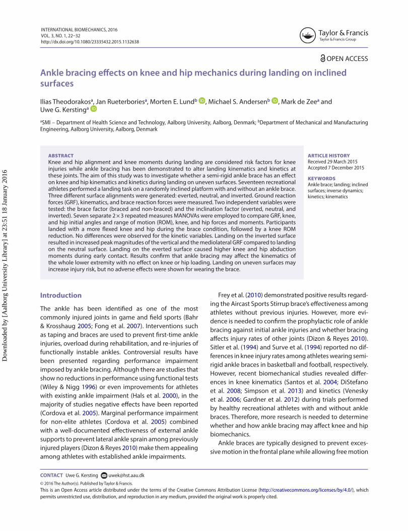
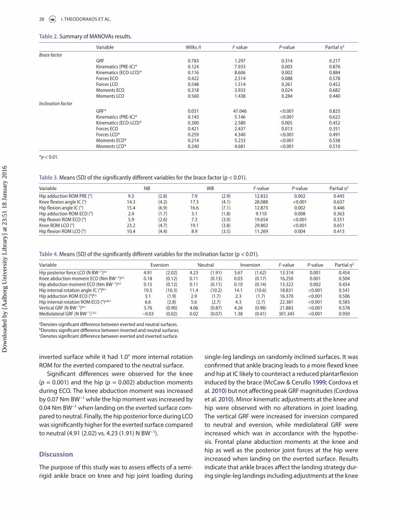
Subsequently, participants were asked to perform a short warm-up of their own choice and execute sin-gle-leg landings from a height of 40 cm repeatedly, until they felt comfortable to perform the task while looking forward without aiming at the platform. They were instructed to start from a position where they were standing on their straight, non-dominant leg with their dominant leg lifted anteriorly (Figure 1(A)). The descent started after a signal from the researcher with only a minimal push-off to ascertain a consistent fall height. Three different surface inclinations were randomly generated: 5° everted, 0° (neutral), and 15° inverted (Figure 1(B)–(D)). The initial position of the platform was at 5° inversion, and it was tilted to the respective landing inclination while the participants were airborne. Each participant performed six successful landings per inclination, with and without a semi-rigid ankle brace (Sports Stirrup Aircast, DJO Nordic A/S, Sweden) (Figure 3(D) and (E)). A trial was discarded if participants failed to maintain their balance on that leg for at least 2 s after IC.
Instrumentation
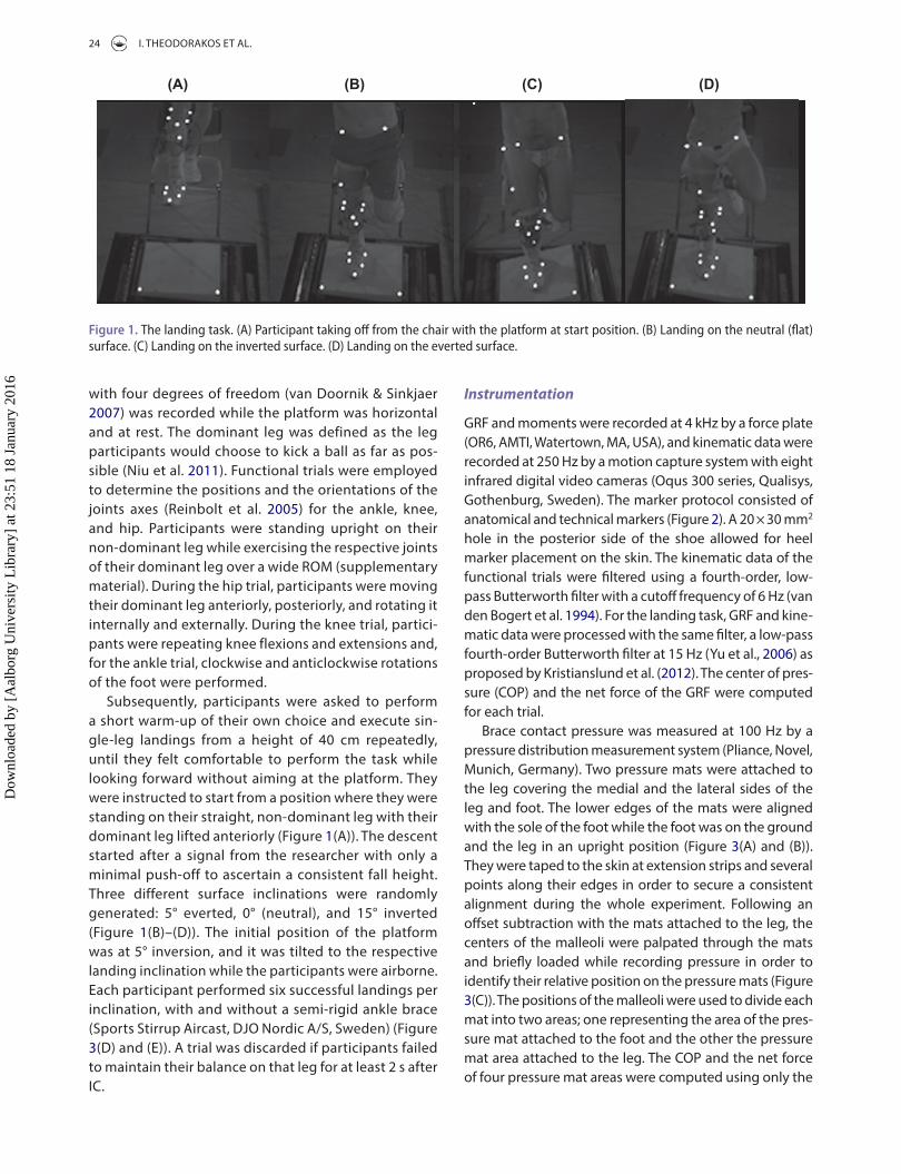
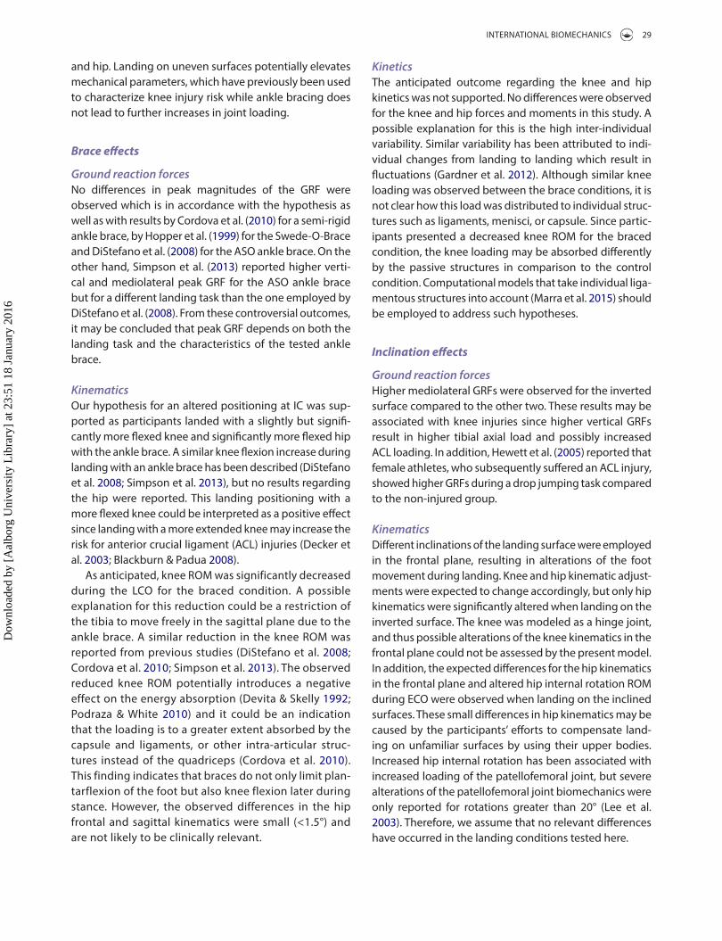
GRF and moments were recorded at 4 kHz by a force plate (OR6, AMTI, Watertown, MA, USA), and kinematic data were recorded at 250 Hz by a motion capture system with eight infrared digital video cameras (Oqus 300 series, Qualisys, Gothenburg, Sweden). The marker protocol consisted of anatomical and technical markers (Figure 2). A 20 × 30 mm2 hole in the posterior side of the shoe allowed for heel marker placement on the skin. The kinematic data of the functional trials were filtered using a fourth-order, low-pass Butterworth filter with a cutoff frequency of 6 Hz (van den Bogert et al. 1994). For the landing task, GRF and kine-matic data were processed with the same filter, a low-pass fourth-order Butterworth filter at 15 Hz (Yu et al., 2006) as proposed by Kristianslund et al. (2012). The center of pres-sure (COP) and the net force of the GRF were computed for each trial.
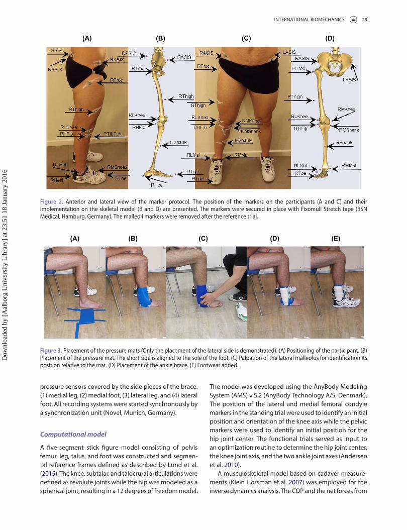
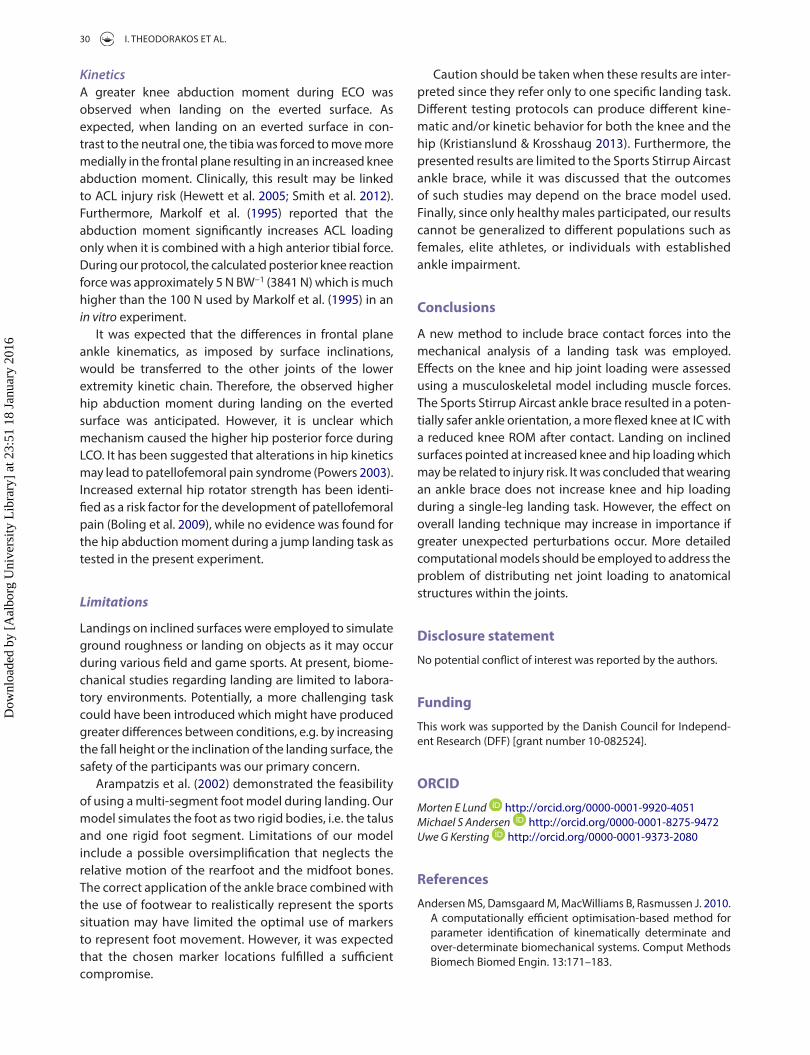
Brace contact pressure was measured at 100 Hz by a pressure distribution measurement system (Pliance, Novel, Munich, Germany). Two pressure mats were attached to the leg covering the medial and the lateral sides of the leg and foot. The lower edges of the mats were aligned with the sole of the foot while the foot was on the ground and the leg in an upright position (Figure 3(A) and (B)). They were taped to the skin at extension strips and several points along their edges in order to secure a consistent alignment during the whole experiment. Following an offset subtraction with the mats attached to the leg, the centers of the malleoli were palpated through the mats and briefly loaded while recording pressure in order to identify their relative position on the pressure mats (Figure 3(C)). The positions of the malleoli were used to divide each mat into two areas; one representing the area of the pres-sure mat attached to the foot and the other the pressure mat area attached to the leg. The COP and the net force of four pressure mat areas were computed using only the
(A) (B) (C) (D)
Figure 1. the landing task. (a) Participant taking off from the chair with the platform at start position. (B) landing on the neutral (flat) surface. (c) landing on the inverted surface. (D) landing on the everted surface.
Dow
nloa
ded
by [
Aal
borg
Uni
vers
ity L
ibra
ry]
at 2
3:51
18
Janu
ary
2016
INTERNATIONAL BIOMECHANICS 25
The model was developed using the AnyBody Modeling System (AMS) v.5.2 (AnyBody Technology A/S, Denmark). The position of the lateral and medial femoral condyle markers in the standing trial were used to identify an initial position and orientation of the knee axis while the pelvic markers were used to identify an initial position for the hip joint center. The functional trials served as input to an optimization routine to determine the hip joint center, the knee joint axis, and the two ankle joint axes (Andersen et al. 2010).
A musculoskeletal model based on cadaver measure-ments (Klein Horsman et al. 2007) was employed for the inverse dynamics analysis. The COP and the net forces from
pressure sensors covered by the side pieces of the brace: (1) medial leg, (2) medial foot, (3) lateral leg, and (4) lateral foot. All recording systems were started synchronously by a synchronization unit (Novel, Munich, Germany).
Computational model
A five-segment stick figure model consisting of pelvis femur, leg, talus, and foot was constructed and segmen-tal reference frames defined as described by Lund et al. (2015). The knee, subtalar, and talocrural articulations were defined as revolute joints while the hip was modeled as a spherical joint, resulting in a 12 degrees of freedom model.
Figure 2. anterior and lateral view of the marker protocol. the position of the markers on the participants (a and c) and their implementation on the skeletal model (B and D) are presented. the markers were secured in place with Fixomull stretch tape (Bsn medical, hamburg, Germany). the malleoli markers were removed after the reference trial.
(A) (B) (C) (D) (E)
Figure 3. Placement of the pressure mats (only the placement of the lateral side is demonstrated). (a) Positioning of the participant. (B) Placement of the pressure mat. the short side is aligned to the sole of the foot. (c) Palpation of the lateral malleolus for identification its position relative to the mat. (D) Placement of the ankle brace. (e) Footwear added.
Dow
nloa
ded
by [
Aal
borg
Uni
vers
ity L
ibra
ry]
at 2
3:51
18
Janu
ary
2016
26 I. THEODORAKOS ET AL.
respectively. Equation (2) expresses the dynamic equilib-rium equations, with C being the coefficient-matrix for the unknown forces, while d contains all known applied loads and inertia forces. Equation (3) states that muscles cannot push, and their capacity is limited by the muscle strength.
Data analysis
The GRFs were computed with respect to a reference frame aligned with the landing surface. The z-axis was perpen-dicular to the landing surface pointing towards the partic-ipant, the x-axis pointed posteriorly, and y-axis laterally for the right leg. The peak GRF were identified for each trial, and scaled to body weight (BW).
Knee and hip ROM were computed over 200 ms before the IC (PRE) to investigate kinematic adjustments prior to landing as it has been reported that the muscle prepara-tion during landing occurs approximately 200 ms before IC (Santello & McDonagh 1998). In order to investigate if differences before and after anticipatory postural adjust-ments were evoked, the contact phase was divided into two periods: 50 ms after IC and denoted early contact (ECO), and 50–200 ms after IC and denoted late contact (LCO). Grüneberg et al. (2003) reported that the short latency responses for the lower leg muscles occur 41.4–45.3 and 41.3–46.1 ms after jump landings from a 30 cm height onto flat and inverted surfaces, respectively. The joint angles at the instant of IC were also computed.
Knee and hip peak reaction forces and moments were computed for ECO and LCO. The knee forces and moments were expressed in a reference frame embedded into the tibia while the hip forces and moments were expressed
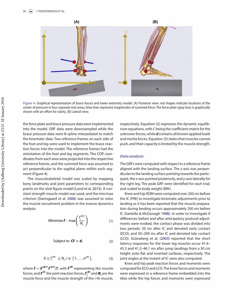
the force plate and brace pressure data were implemented into the model. GRF data were downsampled while the brace pressure data were B-spline interpolated to match the kinematic data. Two reference frames on each side of the foot and leg were used to implement the brace reac-tion forces into the model. The reference frames had the orientation of the foot and leg segments. The COP coor-dinates from each area were projected into the respective reference frames, and the summed force was assumed to act perpendicular to the sagittal plane within each seg-ment (Figure 4).
The musculoskeletal model was scaled by mapping bony landmarks and joint parameters to corresponding points on the stick figure model (Lund et al. 2015). A con-stant strength muscle model was used, and the min/max criterion (Damsgaard et al. 2006) was assumed to solve the muscle recruitment problem in the inverse dynamics analysis:
where f = [f (M)Tf(R)T]T, with f (M) representing the muscle
forces, and f (R) the joint reaction forces, f (M)
i and N
i are the
muscle force and the muscle strength of the i-th muscle,
(1)Minimize f : max
(
f(M)
i
Ni
)
(2)Subject to Cf = d,
(3)0 ≤ f(M)
i≤ N
i, i ∈
{
1,… , n(M)}
,
(B)(A)
Figure 4. Graphical representation of brace forces and lower extremity model. (a) Posterior view: red shapes indicate locations of the center of pressure in four separate mat areas; blue lines represent magnitudes of summed force. the force plate (gray box) is graphically shown with an offset for clarity. (B) lateral view.
Dow
nloa
ded
by [
Aal
borg
Uni
vers
ity L
ibra
ry]
at 2
3:51
18
Janu
ary
2016
INTERNATIONAL BIOMECHANICS 27
Results
No significant interaction between the brace and inclina-tion factors was observed for any of the tested groups.
Brace factor
A significant effect of ankle bracing was revealed for the kinematic variables (Table 2).
Univariate tests (Table 3) showed that the knee (p < 0.001) and the hip (p = 0.002) were more flexed at IC for the braced condition. Furthermore, the knee ROM (p < 0.001) was reduced during LCO. Regarding the hip variables, ankle bracing reduced hip adduction ROM dur-ing PRE (p = 0.002) and ECO (p = 0.008). The hip flexion ROM was increased during ECO (p < 0.001) while it was decreased during LCO (p = 0.004).
Inclination factor
Significant differences were observed for the GRF, the kin-ematic variables, the forces, and the joint moments during LCO (Table 2). Univariate tests showed significant differ-ences for the mediolateral (p < 0.001) and the vertical com-ponent (p < 0.001) of the GRF. Post hoc analysis showed that the mediolateral and vertical GRF were increased by 0.21 and 0.31 N BW−1 for the inverted surface compared to the neutral and everted surfaces, respectively.
Significant differences were observed for the hip inter-nal rotation angle at IC (p < 0.001), the hip adduction ROM (p < 0.001), and the hip internal rotation ROM (p < 0.001) during ECO (Table 4). Participants landed with a 2.7° more internally rotated hip on the inverted surface compared to the neutral surface. During ECO, the hip had 0.5° less adduction ROM and 1.3° less internal rotation ROM for the
in the femoral reference frame. For the right leg, the joint reaction forces were reported as positive in the anterior, proximal, and lateral directions, while adduction, internal rotation, and flexion were reported as positive for the hip moments and landing angles. Adduction, internal rotation, and extension moments were reported as positive for the right knee joint. The joint forces and moments were all scaled to BW.
Statistical analysis
The brace factor (braced and non-braced), and the incli-nation factor (everted, neutral, and inverted) were the independent variables. The dependent variables con-sisted of GRF, kinematic, and kinetic variables of the knee and hip joints and they were grouped according to the analysis period and their type (Table 1). The mean value and the standard deviation of each dependent variable for six successive repetitions per brace and inclination condition were computed. Seven separate 2 × 3 repeated measures MANOVAs were used to assess the effect of the ankle brace and the inclined surface on the grouped variables. When significant differences were observed, univariate two-way (2 × 3) repeated measures ANOVAs were used to identify the significant variables. The cor-rected degrees of freedom by the Greenhouse-Geisser estimates of sphericity were used when the sphericity assumption was violated. Pairwise comparisons of the different inclinations were performed with the Bonferroni adjustment when significant differences were shown for the inclination factor. A commercially available statistical analysis package SPSS v.20 (IBM Corp®, USA) was used for statistical analysis. The significance value was set to p < 0.01 for all analyses.
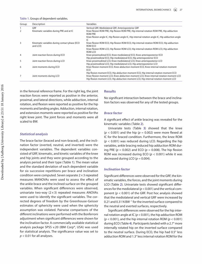
Table 1. Groups of dependent variables.
Group Description Variables1 GrF Vertical GrF, mediolateral GrF, anterioposterior GrF2 Kinematic variables during Pre and at Ic Knee flexion rom Pre, hip flexion rom Pre, hip internal rotation rom Pre, hip adduction
rom PreKnee flexion angle Ic, hip flexion angle Ic, hip internal rotation angle Ic, hip adduction angle Ic
3 Kinematic variables during contact phase (eco and lco)
Knee flexion rom eco, hip flexion rom eco, hip internal rotation rom eco, hip adduction rom ecoKnee flexion rom lco, hip flexion rom lco, hip internal rotation rom lco, hip adduction rom lco
4 Joint reaction forces during eco Knee proximodistal eco, Knee mediolateral eco, Knee anterioposterior ecohip proximodistal eco, hip mediolateral eco, hip anterioposterior eco
5 Joint reaction forces during lco Knee proximodistal lco, Knee mediolateral lco, Knee anterioposterior lcohip proximodistal lco, hip mediolateral lco, hip anterioposterior lco
6 Joint moments during eco Knee flexion moment eco, Knee abduction moment eco, Knee internal rotation moment ecohip flexion moment eco, hip abduction moment eco, hip internal rotation moment eco
7 Joint moments during lco Knee flexion moment lco, Knee abduction moment lco, Knee internal rotation moment lcohip flexion moment lco, hip abduction moment lco, hip internal rotation moment lco
Dow
nloa
ded
by [
Aal
borg
Uni
vers
ity L
ibra
ry]
at 2
3:51
18
Janu
ary
2016
28 I. THEODORAKOS ET AL.
single-leg landings on randomly inclined surfaces. It was confirmed that ankle bracing leads to a more flexed knee and hip at IC likely to counteract a reduced plantarflexion induced by the brace (McCaw & Cerullo 1999; Cordova et al. 2010) but not affecting peak GRF magnitudes (Cordova et al. 2010). Minor kinematic adjustments at the knee and hip were observed with no alterations in joint loading. The vertical GRF were increased for inversion compared to neutral and eversion, while mediolateral GRF were increased which was in accordance with the hypothe-sis. Frontal plane abduction moments at the knee and hip as well as the posterior joint forces at the hip were increased when landing on the everted surface. Results indicate that ankle braces affect the landing strategy dur-ing single-leg landings including adjustments at the knee
inverted surface while it had 1.0° more internal rotation ROM for the everted compared to the neutral surface.
Significant differences were observed for the knee (p = 0.001) and the hip (p = 0.002) abduction moments during ECO. The knee abduction moment was increased by 0.07 Nm BW−1 while the hip moment was increased by 0.04 Nm BW−1 when landing on the everted surface com-pared to neutral. Finally, the hip posterior force during LCO was significantly higher for the everted surface compared to neutral (4.91 (2.02) vs. 4.23 (1.91) N BW−1).
Discussion
The purpose of this study was to assess effects of a semi-rigid ankle brace on knee and hip joint loading during
Table 2. summary of manoVas results.
*p < 0.01.
Variable Wilks Λ F value P-value Partial η2
Brace factorGrF 0.783 1.297 0.314 0.217Kinematics (Pre-Ic)* 0.124 7.933 0.003 0.876Kinematics (eco-lco)* 0.116 8.606 0.002 0.884Forces eco 0.422 2.514 0.088 0.578Forces lco 0.548 1.514 0.261 0.452moments eco 0.318 3.933 0.024 0.682moments lco 0.560 1.438 0.284 0.440
Inclination factorGrF* 0.031 47.046 <0.001 0.825Kinematics (Pre-Ic)* 0.143 5.146 <0.001 0.622Kinematics (eco-lco)* 0.300 2.580 0.005 0.452Forces eco 0.421 2.437 0.013 0.351Forces lco* 0.259 4.340 <0.001 0.491moments eco* 0.214 5.233 <0.001 0.538moments lco* 0.240 4.681 <0.001 0.510
Table 3. means (sD) of the significantly different variables for the brace factor (p < 0.01).
Variable NB WB F-value P-value Partial η2
hip adduction rom Pre (°) 9.3 (2.8) 7.9 (2.9) 12.832 0.002 0.445Knee flexion angle Ic (°) 14.3 (4.2) 17.3 (4.1) 28.088 <0.001 0.637hip flexion angle Ic (°) 15.4 (6.9) 16.6 (7.1) 12.873 0.002 0.446hip adduction rom eco (°) 2.4 (1.7) 3.1 (1.8) 9.110 0.008 0.363hip flexion rom eco (°) 5.9 (2.6) 7.3 (3.0) 19.654 <0.001 0.551Knee rom lco (°) 23.2 (4.7) 19.1 (3.8) 29.802 <0.001 0.651hip flexion rom lco (°) 10.4 (4.4) 8.9 (3.5) 11.269 0.004 0.413
Table 4. means (sD) of the significantly different variables for the inclination factor (p < 0.01).
aDenotes significant difference between everted and neutral surfaces.bDenotes significant difference between inverted and neutral surfaces.cDenotes significant difference between everted and inverted surface.
Variable Eversion Neutral Inversion F-value P-value Partial η2
hip posterior force lco (n BW−1)a,c 4.91 (2.02) 4.23 (1.91) 3.67 (1.62) 13.314 0.001 0.454Knee abduction moment eco (nm BW−1)a,c 0.18 (0.12) 0.11 (0.13) 0.03 (0.17) 16.250 0.001 0.504hip abduction moment eco (nm BW−1)a,c 0.15 (0.12) 0.11 (0.11) 0.10 (0.14) 13.322 0.002 0.454hip internal rotation angle Ic (°)b,c 10.5 (10.3) 11.4 (10.2) 14.1 (10.6) 18.831 <0.001 0.541hip adduction rom eco (°)b,c 3.1 (1.9) 2.9 (1.7) 2.3 (1.7) 16.370 <0.001 0.506hip internal rotation rom eco (°)a,b,c 6.6 (2.8) 5.6 (2.7) 4.3 (2.7) 22.381 <0.001 0.583Vertical GrF (n BW−1)b,c 3.76 (0.90) 4.06 (0.87) 4.26 (0.98) 21.883 <0.001 0.578mediolateral GrF (n BW−1) b,c −0.03 (0.02) 0.02 (0.07) 1.38 (0.41) 301.345 <0.001 0.950
Dow
nloa
ded
by [
Aal
borg
Uni
vers
ity L
ibra
ry]
at 2
3:51
18
Janu
ary
2016
INTERNATIONAL BIOMECHANICS 29
KineticsThe anticipated outcome regarding the knee and hip kinetics was not supported. No differences were observed for the knee and hip forces and moments in this study. A possible explanation for this is the high inter-individual variability. Similar variability has been attributed to indi-vidual changes from landing to landing which result in fluctuations (Gardner et al. 2012). Although similar knee loading was observed between the brace conditions, it is not clear how this load was distributed to individual struc-tures such as ligaments, menisci, or capsule. Since partic-ipants presented a decreased knee ROM for the braced condition, the knee loading may be absorbed differently by the passive structures in comparison to the control condition. Computational models that take individual liga-mentous structures into account (Marra et al. 2015) should be employed to address such hypotheses.
Inclination effects
Ground reaction forcesHigher mediolateral GRFs were observed for the inverted surface compared to the other two. These results may be associated with knee injuries since higher vertical GRFs result in higher tibial axial load and possibly increased ACL loading. In addition, Hewett et al. (2005) reported that female athletes, who subsequently suffered an ACL injury, showed higher GRFs during a drop jumping task compared to the non-injured group.
KinematicsDifferent inclinations of the landing surface were employed in the frontal plane, resulting in alterations of the foot movement during landing. Knee and hip kinematic adjust-ments were expected to change accordingly, but only hip kinematics were significantly altered when landing on the inverted surface. The knee was modeled as a hinge joint, and thus possible alterations of the knee kinematics in the frontal plane could not be assessed by the present model. In addition, the expected differences for the hip kinematics in the frontal plane and altered hip internal rotation ROM during ECO were observed when landing on the inclined surfaces. These small differences in hip kinematics may be caused by the participants’ efforts to compensate land-ing on unfamiliar surfaces by using their upper bodies. Increased hip internal rotation has been associated with increased loading of the patellofemoral joint, but severe alterations of the patellofemoral joint biomechanics were only reported for rotations greater than 20° (Lee et al. 2003). Therefore, we assume that no relevant differences have occurred in the landing conditions tested here.
and hip. Landing on uneven surfaces potentially elevates mechanical parameters, which have previously been used to characterize knee injury risk while ankle bracing does not lead to further increases in joint loading.
Brace effects
Ground reaction forcesNo differences in peak magnitudes of the GRF were observed which is in accordance with the hypothesis as well as with results by Cordova et al. (2010) for a semi-rigid ankle brace, by Hopper et al. (1999) for the Swede-O-Brace and DiStefano et al. (2008) for the ASO ankle brace. On the other hand, Simpson et al. (2013) reported higher verti-cal and mediolateral peak GRF for the ASO ankle brace but for a different landing task than the one employed by DiStefano et al. (2008). From these controversial outcomes, it may be concluded that peak GRF depends on both the landing task and the characteristics of the tested ankle brace.
KinematicsOur hypothesis for an altered positioning at IC was sup-ported as participants landed with a slightly but signifi-cantly more flexed knee and significantly more flexed hip with the ankle brace. A similar knee flexion increase during landing with an ankle brace has been described (DiStefano et al. 2008; Simpson et al. 2013), but no results regarding the hip were reported. This landing positioning with a more flexed knee could be interpreted as a positive effect since landing with a more extended knee may increase the risk for anterior crucial ligament (ACL) injuries (Decker et al. 2003; Blackburn & Padua 2008).
As anticipated, knee ROM was significantly decreased during the LCO for the braced condition. A possible explanation for this reduction could be a restriction of the tibia to move freely in the sagittal plane due to the ankle brace. A similar reduction in the knee ROM was reported from previous studies (DiStefano et al. 2008; Cordova et al. 2010; Simpson et al. 2013). The observed reduced knee ROM potentially introduces a negative effect on the energy absorption (Devita & Skelly 1992; Podraza & White 2010) and it could be an indication that the loading is to a greater extent absorbed by the capsule and ligaments, or other intra-articular struc-tures instead of the quadriceps (Cordova et al. 2010). This finding indicates that braces do not only limit plan-tarflexion of the foot but also knee flexion later during stance. However, the observed differences in the hip frontal and sagittal kinematics were small (<1.5°) and are not likely to be clinically relevant.
Dow
nloa
ded
by [
Aal
borg
Uni
vers
ity L
ibra
ry]
at 2
3:51
18
Janu
ary
2016
30 I. THEODORAKOS ET AL.
Caution should be taken when these results are inter-preted since they refer only to one specific landing task. Different testing protocols can produce different kine-matic and/or kinetic behavior for both the knee and the hip (Kristianslund & Krosshaug 2013). Furthermore, the presented results are limited to the Sports Stirrup Aircast ankle brace, while it was discussed that the outcomes of such studies may depend on the brace model used. Finally, since only healthy males participated, our results cannot be generalized to different populations such as females, elite athletes, or individuals with established ankle impairment.
Conclusions
A new method to include brace contact forces into the mechanical analysis of a landing task was employed. Effects on the knee and hip joint loading were assessed using a musculoskeletal model including muscle forces. The Sports Stirrup Aircast ankle brace resulted in a poten-tially safer ankle orientation, a more flexed knee at IC with a reduced knee ROM after contact. Landing on inclined surfaces pointed at increased knee and hip loading which may be related to injury risk. It was concluded that wearing an ankle brace does not increase knee and hip loading during a single-leg landing task. However, the effect on overall landing technique may increase in importance if greater unexpected perturbations occur. More detailed computational models should be employed to address the problem of distributing net joint loading to anatomical structures within the joints.
Disclosure statement
No potential conflict of interest was reported by the authors.
Funding
This work was supported by the Danish Council for Independ-ent Research (DFF) [grant number 10-082524].
ORCID
Morten E Lund http://orcid.org/0000-0001-9920-4051Michael S Andersen http://orcid.org/0000-0001-8275-9472Uwe G Kersting http://orcid.org/0000-0001-9373-2080
References
Andersen MS, Damsgaard M, MacWilliams B, Rasmussen J. 2010. A computationally efficient optimisation-based method for parameter identification of kinematically determinate and over-determinate biomechanical systems. Comput Methods Biomech Biomed Engin. 13:171–183.
KineticsA greater knee abduction moment during ECO was observed when landing on the everted surface. As expected, when landing on an everted surface in con-trast to the neutral one, the tibia was forced to move more medially in the frontal plane resulting in an increased knee abduction moment. Clinically, this result may be linked to ACL injury risk (Hewett et al. 2005; Smith et al. 2012). Furthermore, Markolf et al. (1995) reported that the abduction moment significantly increases ACL loading only when it is combined with a high anterior tibial force. During our protocol, the calculated posterior knee reaction force was approximately 5 N BW−1 (3841 N) which is much higher than the 100 N used by Markolf et al. (1995) in an in vitro experiment.
It was expected that the differences in frontal plane ankle kinematics, as imposed by surface inclinations, would be transferred to the other joints of the lower extremity kinetic chain. Therefore, the observed higher hip abduction moment during landing on the everted surface was anticipated. However, it is unclear which mechanism caused the higher hip posterior force during LCO. It has been suggested that alterations in hip kinetics may lead to patellofemoral pain syndrome (Powers 2003).Increased external hip rotator strength has been identi-fied as a risk factor for the development of patellofemoral pain (Boling et al. 2009), while no evidence was found for the hip abduction moment during a jump landing task as tested in the present experiment.
Limitations
Landings on inclined surfaces were employed to simulate ground roughness or landing on objects as it may occur during various field and game sports. At present, biome-chanical studies regarding landing are limited to labora-tory environments. Potentially, a more challenging task could have been introduced which might have produced greater differences between conditions, e.g. by increasing the fall height or the inclination of the landing surface, the safety of the participants was our primary concern.
Arampatzis et al. (2002) demonstrated the feasibility of using a multi-segment foot model during landing. Our model simulates the foot as two rigid bodies, i.e. the talus and one rigid foot segment. Limitations of our model include a possible oversimplification that neglects the relative motion of the rearfoot and the midfoot bones. The correct application of the ankle brace combined with the use of footwear to realistically represent the sports situation may have limited the optimal use of markers to represent foot movement. However, it was expected that the chosen marker locations fulfilled a sufficient compromise.
Dow
nloa
ded
by [
Aal
borg
Uni
vers
ity L
ibra
ry]
at 2
3:51
18
Janu
ary
2016
INTERNATIONAL BIOMECHANICS 31
Grüneberg C, Nieuwenhuijzen PH, Duysens J. 2003. Reflex responses in the lower leg following landing impact on an inverting and non-inverting platform. J Physiol. 550:985–993.
Hals TM, Sitler MR, Mattacola CG. 2000. Effect of a semi-rigid ankle stabilizer on performance in persons with functional ankle instability. J Orthop Sports Phys Ther. 30:552–556.
Hewett TE, Myer GD, Ford KR, Heidt RS Jr, Colosimo AJ, McLean SG, van den Bogert AJ, Paterno MV, Succop P. 2005. Biomechanical measures of neuromuscular control and valgus loading of the knee predict anterior cruciate ligament injury risk in female athletes: a prospective study. Am J Sports Med. 33:492–501.
Hodgson B, Tis L, Cobb S, Higbie E. 2005. The effect of external ankle support on vertical ground-reaction force and lower body kinematics. J Sport Rehabil. 14:301–312.
Hopper DM, McNair P, Elliott BC. 1999. Landing in netball: effects of taping and bracing the ankle. Br J Sports Med. 33:409–413.
Klein Horsman MD, Koopman HF, van der Helm FC, Prosé LP, Veeger HE. 2007. Morphological muscle and joint parameters for musculoskeletal modelling of the lower extremity. Clin Biomech (Bristol, Avon). 22:239–247.
Kristianslund E, Krosshaug T. 2013. Comparison of drop jumps and sport-specific sidestep cutting: implications for anterior cruciate ligament injury risk screening. Am J Sports Med. 41:684–688.
Kristianslund E, Krosshaug T, van den Bogert AJ. 2012. Effect of low pass filtering on joint moments from inverse dynamics: Implications for injury prevention. J Biomech. 45:666–671.
Lee TQ, Morris G, Csintalan RP. 2003. The influence of tibial and femoral rotation on patellofemoral contact area and pressure. J Orthop Sports Phys Ther. 33:686–693.
Lund ME, Andersen MS, de Zee M, Rasmussen J. 2015. Scaling of musculoskeletal models from static and dynamic trials. Int J Biomech. 2:1–11.
Markolf KL, Burchfield DM, Shapiro MM, Shepard MF, Finerman GA, Slauterbeck JL. 1995. Combined knee loading states that generate high anterior cruciate ligament forces. J Orthop Res. 13:930–935.
Marra MA, Vanheule V, Fluit R, Koopman BH, Rasmussen J, Verdonschot N, Andersen MS. 2015. A subject-specific musculoskeletal modeling framework to predict in vivo mechanics of total knee arthroplasty. J Biomech Eng. 137:020904.
McCaw ST, Cerullo JF. 1999. Prophylactic ankle stabilizers affect ankle joint kinematics during drop landings. Med Sci Sports Exerc. 31:702–707.
Niu W, Wang Y, He Y, Fan Y, Zhao Q. 2011. Kinematics, kinetics, and electromyogram of ankle during drop landing: a comparison between dominant and non-dominant limb. Hum Mov Sci. 30:614–623.
Podraza JT, White SC. 2010. Effect of knee flexion angle on ground reaction forces, knee moments and muscle co-contraction during an impact-like deceleration landing: Implications for the non-contact mechanism of ACL injury. Knee. 17:291–295.
Powers CM. 2003. The influence of altered lower-extremity kinematics on patellofemoral joint dysfunction: a theoretical perspective. J Orthop Sports Phys Ther. 33:639–646.
Reinbolt JA, Schutte JF, Fregly BJ, Koh BI, Haftka RT, George AD, Mitchell KH. 2005. Determination of patient-specific multi-joint kinematic models through two-level optimization. J Biomech. 38:621–626.
Arampatzis A, Bruggemann GP, Klapsing GM. 2002. A three-dimensional shank-foot model to determine the foot motion during landings. Med Sci Sports Exerc. 34:130–138.
Bahr R, Krosshaug T. 2005. Understanding injury mechanisms: a key component of preventing injuries in sport. Br J Sports Med. 39:324–329.
Blackburn JT, Padua DA. 2008. Influence of trunk flexion on hip and knee joint kinematics during a controlled drop landing. Clin Biomech (Bristol, Avon). 23:313–319.
Boling MC, Padua DA, Marshall SW, Guskiewicz K, Pyne S, Beutler A. 2009. A prospective investigation of biomechanical risk factors for patellofemoral pain syndrome: the joint undertaking to monitor and prevent ACL injury (JUMP-ACL) cohort. Am J Sports Med. 37:2108–2116.
Chen Q, Wortley M, Bhaskaran D, Milner CE, Zhang S. 2012. Is the inverted surface landing more suitable in evaluating ankle braces and ankle inversion perturbation? Clin J Sport Med. 22:214–220.
Cordova ML, Scott BD, Ingersoll CD, Leblanc MJ. 2005. Effects of ankle support on lower-extremity functional performance: a meta-analysis. Med Sci Sports Exerc. 37:635–641.
Cordova ML, Takahashi Y, Kress GM, Brucker JB, Finch AE. 2010. Influence of external ankle support on lower extremity joint mechanics during drop landings. J Sport Rehabil. 19:136–148.
Damsgaard M, Rasmussen J, Christensen ST, Surma E, de Zee M. 2006. Analysis of musculoskeletal systems in the anybody modeling system. Simul Model Pract Th. 14:1100–1111.
Decker MJ, Torry MR, Wyland DJ, Sterett WI, Richard Steadman J. 2003. Gender differences in lower extremity kinematics, kinetics and energy absorption during landing. Clin Biomech (Bristol, Avon). 18:662–669.
Devita P, Skelly WA. 1992. Effect of landing stiffness on joint kinetics and energetics in the lower extremity. Med Sci Sports Exerc. 24:108–115.
DiStefano LJ, Padua DA, Brown CN, Guskiewicz KM. 2008. Lower extremity kinematics and ground reaction forces after prophylactic lace-up ankle bracing. J Athl Train. 43:234–241.
Dizon JM, Reyes JJ. 2010. A systematic review on the effectiveness of external ankle supports in the prevention of inversion ankle sprains among elite and recreational players. J Sci Med Sport. 13:309–317.
van Doornik J, Sinkjaer T. 2007. Robotic platform for human gait analysis. IEEE Trans Biomed Eng. 54:1696–1702.
Fong DT, Hong Y, Chan LK, Yung PS, Chan KM. 2007. A systematic review on ankle injury and ankle sprain in sports. Sports Med. 37:73–94.
Frey C, Feder KS, Sleight J. 2010. Prophylactic ankle brace use in high school volleyball players: a prospective study. Foot Ankle Int. 31:296–300.
Gardner JK, Mccaw ST, Laudner KG, Smith PJ, Stafford LN. 2012. Effect of ankle braces on lower extremity joint energetics in single-leg landings. Med Sci Sports Exerc. 44:1116–1122.
Garrick JG. 1977. The frequency of injury, mechanism of injury, and epidemiology of ankle sprains. Am J Sports Med. 5:241–242.
Griffin LY, Albohm MJ, Arendt EA, Bahr R, Beynnon BD, Demaio M, Dick RW, Engebretsen L, Garrett WE Jr, Hannafin JA, et al. 2006. Understanding and preventing noncontact anterior cruciate ligament injuries: a review of the Hunt Valley II meeting, January 2005. Am J Sports Med. 34:1512–1532.
Gross MT, Liu HY. 2003. The role of ankle bracing for prevention of ankle sprain injuries. J Orthop Sports Phys Ther. 33:572–577.
Dow
nloa
ded
by [
Aal
borg
Uni
vers
ity L
ibra
ry]
at 2
3:51
18
Janu
ary
2016
32 I. THEODORAKOS ET AL.
injury: a review of the literature – Part 1: neuromuscular and anatomic risk. Sports Health. 4:69–78.
Surve I, Schwellnus MP, Noakes T, Lombard C. 1994. A fivefold reduction in the incidence of recurrent ankle sprains in soccer players using the sport-stirrup orthosis. Am J Sports Med. 22:601–606.
van den Bogert AJ, Smith GD, Nigg BM. 1994. In vivo determination of the anatomical axes of the ankle joint complex: an optimization approach. J Biomech. 27:1477 1488.
Vanwanseele B, Stuelcken M, Greene A, Smith R. 2014. The effect of external ankle support on knee and ankle joint movement and loading in netball players. J Sci Med Sport. 17:511–515.
Venesky K, Docherty CL, Dapena J, Schrader J. 2006. Prophylactic ankle braces and knee varus-valgus and internal-external rotation torque. J Athl Train. 41:239–244.
Wiley JP, Nigg BM. 1996. The effect of an ankle orthosis on ankle range of motion and performance. J Orthop Sports Phys Ther. 23:362–369.
Wright IC, Neptune RR, van den Bogert AJ, Nigg BM. 2000. The influence of foot positioning on ankle sprains. J Biomech. 33:513–519.
Yu B, Lin CF, Garrett WE. 2006. Lower extremity biomechanics during the landing of a stop-jump task. Clin Biomech (Bristol, Avon). 21:297–305.
Renstrom P, Ljungqvist A, Arendt E, Beynnon B, Fukubayashi T, Garrett W, Georgoulis T, Hewett TE, Johnson R, Krosshaug T, et al. 2008. Non-contact ACL injuries in female athletes: an International Olympic Committee current concepts statement. Br J Sports Med. 42:394–412.
Santello M, McDonagh MJ. 1998. The control of timing and amplitude of EMG activity in landing movements in humans. Exp Physiol. 83:857–874.
Santos MJ, McIntire K, Foecking J, Liu W. 2004. The effects of ankle bracing on motion of the knee and the hip joint during trunk rotation tasks. Clin Biomech (Bristol, Avon). 19:964–971.
Siegler S, Liu W, Sennett B, Nobilini RJ, Dunbar D. 1997. The three-dimensional passive support characteristics of ankle braces. J Orthop Sports Phys Ther. 26:299–309.
Simpson KJ, Yom JP, Fu YC, Arnett SW, O’Rourke S, Brown CN. 2013. Does wearing a prophylactic ankle brace during drop landings affect lower extremity kinematics and ground reaction forces? J Appl Biomech. 29:205–213.
Sitler M, Ryan J, Wheeler B, McBride J, Arciero R, Anderson J, Horodyski M. 1994. The efficacy of a semirigid ankle stabilizer to reduce acute ankle injuries in basketball: a randomized clinical study at west point. Am J Sports Med. 22:454–461.
Smith HC, Vacek P, Johnson RJ, Slauterbeck JR, Hashemi J, Shultz S, Beynnon BD. 2012. Risk factors for anterior cruciate ligament
Dow
nloa
ded
by [
Aal
borg
Uni
vers
ity L
ibra
ry]
at 2
3:51
18
Janu
ary
2016