Embed Size (px)
Citation preview

PART I
ANIMAL PLEASURES
MlKringelbach_BookPS.indb 25MlKringelbach_BookPS.indb 25 4/27/2009 6:15:27 PM4/27/2009 6:15:27 PM
Copying or circulation without permission are strictly prohibited. This chapter is included in Kringelbach ML and Berridge KC, "Pleasures of the Brain" (c) 2010. Oxford University Press: New York.

MlKringelbach_BookPS.indb 26MlKringelbach_BookPS.indb 26 4/27/2009 6:15:27 PM4/27/2009 6:15:27 PM
Copying or circulation without permission are strictly prohibited. This chapter is included in Kringelbach ML and Berridge KC, "Pleasures of the Brain" (c) 2010. Oxford University Press: New York.
27
able to enhance or stimulate pleasure is slightly dif-ferent from being needed for normal pleasure, and so the contributions of “su0 cient cause” (enhancement) and “necessary cause” (normal) need to be assessed separately. Finally, these and many other brain sub-strates may code the occurrence a pleasurable event by their neural activation. But, for many substrates, the neural activation may be neither necessary nor su0 -cient to produce pleasure (and presumably, those sub-strates instead transfer pleasure information to cause other functions that guide decisions) (Dickinson and Balleine, Chapter 4, this book; Kringelbach, Chapter 12, this book). A particular brain substrate may have all three of these hedonic roles (code, su0 cient cause, and necessary cause), but alternatively, it may code without causing, or it may act as a su0 cient cause but not a necessary cause (Table 1.1). For example, some sites in nucleus accumbens may enhance pleasure but may not be needed for all normal pleasures. It is an important task for aL ective neuroscience to assign pleasure causation to the proper brain substrates. The goal of this chapter is to identify some of the hedonic hotspots in the brain most able to cause enhancements of pleasure.
To identify a neural substrate that causes plea-sure, it is often helpful to manipulate the brain and observe whether this manipulation causes a change in hedonic reactions to a sensory stimulus. Using exper-imental techniques to manipulate neurochemicals in focused brain locations, we have recently mapped sev-eral hedonic hotspots that contribute in causal ways to
A vital question concerning sensory pleasure is how brain mechanisms cause stimuli to become
pleasurable and liked. Pleasure is not an intrinsic fea-ture of any stimulus, but instead reQ ects an aL ective evaluation added to the stimulus by the brain. That is, as Frijda expresses it (Frijda, 2006, Chapter 6, this book), a pleasure gloss or hedonic value must be actively “painted” on sweet or other sensations to make them pleasant. Brain mechanisms of pleasure, whatever they are, must take a mere sensory signal and transform it into a hedonic and ‘liked’ reward.
Finding the brain mechanisms responsible for painting a pleasure gloss is a major challenge for aL ective neuroscience (Barrett and Wager, 2006; Berridge, 2003b; Damasio, 1999; Davidson and Irwin, 1999; Kringelbach, 2005, Chapter 12, this book; LeDoux, 1996; Panksepp, 1991; Peciña et al., 2006). Fortunately, progress on G nding hedonic generators in the brain is being made. In this chapter, we focus speciG cally on the neuroanatomical hedonic hotspots in the brain where neurochemical signals actually contribute causally to the generation of pleasure.
We deG ne a hedonic hotspot as a brain site where pleasure mechanisms are su0 ciently concentrated together in one anatomical locus to cause pleasure enhancement when neurally activated (while rec-ognizing that a hotspot’s contribution to pleasure enhancement depends also on its participation in larger brain circuits). A hotspot might also be a site where natural pleasures are reduced below normal lev-els by neural suppression or damage. However, being
1
Hedonic Hotspots: Generating Sensory Pleasure in the Brain
KYLE S. SMITH, STEPHEN V. MAHLER, SUSANA PECIÑA,
AND KENT C. BERRIDGE
MlKringelbach_BookPS.indb 27MlKringelbach_BookPS.indb 27 4/27/2009 6:15:27 PM4/27/2009 6:15:27 PM
Copying or circulation without permission are strictly prohibited. This chapter is included in Kringelbach ML and Berridge KC, "Pleasures of the Brain" (c) 2010. Oxford University Press: New York.

28 Pleasures of the Brain
(Cabanac, 1971). But how can pleasure be measured in nonverbal animals like rats, in which most research on neurobiological causes must be conducted? The pre-mise that underlies our aL ective neuroscience research on hedonics is that ‘liking’ is a basic evaluative reac-tion of the brain, with objective neural and behavioral indicators that can be quantiG ed by appropriate meth-ods in animals and humans alike.
These objective indicators include emotional facial expressions (Berridge, 2000; Darwin, 1872; Ekman, 1999). Many animals including humans, primates, and
pleasure. These hotspots are scattered across locations that span almost the entire brain and are embedded in a larger pleasure circuit in the brain that operates as a whole to increase hedonic experience.
How Can We Measure Hedonic ‘Liking’ in Animals?
Traditional studies of pleasure ‘liking’ have focused on human adult subjects who can describe their feelings
Table 1.1 Types of Pleasure Mediation: Su0 cient Cause, Necessary Cause, and Code. The phrase “brain struc-ture X mediates pleasure” has three diL erent possible meanings, which may or may not coincide, though they are often meant together. It is useful to distinguish between cause and code, and even to distinguish among diL erent ways of causing (caveats apply to each shorthand distinction). Examples are neither exhaustive nor exclusive (e.g., ventral pallidum also codes pleasure, and orbitofrontal cortex may turn out to cause pleasure); see text for discussion.
‘‘Su2cient Cause’’
‘‘Necessary Cause’’
‘‘Code’’ Neural activation during pleasure
Neural blockade/lesion produces loss of pleasure
Neural stimulation is su2cient to cause increase in pleasure
Types of Roles in Pleasure
Caveat: Code may or may not be cause
Caveat: DeGcit may not always be mirror image of normal functionExample
Ventral Pallidum
Example
Orbitofrontal Cortex
Example
Nucleus Accumbens
Caveat: Causation is distributed beyond stimulated substrate
Some neural activations may cause the pleasure they code.Other neural activations may be instead a consequence of ahedonic reaction generated elsewhere in the brain, rather thancause the hedonic reaction themselves (and presumably helpcause some other psychological function).
Loss of pleasure after a lesion may mean that the substrate wasthe pleasure generator, but alternatively could mean that itsfunction was to facilitate pleasure generation in otherstructures that still remain (e.g. removal of a transistor maymake a radio sequeal, but the transistor’s funtion was not merelya ‘squeal suppressor’).
The stimulated substrate doesn’t contain all causation itself, butrather interacts with other distributed components of a largerbrain circuit to cause pleasure. Condition of other brain substratesand external events may modulate impact of neural activation.
MlKringelbach_BookPS.indb 28MlKringelbach_BookPS.indb 28 4/27/2009 6:15:27 PM4/27/2009 6:15:27 PM
Copying or circulation without permission are strictly prohibited. This chapter is included in Kringelbach ML and Berridge KC, "Pleasures of the Brain" (c) 2010. Oxford University Press: New York.

29 Smith et al.: Hedonic Hotspots
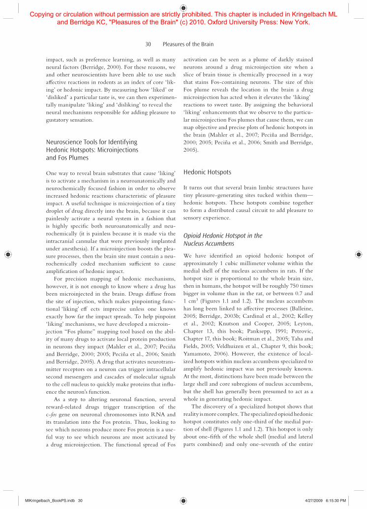
rats exhibit homologous, hedonic, and aversive facial reactions to pleasant and unpleasant tastes. For exam-ple, a human infant, even on its G rst day of life, will rhythmically lick its lips when a drop of sugar water is placed on its tongue (Steiner, 1973). By contrast, a bitter taste like quinine elicits characteristic aver-sive reactions including gaping of the mouth (Steiner, 1973). Like humans, nonhuman primates and rodents display homologous ‘liking’ and ‘disliking’ reactions to sweet and bitter tastes (Steiner et al., 2001). Rats, for example, display the same rhythmic tongue pro-trusions as human infants, as well as paw-licking and related movements, when presented with a sugary solu-tion in the mouth (Grill and Berridge, 1985; Grill and Norgren, 1978a) (Figure 1.1). Similarly, in response to a bitter taste, rats emit the same aversive gaping reac-tions that human infants show, along with headshakes and frantic wiping of the mouth (Figure 1.1).
Importantly, these animal aL ective reactions Q uc-tuate in similar ways to human subjective pleasure when relevant circumstances change (Berridge, 2000).
For example, just as food is more pleasant to us when hungry, sweet tastes elicit more ‘liking’ reactions when rats are hungry than when they are full (Berridge, 2000; Cabanac, 1971). Such homeostatically induced changes in sensory pleasure have been called “allies-thesia” (Cabanac, 1971, Chapter 7, this book; Leknes and Tracey, Chapter 19, this book). Similarly, the intense taste of salt at concentrations higher than sea-water is not pleasant to either people or rats, and nor-mal rats accordingly respond to this taste with gapes and other aversive reactions. However, if one physio-logically depletes a rat of sodium, thus eliciting a state of “salt appetite,” aL ective reactions to this very same salty taste suddenly Q ip from negative to positive and hedonic tongue protrusions are observed instead of aversive gapes (Berridge et al., 1984; Schulkin, 1991; Tindell et al., 2006). Thus ‘liking’ facial reactions to tastes reQ ect not simply the sensory properties of the stimulus, but rather a hedonic evaluation of it that incorporates physiological needs. ‘Liking’ reactions also incorporate psychological inQ uences on hedonic
Figure 1.1 Taste ‘liking’ reactions and contrast map of nucleus accumbens hotspots. Positive ‘liking’ reactions to pleasant sweet tastes shared by human newborn, young orangutan, and adult rat (tongue protrusion; left top), and aversive ‘disliking’ reactions to unpleasant bitter tastes (gape; left bottom). AL ective facial expressions like these provide an objective index of ‘liking’ and ‘disliking’ reactions to the hedonic impact of tastes. Opioid hotspots and coldspots for hedonic ‘liking.’ ‘disliking,’ and motivational ‘wanting’ are mapped and stacked within the nucleus accumbens (medial shell region shown in sagittal view; right). Virtually the entire medial shell stimulates ‘wanting’ for reward (e.g., increased food intake) in response to opioid stimulation (green hexa-gons represent individual microinjection Fos plumes) and so do other nearby structures including the core of nucleus accumbens as well as parts of the ventral neostriatum above the accumbens, and the olfactory tuber-cle beneath the accumbens. The much smaller hedonic hotspot for ‘liking,’ where opioid stimulation actually increases positive hedonic reactions to sucrose taste (red), is contained within the anterior and dorsal quarter of shell. ‘Liking’ reactions to sucrose are reduced by opioid stimulation in a small posterior hedonic coldspot (though still stimulating ‘wanting’; blue), whereas an intermediate region that contains both hotspot and cold-spot mediates opioid suppression of aversive ‘disliking’ for bitter quinine (purple). The hotspot zone map is modiG ed from Peciña and Berridge (2005).
Hedonic Reactions (sweet)
Aversive Reactions (bitter) ‘Wanting’ HotspotEating
Increase
Hedonic Hotspot‘Liking’Increase
Hedonic Coldspot‘Liking’Decrease
Opioid ‘liking’ and ‘wanting’ zones in NAc shell
Sagittal
‘Disliking’Decrease
MlKringelbach_BookPS.indb 29MlKringelbach_BookPS.indb 29 4/27/2009 6:15:28 PM4/27/2009 6:15:28 PM
Copying or circulation without permission are strictly prohibited. This chapter is included in Kringelbach ML and Berridge KC, "Pleasures of the Brain" (c) 2010. Oxford University Press: New York.
30 Pleasures of the Brain
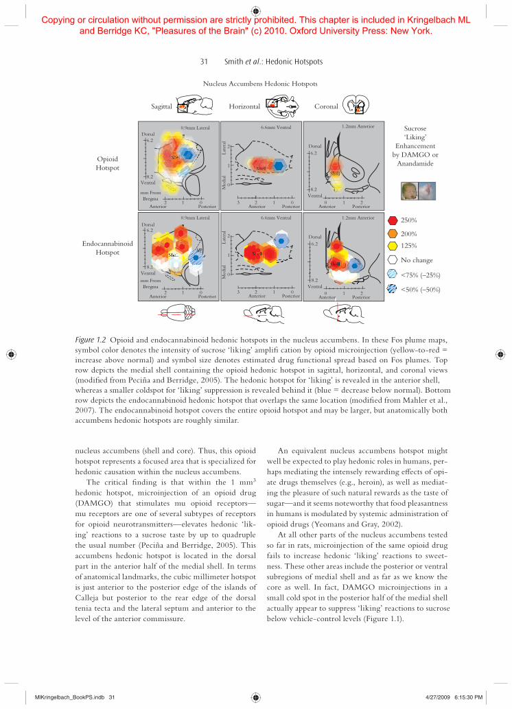
activation can be seen as a plume of darkly stained neurons around a drug microinjection site when a slice of brain tissue is chemically processed in a way that stains Fos-containing neurons. The size of this Fos plume reveals the location in the brain a drug microinjection has acted when it elevates the ‘liking’ reactions to sweet taste. By assigning the behavioral ‘liking’ enhancements that we observe to the particu-lar microinjection Fos plumes that cause them, we can map objective and precise plots of hedonic hotspots in the brain (Mahler et al., 2007; Peciña and Berridge, 2000; 2005; Peciña et al., 2006; Smith and Berridge, 2005).
Hedonic Hotspots
It turns out that several brain limbic structures have tiny pleasure-generating sites tucked within them—hedonic hotspots. These hotspots combine together to form a distributed causal circuit to add pleasure to sensory experience.
Opioid Hedonic Hotspot in the Nucleus Accumbens
We have identiG ed an opioid hedonic hotspot of approximately 1 cubic millimeter volume within the medial shell of the nucleus accumbens in rats. If the hotspot size is proportional to the whole brain size, then in humans, the hotspot will be roughly 750 times bigger in volume than in the rat, or between 0.7 and 1 cm3 (Figures 1.1 and 1.2). The nucleus accumbens has long been linked to aL ective processes (Balleine, 2005; Berridge, 2003b; Cardinal et al., 2002; Kelley et al., 2002; Knutson and Cooper, 2005; Leyton, Chapter 13, this book; Panksepp, 1991; Petrovic, Chapter 17, this book; Roitman et al., 2005; Taha and Fields, 2005; Veldhuizen et al., Chapter 9, this book; Yamamoto, 2006). However, the existence of local-ized hotspots within nucleus accumbens specialized to amplify hedonic impact was not previously known. At the most, distinctions have been made between the large shell and core subregions of nucleus accumbens, but the shell has generally been presumed to act as a whole in generating hedonic impact.
The discovery of a specialized hotspot shows that reality is more complex. The specialized opioid hedonic hotspot constitutes only one-third of the medial por-tion of shell (Figures 1.1 and 1.2). This hotspot is only about one-G fth of the whole shell (medial and lateral parts combined) and only one-seventh of the entire
impact, such as preference learning, as well as many neural factors (Berridge, 2000). For these reasons, we and other neuroscientists have been able to use such aL ective reactions in rodents as an index of core ‘lik-ing’ or hedonic impact. By measuring how ‘liked’ or ‘disliked’ a particular taste is, we can then experimen-tally manipulate ‘liking’ and ‘disliking’ to reveal the neural mechanisms responsible for adding pleasure to gustatory sensation.
Neuroscience Tools for Identifying Hedonic Hotspots: Microinjections and Fos Plumes
One way to reveal brain substrates that cause ‘liking’ is to activate a mechanism in a neuroanatomically and neurochemically focused fashion in order to observe increased hedonic reactions characteristic of pleasure impact. A useful technique is microinjection of a tiny droplet of drug directly into the brain, because it can painlessly activate a neural system in a fashion that is highly speciG c both neuroanatomically and neu-rochemically (it is painless because it is made via the intracranial cannulae that were previously implanted under anesthesia). If a microinjection boosts the plea-sure processes, then the brain site must contain a neu-rochemically coded mechanism su0 cient to cause ampliG cation of hedonic impact.
For precision mapping of hedonic mechanisms, however, it is not enough to know where a drug has been microinjected in the brain. Drugs diL use from the site of injection, which makes pinpointing func-tional ‘liking’ eL ects imprecise unless one knows exactly how far the impact spreads. To help pinpoint ‘liking’ mechanisms, we have developed a microin-jection “Fos plume” mapping tool based on the abil-ity of many drugs to activate local protein production in neurons they impact (Mahler et al., 2007; Peciña and Berridge, 2000; 2005; Peciña et al., 2006; Smith and Berridge, 2005). A drug that activates neurotrans-mitter receptors on a neuron can trigger intracellular second messengers and cascades of molecular signals to the cell nucleus to quickly make proteins that inQ u-ence the neuron’s function.
As a step to altering neuronal function, several reward-related drugs trigger transcription of the c-fos gene on neuronal chromosomes into RNA and its translation into the Fos protein. Thus, looking to see which neurons produce more Fos protein is a use-ful way to see which neurons are most activated by a drug microinjection. The functional spread of Fos
MlKringelbach_BookPS.indb 30MlKringelbach_BookPS.indb 30 4/27/2009 6:15:30 PM4/27/2009 6:15:30 PM
Copying or circulation without permission are strictly prohibited. This chapter is included in Kringelbach ML and Berridge KC, "Pleasures of the Brain" (c) 2010. Oxford University Press: New York.
31 Smith et al.: Hedonic Hotspots
An equivalent nucleus accumbens hotspot might well be expected to play hedonic roles in humans, per-haps mediating the intensely rewarding eL ects of opi-ate drugs themselves (e.g., heroin), as well as mediat-ing the pleasure of such natural rewards as the taste of sugar—and it seems noteworthy that food pleasantness in humans is modulated by systemic administration of opioid drugs (Yeomans and Gray, 2002).
At all other parts of the nucleus accumbens tested so far in rats, microinjection of the same opioid drug fails to increase hedonic ‘liking’ reactions to sweet-ness. These other areas include the posterior or ventral subregions of medial shell and as far as we know the core as well. In fact, DAMGO microinjections in a small cold spot in the posterior half of the medial shell actually appear to suppress ‘liking’ reactions to sucrose below vehicle-control levels (Figure 1.1).
nucleus accumbens (shell and core). Thus, this opioid hotspot represents a focused area that is specialized for hedonic causation within the nucleus accumbens.
The critical G nding is that within the 1 mm3 hedonic hotspot, microinjection of an opioid drug (DAMGO) that stimulates mu opioid receptors—mu receptors are one of several subtypes of receptors for opioid neurotransmitters—elevates hedonic ‘lik-ing’ reactions to a sucrose taste by up to quadruple the usual number (Peciña and Berridge, 2005). This accumbens hedonic hotspot is located in the dorsal part in the anterior half of the medial shell. In terms of anatomical landmarks, the cubic millimeter hotspot is just anterior to the posterior edge of the islands of Calleja but posterior to the rear edge of the dorsal tenia tecta and the lateral septum and anterior to the level of the anterior commissure.
Figure 1.2 Opioid and endocannabinoid hedonic hotspots in the nucleus accumbens. In these Fos plume maps, symbol color denotes the intensity of sucrose ‘liking’ ampliG cation by opioid microinjection (yellow-to-red = increase above normal) and symbol size denotes estimated drug functional spread based on Fos plumes. Top row depicts the medial shell containing the opioid hedonic hotspot in sagittal, horizontal, and coronal views (modiG ed from Peciña and Berridge, 2005). The hedonic hotspot for ‘liking’ is revealed in the anterior shell, whereas a smaller coldspot for ‘liking’ suppression is revealed behind it (blue = decrease below normal). Bottom row depicts the endocannabinoid hedonic hotspot that overlaps the same location (modiG ed from Mahler et al., 2007). The endocannabinoid hotspot covers the entire opioid hotspot and may be larger, but anatomically both accumbens hedonic hotspots are roughly similar.
OpioidHotspot
Nucleus Accumbens Hedonic Hotspots
Horizontal Coronal
Sucrose‘Liking’
Enhancementby DAMGO or
Anandamide
Sagittal
EndocannabinoidHotspot
250%
200%
125%
No change
<75% (–25%)
<50% (–50%)
Dorsal
Ventral
Dorsal
Ventralmm FromBregma
mm FromBregma
Anterior Posterior Anterior
Med
ial
Late
ral
Posterior Anterior Posterior
Shell
Shell Shell
ShellShell
0.9mm Lateral
0.9mm Lateral
6.6mm Ventral 1.2mm Anterior
1.2mm Anterior6.6mm Ventral
6.22
1
12 0
Anterior Posterior12 0
3 2 1 0 0 1 2
0
Anterior
Med
ial
Late
ral
Posterior
2
1
3 2 1 0
0
8.2
Dorsal
Ventral
6.2
8.2
6.2
8.2
Dorsal
Ventral
Anterior Posterior
Shell
0 1 2
6.2
8.2
MlKringelbach_BookPS.indb 31MlKringelbach_BookPS.indb 31 4/27/2009 6:15:30 PM4/27/2009 6:15:30 PM
Copying or circulation without permission are strictly prohibited. This chapter is included in Kringelbach ML and Berridge KC, "Pleasures of the Brain" (c) 2010. Oxford University Press: New York.

32 Pleasures of the Brain
pleasure. Endocannabinoids are another type of nat-ural brain messengers and are chemically similar to plant cannabinoids such as ∆9-THC, a chief psycho-active ingredient in marijuana. An example is anand-amide, a brain endocannabinoid named after the word for bliss in Sanskrit (an endocannabinoid is a natural brain messenger that is chemically similar to a can-nabinoid drug). Cannabinoid drugs have appetite-enhancing eL ects and increase intake of palatable food and sucrose solution in rats and humans (Hart et al., 2002; Kirkham, 2005).
Endocannabinoid and opioid receptors sometimes coexist on the same neurons in the accumbens shell and have been found nearly side by side on the same spine of the same dendrite on neurons in striatum (Pickel et al., 2004; SchoL elmeer et al., 2006). Thus, both opioid and cannabinoid receptors may exist in many of the same synapses within the hedonic hotspot and beyond (Rios et al., 2006; SchoL elmeer et al., 2006). The two signals might also interact in function. For example, opioid blockers (e.g., naloxone) have been shown to prevent many cannabinoid drug eL ects (including food intake enhancements) and vice versa (Tanda et al., 1997; Williams and Kirkham, 2002).
Anandamide signals in the nucleus accumbens participate in generating sensory pleasure similar to opioid signals. We have identiG ed an endocan-nabinoid hedonic hotspot in the nucleus accumbens for enhancing sweetness ‘liking,’ which seems to completely cover the opioid hotspot already described (and possibly extend beyond it) (Mahler et al., 2007) (Figure 1.2). Microinjections of anandamide directly into this 1.6 mm3 hotspot, located in the dorsal por-tion of the medial nucleus accumbens shell, doubled hedonic ‘liking’ reactions to sucrose above normal levels (Figure 1.2).
The endocannabinoid hotspot for ‘liking’ may be slightly larger than the opioid hotspot although dif-ferences in the experiments that mapped them make it di0 cult to compare sizes directly. In any case, in the same endocannabinoid hotspot, anandamide also doubled the amount of food eaten and the time spent engaged in eating behavior. These results indicate that anandamide signals, like mu opioid signals in its over-lapping hotspot in medial shell of accumbens, enhance both hedonic ‘liking’ of tasty rewards and ‘wanting’ to consume those rewards.
The enhancement of aL ective ‘liking’ reactions by anandamide appears speciG c to positive ‘liking’ and not negative ‘disliking.’ In contrast to its ampliG cation of positive hedonic reactions to sucrose, anandamide failed to change aL ective reactivity to a bitter taste of
In contrast to the tight localization of ‘liking’ mech-anisms in the hotspot, motivational ‘wanting’ mecha-nisms appear to be widely distributed throughout almost all of the medial and lateral shell and probably also extend to cover the core of nucleus accumbens and ventral neostriatum (Figure 1.1). For example, DAMGO microinjection robustly stimulates increases in eating behavior and food intake at all of those accumbens sites (Bakshi and Kelley, 1993; Kelley et al., 2002; Peciña and Berridge, 2005; Zhang and Kelley, 2000). Thus, for opioid mechanisms of reward, the nucleus accumbens hotspot generates both ‘liking’ and ‘wanting’ for sweet tastes, whereas other areas of the nucleus accumbens can generate only ‘wanting’ (Figure 1.1).
Of course, a drug microinjection is an unnatural stim-ulus, and brains ordinarily would not experience such intense or localized chemical stimulation. Still, brains do experience many naturally induced increases in nor-mal opioid neurotransmitter release, which might have diL erent eL ects in diL erent locations. Microinjection maps essentially use an artiG cial manipulation to reveal brain mechanisms that paint a pleasure gloss onto sensa-tion in ordinary life. We have focused here on enhanc-ing the ‘liking’ of sweetness, because that is what we are most able to test. A number of questions remain open, such as whether the same mechanisms paint pleasure onto other sensations too, or whether pleasure would be generated even in the absence of any sensory stim-ulus. Current evidence suggests the nucleus accumbens participates in many rewards for people and animals, including sex, music, drugs, social rewards, humor, win-ning money, and so on (Carelli and Wightman, 2004; Gottfried, Chapter 8, this book; Insel and Fernald, 2004; Kalivas and Volkow, 2005; Knutson and Cooper, 2005; Komisaruk and Whipple, 2005; Komisaruk et al., Chapter 10, this book; Leknes and Tracey, Chapter 19, this book; Leyton, Chapter 13, this book; Menon and Levitin, 2005; Mobbs et al., 2003; Robbins and Everitt, 1996; Robinson and Berridge, 1993; Skov, Chapter 16, this book; Wang and Aragona, 2004). Still, more research is needed on hotspot roles in such diverse pleasures. For now, we can only say that, if the brain is organized parsimoniously and uses a “common neural currency” to mediate multiple kinds of pleasures, the answer to questions about other pleasures may well turn out to be “yes.”
Endocannabinoid Hedonic Hotspot in the Nucleus Accumbens
Opioid signals are not the only neurochemical sig-nals in nucleus accumbens that cause increases in
MlKringelbach_BookPS.indb 32MlKringelbach_BookPS.indb 32 4/27/2009 6:15:31 PM4/27/2009 6:15:31 PM
Copying or circulation without permission are strictly prohibited. This chapter is included in Kringelbach ML and Berridge KC, "Pleasures of the Brain" (c) 2010. Oxford University Press: New York.

33 Smith et al.: Hedonic Hotspots
Kalivas and Nakamura, 1999; Mogenson and Yang, 1991 ; Zahm, 2000). Thus, anatomically, the ventral pallidum is in a key position to mediate pleasure sig-nals in the brain.
In fact, it does. In mapping sites where microinjec-tions cause ‘liking’ enhancement, we have found that the ventral pallidum contains its own opioid hedonic hotspot. The ventral pallidum’s hedonic hotspot is an approximately 0.80 mm3 area in its posterior end where mu opioid stimulation magniG es hedonic ‘lik-ing’ (Smith and Berridge, 2005) (Figure 1.3). This hotspot is slightly smaller than the 1 mm3 nucleus accumbens opioid ‘liking’ hotspot, but it is roughly equal in the proportion of the structure that it G lls. The ventral pallidum is only about two-thirds the size of the accumbens medial shell, so both hotspots G ll approximately one-third to one-half of their contain-ing structure.
The hedonic features of the ventral pallidum hotspot are similar to those of the nucleus accumbens. In the posterior hotspot, microinjections of the mu opioid agonist DAMGO roughly double the number of hedonic ‘liking’ reactions to a sucrose taste com-pared to control-vehicle microinjections (Smith and Berridge, 2005) (Figure 1.3). Opioid receptor acti-vation in the ventral pallidum hedonic hotspot also stimulates food ‘wanting’ (eating behavior) as well as ‘liking’ (Shimura et al., 2006; Smith and Berridge,
quinine. Selective ampliG cation of sweet ‘liking’ may possibly suggest a hedonic explanation of why the “marijuana munchies” are often directed toward espe-cially palatable foods, as well as reveal an endogenous brain mechanism for generating the pleasure gloss for natural sensations.
Opioid Hedonic Hotspot in the Posterior Ventral Pallidum
One of the major output structures for nucleus accum-bens reward signals is the ventral pallidum, a fore-brain structure located just posterior to the nucleus accumbens near the bottom of the brain (Heimer and Wilson, 1975). The ventral pallidum is a limbic “G nal common pathway.” It receives projections from a host of reward-related brain areas in addition to the nucleus accumbens, such as amygdala, orbitofrontal cortex, anterior cingulate cortex and infralimbic cor-tex, lateral hypothalamus, ventral tegmental area, and parabrachial nucleus. In turn, the ventral pallidum projects reciprocally to many of them, including the nucleus accumbens, and projects upward to the fore-brain’s mediodorsal nucleus of the thalamus to form a limbic-cortico-limbic loop, connecting to the lim-bic prefrontal cortex and back down to the accum-bens and ventral pallidum (Aldridge and Berridge, Chapter 3, this book; Grove, 1988a,b; Haber et al., 1985;
Figure 1.3 Opioid hedonic hotspot in the ventral pallidum. The ventral pallidum hedonic hotspot is contained in the posterior one-third of ventral pallidum, represented in three planes by red and yellow shading (modiG ed from Smith and Berridge, 2005). The Fos plume map shows the intensity of ‘liking’ ampliG cation caused by opi-oid microinjections (DAMGO), similar to Figure 1.2. Both ‘liking’ and ‘wanting’ are increased simultaneously by opioid stimulation in the hedonic hotspot, whereas both are suppressed together by microinjections in an anterior coldspot (blue area).
Ventral Pallidum Hedonic Hotspot
Sagittal Horizontal Coronal‘Liking’
Enhancementby DAMGO
170%125%
No change
<70% (–30%)
<30% (–70%)
Dorsal
VP
VPVP
VentralMedial Lateral2 3 4
7
8
9
Med
ial
Midline
1Anterior Posteriorbregma
0 –1
Late
ral
3
2
1
Dorsal
7
8
9Ventral
Anterior Posterior0 –1bregma
1.9–2.9mm Lateral 7.8mm Ventral 0.8mm Posterior
MlKringelbach_BookPS.indb 33MlKringelbach_BookPS.indb 33 4/27/2009 6:15:31 PM4/27/2009 6:15:31 PM
Copying or circulation without permission are strictly prohibited. This chapter is included in Kringelbach ML and Berridge KC, "Pleasures of the Brain" (c) 2010. Oxford University Press: New York.

34 Pleasures of the Brain
Is the Caudal Ventral Pallidum Hotspot Also Necessary for ‘liking’?
AmpliG cation of ‘liking’ demonstrates that opioid sig-nals in the hedonic hotspot in ventral pallidum are a su0 cient cause to increase hedonic impact of a sensory pleasure. Other evidence from brain lesions suggests that this same hotspot may also be a necessary cause for normal hedonic reactions to sweet rewards (per-haps consistent with its special role as a G nal common pathway for reward).
It has long been known that aversive ‘disliking’ reactions (e.g., gapes) to normally palatable tastes can accompany the aphagia (failure to eat) caused by very large electrolytic or excitotoxic lesions of lateral hypothalamus, at least if the lesions extend far enough anteriorly and laterally to penetrate the caudal ven-tral pallidum (Anand and Brobeck, 1951; Berridge, 1996; Schallert and Whishaw, 1978; Stellar et al., 1979; Teitelbaum and Epstein, 1962; Teitelbaum and Stellar, 1954).
An early lesion mapping study by Casey Cromwell in our laboratory aimed to better deG ne the site responsible for lesion-increased aversion and found that the only lesions that caused aversion to sucrose taste were those that damaged the ventral pallidum hotspot region, whereas lesions restricted to the lateral hypothalamus did not cause aversion (even if hypotha-lamic lesions caused aphagia or failure to eat as much as pallidal lesions) (Cromwell and Berridge, 1993). Hedonic reactions to a normally ‘liked’ sucrose taste were completely abolished after ventral pallidal lesions that included the hedonic hotspot and replaced by aversive reactions, which are normally evoked by ‘dis-liked’ tastes such as quinine (Cromwell and Berridge, 1993).
Such observations suggest that the same hedonic hotspot in ventral pallidum may contain neural sub-strates that are both a su0 cient cause for pleasure (able to amplify above normal) and a necessary cause (needed for normal pleasure), a hypothesis that studies may test in the future. So far, the ventral pallidum is the only brain site known to be a necessary cause for normal pleasure.
Intriguingly, in a recently reported human case, bilateral partial lesions to the ventral pallidum (over-lapping with external and internal globus) due to a drug overdose left the patient with “a depressed mood” and “anhedonia” (Miller et al., 2006). The patient was a drug addict prior to the lesion, but over the ensuing year “reported the disappearance of all drug cravings and remained abstinent from all recreational drugs
2005). In contrast to these positive eL ects on ‘liking’ and ‘wanting,’ a negative suppression of ‘liking’ and ‘wanting’ is produced if the same DAMGO microin-jections are made in a more anterior coldspot of the ventral pallidum (Figure 1.3). Recently, exciting evi-dence has emerged that humans might share the same ventral pallidum hotspot and coldspot for food plea-sure. Calder and colleagues found that the posterior hotspot of ventral pallidum was activated in people who looked at appetizing pictures of foods like choc-olate cake, whereas their anterior coldspot was acti-vated when looking at disgusting pictures of rotten food (Calder et al., 2007).
The ventral pallidum hotspot uses multiple neuro-chemical signals to generate motivational ‘wanting,’ but not all generate hedonic ‘liking’ as well. For exam-ple, microinjections of a drug (bicuculline) that blocks GABAA signals from accumbens causes increases in ‘wanting’ just as opioid stimulation does, and so makes rats robustly eat more food (Shimura et al., 2006; Smith and Berridge, 2005; Stratford et al., 1999). The GABA-related ‘wanting’ site extends everywhere in the ventral pallidum (roughly two cubic millime-ters), not just the posterior third, and so is much larger than the opioid hotspot. But GABA-related ‘wanting’ never causes an increase in hedonic ‘liking’ reactions to sugar taste, not even in the posterior hotspot, even though the GABA motivational enhancement of food ‘wanting’ is as powerful as the opioid enhancement (Smith and Berridge, 2005). Instead, bicuculline-stimulated eating for ventral pallidum always appears as pure ‘wanting’ without ‘liking.’
Why should blocking GABA receptors in ventral pallidum ever cause increases in ‘wanting’? One expla-nation favored by some neuroscientists is that GABA ordinarily is itself inhibitory (suppressing activity in neurons that receive it) and is released by neurons projecting from the nucleus accumbens to cause inhi-bition of ventral pallidum neuronal activity. Some nucleus accumbens neurons inhibit G ring during a reward or incentive cue, and direct neural inhibition of some accumbens neurons (e.g., by microinjection of a GABA agonist that inhibits neurons) causes psycho-logical excitation of ‘wanting’ and ‘liking’ reward func-tions (Berridge, 2007a; Day and Carelli, 2007; Kelley et al., 2005; Reynolds and Berridge, 2002). It is pos-sible that accumbens inhibition would shut oL the release of GABA in ventral pallidum, and thus free the ventral pallidum neurons to become more active. Our GABA-blocking microinjection would similarly free neurons and might mimic this particular aspect of incentive motivation.
MlKringelbach_BookPS.indb 34MlKringelbach_BookPS.indb 34 4/27/2009 6:15:32 PM4/27/2009 6:15:32 PM
Copying or circulation without permission are strictly prohibited. This chapter is included in Kringelbach ML and Berridge KC, "Pleasures of the Brain" (c) 2010. Oxford University Press: New York.

35 Smith et al.: Hedonic Hotspots
microinjected in the ventral pallidum hotspot (Smith and Berridge, 2007). The same microinjection of naloxone in ventral pallidum feeds back to inhibit the nucleus accumbens, where it reduces the size of the Fos plume caused by a DAMGO microinjection in the accumbens hotspot. That naloxone-induced sup-pression of accumbens neurons seems to reQ ect a sup-pression of the entire ‘liking’ circuit, and so no plea-sure enhancement can be produced.
Yet, despite this suppression of ‘liking’ mechanisms, DAMGO microinjection in the accumbens hotspot still generates an increase in food ‘wanting’ that is as great as if naloxone had not been given into the ventral pallidum at all. This persistent eating enhancement may be due to alternate outgoing opioid-dependent pathways, allowing accumbens ‘wanting’ signals to bypass the ventral pallidum. Accumbens projections to the lateral hypothalamus provide one potential alter-nate ‘wanting’ circuit that might circumvent blockade of the ventral pallidum (Kelley et al., 2005; Smith and Berridge, 2007; Will et al., 2003).
Hedonic Hotspots: Pleasure Valuation Rather Than Motor Expression
Does a hotspot enhancement reQ ect a true magniG ca-tion in pleasure ‘liking’ rather than merely its motor expression? It is an important question, and the answer becomes quite complex. Still, several lines of clear evidence indicate an enhancement of true hedonic ‘liking.’
The enhancement caused by hedonic hotspot activation does not G t any motor category: G rst, the enhancement is not of a single movement of the sort often produced by focused stimulation of a motor structure (because a signature hedonic conG guration of several coordinated reactions is enhanced, not just one reaction); second, it does not directly activate the conG guration as a G xed motor pattern (because the motor reactions are not generated by the microinjec-tion in the absence of a palatable taste stimulus, indi-cating the hotspot did not simply turn on a “hedonic orofacial movement generator”; G nally, it does not increase all movements as a general motor activa-tion (because aversive reactions or other reactions are actually decreased and because hedonic enhancement occurs at diL erent drugs/doses from locomotor move-ment enhancements).
In addition, supporting evidence that hotspot neu-rons are truly hedonic comes from electrophysiology demonstrations that G ring of neurons in the ventral pallidum hotspot tracks the hedonic value of a taste
other than an occasional glass of wine with dinner” and “reported that he no longer experienced pleasure from drinking alcohol” (p. 786). Contrary to our ear-lier description of sensory ‘disliking’ and aphagia in animals with complete lesions of ventral pallidum, the patient also gained 20 lb in weight over the year. However, the extent of bilateral neuron death in ven-tral pallidum is not known for this patient, nor is the precise location of his damage compared with the hedonic hotspot that we have identiG ed in the rat ven-tral pallidum. At the moment, it simply seems strik-ing that ventral pallidum lesions in both humans and other animals appear to induce distortions of hedonic impact and to change the consumption of rewards.
Neurons in the ventral pallidum hotspot of rats code the hedonic impact of taste pleasures in their activation patterns, as well as cause them by aL ect-ing psychological–behavioral hedonic reactions. For example, in collaboration with the laboratory of J. Wayne Aldridge at the University of Michigan, we have found that neuronal G ring rates in the hedonic hotspot of ventral pallidum code the degree of ‘lik-ing’ for sweet and salty tastes (Aldridge and Berridge, Chapter 3, this book; Tindell et al., 2004, 2006). Neurons in the ventral pallidum hotspot G re in a faster burst when a rat tastes a sugar or salt that it ‘likes’ than when it tastes something it ‘dislikes.’ Normally, the neurons G re very little to a ‘disliked’ taste such as an intensely salty taste that is three times saltier than sea-water. But, when rats are put into a physiological state of “salt appetite” by hormone injections that deplete their bodies of salt, the same intense salty taste sud-denly becomes positive and ‘liked.’ Simultaneously, the neurons in the ventral pallidum hotspot may sud-denly G re at least as fast to the intense salt taste as they do to sugar (Aldridge and Berridge, Chapter 3, this book; Tindell et al., 2004, 2006).
Interaction between Accumbens and Pallidum Opioids
How do isolated hotspots in the nucleus accumbens and ventral pallidum combine into integrated brain hedonic circuits? Observations in our laboratory indi-cate that nucleus accumbens and ventral pallidum hotspots exchange information in both directions to form a single integrated circuit that acts to amplify the hedonic impact of a sensory reward (Smith and Berridge, 2007) (Figure 1.4).
‘Liking’ ampliG cation by an opioid microinjec-tion in the accumbens hotspot can be blocked if nal-oxone (an opioid-blocking drug) is simultaneously
MlKringelbach_BookPS.indb 35MlKringelbach_BookPS.indb 35 4/27/2009 6:15:32 PM4/27/2009 6:15:32 PM
Copying or circulation without permission are strictly prohibited. This chapter is included in Kringelbach ML and Berridge KC, "Pleasures of the Brain" (c) 2010. Oxford University Press: New York.

36 Pleasures of the Brain
used to thinking of brainstem areas solely in terms of reQ exive functions. Yet, for over a century, the brainstem has been recognized to participate in the generation of basic aL ective reactions, as well as other psychological functions. John Hughlings Jackson, an innovative 19th century neurologist, proposed that brainstem function provided an essential G rst level in a neural hierarchy of “re-re-representation” of aL ec-tive and other functions. According to this principle, low levels of the brain (the brainstem) generate a basic and concrete representation of events or functions, su0 cient just for basic aL ective reactions and behav-ioral responses.
Examples of basic brainstem ‘liking’ function date back over a century ago when Goltz showed that after surgical removal of its forebrain, a dog would still reject a piece of meat soaked in bitter quinine (Goltz, 1892). Miller and Sherrington subsequently showed that decerebrated cats (surgically transected above the hindbrain) responded with ingestive “elaborate movements of the tongue” to meat-Q avored water but with “retching and reQ exes of rejection” to quinine (p. 167) (Miller and Sherrington, 1915). In the 1970s, Grill and Norgren showed that chronic decerebrate rats, with only a hindbrain and midbrain intact, still
and is not tightly associated to any motor details of reaction movements (Aldridge and Berridge, Chapter 3, this book; Tindell et al., 2006). Such consider-ations lead us to believe that hotspot maps, based on behavioral ‘liking’ reaction studies, truly show the location of brain substrates for hedonic valuation of pleasure rather than simply generators of ‘liking’ movements.
Levels of Pleasure in the Brain
Sensory pleasure does not arise from any one hedonic hotspot, of course. Rather, as indicated by the ‘lik-ing’ circuit between accumbens–pallidum hotspots already described, pleasure results from their connec-tion together into larger hedonic brain circuits that operate as a whole. These integrated circuits stretch across the brain from forebrain to brainstem, forming a hedonic generating system for natural pleasure.
Brainstem Hedonic Roles?
The notion that the brainstem plays any role in sen-sory pleasure might come as a surprise to anyone
Figure 1.4 Hedonic hotspots and hedonic circuits of the brain. Opioid hedonic hotspots are shown in nucleus accumbens, ventral pallidum, and brainstem parabrachial nucleus. Neurochemical signals in each hedonic hotspot can cause ampliG cation of core ‘liking’ reactions to sweetness. Hedonic circuits connect hotspots (red) into integrated loops for causation of ‘liking’ (orange and red loops). Additional forebrain loops relay ‘liking’ signals to limbic regions of prefrontal cortex and back to hotspots, perhaps for translation of core ‘liking’ into conscious feelings of pleasure and cognitive representations (dotted, orange cortex). Dashed, black subcortical lines show mesolimbic dopamine projections, which we suggest fail to cause ‘liking’ after all.
AnteriorCingulate
Orbitofrontal
Insular
NucleusAccumbens
Vent ralPallid um
Thalamus
ParabrachialNucleusVTA
MesolimbicDopamine
Hedonic Hotspots
VentromedialPrefrontal
MlKringelbach_BookPS.indb 36MlKringelbach_BookPS.indb 36 4/27/2009 6:15:32 PM4/27/2009 6:15:32 PM
Copying or circulation without permission are strictly prohibited. This chapter is included in Kringelbach ML and Berridge KC, "Pleasures of the Brain" (c) 2010. Oxford University Press: New York.

37 Smith et al.: Hedonic Hotspots
But, adding one more brain layer called the diencepha-lon or lower forebrain (thalamus, pineal and hypothal-amus) actually unbalances the hierarchy in a negative direction toward complete ‘disliking.’
For example, a surgical preparation that creates this brainstem-plus-lower-forebrain has sometimes been called a “thalamic” animal, involving ablation of everything above the thalamus. It lacks not only neocortex, but also the subcortical upper forebrain, including ventral pallidum, nucleus accumbens, amyg-dala, hippocampus, and neostriatum (all these struc-tures together with neocortex belong to the brain level called the telencephalon). A thalamic rat or cat shows only aversive quinine-like rejection reactions even to a sweet taste and lacks any positive hedonic response to normally pleasant stimuli (Bard, 1934; Grill and Norgren, 1978b). The thalamic animal’s unbalanced aL ective negativity suggests that the diencephalon contains circuitry, which pushes brainstem reactions into ‘disliking’ unless opposed by signals from fore-brain structures further above.
What structure above the thalamus adds enough positive aL ect to Q ip aL ective reactions back to ‘lik-ing’ balance again? That could be answered by add-ing back forebrain structures “one-by-one”, or more practically, taking one or several structures above the thalamus away from normal animals, to G nd out which one is needed for normal ‘liking.’ One might have thought that the answer would be the neocor-tex. However, it turns out that aL ective balance can be restored by merely adding the subneocortical parts of the upper forebrain or telencephalon. Adding the cortex itself beyond that may add little more to basic ‘liking’ reactions. This is shown by observations that “decorticate rats,” which have had the neocortex completely removed but still have all their subneocor-tical forebrain structures, upper as well as lower, show completely normal positive ‘liking’ reactions to sweet tastes and ‘disliking’ to bitter tastes (and can even learn complex tasks to get rewards) (Bard, 1934; Grill and Norgren, 1978b; Wirsig and Grill, 1982).
Of the subcortical upper forebrain structures needed for normal pleasure, we suggest that the ven-tral pallidum might be particularly important; perhaps especially its positive hedonic hotspot due to its nec-essary and su0 cient causal roles in generating ‘liking’ reactions to pleasure (Cromwell and Berridge, 1993; Smith and Berridge, 2005). This would mean that the addition of all of the forebrain to the brainstem, except the ventral pallidum, would actually unbalance aL ect in a negative direction as much as a total “thalamic ablation” of everything above the thalamus.
emitted normal positive tongue protrusions and lip-licking reactions to sucrose taste, but emitted aversive gapes and other rejection reactions to quinine taste (Grill and Norgren, 1978b). In humans, Steiner (1973) showed that anencephalic infants (born without the forebrain due to a congenital malformation but with a normal brainstem) similarly emitted normal tongue protrusions to sucrose taste, but aversive gapes and headshakes in response to bitter tastes (and cried as normal infants do). Thus, the brainstem examined in isolation seems capable of generating elemental forms of aL ective ‘liking’ or ‘disliking’ reactions.
In normal animals with intact brains, however, the brainstem does not react in isolation but rather is wired into a larger brain hierarchy of aL ect generation involving forebrain structures, including the hedonic hotspots in ventral pallidum and nucleus accumbens described earlier. Forebrain levels in a Jacksonian brain hierarchy re-represent and re-re-represent the signals that have been initially represented in brain-stem, taking control of lower functions and adding new abstract features (Hughlings Jackson, 1958). By a hierarchical account, a complete aL ective (or other) function requires the entire system. The full aL ective function cannot be provided by the brainstem alone in the absence of cortex. But conversely, if ‘liking’ is truly organized as a Jacksonian brain hierarchy, then full-blown aL ective function cannot be generated by the cortex alone in the absence of brainstem.
The concept of neural hierarchy and multiple brain levels for aL ect generation is still present in con-temporary thought on emotion, and the brainstem is still posited to make key contributions (Berridge, 2003a; Damasio, 1999; LeDoux, 1996; Panksepp, 1991). For example, Damasio has suggested that the parabrachial nucleus in the pons of the brainstem participates in generating what he calls a “protoself,” a coherent representation of the momentary state of the body used by higher brain levels to generate conscious feelings. A consequence is that brainstem lesions that disrupt generation of protoself functions may uniquely cause coma and loss of conscious aware-ness (Damasio, 1999).
Regarding ‘liking’ reactions, a surprising feature of the aL ective brain hierarchy is that ascending levels can be diL erentially balanced between positive and negative reactions (Grill and Berridge, 1985). As a consequence, less brain can sometimes actually be better aL ectively balanced than more brain. For example, animals with an isolated brainstem (decerebrates) generate balanced ‘liking’ and ‘disliking’ reactions to tastes: positive to sweet but negative to bitter (Grill and Norgren, 1978b).
MlKringelbach_BookPS.indb 37MlKringelbach_BookPS.indb 37 4/27/2009 6:15:32 PM4/27/2009 6:15:32 PM
Copying or circulation without permission are strictly prohibited. This chapter is included in Kringelbach ML and Berridge KC, "Pleasures of the Brain" (c) 2010. Oxford University Press: New York.

38 Pleasures of the Brain
microinjections also make rats ‘want’ to eat more food (Higgs and Cooper, 1996).
The existence of the hedonic hotspot in the parabra-chial nucleus of the pons in the brainstem may explain why a brainstem microinjection of a benzodiazepine causes higher increases in ‘liking’ reactions than fore-brain microinjections of the same drug: the forebrain has no known hotspot for benzodiazepine ampliG ca-tion of hedonic impact (Berridge and Peciña, 1995; Peciña and Berridge, 1996; Soderpalm and Berridge, 2000a). It may also explain why even decerebrate rats, which have only a brainstem (hindbrain and mid-brain), still show an elevation in positive reactions to sucrose taste if given a systemic injection of benzo-diazepine drug to activate their remaining brainstem GABA signals (Berridge, 1988).
The parabrachial nucleus is a relay nucleus where ascending taste sensation signals are processed after leaving the hindbrain nucleus of the solitary tract in the rodent brain (Norgren, 1995; Spector, 2000). In human and other primates, a few studies have indicated that the ascending taste pathway may bypass the parabrachial nucleus on its way to forebrain targets (Beckstead et al., 1980; Pritchard et al., 2000). Until more is known, it is di0 cult to be sure about whether primate brains really lack a parabrachial taste relay. However, even if the parabrachial nucleus is not part of the direct taste pathway, it is still possible that the human parabrachial nucleus contributes indirectly to taste ‘liking.’ That is because the parabrachial nucleus also receives indi-rect descending projections from limbic forebrain sites, which are able to modulate taste sensation (Lundy and Norgren, 2004). Indeed, in humans, taste deG cits can occur with pontine lesions near the parabrachial nucleus and taste intensity discrimination recruits parabrachial activity (Landis et al., 2006; Small et al., 2003).
A retained hedonic role would also be compatible with the suggestion that the parabrachial nucleus medi-ates emotional representations of body states in humans (Damasio, 1999). Thus, although diL erences may exist between rats and people in ascending taste circuits, it is possible that the parabrachial nucleus might still contribute as a hedonic hotspot in humans too.
In addition, some evidence from rat experiments indicates that the parabrachial GABA signal may require opioid signals, perhaps in the forebrain, to amplify ‘liking’ reactions. Pretreatment with an injec-tion of the opioid antagonist naloxone can block the typical 200% elevation of sucrose ‘liking’ reactions that is usually caused by an injection of a benzodi-azepine drug (Richardson et al., 2005). A possible neural explanation for naloxone blocking is if the
Essentially, this anatomical conG guration is what a brain with only ventral pallidum lesions has. Mapping the exact forebrain sites responsible for anhedonia, or loss of normal ‘liking,’ is an interesting goal for future exploration. It is interesting to note that normal levels of ‘liking’ are relatively robust in the face of damage to widespread brain areas. Hedonic robustness may reQ ect the evolutionary importance of pleasure reac-tions, as well as the neural re-re-representation of ‘lik-ing’ function at several levels. Robustness of normal pleasure reactions also contrasts to the relative fragility of ‘liking’ enhancement above normal, which requires unanimous “opioid consent” by multiple forebrain hotspots simultaneously as described earlier (Smith and Berridge, 2007).
In summary, the brainstem has not lost hedonic functions when higher brain areas are present, but rather has been incorporated into a larger neural hier-archy of pleasure controlled by forebrain circuits. Hierarchy means that brainstem has lost its autonomy, so that the forebrain adds new hedonic functions and overrides the preexisting ones (Gallistel, 1980). The hotspots we described in nucleus accumbens and ventral pallidum are examples of forebrain ‘liking’ mechanisms that can override brainstem functions to enhance sensory pleasure.
Benzodiazepine/GABA Hedonic Substrate in the Parabrachial Nucleus
A concrete residue of basic hedonic function in the brainstem is the existence of a hedonic hotspot in the pons of rats. The brainstem hedonic hotspot appears to be located near the parabrachial nucleus of the pons and uses a benzodiazepine/GABA signal to augment hedonics (Peciña and Berridge, 1996; Soderpalm and Berridge, 2000b) (Figure 1.4). Benzodiazepine drugs are probably most famous for their antianxiety and tranquilizing eL ects. However, benzodiazepines also stimulate appetite via separate brain mechanisms and were originally suggested by Cooper in the 1980s to augment the hedonic impact of food rewards (Cooper, 1980; Cooper and Estall, 1985).
Subsequent studies identiG ed the brainstem, par-ticularly its parabrachial nucleus area in the pons, as the chief site where benzodiazepines appear to act to enhance taste palatability and appetite. Microinjections of a benzodiazepine drug into the rat brainstem as a whole or directly into the parabrachial nucleus causes a doubling of the number of ‘liking’ reactions to sugar (Peciña and Berridge, 1996; Soderpalm and Berridge, 2000b) (Figure 1.4). Similar parabrachial
MlKringelbach_BookPS.indb 38MlKringelbach_BookPS.indb 38 4/27/2009 6:15:32 PM4/27/2009 6:15:32 PM
Copying or circulation without permission are strictly prohibited. This chapter is included in Kringelbach ML and Berridge KC, "Pleasures of the Brain" (c) 2010. Oxford University Press: New York.
39 Smith et al.: Hedonic Hotspots
cortex and insular cortex. Cingulate cortex has been observed to be activated by a number of hedonic stimuli, including sexual arousal, taste and olfactory rewards, pleasant music, and rewarding drug stimula-tion (Breiter et al., 1997; Brown et al., 2004; de Araujo et al., 2003; Firestone et al., 1996; Gottfried, Chapter 8, this book; Komisaruk et al., Chapter 10, this book; McCoy et al., 2003; Platt et al., Chapter 5, this book; Rauch et al., 1999; Veldhuizen et al., Chapter 9, this book). The insular cortex has been suggested to con-tain an anterior gustatory site and a posterior hedonic site (Kringelbach et al., 2003; Yaxley et al., 1988). Insular cortex is activated by pleasant tastes or odors in hungry humans and rats, and satiety causes a decline in activation to the same stimuli (de Araujo et al., 2006; Kringelbach et al., 2003; O’Doherty et al., 2000; Small et al., 2001; Small et al., 2003). Insular cortex has been suggested to perhaps be especially important for medi-ating learned likes for initially aversive stimuli, such as the taste of cigarette smoke (Naqvi et al., 2007), and also for learned dislikes such as nausea-induced taste aversions or pictures of rotten foods (Gutierrez et al., 1999).
However, it remains an open question to what extent any of these cortical areas actually cause basic hedonic ‘liking’ reactions to pleasant events beyond coding pleasure for cognitive or other functions (including hedonic consciousness, discussed later). As yet, little direct evidence exists to know if activity in a cortical area is ever su0 cient to generate increases in hedonic impact, or necessary for normal hedonic impact, in the same sense as in hedonic hotspots of subcortical brain structures. Alternatively, cortical hedonic coding may not actually cause basic pleasure, but rather re-represent subcortical pleasure reactions for other functions, such as cognitive representations or even conscious awareness. Cognition and con-sciousness are crucial causal functions too, of course, but distinct from the generation of a basic ‘liking’ reac-tion. Thus, the issue of whether speciG c cortical areas actually cause pleasure ‘liking’ reactions awaits future evidence (Kringelbach, Chapter 12, this book).
Subcortical Hedonic Systems: Conscious or Unconscious?
A related fascinating question concerns how in the brain the consciousness of pleasure arises. Do sub-cortical hedonic hotspots or generating circuits ever directly cause a conscious pleasure feeling, in addition to causing core ‘liking’ reactions? Or is the subjective awareness of pleasure something that must be added by
parabrachial nucleus activates endogenous opioid signals in hedonic hotspots, perhaps in the nucleus accumbens and ventral pallidum, as the next step in the neural circuit for enhancing ‘liking.’ This is con-sistent with the notion that a distributed brain circuit connects together hotspots in brainstem and forebrain and functions as an integrated whole to amplify sen-sory pleasure (Figure 1.4).
Hedonics at the Top End of the Brain: Pleasure-Causing Substrates in the Neocortex?
We have described so far how taste pleasure can arise from a number of hedonic hotspots in brainstem and subcortical forebrain. What about at the very top of the brain? Does the neocortex contain hotspots of its own capable of elevating hedonic reward?
In favor of the possibility, impressive neuroim-aging studies have demonstrated that sites in pre-frontal and related limbic regions of neocortex code positive aL ect and the hedonic impact of many plea-sures (Bechara et al., 2000; Burke et al., Chapter 2, this book; Davidson and Irwin, 1999; Kringelbach, Chapter 12, this book; O’Doherty, 2004; Veldhuizen et al., Chapter 9, this book). Most prominent among cortical sites activated by pleasure may be the orb-itofrontal region of prefrontal cortex (Knutson et al., 2001; Kringelbach, 2005; Rolls, 2000; Small, 2006). In humans, the orbitofrontal cortex, particularly its medial region, is activated by pleasant tastes and odors, pleasant touch sensations, and other pleasant stimuli (de Araujo et al., 2003; Francis et al., 1999; O’Doherty, 2004; Rolls et al., 2003b; Small et al., 2003). Orbitofrontal cortex activity in rats, mon-keys, and humans also tracks changes in pleasure of a constant food stimulus or the alliesthetic reductions in hedonic impact caused by eating foods to satiety (Burke et al., Chapter 2, this book; Faurion et al., 1998; Hollerman et al., 2000; Kringelbach, Chapter 12, this book; Kringelbach et al., 2003; O’Doherty, 2004; Rolls et al., 1989; Simon et al., 2006; Small et al., 2001). For example, the taste of chocolate acti-vates the orbitofrontal cortex in hungry people who like chocolate, but activation declines after subjects eat chocolate to satiety (Veldhuizen et al., Chapter 9, this book; Small et al., 2001). More complex human plea-sures, such as pleasurable music or winning money, have also been reported to activate orbitofrontal cor-tex and other sites (Blood and Zatorre, 2001).
Other cortical regions that might possibly play a role in causing pleasure include anterior cingulate
MlKringelbach_BookPS.indb 39MlKringelbach_BookPS.indb 39 4/27/2009 6:15:32 PM4/27/2009 6:15:32 PM
Copying or circulation without permission are strictly prohibited. This chapter is included in Kringelbach ML and Berridge KC, "Pleasures of the Brain" (c) 2010. Oxford University Press: New York.
40 Pleasures of the Brain
to go on to markedly shift consumption behavior and reactions to a valence-laden stimulus.
Several human clinical cases also seem consistent with the notion of unconscious pleasure under cer-tain conditions. For example, human drug addicts have been reported to self-administer drugs like cocaine even at doses too low to produce subjective eL ects or autonomic responses (Fischman and Foltin, 1992; Hart et al., 2001). Similarly, after gustatory cor-tex damage, a patient has been described to display a clear preference for a sweet beverage over a salty one, yet was unable to tell the two tastes apart in a subjec-tive sensory test and subjectively rated them as equally pleasant (Adolphs et al., 2005).
Some evidence suggests that subliminal stim-uli might trigger core ‘liking’ reactions in the brains of normal people by activating subcortical hedonic hotspots in the absence of conscious awareness. Such studies use neuroimaging measures to show that sub-liminal presentation of positive hedonic stimuli, too brief to be consciously seen, can still activate limbic brain structures such as ventral pallidum or amyg-dala? For example, the ventral pallidum is reported to be activated by subliminal presentation of pictures of happy faces (Whalen et al., 1998) and by sublimi-nal presentation of money cues that signal that a large reward is about to be earned (Pessiglione et al., 2007). Such examples suggest that subjective awareness may not always have access to underlying core aL ective reactions in subcortical brain structures, which might conceivably mediate behavioral manifestations of unconscious ‘liking’ (Winkielman et al., 2005).
We presume that conscious feelings of liking always incorporate these core ‘liking’ reactions, but also involve an additional neural and psychological stage that elaborates the core aL ective reaction into conscious awareness. A traditional and relatively simple brain-based explanation might be that activation of subcorti-cal hedonic hotspots could generate a core ‘liking’ sig-nal that is not itself directly accessible to consciousness and that higher brain systems such as cortex might use coded ‘liking’ signals as an input to generate conscious pleasure experience (liking, without quotes).
Rethinking Old Pleasure Sources: Electrodes and Dopamine
False Pleasure Electrodes?
In contrast to the pleasure substrates described above, some brain substrates once thought to cause pleasure
cortex re-representations? Terminologically, it is easy to distinguish between objective and subjective senses of pleasure. We have always used the term ‘liking’ (in quotes) to mean objective hedonic reactions, whether or not accompanied by subjective feelings (which might not even exist in decerebrates, anencephalics, and similar cases). A ‘liking’ reaction is held to be a core component of normal hedonic feelings, but can sometimes occur by itself without those conscious feelings. By contrast, we use the word liking (with-out quotes) to mean its normal sense of a conscious experience of pleasure. This use helps to distinguish between conscious and unconscious forms of pleasure and to highlight the possibility of unconscious plea-sure in basic ‘liking’ reactions.
Beyond mere words, there is also reason to con-sider conscious pleasure and unconscious pleasure both as real psychological processes with distinct brain mechanisms (Frijda, Chapter 6, this book; Schooler and Mauss, Chapter 14, this book). Although the idea of an unconscious pleasure is counterintuitive to many people, evidence is accumulating that unconscious pleasure processes may exist, often tucked within nor-mal conscious experiences of pleasure and sometimes even on its own.
For example, Winkielman et al. (2005) recently demonstrated that normal human adults can have unconscious ‘liking’ and ‘disliking’ reactions that fail to reach conscious awareness. Participants were sub-liminally shown happy facial expressions (or neutral or angry expressions), followed by a masking stimu-lus in a task designed to wipe out any subjective feel-ings produced by the subliminal expressions, using a modiG cation of subliminal emotional priming pro-cedures (Monahan et al., 2000; Winkielman et al., 1997). Participants then rated their own hedonic and arousal feelings and also sampled and rated a novel fruit beverage. No changes in ratings of conscious hedonic/arousal feelings were produced by sublim-inal exposure to emotional expressions (and partici-pants reported afterwards that they had not seen any emotional expression and were unable to pick the one they saw out of a lineup). Yet, thirsty participants who had subliminally seen a happy subliminal expression poured and drank twice as much of the beverage as those who had seen angry expressions and gave up to four times higher ratings of value to the beverage (Winkielman et al., 2005). These results indicated that under appropriately masked conditions, ordinary peo-ple could have core ‘liking’ and ‘disliking’ reactions to emotional expressions that were completely unfelt at the moment they were caused, yet were strong enough
MlKringelbach_BookPS.indb 40MlKringelbach_BookPS.indb 40 4/27/2009 6:15:33 PM4/27/2009 6:15:33 PM
Copying or circulation without permission are strictly prohibited. This chapter is included in Kringelbach ML and Berridge KC, "Pleasures of the Brain" (c) 2010. Oxford University Press: New York.
41 Smith et al.: Hedonic Hotspots
et al., 2007). One interesting motivational eL ect of such brain stimulation has been to make people and objects in the environment sometimes be perceived as more attractive. For example, one patient developed “fondness” of other people in the clinic and “was in love with two neurologists, and tried to embrace and kiss people” (Herzog et al., 2003). Compulsive pur-suits of objects, stimuli, or activities may also some-times result. For example, the same patient above also “engaged in unrestrained buying of clothing.” Urges to engage in activities, such as the desire to visit par-ticular tourist sites or to take up again former hob-bies that had lapsed, have been reported (Schlaepfer et al., 2008), as has the development of compulsive gambling, compulsive sex, or stealing (Houeto et al., 2002; Mandat et al., 2006).
Why would anyone press a self-stimulation but-ton thousands of times for electrode stimulation if it is not intensely pleasant? Or why engage in compulsive and intense levels of motivated behavior during brain stimulation if the electrode does not make those acts more pleasurable? One possible alternative to pleasure is that the electrode causes incentive salience to be attributed to events associated with the stimulation—such as the button stimulus and the act of pressing it. That might cause people to ‘want’ to press again even if they did not especially ‘like’ it. This incen-tive salience explanation was originally suggested by observations that stimulation of a rewarding electrode also makes rats ‘want’ to eat more food—but does so without ever causing them to ‘like’ the food more (Berridge and Valenstein, 1991). For the rats, the pres-ence of food had been repeatedly paired with the elec-trode stimulation. For humans in self-stimulation situ-ations, the button and pressing it are the events most closely paired with stimulation, and therefore likely to be the target of greatest ‘wanting.’ For the patient who suddenly perceives the whole world as motiva-tionally brighter, other people as more desirable, and certain pursuits as compulsively attractive, the act of button pressing should be even more attractive, espe-cially after pressing it several times. In such cases the button itself could become the greatest motivational magnet. A person therefore could intensely come to ‘want’ to press the button again even if the electrode never caused a hedonic pleasure or ‘liking.’
We hasten to say that our claim that most “pleasure electrodes” failed to generate true ‘liking’ is not to say that none ever did. A few electrode cases sound more plausibly like true pleasure. For example, chronic elec-trode stimulation of the subthalamic nucleus in the fore-brain basal ganglia was described as “morphine-like”
may be turning out not to do so. Perhaps the most famous candidate for a brain substrate that generates pleasure were so-called pleasure electrodes, which used brain electrical stimulation of the subcortical limbic forebrain to reinforce self-administration behavior such as pressing a lever or pushing a button (Delgado, 1969; Green et al., Chapter 18, this book; Heath, 1972; Kringelbach, Chapter 12, this book; Kringelbach et al., 2007; Olds and Milner, 1954; Sem-Jacobsen, 1976). Pleasure electrodes were typically aimed at the septum or lateral hypothalamus though a number of the sites fell within what neuroanatomists would now call the nucleus accumbens, and most electrodes likely activated mesolimbic dopamine systems (Heath, 1972; Hernandez et al., 2006; Olds, 1961; Olds and Milner, 1954) (Figure 1.5). Some patients stimulated these “pleasure electrodes” thousands of times in a single 3-h session (Heath, 1972; Sem-Jacobsen, 1976; Valenstein, 1974). Many textbooks cite these cases as examples of intense brain-induced pleasure. But, despite such dra-matic self-administration, it is questionable whether many of those electrodes ever actually caused pleasure (Berridge, 2003b; Peciña et al., 2006). If one reads the transcripts of verbal responses closely, it is not clear that the patients experienced intense pleasure per se during stimulation.
For example, “B-19,” a young man with chronic electrodes implanted by Heath and colleagues in the 1960s, voraciously self-stimulated his electrode located in septum and nucleus accumbens (Figure 1.5) and protested when the stimulation button was taken away (Heath, 1996). Still, B-19 was never reported to utter exclamations of delight or to say that the elec-trodes caused pleasure thrills. Instead, B19 reported that stimulating his electrode evoked desire to stimu-late again, as well as a strong desire to engage in sex-ual activities. Another Heath patient said his electrode “made him feel as if he were building up to sexual orgasm” but left him “unable to achieve the orgas-tic end point,” an outcome, which often “was frus-trating and produced a “nervous feeling” that seems nearly opposite to pleasure (Heath, 1964). Although stimulation caused patients to become strongly sex-ually aroused, or to want to eat or drink or pursue other incentives, it never produced feelings like sexual orgasm, and it did not serve as a substitute for sexual acts or other reward consumption.
In more recent years, brain electrodes pro-grammed to spontaneously stimulate reward struc-tures have been implanted in a number of patients with Parkinson’s disease in attempts to alleviate prob-lems with movement and low mood (Kringelbach
MlKringelbach_BookPS.indb 41MlKringelbach_BookPS.indb 41 4/27/2009 6:15:33 PM4/27/2009 6:15:33 PM
Copying or circulation without permission are strictly prohibited. This chapter is included in Kringelbach ML and Berridge KC, "Pleasures of the Brain" (c) 2010. Oxford University Press: New York.

42 Pleasures of the Brain
acts to activate a true hedonic hotspot (Kringelbach et al., 2007).
Dopamine: Not a Pleasure Transmitter?
Another false pleasure-causing substrate may be brain dopamine, especially the mesolimbic system that projects from midbrain to nucleus accumbens (which was likely to have been stimulated, directly or indi-rectly, by many of the electrodes described above) (Figure 1.5). Dopamine has been famous as a so-called pleasure neurotransmitter for over 30 years (Hoebel et al., 1999; Shizgal, 1999; Wise and Bozarth, 1985). One reason dopamine was thought to mediate pleasure is that dopamine neurons are turned on by pleasurable
or similar to “sexual climax” (Morgan et al., 2006), which might be a candidate for true pleasure (though even here it is still open as to whether the stimula-tion was truly hedonic in a morphine–euphoric sense or rather a mere sensation of visceral relaxation, and whether it was the hedonic feeling of climax or merely sexual sensations, either of which could be caused by deep forebrain stimulation).
In future research, it would be useful to ask ques-tions that more speciG cally assess the pleasure of elec-trode stimulation. Is the stimulation nice? How nice and compared to what? If the electrode makes a per-son want to eat or drink or engage in sex, then does the stimulation make those targets any more liked when they are consumed? A0 rmative answers to such questions should be found if electrode stimulation
Figure 1.5 Pleasure electrodes or not? Examples of famous so-called pleasure electrode placements in rat (from Olds, 1961) and in human (patient B-19, a young man, from Heath, 1972). Thick black lines show the electrodes (insulated except at tips; red dots indicate their stimulating tips) near the nucleus accumbens. The nearby ventral pallidum is also shown, though it is mostly posterior to the depicted coronal section. Both the rat and the human pressed for electrode stimulation up to thousands of times, but we suggest both electrodes might have produced merely a pure form of ‘wanting’ (incentive salience) rather than actual ‘liking’ (true hedonic pleasure).
WhiteMatter
CortexCaudatePutamen
LateralVentricle
ElectrodeSites
SeptalNucleus
10mm
Rat
1mmVP
NAcCore
NAcShell
VP
Human
Putamen
Caudate
Cortex
WhiteMatter
LateralVentricle
Pleasure Electrodes or Not?
MlKringelbach_BookPS.indb 42MlKringelbach_BookPS.indb 42 4/27/2009 6:15:33 PM4/27/2009 6:15:33 PM
Copying or circulation without permission are strictly prohibited. This chapter is included in Kringelbach ML and Berridge KC, "Pleasures of the Brain" (c) 2010. Oxford University Press: New York.

43 Smith et al.: Hedonic Hotspots
are reported to correlate well with subjective ratings of ‘wanting’ to take more drug, but not with ratings of liking for the same drug (e.g., “Do you like the eL ects you are feeling right now”?) (Evans et al., 2006; Leyton et al., 2002).
Overall in both animals and humans, dopamine now appears neither necessary for generating normal pleasure nor su0 cient for enhancing pleasure (Leknes and Tracey, Chapter 19, this book; Leyton, Chapter 13, this book).
Questions for Future Research
Many questions remain for future research on pleasure generation in the brain. We end simply by highlight-ing a few outstanding ones.
Are there additional hedonic hotspots in the brain? Beyond the hedonic hotspots described here, it seems likely that other brain sites may participate in causal generation of ‘liking’ reactions. Chief among them might be the orbitofrontal cortex in the prefrontal lobe, which is perhaps the most promising candidate for pleasure generation among all cortical structures. Activation of the orbitofrontal cortex appears to show the best cortical correlation with pleasure in humans (Kringelbach, 2005; Kringelbach, Chapter 12, this book; Small et al., 2001; Veldhuizen et al., Chapter 9, this book) and other animals (Burke et al., Chapter 2, this book; Rolls, 2000; Rolls et al., 1989; Schoenbaum and Roesch, 2005). Most intriguingly, orbitofrontal cortex has been suggested to segregate positive and negative aL ective valence into separate areas, cod-ing positive rewards by medial activation and nega-tive aversion by more lateral activation (Kringelbach, 2005; Rolls et al., 2003a; Small et al., 2001).
If orbitofrontal cortex acts to cause basic aL ective reactions, then local stimulation of it might increase positive or negative aL ective reactions, respectively, or focused lesions might disrupt particular aL ective reactions. It would be of great interest to G nd a cor-tical region that exerts clear causal inQ uence on a core ‘liking’ reaction. Other cortical candidates for causal hedonic hotspots might include the insular cortex or anterior cingulate cortex. Other subcortical candidates also remain to be examined more thoroughly: these include the lateral shell of nucleus accumbens (only the medial shell has been thoroughly mapped so far) and perhaps the core of the nucleus accumbens, amygdala nuclei, and the related “extended amygdala” and other limbic structures that are closely wired to hotspots in the nucleus accumbens and ventral pallidum. Discovery of new hedonic hotspots will be useful to extend
stimuli ranging from foods, sex, and drugs to social and cognitive rewards (Ahn and Phillips, 1999; Aragona et al., 2006; Becker et al., 2001; Fiorino et al., 1997; Robinson et al., 2005; Schultz, 1998; Wise, 1998). Further, if dopamine was blocked in animals, all rewards seemed to lose rewarding properties in certain instrumental paradigms, becoming no longer ‘wanted’ in a way that led many neuroscientists to conclude the rewards were no longer ‘liked’ (Hoebel et al., 1999; Shizgal, 1999; Wise and Bozarth, 1985).
But dopamine is probably not a pleasure neuro-transmitter, even if it causes some other component of reward (which we have suggested is incentive salience ‘wanting’) (Berridge, 2007b; Robinson and Berridge, 2003). Dopamine is not needed to cause normal plea-sure of food or drugs of abuse. For example, even mas-sive destruction of ascending dopamine projections does not impair aL ective ‘liking’ reactions elicited by a sweet taste (Berridge and Robinson, 1998; Berridge et al., 1989). Similarly, complete gene-based elimi-nation of dopamine has been suggested to not impair ‘liking’ in dopamine-deG cient mutant mice (Robinson et al., 2005). Nor does dopamine blockade by neuro-leptic drugs reduce ‘liking’ facial reactions of rats to sweetness (Peciña et al., 1997). In humans, the per-ceived pleasantness of chocolate milk is not reduced by the loss of brain dopamine neurons in Parkinson’s dis-ease (Sienkiewicz-Jarosz et al., 2005). Similarly, human subjective ratings of the pleasantness of amphetamine, cocaine, or cigarettes have been reported to persist unsuppressed by dopamine-blocking drugs or dietary-induced dopamine depletion, even when those treat-ments do suppress wanting for more of the same drug (Brauer et al., 2001; Brauer and de Wit, 1997; Leyton, Chapter 13, this book; Leyton et al., 2005).
Elevation of dopamine is not a su0 cient cause for pleasure any more than a reduction of dopamine impairs pleasure as a necessary cause (Leyton, Chapter 13, this book). Elevation of dopamine neurotrans-mission in mutant mice by a gene that raises released dopamine levels to more than one-and-a-half times above normal does not enhance their hedonic ‘liking’ reactions to sweetness, even though the mutant mice appear to ‘want’ food rewards more (working harder, faster, and longer to obtain sweet rewards, and resisting distractions more) (Cagniard et al., 2006; Peciña et al., 2003). Similarly, raising dopamine levels by admin-istering amphetamine, either systemically or directly into the nucleus accumbens, also completely fails to increase hedonic ‘liking’ reactions even when ‘want-ing’ of the same reward is increased (Tindell et al., 2005; Wyvell and Berridge, 2000). Also, in humans, dopamine increases caused by amphetamine or l-Dopa
MlKringelbach_BookPS.indb 43MlKringelbach_BookPS.indb 43 4/27/2009 6:15:33 PM4/27/2009 6:15:33 PM
Copying or circulation without permission are strictly prohibited. This chapter is included in Kringelbach ML and Berridge KC, "Pleasures of the Brain" (c) 2010. Oxford University Press: New York.

44 Pleasures of the Brain
versions of this chapter. The studies described here were supported by grants from the NIH (MH63649 and DA015188).
References
Adolphs, R., Tranel, D., Koenigs, M. and Damasio, A. R. (2005) Preferring one taste over another without recog-nizing either. Nat. Neurosci. 8, 860–861.
Ahn, S. and Phillips, A. G. (1999) Dopaminergic correlates of sensory-speciG c satiety in the medial prefrontal cortex and nucleus accumbens of the rat. J. Neurosci. 19, B1–B6.
Aldridge, J. W. and Berridge, K. C. (this book) Neural cod-ing of pleasure: “Rose-tinted glasses” of the ventral pal-lidum. In: Pleasures of the Brain. Eds. M. L. Kringelbach and K. C. Berridge. Oxford University Press: Oxford.
Anand, B. K. and Brobeck, J. R. (1951) Hypothalamic con-trol of food intake in rats and cats. Yale J. Biol. Med. 24, 123–140.
Aragona, B. J., Liu, Y., Yu, Y. J., Curtis, J. T., Detwiler, J. M., Insel, T. R. and Wang, Z. (2006) Nucleus accum-bens dopamine diL erentially mediates the formation and maintenance of monogamous pair bonds. Nat. Neurosci. 9, 133–139.
Bakshi, V. P. and Kelley, A. E. (1993) Striatal regulation of morphine-induced hyperphagia: an anatomical mapping study. Psychopharmacology (Berl) 111, 207–214.
Balleine, B. W. (2005) Neural bases of food-seeking: AL ect, arousal and reward in corticostriatolimbic circuits. Physiol. Behav. 86, 717–730.
Bard, P. (1934) On emotional expression after decortication with some remarks on certain theoretical views. Part II. Psychol. Rev. 41, 424–449.
Barrett, L. F. and Wager, T. D. (2006) The structure of emo-tion: Evidence from neuroimaging studies. Curr. Dir. Psychol. Sci. 15, 79–83.
Bechara, A., Damasio, H. and Damasio, A. R. (2000) Emotion, decision making and the orbitofrontal cortex. Cereb. Cortex 10, 295–307.
Becker, J. B., Rudick, C. N. and Jenkins, W. J. (2001) The role of dopamine in the nucleus accumbens and striatum during sexual behavior in the female rat. J. Neurosci. 21, 3236–3241.
Beckstead, R. M., Morse, J. R. and Norgren, R. (1980) The nucleus of the solitary tract in the monkey: Projections to the thalamus and brain stem nuclei. J. Comp. Neurol. 190, 259–282.
Berridge, K. C. (1988) Brainstem systems mediate the enhancement of palatability by chlordiazepoxide. Brain Res. 447, 262–268.
Berridge, K. C. (1996) Food reward: Brain substrates of wanting and liking. Neurosci. Biobehav. Rev. 20, 1–25.
Berridge, K. C. (2000) Measuring hedonic impact in ani-mals and infants: Microstructure of aL ective taste reac-tivity patterns. Neurosci. Biobehav. Rev. 24, 173–198.
Berridge, K. C. (2003a) Comparing the emotional brain of humans and other animals. In: Handbook of A& ective Sciences, pp. 25–51. Eds. R. J. Davidson, H. H. Goldsmith and K. Scherer. Oxford University Press: Oxford.
Berridge, K. C. (2003b) Pleasures of the brain. Brain Cogn 52, 106–128.
Berridge, K. C. (2007a) Brain reward systems for food incentives and hedonics in normal appetite and eating
neuroscientiG c understanding of the unique brain cir-cuit that is able to generate ampliG cation of pleasure.
A related wonderful opportunity to assess whether speciG c brain sites in humans can actually cause plea-sure is oL ered by the new crop of deep brain stimula-tion procedures that have recently begun to be reap-plied to pathological conditions such as Parkinson’s disease and depression (Kringelbach et al., 2007). We have argued that some of the classic cases of brain stimulation–induced pleasure may be equivocal at best, but future studies may be more successful at demonstrating true pleasure electrodes (Green et al., Chapter 18, this book; Kringelbach, Chapter 12, this book). Finding stimulation sites that do support plea-sure in this way would be a signiG cant step forward in mapping human hedonic hotspots. It would be useful to have clear evidence that electrode stimulation was truly ‘liked’ in a genuinely hedonic sense, more than merely ‘wanted.’
Beyond G nding more hotspots in the brain, it will also be important in the future to better understand how hotspots work normally to generate pleasure in the brain. This issue touches on an essential question: what does it mean to have a brain-based explanation of pleasure? Something more is needed than merely pointing to neurochemical activation in a crucial brain hotspot. We need a better explanation of why hotspot activation causes pleasure. Tracing that path of neu-ral–psychological causation will require considerably more information on hedonic brain mechanisms and perhaps major conceptual developments as well.
Conclusions
The question of how pleasure is caused in the brain is fundamentally one of how hedonic value gets added to a mere sensation. Modern experimental tools, such as microinjection Fos plume mapping techniques com-bined with behavioral ‘liking’ reaction measures, have revealed an interactive network of hedonic hotspots in the nucleus accumbens, ventral pallidum, and other brain structures (Figure 1.4). These hedonic hotspots appear to be the crucial brain mechanisms that actively paint a gloss of pleasure onto sensations such as sweet-ness and cause them to be ‘liked.’
Acknowledgments
We thank Morten L. Kringelbach, J. Wayne Aldridge, and Sarah W. Newman for helpful comments on earlier
MlKringelbach_BookPS.indb 44MlKringelbach_BookPS.indb 44 4/27/2009 6:15:34 PM4/27/2009 6:15:34 PM
Copying or circulation without permission are strictly prohibited. This chapter is included in Kringelbach ML and Berridge KC, "Pleasures of the Brain" (c) 2010. Oxford University Press: New York.

45 Smith et al.: Hedonic Hotspots
Carelli, R. M. and Wightman, R. M. (2004) Functional microcircuitry in the accumbens underlying drug addic-tion: Insights from real-time signaling during behavior. Curr. Opin. Neurobiol. 14, 763–768.
Cooper, S. J. (1980) Benzodiazepines as appetite-enhancing compounds. Appetite 1, 7–19.
Cooper, S. J. and Estall, L. B. (1985) Behavioural pharma-cology of food, water and salt intake in relation to drug actions at benzodiazepine receptors. Neurosci. Biobehav. Rev. 9, 5–19.
Cromwell, H. C. and Berridge, K. C. (1993) Where does damage lead to enhanced food aversion: the ventral pal-lidum/substantia innominata or lateral hypothalamus? Brain Res. 624, 1–10.
Damasio, A. R. (1999) The Feeling of What Happens: Body and Emotion in the Making of Consciousness. Harcourt Brace: New York.
Darwin, C. (1872) The Expression of the Emotions in Man and Animals (1998 edition: revised and with commentary by P. Ekman). Harper Collins–Oxford University Press: Oxford.
Davidson, R. J. and Irwin, W. (1999) The functional neu-roanatomy of emotion and aL ective style. Trends Cogn Sci 3, 11–21.
Day, J. J. and Carelli, R. M. (2007) The nucleus accum-bens and Pavlovian reward learning. Neuroscientist 13, 148–159.
de Araujo, I. E., Gutierrez, R., Oliveira-Maia, A. J., Pereira, A., Jr., Nicolelis, M. A. and Simon, S. A. (2006) Neural ensemble coding of satiety states. Neuron 51, 483–494.
de Araujo, I. E., Rolls, E. T., Kringelbach, M. L., McGlone, F. and Phillips, N. (2003) Taste-olfactory convergence, and the representation of the pleasantness of Q avour, in the human brain. Eur. J. Neurosci. 18, 2059–2068.
Delgado, J. M. R. (1969) Physical Control of the Mind: Toward a Psychocivilized Society. Harper and Row: New York.
Dickinson, A. and Balleine, B. W. (this book). Hedonics: the cognitive–motivational interface. In: Pleasures of the Brain. Eds. M. L. Kringelbach and K. C. Berridge. Oxford University Press: Oxford.
Ekman, P. (1999) Facial expressions. In: Handbook of Cognition and Emotion, pp. 301–320. John Wiley & Sons Ltd: Chichester.
Evans, A. H., Pavese, N., Lawrence, A. D., Tai, Y. F., Appel, S., Doder, M., Brooks, D. J., Lees, A. J. and Piccini, P. (2006) Compulsive drug use linked to sensitized ventral striatal dopamine transmission. Ann. Neurol. 59, 852–858.
Faurion, A., Cerf, B., Le Bihan, D. and Pillias, A. M. (1998) fMRI study of taste cortical areas in humans. Ann. N Y Acad. Sci. 855, 535–545.
Fiorino, D. F., Coury, A. and Phillips, A. G. (1997) Dynamic changes in nucleus accumbens dopamine eU ux dur-ing the Coolidge eL ect in male rats. J. Neurosci. 17, 4849–4855.
Firestone, L. L., Gyulai, F., Mintun, M., Adler, L. J., Urso, K. and Winter, P. M. (1996) Human brain activity response to fentanyl imaged by positron emission tomography. Anesth Analg 82, 1247–1251.
Fischman, M. W. and Foltin, R. W. (1992) Self-administration of cocaine by humans: a laboratory perspective. Ciba Found Symp 166, 165–173; discussion 173–180.
Francis, S., Rolls, E. T., Bowtell, R., McGlone, F., O’Doherty, J., Browning, A., Clare, S. and Smith, E. (1999) The representation of pleasant touch in the brain and its relationship with taste and olfactory areas. NeuroReport 10, 453–459.
disorders. In: Appetite and Body Weight, pp. 191–216. Eds. T. C. Kirkham and S. J. Cooper. Academic Press: London.
Berridge, K. C. (2007b) The debate over dopamine’s role in reward: the case for incentive salience. Psychopharmacology (Berl) 191, 391–431.
Berridge, K. C., Flynn, F. W., Schulkin, J. and Grill, H. J. (1984) Sodium depletion enhances salt palatability in rats. Behav. Neurosci. 98, 652–660.
Berridge, K. C. and Peciña, S. (1995) Benzodiazepines, appetite, and taste palatability. Neurosci. Biobehav. Rev. 19, 121–131.
Berridge, K. C. and Robinson, T. E. (1998) What is the role of dopamine in reward: Hedonic impact, reward learn-ing, or incentive salience? Brain Res. Brain Res. Rev. 28, 309–369.
Berridge, K. C. and Valenstein, E. S. (1991) What psycho-logical process mediates feeding evoked by electrical stimulation of the lateral hypothalamus? Behav. Neurosci. 105, 3–14.
Berridge, K. C., Venier, I. L. and Robinson, T. E. (1989) Taste reactivity analysis of 6-hydroxydopamine-induced aphagia: Implications for arousal and anhedonia hypoth-eses of dopamine function. Behav. Neurosci. 103, 36–45.
Blood, A. J. and Zatorre, R. J. (2001) Intensely pleasurable responses to music correlate with activity in brain regions implicated in reward and emotion. Proc. Natl Acad. Sci. USA 98, 11818–11823.
Brauer, L. H., Cramblett, M. J., Paxton, D. A. and Rose, J. E. (2001) Haloperidol reduces smoking of both nicotine-containing and denicotinized cigarettes. Psychopharmacology (Berl) 159, 31–37.
Brauer, L. H. and de Wit, H. (1997) High dose pimozide does not block amphetamine-induced euphoria in nor-mal volunteers. Pharmacol Biochem Behav 56, 265–272.
Breiter, H. C., Gollub, R. L., WeisskoL , R. M., Kennedy, D. N., Makris, N., Berke, J. D., Goodman, J. M., Kantor, H. L., Gastfriend, D. R., Riorden, J. P., Mathew, R. T., Rosen, B. R. and Hyman, S. E. (1997) Acute eL ects of cocaine on human brain activity and emotion. Neuron 19, 591–611.
Brown, S., Martinez, M. J. and Parsons, L. M. (2004) Passive music listening spontaneously engages limbic and paral-imbic systems. NeuroReport 15, 2033–2037.
Burke, K. A., Franz, T., Miller, D. and Schoenbaum, G. (this book). Conditioned reinforcement and the specialized role of corticolimbic circuits in the pursuit of happiness and other more speciG c rewards. In: Pleasures of the Brain. Eds. M. L. Kringelbach and K. C. Berridge. Oxford University Press: Oxford.
Cabanac, M. (1971) Physiological role of pleasure. Science 173, 1103–1107.
Cabanac, M. (this book). The dialectics of pleasure. In: Pleasures of the Brain. Eds. M. L. Kringelbach and K. C. Berridge. Oxford University Press: Oxford.
Cagniard, B., Beeler, J. A., Britt, J. P., McGehee, D. S., Marinelli, M. and Zhuang, X. (2006) Dopamine scales performance in the absence of new learning. Neuron 51, 541–547.
Calder, A. J., Beaver, J. D., Davis, M. H., van Ditzhuijzen, J., Keane, J. and Lawrence, A. D. (2007) Disgust sensitivity predicts the insula and pallidal response to pictures of disgusting foods. Eur J Neurosci 25, 3422–3428.
Cardinal, R. N., Parkinson, J. A., Hall, J. and Everitt, B. J. (2002) Emotion and motivation: the role of the amyg-dala, ventral striatum, and prefrontal cortex. Neurosci. Biobehav. Rev. 26, 321–352.
MlKringelbach_BookPS.indb 45MlKringelbach_BookPS.indb 45 4/27/2009 6:15:34 PM4/27/2009 6:15:34 PM
Copying or circulation without permission are strictly prohibited. This chapter is included in Kringelbach ML and Berridge KC, "Pleasures of the Brain" (c) 2010. Oxford University Press: New York.

46 Pleasures of the Brain
Prolonged rewarding stimulation of the rat medial forebrain bundle: Neurochemical and behavioral consequences. Behav. Neurosci. 120, 888–904.
Herzog, J., ReiL , J., Krack, P., Witt, K., Schrader, B., Muller, D. and Deuschl, G. (2003) Manic episode with psychotic symptoms induced by subthalamic nucleus stimulation in a patient with Parkinson’s disease. Mov. Disord. 18, 1382–1384.
Higgs, S. and Cooper, S. J. (1996) Hyperphagia induced by direct administration of midazolam into the parabrachial nucleus of the rat. Eur. J. Pharmacol. 313, 1–9.
Hoebel, B. G., Rada, P. V., Mark, G. P. and Pothos, E. N. (1999) Neural systems for reinforcement and inhibition of behavior: Relevance to eating, addiction, and depres-sion. In: Well-being: the Foundations of Hedonic Psychology, pp. 558–572. Russell Sage Foundation: New York
Hollerman, J. R., Tremblay, L. and Schultz, W. (2000) Involvement of basal ganglia and orbitofrontal cortex in goal-directed behavior. Prog Brain Res 126, 193–215.
Houeto, J. L., Mesnage, V., Mallet, L., Pillon, B., Gargiulo, M., du Moncel, S. T., Bonnet, A. M., Pidoux, B., Dormont, D., Cornu, P. and Agid, Y. (2002) Behavioural disorders, Parkinson’s disease and subthalamic stimula-tion. J. Neurol. Neurosurg. Psychiatry 72, 701–707.
Hughlings Jackson, J. (1958) Selected writings of John Hughlings Jackson, Vols. 1 and 2. Ed. J. Taylor. Staples Press: London.
Insel, T. R. and Fernald, R. D. (2004) How the brain pro-cesses social information: Searching for the social brain. Annu Rev Neurosci 27, 697–722.
Kalivas, P. W. and Nakamura, M. (1999) Neural systems for behavioral activation and reward. Curr. Opin. Neurobiol. 9, 223–227.
Kalivas, P. W. and Volkow, N. D. (2005) The neural basis of addiction: a pathology of motivation and choice. Am. J. Psychiatry 162, 1403–1413.
Kelley, A. E., Bakshi, V. P., Haber, S. N., Steininger, T. L., Will, M. J. and Zhang, M. (2002) Opioid modulation of taste hedonics within the ventral striatum. Physiol. Behav. 76, 365–377.
Kelley, A. E., Baldo, B. A., Pratt, W. E. and Will, M. J. (2005) Corticostriatal–hypothalamic circuitry and food motivation: Integration of energy, action and reward. Physiol. Behav. 86, 773–795.
Kirkham, T. C. (2005) Endocannabinoids in the regula-tion of appetite and body weight. Behav. Pharmacol. 16, 297–313.
Knutson, B. and Cooper, J. C. (2005) Functional magnetic resonance imaging of reward prediction. Curr. Opin. Neurol. 18, 411–417.
Knutson, B., Fong, G. W., Adams, C. M., Varner, J. L. and Hommer, D. (2001) Dissociation of reward anticipation and outcome with event-related fMRI. NeuroReport 12, 3683–3687.
Komisaruk, B. R., Whipple B. and Beyer, C. (this book). Sexual pleasure. In: Pleasures of the Brain. Eds. M. L. Kringelbach and K. C. Berridge. Oxford University Press: Oxford.
Komisaruk, B. R. and Whipple, B. (2005) Functional MRI of the brain during orgasm in women. Annu. Rev. Sex Res. 16, 62–86.
Kringelbach, M. L. (2005) The human orbitofrontal cor-tex: Linking reward to hedonic experience. Nat. Rev. Neurosci. 6, 691–702.
Kringelbach, M. L. (this book). The hedonic brain: a func-tional neuroanatomy of human pleasure. In: Pleasures of
Frijda, N. (this book). On the nature and function of plea-sure In: Pleasures of the Brain. Eds. M. L. Kringelbach and K. C. Berridge. Oxford University Press: Oxford.
Frijda, N. H. (2006) The Laws of Emotion. Lawrence Erlbaum Associates: Mahwah, NJ.
Gallistel, C. R. (1980) The Organization of Action: a New Synthesis. Lawrence Erlbaum Associates: Hillsdale, NJ.
Goltz, F. (1892) Der hund ohne grosshirn. PK ügers Arch. ges Physiol. 51, 570.
Gottfried, J. A. (this book). Olfaction and its pleasures: Human neuroimaging perspectives. In: Pleasures of the Brain. Eds. M. L. Kringelbach and K. C. Berridge. Oxford University Press: Oxford.
Green, A. L., Pereira, E. and Aziz, T. Z. (this book). Deep brain stimulation and pleasure. In: Pleasures of the Brain. Eds. M. L. Kringelbach and K. C. Berridge. Oxford University Press: Oxford.
Grill, H. J. and Berridge, K. C. (1985) Taste reactivity as a measure of the neural control of palatability. In: Progress in Psychobiology and Physiological Psychology, Vol. 2, pp. 1–6. Eds. A. N. Epstein and J. Sprague. Academic Press: New York.
Grill, H. J. and Norgren, R. (1978a) The taste reactivity test. I. Mimetic responses to gustatory stimuli in neurologi-cally normal rats. Brain Res 143, 263–279.
Grill, H. J. and Norgren, R. (1978b) The taste reactivity test. II. Mimetic responses to gustatory stimuli in chronic thalamic and chronic decerebrate rats. Brain Res 143, 281–297.
Grove, E. A. (1988a) EL erent connections of the substantia innominata in the rat. J. Comp. Neurol. 277, 347–364.
Grove, E. A. (1988b) Neural associations of the substantia innominata in the rat: AL erent connections. J. Comp. Neurol. 277, 315–346.
Gutierrez, H., Hernandez-Echeagaray, E., Ramirez-Amaya, V. and Bermudez-Rattoni, F. (1999) Blockade of N-methyl-d-aspartate receptors in the insular cortex disrupts taste aversion and spatial memory formation. Neuroscience 89, 751–758.
Haber, S. N., Groenewegen, H. J., Grove, E. A. and Nauta, W. J. (1985) EL erent connections of the ventral pallidum: Evidence of a dual striato pallidofugal pathway. J Comp Neurol 235, 322–335.
Hart, C. L., Ward, A. S., Haney, M., Comer, S. D., Foltin, R. W. and Fischman, M. W. (2002) Comparison of smoked marijuana and oral Delta(9)-tetrahydrocannabinol in humans. Psychopharmacology (Berl) 164, 407–415.
Hart, C. L., Ward, A. S., Haney, M., Foltin, R. W. and Fischman, M. W. (2001) Methamphetamine self-administration by humans. Psychopharmacology (Berl) 157, 75–81.
Heath, R. G. (1964) The Role of Pleasure in Behavior; a Symposium by 22 Authors. Hoeber Medical Division Harper & Row: New York.
Heath, R. G. (1972) Pleasure and brain activity in man. Deep and surface electroencephalograms during orgasm. J Nerv and Ment Dis 154, 3–18.
Heath, R. G. (1996) Exploring the Mind–Brain Relationship. Moran Printing Inc.: Baton Rouge, LA.
Heimer, L. and Wilson, R. D. (1975) The subcortical projec-tions of allocortex: Similarities in the neural associations of the hippocampus, the periform cortex and the neocor-tex. In: Proceedings of the Golgi Centennial Symposium. Ed. M. Santini, pp. 173–193, New York: Raven Press.
Hernandez, G., Hamdani, S., Rajabi, H., Conover, K., Stewart, J., Arvanitogiannis, A. and Shizgal, P. (2006)
MlKringelbach_BookPS.indb 46MlKringelbach_BookPS.indb 46 4/27/2009 6:15:34 PM4/27/2009 6:15:34 PM
Copying or circulation without permission are strictly prohibited. This chapter is included in Kringelbach ML and Berridge KC, "Pleasures of the Brain" (c) 2010. Oxford University Press: New York.

47 Smith et al.: Hedonic Hotspots
the Brain. Eds. M. L. Kringelbach and K. C. Berridge. Oxford University Press: Oxford.
Kringelbach, M. L., Jenkinson, N., Owen, S. L. and Aziz, T. Z. (2007) Translational principles of deep brain stimula-tion. Nat. Rev. Neurosci. 8, 623–635.
Kringelbach, M. L., O’Doherty, J., Rolls, E. T. and Andrews, C. (2003) Activation of the human orbitofrontal cortex to a liquid food stimulus is correlated with its subjective pleasantness. Cereb. Cortex 13, 1064–1071.
Landis, B. N., Leuchter, I., San Millan Ruiz, D., Lacroix, J. S. and Landis, T. (2006) Transient hemiageusia in cere-brovascular lateral pontine lesions. J. Neurol. Neurosurg. Psychiatry 77, 680–683.
LeDoux, J. (1996) The Emotional Brain: the Mysterious Underpinnings of Emotional Life. Simon & Schuster: New York.
Leknes, S. and Tracey, I. (this book). Pleasure and pain: Masters of mankind. In: Pleasures of the Brain. Eds. M. L. Kringelbach and K. C. Berridge. Oxford University Press: Oxford.
Leyton, M. (this book). The neurobiology of desire. Dopamine and the regulation of mood and motivational states in humans. In: Pleasures of the Brain. Eds. M. L. Kringelbach and K. C. Berridge. Oxford University Press: Oxford.
Leyton, M., Boileau, I., Benkelfat, C., Diksic, M., Baker, G. and Dagher, A. (2002) Amphetamine-induced increases in extracellular dopamine, drug wanting, and novelty seeking: a PET/[11C]raclopride study in healthy men. Neuropsychopharmacology 27, 1027–1035.
Leyton, M., Casey, K. F., Delaney, J. S., Kolivakis, T. and Benkelfat, C. (2005) Cocaine craving, euphoria, and self-administration: a preliminary study of the eL ect of catecholamine precursor depletion. Behav. Neurosci. 119, 1619–1627.
Lundy, R. F., Jr. and Norgren, R. (2004) Activity in the hypothalamus, amygdala, and cortex generates bilateral and convergent modulation of pontine gustatory neu-rons. J. Neurophysiol. 91, 1143–1157.
Mahler, S. V., Smith, K. S. and Berridge, K. C. (2007) Endocannabinoid hedonic hotspot for sensory plea-sure: Anandamide in nucleus accumbens shell enhances “ liking” of sweet reward. Neuropsychopharmacology 32(11), 2267–2278.
Mandat, T. S., Hurwitz, T. and Honey, C. R. (2006) Hypomania as an adverse eL ect of subthalamic nucleus stimulation: Report of two cases. Acta Neurochir. (Wien) 148, 895–897; discussion 898.
McCoy, A. N., Crowley, J. C., Haghighian, G., Dean, H. L. and Platt, M. L. (2003) Saccade reward signals in poste-rior cingulate cortex. Neuron 40, 1031–1040.
Menon, V. and Levitin, D. J. (2005) The rewards of music listening: Response and physiological connectivity of the mesolimbic system. Neuroimage 28, 175–184.
Miller, F. R. and Sherrington, C. S. (1915) Some observa-tions on the bucco-pharyngeal stage of reQ ex deglutition in the Cat. Q. J. Exp. Physiol. 9, 147–186.
Miller, J. M., Vorel, S. R., Tranguch, A. J., Kenny, E. T., Mazzoni, P., van Gorp, W. G. and Kleber, H. D. (2006) Anhedonia after a selective bilateral lesion of the globus pallidus. Am. J. Psychiatry 163, 786–788.
Mobbs, D., Greicius, M. D., Abdel-Azim, E., Menon, V. and Reiss, A. L. (2003) Humor modulates the mesolimbic reward centers. Neuron 40, 1041–1048.
Mogenson, G. J. and Yang, C. R. (1991) The contribution of basal forebrain to limbic-motor integration and the
mediation of motivation to action. Adv. Exp. Med. Biol. 295, 267–290.
Monahan, J. L., Murphy, S. T. and Zajonc, R. B. (2000) Subliminal mere exposure: SpeciG c, general and diL use eL ects. Psychol. Sci. 11, 462–466.
Morgan, J. C., diDonato, C. J., Iyer, S. S., Jenkins, P. D., Smith, J. R. and Sethi, K. D. (2006) Self-stimulatory behavior associated with deep brain stimulation in Parkinson’s disease. Mov. Disord. 21, 283–285.
Naqvi, N. H., Rudrauf, D., Damasio, H. and Bechara, A. (2007) Damage to the insula disrupts addiction to ciga-rette smoking. Science 315, 531–534.
Norgren, R. (1995) Gustatory system. In: The Rat Nervous System, pp. 751–771. Ed. G. Paxinos. Academic Press: San Diego, CA.
O’Doherty, J., Rolls, E. T., Francis, S., Bowtell, R., McGlone, F., Kobal, G., Renner, B. and Ahne, G. (2000) Sensory-speciG c satiety-related olfactory activation of the human orbitofrontal cortex. Neuroreport 11, 399–403.
O’Doherty, J. P. (2004) Reward representations and reward-related learning in the human brain: Insights from neu-roimaging. Curr. Opin. Neurobiol. 14, 769–776.
Olds, J. (1961) Di& erential E& ects of Drives and Drugs on Self-Stimulation at Di& erent Brain Sites. University of Texas Press: Austin, TX.
Olds, J. and Milner, P. (1954) Positive reinforcement pro-duced by electrical stimulation of septal area and other regions of rat brain. J. Comp. Physiol. Psychol. 47, 419–427.
Panksepp, J. (1991) AL ective neuroscience: a conceptual framework for the study of emotions. In: International Reviews of Studies in Emotions, Vol. 1, pp. 59–99. Ed. K. Strongman. Wiley: Chichester.
Peciña, S. and Berridge, K. C. (1996) Brainstem mediates diazepam enhancement of palatability and feeding: Microinjections into fourth ventricle versus lateral ven-tricle. Brain Res 727, 22–30.
Peciña, S. and Berridge, K. C. (2000) Opioid site in nucleus accumbens shell mediates eating and hedonic “liking” for food: Map based on microinjection Fos plumes. Brain Res. 863, 71–86.
Peciña, S. and Berridge, K. C. (2005) Hedonic hot spot in nucleus accumbens shell: Where do mu-opioids cause increased hedonic impact of sweetness? J. Neurosci. 25, 11777–11786.
Peciña, S., Berridge, K. C. and Parker, L. A. (1997) Pimozide does not shift palatability: Separation of anhedonia from sensorimotor suppression by taste reactivity. Pharmacol. Biochem. Behav. 58, 801–811.
Peciña, S., Cagniard, B., Berridge, K. C., Aldridge, J. W. and Zhuang, X. (2003) Hyperdopaminergic mutant mice have higher “wanting” but not “liking” for sweet rewards. J. Neurosci. 23, 9395–9402.
Peciña, S., Smith, K. S. and Berridge, K. C. (2006) Hedonic hot spots in the brain. Neuroscientist 12, 500–511.
Pessiglione, M., Schmidt, L., Draganski, B., Kalisch, R., Lau, H., Dolan, R. J. and Frith, C. D. (2007) How the brain translates money into force: a neuroimaging study of subliminal motivation. Science 316, 904–906.
Petrovic, P. (this book). Placebo analgesia and the brain. In: Pleasures of the Brain. Eds. M. L. Kringelbach and K. C. Berridge. Oxford University Press: Oxford.
Pickel, V. M., Chan, J., Kash, T. L., Rodriguez, J. J. and MacKie, K. (2004) Compartment-speciG c localization of cannabinoid 1 (CB1) and mu-opioid receptors in rat nucleus accumbens. Neuroscience 127, 101–112.
MlKringelbach_BookPS.indb 47MlKringelbach_BookPS.indb 47 4/27/2009 6:15:34 PM4/27/2009 6:15:34 PM
Copying or circulation without permission are strictly prohibited. This chapter is included in Kringelbach ML and Berridge KC, "Pleasures of the Brain" (c) 2010. Oxford University Press: New York.

48 Pleasures of the Brain
SchoL elmeer, A. N., Hogenboom, F., Wardeh, G. and De Vries, T. J. (2006) Interactions between CB1 cannabi-noid and mu opioid receptors mediating inhibition of neurotransmitter release in rat nuclues accumbens core. Neuropharmacology 51, 773–781.
Schooler, J. W. and Mauss, I. (this book). To be happy and to know it: the experience and meta-awareness of plea-sure. In: Pleasures of the Brain. Eds. M. L. Kringelbach and K. C. Berridge. Oxford University Press: Oxford.
Schulkin, J. (1991) Sodium Hunger: the Search for a Salty Taste. Cambridge University Press: New York.
Schultz, W. (1998) Predictive reward signal of dopamine neurons. J. Neurophysiol. 80, 1–27.
Sem-Jacobsen, C. W. (1976) Electrical stimulation and self-stimulation with chronic implanted electrodes: Interpretation and pitfalls of results. In: Brain–Stimulation Reward, pp. 505–520. Eds. A. Wauquier and E. T. Rolls. Elsevier-North Holland: Amsterdam.
Shimura, T., Imaoka, H. and Yamamoto, T. (2006) Neurochemical modulation of ingestive behavior in the ventral pallidum. Eur. J. Neurosci. 23, 1596–1604.
Shizgal, P. (1999) On the neural computation of utility: Implications from studies of brain stimulation reward. In: Well-being: the Foundations of Hedonic Psychology, pp. 500–524. Eds. D. Kahneman, E. Diener and N. Schwarz. Russell Sage Foundation: New York.
Sienkiewicz-Jarosz, H., Scinska, A., Kuran, W., Ryglewicz, D., Rogowski, A., Wrobel, E., Korkosz, A., Kukwa, A., Kostowski, W. and Bienkowski, P. (2005) Taste responses in patients with Parkinson’s disease. J Neurol Neurosurg Psychiatry 76, 40–46.
Simon, S. A., de Araujo, I. E., Gutierrez, R. and Nicolelis, M. A. (2006) The neural mechanisms of gustation: a dis-tributed processing code. Nat. Rev. Neurosci. 7, 890–901.
Skov, M. (this book). Neuroaesthetics. In: Pleasures of the Brain. Eds. M. L. Kringelbach and K. C. Berridge. Oxford University Press: Oxford.
Small, D. M. (2006) Central gustatory processing in humans. Adv. Otorhinolaryngol. 63, 191–220.
Small, D. M., Gregory, M. D., Mak, Y. E., Gitelman, D., Mesulam, M. M. and Parrish, T. (2003) Dissociation of neural representation of intensity and aL ective valuation in human gustation. Neuron 39, 701–711.
Small, D. M., Zatorre, R. J., Dagher, A., Evans, A. C. and Jones-Gotman, M. (2001) Changes in brain activity related to eating chocolate: From pleasure to aversion. Brain 124, 1720–1733.
Smith, K. S. and Berridge, K. C. (2005) The ventral pallidum and hedonic reward: Neurochemical maps of sucrose “liking” and food intake. J. Neurosci. 25, 8637–8649.
Smith, K. S. and Berridge, K. C. (2007) Opioid limbic cir-cuit for reward: Interaction between hedonic hotspots of nucleus accumbens and ventral pallidum. J. Neurosci. 27, 1594–1605.
Soderpalm, A. H. and Berridge, K. C. (2000a) Food intake after diazepam, morphine or muscimol: Microinjections In the nucleus accumbens shell. Pharmacol. Biochem. Behav. 66, 429–434.
Soderpalm, A. H. and Berridge, K. C. (2000b) The hedonic impact and intake of food are increased by midazolam microinjection in the parabrachial nucleus. Brain Res. 877, 288–297.
Spector, A. C. (2000) Linking gustatory neurobiology to behavior in vertebrates. Neurosci. Biobehav. Rev. 24, 391–416.
Pritchard, T. C., Hamilton, R. B. and Norgren, R. (2000) Projections of the parabrachial nucleus in the old world monkey. Exp. Neurol. 165, 101–117.
Rauch, S. L., Shin, L. M., Dougherty, D. D., Alpert, N. M., Orr, S. P., Lasko, M., Macklin, M. L., Fischman, A. J. and Pitman, R. K. (1999) Neural activation during sexual and competitive arousal in healthy men. Psychiatry Res. 91, 1–10.
Reynolds, S. M. and Berridge, K. C. (2002) Positive and negative motivation in nucleus accumbens shell: Bivalent rostrocaudal gradients for GABA-elicited eating, taste “liking”/ “disliking” reactions, place preference/avoid-ance, and fear. J. Neurosci. 22, 7308–7320.
Richardson, D. K., Reynolds, S. M., Cooper, S. J. and Berridge, K. C. (2005) Endogenous opioids are necessary for benzodiazepine palatability enhancement: Naltrexone blocks diazepam-induced increase of sucrose-‘liking’. Pharmacol. Biochem. Behav. 81, 657–663.
Rios, C., Gomes, I. and Devi, L. A. (2006) mu opioid and CB1 cannabinoid receptor interactions: Reciprocal inhibition of receptor signaling and neuritogenesis. Br. J. Pharmacol. 148, 387–395.
Robbins, T. W. and Everitt, B. J. (1996) Neurobehavioural mechanisms of reward and motivation. Curr. Opin. Neurobiol. 6, 228–236.
Robinson, S., Sandstrom, S. M., Denenberg, V. H. and Palmiter, R. D. (2005) Distinguishing whether dopa-mine regulates liking, wanting, and/or learning about rewards. Behav. Neurosci. 119, 5–15.
Robinson, T. E. and Berridge, K. C. (1993) The neural basis of drug craving: an incentive-sensitization theory of addiction. Brain Res. Brain Res. Rev. 18, 247–291.
Robinson, T. E. and Berridge, K. C. (2003) Addiction. Annu. Rev. Psychol. 54, 25–53.
Roitman, M. F., Wheeler, R. A. and Carelli, R. M. (2005) Nucleus accumbens neurons are innately tuned for rewarding and aversive taste stimuli, encode their pre-dictors, and are linked to motor output. Neuron 45, 587–597.
Rolls, E. T. (2000) The orbitofrontal cortex and reward. Cerebral Cortex 10, 384–394.
Rolls, E. T., Kringelbach, M. L. and de Araujo, I. E. (2003a) DiL erent representations of pleasant and unpleasant odours in the human brain. Eur J Neurosci 18, 695–703.
Rolls, E. T., O’Doherty, J., Kringelbach, M. L., Francis, S., Bowtell, R. and McGlone, F. (2003b) Representations of pleasant and painful touch in the human orbitofrontal and cingulate cortices. Cereb. Cortex 13, 308–317.
Rolls, E. T., Sienkiewicz, Z. J. and Yaxley, S. (1989) Hunger modulates the responses to gustatory stimuli of single neurons in the caudolateral orbitofrontal cortex of the macaque monkey. Eur. J. Neurosci. 1, 53–60.
Schallert, T. and Whishaw, I. Q. (1978) Two types of aphagia and two types of sensorimotor impairment after lateral hypothalamic lesions: Observations in normal weight, dieted, and fattened rats. J. Comp. Physiol. Psychol. 92, 720–741.
Schlaepfer, T. E., Cohen, M. X., Frick, C., Kosel, M., Brodesser, D., Axmacher, N., Joe, A. Y., Kreft, M., Lenartz, D. and Sturm, V. (2008) Deep brain stimulation to reward cir-cuitry alleviates anhedonia in refractory major depression. Neuropsychopharmacology. 33(2), 368–377.
Schoenbaum, G. and Roesch, M. (2005) Orbitofrontal cor-tex, associative learning, and expectancies. Neuron 47, 633–636.
MlKringelbach_BookPS.indb 48MlKringelbach_BookPS.indb 48 4/27/2009 6:15:34 PM4/27/2009 6:15:34 PM
Copying or circulation without permission are strictly prohibited. This chapter is included in Kringelbach ML and Berridge KC, "Pleasures of the Brain" (c) 2010. Oxford University Press: New York.

49 Smith et al.: Hedonic Hotspots
Eds. M. L. Kringelbach and K. C. Berridge. Oxford University Press: Oxford.
Whalen, P. J., Rauch, S. L., EtcoL , N. L., McInerney, S. C., Lee, M. B. and Jenike, M. A. (1998) Masked presentations of emotional facial expressions modulate amygdala activ-ity without explicit knowledge. J. Neurosci. 18, 411–418.
Will, M. J., Franzblau, E. B. and Kelley, A. E. (2003) Nucleus accumbens mu-opioids regulate intake of a high-fat diet via activation of a distributed brain network. J. Neurosci. 23, 2882–2888.
Williams, C. M. and Kirkham, T. C. (2002) Reversal of delta 9-THC hyperphagia by SR141716 and naloxone but not dexfenQ uramine. Pharmacol. Biochem. Behav. 71, 333–340.
Winkielman, P., Berridge, K. C. and Wilbarger, J. L. (2005) Unconscious aL ective reactions to masked happy versus angry faces inQ uence consumption behavior and judg-ments of value. Pers Soc Psychol Bull 31, 121–135.
Winkielman, P., Zajonc, R. B. and Schwarz, N. (1997) Subliminal aL ective priming resists attributional inter-ventions. Cognition Emotion 11, 433–465.
Wirsig, C. R. and Grill, H. J. (1982) Contribution of the rat’s neocortex to ingestive control: I. Latent learning for the taste of sodium chloride. J. Comp. Physiol. Psychol. 96, 615–627.
Wise, R. A. (1998) Drug-activation of brain reward path-ways. Drug Alcohol Depend. 51, 13–22.
Wise, R. A. and Bozarth, M. A. (1985) Brain mechanisms of drug reward and euphoria. Psychiatr. Med. 3, 445–460.
Wyvell, C. L. and Berridge, K. C. (2000) Intra-accumbens amphetamine increases the conditioned incentive salience of sucrose reward: Enhancement of reward “wanting” without enhanced “liking” or response reinforcement. J. Neurosci. 20, 8122–8130.
Yamamoto, T. (2006) Neural substrates for the processing of cognitive and aL ective aspects of taste in the brain. Arch. Histol. Cytol. 69, 243–255.
Yaxley, S., Rolls, E. T. and Sienkiewicz, Z. J. (1988) The responsiveness of neurons in the insular gustatory cor-tex of the macaque monkey is independent of hunger. Physiol. Behav. 42, 223–229.
Yeomans, M. R. and Gray, R. W. (2002) Opioid peptides and the control of human ingestive behaviour. Neurosci. Biobehav. Rev. 26, 713–728.
Zahm, D. S. (2000) An integrative neuroanatomical perspec-tive on some subcortical substrates of adaptive respond-ing with emphasis on the nucleus accumbens. Neurosci. Biobehav. Rev. 24, 85–105.
Zhang, M. and Kelley, A. E. (2000) Enhanced intake of high-fat food following striatal mu-opioid stimulation: Microinjection mapping and fos expression. Neuroscience 99, 267–277.
Steiner, J. E. (1973) The gustofacial response: Observation on normal and anencephalic newborn infants. Symp. Oral Sensation Perception 4, 254–278.
Steiner, J. E., Glaser, D., Hawilo, M. E. and Berridge, K. C. (2001) Comparative expression of hedonic impact: AL ective reactions to taste by human infants and other primates. Neurosci. Biobehav. Rev. 25, 53–74.
Stellar, J. R., Brooks, F. H. and Mills, L. E. (1979) Approach and withdrawal analysis of the eL ects of hypothalamic stimulation and lesions in rats. J. Comp. Physiol. Psychol. 93, 446–466.
Stratford, T. R., Kelley, A. E. and Simansky, K. J. (1999) Blockade of GABAA receptors in the medial ventral pallidum elicits feeding in satiated rats. Brain Res. 825, 199–203.
Taha, S. A. and Fields, H. L. (2005) Encoding of palatability and appetitive behaviors by distinct neuronal populations in the nucleus accumbens. J. Neurosci. 25, 1193–1202.
Tanda, G., Pontieri, F. E. and Di Chiara, G. (1997) Cannabinoid and heroin activation of mesolimbic dopa-mine transmission by a common mu1 opioid receptor mechanism. Science 276, 2048–2050.
Teitelbaum, P. and Epstein, A. N. (1962) The lateral hypo-thalamic syndrome: Recovery of feeding and drink-ing after lateral hypothalamic lesions. Psychol. Rev. 69, 74–90.
Teitelbaum, P. and Stellar, E. (1954) Recovery from the fail-ure to eat produced by hypothalamic lesions. Science 120, 894–895.
Tindell, A. J., Berridge, K. C. and Aldridge, J. W. (2004) Ventral pallidal representation of pavlovian cues and reward: Population and rate codes. J. Neurosci. 24, 1058–1069.
Tindell, A. J., Berridge, K. C., Zhang, J., Peciña, S. and Aldridge, J. W. (2005) Ventral pallidal neurons code incentive motivation: AmpliG cation by mesolimbic sensitization and amphetamine. Eur. J. Neurosci. 22, 2617–2634.
Tindell, A. J., Smith, K. S., Peciña, S., Berridge, K. C. and Aldridge, J. W. (2006) Ventral pallidum G ring codes hedonic reward: When a bad taste turns good. J. Neurophysiol. 96, 2399–2409.
Valenstein, E. S. (1974) Brain Control. Wiley: New York.Veldhuizen, M. G., Rudenga, K. J. and Small, D. M. and
(this book). The pleasure of taste, Q avor and food. In: Pleasures of the Brain. Eds. M. L. Kringelbach and K. C. Berridge. Oxford University Press: Oxford.
Wang, Z. and Aragona, B. J. (2004) Neurochemical regula-tion of pair bonding in male prairie voles. Physiol. Behav. 83, 319–328.
Watson, K. K., Shepherd, S.V. and Platt, M. L. (this book). Neuroethology of pleasure. In: Pleasures of the Brain.
MlKringelbach_BookPS.indb 49MlKringelbach_BookPS.indb 49 4/27/2009 6:15:34 PM4/27/2009 6:15:34 PM
Copying or circulation without permission are strictly prohibited. This chapter is included in Kringelbach ML and Berridge KC, "Pleasures of the Brain" (c) 2010. Oxford University Press: New York.