Embed Size (px)
Citation preview

© 2015. Published by The Company of Biologists Ltd | Disease Models & Mechanisms (2015) 8, 195-213 doi:10.1242/dmm.018424
195
ABSTRACTDuchenne muscular dystrophy (DMD) is a progressive muscle-wasting disorder. It is caused by loss-of-function mutations in thedystrophin gene. Currently, there is no cure. A highly promisingtherapeutic strategy is to replace or repair the defective dystrophingene by gene therapy. Numerous animal models of DMD have beendeveloped over the last 30 years, ranging from invertebrate to largemammalian models. mdx mice are the most commonly employedmodels in DMD research and have been used to lay the groundworkfor DMD gene therapy. After ~30 years of development, the field hasreached the stage at which the results in mdx mice can be validatedand scaled-up in symptomatic large animals. The canine DMD(cDMD) model will be excellent for these studies. In this article, wereview the animal models for DMD, the pros and cons of each modelsystem, and the history and progress of preclinical DMD genetherapy research in the animal models. We also discuss the currentand emerging challenges in this field and ways to address thesechallenges using animal models, in particular cDMD dogs.
KEY WORDS: Duchenne muscular dystrophy, Dystrophin, Animalmodel, Canine DMD, Gene therapy
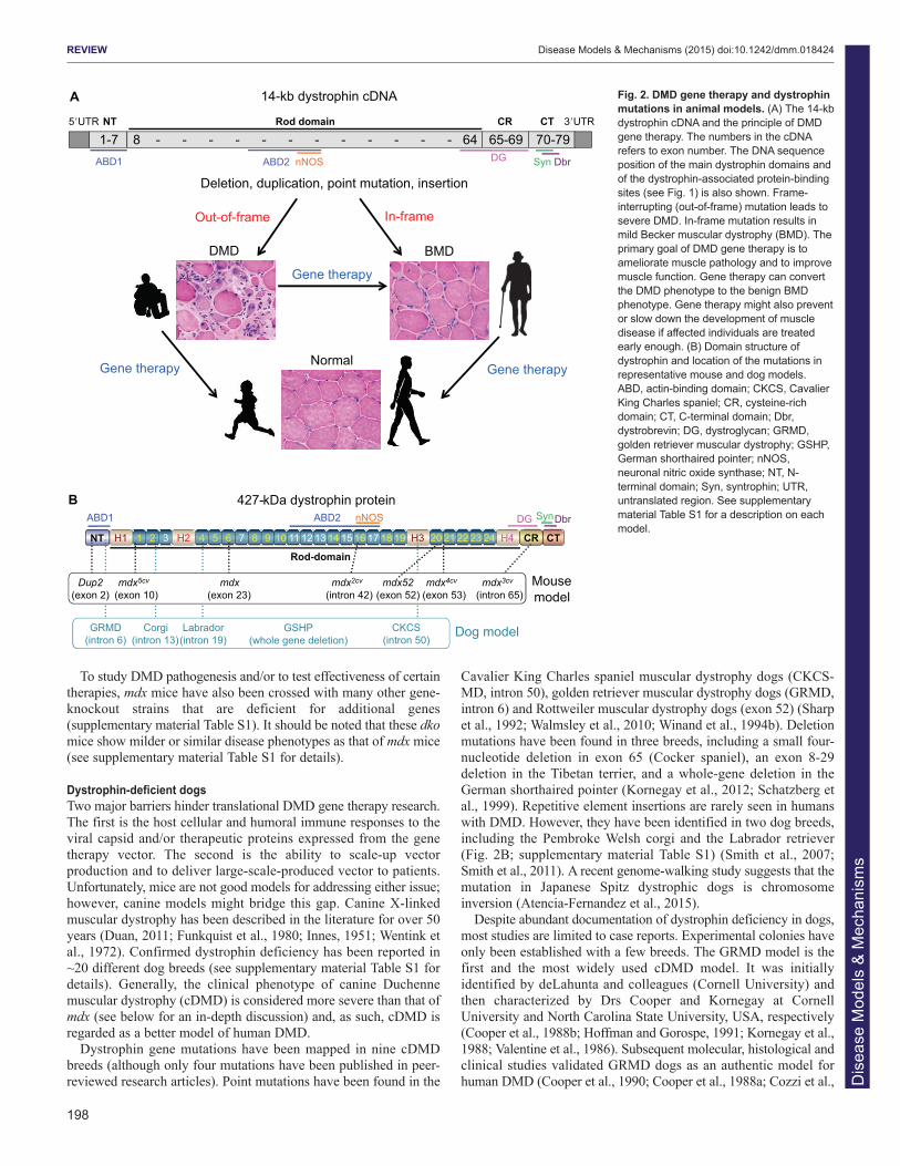
IntroductionDuchenne muscular dystrophy (DMD) is the most commonmuscular dystrophy, with a worldwide incidence of one in 5000 livemale births according to newborn screening (Emery and Muntoni,2003; Mendell and Lloyd-Puryear, 2013). It is caused by the lack ofdystrophin, a critical muscle protein that connects the cytoskeletonand the extracellular matrix (ECM) (Bonilla et al., 1988; Hoffmanet al., 1987). The 2.4-Mb dystrophin gene was discovered in 1986(Kunkel, 2005; Monaco et al., 1986). It contains 79 exons andencodes a ~14-kb cDNA (Koenig et al., 1987). The full-lengthprotein has four functional domains: the N-terminal (NT), rod,cysteine-rich (CR) and C-terminal (CT) domains. Dystrophinassembles several transmembrane (dystroglycan, sarcoglycan,sarcospan) and cytosolic [syntrophin, dystrobrevin and neuronalnitric oxide synthase (nNOS)] proteins into a dystrophin-associatedglycoprotein complex (DAGC) at the sarcolemma (Fig. 1; Box 1 fora glossary of terms) (Ervasti, 2007). Frame-shift mutations of thedystrophin gene abolish protein expression and lead to DMD (Box1). In-frame deletions often generate truncated dystrophin and resultin the milder Becker muscular dystrophy (BMD) (Fig. 2A) (Beggset al., 1991; Hoffman and Kunkel, 1989; Monaco et al., 1988).
REVIEW
1Department of Molecular Microbiology and Immunology, School of Medicine,University of Missouri, Columbia, MO 65212, USA. 2Department of Neurology,School of Medicine, University of Missouri, Columbia, MO 65212, USA.
*Author for correspondence ([email protected])
This is an Open Access article distributed under the terms of the Creative CommonsAttribution License (http://creativecommons.org/licenses/by/3.0), which permits unrestricteduse, distribution and reproduction in any medium provided that the original work is properlyattributed.
The identification of the disease-causing gene and the molecularbasis for the DMD and BMD phenotypes establishes the foundationfor DMD gene therapy (Fig. 2A). To mitigate muscle disease, onecan either restore the full-length transcript or express a truncated butin-frame dystrophin gene (Duan, 2011; Goyenvalle et al., 2011;Konieczny et al., 2013; Mendell et al., 2012; Verhaart and Aartsma-Rus, 2012). Several gene therapy strategies are currently underdevelopment. They include replacing the mutated gene with afunctional candidate gene (gene replacement) or repairing thedefective gene by targeted correction and exon skipping (generepair). Currently, adeno-associated virus (AAV)-mediated genereplacement and antisense oligonucleotide (AON)-mediated exonskipping are at the forefront (see Box 1).
In this Review, we discuss existing DMD animal models and theirapplication in preclinical gene therapy research. We also discusshow to use these models to address the current and emergingchallenges in DMD gene therapy.
Animal modeling of dystrophin deficiencyBoth naturally occurring and laboratory-generated animal modelsare available to study the pathobiology of dystrophin deficiency andto develop innovative therapies for treating DMD. Currently, thereare nearly 60 different animal models for DMD, and the list keepsgrowing (supplementary material Table S1). Non-mammalian (suchas Caenorhabditis elegans, Drosophila melanogaster and zebrafish)and the feline (either hypertrophic or non-hypertrophic) DMDmodels are rarely used in gene therapy studies (Berger and Currie,2012; Chamberlain and Benian, 2000; Kunkel et al., 2006; Lloydand Taylor, 2010; Shelton and Engvall, 2005; Smith, 2011; Winandet al., 1994a), and the newly developed rat and pig DMD modelshave yet to be used in such research (Hollinger et al., 2014; Klymiuket al., 2013; Nakamura et al., 2014; Nonneman et al., 2012). Assuch, we focus this Review on the mouse and dog models (Fig. 2B).We discuss the pros and cons of each system and their use in genetherapy (Table 1).
Dystrophin-deficient miceThe most widely used animal model for DMD research is the mdxmouse. It was discovered in the early 1980s in a colony ofC57BL/10ScSn mice due to elevated serum creatine kinase (CK)and histological evidence of myopathy (Bulfield et al., 1984). Themutation in the mdx mouse is a nonsense point mutation (C-to-Ttransition) in exon 23 that aborted full-length dystrophin expression(Fig. 2B) (Sicinski et al., 1989).
Despite being deficient for dystrophin, mdx mice have minimalclinical symptoms and their lifespan is only reduced by ~25%(Fig. 3; Table 1) (Chamberlain et al., 2007; Li et al., 2009). Incontrast, the lifespan of individuals with DMD is reduced by ~75%(Box 2; Fig. 3B). mdx skeletal muscle disease has several distinctivephases. In the first 2 weeks, mdx muscle is indistinguishable fromthat of normal mice. Between 3 to 6 weeks, it undergoes startling
Animal models of Duchenne muscular dystrophy: from basicmechanisms to gene therapyJoe W. McGreevy1, Chady H. Hakim1, Mark A. McIntosh1 and Dongsheng Duan1,2,*
Dis
ease
Mod
els
& M
echa
nism
s

196
necrosis. Subsequently, the majority of skeletal muscle enters arelatively stable phase owing to robust regeneration. mdx limbmuscles often become hypertrophic during this phase. The onlyexception is the diaphragm, which shows progressive deterioration,as is also seen in affected humans (Box 2) (Stedman et al., 1991).Severe dystrophic phenotypes, such as muscle wasting, scoliosis andheart failure, do not occur until mice are 15 months or older (Bosticket al., 2008b; Bostick et al., 2009; Hakim et al., 2011; Lefaucheur etal., 1995; Lynch et al., 2001; Pastoret and Sebille, 1995). Asignificant portion of aged mdx mice also develops spontaneoussarcoma (Fig. 3A) (Chamberlain et al., 2007; Schmidt et al., 2011;Wang et al., 2014).
The mdx mouse has been crossed to several different geneticbackgrounds, including the albino, BALB/c, C3H, C57BL/6, DBA/2and FVB strains, and several immune-deficient strains. Phenotypicvariation has been observed in different backgrounds(supplementary material Table S1). For example, albino-mdx miceshow more severe neurological dysfunction and higher circulatingcytokines (Stenina et al., 2013). BALB/c-mdx and C3H-mdx miceare less susceptible to sarcoma (Krivov et al., 2009; Schmidt et al.,2011; Stenina et al., 2013). Immune-deficient nude-mdx and scid-mdx mice show less fibrosis (Farini et al., 2007; Morrison et al.,2000). The DBA/2-mdx mice are thought to better represent humandisease because they display more fibrosis and less regeneration(Fukada et al., 2010). However, according to The JacksonLaboratory, the DBA/2 strain is a challenging breeder and it alsocarries mutations in a variety of genes that cause hearing loss andeye abnormalities (http://jaxmice.jax.org/strain/000671.html).
In 1989, four chemical variant (cv) mdx strains were published(Chapman et al., 1989). These mice were generated on the C57BL/6background using the mutagen N-ethyl-N-nitrosourea (ENU) andthey are named as mdx2cv, mdx3cv, mdx4cv and mdx5cv. Each of thesestrains carries a different point mutation (Fig. 2B; supplementarymaterial Table S1) (Cox et al., 1993b; Im et al., 1996). Although theoverall clinical presentation of these mice differs very little from that
of mdx mice, each line has unique features. Specifically, mdx3cv micestill express ~5% of a near-full-length dystrophin protein (Cox et al.,1993b; Li et al., 2008). mdx5cv mice have a more severe skeletalmuscle phenotype (Beastrom et al., 2011). Revertant fibers (see Box1) are rarely seen in mdx4cv and mdx5cv mice (Danko et al., 1992;Partridge and Lu, 2008). In addition to these four strains, severalnew ENU-induced dystrophin-null lines have been recentlygenerated (supplementary material Table S1) (Aigner et al., 2009).
In addition to the above-mentioned strains, several otherdystrophin-deficient lines (Dup2, MD-null, Dp71-null, mdx52 andmdx βgeo) have been created using various genetic engineeringtechniques (see supplementary material Table S1 for details).
Immune-deficient mdx strains are dystrophin-null mice that havebeen crossed to the immune-deficient background. These mice canbe used to study cell or gene therapy without the compoundingeffects of the host immune response. Besides the commonly usednude-mdx and scid-mdx mice (Farini et al., 2007; Morrison et al.,2000), several new lines (NSG-mdx4cv, Rag2–IL2rb–Dmd– and W41mdx) have recently been developed (supplementary materialTable S1) (Arpke et al., 2013; Bencze et al., 2012; Vallese et al.,2013; Walsh et al., 2011). These new lines carry additionalmutations that further compromise the immune system.
Mouse models that recapitulate the DMD phenotypeDystrophin-deficient mice show minimal clinical disease. This couldbe due to the upregulation of compensatory mechanisms or to aspecies-specific property of the muscle. Elimination of compensatorymechanisms or humanization of mdx mice results in mouse modelsthat recapitulate the dystrophic phenotype of human with DMD. Amajor function of dystrophin is to strengthen the sarcolemma bycross-linking the ECM with the cytoskeleton. Two other proteins,utrophin and α7β1-integrin fulfil the same function and theirexpression is upregulated in mdx mice. The genetic elimination ofutrophin and α7-integrin in mdx mice creates utrophin/dystrophin andintegrin/dystrophin double-knockout (dko) mice, respectively
REVIEW Disease Models & Mechanisms (2015) doi:10.1242/dmm.018424
Actin
Actin
Microtubule 3 1 2
12 13 14 15 16 17 18 19 20 21
CT S
yn
Syn
Dbr
Dystrophin
Sarcoglycans
αDG
βDGSarcospan
Laminin
CR Syn
nNOS
H3
H1 H2
Extracellular matrix
Lipid bilayer
Cytoplasm
Fig. 1. Schematic outline of dystrophin and the dystrophin-associated glycoprotein complex (DAGC). Dystrophin contains N-terminal (NT), middle rod,cysteine-rich (CR) and C-terminal (CT) domains. The middle rod domain is composed of 24 spectrin-like repeats (numerical numbers in the cartoon, positivelycharged repeats are marked in white color) and four hinges (H1, H2, H3 and H4). Dystrophin has two actin-binding domains located at NT and repeats 11-15,respectively. Repeats 1-3 interact with the negatively charged lipid bilayer. Repeats 16 and 17 form the neuronal nitric oxide synthase (nNOS)-binding domain.Dystrophin interacts with microtubule through repeats 20-23. Part of H4 and the CR domain bind to the β-subunit of dystroglycan (βDG). The CT domain ofdystrophin interacts with syntrophin (Syn) and dystrobrevin (Dbr). Dystrophin links components of the cytoskeleton (actin and microtubule) to laminin in theextracellular matrix. Sarcoglycans and sarcospan do not interact with dystrophin directly but they strengthen the entire DAGC, which consists of dystrophin,DG, sarcoglycans, sarcospan, Syn, Dbr and nNOS.
Dis
ease
Mod
els
& M
echa
nism
s

(Deconinck et al., 1997a; Grady et al., 1997; Guo et al., 2006; Rooneyet al., 2006). These dko mice are significantly smaller than theirsingle-gene null parents and show much more severe muscle disease(similar to or even worse than that of humans with DMD) (Fig. 3A).
However, they are difficult to generate and care for, and they often dieprematurely (compared with the single knockouts; Fig. 3B). Recentstudies suggest that utrophin heterozygous mdx mice might representan intermediate model between the extreme dko mice and mildlyaffected mdx mice (Rafael-Fortney et al., 2011; van Putten et al.,2012b; Zhou et al., 2008).
Robust skeletal muscle regeneration also explains the slowlyprogressive phenotype of mdx mice. Two different approaches havebeen used to reduce muscle regeneration in mdx mice. Megeney etal. eliminated MyoD, a master myogenic regulator, from mdx mice(Megeney et al., 1996). The resulting MyoD/dystrophin double-mutant mouse shows marked myopathy, dilated cardiomyopathy andpremature death (Fig. 3B) (Megeney et al., 1996; Megeney et al.,1999). Compared with normal muscle, the length of telomere isreduced in DMD muscle (Decary et al., 2000). Sacco et al.hypothesized that the long telomere length in mouse myogenic stemcells contributes to the high regenerative capacity of mouse muscle(Mourkioti et al., 2013; Sacco et al., 2010). Telomerase RNA (mTR)is required for the maintenance of the telomere length. To reducetelomere length in dystrophin-null mice, Sacco et al. crossed mdx4cv
mice with mTR-null mice. These mTR/mdx double-mutant miceshow more severe muscle wasting and cardiac defects (Mourkioti etal., 2013; Sacco et al., 2010). Their lifespan is reduced to ~12months (Fig. 3B).
Other symptomatic dko strains (supplementary material Table S1)have also been generated by mutating genes involved in: (1)cytoskeleton-ECM interactions (such as desmin, laminin and like-glycosyltransferase) (Banks et al., 2014; Gawlik et al., 2014; Martinset al., 2013), (2) the DAGC (such as dystrobrevin and δ-sarcoglycan) (Grady et al., 1999; Li et al., 2009), (3) muscle repair(such as dysferlin) (Grady et al., 1999; Han et al., 2011; Hosur et al.,2012; Li et al., 2009) and (4) inflammation and fibrosis [such asinterleukin-10, a disintegrin and metalloproteinase protein (ADAM)-8, and plasminogen activator inhibitor-1) (Ardite et al., 2012;Nishimura et al., 2015; Nitahara-Kasahara et al., 2014).
Humanization is another method of increasing mdx diseaseseverity. The gene encoding cytidine monophosphate sialic acidhydroxylase (Cmah) is naturally inactivated in humans but not inmice (Varki, 2010). Cmah converts cell-surface sialic acid N-acetylneuraminic acid (Neu5Ac) to N-glycolylneuraminic acid(Neu5Gc). Hence, human cells only have Neu5Ac but no Neu5Gc.Genetic elimination of Cmah humanizes the cell-surface glycanprofile in mice (Hedlund et al., 2007). Interestingly, Cmah-deficientmdx mice show a more severe phenotype (Fig. 3B). Thishumanization process renders Cmah/mdx mice a better modelbecause they more closely recapitulate human disease(Chandrasekharan et al., 2010).
In summary, the large collection of symptomatic double-mutantmouse lines has greatly expanded the armory of potential mousemodels for preclinical studies. Accelerated disease progression inthese dko mice provides an excellent opportunity not only to obtainresults from experimental therapies more rapidly but also to confirmwhether a therapy can indeed ameliorate clinically relevantmanifestations and increase lifespan. Nevertheless, there are alsoimportant limitations. For example, most dko mice are difficult tobreed and are often not commercially available. Importantly, unlikein humans with DMD, all dko mice carry a mutation not only in thedystrophin gene but also in another gene (although because the geneencoding Cmah is inactivated in humans this is not an issue forCmah/mdx mice). This is not the case in affected humans. How thisadditional mutation influences data interpretation remainsincompletely understood.
197
REVIEW Disease Models & Mechanisms (2015) doi:10.1242/dmm.018424
Box 1. GlossaryAdeno-associated virus (AAV): a single-stranded DNA virus identifiedin 1965. AAV has a ~4.7-kb genome and encodes at least three openreading frames (ORFs), one for viral capsid proteins, one for replicationproteins and a third one for the assembly-activating protein. Inrecombinant AAV vectors, viral ORFs are replaced by a reporter ortherapeutic expression cassette. An up to 5-kb vector genome can bepackaged in an AAV vector. At least 13 different AAV serotypes havebeen reported. Hundreds of genetically modified AAV capsids have alsobeen developed. AAV can efficiently transduce post-mitotic tissues andwild-type AAV does not cause human disease. Because of thesefeatures, AAV has been used in numerous clinical trials.Dual and tri-AAV vectors: engineered AAV vector systems that candeliver a 10-kb (dual vector) or 15-kb (tri-vector) expression cassette.Specifically, a large expression cassette is divided into two pieces (dualvectors) or three pieces (tri-vector). An individual piece contains either aregion that overlaps with another piece and/or is engineered with splicingsignals. Each piece is packaged in a single viral particle. Co-delivery ofvectors containing different pieces of the expression cassette results inreconstitution of the original expression cassette in vivo by cellularrecombination mechanisms.Exon skipping: a phenomenon in which one or multiple exons arespliced out and eliminated from the mature mRNA.Frame-shift mutation: a mutation that disrupts the open reading frameof an mRNA transcript.Freezing response: a reflex defense mechanism observed in preyanimals where they freeze or completely stop moving when scared.Hydrodynamic intravascular delivery: a technique used for genedelivery where the hydrostatic pressure is applied to increase thepermeability of the vascular wall. This allows efficient penetration of genetherapy plasmids into the tissue parenchyma.Liposome: an artificially created lipid-bilayer sphere. A DNA plasmid canbe incorporated inside the lipid sphere. The fusion of the lipid bilayer withcell membrane allows delivery of the DNA plasmid into a cell.Microspheres: generic name given to a nanoscale spherical object thatcan be made out of a variety of materials, including lipids, polymers andmetal oxides. They can be used to deliver a DNA plasmid to the cell.Nuclease-based gene editing: DNA gene editing technique that usesendonucleases to make double-stranded breaks in the DNA at a user-specified location to initiate error-prone DNA repair. As a consequence,the DNA sequence at the site of break is altered. These endonucleasesare often linked to sequence-specific targeting proteins, such as zincfingers.Phosphorodiamidate morpholino oligomer (PMO): a syntheticoligonucleotide in which the ribose or deoxyribose backbone is replacedby a morpholine ring and the phosphate replaced byphosphorodiamidate. Any one of the four nucleobases can be attachedto the morpholine ring. Because of the unnatural backbone, PMO is moreresistant than the ordinary antisense oligonucleotide (AON) to nucleasedigestion.Revertant fibers: rarely occurring dystrophin-positive myofibers foundin animals that carry a null mutation in the dystrophin gene. Themolecular mechanisms underlying the formation of revertant fibers arenot completely clear. They might arise from sporadic alternative splicingthat eliminates the mutation from the dystrophin transcript and/or asecond mutation that corrects the original mutation on the DNA.Sarcolemma: muscle-cell plasma membrane.Vivo-morpholino: a morpholino oligomer that has been covalently linkedto an octa-guanidine dendrimer moiety. Conjugation with octa-guanidineincreases cell penetration.WW domain: a protein module of approximately 40 amino acids. Itcontains two preserved tryptophan (W) residues that are spaced 20 to22 amino acids apart. The WW domain folds into a stable, triple-strandedβ-sheet and mediates protein-protein interaction.
Dis
ease
Mod
els
& M
echa
nism
s
198
To study DMD pathogenesis and/or to test effectiveness of certaintherapies, mdx mice have also been crossed with many other gene-knockout strains that are deficient for additional genes(supplementary material Table S1). It should be noted that these dkomice show milder or similar disease phenotypes as that of mdx mice(see supplementary material Table S1 for details).
Dystrophin-deficient dogsTwo major barriers hinder translational DMD gene therapy research.The first is the host cellular and humoral immune responses to theviral capsid and/or therapeutic proteins expressed from the genetherapy vector. The second is the ability to scale-up vectorproduction and to deliver large-scale-produced vector to patients.Unfortunately, mice are not good models for addressing either issue;however, canine models might bridge this gap. Canine X-linkedmuscular dystrophy has been described in the literature for over 50years (Duan, 2011; Funkquist et al., 1980; Innes, 1951; Wentink etal., 1972). Confirmed dystrophin deficiency has been reported in~20 different dog breeds (see supplementary material Table S1 fordetails). Generally, the clinical phenotype of canine Duchennemuscular dystrophy (cDMD) is considered more severe than that ofmdx (see below for an in-depth discussion) and, as such, cDMD isregarded as a better model of human DMD.
Dystrophin gene mutations have been mapped in nine cDMDbreeds (although only four mutations have been published in peer-reviewed research articles). Point mutations have been found in the
Cavalier King Charles spaniel muscular dystrophy dogs (CKCS-MD, intron 50), golden retriever muscular dystrophy dogs (GRMD,intron 6) and Rottweiler muscular dystrophy dogs (exon 52) (Sharpet al., 1992; Walmsley et al., 2010; Winand et al., 1994b). Deletionmutations have been found in three breeds, including a small four-nucleotide deletion in exon 65 (Cocker spaniel), an exon 8-29deletion in the Tibetan terrier, and a whole-gene deletion in theGerman shorthaired pointer (Kornegay et al., 2012; Schatzberg etal., 1999). Repetitive element insertions are rarely seen in humanswith DMD. However, they have been identified in two dog breeds,including the Pembroke Welsh corgi and the Labrador retriever(Fig. 2B; supplementary material Table S1) (Smith et al., 2007;Smith et al., 2011). A recent genome-walking study suggests that themutation in Japanese Spitz dystrophic dogs is chromosomeinversion (Atencia-Fernandez et al., 2015).
Despite abundant documentation of dystrophin deficiency in dogs,most studies are limited to case reports. Experimental colonies haveonly been established with a few breeds. The GRMD model is thefirst and the most widely used cDMD model. It was initiallyidentified by deLahunta and colleagues (Cornell University) andthen characterized by Drs Cooper and Kornegay at CornellUniversity and North Carolina State University, USA, respectively(Cooper et al., 1988b; Hoffman and Gorospe, 1991; Kornegay et al.,1988; Valentine et al., 1986). Subsequent molecular, histological andclinical studies validated GRMD dogs as an authentic model forhuman DMD (Cooper et al., 1990; Cooper et al., 1988a; Cozzi et al.,
REVIEW Disease Models & Mechanisms (2015) doi:10.1242/dmm.018424
Deletion, duplication, point mutation, insertion
Out-of-frame In-frame
BMD
Gene therapy
Gene therapy Gene therapy
A
DMD
14-kb dystrophin cDNA
1-7 8 - - - - - - - - - - - - 64 65-69 70-79 Rod domain
ABD1 ABD2 nNOS DG Syn Dbr
CR CT NT 3�UTR 5�UTR
Normal
mdx5cv (exon 10)
mdx4cv (exon 53)
mdx3cv (intron 65)
mdx (exon 23)
mdx2cv (intron 42)
mdx52 (exon 52)
Dup2 (exon 2)
B
Corgi (intron 13)
Labrador (intron 19)
GSHP (whole gene deletion)
GRMD (intron 6)
CKCS (intron 50)
Mouse model
Dog model
427-kDa dystrophin protein
11 H3 H4 CR CT 10 9 8 7 6 5 4 3 2 1 H2 H1 12 13 14 16 17 18 19 15 21 22 23 24 20
Rod-domain
nNOS ABD1 ABD2
NT
Syn DG Dbr
Fig. 2. DMD gene therapy and dystrophinmutations in animal models. (A) The 14-kbdystrophin cDNA and the principle of DMDgene therapy. The numbers in the cDNArefers to exon number. The DNA sequenceposition of the main dystrophin domains andof the dystrophin-associated protein-bindingsites (see Fig. 1) is also shown. Frame-interrupting (out-of-frame) mutation leads tosevere DMD. In-frame mutation results inmild Becker muscular dystrophy (BMD). Theprimary goal of DMD gene therapy is toameliorate muscle pathology and to improvemuscle function. Gene therapy can convertthe DMD phenotype to the benign BMDphenotype. Gene therapy might also preventor slow down the development of muscledisease if affected individuals are treatedearly enough. (B) Domain structure ofdystrophin and location of the mutations inrepresentative mouse and dog models.ABD, actin-binding domain; CKCS, CavalierKing Charles spaniel; CR, cysteine-richdomain; CT, C-terminal domain; Dbr,dystrobrevin; DG, dystroglycan; GRMD,golden retriever muscular dystrophy; GSHP,German shorthaired pointer; nNOS,neuronal nitric oxide synthase; NT, N-terminal domain; Syn, syntrophin; UTR,untranslated region. See supplementarymaterial Table S1 for a description on eachmodel.
Dis
ease
Mod
els
& M
echa
nism
s
2001; Kornegay et al., 1988; Lanfossi et al., 1999; McCully et al.,1991; Moise et al., 1991; Nguyen et al., 2002; Sharp et al., 1992;Valentine et al., 1989a; Valentine et al., 1990a; Valentine et al., 1991;Valentine and Cooper, 1991; Valentine et al., 1990b; Valentine et al.,1986; Valentine et al., 1988; Valentine et al., 1989b; Valentine et al.,1989c; Valentine et al., 1989d; Valentine et al., 1992). Currently,GRMD dogs are maintained in several colonies throughout the USA(including the University of Missouri and Texas A&M University,among others), and in France, Brazil and Australia. The GRMDmutation has also been crossed to the Beagle background and acolony is now maintained in Japan; these dogs are called canine X-linked muscular dystrophy in Japan or CXMDJ (Shimatsu et al.,2003; Valentine et al., 1988). Recently, we and others have createdhybrid strains that are on mixed genetic backgrounds and/or containmutations of different breeds (Cotten et al., 2013; Fine et al., 2011;Miyazato et al., 2011; Shin et al., 2013a; Yang et al., 2012). BesidesGRMD-based colonies, research colonies have also been generatedfrom affected Pembroke Welsh corgis and Labrador retrievers(Auburn University and University of Missouri), and CKCS-MD(Royal Veterinary College, UK) (Smith et al., 2007; Smith et al.,2011; Walmsley et al., 2010). The CKCS-MD model is especiallyinteresting because the mutation in this breed corresponds to a majordeletion hot spot (exons 45-53) in humans with DMD (Aartsma-Ruset al., 2006; Flanigan et al., 2009b; Tuffery-Giraud et al., 2009).
Affected dogs share a remarkably similar clinical course to thatof DMD boys (Box 2; Fig. 3; Table 1) (Shimatsu et al., 2005; Smithet al., 2011; Valentine et al., 1988). Limb weakness and exerciseintolerance start around 2 to 3 months of age (analogous to ~3 yearsof age in humans) (Valentine et al., 1988). Muscle atrophy, jointcontracture, hypersalivation, dysphagia, abnormal gait and signs ofcardiac involvement become apparent at ~6 months (Fig. 3A; Table
1) (Fan et al., 2014; Fine et al., 2011; Valentine et al., 1988;Valentine et al., 1989c; Yugeta et al., 2006). At around 6 to 10months, disease progression enters a relatively stable ‘honeymoon’period (Fan et al., 2014; Shimatsu et al., 2005; Valentine et al.,1988). Death often occurs around 3 years of age (a ~75% reductionof the lifespan) (Fig. 3B). Humans with DMD show heterogeneityin their clinical manifestation (Box 2) (Ashwath et al., 2014;Desguerre et al., 2009; Sifringer et al., 2004). cDMD dogs also showvariation in their symptoms. In extreme cases, affected subjects areessentially asymptomatic despite the lack of dystrophin in theirmuscles (Ambrósio et al., 2008; Dubowitz, 2006; Hattori et al.,1999; Wakefield et al., 2009; Zatz et al., 2014; Zucconi et al., 2010).
Besides clinical resemblance, cDMD dogs also have histologicallesions similar to affected humans. For example, limb musclefibrosis is a salient disease feature in humans with DMD and inaffected dogs but not in mdx mice (C.H.H. and D.D., unpublishedobservations). Vigorous regeneration in mouse muscle contributessubstantially to the mild phenotype of mdx mice. This regenerationis evident by high proportions of centrally nucleated myofibers inmdx mice. Similar to humans with DMD, cDMD dogs have muchfewer myofibers containing central nucleation (Cozzi et al., 2001;Shin et al., 2013b; Smith et al., 2011; Yang et al., 2012).
It should be noted that the clinical presentation of cDMD dogs isnot identical to that of humans with DMD (Table 1). About 20-30%of cDMD puppies die within 2 weeks of birth likely due todiaphragm failure (Ambrósio et al., 2008; Nakamura et al., 2013;Shimatsu et al., 2005; Valentine et al., 1988). However, this neonataldeath is not seen in newborn DMD boys. Growth retardation isanother canine-specific symptom (West et al., 2013). Body weightat birth is similar between normal and affected cDMD puppies(Smith et al., 2011). However, at 1 and 6 months of age, the body
199
REVIEW Disease Models & Mechanisms (2015) doi:10.1242/dmm.018424
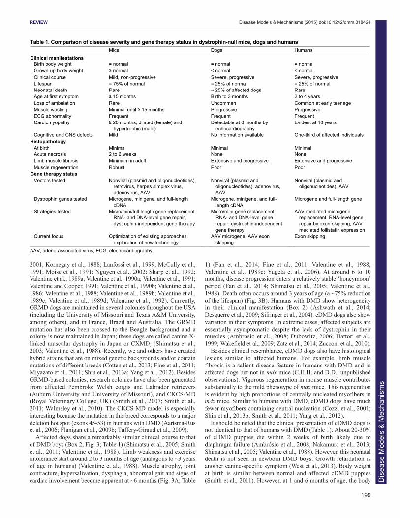
Table 1. Comparison of disease severity and gene therapy status in dystrophin-null mice, dogs and humans Mice Dogs Humans Clinical manifestations
Birth body weight = normal = normal = normal Grown-up body weight normal < normal < normal Clinical course Mild, non-progressive Severe, progressive Severe, progressive Lifespan = 75% of normal = 25% of normal = 25% of normal Neonatal death Rare ~ 25% of affected dogs Rare Age at first symptom 15 months Birth to 3 months 2 to 4 years Loss of ambulation Rare Uncomman Common at early teenage Muscle wasting Minimal until 15 months Progressive Progressive ECG abnormality Frequent Frequent Frequent Cardiomyopathy 20 months; dilated (female) and
hypertrophic (male) Detectable at 6 months by
echocardiography Evident at 16 years
Cognitive and CNS defects Mild No information available One-third of affected individuals Histopathology
At birth Minimal Minimal Minimal Acute necrosis 2 to 6 weeks None None Limb muscle fibrosis Minimum in adult Extensive and progressive Extensive and progressive Muscle regeneration Robust Poor Poor
Gene therapy status Vectors tested Nonviral (plasmid and oligonucleotides),
retrovirus, herpes simplex virus, adenovirus, AAV
Nonviral (plasmid and oligonucleotides), adenovirus, AAV
Nonviral (plasmid and oligonucleotides), AAV
Dystrophin genes tested Microgene, minigene, and full-length cDNA
Microgene, minigene, and full-length cDNA
Microgene and full-length gene
Strategies tested Micro/mini/full-length gene replacement, RNA- and DNA-level gene repair, dystrophin-independent gene therapy
Micro/mini-gene replacement, RNA- and DNA-level gene repair, dystrophin-independent gene therapy
AAV-mediated microgene replacement, RNA-level gene repair by exon-skipping, AAV-mediated follistatin expression
Current focus Optimization of existing approaches, exploration of new technology
AAV microgene; AAV exon skipping
Exon skipping
AAV, adeno-associated virus; ECG, electrocardiography.
Dis
ease
Mod
els
& M
echa
nism
s

200
weight of affected puppies reaches only ~80% and ~60% of normal,respectively (C.H.H. and D.D., unpublished observation from n>50dogs). Finally, untreated humans with DMD usually lose ambulationduring the early teenage years. However, complete loss ofambulation is not a clinical feature in young cDMD dogs (Duan etal., 2015; Valentine et al., 1988).
Overall, cDMD dogs share many features with that of humanswith DMD. These features make cDMD dogs an excellent model toconduct preclinical gene therapy studies (Duan, 2011; Duan, 2015).Nevertheless, mdx mice remain the most commonly used model inDMD gene therapy studies owing to the low cost and easy access.Any discussion of DMD models in gene therapy that lacked mentionof mdx mice would not be complete.
Establishing the foundations of gene therapy: transgenicmdx miceThe successful development of a gene therapy requires research toidentify the therapeutic candidate gene, the level of expressionneeded to produce a therapeutic effect and the tissue that should betargeted (Chamberlain, 2002; Duan, 2006). As we discuss in this
section, for DMD gene therapy research, these fundamentalquestions have been addressed using transgenic mdx mice.
Therapeutic potential of truncated dystrophin genesNaturally occurring small dystrophin isoformsThe enormous size of the full-length dystrophin gene poses one ofthe biggest challenges for gene therapy because it exceeds thepackaging limit of most viral vectors. For this reason, identifying asmaller but functional gene has been an ongoing goal in thedevelopment of a dystrophin gene-replacement therapy. Earlystudies showed that, besides the 427-kDa full-length protein, thedystrophin gene also encodes a number of smaller N-terminal-truncated non-muscle isoforms (Ahn and Kunkel, 1993; Blake et al.,2002; Ervasti, 2007). These include Dp260, Dp140, Dp116, Dp71and Dp40 (numbers refer to the molecular weight) (Fig. 4A). Withthe exception of Dp40 (Fujimoto et al., 2014), they all contain theCT and CR domains but are missing the NT actin-binding domain.To determine whether these miniature isoforms are therapeuticallyrelevant, the Chamberlain lab, as well as others, made transgenicmdx mice for Dp260, Dp116 and Dp71 (see supplementary material
REVIEW Disease Models & Mechanisms (2015) doi:10.1242/dmm.018424
BL1
0
mdx
6-m-old male
BL1
0
dko B
L6
dko
1-m-old male 12-day-old male
5-m-old affected male 2-yr-old normal and affected male
Affected Normal
ale 23-m-old male
mdx
A
Utrophin/dystrophin dko
Integrin/ dystrophin dko
5-m-old a5 ld fffected male f t d lfff
Affected
B
~ 20 yr
50 25 0 100 75
~ 3 yr
% of normal
~ 0.5 m
~ 1 m
~ 2.5 m
~3 m
~ 9 m
~ 10 m
~ 11 m
~ 12 m
~ 12 m
~ 22 m
~27 m
1
1
1
1
mm
~ 12 yr
~ 80 yr Normal human
Normal mouse
mdx
myoD/mdx
Utrophin/mdx
Integrin/mdx
mTR/mdx
Cmah/mdx
γ-sarcoglyan/mdx
Desmin/mdx
Dystrobrevin/mdx
Laminin/mdx
Normal dog
cDMD
DMD
Fig. 3. Representative animal models for DMD.(A) Representative pictures of selected DMD mouse and dogmodels. mdx mice do not show symptoms (see 6-month-oldphoto) until very old (see 23-month-old photo). Aged mdx mice arealso prone to rhabdomyosarcoma (a tumor of muscle origin; redarrow). Utrophin/dystrophin and integrin/dystrophin double-knockout (dko) mice are much smaller than the age-matched wild-type (BL10 and BL6) mice. A 5-month-old affected dog shows limbmuscle atrophy and is reluctant to exercise. At the age of 2 yearsold, the affected dog displays severe clinical disease, whereas itsnormal sibling remains healthy. (B) Lifespan comparison amongaffected humans, affected dogs and various mouse models.
Dis
ease
Mod
els
& M
echa
nism
s

Table S1 for details) (Cox et al., 1994; Gaedigk et al., 2006;Greenberg et al., 1994; Judge et al., 2011; Judge et al., 2006; Warneret al., 2002).
Dp71 is the most abundant non-muscle dystrophin isoform. Itcontains only the CR and CT domains (Fig. 4A). Because the CTdomain carries the binding sites for syntrophin and dystrobrevin, itwas initially thought that Dp71 might restore some of the signalingfunctions of dystrophin. Surprisingly, however, transgenicoverexpression of Dp71 results in more severe muscle disease inmdx mice (Cox et al., 1994; Greenberg et al., 1994) and myopathyin normal mice (Leibovitz et al., 2002). The WW domain of hinge4 (H4; see Fig. 1A and Box 1), which is partially truncated in Dp71,participates in dystrophin-dystroglycan interaction (Huang et al.,2000). To fully appreciate the contribution of dystroglycan bindingand dystrophin signaling in DMD pathogenesis, Judge et al.generated Dp116 transgenic mdx mice. Dp116 is a Schwann-cell-specific dystrophin isoform. It contains the last three spectrin-likerepeats, H4, and the CR and CT domains (Fig. 4A). Dp116expression does not improve muscle disease in mdx4cv mice nor doesit reduce the histopathology in utrophin/dystrophin dko mice (Judgeet al., 2011; Judge et al., 2006). Interestingly, the lifespan ofutrophin/dystrophin dko mice was significantly increased bytransgenic Dp116 expression (Judge et al., 2011).
Two independent strains of Dp260 transgenic mice have beenstudied (Gaedigk et al., 2006; Warner et al., 2002). Although Dp260(also known as the retinal isoform of dystrophin) does not carry theNT domain, it contains the ABD2 domain (Fig. 2B; Fig. 4A). Itsoverexpression significantly reduces the dystrophic phenotype ofmdx and utrophin/dystrophin dko mice but does not completelyprevent muscle degeneration, inflammation and fibrosis (Gaedigk etal., 2006; Warner et al., 2002). In summary, transgenic analyses ofnaturally occurring dystrophin isoforms suggest that the N-terminaldomain is required for maximum muscle protection and that acomplete dystroglycan-binding domain (including the WW domainin H4 and the CR domain) is important.
Synthetic mini- and micro-dystrophin genesAn alternative approach to developing a smaller but functionaldystrophin gene is through genetic engineering. To achieve this, oneneeds to know which regions of the dystrophin gene are dispensable
for its normal functions. The first clue about this came from a mildlyaffected individual, who was ambulant at age 61 (England et al.,1990). This person carries a large in-frame deletion (∆17-48) in therod domain, which eliminates 46% of the coding sequence.Transgenic expression of the ∆17-48 minigene in mdx micesignificantly reduced skeletal muscle pathology and increasedspecific muscle force (Phelps et al., 1995; Wells et al., 1995).Subsequent optimization by removing residue repeat 19 in the ∆17-48 minigene resulted in a more protective ∆H2-R19 minigene(Fig. 4B) (Harper et al., 2002).
An important function of dystrophin is to recruit nNOS to thesarcolemma. Failure to do so results in functional ischemia andaggravates muscle disease (Thomas, 2013). We recently identifiedthe dystrophin nNOS-binding site at R16/17 of the rod domain(Fig. 1; Fig. 2B) (Lai et al., 2009; Lai et al., 2013). Inclusion of thisbinding site in synthetic dystrophins (Fig. 4B) significantly enhancesmuscle protection and exercise capacity (Lai et al., 2009; Zhang etal., 2013).
The mini-dystrophin gene is ~6 to 8 kb. One drawback is that itcannot fit into the 5-kb packaging limit of AAV, the most efficientmuscle gene-transfer vector. A pivotal transgenic study from theChamberlain lab opened the door to further reducing the size of thedystrophin gene by deleting the entire CT domain (Crawford et al.,2000). Specifically, Chamberlain and colleagues showed that a C-terminal-truncated dystrophin gene successfully restored syntrophinand dystrobrevin to the sarcolemma and completely protected youngadult mdx mice (Crawford et al., 2000). Consistent with thistransgenic study, a subset of affected individuals who have partialor complete CT-domain deletion also show mild disease (Aartsma-Rus et al., 2006; McCabe et al., 1989; Patria et al., 1996; Tuffery-Giraud et al., 2009). Collectively, the existing data suggest that themajority of the rod domain (except for R16/17) and the entire C-terminal domain are not essential for dystrophin function. Based onthis understanding, several versions of highly abbreviated syntheticmicro-dystrophin genes (<4 kb) have been engineered (Fig. 4B)(Harper et al., 2002; Lai et al., 2009; Wang et al., 2000). Thesemicrogenes greatly prevent muscle damage in transgenic mdx mice(Hakim and Duan, 2013; Harper et al., 2002; Li et al., 2011).
Level of expressionTwo essential questions in DMD gene therapy are: (1) how muchdystrophin is too much, and (2) how much dystrophin is enough toameliorate disease? In transgenic mdx mice, Chamberlain andcolleagues found that 50-fold overexpression of full-lengthdystrophin was not toxic to skeletal muscle, thus providing a highsafety margin (Cox et al., 1993a). Studies in transgenic mdx micehave also revealed the threshold for histological and physiologicalprotection (Phelps et al., 1995; Wells et al., 1995). Dystrophinexpression at ~20% of the wild-type level significantly mitigatedmuscle pathology and enhanced muscle contractility (Phelps et al.,1995; Wells et al., 1995). mdx3cv mice express ~5% of a near-full-length dystrophin protein and mdx-Xist∆hs mice express variable lowlevels of dystrophin (supplementary material Table S1) (Cox et al.,1993b; van Putten et al., 2012a). Recent studies in mdx3cv and mdx-Xist∆hs mice suggest that dystrophin expression at a 5% level stillpreserves some muscle function in mdx mice and extends thelifespan of utrophin/dystrophin dko mice (Li et al., 2008; Li et al.,2010; van Putten et al., 2012a; van Putten et al., 2013). A clearcorrelation between the dystrophin level and clinical manifestationhas also been noticed in humans with DMD (Nicholson et al.,1993a; Nicholson et al., 1993b). Affected individuals with ≥20%wild-type dystrophin protein expression are often ambulant beyond
201
REVIEW Disease Models & Mechanisms (2015) doi:10.1242/dmm.018424
Box 2. Clinical features of DMDLarge-scale population studies have outlined the natural diseaseprogression in affected humans (Table 1) (Bushby and Connor, 2011;Henricson et al., 2013; Magri et al., 2011; McDonald et al., 2013a;McDonald et al., 2013b; Spurney et al., 2014). The first clinical signusually appears around age 3. Between ages 5 and 8, symptoms areoften stabilized or even slightly improved (known as the ‘honeymoon’period) in the absence of any treatment (Bushby and Connor, 2011;McDonald et al., 2013a; McDonald et al., 2010). Rapid clinicaldeterioration starts around 7 to 8 years of age (Mercuri and Muntoni,2013). Individuals with DMD lose their ambulation at approximately age10, develop cardiomyopathy at about age 16 and die around age 20 (lifeexpectancy is reduced by ~75%). With the use of steroids, symptommanagement and multidisciplinary care (especially nocturnal ventilation),the lifespan of an affected individual is now extended to 30 to 40 yearsof age. In these individuals, cardiac complications (cardiomyopathyand/or cardiac arrhythmia) have emerged as a major source of morbidityand mortality. Despite the overall trend of disease progression throughoutlife, affected individuals actually show heterogeneity in clinicalmanifestations. One retrospective study of 75 drug-naïve affectedindividuals classified DMD into four distinctive groups (infantile, classical,moderate pure motor and severe pure motor) based on the intellectualand motor outcome (Desguerre et al., 2009).
Dis
ease
Mod
els
& M
echa
nism
s

202
age 20 (Bulman et al., 1991; Byers et al., 1992; Hoffman et al.,1989). An affected individual with 30% dystrophin proteinexpression, measured through western blot, was even free of skeletalmuscle disease at age 23 (Neri et al., 2007). Where gene therapy isconcerned, there is no doubt that restoring ≥20% protein expressionwill be needed to achieve clinically meaningful improvement.Nonetheless, mouse data suggest that even a low level of expression(~5%) might still be beneficial.
Target tissue: skeletal muscle versus heartHumans with DMD suffer from both skeletal muscle disease andcardiomyopathy. It thus seems obvious that both skeletal and heartmuscle should be treated. However, many existing gene therapyapproaches (such as some AONs used for exon skipping and AAVserotype-9-mediated systemic gene transfer in newborn dogs)cannot efficiently reach the heart (Alter et al., 2006; Hakim et al.,2014; Yokota et al., 2009; Yue et al., 2008). Will skeletal-muscle-centered therapy benefit individuals with DMD? An early study inyoung (4- to 5-month-old) transgenic mdx4cv mice suggests thattargeted repair of skeletal muscle accelerates heart disease(Townsend et al., 2008). However, the interpretation of the heartfunction data in this study has been questioned (Wasala et al.,
2013). Using a different approach, Crisp et al. reached acompletely opposite conclusion in adult (6- to 9-month-old) mdxmice and neonatal (10-day-old) utrophin/dystrophin dko mice(Crisp et al., 2011). They concluded that skeletal muscle rescuecan prevent cardiomyopathy (Crisp et al., 2011). Because mdxmice do not develop clinically evident cardiomyopathy until theyare 21-months old (Bostick et al., 2008b; Bostick et al., 2009), werecently re-evaluated this issue in a similar transgenic strain usedby Townsend et al. (Townsend et al., 2008; Wasala et al., 2013).Surprisingly, skeletal-muscle-rescued mdx mice showed theidentical heart disease as that of non-transgenic mdx mice at theage of 23 months (Wasala et al., 2013). In summary, skeletalmuscle rescue might neither aggravate nor completely alleviatecardiomyopathy. As such, we believe that gene therapy shouldtreat both skeletal and cardiac muscles.
Gene replacement therapyA straightforward approach to treating DMD is to add back afunctional dystrophin gene. This can be achieved using a variety ofgene-transfer vectors, including nonviral, retroviral, adenoviral,herpes simplex viral and AAV vectors. The candidate gene can bethe full-length cDNA or an abbreviated synthetic gene.
REVIEW Disease Models & Mechanisms (2015) doi:10.1242/dmm.018424
Dp427 11 H3 H4 CR CT 10 9 8 7 6 5 4 3 2 1 H2 H1 NT 12 13 14 16 17 18 19 15 21 22 23 24 20
A
CR CT Dp71
H4 CR CT 22 23 24 Dp116
11 H3 H4 CR CT 10 12 13 14 16 17 18 19 15 21 22 23 24 20 Dp260
CR Dp40
Dp427(B) Dp260 (intron 29)
Dp140 (intron 44)
Dp116 (intron 55)
Dp71 & Dp40 (intron 62) Dp427 (M)
Dp427 (P)
Dp140 H3 H4 CR CT 19 21 22 23 24 20 19
�17-48 (Davies lab, from patient) H3 H4 CR CT 19 21 22 23 24 20 19 16 17 18 3 2 1 H1 11 10 9 8 7 6 5 4 H2 12 13 14 15 1
�H2-R19 (Chamberlain lab, synthetic) H3 H4 CR CT 21 22 23 24 20 16 17 18 3 2 1 H1 11 10 9 8 7 6 5 4 H2 12 13 14 15
�H2-R15 ( Duan lab, synthetic) H3 H4 CR CT 21 22 23 24 20 3 2 1 H1 11 10 9 8 7 6 5 4 H2 12 13 14 15
�R4-R23/C (Chamberlain lab, synthetic) H4 CR 3 2 1 H2 H1 24
�R3-19/20-21/C (also called �3990) (Xiao lab, synthetic) H3 H4 CR 3 2 1 H1 22 23 24
�R2-15/R18-19/R20-23/C ( Duan lab, synthetic) H3 H4 CR 1 H1 16 17 24
Full-length dystrophin
Min
i-dys
troph
in
(6 to
8 k
b)
Mic
ro-d
ystro
phin
(<
4 kb
)
B
11 H3 H4 CR CT 10 9 8 7 6 5 4 3 2 1 H2 H1 12 13 14 16 17 18 19 15 21 22 23 24 20
Rod domain
nNOS AB1 AB2
NT
NT
NT
NT
NT
NT
NT
18 17 16 19
Syn DG Dbr
Fig. 4. Structure of abbreviateddystrophins. (A) Naturally occurringdystrophin isoforms. In the topmost schematic,blue boxes denote exons. The full-lengthdystrophin (Dp427) transcripts have threeisoforms, including brain Dp427 (B), muscleDp427 (M) and Purkinje cell Dp427 (P).Smaller dystrophin isoforms are producedfrom promoters located in different introns(intron positions are marked for each isoform).Dp260 is expressed in the retina, Dp140 in thebrain and kidney, Dp116 in Schwann cells,and Dp71 and Dp40 are expressed from thesame promoter except Dp71 is ubiquitouslyexpressed whereas Dp40 only exists in thebrain. Except for Dp140, all other dystrophinisoforms have unique N-terminal sequencesnot present in the full-length protein.(B) Structure of representative mini- andmicro-dystrophins. The full-length dystrophinprotein is shown uppermost, and features thesame terminology as that used in Fig. 1.
Dis
ease
Mod
els
& M
echa
nism
s
Replacement with the full-length dystrophin coding sequenceSeveral strategies have been explored to deliver the 14-kb, full-length dystrophin cDNA. Direct plasmid injection was tested in mdxmice soon after the discovery of the dystrophin gene (Acsadi et al.,1991). A number of different nonviral delivery approaches havesince been evaluated in mdx mice. These include the use ofliposomes, microspheres, electroporation and hydrodynamicintravascular delivery (see Box 1). Direct plasmid injection has alsobeen tested in the GRMD model and in a Phase 1 human trial(Braun, 2004; Duan, 2008). However, poor transduction andtransient expression have limited further development of theseplasmid-based therapeutic strategies.
The gutted adenoviral vector does not carry any viral genes andcan package a 35-kb genome. It has been used to express the full-length dystrophin cDNA (Haecker et al., 1996; Kochanek et al.,1996; Kumar-Singh and Chamberlain, 1996). Tests conducted inmdx and utrophin/dystrophin dko mice have yielded promisingresults (Clemens et al., 1996; DelloRusso et al., 2002; Ishizaki et al.,2011; Kawano et al., 2008). The current challenges are the hostimmune response to the adenoviral capsid and the contaminatingwild-type adenovirus. Herpes simplex virus also has an extremelylarge capacity (~150 kb) and has been used to package the full-length dystrophin cDNA (Akkaraju et al., 1999; Liu et al., 2006).However, there have been very few animal studies performed withit due to the toxicity of the virus.
Recently, tri-AAV vectors were used to deliver the full-lengthdystrophin cDNA (see Box 1) (Koo et al., 2014; Lostal et al., 2014).In this system, the full-length cDNA expression cassette is split intothree fragments and separately packaged in an AAV vector. Co-infection with all three AAV vectors results in the production of afull-length dystrophin protein. This approach has been tested in mdxand mdx4cv mice by direct muscle injection. The therapeutic benefitsof this system await substantial improvement in transductionefficiency.
Replacement with small synthetic dystrophin genesThe 6- to 8-kb minigenes discussed earlier in the Review have beentested with plasmid, retrovirus, adenovirus and AAV. Retroviraldelivery is very inefficient because the virus does not transduce post-mitotic muscle cells (Dunckley et al., 1993). The first-generationE1-deleted adenovirus was used to deliver the ∆17-48 minigene tomdx mice and GRMD dogs (Howell et al., 1998; Ragot et al., 1993).Although this vector is more efficient than a retroviral vector, itinduces a strong cellular immune response in mdx mice (Howell etal., 1998; Ragot et al., 1993). The Chamberlain and Duan labs havetested dual-AAV-vector-mediated mini-dystrophin therapy in mdxmice using local and systemic gene transfer (see Box 1) (Ghosh etal., 2008; Lai et al., 2005; Odom et al., 2011; Zhang and Duan,2012; Zhang et al., 2013). In the dual AAV vector system, mini-dystrophin expression is achieved with a pair of AAV vectors, eachcarrying half of the minigene. These studies have shown asignificant improvement of histology and function in treated mdxmice. Noticeably, the use of the R16/17-containing mini-dystrophindual AAV vectors has successfully restored sarcolemmal nNOSexpression and ameliorated functional ischemia (Zhang and Duan,2012; Zhang et al., 2013).
AAV-mediated, micro-dystrophin gene therapy is currently at thecutting edge of DMD gene-replacement therapy. Local injectionstudies performed in the Chamberlain, Dickson, Duan, Takeda andXiao laboratories suggest that a rationally designed dystrophinmicrogene can protect limb muscles and the heart in mdx micedespite the absence of ~70% of the coding sequence (Harper et al.,
2002; Wang et al., 2000; Yoshimura et al., 2004; Yue et al., 2003).Using the newly developed AAV serotype-6 and -8 vectors (Gao etal., 2002; Rutledge et al., 1998), the Chamberlain and Xiao labsachieved widespread whole-body muscle gene transfer in the rodentmodels of muscular dystrophies (Gregorevic et al., 2004; Wang etal., 2005). Later, it was found that AAV serotype-9 can also provideefficient systemic muscle delivery (Bostick et al., 2007; Pacak et al.,2006). More recent studies suggest that AAV-8 and AAV-9 can alsoproduce robust body-wide muscle gene transfer in neonatal dogs(Hakim et al., 2014; Kornegay et al., 2010; Pan et al., 2013; Yue etal., 2008).
The first systemic gene therapy test was performed in mdx miceby Gregorevic et al. (Gregorevic et al., 2004) and subsequently inutrophin/dystrophin and myoD/dystrophin dko mice (Gregorevic etal., 2006; Lai et al., 2009). In these studies, micro-dystrophin genetherapy significantly ameliorated the histological and physiologicalsigns of muscular dystrophy, reduced CK levels and extendedlifespan. To further improve therapeutic efficacy, several labs madeadditional changes to the existing micro-dystrophin constructs.Dickson and colleagues found that codon-optimization and inclusionof the syntrophin/dystrobrevin-binding site resulted in better rescue(Foster et al., 2008; Koo et al., 2011a). The Chamberlain lab foundthat the rigid poly-proline site in hinge 2 compromised micro-dystrophin function (Banks et al., 2010). Our studies have suggestedthat R16/17 should be incorporated in the microgene design tonormalize nNOS expression (Harper, 2013; Lai et al., 2009; Lai etal., 2013; Li et al., 2011; Shin et al., 2013b).
In an effort to translate AAV microgene therapy to largemammals, several groups have extended research into cDMDmodels (Koo et al., 2011b; Kornegay et al., 2010; Shin et al., 2012a;Shin et al., 2012b; Wang et al., 2007). These studies uncovered twoimportant issues that were not encountered during mouse studies.First, intramuscular injection results in a strong cellular immuneresponse (Ohshima et al., 2009; Wang et al., 2007; Yuasa et al.,2007; Yue et al., 2008). As a result, transient immune suppressionis necessary for persistent transduction in dog muscle (Shin et al.,2012b; Wang et al., 2007). Second, a microgene that reduces muscledisease in mice might not work effectively in dogs (Kornegay et al.,2010; Sampaolesi et al., 2006). Specifically, the ∆R4-23/Cdystrophin microgene did not improve muscle histology when testedin a cell therapy study (Sampaolesi et al., 2006). Newborn GRMDdogs developed more severe disease after treatment with the ∆R3-19/20-21/C (also called ∆3990) microgene (Kornegay et al., 2010).Currently, convincing physiological improvement has only beendemonstrated in the ∆R2-15/R18-19/R20-23/C microgene-treateddogs (Shin et al., 2013b).
Gene repair therapyTherapeutic approaches that aim to repair or correct a DMD genemutation have been conducted at both the RNA and DNA levelusing oligonucleotides or engineered endonucleases (Aartsma-Rus,2012; Bertoni, 2014). Although AON-mediated exon skipping hasalready reached Phase 3 human trials, endonuclease-based generepair has just begun to emerge (Koo and Wood, 2013; Lu et al.,2011).
Repairing the dystrophin transcriptTherapeutic RNA targeting using exon skipping is by far the mostadvanced DMD gene therapy technology developed to date. In exonskipping, AONs are used to modulate the splicing of the RNAtranscript such that one or several exons are excluded. As a result,an out-of-frame mRNA is converted into an in-frame transcript or
203
REVIEW Disease Models & Mechanisms (2015) doi:10.1242/dmm.018424
Dis
ease
Mod
els
& M
echa
nism
s

204
an exon that contains a premature stop codon is removed from thetranscript (Spitali and Aartsma-Rus, 2012). An internally deleted butpartially functional dystrophin produced from exon skipping isexpected to convert severe DMD to the milder Becker phenotype.This approach represents an excellent example of how a rationallydesigned strategy can rapidly move from bench to bedside.
The initial proof-of-principle study for exon skipping wasconducted in cultured mdx mouse muscle cells (Dunckley et al.,1998). Subsequent in vivo tests in mdx mice showed that thisapproach produced a highly efficient restoration of dystrophinexpression and improved muscle function, following local orsystemic injection (Alter et al., 2006; Gebski et al., 2003; Lu et al.,2003; Lu et al., 2005; Mann et al., 2001). Similarly, exon skipping(Fig. 5) has been achieved in cultured cDMD muscle cells and inCXMDJ dogs by local and systemic delivery (McClorey et al., 2006;Walmsley et al., 2010; Yokota et al., 2009). Several clinical trialshave been initiated based on the results of animal studies (Koo andWood, 2013; Opar, 2012). Data from the Phase 1 and 2 trials arehighly promising (Cirak et al., 2011; Goemans et al., 2011; Kinaliet al., 2009; Mendell et al., 2013; van Deutekom et al., 2007).However, the expected efficacy remains to be confirmed in a Phase3 study (Hoffman and McNally, 2014; Wood, 2013).
Early exon-skipping studies used AONs based on 2′-O-methylated phosphorothioate (2OMe-PS) or phosphorodiamidatemorpholino oligomers (PMOs) (Box 1). An important limitation ofthese AONs is that they cannot reach the heart. To overcome thishurdle, a variety of conjugated PMOs have been developed (Aoki etal., 2012; Jearawiriyapaisarn et al., 2008; Wu et al., 2009; Wu et al.,2008; Yin et al., 2008; Yin et al., 2011). In these PMOs,oligonucleotides are covalently linked to a cell-penetrating peptideor an octa-guanidine dendrimer, which can enhance cell penetration(the octa-guanidine-modified PMO is called vivo-morpholino; seeBox 1). Systemic delivery of conjugated AONs in mdx miceproduced robust exon skipping in the heart and the restoration ofcardiac function (Wu et al., 2008; Wu et al., 2011). Anotherdrawback of AON therapy is the rapid turnover of the therapeutic
oligonucleotides. To solve this problem, investigators have begun touse the AAV vector to achieve persistent AON delivery in vivo inmdx mice (Denti et al., 2006; Goyenvalle et al., 2004). Recently,AAV-based exon skipping has been shown to significantly improvethe dystrophic phenotype in utrophin/dystrophin dko mice and inGRMD dogs (Barbash et al., 2013; Bish et al., 2012; Goyenvalle etal., 2012; Le Guiner et al., 2014; Vulin et al., 2012).
mdx mice and GRMD dogs carry point mutations in thedystrophin gene. However, ~ 60% of DMD is due to deletions inexons 45-53 or duplications in exon 2 (Flanigan et al., 2009b).mdx52 and dup2 mice carry mutations that resemble the deletionsand duplications in affected humans, respectively. Hence, they areexcellent models for preclinical testing. Aoki et al. delivered acocktail of ten vivo-morpholino AONs to mdx52 mice and achievedefficient multiple-exon skipping (exons 45-55) (Fig. 5) (Aoki et al.,2012). The resulting ∆45-55 dystrophin transcript is highlyprotective and significantly improves muscle strength and histologywithout causing any toxicity (Aoki et al., 2012). The duplication ofexon 2 is a more challenging error to correct because a completeskipping of exon 2 leads to an out-of-frame transcript. Wein et al.recently tested exon 2 skipping in the dup2 model using an AAV-based exon-skipping system (Wein et al., 2014). The treatmentgenerated a ∆2 transcript with a premature stop codon in exon 3.Surprisingly, however, the dystrophic phenotype was significantlyameliorated. Further investigation suggests that the removal of exon2 activates a downstream internal ribosome entry site in exon 5.Translation from this site yields a highly functional protein (Wein etal., 2014). The results of the Aoki et al. and Wein et al. studies areespecially appealing because humans who carry similar transcriptsare often asymptomatic (Ferreiro et al., 2009; Flanigan et al., 2009a;Nakamura et al., 2008). Therapies based on the same principle mighttherefore yield dramatic clinical improvement in boys with DMD.
Repair at the DNA levelCompared to exon skipping, approaches to correct the mutateddystrophin gene are less developed (Bertoni, 2014). Initial DNA-
REVIEW Disease Models & Mechanisms (2015) doi:10.1242/dmm.018424
9 Exon 5 7 8 6
9 Exon 5 7 8 6
9 Exon 5 8 6
Exon 5 8 6
Exon 5 8 6
Normal
GRMD
GRMD +exon
skipping
44
Premature stop
45 45 46 47 48 49 50 51 52 53 54 55 56 57
Exon 52 deletion
Normal
mdx52
mdx52 +exon
skipping
Exon 53 out-of-frame
7
9 Exon 5
Exon 7 spliced out
Splice acceptor mutation
Exon 8 out-of-frame
7
Multi-exon skipping
Multi-exon skipping
cDNA
cDNA
9
44 45 45 46 47 48 49 50 51 52 53 54 55 56 57
44 45 45 46 47 48 49 50 51 53 54 55 56 57
44 45 45 46 47 48 49 50 51 53
44 45 45 46 47 48 49 50 51 53 54 55 56 57
44 45 45 55 56 57
Fig. 5. Multiple-exon skipping. Theuppermost diagram is the intron/exonstructure of the dystrophin gene. Blueboxes denote exons. The top boxshows the golden retriever musculardystrophy dog (GRMD) mutation andexon skipping for GRMD. A pointmutation in intron 6 alters normalsplicing, and the resulting transcript(gray) is out-of-frame. Skipping exons6, 7 and 8 yields an in-frame transcript.The bottom box shows the mdx52mutation and exon skipping in mdx52.Deletion of exon 52 disrupts thereading frame and results in apremature stop. Removing exons 45 to55 from the mutated transcriptgenerates an in-frame transcript.
Dis
ease
Mod
els
& M
echa
nism
s

repair strategies used oligonucleotides that are homologous to thetarget DNA. This approach has resulted in gene correction in mdxand mdx5cv mice, and in one GRMD dog, but the efficiency was toolow for clinical application (Bartlett et al., 2000; Kayali et al., 2010;Rando et al., 2000). Nuclease-based gene editing is a powerfultechnology to correct DNA defects (Box 1). Briefly, a nuclease isused as a pair of molecular scissors to cut DNA at the target site.When a double-strand DNA break is repaired by cellularmechanisms, insertions and/or deletions are introduced at the breakpoint. Some of these modifications yield the wild-type sequence,hence gene correction. Four families of engineered nucleases havebeen recently developed, including meganuclease, zinc-fingernuclease, TALEN (transcription activator-like effector nuclease) andthe CRISPR/Cas (clustered regularly interspaced short palindromicrepeat/CRISPR-associated nuclease/helicase) system. These have allbeen explored for use in DMD therapy; however, the majority of thestudies are currently limited to cultured cells (Chapdelaine et al.,2010; Long et al., 2014; Ousterout et al., 2014; Ousterout et al.,2013; Rousseau et al., 2011). Future studies are needed to validatethese highly promising gene-editing strategies in animal models ofDMD.
Gene therapy for cardiomyopathy and neuronal defectsCardiomyopathy and neuronal defects are two other prominentclinical features of DMD. Gene therapy for the heart and centralnervous system (CNS) requires special consideration (Anderson etal., 2002; Duan, 2006; Lai and Duan, 2012; Nardes et al., 2012;Ricotti et al., 2011; Shin et al., 2010; Snow et al., 2013) becausethese organs differ from skeletal muscle in their anatomy andphysiology. Importantly, dystrophin deficiency produces a uniquedisease profile in the heart and CNS.
Duchenne cardiomyopathy gene therapyThe characteristic cardiac manifestation of DMD is dilatedcardiomyopathy (Duan, 2006; Finsterer and Cripe, 2014). Heartdamage is also a prominent phenotype in various strains of dkomice, including the utrophin/dystrophin dko, α7-integrin/dystrophindko, myoD/dystrophin dko and mTR/dystrophin dko mice (Grady etal., 1997; Guo et al., 2006; Megeney et al., 1999; Mourkioti et al.,2013) (supplementary material Table S1). However, aged femalemdx mice are by far the best mouse models for studying Duchennedilated cardiomyopathy because they are genetically andphenotypically identical to affected humans (Bostick et al., 2010;Bostick et al., 2008b).
Most Duchenne cardiomyopathy gene therapy studies have beenconducted in the mdx model. Using dystrophin heterozygous mice,Duan and colleagues demonstrated that dystrophin expression in50% of cardiomyocytes was sufficient to mitigate heart injury inmdx mice (Bostick et al., 2008b; Yue et al., 2004). The first cardiacgene therapy study was performed in neonatal mdx mice using anAAV-5 ∆R4-23/∆C microgene vector in our laboratory (Fig. 4B).This micro-dystrophin gene therapy restores the DAGC andincreases the strength of the cardiomyocyte membrane (Yue et al.,2003). Subsequent studies using the same microgene normalizedthe electrocardiography (ECG) defects and improved cardiachemodynamics in young and adult mdx mice (Bostick et al.,2008a; Schinkel et al., 2012; Shin et al., 2011b; Townsend et al.,2007). To further explore the therapeutic potential, Bostick et al.treated aged female mdx mice with an AAV-9 ∆R4-23/∆Cmicrogene vector (Bostick et al., 2011; Bostick et al., 2012). Theyachieved efficient whole-heart gene transfer despite the presenceof extensive myocardial fibrosis. In near-terminal-age mice (16- to
20 months old), fibrosis was significantly reduced andhemodynamic performance significantly enhanced (Bostick et al.,2011). However, such improvements were not observed interminal-age mice (>21 months old) (Bostick et al., 2012). Thecardiac protection of the mini-dystrophin gene has only beenexamined using the 6-kb ∆H2-R19 minigene in transgenic mdxmice (Fig. 4B) (Bostick et al., 2009). This minigene completelynormalizes skeletal muscle force in transgenic mdx mice (Harperet al., 2002). However, it does not lead to a full recovery of heartfunction (Bostick et al., 2009).
Exon skipping has also been explored for treating mdx heartdisease. The original 2OMe-PS and PMO AONs cannot reach theheart (Alter et al., 2006). However, conjugated PMOs developed inthe Lu and Wood labs have significantly increased cardiac exonskipping and heart contractility in mdx mice (Wu et al., 2009; Wu etal., 2008; Wu et al., 2011; Yin et al., 2008; Yin et al., 2011).Recently, two groups tested AAV-based exon skipping in GRMDdogs. Sweeney and colleagues delivered the vector to the heart viafluoroscopy-guided trans-endocardial injection (Bish et al., 2012).This treatment restored dystrophin expression in the heart, reducedfibrosis and improved left ventricular function (Bish et al., 2012).Using X-ray-fused magnetic resonance, Barbash et al. have furtherimproved the transendocardial gene-delivery method and achieveddystrophin expression in the GRMD heart (Barbash et al., 2013).
Correcting neuronal defects with gene therapyAbout one-third of individuals with DMD display cognitivedeficiency and other CNS symptoms (Anderson et al., 2002;D’Angelo and Bresolin, 2006; Nardes et al., 2012; Ricotti et al., 2011;Snow et al., 2013). Although all dystrophin isoforms have beendetected in the nervous system (Lidov, 1996; Tozawa et al., 2012),only Dp140 and Dp71 have been implicated in neuronal abnormalitiesin humans with DMD (Bardoni et al., 2000; Bardoni et al., 1999;Daoud et al., 2009a; Daoud et al., 2009b; Felisari et al., 2000; Moizardet al., 1998; Moizard et al., 2000; Pane et al., 2012; Taylor et al.,2010). Among all DMD models, only mdx3cv and mdx βgeo mice donot express Dp140 and Dp70. Surprisingly, neurocognitive behaviorsof mdx3cv mice are only slightly different from those of mdx mice(Muntoni et al., 1991; Vaillend et al., 1998; Vaillend et al., 2004;Vaillend et al., 1995; Vaillend and Ungerer, 1999; Yamamoto et al.,2010). Dp71-specific knockout mice have also been generated and,interestingly, they show more severe learning impairment than mdxmice (Daoud et al., 2009b; Sarig et al., 1999). It is very likely thatnone of the existing mouse models can fully recapitulate theneurocognitive impairments of humans with DMD (D’Angelo andBresolin, 2006). Nevertheless, most investigators have used mdx miceto dissect the molecular and cellular consequences of dystrophindeficiency in the brain. Collectively, these studies have revealedabnormalities in the hippocampus and in several other regions of thebrain (Ghedini et al., 2012; Graciotti et al., 2008; Miranda et al., 2011;Miranda et al., 2009; Parames et al., 2014; Vaillend et al., 2004;Vaillend et al., 1999). So far, only exon skipping has been explored totreat CNS defects. Vaillend and colleagues injected an AAV exon-skipping vector to the mdx brain and found improvement ofhippocampus function (Dallérac et al., 2011; Vaillend et al., 2010).Sekiguchi et al. ameliorated the abnormal freezing response (see Box1) seen in mdx mice by injecting PMO AON to the ventricles of thebrain (Sekiguchi et al., 2009). Utrophin has been considered as ahighly promising replacement for dystrophin (see next section fordetails). Interestingly, a recent study suggested that utrophinupregulation in the brain might not rescue behavioral deficiency inmdx mice (Perronnet et al., 2012).
205
REVIEW Disease Models & Mechanisms (2015) doi:10.1242/dmm.018424
Dis
ease
Mod
els
& M
echa
nism
s

206
Dystrophin-independent gene therapy for DMD: lessons fromanimal modelsThe striking phenotypic differences between dystrophin-deficientmice and affected humans have stimulated much interest inidentifying the genes that modify DMD phenotypes. Compared withdystrophin-based therapy, the modulation of genes that already existin the body has clear immunological advantages; the therapeuticexpression of these genes is unlikely to induce immune rejectionbecause they are considered as self (Ebihara et al., 2000).
Utrophin and α7β1-integrin are among the most obviouscandidates to consider because: (1) similarly to dystrophin, theystrengthen the sarcolemma by cross-linking the ECM and thecytoskeleton; (2) their expression is upregulated in mdx mice; (3)genetic elimination of either gene aggravates dystrophicmanifestations in mdx mice; and (4) overexpression of either geneameliorates muscle disease in mdx mice (Burkin et al., 2005; Burkinet al., 2001; Deconinck et al., 1997a; Deconinck et al., 1997b; Gradyet al., 1997; Guo et al., 2006; Rafael et al., 1998; Rooney et al.,2006; Tinsley et al., 1998; Tinsley et al., 1996). As a result, genetherapy studies have been conducted in dystrophic mice (and somedogs) using full-length utrophin (Deol et al., 2007), mini-utrophin(Cerletti et al., 2003; Gilbert et al., 1999; Wakefield et al., 2000),micro-utrophin (Odom et al., 2008) and α7-integrin (Heller et al.,2013). As predicted from knockout and transgenic experiments, thedystrophic phenotype was significantly reduced by utrophin orintegrin gene therapy.
Myostatin inhibition is another example of dystrophin-independent therapy for DMD. Myostatin is an endogenous muscle-growth inhibitor (Lee, 2004; McPherron et al., 1997). Mutations inthe myostatin gene cause hypermuscularity in mouse, cattle, sheep,dog and humans (Stinckens et al., 2011). Elimination of themyostatin gene protects mdx mice by reducing fibrosis andincreasing muscle strength (Wagner et al., 2002). These observationsprovide compelling justification to explore myostatin inhibition genetherapy in animal models and, more recently, in BMD patients(Mendell et al., 2015; Rodino-Klapac et al., 2009).
Evidence from preclinical studies is opening up new lines ofinvestigation concerning how other endogenous genes could be usedin DMD gene therapy. These include genes encoding cytotoxic T-cell GalNAc transferase (Xu et al., 2007), nNOS (Lai et al., 2014),sarcoplasmic reticulum calcium ATPase 2a (Shin et al., 2011a),peroxisome proliferator-activated receptor gamma coactivator 1-alpha (Selsby et al., 2012) and sarcospan (Marshall et al., 2013).
Conclusions and perspectiveAnimal models have greatly enriched our understanding of thebiological function of dystrophin and the pathology of DMD,providing excellent platforms for investigating the efficacy andtoxicity of experimental gene therapies. Considerable progress hasbeen made in model development in the last three decades. We nowhave a large (and still expanding) collection of animal models(supplementary material Table S1). Although this offers anunprecedented opportunity for cross-species comparison andtranslation (Poussin et al., 2014), it also adds complexity anddifficulty in model selection for preclinical studies. The advantagesand limitations of each model system can vary depending on thestudy question. Some aspects of the DMD pathology (such asneurocognitive deficiency) remain difficult to model. Furthermore,animals are not humans. The findings from animal studies mayguide but not completely predict the outcome of clinical studies.Nevertheless, the value of animal models should never beunderestimated. The development of an effective gene therapy for
DMD has relied heavily, and will continue to rely, on animal models(Duan, 2011). Animal studies not only establish the proof-of-principle, they are also crucial for protocol optimization before andduring human tests. Certain studies that cannot be performed inaffected individuals (such as necropsy, in situ and ex vivo single-muscle force measurement) will have to be carried out in animalmodels. The field has surmounted many obstacles in thedevelopment of DMD models. The mild mdx mice are nowcomplemented by numerous background and mutation variants thatcan better mimic affected humans. However, as therapies that havebeen in development for the last decade enter clinical trials, newquestions are emerging. Many of these new questions (such as theimmune response to the AAV vector and scaling-up of systemicgene transfer) might be better answered with cDMD dogs, a modelthat remains to be fully characterized (Duan, 2011; Duan, 2015).
AcknowledgementsWe thank Mitchell C. Tarka for help with the Fig. 1 illustration.
Competing interestsD.D. is a member of the scientific advisory board for Solid GT, a subsidiary of SolidVentures.
FundingDMD research in the Duan lab is supported by the National Institutes of Health(AR-49419, HL-91883), Department of Defense (MD-13), Muscular DystrophyAssociation, Parent Project Muscular Dystrophy, Jesse’s Journey-The Foundationfor Gene and Cell Therapy, Hope for Javier, Kansas City Area Life SciencesInstitute and the University of Missouri.
Supplementary materialSupplementary material available online athttp://dmm.biologists.org/lookup/suppl/doi:10.1242/dmm.018424/-/DC1
ReferencesAartsma-Rus, A. (2012). Overview on DMD exon skipping. Methods Mol. Biol. 867,
97-116. Aartsma-Rus, A., Van Deutekom, J. C., Fokkema, I. F., Van Ommen, G. J. and Den
Dunnen, J. T. (2006). Entries in the Leiden Duchenne muscular dystrophy mutationdatabase: an overview of mutation types and paradoxical cases that confirm thereading-frame rule. Muscle Nerve 34, 135-144.
Acsadi, G., Dickson, G., Love, D. R., Jani, A., Walsh, F. S., Gurusinghe, A., Wolff,J. A. and Davies, K. E. (1991). Human dystrophin expression in mdx mice afterintramuscular injection of DNA constructs. Nature 352, 815-818.
Ahn, A. H. and Kunkel, L. M. (1993). The structural and functional diversity ofdystrophin. Nat. Genet. 3, 283-291.
Aigner, B., Rathkolb, B., Klaften, M., Sedlmeier, R., Klempt, M., Wagner, S.,Michel, D., Mayer, U., Klopstock, T., de Angelis, M. H. et al. (2009). Generation ofN-ethyl-N-nitrosourea-induced mouse mutants with deviations in plasma enzymeactivities as novel organ-specific disease models. Exp. Physiol. 94, 412-421.
Akkaraju, G. R., Huard, J., Hoffman, E. P., Goins, W. F., Pruchnic, R., Watkins, S.C., Cohen, J. B. and Glorioso, J. C. (1999). Herpes simplex virus vector-mediateddystrophin gene transfer and expression in mdx mouse skeletal muscle. J. GeneMed. 1, 280-289.
Alter, J., Lou, F., Rabinowitz, A., Yin, H., Rosenfeld, J., Wilton, S. D., Partridge, T.A. and Lu, Q. L. (2006). Systemic delivery of morpholino oligonucleotide restoresdystrophin expression bodywide and improves dystrophic pathology. Nat. Med. 12,175-177.
Ambrósio, C. E., Valadares, M. C., Zucconi, E., Cabral, R., Pearson, P. L., Gaiad, T.P., Canovas, M., Vainzof, M., Miglino, M. A. and Zatz, M. (2008). Ringo, a GoldenRetriever Muscular Dystrophy (GRMD) dog with absent dystrophin but normalstrength. Neuromuscul. Disord. 18, 892-893.
Anderson, J. L., Head, S. I., Rae, C. and Morley, J. W. (2002). Brain function inDuchenne muscular dystrophy. Brain 125, 4-13.
Aoki, Y., Yokota, T., Nagata, T., Nakamura, A., Tanihata, J., Saito, T., Duguez, S. M.,Nagaraju, K., Hoffman, E. P., Partridge, T. et al. (2012). Bodywide skipping ofexons 45-55 in dystrophic mdx52 mice by systemic antisense delivery. Proc. Natl.Acad. Sci. USA 109, 13763-13768.
Ardite, E., Perdiguero, E., Vidal, B., Gutarra, S., Serrano, A. L. and Muñoz-Cánoves, P. (2012). PAI-1-regulated miR-21 defines a novel age-associatedfibrogenic pathway in muscular dystrophy. J. Cell Biol. 196, 163-175.
Arpke, R. W., Darabi, R., Mader, T. L., Zhang, Y., Toyama, A., Lonetree, C. L., Nash,N., Lowe, D. A., Perlingeiro, R. C. and Kyba, M. (2013). A new immuno-,dystrophin-deficient model, the NSG-mdx4cv mouse, provides evidence forfunctional improvement following allogeneic satellite cell transplantation. Stem Cells31, 1611-1620.
REVIEW Disease Models & Mechanisms (2015) doi:10.1242/dmm.018424
Dis
ease
Mod
els
& M
echa
nism
s

Ashwath, M. L., Jacobs, I. B., Crowe, C. A., Ashwath, R. C., Super, D. M. andBahler, R. C. (2014). Left ventricular dysfunction in Duchenne muscular dystrophyand genotype. Am. J. Cardiol. 114, 284-289.
Atencia-Fernandez, S., Shiel, R. E., Mooney, C. T. and Nolan, C. M. (2015).Muscular dystrophy in the Japanese Spitz: an inversion disrupts the DMD andRPGR genes. Anim. Genet. [Epub ahead of print] doi: 10.1111/age.12266.
Banks, G. B., Judge, L. M., Allen, J. M. and Chamberlain, J. S. (2010). Thepolyproline site in hinge 2 influences the functional capacity of truncated dystrophins.PLoS Genet. 6, e1000958.
Banks, G. B., Combs, A. C., Odom, G. L., Bloch, R. J. and Chamberlain, J. S.(2014). Muscle structure influences utrophin expression in mdx mice. PLoS Genet.10, e1004431.
Barbash, I. M., Cecchini, S., Faranesh, A. Z., Virag, T., Li, L., Yang, Y., Hoyt, R. F.,Kornegay, J. N., Bogan, J. R., Garcia, L. et al. (2013). MRI roadmap-guidedtransendocardial delivery of exon-skipping recombinant adeno-associated virusrestores dystrophin expression in a canine model of Duchenne muscular dystrophy.Gene Ther. 20, 274-282.
Bardoni, A., Sironi, M., Felisari, G., Comi, G. P. and Bresolin, N. (1999). Absence ofbrain Dp140 isoform and cognitive impairment in Becker muscular dystrophy. Lancet353, 897-898.
Bardoni, A., Felisari, G., Sironi, M., Comi, G., Lai, M., Robotti, M. and Bresolin, N.(2000). Loss of Dp140 regulatory sequences is associated with cognitive impairmentin dystrophinopathies. Neuromuscul. Disord. 10, 194-199.
Bartlett, R. J., Stockinger, S., Denis, M. M., Bartlett, W. T., Inverardi, L., Le, T. T., thiMan, N., Morris, G. E., Bogan, D. J., Metcalf-Bogan, J. et al. (2000). In vivotargeted repair of a point mutation in the canine dystrophin gene by a chimericRNA/DNA oligonucleotide. Nat. Biotechnol. 18, 615-622.
Beastrom, N., Lu, H., Macke, A., Canan, B. D., Johnson, E. K., Penton, C. M.,Kaspar, B. K., Rodino-Klapac, L. R., Zhou, L., Janssen, P. M. et al. (2011).Mdx5cv mice manifest more severe muscle dysfunction and diaphragm force deficitsthan do mdx Mice. Am. J. Pathol. 179, 2464-2474.
Beggs, A. H., Hoffman, E. P., Snyder, J. R., Arahata, K., Specht, L., Shapiro, F.,Angelini, C., Sugita, H. and Kunkel, L. M. (1991). Exploring the molecular basis forvariability among patients with Becker muscular dystrophy: dystrophin gene andprotein studies. Am. J. Hum. Genet. 49, 54-67.
Bencze, M., Negroni, E., Vallese, D., Yacoub-Youssef, H., Chaouch, S., Wolff, A.,Aamiri, A., Di Santo, J. P., Chazaud, B., Butler-Browne, G. et al. (2012).Proinflammatory macrophages enhance the regenerative capacity of humanmyoblasts by modifying their kinetics of proliferation and differentiation. Mol. Ther.20, 2168-2179.
Berger, J. and Currie, P. D. (2012). Zebrafish models flex their muscles to shed lighton muscular dystrophies. Dis. Model. Mech. 5, 726-732.
Bertoni, C. (2014). Emerging gene editing strategies for Duchenne musculardystrophy targeting stem cells. Front. Physiol. 5, 148.
Bish, L. T., Sleeper, M. M., Forbes, S. C., Wang, B., Reynolds, C., Singletary, G. E.,Trafny, D., Morine, K. J., Sanmiguel, J., Cecchini, S. et al. (2012). Long-termrestoration of cardiac dystrophin expression in golden retriever muscular dystrophyfollowing rAAV6-mediated exon skipping. Mol. Ther. 20, 580-589.
Blake, D. J., Weir, A., Newey, S. E. and Davies, K. E. (2002). Function and geneticsof dystrophin and dystrophin-related proteins in muscle. Physiol. Rev. 82, 291-329.
Bonilla, E., Samitt, C. E., Miranda, A. F., Hays, A. P., Salviati, G., DiMauro, S.,Kunkel, L. M., Hoffman, E. P. and Rowland, L. P. (1988). Duchenne musculardystrophy: deficiency of dystrophin at the muscle cell surface. Cell 54, 447-452.
Bostick, B., Ghosh, A., Yue, Y., Long, C. and Duan, D. (2007). Systemic AAV-9transduction in mice is influenced by animal age but not by the route ofadministration. Gene Ther. 14, 1605-1609.
Bostick, B., Yue, Y., Lai, Y., Long, C., Li, D. and Duan, D. (2008a). Adeno-associatedvirus serotype-9 microdystrophin gene therapy ameliorates electrocardiographicabnormalities in mdx mice. Hum. Gene Ther. 19, 851-856.
Bostick, B., Yue, Y., Long, C. and Duan, D. (2008b). Prevention of dystrophin-deficient cardiomyopathy in twenty-one-month-old carrier mice by mosaic dystrophinexpression or complementary dystrophin/utrophin expression. Circ. Res. 102, 121-130.
Bostick, B., Yue, Y., Long, C., Marschalk, N., Fine, D. M., Chen, J. and Duan, D.(2009). Cardiac expression of a mini-dystrophin that normalizes skeletal muscleforce only partially restores heart function in aged mdx mice. Mol. Ther. 17, 253-261.
Bostick, B., Yue, Y. and Duan, D. (2010). Gender influences cardiac function in themdx model of Duchenne cardiomyopathy. Muscle Nerve 42, 600-603.
Bostick, B., Shin, J.-H., Yue, Y. and Duan, D. (2011). AAV-microdystrophin therapyimproves cardiac performance in aged female mdx mice. Mol. Ther. 19, 1826-1832.
Bostick, B., Shin, J. H., Yue, Y., Wasala, N. B., Lai, Y. and Duan, D. (2012). AAVmicro-dystrophin gene therapy alleviates stress-induced cardiac death but notmyocardial fibrosis in >21-m-old mdx mice, an end-stage model of Duchennemuscular dystrophy cardiomyopathy. J. Mol. Cell. Cardiol. 53, 217-222.
Braun, S. (2004). Naked plasmid DNA for the treatment of muscular dystrophy. Curr.Opin. Mol. Ther. 6, 499-505.
Bulfield, G., Siller, W. G., Wight, P. A. and Moore, K. J. (1984). X chromosome-linked muscular dystrophy (mdx) in the mouse. Proc. Natl. Acad. Sci. USA 81, 1189-1192.
Bulman, D. E., Murphy, E. G., Zubrzycka-Gaarn, E. E., Worton, R. G. and Ray, P. N.(1991). Differentiation of Duchenne and Becker muscular dystrophy phenotypes withamino- and carboxy-terminal antisera specific for dystrophin. Am. J. Hum. Genet. 48,295-304.
Burkin, D. J., Wallace, G. Q., Nicol, K. J., Kaufman, D. J. and Kaufman, S. J.(2001). Enhanced expression of the alpha 7 beta 1 integrin reduces musculardystrophy and restores viability in dystrophic mice. J. Cell Biol. 152, 1207-1218.
Burkin, D. J., Wallace, G. Q., Milner, D. J., Chaney, E. J., Mulligan, J. A. andKaufman, S. J. (2005). Transgenic expression of alpha7beta1 integrin maintainsmuscle integrity, increases regenerative capacity, promotes hypertrophy, andreduces cardiomyopathy in dystrophic mice. Am. J. Pathol. 166, 253-263.
Bushby, K. and Connor, E. (2011). Clinical outcome measures for trials in Duchennemuscular dystrophy: report from International Working Group meetings. Clin.Investig. (Lond) 1, 1217-1235.
Byers, T. J., Neumann, P. E., Beggs, A. H. and Kunkel, L. M. (1992). ELISAquantitation of dystrophin for the diagnosis of Duchenne and Becker musculardystrophies. Neurology 42, 570-576.
Cerletti, M., Negri, T., Cozzi, F., Colpo, R., Andreetta, F., Croci, D., Davies, K. E.,Cornelio, F., Pozza, O., Karpati, G. et al. (2003). Dystrophic phenotype of canineX-linked muscular dystrophy is mitigated by adenovirus-mediated utrophin genetransfer. Gene Ther. 10, 750-757.
Chamberlain, J. S. (2002). Gene therapy of muscular dystrophy. Hum. Mol. Genet. 11,2355-2362.
Chamberlain, J. S. and Benian, G. M. (2000). Muscular dystrophy: the worm turns togenetic disease. Curr. Biol. 10, R795-R797.
Chamberlain, J. S., Metzger, J., Reyes, M., Townsend, D. and Faulkner, J. A.(2007). Dystrophin-deficient mdx mice display a reduced life span and aresusceptible to spontaneous rhabdomyosarcoma. FASEB J. 21, 2195-2204.
Chandrasekharan, K., Yoon, J. H., Xu, Y., deVries, S., Camboni, M., Janssen, P.M., Varki, A. and Martin, P. T. (2010). A human-specific deletion in mouse Cmahincreases disease severity in the mdx model of Duchenne muscular dystrophy. Sci.Transl. Med. 2, 42ra54.
Chapdelaine, P., Pichavant, C., Rousseau, J., Pâques, F. and Tremblay, J. P.(2010). Meganucleases can restore the reading frame of a mutated dystrophin.Gene Ther. 17, 846-858.
Chapman, V. M., Miller, D. R., Armstrong, D. and Caskey, C. T. (1989). Recovery ofinduced mutations for X chromosome-linked muscular dystrophy in mice. Proc. Natl.Acad. Sci. USA 86, 1292-1296.
Cirak, S., Arechavala-Gomeza, V., Guglieri, M., Feng, L., Torelli, S., Anthony, K.,Abbs, S., Garralda, M. E., Bourke, J., Wells, D. J. et al. (2011). Exon skipping anddystrophin restoration in patients with Duchenne muscular dystrophy after systemicphosphorodiamidate morpholino oligomer treatment: an open-label, phase 2, dose-escalation study. Lancet 378, 595-605.
Clemens, P. R., Kochanek, S., Sunada, Y., Chan, S., Chen, H. H., Campbell, K. P.and Caskey, C. T. (1996). In vivo muscle gene transfer of full-length dystrophin withan adenoviral vector that lacks all viral genes. Gene Ther. 3, 965-972.
Cooper, B. J., Valentine, B. A., Wilson, S., Patterson, D. F. and Concannon, P. W.(1988a). Canine muscular dystrophy: confirmation of X-linked inheritance. J. Hered.79, 405-408.
Cooper, B. J., Winand, N. J., Stedman, H., Valentine, B. A., Hoffman, E. P., Kunkel,L. M., Scott, M. O., Fischbeck, K. H., Kornegay, J. N., Avery, R. J. et al. (1988b).The homologue of the Duchenne locus is defective in X-linked muscular dystrophy ofdogs. Nature 334, 154-156.
Cooper, B. J., Gallagher, E. A., Smith, C. A., Valentine, B. A. and Winand, N. J.(1990). Mosaic expression of dystrophin in carriers of canine X-linked musculardystrophy. Lab. Invest. 62, 171-178.
Cotten, S. W., Kornegay, J. N., Bogan, D. J., Wadosky, K. M., Patterson, C. andWillis, M. S. (2013). Genetic myostatin decrease in the golden retriever musculardystrophy model does not significantly affect the ubiquitin proteasome systemdespite enhancing the severity of disease. Am. J. Transl. Res. 6, 43-53.
Cox, G. A., Cole, N. M., Matsumura, K., Phelps, S. F., Hauschka, S. D., Campbell,K. P., Faulkner, J. A. and Chamberlain, J. S. (1993a). Overexpression ofdystrophin in transgenic mdx mice eliminates dystrophic symptoms without toxicity.Nature 364, 725-729.
Cox, G. A., Phelps, S. F., Chapman, V. M. and Chamberlain, J. S. (1993b). New mdxmutation disrupts expression of muscle and nonmuscle isoforms of dystrophin. Nat.Genet. 4, 87-93.
Cox, G. A., Sunada, Y., Campbell, K. P. and Chamberlain, J. S. (1994). Dp71 canrestore the dystrophin-associated glycoprotein complex in muscle but fails to preventdystrophy. Nat. Genet. 8, 333-339.
Cozzi, F., Cerletti, M., Luvoni, G. C., Lombardo, R., Brambilla, P. G., Faverzani, S.,Blasevich, F., Cornelio, F., Pozza, O. and Mora, M. (2001). Development of musclepathology in canine X-linked muscular dystrophy. II. Quantitative characterization ofhistopathological progression during postnatal skeletal muscle development. ActaNeuropathol. 101, 469-478.
Crawford, G. E., Faulkner, J. A., Crosbie, R. H., Campbell, K. P., Froehner, S. C.and Chamberlain, J. S. (2000). Assembly of the dystrophin-associated proteincomplex does not require the dystrophin COOH-terminal domain. J. Cell Biol. 150,1399-1410.
Crisp, A., Yin, H., Goyenvalle, A., Betts, C., Moulton, H. M., Seow, Y., Babbs, A.,Merritt, T., Saleh, A. F., Gait, M. J. et al. (2011). Diaphragm rescue alone preventsheart dysfunction in dystrophic mice. Hum. Mol. Genet. 20, 413-421.
D’Angelo, M. G. and Bresolin, N. (2006). Cognitive impairment in neuromusculardisorders. Muscle Nerve 34, 16-33.
Dallérac, G., Perronnet, C., Chagneau, C., Leblanc-Veyrac, P., Samson-Desvignes, N., Peltekian, E., Danos, O., Garcia, L., Laroche, S., Billard, J. M. etal. (2011). Rescue of a dystrophin-like protein by exon skipping normalizes synapticplasticity in the hippocampus of the mdx mouse. Neurobiol. Dis. 43, 635-641.
Danko, I., Chapman, V. and Wolff, J. A. (1992). The frequency of revertants in mdxmouse genetic models for Duchenne muscular dystrophy. Pediatr. Res. 32, 128-131.
Daoud, F., Angeard, N., Demerre, B., Martie, I., Benyaou, R., Leturcq, F., Cossée,M., Deburgrave, N., Saillour, Y., Tuffery, S. et al. (2009a). Analysis of Dp71
207
REVIEW Disease Models & Mechanisms (2015) doi:10.1242/dmm.018424
Dis
ease
Mod
els
& M
echa
nism
s

208
contribution in the severity of mental retardation through comparison of Duchenneand Becker patients differing by mutation consequences on Dp71 expression. Hum.Mol. Genet. 18, 3779-3794.
Daoud, F., Candelario-Martínez, A., Billard, J. M., Avital, A., Khelfaoui, M.,Rozenvald, Y., Guegan, M., Mornet, D., Jaillard, D., Nudel, U. et al. (2009b). Roleof mental retardation-associated dystrophin-gene product Dp71 in excitatorysynapse organization, synaptic plasticity and behavioral functions. PLoS ONE 4,e6574.
Decary, S., Hamida, C. B., Mouly, V., Barbet, J. P., Hentati, F. and Butler-Browne,G. S. (2000). Shorter telomeres in dystrophic muscle consistent with extensiveregeneration in young children. Neuromuscul. Disord. 10, 113-120.
Deconinck, A. E., Rafael, J. A., Skinner, J. A., Brown, S. C., Potter, A. C.,Metzinger, L., Watt, D. J., Dickson, J. G., Tinsley, J. M. and Davies, K. E.(1997a). Utrophin-dystrophin-deficient mice as a model for Duchenne musculardystrophy. Cell 90, 717-727.
Deconinck, N., Tinsley, J., De Backer, F., Fisher, R., Kahn, D., Phelps, S., Davies,K. and Gillis, J. M. (1997b). Expression of truncated utrophin leads to majorfunctional improvements in dystrophin-deficient muscles of mice. Nat. Med. 3, 1216-1221.
DelloRusso, C., Scott, J. M., Hartigan-O’Connor, D., Salvatori, G., Barjot, C.,Robinson, A. S., Crawford, R. W., Brooks, S. V. and Chamberlain, J. S. (2002).Functional correction of adult mdx mouse muscle using gutted adenoviral vectorsexpressing full-length dystrophin. Proc. Natl. Acad. Sci. USA 99, 12979-12984.
Denti, M. A., Rosa, A., D’Antona, G., Sthandier, O., De Angelis, F. G., Nicoletti, C.,Allocca, M., Pansarasa, O., Parente, V., Musarò, A. et al. (2006). Body-wide genetherapy of Duchenne muscular dystrophy in the mdx mouse model. Proc. Natl. Acad.Sci. USA 103, 3758-3763.
Deol, J. R., Danialou, G., Larochelle, N., Bourget, M., Moon, J. S., Liu, A. B.,Gilbert, R., Petrof, B. J., Nalbantoglu, J. and Karpati, G. (2007). Successfulcompensation for dystrophin deficiency by a helper-dependent adenovirusexpressing full-length utrophin. Mol. Ther. 15, 1767-1774.
Desguerre, I., Christov, C., Mayer, M., Zeller, R., Becane, H. M., Bastuji-Garin, S.,Leturcq, F., Chiron, C., Chelly, J. and Gherardi, R. K. (2009). Clinicalheterogeneity of duchenne muscular dystrophy (DMD): definition of sub-phenotypesand predictive criteria by long-term follow-up. PLoS ONE 4, e4347.
Duan, D. (2006). Challenges and opportunities in dystrophin-deficient cardiomyopathygene therapy. Hum. Mol. Genet. 15, R253-R261.
Duan, D. (2008). Myodys, a full-length dystrophin plasmid vector for Duchenne andBecker muscular dystrophy gene therapy. Curr. Opin. Mol. Ther. 10, 86-94.
Duan, D. (2011). Duchenne muscular dystrophy gene therapy: lost in translation? Res.Rep. Biol. 2011, 31-42.
Duan, D. (2015). Duchenne muscular dystrophy gene therapy in the canine model.Hum. Gene Ther. Clin. Dev. [Epub ahead of print] doi:10.1089/hum.2015.006.
Duan, D., Hakim, C. H., Ambrosio, C. E., Smith, B. F. and Sweeney, H. L. (2015).Early loss of ambulation is not a representative clinical feature in Duchennemuscular dystrophy dogs: remarks on the article of Barthélémy et al. Dis. Model.Mech. 8, 193-194.
Dubowitz, V. (2006). Enigmatic conflict of clinical and molecular diagnosis inDuchenne/Becker muscular dystrophy. Neuromuscul. Disord. 16, 865-866.
Dunckley, M. G., Wells, D. J., Walsh, F. S. and Dickson, G. (1993). Direct retroviral-mediated transfer of a dystrophin minigene into mdx mouse muscle in vivo. Hum.Mol. Genet. 2, 717-723.
Dunckley, M. G., Manoharan, M., Villiet, P., Eperon, I. C. and Dickson, G. (1998).Modification of splicing in the dystrophin gene in cultured mdx muscle cells byantisense oligoribonucleotides. Hum. Mol. Genet. 7, 1083-1090.
Ebihara, S., Guibinga, G. H., Gilbert, R., Nalbantoglu, J., Massie, B., Karpati, G.and Petrof, B. J. (2000). Differential effects of dystrophin and utrophin gene transferin immunocompetent muscular dystrophy (mdx) mice. Physiol. Genomics 3, 133-144.
Emery, A. E. H. and Muntoni, F. (2003). Duchenne muscular dystrophy. Oxford; NewYork, NY: Oxford University Press.
England, S. B., Nicholson, L. V., Johnson, M. A., Forrest, S. M., Love, D. R.,Zubrzycka-Gaarn, E. E., Bulman, D. E., Harris, J. B. and Davies, K. E. (1990).Very mild muscular dystrophy associated with the deletion of 46% of dystrophin.Nature 343, 180-182.
Ervasti, J. M. (2007). Dystrophin, its interactions with other proteins, and implicationsfor muscular dystrophy. Biochim. Biophys. Acta 1772, 108-117.
Fan, Z., Wang, J., Ahn, M., Shiloh-Malawsky, Y., Chahin, N., Elmore, S., Bagnell,C. R., Jr, Wilber, K., An, H., Lin, W. et al. (2014). Characteristics of magneticresonance imaging biomarkers in a natural history study of golden retriever musculardystrophy. Neuromuscul. Disord. 24, 178-191.
Farini, A., Meregalli, M., Belicchi, M., Battistelli, M., Parolini, D., D’Antona, G.,Gavina, M., Ottoboni, L., Constantin, G., Bottinelli, R. et al. (2007). T and Blymphocyte depletion has a marked effect on the fibrosis of dystrophic skeletalmuscles in the scid/mdx mouse. J. Pathol. 213, 229-238.
Felisari, G., Martinelli Boneschi, F., Bardoni, A., Sironi, M., Comi, G. P., Robotti,M., Turconi, A. C., Lai, M., Corrao, G. and Bresolin, N. (2000). Loss of Dp140dystrophin isoform and intellectual impairment in Duchenne dystrophy. Neurology55, 559-564.
Ferreiro, V., Giliberto, F., Muñiz, G. M., Francipane, L., Marzese, D. M., Mampel, A.,Roqué, M., Frechtel, G. D. and Szijan, I. (2009). Asymptomatic Becker musculardystrophy in a family with a multiexon deletion. Muscle Nerve 39, 239-243.
Fine, D. M., Shin, J. H., Yue, Y., Volkmann, D., Leach, S. B., Smith, B. F., McIntosh,M. and Duan, D. (2011). Age-matched comparison reveals early
electrocardiography and echocardiography changes in dystrophin-deficient dogs.Neuromuscul. Disord. 21, 453-461.
Finsterer, J. and Cripe, L. (2014). Treatment of dystrophin cardiomyopathies. Nat.Rev. Cardiol. 11, 168-179.
Flanigan, K. M., Dunn, D. M., von Niederhausern, A., Howard, M. T., Mendell, J.,Connolly, A., Saunders, C., Modrcin, A., Dasouki, M., Comi, G. P. et al. (2009a).DMD Trp3X nonsense mutation associated with a founder effect in North Americanfamilies with mild Becker muscular dystrophy. Neuromuscul. Disord. 19, 743-748.
Flanigan, K. M., Dunn, D. M., von Niederhausern, A., Soltanzadeh, P., Gappmaier,E., Howard, M. T., Sampson, J. B., Mendell, J. R., Wall, C., King, W. M. et al.;United Dystrophinopathy Project Consortium (2009b). Mutational spectrum ofDMD mutations in dystrophinopathy patients: application of modern diagnostictechniques to a large cohort. Hum. Mutat. 30, 1657-1666.
Foster, H., Sharp, P. S., Athanasopoulos, T., Trollet, C., Graham, I. R., Foster, K.,Wells, D. J. and Dickson, G. (2008). Codon and mRNA sequence optimization ofmicrodystrophin transgenes improves expression and physiological outcome indystrophic mdx mice following AAV2/8 gene transfer. Mol. Ther. 16, 1825-1832.
Fujimoto, T., Itoh, K., Yaoi, T. and Fushiki, S. (2014). Somatodendritic and excitatorypostsynaptic distribution of neuron-type dystrophin isoform, Dp40, in hippocampalneurons. Biochem. Biophys. Res. Commun. 452, 79-84.
Fukada, S., Morikawa, D., Yamamoto, Y., Yoshida, T., Sumie, N., Yamaguchi, M.,Ito, T., Miyagoe-Suzuki, Y., Takeda, S., Tsujikawa, K. et al. (2010). Geneticbackground affects properties of satellite cells and mdx phenotypes. Am. J. Pathol.176, 2414-2424.
Funkquist, B., Haraldsson, I. and Stahre, L. (1980). Primary progressive musculardystrophy in the dog. Vet. Rec. 106, 341-343.
Gaedigk, R., Law, D. J., Fitzgerald-Gustafson, K. M., McNulty, S. G., Nsumu, N. N.,Modrcin, A. C., Rinaldi, R. J., Pinson, D., Fowler, S. C., Bilgen, M. et al. (2006).Improvement in survival and muscle function in an mdx/utrn(-/-) double mutant mouseusing a human retinal dystrophin transgene. Neuromuscul. Disord. 16, 192-203.
Gao, G. P., Alvira, M. R., Wang, L., Calcedo, R., Johnston, J. and Wilson, J. M.(2002). Novel adeno-associated viruses from rhesus monkeys as vectors for humangene therapy. Proc. Natl. Acad. Sci. USA 99, 11854-11859.
Gawlik, K. I., Holmberg, J. and Durbeej, M. (2014). Loss of dystrophin and β-sarcoglycan significantly exacerbates the phenotype of laminin α2 chain-deficientanimals. Am. J. Pathol. 184, 740-752.
Gebski, B. L., Mann, C. J., Fletcher, S. and Wilton, S. D. (2003). Morpholinoantisense oligonucleotide induced dystrophin exon 23 skipping in mdx mousemuscle. Hum. Mol. Genet. 12, 1801-1811.
Ghedini, P. C., Avellar, M. C., De Lima, T. C., Lima-Landman, M. T., Lapa, A. J. andSouccar, C. (2012). Quantitative changes of nicotinic receptors in the hippocampusof dystrophin-deficient mice. Brain Res. 1483, 96-104.
Ghosh, A., Yue, Y., Lai, Y. and Duan, D. (2008). A hybrid vector system expandsadeno-associated viral vector packaging capacity in a transgene-independentmanner. Mol. Ther. 16, 124-130.
Gilbert, R., Nalbantoglu, J., Petrof, B. J., Ebihara, S., Guibinga, G. H., Tinsley, J.M., Kamen, A., Massie, B., Davies, K. E. and Karpati, G. (1999). Adenovirus-mediated utrophin gene transfer mitigates the dystrophic phenotype of mdx mousemuscles. Hum. Gene Ther. 10, 1299-1310.
Goemans, N. M., Tulinius, M., van den Akker, J. T., Burm, B. E., Ekhart, P. F.,Heuvelmans, N., Holling, T., Janson, A. A., Platenburg, G. J., Sipkens, J. A. etal. (2011). Systemic administration of PRO051 in Duchenne’s muscular dystrophy.N. Engl. J. Med. 364, 1513-1522.
Goyenvalle, A., Vulin, A., Fougerousse, F., Leturcq, F., Kaplan, J. C., Garcia, L.and Danos, O. (2004). Rescue of dystrophic muscle through U7 snRNA-mediatedexon skipping. Science 306, 1796-1799.
Goyenvalle, A., Seto, J. T., Davies, K. E. and Chamberlain, J. (2011). Therapeuticapproaches to muscular dystrophy. Hum. Mol. Genet. 20, R69-R78.
Goyenvalle, A., Babbs, A., Wright, J., Wilkins, V., Powell, D., Garcia, L. andDavies, K. E. (2012). Rescue of severely affected dystrophin/utrophin-deficient micethrough scAAV-U7snRNA-mediated exon skipping. Hum. Mol. Genet. 21, 2559-2571.
Graciotti, L., Minelli, A., Minciacchi, D., Procopio, A. and Fulgenzi, G. (2008).GABAergic miniature spontaneous activity is increased in the CA1 hippocampalregion of dystrophic mdx mice. Neuromuscul. Disord. 18, 220-226.
Grady, R. M., Teng, H., Nichol, M. C., Cunningham, J. C., Wilkinson, R. S. andSanes, J. R. (1997). Skeletal and cardiac myopathies in mice lacking utrophin anddystrophin: a model for Duchenne muscular dystrophy. Cell 90, 729-738.
Grady, R. M., Grange, R. W., Lau, K. S., Maimone, M. M., Nichol, M. C., Stull, J. T.and Sanes, J. R. (1999). Role for alpha-dystrobrevin in the pathogenesis ofdystrophin-dependent muscular dystrophies. Nat. Cell Biol. 1, 215-220.
Greenberg, D. S., Sunada, Y., Campbell, K. P., Yaffe, D. and Nudel, U. (1994).Exogenous Dp71 restores the levels of dystrophin associated proteins but does notalleviate muscle damage in mdx mice. Nat. Genet. 8, 340-344.
Gregorevic, P., Blankinship, M. J., Allen, J. M., Crawford, R. W., Meuse, L., Miller,D. G., Russell, D. W. and Chamberlain, J. S. (2004). Systemic delivery of genes tostriated muscles using adeno-associated viral vectors. Nat. Med. 10, 828-834.
Gregorevic, P., Allen, J. M., Minami, E., Blankinship, M. J., Haraguchi, M., Meuse,L., Finn, E., Adams, M. E., Froehner, S. C., Murry, C. E. et al. (2006). rAAV6-microdystrophin preserves muscle function and extends lifespan in severelydystrophic mice. Nat. Med. 12, 787-789.
Guo, C., Willem, M., Werner, A., Raivich, G., Emerson, M., Neyses, L. and Mayer,U. (2006). Absence of alpha 7 integrin in dystrophin-deficient mice causes amyopathy similar to Duchenne muscular dystrophy. Hum. Mol. Genet. 15, 989-998.
REVIEW Disease Models & Mechanisms (2015) doi:10.1242/dmm.018424
Dis
ease
Mod
els
& M
echa
nism
s

Haecker, S. E., Stedman, H. H., Balice-Gordon, R. J., Smith, D. B., Greelish, J. P.,Mitchell, M. A., Wells, A., Sweeney, H. L. and Wilson, J. M. (1996). In vivoexpression of full-length human dystrophin from adenoviral vectors deleted of all viralgenes. Hum. Gene Ther. 7, 1907-1914.
Hakim, C. H. and Duan, D. (2013). Truncated dystrophins reduce muscle stiffness inthe extensor digitorum longus muscle of mdx mice. J. Appl. Physiol. 114, 482-489.
Hakim, C. H., Grange, R. W. and Duan, D. (2011). The passive mechanical propertiesof the extensor digitorum longus muscle are compromised in 2- to 20-mo-old mdxmice. J. Appl. Physiol. 110, 1656-1663.
Hakim, C. H., Yue, Y., Shin, J. H., Williams, R. R., Zhang, K., Smith, B. F. and Duan,D. (2014). Systemic gene transfer reveals distinctive muscle transduction profile oftyrosine mutant AAV-1, -6, and -9 in neonatal dogs. Mol Ther Methods Clin Dev 1,14002.
Han, R., Rader, E. P., Levy, J. R., Bansal, D. and Campbell, K. P. (2011). Dystrophindeficiency exacerbates skeletal muscle pathology in dysferlin-null mice. SkeletMuscle 1, 35.
Harper, S. Q. (2013). Molecular dissection of dystrophin identifies the docking site fornNOS. Proc. Natl. Acad. Sci. USA 110, 387-388.
Harper, S. Q., Hauser, M. A., DelloRusso, C., Duan, D., Crawford, R. W., Phelps, S.F., Harper, H. A., Robinson, A. S., Engelhardt, J. F., Brooks, S. V. et al. (2002).Modular flexibility of dystrophin: implications for gene therapy of Duchenne musculardystrophy. Nat. Med. 8, 253-261.
Hattori, N., Kaido, M., Nishigaki, T., Inui, K., Fujimura, H., Nishimura, T., Naka, T.and Hazama, T. (1999). Undetectable dystrophin can still result in a relatively benignphenotype of dystrophinopathy. Neuromuscul. Disord. 9, 220-226.
Hedlund, M., Tangvoranuntakul, P., Takematsu, H., Long, J. M., Housley, G. D.,Kozutsumi, Y., Suzuki, A., Wynshaw-Boris, A., Ryan, A. F., Gallo, R. L. et al.(2007). N-glycolylneuraminic acid deficiency in mice: implications for human biologyand evolution. Mol. Cell. Biol. 27, 4340-4346.
Heller, K. N., Montgomery, C. L., Janssen, P. M., Clark, K. R., Mendell, J. R. andRodino-Klapac, L. R. (2013). AAV-mediated overexpression of human α7 integrinleads to histological and functional improvement in dystrophic mice. Mol. Ther. 21,520-525.
Henricson, E. K., Abresch, R. T., Cnaan, A., Hu, F., Duong, T., Arrieta, A., Han, J.,Escolar, D. M., Florence, J. M., Clemens, P. R. et al.; CINRG Investigators(2013). The cooperative international neuromuscular research group Duchennenatural history study: glucocorticoid treatment preserves clinically meaningfulfunctional milestones and reduces rate of disease progression as measured bymanual muscle testing and other commonly used clinical trial outcome measures.Muscle Nerve 48, 55-67.
Hoffman, E. P. and Gorospe, J. R. M. (1991). The animal models of Duchennemuscular dystrophy: windows on the pathophsiological consequences of dystrophindeficiency. Curr. Top. Membr. 38, 113-154.
Hoffman, E. P. and Kunkel, L. M. (1989). Dystrophin abnormalities inDuchenne/Becker muscular dystrophy. Neuron 2, 1019-1029.
Hoffman, E. P. and McNally, E. M. (2014). Exon-skipping therapy: a roadblock, detour,or bump in the road? Sci. Transl. Med. 6, 230fs14.
Hoffman, E. P., Brown, R. H., Jr and Kunkel, L. M. (1987). Dystrophin: the proteinproduct of the Duchenne muscular dystrophy locus. Cell 51, 919-928.
Hoffman, E. P., Kunkel, L. M., Angelini, C., Clarke, A., Johnson, M. and Harris, J.B. (1989). Improved diagnosis of Becker muscular dystrophy by dystrophin testing.Neurology 39, 1011-1017.
Hollinger, K., Yang, C. X., Montz, R. E., Nonneman, D., Ross, J. W. and Selsby, J.T. (2014). Dystrophin insufficiency causes selective muscle histopathology and lossof dystrophin-glycoprotein complex assembly in pig skeletal muscle. FASEB J. 28,1600-1609.
Hosur, V., Kavirayani, A., Riefler, J., Carney, L. M., Lyons, B., Gott, B., Cox, G. A.and Shultz, L. D. (2012). Dystrophin and dysferlin double mutant mice: a novelmodel for rhabdomyosarcoma. Cancer Genet 205, 232-241.
Howell, J. M., Lochmüller, H., O’Hara, A., Fletcher, S., Kakulas, B. A., Massie, B.,Nalbantoglu, J. and Karpati, G. (1998). High-level dystrophin expression afteradenovirus-mediated dystrophin minigene transfer to skeletal muscle of dystrophicdogs: prolongation of expression with immunosuppression. Hum. Gene Ther. 9, 629-634.
Huang, X., Poy, F., Zhang, R., Joachimiak, A., Sudol, M. and Eck, M. J. (2000).Structure of a WW domain containing fragment of dystrophin in complex with beta-dystroglycan. Nat. Struct. Biol. 7, 634-638.
Im, W. B., Phelps, S. F., Copen, E. H., Adams, E. G., Slightom, J. L. andChamberlain, J. S. (1996). Differential expression of dystrophin isoforms in strainsof mdx mice with different mutations. Hum. Mol. Genet. 5, 1149-1153.
Innes, J. R. (1951). Myopathies in animals; a record of some cases includingprogressive muscular dystrophy (pseudo-hypertrophic) (dog), “weisses Fleisch”(lamb), neuropathic muscular atrophy (sheep) and lymphocytic/histiocytic myositis,neuritis, radiculitis (dog). Br. Vet. J. 107, 131-143.
Ishizaki, M., Maeda, Y., Kawano, R., Suga, T., Uchida, Y., Uchino, K., Yamashita,S., Kimura, E. and Uchino, M. (2011). Rescue from respiratory dysfunction bytransduction of full-length dystrophin to diaphragm via the peritoneal cavity inutrophin/dystrophin double knockout mice. Mol. Ther. 19, 1230-1235.
Jearawiriyapaisarn, N., Moulton, H. M., Buckley, B., Roberts, J., Sazani, P.,Fucharoen, S., Iversen, P. L. and Kole, R. (2008). Sustained dystrophin expressioninduced by peptide-conjugated morpholino oligomers in the muscles of mdx mice.Mol. Ther. 16, 1624-1629.
Judge, L. M., Haraguchiln, M. and Chamberlain, J. S. (2006). Dissecting thesignaling and mechanical functions of the dystrophin-glycoprotein complex. J. CellSci. 119, 1537-1546.
Judge, L. M., Arnett, A. L., Banks, G. B. and Chamberlain, J. S. (2011). Expressionof the dystrophin isoform Dp116 preserves functional muscle mass and extendslifespan without preventing dystrophy in severely dystrophic mice. Hum. Mol. Genet.20, 4978-4990.
Kawano, R., Ishizaki, M., Maeda, Y., Uchida, Y., Kimura, E. and Uchino, M. (2008).Transduction of full-length dystrophin to multiple skeletal muscles improves motorperformance and life span in utrophin/dystrophin double knockout mice. Mol. Ther.16, 825-831.
Kayali, R., Bury, F., Ballard, M. and Bertoni, C. (2010). Site-directed gene repair ofthe dystrophin gene mediated by PNA-ssODNs. Hum. Mol. Genet. 19, 3266-3281.
Kinali, M., Arechavala-Gomeza, V., Feng, L., Cirak, S., Hunt, D., Adkin, C.,Guglieri, M., Ashton, E., Abbs, S., Nihoyannopoulos, P. et al. (2009). Localrestoration of dystrophin expression with the morpholino oligomer AVI-4658 inDuchenne muscular dystrophy: a single-blind, placebo-controlled, dose-escalation,proof-of-concept study. Lancet Neurol. 8, 918-928.
Klymiuk, N., Blutke, A., Graf, A., Krause, S., Burkhardt, K., Wuensch, A., Krebs,S., Kessler, B., Zakhartchenko, V., Kurome, M. et al. (2013). Dystrophin-deficientpigs provide new insights into the hierarchy of physiological derangements ofdystrophic muscle. Hum. Mol. Genet. 22, 4368-4382.
Kochanek, S., Clemens, P. R., Mitani, K., Chen, H. H., Chan, S. and Caskey, C. T.(1996). A new adenoviral vector: Replacement of all viral coding sequences with 28kb of DNA independently expressing both full-length dystrophin and beta-galactosidase. Proc. Natl. Acad. Sci. USA 93, 5731-5736.
Koenig, M., Hoffman, E. P., Bertelson, C. J., Monaco, A. P., Feener, C. and Kunkel,L. M. (1987). Complete cloning of the Duchenne muscular dystrophy (DMD) cDNAand preliminary genomic organization of the DMD gene in normal and affectedindividuals. Cell 50, 509-517.
Konieczny, P., Swiderski, K. and Chamberlain, J. S. (2013). Gene and cell-mediatedtherapies for muscular dystrophy. Muscle Nerve 47, 649-663.
Koo, T. and Wood, M. J. (2013). Clinical trials using antisense oligonucleotides inDuchenne muscular dystrophy. Hum. Gene Ther. 24, 479-488.
Koo, T., Malerba, A., Athanasopoulos, T., Trollet, C., Boldrin, L., Ferry, A.,Popplewell, L., Foster, H., Foster, K. and Dickson, G. (2011a). Delivery ofAAV2/9-microdystrophin genes incorporating helix 1 of the coiled-coil motif in the C-terminal domain of dystrophin improves muscle pathology and restores the level ofα1-syntrophin and α-dystrobrevin in skeletal muscles of mdx mice. Hum. Gene Ther.22, 1379-1388.
Koo, T., Okada, T., Athanasopoulos, T., Foster, H., Takeda, S. and Dickson, G.(2011b). Long-term functional adeno-associated virus-microdystrophin expression inthe dystrophic CXMDj dog. J. Gene Med. 13, 497-506.
Koo, T., Popplewell, L., Athanasopoulos, T. and Dickson, G. (2014). Triple trans-splicing adeno-associated virus vectors capable of transferring the coding sequencefor full-length dystrophin protein into dystrophic mice. Hum. Gene Ther. 25, 98-108.
Kornegay, J. N., Tuler, S. M., Miller, D. M. and Levesque, D. C. (1988). Musculardystrophy in a litter of golden retriever dogs. Muscle Nerve 11, 1056-1064.
Kornegay, J. N., Li, J., Bogan, J. R., Bogan, D. J., Chen, C., Zheng, H., Wang, B.,Qiao, C., Howard, J. F., Jr and Xiao, X. (2010). Widespread muscle expression ofan AAV9 human mini-dystrophin vector after intravenous injection in neonataldystrophin-deficient dogs. Mol. Ther. 18, 1501-1508.
Kornegay, J. N., Bogan, J. R., Bogan, D. J., Childers, M. K., Li, J., Nghiem, P.,Detwiler, D. A., Larsen, C. A., Grange, R. W., Bhavaraju-Sanka, R. K. et al.(2012). Canine models of Duchenne muscular dystrophy and their use in therapeuticstrategies. Mamm. Genome 23, 85-108.
Krivov, L. I., Stenina, M. A., Yarygin, V. N., Polyakov, A. V., Savchuk, V. I.,Obrubov, S. A. and Komarova, N. V. (2009). A new genetic variant of mdx mice:study of the phenotype. Bull. Exp. Biol. Med. 147, 625-629.
Kumar-Singh, R. and Chamberlain, J. S. (1996). Encapsidated adenovirusminichromosomes allow delivery and expression of a 14 kb dystrophin cDNA tomuscle cells. Hum. Mol. Genet. 5, 913-921.
Kunkel, L. M. (2005). 2004 William Allan award address. cloning of the DMD gene.Am. J. Hum. Genet. 76, 205-214.
Kunkel, L. M., Bachrach, E., Bennett, R. R., Guyon, J. and Steffen, L. (2006).Diagnosis and cell-based therapy for Duchenne muscular dystrophy in humans,mice, and zebrafish. J. Hum. Genet. 51, 397-406.
Lai, Y. and Duan, D. (2012). Progress in gene therapy of dystrophic heart disease.Gene Ther. 19, 678-685.
Lai, Y., Yue, Y., Liu, M., Ghosh, A., Engelhardt, J. F., Chamberlain, J. S. and Duan,D. (2005). Efficient in vivo gene expression by trans-splicing adeno-associated viralvectors. Nat. Biotechnol. 23, 1435-1439.
Lai, Y., Thomas, G. D., Yue, Y., Yang, H. T., Li, D., Long, C., Judge, L., Bostick, B.,Chamberlain, J. S., Terjung, R. L. et al. (2009). Dystrophins carrying spectrin-likerepeats 16 and 17 anchor nNOS to the sarcolemma and enhance exerciseperformance in a mouse model of muscular dystrophy. J. Clin. Invest. 119, 624-635.
Lai, Y., Zhao, J., Yue, Y. and Duan, D. (2013). α2 and α3 helices of dystrophin R16and R17 frame a microdomain in the α1 helix of dystrophin R17 for neuronal NOSbinding. Proc. Natl. Acad. Sci. USA 110, 525-530.
Lai, Y., Zhao, J., Yue, Y., Wasala, N. B. and Duan, D. (2014). Partial restoration ofcardiac function with ΔPDZ nNOS in aged mdx model of Duchenne cardiomyopathy.Hum. Mol. Genet. 23, 3189-3199.
Lanfossi, M., Cozzi, F., Bugini, D., Colombo, S., Scarpa, P., Morandi, L., Galbiati,S., Cornelio, F., Pozza, O. and Mora, M. (1999). Development of muscle pathologyin canine X-linked muscular dystrophy. I. Delayed postnatal maturation of affectedand normal muscle as revealed by myosin isoform analysis and utrophin expression.Acta Neuropathol. 97, 127-138.
209
REVIEW Disease Models & Mechanisms (2015) doi:10.1242/dmm.018424
Dis
ease
Mod
els
& M
echa
nism
s

210
Le Guiner, C., Montus, M., Servais, L., Cherel, Y., Francois, V., Thibaud, J. L.,Wary, C., Matot, B., Larcher, T., Guigand, L. et al. (2014). Forelimb treatment in alarge cohort of dystrophic dogs supports delivery of a recombinant AAV for exonskipping in Duchenne patients. Mol. Ther. 22, 1923-1935.
Lee, S. J. (2004). Regulation of muscle mass by myostatin. Annu. Rev. Cell Dev. Biol.20, 61-86.
Lefaucheur, J. P., Pastoret, C. and Sebille, A. (1995). Phenotype of dystrophinopathyin old mdx mice. Anat. Rec. 242, 70-76.
Leibovitz, S., Meshorer, A., Fridman, Y., Wieneke, S., Jockusch, H., Yaffe, D. andNudel, U. (2002). Exogenous Dp71 is a dominant negative competitor of dystrophinin skeletal muscle. Neuromuscul. Disord. 12, 836-844.
Li, D., Yue, Y. and Duan, D. (2008). Preservation of muscle force in Mdx3cv micecorrelates with low-level expression of a near full-length dystrophin protein. Am. J.Pathol. 172, 1332-1341.
Li, D., Long, C., Yue, Y. and Duan, D. (2009). Sub-physiological sarcoglycanexpression contributes to compensatory muscle protection in mdx mice. Hum. Mol.Genet. 18, 1209-1220.
Li, D., Yue, Y. and Duan, D. (2010). Marginal level dystrophin expression improvesclinical outcome in a strain of dystrophin/utrophin double knockout mice. PLoS ONE5, e15286.
Li, D., Yue, Y., Lai, Y., Hakim, C. H. and Duan, D. (2011). Nitrosative stress elicited bynNOSμ delocalization inhibits muscle force in dystrophin-null mice. J. Pathol. 223,88-98.
Lidov, H. G. (1996). Dystrophin in the nervous system. Brain Pathol. 6, 63-77. Liu, Q., Perez, C. F. and Wang, Y. (2006). Efficient site-specific integration of large
transgenes by an enhanced herpes simplex virus/adeno-associated virus hybridamplicon vector. J. Virol. 80, 1672-1679.
Lloyd, T. E. and Taylor, J. P. (2010). Flightless flies: Drosophila models ofneuromuscular disease. Ann. N. Y. Acad. Sci. 1184, e1-e20.
Long, C., McAnally, J. R., Shelton, J. M., Mireault, A. A., Bassel-Duby, R. andOlson, E. N. (2014). Prevention of muscular dystrophy in mice by CRISPR/Cas9-mediated editing of germline DNA. Science 345, 1184-1188.
Lostal, W., Kodippili, K., Yue, Y. and Duan, D. (2014). Full-length dystrophinreconstitution with adeno-associated viral vectors. Hum. Gene Ther. 25, 552-562.
Lu, Q. L., Mann, C. J., Lou, F., Bou-Gharios, G., Morris, G. E., Xue, S. A., Fletcher,S., Partridge, T. A. and Wilton, S. D. (2003). Functional amounts of dystrophinproduced by skipping the mutated exon in the mdx dystrophic mouse. Nat. Med. 9,1009-1014.
Lu, Q. L., Rabinowitz, A., Chen, Y. C., Yokota, T., Yin, H., Alter, J., Jadoon, A., Bou-Gharios, G. and Partridge, T. (2005). Systemic delivery of antisenseoligoribonucleotide restores dystrophin expression in body-wide skeletal muscles.Proc. Natl. Acad. Sci. USA 102, 198-203.
Lu, Q. L., Yokota, T., Takeda, S., Garcia, L., Muntoni, F. and Partridge, T. (2011).The status of exon skipping as a therapeutic approach to duchenne musculardystrophy. Mol. Ther. 19, 9-15.
Lynch, G. S., Hinkle, R. T., Chamberlain, J. S., Brooks, S. V. and Faulkner, J. A.(2001). Force and power output of fast and slow skeletal muscles from mdx mice 6-28 months old. J. Physiol. 535, 591-600.
Magri, F., Govoni, A., D’Angelo, M. G., Del Bo, R., Ghezzi, S., Sandra, G., Turconi,A. C., Sciacco, M., Ciscato, P., Bordoni, A. et al. (2011). Genotype and phenotypecharacterization in a large dystrophinopathic cohort with extended follow-up. J.Neurol. 258, 1610-1623.
Mann, C. J., Honeyman, K., Cheng, A. J., Ly, T., Lloyd, F., Fletcher, S., Morgan, J.E., Partridge, T. A. and Wilton, S. D. (2001). Antisense-induced exon skipping andsynthesis of dystrophin in the mdx mouse. Proc. Natl. Acad. Sci. USA 98, 42-47.
Marshall, J. L., Kwok, Y., McMorran, B. J., Baum, L. G. and Crosbie-Watson, R. H.(2013). The potential of sarcospan in adhesion complex replacement therapeuticsfor the treatment of muscular dystrophy. FEBS J. 280, 4210-4229.
Martins, P. C., Ayub-Guerrieri, D., Martins-Bach, A. B., Onofre-Oliveira, P.,Malheiros, J. M., Tannus, A., de Sousa, P. L., Carlier, P. G. and Vainzof, M.(2013). Dmdmdx/Largemyd: a new mouse model of neuromuscular diseases useful forstudying physiopathological mechanisms and testing therapies. Dis. Model. Mech. 6,1167-1174.
McCabe, E. R., Towbin, J., Chamberlain, J., Baumbach, L., Witkowski, J., vanOmmen, G. J., Koenig, M., Kunkel, L. M. and Seltzer, W. K. (1989).Complementary DNA probes for the Duchenne muscular dystrophy locusdemonstrate a previously undetectable deletion in a patient with dystrophicmyopathy, glycerol kinase deficiency, and congenital adrenal hypoplasia. J. Clin.Invest. 83, 95-99.
McClorey, G., Moulton, H. M., Iversen, P. L., Fletcher, S. and Wilton, S. D. (2006).Antisense oligonucleotide-induced exon skipping restores dystrophin expression invitro in a canine model of DMD. Gene Ther. 13, 1373-1381.
McCully, K., Giger, U., Argov, Z., Valentine, B., Cooper, B., Chance, B. and Bank,W. (1991). Canine X-linked muscular dystrophy studied with in vivo phosphorusmagnetic resonance spectroscopy. Muscle Nerve 14, 1091-1098.
McDonald, C. M., Henricson, E. K., Han, J. J., Abresch, R. T., Nicorici, A.,Atkinson, L., Elfring, G. L., Reha, A. and Miller, L. L. (2010). The 6-minute walktest in Duchenne/Becker muscular dystrophy: longitudinal observations. MuscleNerve 42, 966-974.
McDonald, C. M., Henricson, E. K., Abresch, R. T., Florence, J. M., Eagle, M.,Gappmaier, E., Glanzman, A. M., Spiegel, R., Barth, J., Elfring, G. et al.;PTC124-GD-007-DMD Study Group (2013a). The 6-minute walk test and otherendpoints in Duchenne muscular dystrophy: longitudinal natural history observationsover 48 weeks from a multicenter study. Muscle Nerve 48, 343-356.
McDonald, C. M., Henricson, E. K., Abresch, R. T., Han, J. J., Escolar, D. M.,Florence, J. M., Duong, T., Arrieta, A., Clemens, P. R., Hoffman, E. P. et al.;Cinrg Investigators (2013b). The cooperative international neuromuscular researchgroup Duchenne natural history study—a longitudinal investigation in the era ofglucocorticoid therapy: design of protocol and the methods used. Muscle Nerve 48,32-54.
McPherron, A. C., Lawler, A. M. and Lee, S. J. (1997). Regulation of skeletal musclemass in mice by a new TGF-beta superfamily member. Nature 387, 83-90.
Megeney, L. A., Kablar, B., Garrett, K., Anderson, J. E. and Rudnicki, M. A. (1996).MyoD is required for myogenic stem cell function in adult skeletal muscle. GenesDev. 10, 1173-1183.
Megeney, L. A., Kablar, B., Perry, R. L., Ying, C., May, L. and Rudnicki, M. A.(1999). Severe cardiomyopathy in mice lacking dystrophin and MyoD. Proc. Natl.Acad. Sci. USA 96, 220-225.
Mendell, J. R. and Lloyd-Puryear, M. (2013). Report of MDA muscle diseasesymposium on newborn screening for Duchenne muscular dystrophy. Muscle Nerve48, 21-26.
Mendell, J. R., Rodino-Klapac, L., Sahenk, Z., Malik, V., Kaspar, B. K., Walker, C.M. and Clark, K. R. (2012). Gene therapy for muscular dystrophy: lessons learnedand path forward. Neurosci. Lett. 527, 90-99.
Mendell, J. R., Rodino-Klapac, L. R., Sahenk, Z., Roush, K., Bird, L., Lowes, L. P.,Alfano, L., Gomez, A. M., Lewis, S., Kota, J. et al.; Eteplirsen Study Group(2013). Eteplirsen for the treatment of Duchenne muscular dystrophy. Ann. Neurol.74, 637-647.
Mendell, J. R., Sahenk, Z., Malik, V., Gomez, A. M., Flanigan, K. M., Lowes, L. P.,Alfano, L. N., Berry, K., Meadows, E., Lewis, S. et al. (2015). A phase 1/2afollistatin gene therapy trial for Becker muscular dystrophy. Mol. Ther. 23, 192-201.
Mercuri, E. and Muntoni, F. (2013). Muscular dystrophy: new challenges and reviewof the current clinical trials. Curr. Opin. Pediatr. 25, 701-707.
Miranda, R., Sébrié, C., Degrouard, J., Gillet, B., Jaillard, D., Laroche, S. andVaillend, C. (2009). Reorganization of inhibitory synapses and increased PSDlength of perforated excitatory synapses in hippocampal area CA1 of dystrophin-deficient mdx mice. Cereb. Cortex 19, 876-888.
Miranda, R., Nudel, U., Laroche, S. and Vaillend, C. (2011). Altered presynapticultrastructure in excitatory hippocampal synapses of mice lacking dystrophins Dp427or Dp71. Neurobiol. Dis. 43, 134-141.
Miyazato, L. G., Moraes, J. R., Beretta, D. C. and Kornegay, J. N. (2011). Musculardystrophy in dogs: does the crossing of breeds influence disease phenotype? Vet.Pathol. 48, 655-662.
Moise, N. S., Valentine, B. A., Brown, C. A., Erb, H. N., Beck, K. A., Cooper, B. J.and Gilmour, R. F. (1991). Duchenne’s cardiomyopathy in a canine model:electrocardiographic and echocardiographic studies. J. Am. Coll. Cardiol. 17, 812-820.
Moizard, M. P., Billard, C., Toutain, A., Berret, F., Marmin, N. and Moraine, C.(1998). Are Dp71 and Dp140 brain dystrophin isoforms related to cognitiveimpairment in Duchenne muscular dystrophy? Am. J. Med. Genet. 80, 32-41.
Moizard, M. P., Toutain, A., Fournier, D., Berret, F., Raynaud, M., Billard, C.,Andres, C. and Moraine, C. (2000). Severe cognitive impairment in DMD: obviousclinical indication for Dp71 isoform point mutation screening. Eur. J. Hum. Genet. 8,552-556.
Monaco, A. P., Neve, R. L., Colletti-Feener, C., Bertelson, C. J., Kurnit, D. M. andKunkel, L. M. (1986). Isolation of candidate cDNAs for portions of the Duchennemuscular dystrophy gene. Nature 323, 646-650.
Monaco, A. P., Bertelson, C. J., Liechti-Gallati, S., Moser, H. and Kunkel, L. M.(1988). An explanation for the phenotypic differences between patients bearingpartial deletions of the DMD locus. Genomics 2, 90-95.
Morrison, J., Lu, Q. L., Pastoret, C., Partridge, T. and Bou-Gharios, G. (2000). T-cell-dependent fibrosis in the mdx dystrophic mouse. Lab. Invest. 80, 881-891.
Mourkioti, F., Kustan, J., Kraft, P., Day, J. W., Zhao, M. M., Kost-Alimova, M.,Protopopov, A., DePinho, R. A., Bernstein, D., Meeker, A. K. et al. (2013). Role oftelomere dysfunction in cardiac failure in Duchenne muscular dystrophy. Nat. CellBiol. 15, 895-904.
Muntoni, F., Mateddu, A. and Serra, G. (1991). Passive avoidance behaviour deficitin the mdx mouse. Neuromuscul. Disord. 1, 121-123.
Nakamura, A., Yoshida, K., Fukushima, K., Ueda, H., Urasawa, N., Koyama, J.,Yazaki, Y., Yazaki, M., Sakai, T., Haruta, S. et al. (2008). Follow-up of three patientswith a large in-frame deletion of exons 45-55 in the Duchenne muscular dystrophy(DMD) gene. J. Clin. Neurosci. 15, 757-763.
Nakamura, A., Kobayashi, M., Kuraoka, M., Yuasa, K., Yugeta, N., Okada, T. andTakeda, S. (2013). Initial pulmonary respiration causes massive diaphragm damageand hyper-CKemia in Duchenne muscular dystrophy dog. Sci. Rep. 3, 2183.
Nakamura, K., Fujii, W., Tsuboi, M., Tanihata, J., Teramoto, N., Takeuchi, S., Naito,K., Yamanouchi, K. and Nishihara, M. (2014). Generation of muscular dystrophymodel rats with a CRISPR/Cas system. Sci. Rep. 4, 5635.
Nardes, F., Araújo, A. P. and Ribeiro, M. G. (2012). Mental retardation in Duchennemuscular dystrophy. J. Pediatr. (Rio J.) 88, 6-16.
Neri, M., Torelli, S., Brown, S., Ugo, I., Sabatelli, P., Merlini, L., Spitali, P., Rimessi, P.,Gualandi, F., Sewry, C. et al. (2007). Dystrophin levels as low as 30% are sufficient toavoid muscular dystrophy in the human. Neuromuscul. Disord. 17, 913-918.
Nguyen, F., Cherel, Y., Guigand, L., Goubault-Leroux, I. and Wyers, M. (2002).Muscle lesions associated with dystrophin deficiency in neonatal golden retrieverpuppies. J. Comp. Pathol. 126, 100-108.
Nicholson, L. V., Johnson, M. A., Bushby, K. M. and Gardner-Medwin, D. (1993a).Functional significance of dystrophin positive fibres in Duchenne musculardystrophy. Arch. Dis. Child. 68, 632-636.
REVIEW Disease Models & Mechanisms (2015) doi:10.1242/dmm.018424
Dis
ease
Mod
els
& M
echa
nism
s

Nicholson, L. V., Johnson, M. A., Bushby, K. M., Gardner-Medwin, D., Curtis, A.,Ginjaar, I. B., den Dunnen, J. T., Welch, J. L., Butler, T. J., Bakker, E. et al.(1993b). Integrated study of 100 patients with Xp21 linked muscular dystrophy usingclinical, genetic, immunochemical, and histopathological data. Part 1. Trends acrossthe clinical groups. J. Med. Genet. 30, 728-736.
Nishimura, D., Sakai, H., Sato, T., Sato, F., Nishimura, S., Toyama-Sorimachi, N.,Bartsch, J. W. and Sehara-Fujisawa, A. (2015). Roles of ADAM8 in elimination of injured muscle fibers prior to skeletal muscle regeneration. Mech. Dev. 135, 58-67.
Nitahara-Kasahara, Y., Hayashita-Kinoh, H., Chiyo, T., Nishiyama, A., Okada, H.,Takeda, S. and Okada, T. (2014). Dystrophic mdx mice develop severe cardiac andrespiratory dysfunction following genetic ablation of the anti-inflammatory cytokineIL-10. Hum. Mol. Genet. 23, 3990-4000.
Nonneman, D. J., Brown-Brandl, T., Jones, S. A., Wiedmann, R. T. and Rohrer, G.A. (2012). A defect in dystrophin causes a novel porcine stress syndrome. BMCGenomics 13, 233.
Odom, G. L., Gregorevic, P., Allen, J. M., Finn, E. and Chamberlain, J. S. (2008).Microutrophin delivery through rAAV6 increases lifespan and improves musclefunction in dystrophic dystrophin/utrophin-deficient mice. Mol. Ther. 16, 1539-1545.
Odom, G. L., Gregorevic, P., Allen, J. M. and Chamberlain, J. S. (2011). Genetherapy of mdx mice with large truncated dystrophins generated by recombinationusing rAAV6. Mol. Ther. 19, 36-45.
Ohshima, S., Shin, J. H., Yuasa, K., Nishiyama, A., Kira, J., Okada, T. and Takeda,S. (2009). Transduction efficiency and immune response associated with theadministration of AAV8 vector into dog skeletal muscle. Mol. Ther. 17, 73-80.
Opar, A. (2012). Exon-skipping drug pulls ahead in muscular dystrophy field. Nat. Med.18, 1314.
Ousterout, D. G., Perez-Pinera, P., Thakore, P. I., Kabadi, A. M., Brown, M. T., Qin,X., Fedrigo, O., Mouly, V., Tremblay, J. P. and Gersbach, C. A. (2013). Readingframe correction by targeted genome editing restores dystrophin expression in cellsfrom Duchenne muscular dystrophy patients. Mol. Ther. 21, 1718-1726.
Ousterout, D. G., Kabadi, A. M., Thakore, P. I., Perez-Pinera, P., Brown, M. T.,Majoros, W. H., Reddy, T. E. and Gersbach, C. A. (2014). Correction of dystrophinexpression in cells from Duchenne Muscular Dystrophy patients through genomicexcision of Exon 51 by zinc finger nucleases. Mol. Ther. [Epub ahead of print] doi:10.1038/mt.2014.234.
Pacak, C. A., Mah, C. S., Thattaliyath, B. D., Conlon, T. J., Lewis, M. A., Cloutier,D. E., Zolotukhin, I., Tarantal, A. F. and Byrne, B. J. (2006). Recombinant adeno-associated virus serotype 9 leads to preferential cardiac transduction in vivo. Circ.Res. 99, e3-e9.
Pan, X., Yue, Y., Zhang, K., Lostal, W., Shin, J. H. and Duan, D. (2013). Long-termrobust myocardial transduction of the dog heart from a peripheral vein by adeno-associated virus serotype-8. Hum. Gene Ther. 24, 584-594.
Pane, M., Lombardo, M. E., Alfieri, P., D’Amico, A., Bianco, F., Vasco, G., Piccini,G., Mallardi, M., Romeo, D. M., Ricotti, V. et al. (2012). Attention deficithyperactivity disorder and cognitive function in Duchenne muscular dystrophy:phenotype-genotype correlation. J. Pediatr. 161, 705-709 e1.
Parames, S. F., Coletta-Yudice, E. D., Nogueira, F. M., Nering de Sousa, M. B.,Hayashi, M. A., Lima-Landman, M. T., Lapa, A. J. and Souccar, C. (2014). Alteredacetylcholine release in the hippocampus of dystrophin-deficient mice. Neuroscience269, 173-183.
Partridge, T. and Lu, Q. L. (2008). The enigma of the ‘dystrophin revertant’ musclefibre. In Recent Advances in Skeletal Muscle Differentiation (ed. T. Kunihiro and S.Takeda), pp. 93-107. Kerala: Research Signpost.
Pastoret, C. and Sebille, A. (1995). Mdx mice show progressive weakness andmuscle deterioration with age. J. Neurol. Sci. 129, 97-105.
Patria, S. Y., Alimsardjono, H., Nishio, H., Takeshima, Y., Nakamura, H. andMatsuo, M. (1996). A case of Becker muscular dystrophy resulting from the skippingof four contiguous exons (71-74) of the dystrophin gene during mRNA maturation.Proc. Assoc. Am. Physicians 108, 308-314.
Perronnet, C., Chagneau, C., Le Blanc, P., Samson-Desvignes, N., Mornet, D.,Laroche, S., De La Porte, S. and Vaillend, C. (2012). Upregulation of brainutrophin does not rescue behavioral alterations in dystrophin-deficient mice. Hum.Mol. Genet. 21, 2263-2276.
Phelps, S. F., Hauser, M. A., Cole, N. M., Rafael, J. A., Hinkle, R. T., Faulkner, J. A.and Chamberlain, J. S. (1995). Expression of full-length and truncated dystrophinmini-genes in transgenic mdx mice. Hum. Mol. Genet. 4, 1251-1258.
Poussin, G., Mathis, C., Alexopoulos, L. G., Messinis, D. E., Dulize, R. H. J.,Belcastro, V., Melas, I. N., Sakellaropoulos, T., Rhrissorrakrai, K., Bilal, E. et al.(2014). The species translation challenge – a systems biology perspective on humanand rat bronchial epithelial cells. Sci. Data 1, 140009.
Rafael, J. A., Tinsley, J. M., Potter, A. C., Deconinck, A. E. and Davies, K. E.(1998). Skeletal muscle-specific expression of a utrophin transgene rescuesutrophin-dystrophin deficient mice. Nat. Genet. 19, 79-82.
Rafael-Fortney, J. A., Chimanji, N. S., Schill, K. E., Martin, C. D., Murray, J. D.,Ganguly, R., Stangland, J. E., Tran, T., Xu, Y., Canan, B. D. et al. (2011). Earlytreatment with lisinopril and spironolactone preserves cardiac and skeletal muscle inDuchenne muscular dystrophy mice. Circulation 124, 582-588.
Ragot, T., Vincent, N., Chafey, P., Vigne, E., Gilgenkrantz, H., Couton, D., Cartaud,J., Briand, P., Kaplan, J. C., Perricaudet, M. et al. (1993). Efficient adenovirus-mediated transfer of a human minidystrophin gene to skeletal muscle of mdx mice.Nature 361, 647-650.
Rando, T. A., Disatnik, M. H. and Zhou, L. Z. (2000). Rescue of dystrophinexpression in mdx mouse muscle by RNA/DNA oligonucleotides. Proc. Natl. Acad.Sci. USA 97, 5363-5368.
Ricotti, V., Roberts, R. G. and Muntoni, F. (2011). Dystrophin and the brain. Dev.Med. Child Neurol. 53, 12.
Rodino-Klapac, L. R., Haidet, A. M., Kota, J., Handy, C., Kaspar, B. K. andMendell, J. R. (2009). Inhibition of myostatin with emphasis on follistatin as atherapy for muscle disease. Muscle Nerve 39, 283-296.
Rooney, J. E., Welser, J. V., Dechert, M. A., Flintoff-Dye, N. L., Kaufman, S. J. andBurkin, D. J. (2006). Severe muscular dystrophy in mice that lack dystrophin andalpha7 integrin. J. Cell Sci. 119, 2185-2195.
Rousseau, J., Chapdelaine, P., Boisvert, S., Almeida, L. P., Corbeil, J., Montpetit,A. and Tremblay, J. P. (2011). Endonucleases: tools to correct the dystrophin gene.J. Gene Med. 13, 522-537.
Rutledge, E. A., Halbert, C. L. and Russell, D. W. (1998). Infectious clones andvectors derived from adeno-associated virus (AAV) serotypes other than AAV type 2.J. Virol. 72, 309-319.
Sacco, A., Mourkioti, F., Tran, R., Choi, J., Llewellyn, M., Kraft, P., Shkreli, M.,Delp, S., Pomerantz, J. H., Artandi, S. E. et al. (2010). Short telomeres and stemcell exhaustion model Duchenne muscular dystrophy in mdx/mTR mice. Cell 143,1059-1071.
Sampaolesi, M., Blot, S., D’Antona, G., Granger, N., Tonlorenzi, R., Innocenzi, A.,Mognol, P., Thibaud, J. L., Galvez, B. G., Barthélémy, I. et al. (2006).Mesoangioblast stem cells ameliorate muscle function in dystrophic dogs. Nature444, 574-579.
Sarig, R., Mezger-Lallemand, V., Gitelman, I., Davis, C., Fuchs, O., Yaffe, D. andNudel, U. (1999). Targeted inactivation of Dp71, the major non-muscle product ofthe DMD gene: differential activity of the Dp71 promoter during development. Hum.Mol. Genet. 8, 1-10.
Schatzberg, S. J., Olby, N. J., Breen, M., Anderson, L. V., Langford, C. F., Dickens,H. F., Wilton, S. D., Zeiss, C. J., Binns, M. M., Kornegay, J. N. et al. (1999).Molecular analysis of a spontaneous dystrophin ‘knockout’ dog. Neuromuscul.Disord. 9, 289-295.
Schinkel, S., Bauer, R., Bekeredjian, R., Stucka, R., Rutschow, D., Lochmüller, H.,Kleinschmidt, J. A., Katus, H. A. and Müller, O. J. (2012). Long-term preservationof cardiac structure and function after adeno-associated virus serotype 9-mediatedmicrodystrophin gene transfer in mdx mice. Hum. Gene Ther. 23, 566-575.
Schmidt, W. M., Uddin, M. H., Dysek, S., Moser-Thier, K., Pirker, C., Höger, H.,Ambros, I. M., Ambros, P. F., Berger, W. and Bittner, R. E. (2011). DNA damage,somatic aneuploidy, and malignant sarcoma susceptibility in muscular dystrophies.PLoS Genet. 7, e1002042.
Sekiguchi, M., Zushida, K., Yoshida, M., Maekawa, M., Kamichi, S., Yoshida, M.,Sahara, Y., Yuasa, S., Takeda, S. and Wada, K. (2009). A deficit of brain dystrophinimpairs specific amygdala GABAergic transmission and enhances defensivebehaviour in mice. Brain 132, 124-135.
Selsby, J. T., Morine, K. J., Pendrak, K., Barton, E. R. and Sweeney, H. L. (2012).Rescue of dystrophic skeletal muscle by PGC-1α involves a fast to slow fiber typeshift in the mdx mouse. PLoS ONE 7, e30063.
Sharp, N. J., Kornegay, J. N., Van Camp, S. D., Herbstreith, M. H., Secore, S. L.,Kettle, S., Hung, W. Y., Constantinou, C. D., Dykstra, M. J., Roses, A. D. et al.(1992). An error in dystrophin mRNA processing in golden retriever musculardystrophy, an animal homologue of Duchenne muscular dystrophy. Genomics 13,115-121.
Shelton, G. D. and Engvall, E. (2005). Canine and feline models of human inheritedmuscle diseases. Neuromuscul. Disord. 15, 127-138.
Shimatsu, Y., Katagiri, K., Furuta, T., Nakura, M., Tanioka, Y., Yuasa, K., Tomohiro,M., Kornegay, J. N., Nonaka, I. and Takeda, S. (2003). Canine X-linked musculardystrophy in Japan (CXMDJ). Exp. Anim. 52, 93-97.
Shimatsu, Y., Yoshimura, M., Yuasa, K., Urasawa, N., Tomohiro, M., Nakura, M.,Tanigawa, M., Nakamura, A. and Takeda, S. (2005). Major clinical andhistopathological characteristics of canine X-linked muscular dystrophy in Japan,CXMDJ. Acta Myol. 24, 145-154.
Shin, J.-H., Bostick, B., Yue, Y. and Duan, D. (2010). Duchenne cardiomyopathygene therapy. In Muscle Gene Therapy (ed. D. Duan), pp. 141-162. New York, NY:Springer Science + Business Media, LLC.
Shin, J. H., Bostick, B., Yue, Y., Hajjar, R. and Duan, D. (2011a). SERCA2a genetransfer improves electrocardiographic performance in aged mdx mice. J. Transl.Med. 9, 132.
Shin, J. H., Nitahara-Kasahara, Y., Hayashita-Kinoh, H., Ohshima-Hosoyama, S.,Kinoshita, K., Chiyo, T., Okada, H., Okada, T. and Takeda, S. (2011b).Improvement of cardiac fibrosis in dystrophic mice by rAAV9-mediatedmicrodystrophin transduction. Gene Ther. 18, 910-919.
Shin, J. H., Yue, Y., Smith, B. and Duan, D. (2012a). Humoral immunity to AAV-6, 8,and 9 in normal and dystrophic dogs. Hum. Gene Ther. 23, 287-294.
Shin, J. H., Yue, Y., Srivastava, A., Smith, B., Lai, Y. and Duan, D. (2012b). Asimplified immune suppression scheme leads to persistent micro-dystrophinexpression in Duchenne muscular dystrophy dogs. Hum. Gene Ther. 23, 202-209.
Shin, J. H., Greer, B., Hakim, C. H., Zhou, Z., Chung, Y. C., Duan, Y., He, Z. andDuan, D. (2013a). Quantitative phenotyping of Duchenne muscular dystrophy dogsby comprehensive gait analysis and overnight activity monitoring. PLoS ONE 8,e59875.
Shin, J. H., Pan, X., Hakim, C. H., Yang, H. T., Yue, Y., Zhang, K., Terjung, R. L. andDuan, D. (2013b). Microdystrophin ameliorates muscular dystrophy in the caninemodel of duchenne muscular dystrophy. Mol. Ther. 21, 750-757.
Sicinski, P., Geng, Y., Ryder-Cook, A. S., Barnard, E. A., Darlison, M. G. andBarnard, P. J. (1989). The molecular basis of muscular dystrophy in the mdx mouse:a point mutation. Science 244, 1578-1580.
211
REVIEW Disease Models & Mechanisms (2015) doi:10.1242/dmm.018424
Dis
ease
Mod
els
& M
echa
nism
s

212
Sifringer, M., Uhlenberg, B., Lammel, S., Hanke, R., Neumann, B., von Moers, A.,Koch, I. and Speer, A. (2004). Identification of transcripts from a subtraction librarywhich might be responsible for the mild phenotype in an intrafamilially variablecourse of Duchenne muscular dystrophy. Hum. Genet. 114, 149-156.
Smith, K. (2011). Feline muscular dystrophy: parallels between cats and people. Vet.Rec. 168, 507-508.
Smith, B. F., Kornegay, J. N. and Duan, D. (2007). Independent canine models ofDuchenne muscular dystrophy due to intronic insertions of repetitive DNA. Mol. Ther.15, S51.
Smith, B. F., Yue, Y., Woods, P. R., Kornegay, J. N., Shin, J. H., Williams, R. R. andDuan, D. (2011). An intronic LINE-1 element insertion in the dystrophin gene abortsdystrophin expression and results in Duchenne-like muscular dystrophy in the corgibreed. Lab. Invest. 91, 216-231.
Snow, W. M., Anderson, J. E. and Jakobson, L. S. (2013). Neuropsychological andneurobehavioral functioning in Duchenne muscular dystrophy: a review. Neurosci.Biobehav. Rev. 37, 743-752.
Spitali, P. and Aartsma-Rus, A. (2012). Splice modulating therapies for humandisease. Cell 148, 1085-1088.
Spurney, C., Shimizu, R., Morgenroth, L. P., Kolski, H., Gordish-Dressman, H.,Clemens, P. R.; CINRG Investigators (2014). Cooperative InternationalNeuromuscular Research Group Duchenne Natural History Study demonstratesinsufficient diagnosis and treatment of cardiomyopathy in Duchenne musculardystrophy. Muscle Nerve 50, 250-256.
Stedman, H. H., Sweeney, H. L., Shrager, J. B., Maguire, H. C., Panettieri, R. A.,Petrof, B., Narusawa, M., Leferovich, J. M., Sladky, J. T. and Kelly, A. M. (1991).The mdx mouse diaphragm reproduces the degenerative changes of Duchennemuscular dystrophy. Nature 352, 536-539.
Stenina, M. A., Krivov, L. I., Voevodin, D. A. and Yarygin, V. N. (2013). Phenotypicdifferences between mdx black mice and mdx albino mice. Comparison of cytokinelevels in the blood. Bull. Exp. Biol. Med. 155, 376-379.
Stinckens, A., Georges, M. and Buys, N. (2011). Mutations in the myostatin geneleading to hypermuscularity in mammals: indications for a similar mechanism in fish?Anim. Genet. 42, 229-234.
Taylor, P. J., Betts, G. A., Maroulis, S., Gilissen, C., Pedersen, R. L., Mowat, D. R.,Johnston, H. M. and Buckley, M. F. (2010). Dystrophin gene mutation location andthe risk of cognitive impairment in Duchenne muscular dystrophy. PLoS ONE 5,e8803.
Thomas, G. D. (2013). Functional muscle ischemia in Duchenne and Becker musculardystrophy. Front. Physiol. 4, 381.
Tinsley, J. M., Potter, A. C., Phelps, S. R., Fisher, R., Trickett, J. I. and Davies, K.E. (1996). Amelioration of the dystrophic phenotype of mdx mice using a truncatedutrophin transgene. Nature 384, 349-353.
Tinsley, J., Deconinck, N., Fisher, R., Kahn, D., Phelps, S., Gillis, J. M. and Davies,K. (1998). Expression of full-length utrophin prevents muscular dystrophy in mdxmice. Nat. Med. 4, 1441-1444.
Townsend, D., Blankinship, M. J., Allen, J. M., Gregorevic, P., Chamberlain, J. S.and Metzger, J. M. (2007). Systemic administration of micro-dystrophin restorescardiac geometry and prevents dobutamine-induced cardiac pump failure. Mol. Ther.15, 1086-1092.
Townsend, D., Yasuda, S., Li, S., Chamberlain, J. S. and Metzger, J. M. (2008).Emergent dilated cardiomyopathy caused by targeted repair of dystrophic skeletalmuscle. Mol. Ther. 16, 832-835.
Tozawa, T., Itoh, K., Yaoi, T., Tando, S., Umekage, M., Dai, H., Hosoi, H. andFushiki, S. (2012). The shortest isoform of dystrophin (Dp40) interacts with a groupof presynaptic proteins to form a presumptive novel complex in the mouse brain.Mol. Neurobiol. 45, 287-297.
Tuffery-Giraud, S., Béroud, C., Leturcq, F., Yaou, R. B., Hamroun, D., Michel-Calemard, L., Moizard, M. P., Bernard, R., Cossée, M., Boisseau, P. et al. (2009).Genotype-phenotype analysis in 2,405 patients with a dystrophinopathy using theUMD-DMD database: a model of nationwide knowledgebase. Hum. Mutat. 30, 934-945.
Vaillend, C. and Ungerer, A. (1999). Behavioral characterization of mdx3cv micedeficient in C-terminal dystrophins. Neuromuscul. Disord. 9, 296-304.
Vaillend, C., Rendon, A., Misslin, R. and Ungerer, A. (1995). Influence of dystrophin-gene mutation on mdx mouse behavior. I. Retention deficits at long delays inspontaneous alternation and bar-pressing tasks. Behav. Genet. 25, 569-579.
Vaillend, C., Billard, J. M., Claudepierre, T., Rendon, A., Dutar, P. and Ungerer, A.(1998). Spatial discrimination learning and CA1 hippocampal synaptic plasticity inmdx and mdx3cv mice lacking dystrophin gene products. Neuroscience 86, 53-66.
Vaillend, C., Ungerer, A. and Billard, J. M. (1999). Facilitated NMDA receptor-mediated synaptic plasticity in the hippocampal CA1 area of dystrophin-deficientmice. Synapse 33, 59-70.
Vaillend, C., Billard, J. M. and Laroche, S. (2004). Impaired long-term spatial andrecognition memory and enhanced CA1 hippocampal LTP in the dystrophin-deficientDmd(mdx) mouse. Neurobiol. Dis. 17, 10-20.
Vaillend, C., Perronnet, C., Ros, C., Gruszczynski, C., Goyenvalle, A., Laroche, S.,Danos, O., Garcia, L. and Peltekian, E. (2010). Rescue of a dystrophin-like proteinby exon skipping in vivo restores GABAA-receptor clustering in the hippocampus ofthe mdx mouse. Mol. Ther. 18, 1683-1688.
Valentine, B. A. and Cooper, B. J. (1991). Canine X-linked muscular dystrophy:selective involvement of muscles in neonatal dogs. Neuromuscul. Disord. 1, 31-38.
Valentine, B. A., Cooper, B. J., Cummings, J. F. and deLahunta, A. (1986).Progressive muscular dystrophy in a golden retriever dog: light microscope andultrastructural features at 4 and 8 months. Acta Neuropathol. 71, 301-310.
Valentine, B. A., Cooper, B. J., de Lahunta, A., O’Quinn, R. and Blue, J. T. (1988).Canine X-linked muscular dystrophy. An animal model of Duchenne musculardystrophy: clinical studies. J. Neurol. Sci. 88, 69-81.
Valentine, B. A., Blue, J. T. and Cooper, B. J. (1989a). The effect of exercise oncanine dystrophic muscle. Ann. Neurol. 26, 588.
Valentine, B. A., Cooper, B. J. and Gallagher, E. A. (1989b). Intracellular calcium incanine muscle biopsies. J. Comp. Pathol. 100, 223-230.
Valentine, B. A., Cummings, J. F. and Cooper, B. J. (1989c). Development ofDuchenne-type cardiomyopathy. Morphologic studies in a canine model. Am. J.Pathol. 135, 671-678.
Valentine, B. A., Kornegay, J. N. and Cooper, B. J. (1989d). Clinicalelectromyographic studies of canine X-linked muscular dystrophy. Am. J. Vet. Res.50, 2145-2147.
Valentine, B. A., Blue, J. T., Shelley, S. M. and Cooper, B. J. (1990a). Increasedserum alanine aminotransferase activity associated with muscle necrosis in the dog.J. Vet. Intern. Med. 4, 140-143.
Valentine, B. A., Cooper, B. J., Cummings, J. F. and de Lahunta, A. (1990b).Canine X-linked muscular dystrophy: morphologic lesions. J. Neurol. Sci. 97, 1-23.
Valentine, B. A., Chandler, S. K., Cummings, J. F. and Cooper, B. J. (1991). In vitrocharacteristics of normal and dystrophic skeletal muscle from dogs. Am. J. Vet. Res.52, 104-107.
Valentine, B. A., Winand, N. J., Pradhan, D., Moise, N. S., de Lahunta, A.,Kornegay, J. N. and Cooper, B. J. (1992). Canine X-linked muscular dystrophy asan animal model of Duchenne muscular dystrophy: a review. Am. J. Med. Genet. 42,352-356.
Vallese, D., Negroni, E., Duguez, S., Ferry, A., Trollet, C., Aamiri, A., Vosshenrich,C. A., Füchtbauer, E. M., Di Santo, J. P., Vitiello, L. et al. (2013). TheRag2–Il2rb–Dmd– mouse: a novel dystrophic and immunodeficient model to assessinnovating therapeutic strategies for muscular dystrophies. Mol. Ther. 21, 1950-1957.
van Deutekom, J. C., Janson, A. A., Ginjaar, I. B., Frankhuizen, W. S., Aartsma-Rus, A., Bremmer-Bout, M., den Dunnen, J. T., Koop, K., van der Kooi, A. J.,Goemans, N. M. et al. (2007). Local dystrophin restoration with antisenseoligonucleotide PRO051. N. Engl. J. Med. 357, 2677-2686.
van Putten, M., Hulsker, M., Nadarajah, V. D., van Heiningen, S. H., van Huizen, E.,van Iterson, M., Admiraal, P., Messemaker, T., den Dunnen, J. T., ’t Hoen, P. A.et al. (2012a). The effects of low levels of dystrophin on mouse muscle function andpathology. PLoS ONE 7, e31937.
van Putten, M., Kumar, D., Hulsker, M., Hoogaars, W. M., Plomp, J. J., van Opstal,A., van Iterson, M., Admiraal, P., van Ommen, G. J., ’t Hoen, P. A. et al. (2012b).Comparison of skeletal muscle pathology and motor function of dystrophin andutrophin deficient mouse strains. Neuromuscul. Disord. 22, 406-417.
van Putten, M., Hulsker, M., Young, C., Nadarajah, V. D., Heemskerk, H., van derWeerd, L., ’t Hoen, P. A., van Ommen, G. J. and Aartsma-Rus, A. M. (2013). Lowdystrophin levels increase survival and improve muscle pathology and function indystrophin/utrophin double-knockout mice. FASEB J. 27, 2484-2495.
Varki, A. (2010). Colloquium paper: uniquely human evolution of sialic acid geneticsand biology. Proc. Natl. Acad. Sci. USA 107 Suppl. 2, 8939-8946.
Verhaart, I. E. and Aartsma-Rus, A. (2012). Gene therapy for Duchenne musculardystrophy. Curr. Opin. Neurol. 25, 588-596.
Vulin, A., Barthélémy, I., Goyenvalle, A., Thibaud, J. L., Beley, C., Griffith, G.,Benchaouir, R., le Hir, M., Unterfinger, Y., Lorain, S. et al. (2012). Muscle functionrecovery in golden retriever muscular dystrophy after AAV1-U7 exon skipping. Mol.Ther. 20, 2120-2133.
Wagner, K. R., McPherron, A. C., Winik, N. and Lee, S. J. (2002). Loss of myostatinattenuates severity of muscular dystrophy in mdx mice. Ann. Neurol. 52, 832-836.
Wakefield, P. M., Tinsley, J. M., Wood, M. J., Gilbert, R., Karpati, G. and Davies, K.E. (2000). Prevention of the dystrophic phenotype in dystrophin/utrophin-deficientmuscle following adenovirus-mediated transfer of a utrophin minigene. Gene Ther. 7,201-204.
Wakefield, S. E., Dimberg, E. L., Moore, S. A. and Tseng, B. S. (2009).Dystrophinopathy presenting with arrhythmia in an asymptomatic 34-year-old man: acase report. J. Med. Case Rep. 3, 8625.
Walmsley, G. L., Arechavala-Gomeza, V., Fernandez-Fuente, M., Burke, M. M.,Nagel, N., Holder, A., Stanley, R., Chandler, K., Marks, S. L., Muntoni, F. et al.(2010). A Duchenne muscular dystrophy gene hot spot mutation in dystrophin-deficient Cavalier King Charles spaniels is amenable to exon 51 skipping. PLoSONE 5, e8647.
Walsh, S., Nygren, J., Pontén, A. and Jovinge, S. (2011). Myogenic reprogrammingof bone marrow derived cells in a W41Dmd(mdx) deficient mouse model. PLoS ONE6, e27500.
Wang, B., Li, J. and Xiao, X. (2000). Adeno-associated virus vector carrying humanminidystrophin genes effectively ameliorates muscular dystrophy in mdx mousemodel. Proc. Natl. Acad. Sci. USA 97, 13714-13719.
Wang, Z., Zhu, T., Qiao, C., Zhou, L., Wang, B., Zhang, J., Chen, C., Li, J. and Xiao,X. (2005). Adeno-associated virus serotype 8 efficiently delivers genes to muscleand heart. Nat. Biotechnol. 23, 321-328.
Wang, Z., Kuhr, C. S., Allen, J. M., Blankinship, M., Gregorevic, P., Chamberlain,J. S., Tapscott, S. J. and Storb, R. (2007). Sustained AAV-mediated dystrophinexpression in a canine model of Duchenne muscular dystrophy with a brief course ofimmunosuppression. Mol. Ther. 15, 1160-1166.
Wang, Y., Marino-Enriquez, A., Bennett, R. R., Zhu, M., Shen, Y., Eilers, G., Lee, J.C., Henze, J., Fletcher, B. S., Gu, Z. et al. (2014). Dystrophin is a tumor suppressorin human cancers with myogenic programs. Nat. Genet. 46, 601-606.
REVIEW Disease Models & Mechanisms (2015) doi:10.1242/dmm.018424
Dis
ease
Mod
els
& M
echa
nism
s

Warner, L. E., DelloRusso, C., Crawford, R. W., Rybakova, I. N., Patel, J. R.,Ervasti, J. M. and Chamberlain, J. S. (2002). Expression of Dp260 in muscletethers the actin cytoskeleton to the dystrophin-glycoprotein complex and partiallyprevents dystrophy. Hum. Mol. Genet. 11, 1095-1105.
Wasala, N. B., Bostick, B., Yue, Y. and Duan, D. (2013). Exclusive skeletal musclecorrection does not modulate dystrophic heart disease in the aged mdx model ofDuchenne cardiomyopathy. Hum. Mol. Genet. 22, 2634-2641.
Wein, N., Vulin, A., Falzarano, M. S., Szigyarto, C. A., Maiti, B., Findlay, A., Heller,K. N., Uhlén, M., Bakthavachalu, B., Messina, S. et al. (2014). Translation from aDMD exon 5 IRES results in a functional dystrophin isoform that attenuatesdystrophinopathy in humans and mice. Nat. Med. 20, 992-1000.
Wells, D. J., Wells, K. E., Asante, E. A., Turner, G., Sunada, Y., Campbell, K. P.,Walsh, F. S. and Dickson, G. (1995). Expression of human full-length andminidystrophin in transgenic mdx mice: implications for gene therapy of Duchennemuscular dystrophy. Hum. Mol. Genet. 4, 1245-1250.
Wentink, G. H., van der Linde-sipman, J. S., Meijer, A. E. F. H., Kamphuisen, H. A.C., van Vorstenbosch, C. J. A. H. V., Hartman, W. and Hendriks, H. J. (1972).Myopathy with a possible recessive X-linked inheritance in a litter of Irish Terriers.Vet. Pathol. 9, 328-349.
West, N. A., Yang, M. L., Weitzenkamp, D. A., Andrews, J., Meaney, F. J., Oleszek,J., Miller, L. A., Matthews, D. and DiGuiseppi, C. (2013). Patterns of growth inambulatory males with Duchenne muscular dystrophy. J. Pediatr. 163, 1759-1763 e1.
Winand, N. J., Edwards, M., Pradhan, D., Berian, C. A. and Cooper, B. J. (1994a).Deletion of the dystrophin muscle promoter in feline muscular dystrophy.Neuromuscul. Disord. 4, 433-445.
Winand, N. J., Pradhan, D. and Cooper, B. J. (1994b). Molecular characterization ofsevere Duchenne-type muscular dystrophy in a family of Rottweiler dogs. InMolecular Mechanism of Neuromuscular Disease. Tucson, AZ: Muscular DystrophyAssociation.
Wood, M. J. (2013). To skip or not to skip: that is the question for Duchenne musculardystrophy. Mol. Ther. 21, 2131-2132.
Wu, B., Moulton, H. M., Iversen, P. L., Jiang, J., Li, J., Li, J., Spurney, C. F., Sali, A.,Guerron, A. D., Nagaraju, K. et al. (2008). Effective rescue of dystrophin improvescardiac function in dystrophin-deficient mice by a modified morpholino oligomer.Proc. Natl. Acad. Sci. USA 105, 14814-14819.
Wu, B., Li, Y., Morcos, P. A., Doran, T. J., Lu, P. and Lu, Q. L. (2009). Octa-guanidinemorpholino restores dystrophin expression in cardiac and skeletal muscles andameliorates pathology in dystrophic mdx mice. Mol. Ther. 17, 864-871.
Wu, B., Xiao, B., Cloer, C., Shaban, M., Sali, A., Lu, P., Li, J., Nagaraju, K., Xiao, X.and Lu, Q. L. (2011). One-year treatment of morpholino antisense oligomerimproves skeletal and cardiac muscle functions in dystrophic mdx mice. Mol. Ther.19, 576-583.
Xu, R., Camboni, M. and Martin, P. T. (2007). Postnatal overexpression of the CTGalNAc transferase inhibits muscular dystrophy in mdx mice without altering musclegrowth or neuromuscular development: evidence for a utrophin-independentmechanism. Neuromuscul. Disord. 17, 209-220.
Yamamoto, K., Yamada, D., Kabuta, T., Takahashi, A., Wada, K. and Sekiguchi, M.(2010). Reduction of abnormal behavioral response to brief restraint by informationfrom other mice in dystrophin-deficient mdx mice. Neuromuscul. Disord. 20, 505-511.
Yang, H. T., Shin, J. H., Hakim, C. H., Pan, X., Terjung, R. L. and Duan, D. (2012).Dystrophin deficiency compromises force production of the extensor carpi ulnarismuscle in the canine model of Duchenne muscular dystrophy. PLoS ONE 7, e44438.
Yin, H., Moulton, H. M., Seow, Y., Boyd, C., Boutilier, J., Iverson, P. and Wood, M.J. (2008). Cell-penetrating peptide-conjugated antisense oligonucleotides restoresystemic muscle and cardiac dystrophin expression and function. Hum. Mol. Genet.17, 3909-3918.
Yin, H., Saleh, A. F., Betts, C., Camelliti, P., Seow, Y., Ashraf, S., Arzumanov, A.,Hammond, S., Merritt, T., Gait, M. J. et al. (2011). Pip5 transduction peptides directhigh efficiency oligonucleotide-mediated dystrophin exon skipping in heart andphenotypic correction in mdx mice. Mol. Ther. 19, 1295-1303.
Yokota, T., Lu, Q. L., Partridge, T., Kobayashi, M., Nakamura, A., Takeda, S. andHoffman, E. (2009). Efficacy of systemic morpholino exon-skipping in Duchennedystrophy dogs. Ann. Neurol. 65, 667-676.
Yoshimura, M., Sakamoto, M., Ikemoto, M., Mochizuki, Y., Yuasa, K., Miyagoe-Suzuki, Y. and Takeda, S. (2004). AAV vector-mediated microdystrophin expressionin a relatively small percentage of mdx myofibers improved the mdx phenotype. Mol.Ther. 10, 821-828.
Yuasa, K., Yoshimura, M., Urasawa, N., Ohshima, S., Howell, J. M., Nakamura, A.,Hijikata, T., Miyagoe-Suzuki, Y. and Takeda, S. (2007). Injection of a recombinantAAV serotype 2 into canine skeletal muscles evokes strong immune responsesagainst transgene products. Gene Ther. 14, 1249-1260.
Yue, Y., Li, Z., Harper, S. Q., Davisson, R. L., Chamberlain, J. S. and Duan, D.(2003). Microdystrophin gene therapy of cardiomyopathy restores dystrophin-glycoprotein complex and improves sarcolemma integrity in the mdx mouse heart.Circulation 108, 1626-1632.
Yue, Y., Skimming, J. W., Liu, M., Strawn, T. and Duan, D. (2004). Full-lengthdystrophin expression in half of the heart cells ameliorates beta-isoproterenol-induced cardiomyopathy in mdx mice. Hum. Mol. Genet. 13, 1669-1675.
Yue, Y., Ghosh, A., Long, C., Bostick, B., Smith, B. F., Kornegay, J. N. and Duan,D. (2008). A single intravenous injection of adeno-associated virus serotype-9 leadsto whole body skeletal muscle transduction in dogs. Mol. Ther. 16, 1944-1952.
Yugeta, N., Urasawa, N., Fujii, Y., Yoshimura, M., Yuasa, K., Wada, M. R., Nakura,M., Shimatsu, Y., Tomohiro, M., Takahashi, A. et al. (2006). Cardiac involvementin Beagle-based canine X-linked muscular dystrophy in Japan (CXMDJ):electrocardiographic, echocardiographic, and morphologic studies. BMC Cardiovasc.Disord. 6, 47.
Zatz, M., Pavanello, R. C., Lazar, M., Yamamoto, G. L., Lourenço, N. C., Cerqueira,A., Nogueira, L. and Vainzof, M. (2014). Milder course in Duchenne patients withnonsense mutations and no muscle dystrophin. Neuromuscul. Disord. 24, 986-989.
Zhang, Y. and Duan, D. (2012). Novel mini-dystrophin gene dual adeno-associatedvirus vectors restore neuronal nitric oxide synthase expression at the sarcolemma.Hum. Gene Ther. 23, 98-103.
Zhang, Y., Yue, Y., Li, L., Hakim, C. H., Zhang, K., Thomas, G. D. and Duan, D.(2013). Dual AAV therapy ameliorates exercise-induced muscle injury and functionalischemia in murine models of Duchenne muscular dystrophy. Hum. Mol. Genet. 22,3720-3729.
Zhou, L., Rafael-Fortney, J. A., Huang, P., Zhao, X. S., Cheng, G., Zhou, X.,Kaminski, H. J., Liu, L. and Ransohoff, R. M. (2008). Haploinsufficiency ofutrophin gene worsens skeletal muscle inflammation and fibrosis in mdx mice. J.Neurol. Sci. 264, 106-111.
Zucconi, E., Valadares, M. C., Vieira, N. M., Bueno, C. R., Jr, Secco, M., Jazedje, T.,da Silva, H. C., Vainzof, M. and Zatz, M. (2010). Ringo: discordance between themolecular and clinical manifestation in a golden retriever muscular dystrophy dog.Neuromuscul. Disord. 20, 64-70.
213
REVIEW Disease Models & Mechanisms (2015) doi:10.1242/dmm.018424
Dis
ease
Mod
els
& M
echa
nism
s