Embed Size (px)
Citation preview

Supplementary information for Angle Sensing in Magnetotaxis of
Magnetospirillum magneticum AMB-1
SI materials and methods
Construction of mutant and complementary strains. About 701 bp upstream of
amb0995 and 700 bp downstream region of amb0994 were amplified from
chromosome and ligated flanked the coding sequence of gentamycin marker gene in
pAK11. The resultant plasmid used for homologous recombination was conjugated
into wild type Magnetospirillum magneticum AMB-1 by the donor strain Escherichia
coli WM3064. Colonies resistant to gentamycin were further screened for kanamycin
sensitivity to obtain the amb0994-0995 mutant strain.
For verifying amb0994-0995 mutant strain, we amplified the region adjacent to the
genes with the primers at the locus of 1624 bp upstream of amb0995 and 1420 bp
downstream of amb0994. The two primers (Table S1) matched well with the expected
regions and had no similarity to anywhere in the chromosome. As we expected, the
PCR product from mutant stain was about 1000 bp shorter than that from wild type
(Fig. S10). Then the PCR products were sequenced for checking whether
amb0994-0995 was replaced by gentamycin marker precisely while leaving the
flanking sequences intact.
Insertion mutagenesis was used to disrupt the gene amb2196. Briefly, an internal
fragment of Amb2196 coding sequence was amplified and ligated into pAK0
generating pAK0-21962. The plasmid was also transconjugated into wild type strain to
create amb2196. Anchored PCR was performed to confirm that the target gene was
inserted with the plasmid (Fig. S10).
For checking the maintenance of the magnetosome island (MAI) in each mutant strain,
four makers within MAI were amplified using primer pairs denoted in Fig. S11. Since
Electronic Supplementary Material (ESI) for Integrative Biology.This journal is © The Royal Society of Chemistry 2014

four representative segments can be amplified (Fig.S11) in all mutants and wild type
strains, the MAI were maintained and the behaviors were indeed due to deleted genes
rather than lacking or rearrangement of MAI.
For complementation of amb0994-0995 (Camb0994-0995), full length of
amb0994-0995 with its own promoter were amplified and constructed into
pBBR1MCS-5. The resultant plasmid was conjugated into amb0994-0995 strain.
Preparing vigorous swimming cells. In all motility assays, cells of wild type, mutant
and complementation were applied to geomagnetic field3 and conducted no bias
separation for 10 min to obtain the most vigorous cells3.
Detection of bacteria growth and magnetism
M. magneticum AMB-1 was cultured at 28℃ in an enriched Magnetospirillum
growth medium (EMSGM)4. Briefly, for investigating the growth and magnetism,
strains were inoculated into stoppered 300 mL serum bottles containing 250 mL of
culture medium without any aeration and agitation and kept statically, the dissolved
oxygen in the medium will be gradually dropped to a microaerobic condition due to
bacteria growth5. Cmag values were measured to account for the magnetism using
magneto-spectrophotometer as described previously6 and the external magnetic field
used was 10mT.
Transmission Electron Microscopy (TEM). M. magneticum cells grown to early
stationary phase were collected, washed twice with ddH2O, and adsorbed onto
carbon-coated copper grids. The transmission images of the cells were performed with
tecnai F30 at acceleration voltage of 120 kV. The average magnetosome number per
cell was determined directly by counting magnetosome particles in at least 100
individual cells.
Microfluidic device and calibration. Since high concentration of dissolved oxygen
is harmful and lethal to anaerobic AMB-1 but low concentration is required for its
growth, we designed the device that can control the O2 concentration on chip as well
as establish a concentration gradient to test the aerotaxis behavior. The mold of our
chips were fabricated using standard photolithography7. As shown in Fig.S9, the
observation window was designed 400m×400 m to fit the field of view using 20X
objective lens and fabricated to be 25m in height considering both the depth of field
and the swimming of bacteria. The gas channel has the width of 300m and height of
25m. The distance between gas channel and boundary of observation window is
40m to achieve fast diffusion. The PDMS chips were assembled to be three layer
that one piece of polyvinylidene chloride (PVDC) membrane was embedded in two
PDMS layers7.
Gas concentration inside observation chamber was detected by adding 1mg/mL
Ruthenium tris (2,2’-bipyridil) dichloride hexahydrate (RTDP, sigma). When two gas
supply channels were fed with the same gas, the gas in observation window was kept
homogeneous. Alternatively, when gas supply channels fed with two different kinds of
gas, gas gradient can be established in observation window. In our experiment two
kinds of gas conditions were established, homogeneous N2 and gas gradient of
1.75%-19.25% O2.
SI results
Details of the three-states swimming pattern. Three states can be distinguished
statistically. For WT cell under control condition, the average value of instantaneous
speed for run, reverse and tumble is (36.08±11.59) m/s, (18.03±10.01)m/s and
(8.17±5.44) m/s respectively. The correspondence average value of motion time is
(3.25±3.77) s, (0.53±0.36) s and (0.10±0.09) s. Therefore, episodes of each state
display significant differences on the mean speed - motion time 2D plot (Fig. S12).
The directional change within 0.05s is 97.43±55.53° during tumble, indicating the
special property of tumble beside extreme low speed is continuous changing of

direction. Comparatively, the average value angular velocity in each episode of run
and reverse is 34.61°/s and 37.91°/s respectively. Considering the AMB-1 can be
treated as rod shape, with long axis a~2.5m, short axis b~0.5m, the rotational
diffusion is calculated to be Dr=kBT*(ln(2a/b)-0.5)/8a3/3), considering 2D focus,
the average rotational diffusion angle <>=sqrt(4Drt)=30.61°/s, which is in
consistency with the experimental results of run and reverse.
Considering the swimming of bacteria, translational diffusion effect can be evaluated
as where the displacement of cell under time interval is s, and is
the cosine of the mean angle between successive runs8. Therefore, larger speed
induced more translational diffusional constant.
The angular shift of run-(tumble)-reverse, reverse-(tumble)-run and run-tumble-run is
plotted in Fig. S13, with average value of (137.18±44.57) °, (128.30±51.71) ° and
(35.33±42.50) °. We did not observed reverse-tumble-reverse event in control
experiments. Under 1.0mT external magnetic field cells behave reverse-tumble-
reverse with a low probability.
The handness and CW/CCW bias of AMB-1. The swimming pattern of
amphitrichous flagellated bacteria is never investigated before, and we failed to stain
AMB-1flagella. Therefore, dynamic coupling of flagella with swimming pattern is not
provided by experiment but by reasonable deduction from other organism. Previous
flagella model from peritrichous Escherichia coli and monotrichous Vibrio
alginolyticus are well studied, both of which use the reversion of motor to adjust
motility state. In E. coli, a set of flagella forms a bundle when motor runs in the
counterclockwise (CCW) direction. The bundle dispersed when one or more motors
turns clockwisely (CW). Chemotaxis is conducted by modulating the bias of CCW
and CW9. Similarly, in V. alginolyticus, the CCW rotated motor push the cell forward
while reversed motor pull the cell backward. Between reverse and run are flicks10.
Motor switch change the motility status in both bacteria. Therefore, we deduced that

the changes between three motion states are the result from alteration of motor
rotation. Whether the two flagella form bundle is unknown, but does not affect any
conclusion in this paper.
The handness of bacteria can be obtained by observing swimming path near surface11.
In our experiment, we found (1) the smooth-swimming bacteria moved in a CCW
direction at the lower surface of the chamber and in a CW direction at the upper
surface because of the frictional dragging force near surface. (2) Tethered bacteria
rotate CW direction. Both observations are in agreement with the character of
right-handed bacteria (Fig. S14)11. Therefore, it is plausible that AMB-1 perform run
when both flagella rotate CW, while reverse when both flagella rotate CCW.
Compared to other bacteria also using motor reversal to change direction, Vibrio
alginolyticus have a sequential run-reverse-tumble pattern with comparable motion
time in runs and reverses10, and E. coli maintain two-state stochastic switch between
runs and tumbles12. Comparatively AMB-1 cells have larger translational speed and
longer swimming intervals at run state (Fig. 2A-D), and the switch between the three
states are randomly. These combinatorial properties in AMB-1 show the swimming
pattern of amphitrichous flagellated bacteria is mixture of monotrichously and
peritrichously flagellated bacteria.
Aerotactic behavior of AMB-1. We established a stable air gradient on-chip by
constant injecting air and N2 to gas channel respectively. In this way oxygen
concentration is linear distributed between two gas channels, with the concentration
range of 1.75%-19.25% in observation area (Fig. S9B). WT and two knockout strains,
amb0994-0995 and amb2196 were placed under air gradient and the position of
~100 cells was marked to calculate the population distribution (Fig. S8). All strains
peaked at the oxygen concentration of 1.75% and maintained the same distribution.
This result suggests WT, amb0994-0995 and amb2196 strains have the same
aerotaxis behavior and deletion of amb0995 does not affect aerotactic behavior.

AMB-1 has at least three kinds of aerotaxis MCPs, Heme-MCP, PAS-MCP and
Hemerythrin-MCP. Each kind has more than one proteins. The recessive phenotype of
amb0995 knockout strain suggest other aerotactic MCP still function after deletion of
PAS domain containing amb0995.
Supplementary table
Table S1. Primers used in this study
Number Primer sequence from 5’ to 3’ Use
1 ATTGGGCCCCACTTCGATGCGTCTGATCT Cloning of upstream flank of
amb0994-0995 2 CGCGGATCCAAACACGTCGTTGACGTAGGTC
3 TGCTCTAGAGCGATTTTGGTTTTCAATGTGAAT Cloning of downstream flank of
amb0994-0995 4 CCCGAGCTCATGCCTTAAGAGTTTCATG
5 GATAGGGCGATGTACTTGGCAA Forward and reverse primers anchored in
the chromosome for checking the deletion
of amb0994-0995
6 ATGCGGCGTCCACCTACGATT
7 ATTGGGCCCATTGGAGCCTTGCAACACAG
Cloning of internal fragment of amb2196
8 CCCGAGCTCGATGATCGCTGAGGAAACCG
9 GGGGGTTGAAAATGGGTTTCA Primers anchored in the chromosome for
checking the insertion sites of amb2196 10 CGATTACGAGCTTGCTGCTCG
11 CCGTAAAGCACTAAATCGGAAC Forward and reverse primers anchored in
the insertion plasmid 12 AGTGAGCGCAACGCAATTAAT
Supplementary figures

Fig. S1 log-log plot of run time and reverse time distribution in Fig.2F&G in main
text.
Fig. S2 Deletion of amb0994 or amb2196 in AMB-1 did not affect bacterial growth
(OD600) and magnetism (Cmag). Growth curve (a) and corresponding Cmag value (b)
of wild type and deletion mutant strains under microaerobic condition.
Fig. S3 Transmission electron microscopy (TEM) showed the well-arrangement of
magnetosome chain (A-C) and the distribution of the number of magnetosomes in
different strains (D-F). The number of magnetosomes in each cell was 17.1±7.1,
17.7±6.6, and 18.0±6.4 for WT, amb0994-0995 and amb2196 respectively. Each
statistics includes 100 bacteria.

Fig. S4 The blast result of Amb0994 and Amb2196. Amb2196 shares highest
similarity among all AMB-1 proteins, which is 97% at protein level.
Fig. S5 Scatter plot of run time vs mean v-B angle of each episode in different strains.
Notice that the y-axis has different scales in subfigures.

Fig. S6 Scatter plot of reverse time vs mean v-B angle of each episode in different
strains. Notice that the y-axis has different scales in subfigures.
Fig. S7 The correlation between the overall motion and the v-B angle. Run states and
reverse states are not distinguished here.

Fig. S8 The bacteria distribution under oxygen gradient.
Fig. S9 Microfluidic setup and oxygen concentration calibration. (A) Top and side
view of the device. Calibration was done by measuring the mean fluorescence
intensity of three locations in observation area: 7.2 ±4.0μm from window boundary
near gas channel1 (blue line); 7.2 ±4.0μm from window boundary near gas channel2
(red line) and 200.0±4.0μm from either boundary (green line). (B) The fluorescence
intensity of RTDP due to oxygen quenching. Gases fed were marked by different
colors: N2 and O2 were highlighted at background by blue and red respectively.
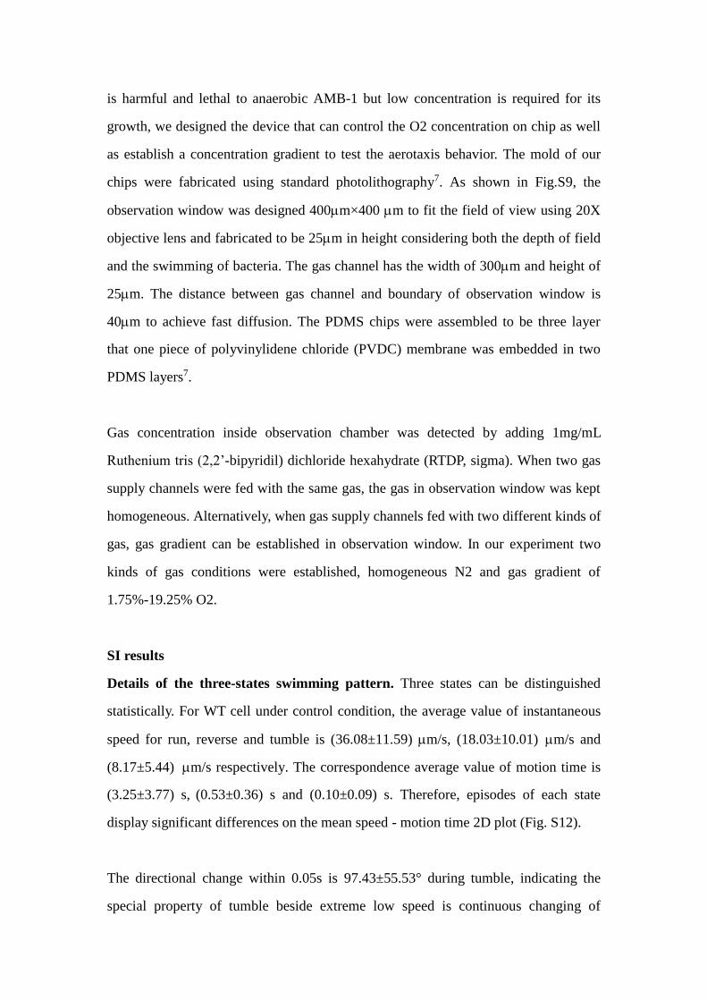
Fig.S10 Confirmation of target disruption of amb0994-0995 and amb2196. Lane 1-3:
PCR results using anchored primer F (upstream of amb0995) and R (downstream of
amb0994) for wild type, amb0994-0995 and amb2196 strains to check the deletion
of amb0994-0995. Lane 4 and 5: PCR results using anchored primer in upstream of
amb2196 on chromosome and primer in insertion plasimid, and downstream of
amb2196 on chromosome and primer in insertion plasmid for amb2196 strain.
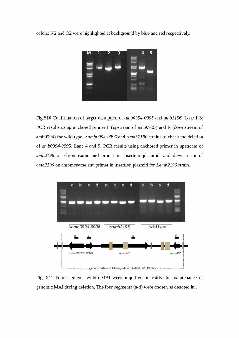
Fig. S11 Four segments within MAI were amplified to testify the maintenance of
genomic MAI during deletion. The four segments (a-d) were chosen as denoted in1.
M 1 2 3 4 5

Fig. S12 Scatter of motion duration as a function of velocity for each state and the
corresponding margin distribution for motion time and velocity of each episode. All
data were collected from WT control condition.
Fig. S13 The angular shift of three types of transition. The transition from reverse to
reverse was not observed in experiment of WT under control condition.

Fig. S14 A schematic drawing illustrating the swimming of a cell above a surface. The
light blue curved arrows indicate the rotation directions of the cell body and flagella
filament, and the straight arrows indicate the net lateral drag forces on them. The
dashed dark blue arrow depicts the circular trajectory ( derived from13).
SI movie
Time-lapse movie of AMB-1 motion. Here the objective lens is 100X, while time
resolution is 0.05s.
SI References
1. X. Ge, K. Wang, T. Bo, Y. Kou, W. Liu, and G. Chen, FEMS microbiology
letters, 2011, 320, 118–27.
2. K. Wang, X. Ge, T. Bo, Q. Chen, G. Chen, and W. Liu, Letters in applied
microbiology, 2011, 53, 55–62.
3. J. H. Li, X. Ge, X. K. Zhang, G. J. Chen, and Y. X. Pan, Chinese Journal Of
Oceanology And Limnology, 2010, 28, 826–831.
4. C.-D. Yang, H. Takeyama, T. Tanaka, and T. Matsunaga, Enzyme and
Microbial Technology, 2001, 29, 13–19.
5. J.-B. Sun, F. Zhao, T. Tang, W. Jiang, J. Tian, Y. Li, and J.-L. Li, Applied
microbiology and biotechnology, 2008, 79, 389–97.
6. D. Schüler, R. Uhl, and E. Bäuerlein, FEMS Microbiology Letters, 1995, 132,
139–145.

7. N. Li, C. Luo, X. Zhu, Y. Chen, Q. Ouyang, and L. Zhou, Microelectronic
Engineering, 2011, 88, 1698–1701.
8. P. S. Lovely and F. W. Dahlquist, Journal of Theoretical Biology, 1975, 50,
477–496.
9. P. D. Frymier, R. M. Ford, H. C. Berg, and P. T. Cummings, Proceedings of
the National Academy of Sciences of the United States of America, 1995, 92,
6195–6199.
10. L. Xie, T. Altindal, S. Chattopadhyay, and X.-L. L. Wu, Proceedings of the
National Academy of Sciences of the United States of America, 2011, 108,
2246–2251.
11. B. L. Taylor and D. E. Koshland, Journal of bacteriology, 1974, 119, 640–2.
12. H. C. Berg and D. A. Brown, Nature, 1972, 239, 500–504.
13. G. Li, L.-K. K. Tam, and J. X. Tang, Proceedings of the National Academy of
Sciences of the United States of America, 2008, 105, 18355–18359.