Embed Size (px)
Citation preview

the mechanism of TMR is still controversial, a growingamount of clinical and experimental evidence suggeststhat TMR-induced angiogenesis plays an important rolein this therapeutic approach.
Clinical development of TMR and its subsequentgovernmental approval has benefited immensely fromclose collaborations and financial support from the laserindustry. In return, all clinical studies of TMR haveused purposely designed laser devices since the early1980s. However, the initially proposed advantage oflaser, that is, improved long-term channel patency, is nolonger supported by more recently available data. Infact, most investigators now believe that TMR achievesits therapeutic benefits independently of long-termchannel patency.
Alternatively, it has been proposed that TMR induces
T ransmyocardial revascularization (TMR) is a newlydeveloped surgical treatment for patients with
symptomatic end-stage coronary artery diseases whohave exhausted other treatment alternatives. Although
Background: Angiogenesis is the proposed mechanism of transmyocar-dial revascularization. We evaluated mechanical transmyocardial revas-cularization in a chronically ischemic porcine model by measuringmyocardial angiogenic response. Methods: Ameroid constrictors wereimplanted 6 weeks before mechanical transmyocardial revasculariza-tion. Group I (n = 5) and group II (n = 3) animals received 30 punctureswith an 18-gauge needle and samples were harvested at 1 and 4 weeks,respectively, after the operation. Group III (n = 5) had sternotomy onlyand served as the control group. Myocardial samples were immunohis-tochemically stained for vascular endothelial growth factor (VEGF),basic fibroblast growth factor (bFGF), and transforming growth factorβ (TGF-β) using specific antibodies. Growth factor expression wasquantified by means of computer-assisted morphometry. Vascular den-sity was assessed by immunohistochemical stain for VEGF and factorVIII. Results:Compared with group III, increased angiogenic factor lev-els were found in group I (VEGF 0.47 ± 0.03 mm2 vs 0.05 ± 0.05 mm2, P= .000; bFGF 0.67 ± 0.14 mm2 vs 0.03 ± 0.03 mm2, P = .000; TGF-β 1.40± 0.18 mm2 vs 0.09 ± 0.06 mm2, P = 0.000), and in group II (VEGF 0.34± 0.06 mm2 vs 0.05 ± 0.05 mm2, P = .003; bFGF 0.06 ± 0.02 mm2 vs 0.03± 0.03 mm2, P = .135; TGF-β 0.28 ± 0.09 mm2 vs 0.09 ± 0.06 mm2, P =.042). Vascular densities after mechanical transmyocardial revascular-ization were also increased (group I, VEGF stain 8.1 ± 0.6 vs 1.1 ± 0.5,P= .000; factor VIII stain 5.1 ± 2.7 vs 0.4 ± 0.3,P = .018; group II, VEGFstain 1.9 ± 0.5 vs 1.1 ± 0.5,P = 0.107; factor VIII stain 2.3 ± 0.4 vs 0.4 ±0.3, P = .004). Conclusions:Mechanical transmyocardial revasculariza-tion is associated with increased angiogenic factor expression and con-comitant neovascularization at up to 4 weeks. These changes are indis-tinguishable from those of laser transmyocardial revascularization.Myocardial perfusion studies are needed to establish the functional sig-nificance of these angiogenic changes. (J Thorac Cardiovasc Surg 1999;118:849-56)
Victor Chu, MDa
Jin-qiang Kuang, MDa
Amy McGinna
Adel Giaid, PhDb
Stephen Korkola, MDa
Ray C.-J. Chiu, MD, PhDa
From the Division of Cardiothoracic Surgerya and the Department ofPathology, McGill University,b Montreal, Quebec, Canada.
Read at the Seventy-ninth Annual Meeting of The AmericanAssociation for Thoracic Surgery, New Orleans, La, April 18-21,1999.
Received for publication March 19, 1999; revisions requested May 4,1999; revisions received July 8, 1999; accepted for publicationJuly 13, 1999.
Address for reprints: Ray C.-J. Chiu, MD, Room C9.169, MontrealGeneral Hospital, 1650 Cedar Ave, Montreal, Quebec, H3G 1A4,Canada (E-mail: [email protected]).
Copyright © 1999 by Mosby, Inc.0022-5223/99 $8.00 + 012/6/101413
849
ANGIOGENIC RESPONSE INDUCED BY MECHANICAL TRANSMYOCARDIAL REVASCULARIZATION
angiogenesis and increases myocardial collateral circu-lation through a tissue injury/wound healingprocess. Alogical extension of such a hypothesis is that angiogen-esis is a nonspecificresponse to tissue injury, which canbe created in a variety of ways. Indeed, in addition to awide array of laser sources, radio frequency and simpleneedle punctures1 have also been shown to inducemyocardial angiogenesis in animal models.
Mechanical needle puncture deserves further consid-eration, not only for its simplicity. It has also beenshown by Sen and coworkers2 to be effective in thetreatment of acute myocardial ischemia. More recently,Whittaker, Rakusan, and Kloner3 demonstrated thatneedle punctures were more effective than laser chan-nels in protecting acute ischemia in an animal model.Our earlier study4 also indicated that needle TMR com-pared favorably with a carbon dioxide laser. Neverthe-less, the effectiveness of needle TMR in promotingmyocardial angiogenesis and its potential as a simpleand low-cost alternative to laser procedures have neverbeen evaluated.
We performed needle mechanical TMR in a chroni-cally ischemic porcine model. The purpose of thisstudy is to further characterize the angiogenic responsefollowing needle TMR, providing a more detailedanalysis with multiple growth factors over differenttime points.
MethodsAll experimental animals were cared for in accordance
with institutional guidelines and the “Guide for the Care andUse of Laboratory Animals” prepared by the Institute ofLaboratory Animal Resources, National Research Council,and published by the National Academy Press, revised 1996.
Animal model with chronic ischemia. Fifteen Yorkshirepigs weighing 15 to 20 kg were premedicated with intramus-cular ketamine (15 mg/kg) and were anesthetized with intra-venous injection of thiopental sodium (15 mg/kg). After oralendotracheal intubation, anesthesia was maintained with0.5% to 2.0% isoflurane in room air. Oxygen saturation wascontinuously monitored with a transcutaneous oximeterprobe. Cefazolin, 500 mg, was given intravenously before theskin incision.
Animals were placed in the right lateral decubitus position.The thoracic area was prepared and draped in a sterile fash-ion. Exposure of the proximal left circumflex artery wasachieved via a minithoracotomy through the 4th intercostalspace. A 1-cm segment of the left circumflex artery before thefirst obtuse marginal branch was dissected free with bothsharp and blunt dissections. Care was taken to minimizedirect manipulation of the artery itself to avoid vessel spasm.An ameroid constrictor (2.75 mm, Research Instruments andManufacturing, Corvallis, Ore) was placed around the left cir-cumflex artery. The pericardium and the chest were closed in
layers and the anesthesia reversed. The animals were kept for6 weeks to allow time for gradual occlusion of the left cir-cumflex artery by ameroid constrictors.
TMR. Six weeks after insertion of the ameroid constrictors,animals were randomly assigned to 3 groups (n = 5 each).Groups I and II received 30 needle punctures whereas groupIII underwent sternotomy only. Tissue samples from groups Iand III were harvested 1 week after the operation and samplesfrom group II were harvested 4 weeks after TMR.
All TMR operations were performed through a mediansternotomy. Anesthesia and intubation were performed in thesame fashion as the first operation. All animals received aprophylactic intravenous lidocaine bolus (2 mg/kg) and weremaintained on a lidocaine infusion (1 mg/min) throughout theoperation. Median sternotomies were performed and thehearts were exposed by opening the pericardium and careful-ly dissecting away pericardial adhesions. Needle punctureswere performed in an area measuring approximately 2 × 2 cmbetween the 1st and 2nd obtuse marginal arteries with 18-gauge hypodermic needles. Transmural penetration was con-firmed by noting pulsatile flow of arterial blood through theneedle. Bleeding was controlled with finger pressure or 4-0Prolene sutures (Ethicon, Inc, Somerville, NJ), which alsoserved as markers of puncture sites at the time of tissue har-vest. Sternums were then closed with steel wires and the inci-sions closed in layers. Anesthesia was reversed and the ani-mal allowed to recover.
Sample harvest and cryopreservation. At the time of tis-sue harvesting, repeat sternotomies were performed throughthe same incision. Hearts were isolated by careful dissectionof adhesions. Animals were put to death with an overdose ofpentobarbital and potassium chloride. The ascending aortaswere crossclamped and the hearts fixed in situ by injecting 1L of ice cold 4% paraformaldehyde through the aortic root.Full-thickness slices of myocardium from the TMR-treatedarea (or corresponding ischemic area in the control group)were removed and immediately immersed in 4%paraformaldehyde in phosphate-buffered saline solution.These were kept at 4°C for 12 hours. The specimens werethen transferred into 15% sucrose in phosphate-bufferedsaline solution and kept at 4°C for 3 days. Afterward, sampleswere embedded in OCT compound (Tissue-Tek, SakuraFinetek Inc, Torrance, Calif), snap frozen with liquid nitro-gen, and kept at –80°C.
All ameroid constrictors were retrieved from the heart andinspected to confirm vessel occlusion.
Sample analysisImmunohistochemistry. Cryostat sections of tissue samples
were mounted on glass slides and immunostained with anti-sera to vascular endothelial growth factor (VEGF), basicfibroblast growth factor (bFGF), transforming growth factorβ (TGF-β) (Santa Cruz Biotechnology Inc, Santa Cruz,Calif), or factor VIII ligands (Sigma-Aldrich Canada Ltd,Oakville, Ontario, Canada) with a modified avidin biotin-per-oxidase method.5 Tissue sections were made permeable withoctylphenoxypolyethoxyethanol (Triton X-100; UnionCarbide Corporation, Danbury, Conn), incubated in hydrogen
850 Chu et al The Journal of Thoracic andCardiovascular Surgery
November 1999

peroxide to block endogenous peroxidase activity. They werethen incubated first with normal goat serum for 30 minutesand followed with the primary antibody for 16 hours at 4°C.Afterward, they were incubated with biotinylated immuno-globulin G and stained with an immmunoperoxidase tech-nique according to the manufacturer’s instructions (VectastainABC Elite Kit; Vector Laboratories, Burlingame, Calif).
Angiogenic growth factor expression. Growth factorexpression was quantified by measuring the area of tissuesections positively stained for VEGF, bFGF, or TGF-β ineach high-power field (400×). Measurements were performedaround TMR puncture sites, which were identified by the fol-lowing criteria: (1) identifiable needle or laser puncture scarunder low-power view (100×), (2) presence of inflammatorycells and granulation tissue, and (3) loss of normal myocyteappearance and homogeneity. With the use of the samplingmethod of “systematic sampling with a random start,”6 8sampling sites from each tissue section were photographedwith a still video camera and digitized into tagged image fileformat (TIFF) files. Quantitative measurements of stainedarea were performed with an IBM compatible personal com-puter using Matrox Inspector 2.1 (Matrox Inc, Montreal,Quebec, Canada). Total amount of growth factor expressionfor each animal was reported as mean area of positive stain(square millimeters).
Vascular density. TMR-induced angiogenesis was quanti-fied by measuring vascular density of VEGF- and factorVIII–stained blood vessels per high-power field around punc-ture sites. Positively stained vessels were defined as roundstructures with a central lumen, which was lined by a thinlayer of endothelium stained positively for VEGF or factorVIII. Eight measurements were taken for each tissue sectionby means of the same sampling method. Results of angio-genesis for each animal were reported as mean number ofvessels per high-power field.
Statistical analysis. All numeric data were reported asmean ± 1 standard deviation where applicable. Data analyseswith the Student t test were performed with SPSS 7.5.2 forWindows software (SPSS Inc, Chicago, Ill).
Results
Mortality. Two deaths occurred in group II on post-operative day 2 after TMR. Autopsy showed severepulmonary congestion and tissue edema with no evi-dence of myocardial infarction. The presumed cause ofdeath was congestive heart failure.
Histology. All animals had complete occlusion of theleft circumflex coronary artery at the time of sampleharvesting. In only 1 of the 15 animals, a small area oftransmural scar was noticed at the 2nd operation beforeTMR. This was probably the result of early occlusionof the ameroid constrictor. In TMR-treated specimens,the areas of transmural punctures were easily identifiedby the presence of numerous fibrous scars on the endo-cardium.
Under low-power light microscopic examinations,transmural puncture sites could be identified as centralfibrous tracts surrounded by inflammatory changes. Allchannels were completely occluded by fibrosis. Oneweek after TMR (group I), the fibrous tracts consistedmainly of fibroblasts and collagen material with occa-sional small blood vessels. The surrounding area consist-ed of granulation tissue and damaged myocardium withinfiltrating lymphocytes and macrophages. These weresimilar to the typical inflammatory changes during thenormal tissue healing process. Numerous small vascularstructures were also found in the area of tissue inflam-mation. These vessels were morphologically indistin-guishable from native myocardial capillaries except fortheir endothelium, which was positively stained forVEGF or factor VIII (Fig 1). Most of these vessels weresmaller than 10 µm in diameter and were believed to beat various stages of angiogenic development.
Inflammatory changes related to needle punctureswere limited to their immediate vicinity. Each puncturesite was separated from others by normal-lookingmyocardium indistinguishable from the control speci-men under light microscopy.
Four weeks after needle puncture (group II), the trans-mural channels were completely replaced by fibrousscar tissue. Significantly less inflammatory cellularinfiltrate was observed in the granulation tissue sur-rounding needle tracts. Compared with group I, fewervessels with positively stained endothelium were foundin group II. However, the remaining vessels were moremature looking and had a somewhat larger diameter.
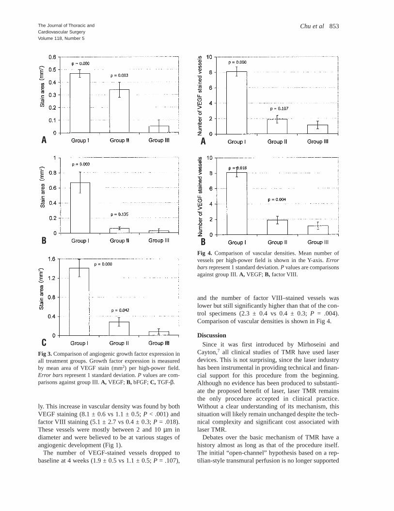
Angiogenic growth factor expression. Parallelcomparison revealed that different angiogenic growthfactors had distinct stain patterns (Fig 2). In samplestaken 1 week after needle puncture (group I), severalcell types stained positively for VEGF, includingendothelial cells, macrophages, fibroblasts, andmyocytes. In general, positive stains were limited toareas adjacent to the puncture sites. Endothelium andmacrophages gave the most intense stains but repre-sented only a small portion of the total area as mea-sured by computer-assisted morphometry. On the otherhand, myocytes and fibroblasts produced a more dif-fused staining pattern and represented most of our mea-surements for VEGF expression (0.47 ± 0.03 mm2 vs0.05 ± 0.05 mm2; P < .001; Fig 3,A). Staining wasminimal in areas away from puncture sites, and mea-surements from these areas were not significantly high-er than the baseline.
Four weeks after TMR (group II), there was lessVEGF staining in the endothelial cells and macro-phages. However, positive stain from myocytes and
The Journal of Thoracic andCardiovascular SurgeryVolume 118, Number 5
Chu et al 851

fibroblasts persisted, and the overall level as measuredby morphometric analysis was still significantly higherthan that of control specimens (0.34 ± 0.06 mm2 vs0.05 ± 0.05 mm2; P = .003; Fig 3,A).
Basic FGF stained much differently from VEGF. Oneweek after TMR (group I), there was strong staining offibroblasts along the needle tracts by anti-bFGF anti-sera (0.67 ± 0.14 mm2 vs 0.03 ± 0.03 mm2; P < 0.001;Fig 3,B). Very little stain was found in other cell types.The level of bFGF stain dropped to baseline at 4 weeks(group II, 0.06 ± 0.02 mm2 vs 0.03 ± 0.03 mm2; P =.135; Fig 3,B).
TGF-β stain followed a pattern similar to that ofVEGF 1 week after TMR (group I), that is, endothelialcells, macrophages, myocytes, and fibroblasts. Again,most of the morphometric measurement was frommyocytes and fibroblasts, which gave a more diffusestain pattern (1.40 ± 0.18 mm2 vs 0.09 ± 0.06 mm2; P< .001; Fig 3,C). At 4 weeks (group II), the level of
stain had decreased considerably but was still signifi-cantly higher than that of the baseline (0.28 ± 0.09 mm2
vs 0.09 ± 0.06 mm2; P = .042; Fig 3,C).Angiogenesis. One week after needle puncture
(group I), the number of vascular structures in thevicinity of needle punctures had increased significant-
852 Chu et al The Journal of Thoracic andCardiovascular Surgery
November 1999
Fig 1. Representative images from TMR-treated area (200×)illustrating increased number of small vessels with endothe-lium stained for VEGF (A) or factor VIII (B).
Fig 2. Representative digitized image of TMR-treated tissuesection (200×) stained for VEGF (A), bFGF (B), and TGF-β (C).
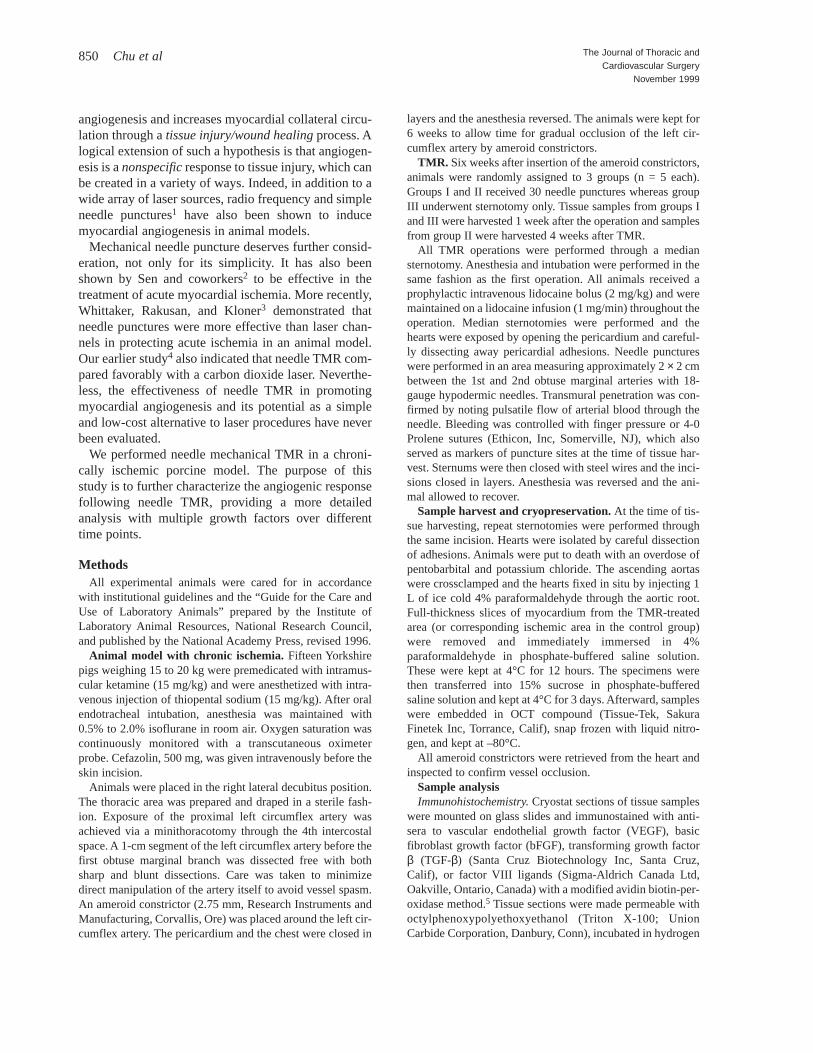
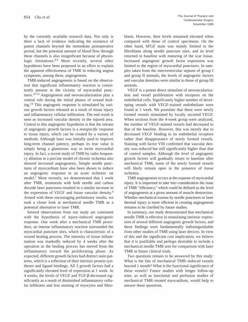
ly. This increase in vascular density was found by bothVEGF staining (8.1 ± 0.6 vs 1.1 ± 0.5; P < .001) andfactor VIII staining (5.1 ± 2.7 vs 0.4 ± 0.3; P = .018).These vessels were mostly between 2 and 10 µm indiameter and were believed to be at various stages ofangiogenic development (Fig 1).
The number of VEGF-stained vessels dropped tobaseline at 4 weeks (1.9 ± 0.5 vs 1.1 ± 0.5; P = .107),
and the number of factor VIII–stained vessels waslower but still significantly higher than that of the con-trol specimens (2.3 ± 0.4 vs 0.4 ± 0.3; P = .004).Comparison of vascular densities is shown in Fig 4.
DiscussionSince it was first introduced by Mirhoseini and
Cayton,7 all clinical studies of TMR have used laserdevices. This is not surprising, since the laser industryhas been instrumental in providing technical and finan-cial support for this procedure from the beginning.Although no evidence has been produced to substanti-ate the proposed benefit of laser, laser TMR remainsthe only procedure accepted in clinical practice.Without a clear understanding of its mechanism, thissituation will likely remain unchanged despite the tech-nical complexity and significant cost associated withlaser TMR.
Debates over the basic mechanism of TMR have ahistory almost as long as that of the procedure itself.The initial “open-channel” hypothesis based on a rep-tilian-style transmural perfusion is no longer supported
The Journal of Thoracic andCardiovascular SurgeryVolume 118, Number 5
Chu et al 853
Fig 3. Comparison of angiogenic growth factor expression inall treatment groups. Growth factor expression is measuredby mean area of VEGF stain (mm2) per high-power field.Error bars represent 1 standard deviation. P values are com-parisons against group III. A, VEGF; B, bFGF; C, TGF-β.
A
B
C
A
BFig 4. Comparison of vascular densities. Mean number ofvessels per high-power field is shown in the Y-axis. Errorbars represent 1 standard deviation. P values are comparisonsagainst group III. A, VEGF; B, factor VIII.
by the currently available research data. Not only isthere a lack of evidence indicating the existence ofpatent channels beyond the immediate postoperativeperiod, but the potential amount of blood flow throughthese channels is also insignificant because of physio-logic limitations.8,9 More recently, several otherhypotheses have been proposed in an effort to explainthe apparent effectiveness of TMR in reducing anginasymptoms, among these, angiogenesis.
TMR-induced angiogenesis is based on the observa-tion that significant inflammatory reaction is consis-tently present in the vicinity of myocardial punc-tures.10-13 Angiogenesis and neovascularization play acentral role during the initial phases of wound heal-ing.14 This angiogenic response is stimulated by vari-ous growth factors released as a result of tissue injuryand inflammatory cellular infiltration. The end result isseen as increased vascular density in the injured area.Central to this angiogenic hypothesis is that the releaseof angiogenic growth factors is a nonspecificresponseto tissue injury, which can be created by a variety ofmethods. Although laser was initially used to improvelong-term channel patency, perhaps its true value issimply being a glamorous way to incite myocardialinjury. In fact, a recent study of TMR by radio frequen-cy ablation in a porcine model of chronic ischemia alsoshowed increased angiogenesis. Simple needle punc-tures of myocardium have also been shown to inducean angiogenic response in an acute ischemic ratmodel.1 More recently, we demonstrated that 1 weekafter TMR, treatments with both needle and carbondioxide laser punctures resulted in a similar increase inthe expression of VEGF and tissue vascular density.4
Armed with these encouraging preliminary results, wetook a closer look at mechanical needle TMR as apotential alternative to laser TMR.
Several observations from our study are consistentwith the hypothesis of injury-induced angiogenicresponse. One week after a mechanical TMR proce-dure, an intense inflammatory reaction surrounded themyocardial puncture sites, which is characteristic of awound healing process. The intensity of tissue inflam-mation was markedly reduced by 4 weeks after theoperation as the healing process has moved from theinflammatory toward the proliferating phase. Asexpected, different growth factors had distinct stain pat-terns, which is a reflection of their intrinsic protein syn-theses and ligand bindings. All 3 growth factors had asignificantly elevated level of expression at 1 week. At4 weeks, the levels of VEGF and TGF-β decreased sig-nificantly as a result of diminished inflammatory cellu-lar infiltrates and less staining of myocytes and fibro-
blasts. However, their levels remained elevated whencompared with those of control specimens. On theother hand, bFGF stain was mainly limited to thefibroblasts along needle puncture sites, and its levelreturned to baseline with maturing of the scar tissue.Increased angiogenic growth factor expression waslimited to the region of myocardial punctures. In sam-ples taken from the interventricular septum of group Iand group II animals, the levels of angiogenic factorsand vascular densities were similar to those of group IIIanimals.
VEGF is a potent direct stimulant of neovasculariza-tion and vessel proliferation with receptors on theendothelial cells. Significantly higher number of devel-oping vessels with VEGF-stained endothelium werefound at 1 week. We postulate that these were newlyformed vessels stimulated by locally secreted VEGF.When sections from the 4-week group were analyzed,the number of VEGF-stained vessels had decreased tothat of the baseline. However, this was mostly due todecreased VEGF binding to its endothelial receptorsrather than disappearance of newly formed vessels.Staining with factor VIII confirmed that vascular den-sity was reduced but still significantly higher than thatof control samples. Although the level of angiogenicgrowth factors will gradually return to baseline aftermechanical TMR, some of the newly formed vesselswill likely remain open in the presence of tissueischemia.
TMR angiogenesis occurs at the expense of myocardialinjury. It is important to take into consideration the issueof TMR “efficiency,” which could be defined as the levelof angiogenesis at a given amount of muscle destruction.Whether mechanical trauma by needle punctures or laserthermal injury is more efficient in creating angiogenesisremains to be clarified by future studies.
In summary, our study demonstrated that mechanicalneedle TMR is effective in stimulating intrinsic expres-sion of several different angiogenic growth factors, andthese findings were fundamentally indistinguishablefrom other studies of TMR using laser devices. In viewof this and the significant cost implication, we believethat it is justifiable and perhaps desirable to include amechanical needle TMR arm for comparison with laserTMR in future clinical trials.
Two questions remain to be answered by this study:What is the fate of mechanical TMR–induced vesselsbeyond 1 month? What is the functional significance ofthese vessels? Future studies with longer follow-uptime, as well as functional and perfusion studies ofmechanical TMR–treated myocardium, would help toanswer these questions.
854 Chu et al The Journal of Thoracic andCardiovascular Surgery
November 1999

We thank Dianne Murray and Marlene Brydon for theirdedication in animal care and Jean-Yves Latreille, RN, fortechnical assistance in operating laser equipment.
R E F E R E N C E S1. Pelletier MP, Giaid A, Sivaraman S, et al. Angiogenesis and
growth factor expression in a model of transmyocardial revascu-larization. Ann Thorac Surg 1998;66:12-8.
2. Sen PK, Daulatram J, Kinare SG, et al. Further studies in multi-ple transmyocardial acupuncture as a method of myocardialrevascularization. Surgery 1968;64:861-70.
3. Whittaker P, Rakusan K, Kloner RA. Transmural channels canprotect ischemic tissue: assessment of long-term myocardialresponse to laser- and needle-made channels. Circulation 1996;93:143-52.
4. Chu VF, Giaid A, Kuang J-Q, et al. Angiogenic response in trans-myocardial revascularization: comparison of laser vs. mechanicalpunctures. Ann Thorac Surg. In press.
5. Hsu S, Raine L, Fanger H. Use of avidin-biotin-peroxidase com-plex (ABC) in immunoperoxidase techniques: a comparisonbetween ABC and unlabeled antibody (PA) procedures. J Histo-chem Cytochem 1981;29:577-80.
6. Weibel E. Stereological methods. 1st ed. Vol 1. New York:Academic Press; 1979.
7. Mirhoseini M, Cayton MM. Revascularization of the heart bylaser. J Microsurg 1981;2:253-60.
8. Hardy RI, James FW, Millard RW, Kaplan S. Regional myocar-dial blood flow and cardiac mechanics in dog hearts with CO2laser–induced intramyocardial revascularization. Basic ResCardiol 1990;85:179-97.
9. Kohmoto T, Fisher PE, Gu A, et al. Does blood flow throughholmium:YAG transmyocardial laser channels? Ann Thorac Surg1996;61:861-8.
10. Jansen ED, Frenz M, Kadipasaoglu KA, et al. Laser-tissue inter-action during transmyocardial laser revascularization. AnnThorac Surg 1997;63:640-7.
11. Whittaker P. Detection and assessment of laser-mediated injuryin transmyocardial revascularization. J Clin Laser Med Surg1997;15:261-7.
12. Krabatsch T, Schaper F, Leder C, et al. Histological findings aftertransmyocardial laser revascularization. J Card Surg 1996;11:326-31.
13. Fleischer KJ, Goldschmidt-Clermont PJ, Fonger JD, et al. One-month histologic response of transmyocardial laser channels withmolecular intervention. Ann Thorac Surg 1996;62:1051-8.
14. Davidson J. Wound repair. In Gallin J, Goldstein I, Snyderman R,editors. Inflammation: basic principles and clinical correlates.New York: Raven Press; 1992. p. 809-19.
DiscussionDr Ernst Wolner (Vienna, Austria). In 1969 I published an
experimental study very similar to yours, with ameroid con-strictors, but I did not use this puncture. That was the time ofthe Vineberg operation. I used strips from the jugular veinand a curved needle, and I positioned 7 to 8 of these strips onthe left anterior wall, from the outside to the inside of the cav-ity and back to the outside. At that time we did not havegrowth factor, but we did find some new vessels.
Using xenon 133, we also made some overall measure-
ments of blood flow on the anterior wall of the left ventricle.We found an increase in the overall blood supply or bloodperfusion of the anterior wall. Thus these old experiments arein complete accordance with your data, indicating that itseems to be enough to create an injury to get some changesand not to use this very expensive laser.
Dr Chu. Thank you, Dr Wolner. Dr Thomas M. Egan (Chapel Hill, NC). That was a very
nice presentation. I wonder about the accuracy of usingimmunohistochemical staining for your outcome measures.Specifically, how did you deal with background expression?Have you made an attempt to measure messenger RNAinstead of relying on expression?
Dr Chu. These are very good points. First of all, to exam-ine the presence of background angiographic expression, westained tissue sections not only from sham-operated animals,which is our control group, but also from unpunctured areasin our treatment groups. Although these results were notshown here, we found that the levels of immunostaining fromunpunctured areas were consistently low and similar to thoseof our control group. It seems that background expression isnot a big problem.
Messenger RNA level was not measured in this particularstudy.
Dr Beat H. Walpoth (Bern, Switzerland). It seems thatyou did not do TMR and needle puncture in the same animalbut rather did a historical comparison. You did not presentresults comparing the 2 techniques. You just showed that nee-dle puncture will induce growth factors. I think this point isworth mentioning, although it has been noted before.
An interesting question, however, is to know whether laser-induced injury, yielding a stronger and bigger injury, willinduce quantitatively more growth factors. Can you comment?
Dr Chu. Absolutely. In fact, this study was an extension ofour preliminary experiments, which were presented beforethe Society for Thoracic Surgeons meeting in San Antonio. Inthat study, we directly compared the effect of needle punc-tures versus carbon dioxide laser TMR. I agree that individ-ual laser puncture causes much more tissue damage andtherefore more angiogenic factor expression per puncture.However, we also showed that such difference was onlyquantitative and could be fully compensated by simplyincreasing the number of needle punctures.
There is a balance between tissue damage and angiogenicresponse, and I cannot tell you whether needle or laser ismore efficient. However, the bottom line is that the samelevel of angiogenesis can be achieved using simple needlepunctures.
Dr Jakob Vinten-Johansen (Atlanta, Ga). Is there acause-and-effect relationship between the local expression ofangiogenic factors and the actual capillary density that isobserved?
Dr Chu. We definitely found that tissue capillary densitiesparallel the levels of various angiogenic factors. In addition,we found an increased number of vessels in needle-treatedareas with VEGF-stained endothelium. We think these couldbe locally produced protein products binding to endothelial
The Journal of Thoracic andCardiovascular SurgeryVolume 118, Number 5
Chu et al 855

receptors. Yes, we believe there is a cause-and-effectrelationship.
Dr Paul Kurlansky (Miami Beach, Fla). Dr Chu, I am fol-lowing your work with interest. I saw your presentation at theSociety for Thoracic Surgeons meeting. My question regard-ing needle puncture versus laser TMR is as follows: Have youdone any studies on the time course and the quality of theeffect in addition to the work you have shown us on the quan-tity of the effect? In other words, is there a similar time courseas regards neovascularization and VEGF expression, and arethere other growth factors that are expressed differentiallywith one technique versus the other?
Dr Chu. Thank you very much. This study was meant tofurther characterize angiogenesis of mechanical needle TMRonly. We included 2 animal groups at 1 and 4 weeks postop-eratively for the purpose of time-course study. We also lookedat several different angiogenic factors, as well as includingfactor VIII for measuring vascular density. Indeed, the pat-terns of expression and the time course of different angio-genic factors were quite distinct from one another.
I think an important question here is whether the increasedvascular density has any functional significance. This will beanswered by myocardial perfusion studies currently inprogress in our laboratory.
856 Chu et al The Journal of Thoracic andCardiovascular Surgery
November 1999