Embed Size (px)
Citation preview

Angiogenesis and Metastasis
(RPN 530 11/3 lecture)
Tariq Bhat (Immunology)

Angiogenesis

• Blood vessels in the body- composed of macro (artery/vein) and micro-vessels.
• Blood vessels- supply oxygen and nutrition, and removal of wastes.
• Lymphatic vessel- drain the tissue fluid to blood circulation and protect from germs by immunity at lymph nodes
Capillaries Lymph ductsEndothelial cells Pericyte
Macro-vessel Micro-vessel

Angiogenesis

Vasculogenesis - Angiogenesis - Arteriogenesis Vasculogenesis Formation of blood vessels by differentiation from (hem)angioblasts
Sprouting angiogenesis Sprouting of cells from mature endothelial cells of the vessel wall
Arteriogenesis growth of large arteries from pre-existing small vessels/capillaries
Lymphangiogenesis Formation of the lymphatic vasculature
Definitions

Vasculogenesis
Formation of vessels by differentiation of cells from angioblasts in the yolk sac of the embryo:
Is differentiation and proliferation of endothelial cells in a non-vascularized tissue
Leads to formation of a primitive tubular network
Has to undergo angiogenic remodeling to stable vascular system

Hemangioblast Angioblast EC
Postnatal vasculogenesis
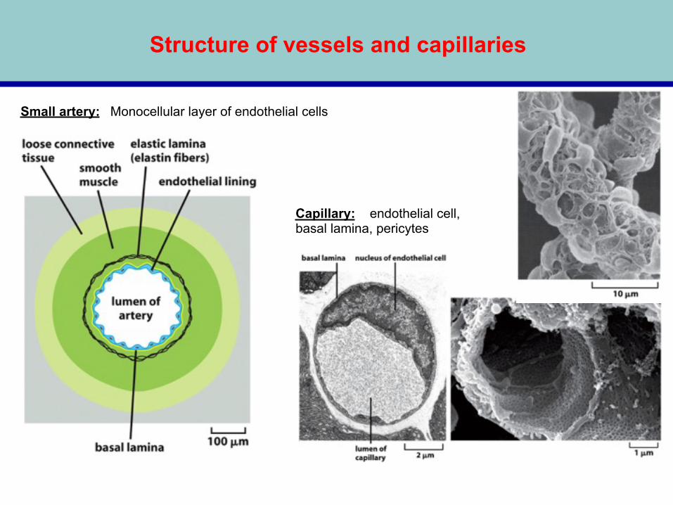
Structure of vessels and capillaries
Monocellular layer of endothelial cellsSmall artery:
Capillary: endothelial cell, basal lamina, pericytes

The 4 major steps of endothelial cells in angiogenesis
1.Breaking through of the basal lamina that envelops existing blood vessels
2. Migration toward a source signal
3. Proliferation
4. Formation of tubes
Angiogenesis is a multi-step process

Key Stage Markers
Stage One: Endothelial cell activation in response to angiogenic factors.
Basic Fibroblast Growth Factor (bFGF): a potent stimulatory factor for endothelial cell migration and proliferation. Vascular Endothelial Growth Factor (VEGF): initiates cell proliferation and migration.
Stage Two: Degradation of the capillary wall by extracellular proteinases.
Matrix Metalloproteinases (MMPs): MMP1 (a collagenase) and MMP2 are expressed during angiogenesis and act to degrade extracellular matrix components.
Stage Three: Formation of a branch point in the vessel wall.
Integrins: expressed on newly forming vessels.
Stage Four: Migration of endothelial cells into the extracellular matrix towards the angiogenic stimulus.
Integrins: allow migrating endothelial cells to interact with specific components of the surrounding matrix. MMPs and urokinase: aid migration of endothelial cells into the surrounding matrix.
Stage Five: Re-organisation of endothelial cells to form tubules with a central lumen.
Angiopoietin (Ang 1): produced by surrounding stromal cells; facilitates endothelial cell survival and stabilisation of new capillary tubes.
Stage Six: Interconnection of the new tubules to form a network (anastomosis).
Platelet Derived Growth Factor (PDGF): produced by endothelial cells of the new capillaries; recruits pericytes which stabilize the new vessels.
Conti…
Life time of endothelial cells (Major Players):
months (lung, liver) to years (brain, muscle)
Slow repair and renewal of vascular wall
New vessel formation:
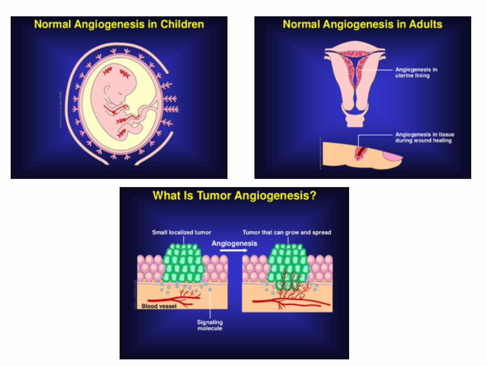
Embryo, growth In uterus, during menstruation cycle Wound repair

Angiogenesis:
Sprouting of cells from mature endothelial cells of the vessel wall
secretion of proteases, resolution of basal lamina, migration towards chemotactic gradient, proliferation, tube formation
VEGF is factor largely specific for endothelial cells, bFGF can also induce, not specific for EC
tip cell
stem
Angiogenesisisinvolvedinformation,maturationanddifferentiationofbloodvesselsfrompre-existingvessels.AngiogenesiscanbeobservedinPhysiologicalandpathologicalconditionsincludinggrowth,injury,inflammationandcancer.Occasionally,angiogenesisiscalledneovascularization. (Extendandexpandbloodvessels)
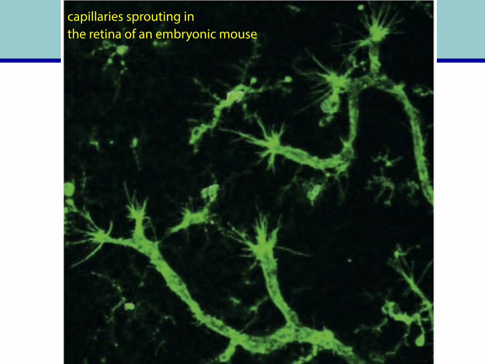
capillaries sprouting in the retina of an embryonic mouse


VEGF conti..

Chorioallantoic Membrane Assay (CAM)
Serum free-media Serum free-media plus VEGF

Flt- FMS-like tyrosine kinase KDR- Kinase insert domain receptor (KDR, a type III receptor tyrosine kinase)

VEGF-VEGFR signaling
Angiogenesis
EC proliferation, survival, migration and invasion

VEGF Is a Key Mediator of Angiogenesis
Upstream activators of VEGF synthesis
Downstream signaling pathways

Role of hypoxia in angiogenesis: (Hypoxia - HIF – VEGF module)
Like VEGF
VBC: Von Hippel-Lindau (VHL)-containing VHL-elongin BC

Role of hypoxia in angiogenesis: (Hypoxia - HIF – VEGF module) conti…
HIF: hypoxia inducible factor VEGF: vascular endothelial growth factor

VEGF-gene expression: Regulated by HIF, HIF is continuously produced, ubiquitinylated, degraded in proteasome, therefore low concentration;
Ubiquitinylation is dependent on Hippel-Lindau tumor suppressor (part of an E3 ubiquitin-ligase complex)
HIF1α is modified by a prolyl hydroxylase, then better interaction with vHL protein, high turnover; Hydroxylase is regulated by O2
FIH: Factor inhibiting HIF1α- Aaparaginyl hydroxulation leading to HIF inactivation
Von Hippel-Lindau Tumor Suppressor, HIF and VEGF

Angiogenesis-dependent diseases
Excess: •Cancer
•Infantile hemangiomas
•Autoimmune diseases, chronic inflammatory diseases:
•Rheumatoid arthritis
•Psoriasis
•Age-related macular degeneration
•Atherosclerosis
Deficiency: •Limb ischemia
•Myocardial ischemia

Angiogenic inhibitors: •During the process of wound healing, the burst of angiogenesis must be shut down once the newly formed capillaries have reached a certain density.
TSP-1 produced by stromal fibroblasts, ECs and immune cells suppresses tumor progression by inhibiting angiogenesis through direct effects on EC migration and survival and through indirect effects on growth factor mobilization.


Inhibits endothelial cell migration, proliferation and induces EC apoptosis
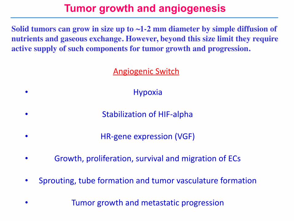
Tumor growth and angiogenesis
Solid tumors can grow in size up to ~1-2 mm diameter by simple diffusion of nutrients and gaseous exchange. However, beyond this size limit they require active supply of such components for tumor growth and progression.
AngiogenicSwitch
• Hypoxia
• StabilizationofHIF-alpha
• HR-geneexpression(VGF)
• Growth,proliferation,survivalandmigrationofECs
• Sprouting,tubeformationandtumorvasculatureformation
• Tumorgrowthandmetastaticprogression


Featuresoftumorangiogenesis
• Extremeandchaoticexpressionofangiogenicfactors
• DisorganizedvascularstructureLowadhesionandpericytecoverage
• Hypoxicstress,metabolicchanges,cancercellintravasationandlesseffectofchemotherapy

What Is Tumor Angiogenesis?
Tumor angiogenesis Proliferation of a network of blood vessels that penetrates into cancerous growths. Function Supplying nutrients and oxygen and removing waste products. Mechanism Cance cells releas molecules that send signals to surrounding normal host tissue. This signaling activates certain genes in the host tissue that, in turn, make proteins to encourage growth of new blood vessels.

Stroma contributes to tumor angiogenesis
TumorMicroenvironment(Tumor-associatedstroma):inducedbycytokinesandchemokinessecretedfromtumorcells
• Macrophage: Tumor--Associated Macrophages (TAMs)
• Fibroblast: Carcinoma--Associated Fibroblasts (CAFs)
• Myeloid cell: Bone Marrow Derived Cells (BMDCs)
• Extracellular matrix (ECM)
Tumor microenvironment complexity and degree of infiltration of various components correlates with the tumor
angiogenesis and invasiveness

Macrophage and tumor angiogenesis


Anti-angiogenic therapy
Dr. Judah Folkman proposed the concept of anti-angiogenic therapy (NEJM.1971).


Antiangiogenic Therapies: Potential Targets
• Block pro-angiogenic molecules (e.g., VEGF)
• Add anti-angiogenic regulators (e.g. angiostatin, endostatin, TSP-1)
• Inhibit stroma-degrading enzymes (e.g., MMPIs)
• Target vascular antigens (e.g., avb3 integrin)
• Attack pericytes


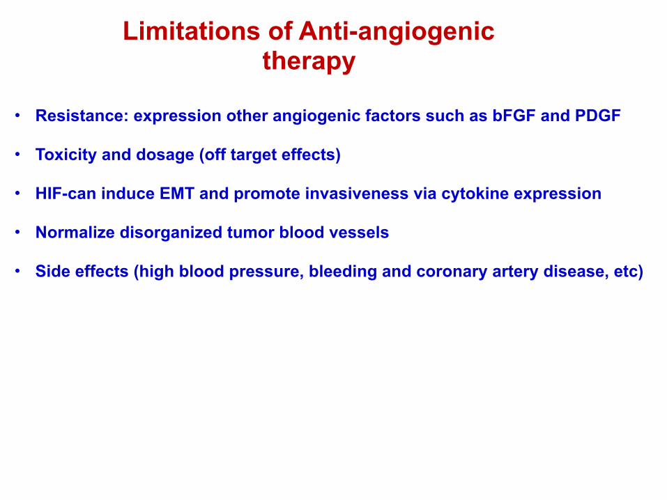
Limitations of Anti-angiogenic therapy
• Resistance: expression other angiogenic factors such as bFGF and PDGF
• Toxicity and dosage (off target effects)
• HIF-can induce EMT and promote invasiveness via cytokine expression
• Normalize disorganized tumor blood vessels
• Side effects (high blood pressure, bleeding and coronary artery disease, etc)


Metastasis

When does metastasis begin?
Commitment to the metastatic phenotype: • How early does it occur? • Can it be reversed?
Progenitor lesions: • What are the key progenitor lesions? • What is the efficiency of transition to invasion? • Are all metastasis precursors clonal?

What is the role of the host?
• Under what conditions does the host drive or suppress the process?
• Does the transition from pre-invasive to invasive lesions require host participation?
• If so what are the molecular and cellular players that are functionally important?
• The circuitry of the tumor host communication may be the key to prevention of invasion.
Physiologic basis of metastasis
• Is metastasis a normal physiologic program which is disregulated or inappropriately activated?
• Does a physiologic motility and invasion program exist for development, angiogenesis morphogenesis and wound healing?
• Is metastasis colony formation a natural ongoing process conducted by stem cells?

What is the driving force?
• Is the metastatic phenotype pre determined within the primary tumor? Within the host microenvironment?
• Are malignant cells a product of adaptation and selection?
• What is the selection factor? If malignant cells are survival of the fittest, then what is the fitness test?
• Is cell survival in a foreign (non home) tissue the ultimate selection factor?
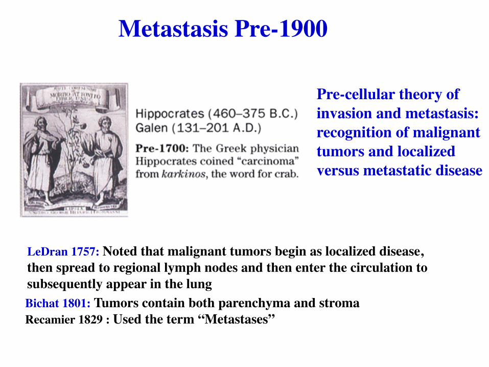
Pre-cellular theory of invasion and metastasis: recognition of malignant tumors and localized versus metastatic disease
LeDran 1757: Noted that malignant tumors begin as localized disease, then spread to regional lymph nodes and then enter the circulation to subsequently appear in the lungBichat 1801: Tumors contain both parenchyma and stromaRecamier 1829 : Used the term “Metastases”
Metastasis Pre-1900

Validation of the cellular theory ofcancer metastasis

Potential molecular mechanisms: a) Preferential adhesion in the vessels of the target organb) Selective extravasationc) Organ attractantsd) Organ specific survival and growth
The organ pattern of metastasis is characteristic of the tumor type and tissue of origin. 50-70% of the metastatic pattern can be predicted by the venous drainage blood flow. The remaining 30-50 % may be caused by specific molecular homing mechanisms.

Pre-metastaticnicheformationSomethingsecretedfromprimarytumorandchangingthebehaviorof
hosttissueatdistantsites

Cancerdevelopsthroughgradualchangesincellmorphologyandproperties
benigntumor
malignanttumor

Wheredotheygo?
Figure 14.42 The Biology of Cancer (© Garland Science 2007)
Metastatictropism

Figure 14.17b The Biology of Cancer (© Garland Science 2007)
Metastasis

Anorganiscomposedofseveraltissues
Epithelialcells
Connectivetissue
Muscletissue
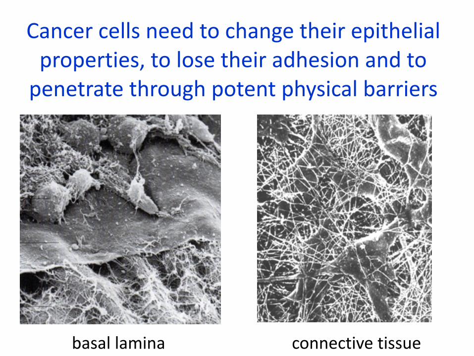
Cancercellsneedtochangetheirepithelialproperties,tolosetheiradhesionandtopenetratethroughpotentphysicalbarriers
basallamina connectivetissue

Intravasation

Once lodged in the blood vessels of various tissues, cancer cells must escape from the lumina of these vessels and penetrate into the surrounding tissue- the step termed extravasation.

Formation of microthrombus (attachment of platelets) and Proliferation in the lumen of the capillary
Platelet-mediated tumor cell extravasation

Theblood:ahostileenvironment
Figure 14.7b The Biology of Cancer (© Garland Science 2007)
http://www.cancerquest.org/
-Cellsarenormallyanchorage-dependent(anoikis)-Shearforcestearcellsapart

Chemokines regulate leukocyte recirculation and trafficking to sites of inflammation and infection

Premise:Metastasis homing is dictated by relative abundance of chemokines and cognate receptors on the tumor cell.
Why do the tumor cells express the chemokine receptors in the primary tumor prior to dissemination?
Therapeutic utility is limited because dissemination has already occurred at the time of diagnosis

Figure 14.10a The Biology of Cancer (© Garland Science 2007)
Colonization
First,micrometasteses

Figure 14.12 The Biology of Cancer (© Garland Science 2007)
Dormantmicrometastesesareviable
Figure 14.50a The Biology of Cancer (© Garland Science 2007)

SteegNatureMed06
Angiogenesis
Eventually:macrometastases
Intravasation
Latency
Colonization

Nguyen,NatureRev.Cancer2009

Figure 20-44 Molecular Biology of the Cell (© Garland Science 2008)
Asequenceofinefficientsteps
Metastaticinefficiency

Metastasis Promoting Genes - I
Gene Tissue Site FunctionARM-1 Lymphoma Promotes adhesion of tumor cells to the
endotheliumATX Breast, Liver, Lung, Melanoma,
Teratocarcinomacytoskeletal reorganization and motility; G-protein coupled receptor activation
CD44 Multiple sites cell-cell interactions; activates HGF/c-Met pathway
Cox2 Breast, Colorectal, Gastric Prostaglandin synthase; induces VEGFCyr61 Breast Mediates adhesion; Erb-B2/3/4 pathwayEzrin Liver, Ovary, Pancreas, Prostate,
UterusMembrane-cytoskeletal linker; RHO and RAC interactions
HMG-I(Y) Breast, Cervical, Colorectal, Prostate, Skin, Thyroid, Uterus
Regulated by EGF and MMP-9
Laminin-5 Multiple sites EGF and TGF-α induce expression of laminin subunits; cell adhesion, motility
c-Met Multiple sites Activated by HGF; Modulates Ras and PI3 kinase

Metastasis Promoting Genes - II
Gene Tissue Site FunctionMTA1 Breast, Cervix, Melanoma,
OvaryNeucleosome remodeling; histone deacetylase complex
Oncostatin M Lung Activates PKA-dependent pathwayPP2A Not determined Activated by p38/MAPK; inhibits MEK1,
MEK2, and MMP-1RAGE Gastric, Lung, Pancreatic,
Renaltransmembrane receptor; activates p21, MAPKs, NF-6B, cdc42/rac
S100A4 Breast, Colorectal, Prostate Calcium-binding protein; activates c-erbB-2S100A9 Colon, Gastric, Skin Calcium-binding protein; Modulates Mac-1
integrin receptor through G-proteinSemaphorins Gastric, Leukemia, Lung, Skin cell-cell interactions; Receptor crosstalk with
c-Met binding semaphorin receptor, plexin Thymosin-β15 Prostate actin binding; motilityWnt-5a Breast, Colon, Lung,
Melanoma, Pancreas, ProstatePKC activation with associated changes in cytoskeleton, cell adhesion, and motility

Metastasis Suppressor Genes - IGene Tissue Site FunctionAnnexin7 Prostate calcium-dependent GTPase; substrate for PKC
and other kinases associated with proliferationBRMS1 Breast, Melanoma gap-junctional communicationCC3 Colon, Lung serine/threonine kinaseCEACAM1-4S Breast, Colon Bax pathwayCRSP3 Melanoma transcriptional co-activatorDAP-kinase Multiple sites calcium/calmodulin-dependent serine/threonine
kinase; pro-apoptotic pathwayE-cadherin Multiple sites Wnt signaling; cytoskeleton; cell-cell adhesionHEPSIN Ovarian, Prostate, Renal transmembrane serine proteaseHPIHSα Breast non-histone heterochromatin-associated proteinKAI-1 Breast, Prostate Transmembrane tetraspondin; role in adhesion,
motility, growth regulation, and differentiation; integrin interaction
KiSS1 Breast, Melanoma Modulates Rho, Rac, and MAPK signaling Maspin Breast, Colon, Oral
Squamous Cell, ProstateSerine protease inhibitor; binds collagen and can modulate integrins
Melastatin Melanoma Calcium channel protein

Metastasis Suppressor Genes - IIGene Tissue Site FunctionMKK4 Ovary, Prostate MAPK; phosphorylates and activates p38 and
JNK kinasesNESH Lung, Prostate src homology 3 adapter protein; down
regulates p21 pathwayNM23-H1 Breast, Colon, Melanoma,
Oral Squamous Cellhistidine kinase; phosphorylates KSR, which might reduce ERK 1/2 activation
PTEN Multiple sites phosphatase; growth regulation, cell motilityRhoGD12 Bladder Inhibits GTP binding; regulates RHO and RACSFRP1 Breast, Colorectal Modulates Wnt signaling pathwaySHPS-1 Breast, Leukemia glycoprotein; may regulate RAS-MAPK
signaling; suppresses anchorage independent growth
Syk Breast, Colon, Pancreas, Skin
Tyrosine kinase; inhibits PI3 kinase; necessary for MAPK activation
TSP-1 Multiple sites inhibits endothelial cell proliferation and migration; c-Myc expression inhibits TSP-1
tropomyosins Breast interacts with e-cadherin/catenin complexVDUP1 Melanoma Thioredoxin inhibitor; upregulates KiSS1;
interacts with CRSPs

Howdocellsbecomeinvasive???

Figure 14.13a The Biology of Cancer (© Garland Science 2007)
EMTEpithelialtoMesenchymalTransition
seaurchinembryo
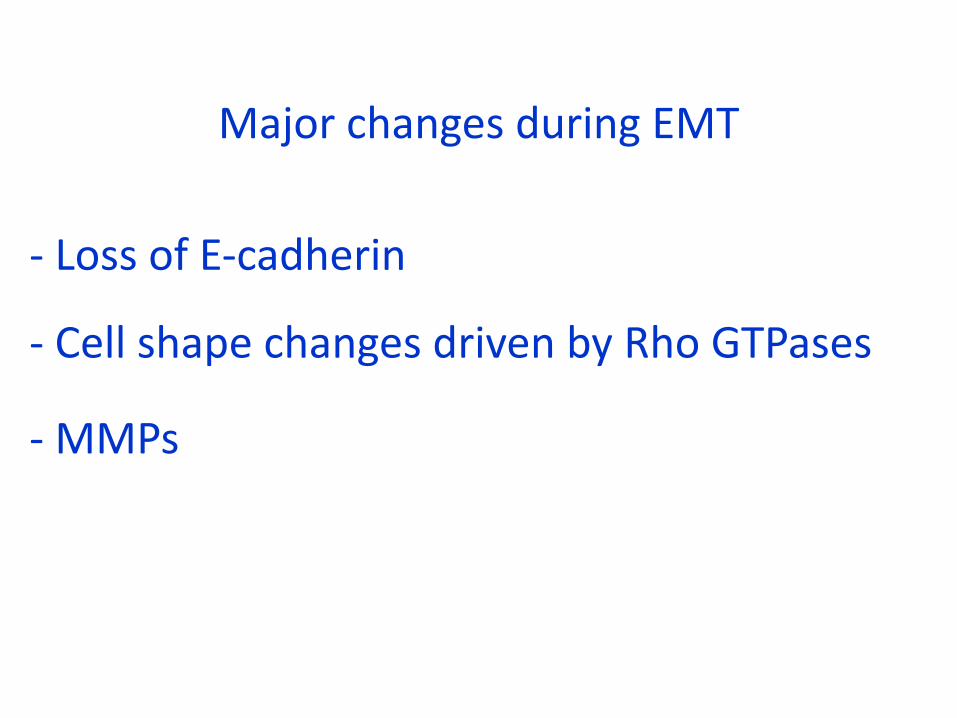
MajorchangesduringEMT
-LossofE-cadherin
-CellshapechangesdrivenbyRhoGTPases
-MMPs


EMTinTumorProgression
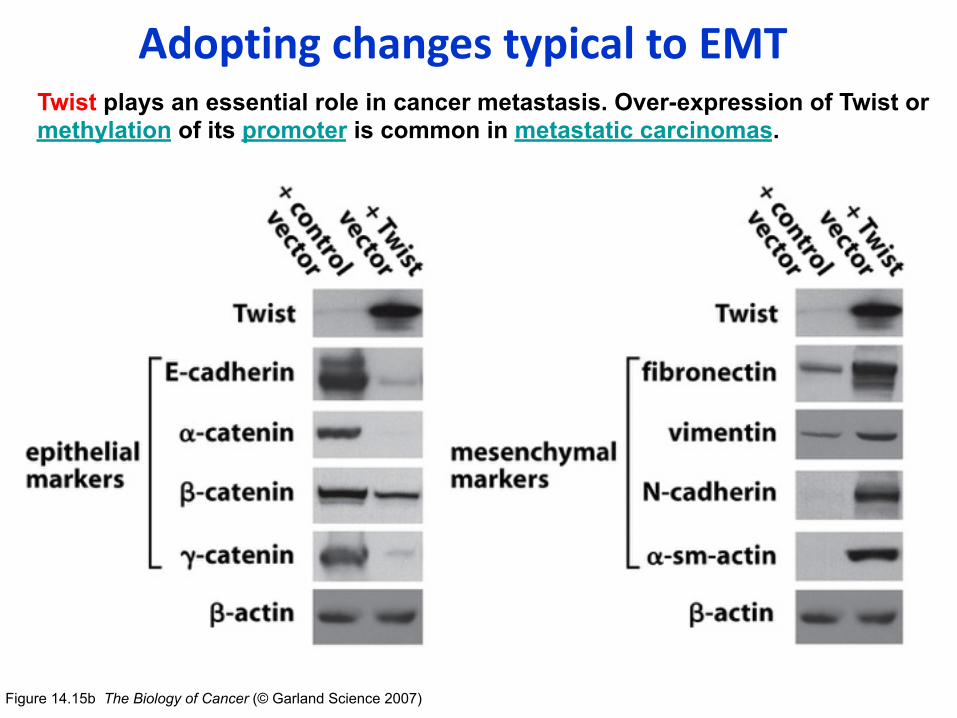
Figure 14.15b The Biology of Cancer (© Garland Science 2007)
AdoptingchangestypicaltoEMTTwist plays an essential role in cancer metastasis. Over-expression of Twist or methylation of its promoter is common in metastatic carcinomas.

MMPs(matrixmetalloproteinases)helpthecancercellstoinvadetheECM
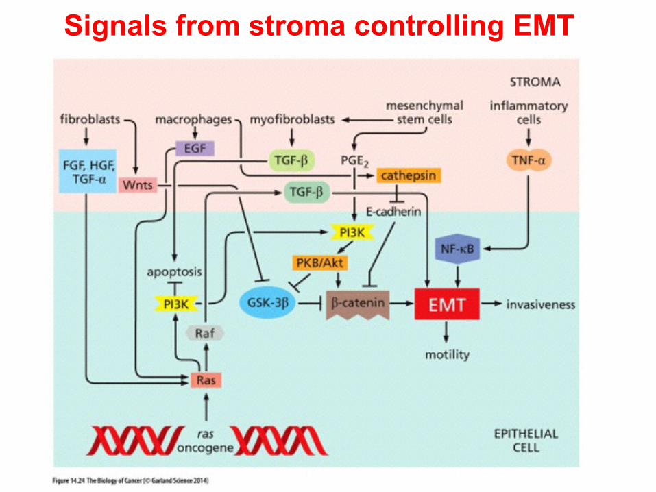
Signals from stroma controlling EMT

Epithelial Markers
E-cadherin Claudins Occludins
Desmoplakin Cytokeratins
Mesenchymal markers
Fibronectin Vitronectin Vimentin
Cell shape changes Cell movements, invasion
RhoB MMPs
Proliferation
Cyclin D CDK4
Rb phosph p21
Survival
PI3K activity ERK activity Caspases
P53 BID
Snail or Slug functions

Small GTPase family play a key role of cancer cell motility
Cytokines Chemokines Extracellular
matrix
Rho family •Rho •Rac
•Cdc42
Stress fiber Focal
adhesion Lamellipodia
Filopodia
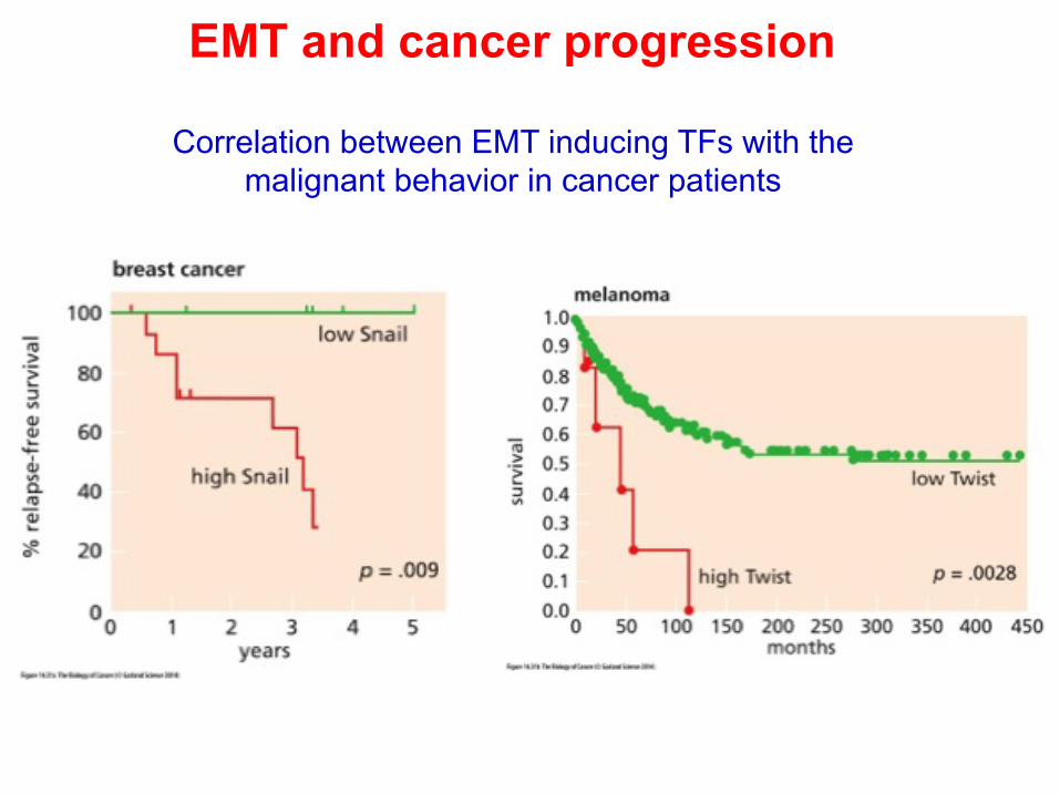
EMT and cancer progression
Correlation between EMT inducing TFs with the malignant behavior in cancer patients


![cancer lecture slides metastasis [Read-Only] · Several anti-angiogenesis inhibitors are in development. They differ in their specificity, target, and mode of action. PHILIPPINE CANCER](https://img.dokumen.tips/doc/110x75/5fc71874908f7264ec060b84/cancer-lecture-slides-metastasis-read-only-several-anti-angiogenesis-inhibitors.jpg)


![Plant Lectins in Therapeutic and Diagnostic Cancer Research · metastasis by increasing apoptosis or type I programmed cell death and inhibiting angiogenesis [18]. A number of cellular](https://img.dokumen.tips/doc/110x75/5f05e2a77e708231d415349e/plant-lectins-in-therapeutic-and-diagnostic-cancer-research-metastasis-by-increasing.jpg)