Embed Size (px)
Citation preview
Crit Care Shock (2019) 22:131-146
Anesthesia and intensive care management in acute ischemic stroke patient Hamzah, Prananda Surya Airlangga, Abdulloh Machin, Nancy Margarita Rehatta
Abstract Acute ischemic stroke is a brain functional dis-order, which cause high disability and mortality rate worldwide, the second most common cause of dementia, and the third leading cause of death. It has enormous clinical, social, and eco-nomic implications and demands a significant effort from both basic scientists and clinicians in the quest for understanding the underlying pathomechanisms and producing suitable pre-ventive measures and successful therapies. Management of acute ischemic stroke has been revolutionized by the introduction of several interventions, such as prehospital and stroke .
unit care, intravenous tissue plasminogen acti-vator therapy within 4.5 hours of stroke onset, aspirin therapy within 48 hours of stroke onset, decompressive craniectomy for supratentorial malignant hemispheric cerebral infarct, and more recently endovascular therapy for anteri-or circulating stroke. Also, special attention in management of vital systemic physiological var-iables, including oxygenation, blood pressure, temperature, and serum glucose. In line with this, the role of neuroanesthesiologists and neu-ro critical care in managing acute ischemic stroke become more prominent.
Key words: Acute ischemic stroke, acute ischemic stroke management, neuroanethesiologists, neuro critical care.
Crit Care Shock 2019 Vol. 22 No. 3 131
Address for correspondence: Hamzah Department of Anestesiology and Intensive Care, Faculty of Medicine Airlangga University - Dr. Soetomo General Hospital, Surabaya, Indonesia Jalan Mayjend Prof. Dr. Moestopo No. 6-8, Airlangga, Gu-beng, Surabaya, East Java 60285, Indonesia Tel: +62315501503, +62315501504 Email: [email protected]
From Department of Anestesiology and Intensive Care, Faculty of Medicine Airlangga University - Dr. Soetomo General Hospital, Surabaya (Hamzah, Prananda Surya Air-langga, Nancy Margarita Rehatta) and Departement of Neurology, Faculty of Medicine Airlangga University - Airlanga University Hospital, Surabaya (Abdulloh Machin).
Introduction Acute ischemic stroke (AIS) is a condition that causing focal and global brain physiology disfunction with high morbidity and mortality rate. (1,2) Focal AIS is caused by thrmbosis or cerebral arthery occlusion. (3) Nowadays, the amout of AIS patients are increasing and 15-20% of them may .
require intensive care management and monitoring. (1,4) Recent AIS management has developed progressively, from intravenous administration of recombinant tissue-type plasminogen activator (rtPA) within 4.5 hours from the onset, until the other new arterial intervension such as intra-arthery thrombolysis, angioplasty stenting, and decompression craniectomy in malignant brain infarction that may induce severe brain oedema and mass-like effect. (5) There are some new level 1 evidence-based medicine regarding the success-ful outcome of endovascular treatment in anterior cerebral stroke. (6,7) Rapid management in various systemic problems, such as blood pressure control, body temperature, blood glucose level, and oxygenation would need a closed monitoring by a neuroanesthetist - neuro critical care doctor in the ICU. Clinical improvement is an indicator of a success patient management. Neuroprotective concept that was proposed after many experimental researches before, failed to improve patient clinical outcome. This condition may occur because there are too .

132 Crit Care Shock 2019 Vol. 22 No. 3
many risk factors that associate with AIS, such as heterogeneity of AIS in human, and lacking of standardise clinical and preclinical research method. These last two decades research showed that endovascular intervention in AIS patients could significantly improve patient outcome. (2,8,9) This review article would describe the intensive care management for AIS patients based on the latest studies. The writers hoped that through this article they can provide a treatment guidelines for AIS patients and open up research opportunities in this sector. Stratification and etiology of AIS Since 1980-1990, many brain imaging modalities have emerged, including echocardiography and doppler ultrasound, which help clinicians diagnose a lacunar infarct, big artheroscleotic plaque, and other source of cardioemboli stroke. (10) Stroke was classified by stroke data bank and Trial of ORG 10172 in Acute Stroke Treatment (TOAST) into: - Large artery atherosclerosis - Cardiac embolism - Small artery occlusion - Stroke of other determined etiology - Stroke of undetermined etiology This system helps the clinician to group AIS patients based on the etiology of stroke. TOAST stratification method is not only useful in clinical and epidemiological studies, but also in accomodating all AIS patients. (10,11) Large artery atherosclerosis Based on its pathological study, atherosclerosis lesions can be found in bifurcations and big arterial arches. The more proximal the lesion is, the more dangerous the atherosclerosis lesion becomes. Atherosclerosis lesion can cause intravascular stenosis, while the rupture of atherosclerotic plaque can induce inflammation process, which leads to complete occlusion of the blood vessel. (12) One of the most common locations of large artery to become atherosclerotic is the carotid artery. At first, stenosis in the carotid artery is usually asymptomatic and preceded by infarction, which occurs in the left anterior descending artery (LAD). Perfusion disturbance in the carotid artery may interfere or cause an occlusion in the distal vessel(s) and increases the risk of infarction in the LAD. In carotid artery occlusion, the affected areas are usually supra-sylvii, frontal, central, and parie-tal area. (11,12)
The cause of perfusion disturbance is commonly emboli fragment, released from atheroma ulcers, which clogs the distal arteries. Another cause of perfusion impairment is a sudden blockage in the stenotic area, which causes decrease perfusion to the distal area. (13) Cardioembolic stroke Cardioemboli occur in about 14-30% of ischemic stroke patients. (14) Stroke that’s caused by embo-lism mostly happens in the middle cerebral artery (MCA). (15) Blood clots in the heart are usually caused by blood flow stasis or clots in prostheses. (11,16) In neuroimaging, cardioembolic strokes may appear in more than one artery. It is more common in the anterior circulation than the posterior one, but it can occur bilaterally. (11,12) Small vessel disease This mechanism happens due to the occlusion of microcirculation in the small blood vessels. (17,18) The etiology of this stroke subtype is the presence of lipohyalunosis in the vessel wall, while an in-crease in microatheroma also contributes to this pathology. (17,18) The most common location of this stroke is the deep region in the substantia alba, internal capsule, and its penetrating branches, pons, brainstem, and thalamus. Infarction caused by this mechanism is usually small (<1.5 cm) and the symptoms depend on the location in which the infarction occurs. The stroke is called the “lacunar syndrome” when charactherized by: 1) pure motor symptoms, 2) pure sensory symptoms, 3) sen-sorimotor mixture, 4) ataxia hemiparese, and 5) dysarthria-clumsy hand syndrome. (12) Other determined etiology The causes that include in this category are extra-cranial artery dissection, strokes that are caused by vasospasm associated with subarachnoid hemor-rhage (SAH), hereditary arteriopathy (such as Moyamoya disease), vasculitis caused by infec-tions, and strokes caused by coagulopathy. (13) Undetermined causes Patients who have undergone complete screening for heart problems, large artery diseases, coag-ulopathy, and other possible conditions, but the cause of the stroke remains unknown. (11) Pathophysiology of AIS The pathophysiology of AIS is very complex, but in general there is a regional obstruction in cerebral blood flow resulting in an ischemic area in the .

Crit Care Shock 2019 Vol. 22 No. 3 133
brain tissue (ischemic core), while the surrounding area experiences hypoperfusion known as the pe-numbra region. (19) The ischemic nucleus is con-sidered to be an irreversible area and the penumbra is considered to be an area that has good potential for a complete recovery. (20,21) AIS diagnosis Diagnosis of AIS is generally made through careful and thorough history-taking, neurological, and physical examination, as well as radiological imaging. (22,23) Anamnesis The most important information to be obtained from the AIS history is the onset. When the last time the patient felt normal? If the patient is un-conscious, when the last time the patient was symptoms free? Another history that needs to be explored is the condition around the patient when the symptoms appeared. Even though it is not al-ways accurate, it can help the clinician to find an-other diagnosis or get rid of the differential diagno-sis. It is also important to ask about the risk factors such as history of atherosclerosis, heart disease, history of drug abuse, migraines, seizures, infec-tions, trauma, and pregnancy. Obtaining these data is crucial in determining the treatment options. (24) Physical examination After the primary survey is carried out, a further general physical examination is done to ascertain the potential causes of AIS, other comorbid fac-tors, or other matters that might affect the selection of AIS therapy later. Neurological examination The initial neurological examination must be brief, but thorough. Every ischemic region would present a distinct symptom. A standardized neurological examination will help the clinician in performing neurological examination component precisely, quickly, and uniformly. The commonly used standard is the National Institute of Health Stroke Scale (NIHSS). Based on this scoring system, the clinical severity of the disease can be determined, where the higher the score the poorer the outcome would be. At the present, NIHSS (Table 1) is also one of the criteria for thrombolysis therapy and the best predictor of large vessel occlusion. (25) Stroke is one of the most common cause of disabil-ities in adult patients, therefore efforts have been made to quantify the level of disability in adults through modified Rankin Scale (mRS). (26) Val-ues ranging from 0-2 in this table are considered as .
good outcomes after an AIS episode. (27) Diagnostic test Some diagnostic tests need to be done routinely in all patients suspected with AIS to rule out other differential diagnoses, especially hemorrhagic stroke. Diagnostic tests can help the clinician to identify other comorbid diseases, to determine treatment options and to find out the possible com-plications that may occur. These tests may include blood glucose, serum electrolytes, kidney function tests, complete blood count with platelet count, cardiac markers, prothrombin time (PT), interna-tional normalized ratio (INR), activated partial thromboplastin time (aPTT), and electrocardio-gram (ECG). (28) Blood glucose levels are important to be checked before the administration of rtPA, because hypo-glycemia can cause focal signs and symptoms that resemble AIS. (29) Platelet count and PT/INR in AIS patients are important especially in patients with thrombocytopenia, impaired blood clotting function, liver dysfunction, or patient with antico-agulant medication. (24) Cardiac examinations such as electrocardiogram and cardiac markers are essential because of the strong association between stroke and cardiac ab-normalities. An increase in cardiac markers occurs in 5 to 34% of patients, whereas an increase in tro-ponin T is associated with severity, outcome and mortality. (24) Before giving thrombolysis therapy, the only laboratory test that need to be checked is blood glucose level, except in certain cases when the patient has blood coagulopathy, the doctor may need to order some more tests. (25) Diagnostic radiological imaging Brain imaging is not only capable of identifying the type and cause of stroke, but also distinguish-ing between ischemic and hemorrhagic stroke. In addition, it can also differentiate an irreversibly damaged tissue from areas that may be recovered, thus it is useful to determine the patient’s therapy in emergency situation and to predict outcomes. Vascular imaging can identify the location and the cause of arterial obstruction, in addition to identi-fying high risk patients. (28) Computed tomography scan (CT scan) All patients suspected with transient ischemic attack (TIA) or stroke are recommended to get an emergency head CT scan. (25,27,28) Patients sus-pected with TIA are equally important to get a CT scan because up to 10% of TIA patients suffer from strokes in the next 48 hours. (28)

134 Crit Care Shock 2019 Vol. 22 No. 3
Stroke patients are more prioritized than other pa-tients to get a brain imaging, because the outcome of the patient is determined by how soon the pa-tient can get a proper treatment. Head CT scan for patients suspected with stroke has proven to be the most cost-effective diagnostic strategy. In the hy-peracute phase, CT scan without contrast is the main investigative method to eliminate other pos-sible diseases such as stroke-like symptoms (stroke mimics) and bleeding lesions, which will influence the physician decision regarding the patient man-agement. That’s why head CT scan is the first choice of radiological imaging modality in stroke patients. (25,27) Initial CT in ischemic stroke will show some ab-normalities such as hypodensity in more than 1/3 cerebral parenchyma in areas that receive blood flow from the middle cerebral artery, hypodensity in lenticular nucleus, blurring of the cortical sul-cus, focal parenchymal hypodensity, loss of differ-ences in gray matter in the basal ganglia area, large vessel hyperdensity (e.g. middle cerebral artery hyperdensity, which indicates the presence of in-traluminal thrombus), and cerebral edema with blurring of cerebrospinal fluid space. (27,28) CT scan is very specific in identifying the signs of is-chemic brain damage, but sensitivity is only 61%. Two-thirds of patients with moderate to severe strokes have marked ischemic changes within the first few hours, but less than 50% of patients with mild strokes have ischemic lesions that appear rel-evant on CT scans, especially in the first few hours after the onset. The initial changes in brain CT scan are correlated with patient poor outcome and the success rate of reperfusion therapy. However, currently there is insufficient evidence for signs of early stroke changes being the reason of delaying thrombolysis therapy. (25) Sometimes, changes in the initial CT scan of AIS are missed, that’s why a scoring system is devel-oped to improve the detection of early ischemic changes. (30) One of the most widely used stand-ardized methods is the Alberta Stroke Program Early CT Score (ASPECTS). (30) ASPECTS has been developed to recognize the early signs of is-chemia. This scale assesses two axial pieces on the CT scan: the first one is at the level of the thalamus and the basal core, the other is at the basal level of the superior ganglia. The method of assessment is as follows: The middle cerebral artery is divided into ten regions. Subcortical structures have a score of 3, each divided into caudate, lentiform nucleus, and internal capsule. The cortex associated with the middle cerebral artery has a score of 7; 4 points derived from axial pieces at the level of the basal .
ganglia for the insular cortex, area M1, M2, and M3; 3 points originating from subsequent cuts for regions M4, M5, and M6. One point is reduced for each area with signs of initial ischemia (hypodensi-ty or edema). (31) ASPECTS is very helpful in predicting patient out-comes, especially for endovascular management in the acute phase, but this scoring system cannot be applied to lacunar strokes, ischemia in the mid-brain, or in other ischemic lesions involving arteri-al areas other than MCA. (27) CT angiography This method is used to evaluate the lumen of the intracranial artery and its branches. CT scans with contrast can give an insight of how contrast media is distributed in the area of the cranial blood ves-sels and show the clogged artery as having filling defects. This method can also assess the patency of the extracranial carotid system while seeking ste-nosis or a barrier that can cause AIS. Therefore, this method becomes important for the provision of potential therapy in AIS, especially for mechanical thrombectomy. (6) Noninvasive vascular imaging is recommended during the initial evaluation of patients who are planned to receive endovascular therapy, especially for patients with large vessel occlusion. But this diagnostic modality should not be a reason to delay thrombolysis therapy. (25) CT perfusion This method performs repeated imaging of the same cerebral parenchyma as bolus of intravenous contrast media is given. The goal of this imaging study is to map the overall volume of brain perfu-sion. The images need to be analyzed and inter-preted where the cerebral ischemic area will appear hypodense. (32) This method can also estimate the cerebral blood flow (CBF), cerebral blood volume (CBV), and mean transit time (MTT) needed for blood to flow from the vascular compartment to cerebral tissue by quantitative analysis of contrast media kinetics. (33) CBF and CBV boundary val-ues can be used to predict whether the brain paren-chyma may survive or not. ASPECTS has been used in mapping CBF or MTT to identify the max-imum expansion of ischemia region by assessing the absence of reperfusion, and the difference be-tween CBV and CBF (or MTT) to identify the area of ischemic penumbra. (6) Magnetic resonance imaging Immediate CT scan is the most cost-effective im-aging strategy for acute stroke patients, but it is insensitive for prolonged bleeding. That’s where .

Crit Care Shock 2019 Vol. 22 No. 3 135
magnetic resonance imaging (MRI) can be used as an alternative modality when non-contrast CT le-sions are not seen or when the clinical location of stroke is unclear at the time of stenting. However, delaying the administration of thrombolysis thera-py because of waiting for the MRI result is not recommended. (25) MRI is time consuming in obtaining and recon-structing the images. It is less suitable for restless patients or patients with possibility of vomiting and aspiration. If needed, emergency life assistance should be provided during the MRI process, as AIS patients (especially those with severe stroke) tend to become hypoxic when lying supine during im-aging process. Therefore, MRI is rarely used in emergencies and is not included in acute stroke protocols. However, MRI is a radiological modali-ty that has 100% diagnostic accuracy in detecting bleeding lesions. In addition, a certain sequence such as gradient echo (GRE) can help physician in distinguish acute changes from chronic forms. (34) DWI and PWI Diffusion-weighted imaging (DWI) is a method based on the ability of MRI to detect signals from the movement of water molecules based on differ-ences in Brownian motion. (35) This method can detect anomalies associated with cerebral ischemia within 3-30 minutes of the onset of symptoms, when there are no abnormal changes in traditional MRI and CT scans. (35) DWI can distinguish be-tween cytotoxic and vasogenic edema using appar-ent diffusion coefficient (ADC). The ADC can measure the level of water diffusion. In this con-text, hypointense signals in ADC mapping are in accordance with cytotoxic edema, while hyperin-tense signals represent vasogenic edema. This method also gives a predictive value of clinical and functional outcomes. (36) Tissues that appear ab-normal on DWI are identified as having ischemic damage, where tissue with a slight reduced ADC can be permanently damaged; but, there is no ADC threshold can be relied upon to distinguish dead tissue from living tissue. (37) Perfusion-weighted imaging (PWI) can identify areas of ischemia through MRI by measuring the amount of contrast that reaches brain tissue. (38) Map of CBF, CBV, and MTT are processed through several stages of scanning in the same brain parenchyma area. This method can be used to assess whether the brain tissue is still salvageable or not, assess the severity of lesions, considering the choice of therapy where reversible ischemic brain damage is a key factor in selecting patients for reperfusion, and monitoring patients. (36) The pattern of missed match between .
DWI and PWI highlights the area of cerebral pa-renchyma that can still be saved. Specifically, while DWI detects irreversible-damaged brain pa-renchyma (ischemic nucleus), PWI can show hy-poperfusion areas (ischemic penumbra region) by reducing the irreversible areas seen on DWI. (39) MR angiography This method could show any signs of stenosis or occlusion in extra- and intracranial vessels. The sensitivity and specificity in detecting vascular lesions vary from 86 to 97% for CT angiography and 62 to 91% for MRI angiography. Acute thrombolytic occlusion is usually shown as a hy-pointense image in a large vessel (middle or carot-id cerebral artery). (40) However this method is not recommended for AIS patients who are admitted to the hospital in less than 6 hours after the onset. (25) Neurosonology Carotid color Doppler and transcranial Doppler are two noninvasive methods that can be used for neu-rovascular assessment. This method can assess se-rious vascular stenosis or identify the cause of a stroke such as vascular occlusion, or for carotid and vertebral arteries surgery. Transcranial Dop-pler works by utilizing the emission of low fre-quency pulsating sounds that can penetrate the bone, allowing identification of stenosis, occlusion, collateral circuits, and possible reperfusion after thrombolytic treatment. This method has some advantages such as practical and as a bedside diagnostic modality, but it re-quires an experienced operator. The combination of duplex ultrasound imaging techniques and magnetic resonance angiography (MRA), which is relatively risk-free, can diagnose carotid artery ste-nosis. Digital subtraction angiography (DSA), which has 1-3% risk of stroke in patients with ca-rotid lesions, might be used as well. (41,42) Tran-scranial doppler is the only technique that could detect circulating intracranial embolism, which is very common in patients with large arterial dis-ease. (43) Symptomatic carotid artery stenosis is a strong independent predictor of recurrent acute ischemic stroke and TIA. That’s why it should be used to evaluate antiplatelet therapy. (44) Ultrasonography has some limitation such as the difficulty in finding a bone window and inability to provide a good image of posterior circulation thet need to be measure repeatedly. In this condition, CT angiography is preferred in clinical practice, especially in urgent situations. (28)
136 Crit Care Shock 2019 Vol. 22 No. 3
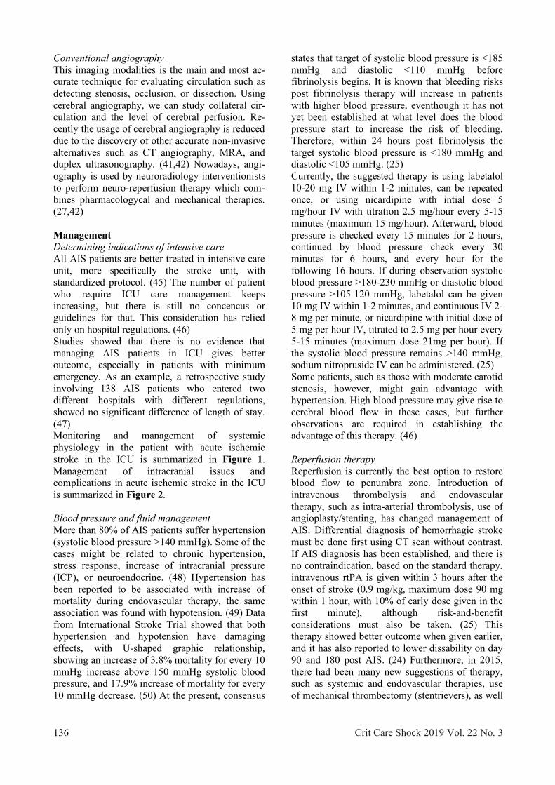
Conventional angiography This imaging modalities is the main and most ac-curate technique for evaluating circulation such as detecting stenosis, occlusion, or dissection. Using cerebral angiography, we can study collateral cir-culation and the level of cerebral perfusion. Re-cently the usage of cerebral angiography is reduced due to the discovery of other accurate non-invasive alternatives such as CT angiography, MRA, and duplex ultrasonography. (41,42) Nowadays, angi-ography is used by neuroradiology interventionists to perform neuro-reperfusion therapy which com-bines pharmacologycal and mechanical therapies. (27,42) Management Determining indications of intensive care All AIS patients are better treated in intensive care unit, more specifically the stroke unit, with standardized protocol. (45) The number of patient who require ICU care management keeps increasing, but there is still no concencus or guidelines for that. This consideration has relied only on hospital regulations. (46) Studies showed that there is no evidence that managing AIS patients in ICU gives better outcome, especially in patients with minimum emergency. As an example, a retrospective study involving 138 AIS patients who entered two different hospitals with different regulations, showed no significant difference of length of stay. (47) Monitoring and management of systemic physiology in the patient with acute ischemic stroke in the ICU is summarized in Figure 1. Management of intracranial issues and complications in acute ischemic stroke in the ICU is summarized in Figure 2. Blood pressure and fluid management More than 80% of AIS patients suffer hypertension (systolic blood pressure >140 mmHg). Some of the cases might be related to chronic hypertension, stress response, increase of intracranial pressure (ICP), or neuroendocrine. (48) Hypertension has been reported to be associated with increase of mortality during endovascular therapy, the same association was found with hypotension. (49) Data from International Stroke Trial showed that both hypertension and hypotension have damaging effects, with U-shaped graphic relationship, showing an increase of 3.8% mortality for every 10 mmHg increase above 150 mmHg systolic blood pressure, and 17.9% increase of mortality for every 10 mmHg decrease. (50) At the present, consensus .
states that target of systolic blood pressure is <185 mmHg and diastolic <110 mmHg before fibrinolysis begins. It is known that bleeding risks post fibrinolysis therapy will increase in patients with higher blood pressure, eventhough it has not yet been established at what level does the blood pressure start to increase the risk of bleeding. Therefore, within 24 hours post fibrinolysis the target systolic blood pressure is <180 mmHg and diastolic <105 mmHg. (25) Currently, the suggested therapy is using labetalol 10-20 mg IV within 1-2 minutes, can be repeated once, or using nicardipine with intial dose 5 mg/hour IV with titration 2.5 mg/hour every 5-15 minutes (maximum 15 mg/hour). Afterward, blood pressure is checked every 15 minutes for 2 hours, continued by blood pressure check every 30 minutes for 6 hours, and every hour for the following 16 hours. If during observation systolic blood pressure >180-230 mmHg or diastolic blood pressure >105-120 mmHg, labetalol can be given 10 mg IV within 1-2 minutes, and continuous IV 2-8 mg per minute, or nicardipine with initial dose of 5 mg per hour IV, titrated to 2.5 mg per hour every 5-15 minutes (maximum dose 21mg per hour). If the systolic blood pressure remains >140 mmHg, sodium nitropruside IV can be administered. (25) Some patients, such as those with moderate carotid stenosis, however, might gain advantage with hypertension. High blood pressure may give rise to cerebral blood flow in these cases, but further observations are required in establishing the advantage of this therapy. (46) Reperfusion therapy Reperfusion is currently the best option to restore blood flow to penumbra zone. Introduction of intravenous thrombolysis and endovascular therapy, such as intra-arterial thrombolysis, use of angioplasty/stenting, has changed management of AIS. Differential diagnosis of hemorrhagic stroke must be done first using CT scan without contrast. If AIS diagnosis has been established, and there is no contraindication, based on the standard therapy, intravenous rtPA is given within 3 hours after the onset of stroke (0.9 mg/kg, maximum dose 90 mg within 1 hour, with 10% of early dose given in the first minute), although risk-and-benefit considerations must also be taken. (25) This therapy showed better outcome when given earlier, and it has also reported to lower dissability on day 90 and 180 post AIS. (24) Furthermore, in 2015, there had been many new suggestions of therapy, such as systemic and endovascular therapies, use of mechanical thrombectomy (stentrievers), as well .

Crit Care Shock 2019 Vol. 22 No. 3 137
as arguments of rtPA administration time limits. Current endovascular therapy can be summarized into: 1. CT scan is compulsary to eliminate hemorrage,
no matter how small. 2. Rdiological imaging with contrast (CT scan or
MRI) shows proximal occlusion from major artery branch and the anterior circulation.
3. Procedure must be done in a hospital, which is facilitated by cardiovascular intervention diagnostic installation, especially for performing stent retreiver.
4. Mechanical thrombectomy must be performed in less than 6 hours of period. (2)
Choices of anesthesia techniques during endovascular therapy Anethesiologists play an important role in the management of endovascular AIS. The choice of anesthesia technique is made based on several factors, including airway, breathing, hemo-dynamics, level of consciousness, agitation, and ability to lie still. The choice has to be made quickly because 30-minute delay in starting thrombolysis may reduce 3-month success rate up to 10%. Endovascular therapy can be performed using general anesthesia (GA) or measured anaes-thesia care (MAC), which uses local anethesia, with or without sedation. Retrospective studies so far showed that MAC gave better results compared to GA. This result, however, may be due to the fact that in GA group, many involve stroke in the posterior circulation, which is associated with poorer outcome. In addition, most of the patients in the GA group had worse conditions in the beginning. Therefore, more studies are required to determine the standard choice of anethesia technique during endovascular therapy. Oxygenation and ventilation management Hypoxia is commonly found in AIS and very influential to the patient care outcome. It is, therefore, very important to identify the causes of hypoxia, which may include neurological and non-neurological factors, such as aspiration pneumonia, acute lung injury, pulmonary emboli, and central respiration arrhytmia. Established studies so far showed that hyperbaric therapy (51,52) and routine oxygenation supple-mentation in non-hypoxic patients (41) worsen their stroke severity and condition. Current guidelines from American Stroke Association and European Stroke Organisation recommend oxygen supplementation only in patients with SpO2<94%, who can breathe on their own. (24,28)
Continuous monitoring of arterial oxygen with pulse oximeter is required in all AIS patients in ICU, while blood gas analysis is measured regularly during the use of mechanical ventilation, in order to observe and manage PaO2 (>80 mmHg) optimally. Hypocapnia (PaCO2<35 mmHg) is associated with poor outcome post stroke attack, therefore, normocapnia (PaCO2 35 to 45 mmHg) is the target for intubated patients. For patients with spontaneous ventilation, SpO2 is suggested to be maintained at >92%, (53) while SpO2 levels during endovascular therapy and ICU stay is maintained at > 94%. (1) More than a third of patients with severe AIS need tracheostomy. Indications of tracheostomy includ-ing the use of long-term mechanical ventilator (for 2 weeks or more), severe dysphagia and/or bulbar palsies due to extensive brain and brain stem infarction. (54) Based on a systematical evaluation of 11 studies involving 886 patients, the best method to detect dysphagia in patients who are not intubated is through water test, which has 73%-98% sensitivity and 63%-76% specificity. This test is done by giving an aliquot of water, which has been determined before, to evaluate the ability to swallow using coughing, choking, and voice changing as markers, while continuous monitoring using pulse oximeter is done. (55) The time when tracheostomy is needed is debatable. Generally, tracheostomy is considered after 1 week use of mechanical ventilator in stroke patients. (56) However, some studies showed that patients who received tracheostomy 7 and 14 days after mechanical ventilator use didn’t have any com-plications due to tracheostomy, using the length of ICU stay, sepsis severity, and bleeding as indi-cators. (54) Glucose serum management More than 40% of patients suffer hyperglicemia in acute phase of AIS. The high level of serum glucose is an indicator of disease severity, which is associated with increase of cortex toxicity, larger infarct volume, poorer outcome, and infection risk. (57) Pathophysiology of hyperglicemia associated with death in AIS has not been fully understood, but is suspected involving many factors, and the failure of recovery in ischemic penumbra zone to be the key. Other mechanisms, which are sug-gested to play a role including increase of coag-ulation and decrease of fibrinolytic activity, which inhibit recanalization, vasodilation inhibition, which causes reduced cerebral blood flow (CBF), reperfusion injury duse to oxydative stress and in-flammation. (47) Hyperglycemic management af- .
138 Crit Care Shock 2019 Vol. 22 No. 3
ter AIS currently targets serum glucose level at 70-140 mg/dl during ICU care. (1) Hypoglycemia in AIS patients must be dealt with when the serum glucose level reaches <60 mg/dl. (25) Suggested method in the present is by continuous glucose infusion to avoid extreme low serum glucose levels. Temperature management and hypothermia therapy Fever can be found among up to 35.5% AIS patients and is associated with poor outcome. (58) Infection, which causes the fever, must be found and treated, but it can also be caused by central inflammatory response without the presence of in-fection. Fever must be treated aggressively. Man-agement may include the use of acetaminophen as the first line, metamizole, rapid cold saline (4 oC) infusion, or automatic cooling system. Suggested target temperature is 35 oC to 37 oC in patients with endovascular therapy and <37.5 oC in ICU patients. (1) Different to hyperthermia, hypothermia in early care post AIS is associated with better outcome. (59) Therapeutic hypothermia (TH) has shown many neuroprotective effects. (60) However, there are some risks associated with TH, including higher pneumonia incidence, shivering, electrolyte imbalance, and cardiac dysfunction. (25) Addi-tional airway management has become a challenge in doing TH after AIS, and may be an indicator itself to have an ICU treatment care. (61) Although local cooling (in head only) through nasal air flow, conduction through nasal catheter, air, gel caps, and other methods can reduce systemic risk, (62) there are also some reports of higher complication events and equal mortality with conventional therapy, which suggests the need of more research about its use outside trials. (63) In vitro studies showed that lower temperature (<37 oC) may dis-turb the performance of rtPA, (64) therefore, more studies in clinical trials context are required to observe the effects of TH in thrombolytic therapy. (25) Anticoagulation, antiplatelet therapy, and thromboprophylaxis Anticoagulation, antiplatelet therapy, and throm-boprophylaxis after AIS is made harder by intravenous thrombolysis and endovascular therapies. Data available in this topic is not specific for endovascular therapy or ICU treatment care. Moreover, the optimal agent and dose have not been clearly stated. Currently, the use of these agents are avoided during the first 24 hour after .
thrombolysis, but this approach still needs further evaluation. (2) Class I evidence supports the use of high dose aspirin (160-325 mg) in 24-48 hours after AIS begins. (24) However, this approach does not substitute endovascular therapy. Evidence sup-porting other antiplatelet such as clopidrogel is still weak. History of antiplatelet use also showed an increase of symptomatic ICH incidence. (65) Heparin is used during endovascular therapy to minimize thrombosis and emboli risks due to catheterization. Moreover, combination of aspirin and clopidrogel are often used during endovascular stent insertion. (66,67) Until now there is no strong and consistent evidence stating the correlation between the use of oral anticoagulant and bleeding post AIS, including the new anticoagulants such as dabigatran and rivaroxaban. The choice to use of anticoagulants in these cases, therefore, is made by the doctors with considerations of its risks and benefits. (2) Thromboembolism prevention can be facilitated by early hydration and mobilization. Graduated compression stocking does not reduce the incidence of thromboembolism. (68) Other studies showed that intermittent pneumatic calf compression reduced risks of deep vein thrombosis in 30 days after trial entry, with improvement of survivability in 6 months, but without significant increase of function and quality of life. (69) The use of low molecular weight heparin (LMWH) in vein thromboembolism prevention is supported by class I evidence. The type and dose of LMWH chosen must be adjusted by local guideline. Current evidence showed that the best option is using subcutaneous enoxaparin 40 mg single dose as reported in PREvention of Venous thromboem-bolism after Acute Ischemic stroke with Low mo-lecular weight heparin (PREVAIL) study. (70) There is no clear guideline on when to start giving LMWH, but reports suggested that it is better to start early with interlude of 24 hour minimum after reperfusion therapy to avoid intracranial bleeding. Bleeding management Hemorrhagic transformation may occur among 5-6% of patients who receive intravenous rtPA and intra-arterial recanalization therapy, even in some patients who do not undergo reperfusion therapy. There was no significant difference of ICH incidence between patients who received intra-venous thromobolytic therapy and endovascular therapy. (6,7,71-73) Presently, there is still no guidelines in managing hemorrhagic transfor-mation, although combination of cryoprecipitate, .

Crit Care Shock 2019 Vol. 22 No. 3 139
fresh frozen plasma, recombinant factor VII, and prothrombin complex concentrate can be used with local guidelines and expert’s instructions. The administration of rtPA must be stopped immediately if hemorrhagic transformation happens and protamin should be given to patients who receive heparin during endovascular therapy. Consultation with neurosurgeon regarding the indications of the surgery may be needed, expecially in patients with extensive hematoma. (2) Seizure and anticonvulsants Unlike other acute brain injuries, convulsive seizure rarely happens after AIS, even in the cases related to thrombolysis. (74) Previously known seizure must be treated aggresively, but the role of anticonvulsants prophylaxis is yet to be elucidated. (75) There is no specific data describing seizure management during ICU treatment care or endovascular therapy in AIS, but in general evidence showed that the use of anticonvulsants is associated with long-term poor outcome. (76) However, most of the studies used phenytoin, therefore, it is unclear whether the same results should be expected with the new agents as well. Even though phenytoin is commonly considered as first line anticonvulsant. Levetiracetam is more preferable nowadays in medical centers. Non-convulsive seizures is also rarely found in AIS compared to other types of acute brain injuries. It is also still unclear if application of anticonvulsant therapy as medication or prophylaxis can better the outcome. Consensus reccommended the use of electroencephalography (EEG) to eliminate diagnosis of non-convulsive seizure in all AIS patients with change of consciousness levels, which is unexplainable and/or persistant. (76) Role of neuromonitoring Basic of neurological deterioration detection in-cludes monitoring of conscious and cooperative patients, combination of clinical evaluation and neurologic physical examination, and radiological imaging. This also becomes the determinant of next steps in managing AIS. This applies to patients in the emergency room, reperfusion therapy patients, those treated in the ICU and in stroke unit. The intracranial pressure (ICP) of AIS patients, even those with extensive infarct and edema, is commonly normal (<20 mmHg). Routine examination of ICP is therefore not reccommended. (77) Non-invasive neurological monitoring tools seem to be more relevant to use, where patients do not .
require sedation. One of the most useful tools is transcranial doppler ultrasonography, which can facilitate AIS diagnosis by detecting the response of acute MCA occlusion towards thrombolysis, aiding in determining patient’s prognosis based on severity of arterial occlusion, evaluating brain blood flow and vasoreactivity, and when combined with continuous blood pressure measurements can evaluate cerebral autoregulations. (2) Sugery Approximately 10% of AIS involves extensive edema in the hemisphere, more commonly found in patients with internal carotid artery or middle cerebral artery or both occlusions. The most common symptoms are hemiplegia, head or eye deviation, aphasia, and/or contralateral neglect syndrome. Even the treatment with hyperventilation, manitol, barbiturate coma, and hypothermia, the mortality rate is still high (estimated between 50-78%). Decrease of consciousness is often found after 48 hours and followed by transtentorial herniation during 48-96 hours afterwards. (27) The role of decompressive craniectomy in patients with malignant infarct from MCA has been vastly studied. Indication for surgery is clear, (1) and this surgery for patients aged 18 to 60 years old with MCA malignant infarct within 48 hours after the first symptoms appear can reduce mortality up to 29-78% with improvements of patient’s functions. (10) Although this surgery can increase patient’s survival, and that the timing should be done as early as possible, often patients end up suffering from severe dissabilities. Therefore, careful considerations from both patients and their family must be taken. Conclusion Introduction of several interventions, including reperfusion therapy and decompressive craniectomy, have improved AIS management in the two decades. Furthermore, management of essential systemic physiological variables, such as oxygenation, blood pressure, temperature, and glycemic control, has been appreciated. In relation to class I evidence, which supported the role of endovascular therapy, management of AIS has become more aggressive, which involves greater contribution from anesthesiologists and intensive care. Many challenges and questions in managing AIS have not been successfully answered due to lack of data regarding AIS specific management using endovascular therapy and intensive care. Consensus guidelines in anesthesy care from endo- .

Table 1. National Institutes of Health Stroke Scale (NIHSS) (78)
140 Crit Care Shock 2019 Vol. 22 No. 3
vascular therapy has been published by the Society for Neuroscience in Anesthesiology and Critical Care. (53) In ICU setting, several matters that need careful observation include airway management, ventilation, optimalization from hemodynamic and fluid parameters, as well as monitoring and control of temperature, glycemic status, anticoagulant .
management, antiplatelet and thrombophylaxis, management of complications associated with reperfusion therapy, detection and management of seizure, and neurosurgeries. (1) Therefore, there are still plenty more specific studies needed for AIS management using intensive and endovascular therapies.

Figure 1. Monitoring and management of systemic physiology in the patient with acute ischemic stroke in the intensive care unit (1)
Crit Care Shock 2019 Vol. 22 No. 3 141

Figure 2. Management of intracranial issues and complications in acute ischemic stroke in the intensive care unit (1)
142 Crit Care Shock 2019 Vol. 22 No. 3
1. Kirkman MA, Citerio G, Smith M. The inten-sive care management of acute ischemic stroke: An overview. Intensive Care Med 2014;40:640-53.
2. Kirkman MA, Lambden S, Smith M. Chal-lenges in the Anesthetic and Intensive Care Management of Acute Ischemic Stroke. J Neurosurg Anesthesiol 2016;28:214-32.
3. Kanyal N. The Science of Ischemic Stroke: Pathophysiology & Pharmacological Treat-ment. Int J Pharma Res Rev (IJPRR) 2015; 4:65-84.
4. Zazulia AR. Critical Care Management of A-cute Ischemic Stroke. Continuum (Minneap Minn) 2009;15:68-82.
5. Vahedi K, Hofmeijer J, Juettler E, Vicaut E, George B, Algra A, et al. Early decompressive surgery in malignant infarction of the middle cerebral artery: a pooled analysis of three ran-domised controlled trials. Lancet Neurol 2007;6:215-22.
6. Goyal M, Demchuk AM, Menon BK, Eesa M, Rempel JL, Thornton J, et al. Randomized Assessment of Rapid Endovascular Treatment of Ischemic Stroke. N Engl J Med 2015;372: 1019-30.
7. Jovin TG, Chamorro A, Cobo E, de Miquel MA, Molina CA, Rovira A, et al. Thrombec-tomy within 8 Hours after Symptom Onset in Ischemic Stroke. N Engl J Med 2015;372: 2296-306.
8. Sutherland BA, Minnerup J, Balami JS, Arba F, Buchan AM, Kleinschnitz C. Neuropro-tection for ischaemic stroke: Translation from the bench to the bedside. Int J Stroke 2012; 7:407-18.
9. Alkhalili K, Chalouhi N, Tjoumakaris S, Hasan D, Starke RM, Zanaty M, et al. Endovascular Intervention for Acute Ischemic Stroke in Light of Recent Trials. Scientific-WorldJournal 2014;2014:429549.
10. Adams HP, Bendixen BH, Kappelle LJ, Biller J, Love BB, Gordon DL, et al. Classification of subtype of acute ischemic stroke. Definitions for use in a multicenter clinical trial. TOAST. Trial of Org 10172 in Acute Stroke Treatment. Stroke 1993;24:35-41.
11. Chung JW, Park SH, Kim N, Kim WJ, Park JH, Ko Y, et al. Trial of ORG 10172 in Acute Stroke Treatment (TOAST) Classification and Vascular Territory of Ischemic Stroke Lesions Diagnosed by Diffusion-Weighted Imaging. J .
Crit Care Shock 2019 Vol. 22 No. 3 143
Am Heart Assoc 2014;3:e001119. 12. Grotta JC, Albers GW, Broderick JP, Kasner
SE, Lo EH, Mendelow AD, et al. Stroke: pathophysiology, diagnosis, and management. Elsevier Inc.; 2015.
13. Ropper AH. Cerebrovascular diseases. In: Sydor AM, Davis KJ, editors. Adams and Victor’s Principles of Neurology. 10th ed. New York: McGraw-Hill Education; 2014.
14. Arboix A, Alio J. Cardioembolic Stroke: Clinical Features, Specific Cardiac Disorders and Prognosis. Curr Cardiol Rev 2010;6:150-61.
15. Zheng M, Fan DS. Acute Cardioembolic and Thrombotic Middle Cerebral Artery Occlusions Have Different Morphological Susceptibility Signs on T2 -Weighted Magnetic Resonance Images. Biomed Res Int 2015;2015:839820.
16. Kamel H, Healey JS. Cardioembolic Stroke. Circ Res 2017;120:514-26.
17. Joutel A, Faraci FM. Cerebral Small Vessel Disease: Insights and Opportunities From Mouse Models of Collagen IV–Related Small Vessel Disease and Cerebral Autosomal Dominant Arteriopathy With Subcortical Infarcts and Leukoencephalopathy. Stroke 2014;45:1215-21.
18. Shi Y, Wardlaw JM. Update on cerebral small vessel disease: A dynamic whole-brain disease. Stroke Vasc Neurol 2016;1:83-92.
19. Deb P, Sharma S, Hassan KM. Pathophysiologic mechanisms of acute ischemic stroke: An overview with emphasis on therapeutic significance beyond thrombolysis. Pathophysiology 2010;17:197-218.
20. Jauss M, Krieger D, Hornig C, Schramm J, Busse O. Surgical and medical management of patients with massive cerebellar infarctions: Results of the German-Austrian Cerebellar Infarction Study. J Neurol 1999;246:257-64.
21. Dirnagl U. Pathobiology of injury after stroke: The neurovascular unit and beyond. Ann N Y Acad Sci 2012;1268:21-5.
22. Musuka TD, Wilton SB, Traboulsi M, Hill MD. Diagnosis and management of acute ischemic stroke: speed is critical. CMAJ 2015;187:887-93.
23. Nentwich LM. Diagnosis of Acute Ischemic Stoke. Emerg Med Clin North Am 2016;34: 837-59.
References
24. Jauch EC, Saver JL, Adams HP, Bruno A, Connors JJB, Demaerschalk BM, et al. Guidelines for the early management of patients with acute ischemic stroke: A guideline for healthcare professionals from the American Heart Association/American Stroke Association. Stroke 2013;44:870-947.
25. Powers WJ, Rabinstein AA, Ackerson T, Adeoye OM, Bambakidis NC, Becker K, et al. 2018 Guidelines for the Early Management of Patients With Acute Ischemic Stroke: A Guideline for Healthcare Professionals From the American Heart Association/American Stroke Association. Stroke 2018;49:e46-110.
26. Broderick JP, Adeoye O, Elm J. Evolution of the Modified Rankin Scale and Its Use in Future Stroke Trials. Stroke 2017;48:2007-12.
27. D’Aliberti G, Longoni M, Motto C, Oppo V, Perini V, Valvassori L, et al. Ischemic Stroke. In: Agostoni E, editor. Emergency Manage-ment in Neurology. Springer International Publishing; 2017. P. 1-91.
28. European Stroke Organisation (ESO) Executive Committee, ESO Writing Committee. Guidelines for management of ischaemic stroke and transient ischaemic attack 2008. Cerebrovasc Dis 2008;25:457-507.
29. Hafez S, Coucha M, Bruno A, Fagan SC, Ergul A. Hyperglycemia, Acute Ischemic Stroke, and Thrombolytic Therapy. Transl Stroke Res 2014;5:442-53.
30. Pexman JH, Barber PA, Hill MD, Sevick RJ, Demchuk AM, Hudon ME, et al. Use of the Alberta Stroke Program Early CT Score (ASPECTS) for Assessing CT Scans in Patients with Acute Stroke. AJNR Am J Neu-roradiol 2001;22:1534-42.
31. Barber PA, Demchuk AM, Zhang JJ, Buchan AM. Validity and reliability of a quantitative computed tomography score in predicting outcome of hyperacute stroke before thrombolytic therapy. ASPECTS Study Group. Alberta StrokeProgramme Early CT Score. Lancet 2000;355:1670-4.
32. Allmendinger AM, Tang ER, Lui YW, Spektor V. Imaging of Stroke: Part 1, Perfusion CT--Overview of Imaging Technique, Interpre-tation Pearls, and Common Pitfalls. AJR Am J Roentgenol 2012;198:52-62.
33. Lev MH. Perfusion Imaging of Acute Stroke: Its Role in Current and Future Clinical Practice. Radiology 2013;266:22-7.
34. Hand PJ, Wardlaw JM, Rowat AM, Haisma JA, Lindley RI, Dennis MS. Magnetic resonance brain imaging in patients with acute .
144 Crit Care Shock 2019 Vol. 22 No. 3
stroke: Feasibility and patient related difficulties. J Neurol Neurosurg Psychiatry 2005;76:1525-7.
35. Baliyan V, Das CJ, Sharma R, Gupta AK. Diffusion weighted imaging: Technique and applications. World J Radiol 2016;8:785-98.
36. Le Bihan D, Johansen-Berg H. Diffusion MRI at 25: Exploring brain tissue structure and function. Neuroimage 2012;61:324-41.
37. Oppenheim C, Lamy C, Touzé E, Calvet D, Hamon M, Mas JL, et al. Do transient ischemic attacks with diffusion-weighted imaging abnormalities correspond to brain infarctions? AJNR Am J Neuroradiol 2006;27:1782-7.
38. Essig M, Shiroishi MS, Nguyen TB, Saake M, Provenzale JM, Enterline D, et al. Perfusion MRI: The five most frequently asked technical questions. AJR Am J Roentgenol 2013;200:24-34.
39. Wardlaw J, Murray V, Berge E, del Zoppo G. Thrombolysis for acute ischaemic stroke. Cochrane Database Syst Rev 2014;29: CD000213.
40. Smith EE, Abdullah AR, Petkovska I, Rosenthal E, Koroshetz WJ, Schwamm LH. Poor outcomes in patients who do not receive intravenous tissue plasminogen activator because of mild or improving ischemic stroke. Stroke 2005;36:2497-9.
41. Nederkoorn PJ, van der Graaf Y, Hunink MG. Duplex ultrasound and magnetic resonance angiography compared with digital subtraction angiography in carotid artery stenosis: A systematic review. Stroke 2003;34:1324-32.
42. Forsting M, Wanke I. Funeral for a friend. Stroke 2003;34:1324-32.
43. Markus HS, MacKinnon A. Asymptomatic embolization detected by Doppler ultrasound predicts stroke risk in symptomatic carotid artery stenosis. Stroke 2005;36:971-5.
44. Markus HS, Droste DW, Kaps M, Larrue V, Lees KR, Siebler M, et al. Dual Antiplatelet Therapy With Clopidogrel and Aspirin in Symptomatic Carotid Stenosis Evaluated Using Doppler Embolic Signal Detection: the Clopidogrel and Aspirin for Reduction of Em-boli in Symptomatic Carotid Stenosis (CA-RESS) trial. Circulation 2005;111:2233-40.
45. Stroke Unit Trialists’ Collaboration. Organised inpatient (stroke unit) care for stroke. Cochrane Database Syst Rev 2013;(9): CD000197.
46. Bevers MB, Kimberly WT. Critical Care Management of Acute Ischemic Stroke. Curr Treat Options Cardiovasc Med 2017;19:41.
47. Briggs DE, Felberg RA, Malkoff MD, Bratina P, Grotta JC. Should mild or moderate stroke patients be admitted to an intensive care unit? Stroke 2001;32:871-6.
48. Abou-Chebl A, Zaidat OO, Castonguay AC, Gupta R, Sun CH, Martin CO, et al. North American SOLITAIRE stent-retriever acute stroke registry: Choice of anesthesia and outcomes. Stroke 2014;45:1396-401.
49. Wohlfahrt P, Krajcoviechova A, Jozifova M, Mayer O, Vanek J, Filipovsky J, et al. Low blood pressure during the acute period of ischemic stroke is associated with decreased survival. J Hypertens 2015;33:339-45.
50. Leonardi-Bee J, Bath PM, Phillips SJ, Sandercock PA, IST Collaborative Group. Blood pressure and clinical outcomes in the International Stroke Trial. Stroke 2002;33:155-20.
51. Ali K, Warusevitane A, Lally F, Sim J, Sills S, Pountain S, et al. The Stroke Oxygen Pilot Study: A Randomized Controlled Trial of the Effects of Routine Oxygen Supplementation Early after Acute Stroke-Effect on Key Outcomes at Six Months. PLoS One 2013; 8:e59274.
52. Rincon F, Kang J, Maltenfort M, Vibbert M, Urtecho J, Athar MK, et al. Association Between Hyperoxia and Mortality After Stroke: a multicenter cohort study. Crit Care Med 2014;42:387-96.
53. Talke PO, Sharma D, Heyer EJ, Bergese SD, Blackham KA, Stevens RD. Society for neuroscience in anesthesiology and critical care expert consensus statement: Anesthetic management of endovascular treatment for acute ischemic stroke: Endorsed by the society of NeuroInterventional surgery and the neurocritical care society. J Neurosurg Anesthesiol 2014;26:95-108.
54. Bosel J, Schiller P, Hook Y, Andes M, Neumann JO, Poli S, et al. Stroke-Related Early Tracheostomy Versus Prolonged Orotracheal Intubation in Neurocritical Care Trial (SETPOINT): A Randomized Pilot Trial. Stroke 2013;44:21-8.
55. Bours GJ, Speyer R, Lemmens J, Limburg M, de Wit R. Bedside screening tests vs. videofluoroscopy or fibreoptic endoscopic evaluation of swallowing to detect dysphagia in patients with neurological disorders: sys-tematic review. J Adv Nurs 2009;65:477-93.
56. Kirkman MA, Citerio G, Smith M. The intensive care management of acute ischemic stroke: an overview. Intensive Care Med 2014; .
Crit Care Shock 2019 Vol. 22 No. 3 145
40:640-53. 57. Alvarez-Sabín J, Molina CA, Montaner J,
Arenillas JF, Huertas R, Ribo M, et al. Effects of admission hyperglycemia on stroke outcome in reperfused tissue plasminogen activator-treated patients. Stroke 2003;34:1235-41.
58. Phipps MS, Desai RA, Wira C, Bravata DM. Epidemiology and Outcomes of Fever Burden Among Patients With Acute Ischemic Stroke. Stroke 2011;42:3357-62.
59. Kammersgaard LP, Jorgensen HS, Rungby JA, Reith J, Nakayama H, Weber UJ, et al. Admission Body Temperature Predicts Long-Term Mortality After Acute Stroke: The Copenhagen Stroke Study. Stroke 2002; 33:1759-62.
60. Yenari MA, Han HS. Neuroprotective mechanisms of hypothermia in brain ischaemia. Nat Rev Neurosci 2012;13:267-78.
61. Choi HA, Badjatia N, Mayer SA. Hypothermia for acute brain injury—mechanisms and practical aspects. Nat Rev Neurol 2012;8:214-22.
62. Qiu WS, Wang WM, Du HY, Liu WG, Shen H, Shen LF, et al. Thrombocytopenia after therapeutic hypothermia in severe traumatic brain injury. Chin J Traumatol 2006;9:238-41.
63. Harris B, Andrews PJ, Murray GD, Forbes J, Moseley O. Systematic review of head cooling in adults after traumatic brain injury and stroke. Health technol assess 2012;16:1-175.
64. Meunier JM, Chang WT, Bluett B, Wenker E, Lindsell CJ, Shaw GJ. Temperature Affects Thrombolytic Efficacy Using rt-PA and Eptifibatide, an In Vitro Study. Ther Hypothermia Temp Manag 2012;2:112-8.
65. Pan X, Zhu Y, Zheng D, Liu Y, Yu F, Yang J. Prior antiplatelet agent use and outcomes after intravenous thrombolysis with recombinant tissue plasminogen activator in acute ischemic stroke: a meta-analysis of cohort studies and randomized controlled trials. Int J Stroke 2015;10:317-23.
66. Fiorella D. Anti-thrombotic medications for the neurointerventionist: Aspirin and clopidogrel. J Neurointerv Surg 2010;2:44-9.
67. Natarajan SK, Snyder KV, Siddiqui AH, Ionita CC, Hopkins LN, Levy EI. Safety and effectiveness of endovascular therapy after 8 hours of acute ischemic stroke onset and wake-up strokes. Stroke 2009;40:3269-74.
68. CLOTS Trials Collaboration, Dennis M, Sandercock P, Reid J, Graham C, Murray G, et al. The Effect of Graduated Compression Stockings on Long-term Outcomes After .

Stroke: The CLOTS Trials 1 and 2. Stroke 2013;44:1075-9.
69. Dennis M, Sandercock P, Graham C, Forbes J, CLOTS (Clots in Legs Or sTockings af-ter Stroke) Trials Collaboration, Smith J. The Clots in Legs Or sTockings after Stroke (CLOTS) 3 trial: a randomised controlled trial to determine whether or not intermittent pneumatic compression reduces the risk of post-stroke deep vein thrombosis and to estimate its cost-effectiveness. Health Technol Assess 2015;19:1-90.
70. Sherman DG, Albers GW, Bladin C, Fieschi C, Gabbai AA, Kase CS, et al. The efficacy and safety of enoxaparin versus unfractionated heparin for the prevention of venous thromboembolism after acute ischaemic stroke (PREVAIL Study): an open-label randomised comparison. Lancet 2007;369:1347-55.
71. Berkhemer OA, Fransen PS, Beumer D, van den Berg LA, Lingsma HF, Yoo AJ, et al. A Randomized Trial of Intraarterial Treatment for Acute Ischemic Stroke. N Engl J Med 2015;372:11-20.
72. Campbell BCV, Mitchell PJ, Kleinig TJ, Dewey HM, Churilov L, Yassi N, et al. Endovascular Therapy for Ischemic Stroke with Perfusion-Imaging Selection. N Engl J Med 2015;372:1009-18.
146 Crit Care Shock 2019 Vol. 22 No. 3
73. Saver JL, Goyal M, Bonafe A, Diener HC, Levy EI, Pereira VM, et al. Stent-Retriever Thrombectomy after Intravenous t-PA vs. t-PA Alone in Stroke. N Engl J Med 2015;372:2285-95.
74. Alvarez V, Rossetti AO, Papavasileiou V, Michel P. Acute seizures in acute ischemic stroke: Does thrombolysis have a role to play? J Neurol 2013;260:55-61.
75. van Tuijl JH, van Raak EP, de Krom MC, Lodder J, Aldenkamp AP. Early treatment after stroke for the prevention of late epileptic seizures: a report on the problems performing a randomised placebo-controlled double-blind trial aimed at anti-epileptogenesis. Seizure 2011;20:285-91.
76. Camilo O, Goldstein LB. Seizures and epilepsy after ischemic stroke. Stroke 2004;35:1769-75.
77. Poca MA, Benejam B, Sahuquillo J, Riveiro M, Frascheri L, Merino MA, et al. Monitoring intracranial pressure in patients with malignant middle cerebral artery infarction: is it useful? J Neurosurg 2010;112:648-57.
78. Richardson J, Murray D, House C, Lowenkopf T. Successful implementation of the National Institutes of Health Stroke Scale on a stroke/neurovascular unit. J Neurosci Nurs 2006;38:309-15.