Embed Size (px)
Citation preview
STUDIES WITH RADIOACTIVE DI-AZO DYES. 1. THE LOCALIZA-TION OF RADIOACTIVE DI-BROM TRYPAN BLUE
IN INFLAMMATORYLESIONS
By FRANCIS D. MOORE1AND LESTER H. TOBIN
(From the Medical Laboratories of the Collis P. Huntington Memorial Hospital, HarvardUniversity, and the Surgical Services of the Massachusetts General Hospital, Boston)
(Received for publication February 26, 1942)
The localization of circulating colloidal dyes inareas of increased capillary permeability, andtheir ingestion there by phagocytic cells, has beenunder investigation for the past 30 years. Firstobserved by Goldmann (1) during his earlystudies with the vital stains, it later became thesubject of study by investigators working with theinflammatory process, who found that many typesof inflammation caused sufficient increase in per-meability of the capillaries to produce local accu-mulations of dye (2, 3, 4).
Evans (5) described the macrophage system asa separate entity as a result of its delineation bysuch dyes, following an extensive study of theintra-vitam reactions of many dyes of the benzi-dine series, such as trypan blue (6). More re-cently, many workers (7 to 10) have used dye-accumulation as an index of the extent of permea-bility changes following injections of various ir-ritants, fractions of inflammatory exudates, ororgan-extracts, in an effort to elucidate the detailsof the inflammatory sequence. Burrows (11)provides an excellent review of the work in thisfield prior to 1932.
Keith, Rowntree and Geraghty (12) exploredthe use of colloidal dyes in blood-volume deter-minations, this application arising from the factthat many of the acid azo dyes remain intravascu-lar for fairly prolonged periods of time, and ap-pear to form a bond with plasma protein (13).They, and others who followed in the same field(Dawson, Evans and Whipple (14), and Greger-son, Gibson and Stead (15)) have added much toour knowledge of the intravascular reactions ofthe dyes through their quantitative observationson serum concentrations, rates of elimination fromthe blood stream, and toxicity. Through this
1 This study was carried out under a Fellowship fromthe National Research Council.
This is reprint number 557 of the Harvard CancerCommission.
work a new dye came into common use, calledEvans blue (so named because its biological prop-erties were first described by Prof. H. M. Evans(14)), otherwise known as T-1824. The lattername is based on the structure of the dye, indicat-ing that tolidine is coupled with the "1-8-2-4-"acid (1-amino-8-naphthol-2, 4-di-sulfonic acid).This dye is isomeric with trypan blue, which bythe same type of nomenclature would be "T-1836."
In the past five years, work has begun to appearin the literature bearing on the potential clinicalapplication of the property of these dyes of con-centrating in abscesses. Menkin (9) saw the po-tentialities of this field and wrote that trypan bluemight be used to enhance the roentgen appearanceof abscesses. Strauss, et al. (16) conceived theidea that some substance which would concentratein abscesses might be made radioactive and so beuseful as a means of localizing them clinically.This group has studied various radioactive andnon-radioactive substances including a non-radio-active brominated Evans blue, and a radioactivebrominated "H-acid" (the 1-8-3-6 sulfonic acidcomprising a portion of the trypan blue molecule)(17, 18). They did not employ very large dosesof radioactivity and their radioactive readingsfrom intact animals were quite low. Work is notreported with an actual radioactive dye; H-acidis neither colloidal nor a dye, although it is a dye-intermediate used in the manufacture of trypanblue. They conclude that the method holds somepromise and deserves further work.
A colloidal dye such as trypan blue, by virtueof its property of accumulation in abscesses, of-fers us a means of diagnosing localized inflamma-
. tion if we can make such a dye radioactive. Thedye must be radioactive to a degree permitting itsdetection from outside the patient with a Geigercounter. Furthermore, the radioactive elementmust be firmly affixed to the molecule by a non-
471

FRANCIS D. MOOREAND LESTER H. TOBIN
dissociating bond so that the radioactivity is car-ried throughout its course in the body by thelarger organic molecule. For example, a sodiumsalt of such a dye made with radioactive sodiumwould be useless for such a purpose because thesodium is attached to the molecule by a polar bond,and dissociates in solution. Therefore, the radio-active sodium would exchange with stable sodiumimmediately upon introduction into the circulationand the dye would no longer, in any sense, beradioactive.
Were this diagnostic tool to be perfected, lesionssuch as appendiceal, sub-diaphragmatic or brainabscesses would become more certain of diagnosisand more susceptible to accurate treatment.
We wish herein to report the study of thelocalization in inflammatory lesions of a radio-active colloidal di-azo acid dye, di-brom trypanblue.
EXPERIMENTALPROCEDURE
1. ChemistryThe trypan blue molecule consists of a di-phenyl di-
amine (ortho-tolidine) coupled through two azo linkages
to two equivalents of H-acid (l-amino--naphkhol-3, 6-di-sulfonic acid). We have rendered the molecule radio-active by adding two atoms of radio-bromine to theortho-tolidine, and then coupling it with the acid to makethe finished dye. The structural formula of the dye andthe position of the bromines is shown in Figure 1.
Radio-bromine is chosen to confer radioactivity on themolecule because bromine is reactive chemically and Br'has a penetrating gamma-ray.2 The half-life of Br' is34 hours (19);
The brominated dye is red rather than blue; it is col-loidal and will pass through a cellophane membrane onlyvery slowly (4 per cent in 24 hours); it conducts itselfin animals substantially as trypan blue does. Because ofthe identification of the di-brom tolidine, we are assuredof the position of the radio-bromines in the molecule.This radioactivity is firmly affixed to the molecule be-cause bromine on an aromatic ring does not ionize orbecome dissociated under ordinary conditions.
However, adding two bromines to this molecule has
2 The authors wish to express their gratitude to Prof.Robley D. Evans and the Cyclotron Group at the Massa-chusetts Institute of Technology, and Dr. Baldwin Curtisand the Cyclotron Staff of Harvard University, for pro-viding them with radio-bromine. Dr. Arthur Kip of theMassachusetts Institute of Technology has given theauthors much assistance with the physical detection equip-ment used in this study.
Ia
Br*
FIG. 1. THE STRUCTURALFORMULAOF TRYPANBLUE, SHOWINGTHE POINTSAT WHICHRADIOACTIVITY IS CONFERREDUPONTHE MOLECULEBY THE ADDITIONOF Two ATOMSOF RADIOACTIVE BROMINE
472
B
STUDIES WITH RADIOACTIVE DI-AZO DYES
increased the molecular weight by 16.8 per cent and it isto be expected that chemically and biologically the dyemay differ somewhat from trypan blue.3
2. Animal techniques
Our first experiments were done on rats, employingconcentrated sterile broth injected intradermally as ameans of producing the inflammatory process (9). Theanimals were given their dye intravenously, coincidentwith the induction of the inflammation, and two hourslater anaesthetized with intraperitoneal Nembutal, andstudied with an unscreened Geiger counter.
In an attempt to approximate more closely the condi-tions present in human inflammation, experiments werethen done on rabbits using bacterial abscesses. Theselesions were produced by the injection of virulent staphy-lococci around a mass of agar, which had been injectedwarm and allowed to gelatinate in the subcutaneous tis-sues. This gelatinous mass provides a nidus around whichthe infection may develop. Using such a technique, grosssigns of inflammation are evident in 1 to 2 hours; in 6to 12 hours the area becomes edematous. By 24 to 48hours the process has either resolved or gone on to forma localized chronic abscess which may rupture and becomeulcerated.
The staphylococci were grown on blood agar for 48hours, with frequent transplants as a means of increasingtheir virulence. But even with these precautions therewas sufficient variation in virulence and animal resistanceto become a factor in interpretation of the results.
During the 6 to 12 hour period in the development ofthe abscess, radioactive di-brom trypan blue was injectedintravenously, usually in fractional doses over a period of4 to 6 hours. An hour after the last dose, the animalswere anaesthetized with ether or intramuscular Nem-butal and taken to the counter to study the distributionof the radioactive dye.
3. Dosage and counting techniqueIn rats, 0.1 to 0.5 pC.4 was a sufficient dose of radio-
active dye to give satisfactory readings. For rabbits, 1.5to 3.0 p&c. was found to be satisfactory. The dye was usedin concentrations of 0.25 per cent to 0.5 per cent, and from6 to 40 cc. of dye were necessary to administer the properamount of radioactivity, depending on what period oftime had elapsed after the standardization measurements
8 Methods of production of free bromine from thecyclotron targets, the synthesis of the dye, its purificationand properties are to be described in a subsequent paper(20).
4The unit pc. (micro-curie), as used here, is expressedin reference to a uranium standard. It represents a radio-activity reading equivalent to that given by one micro-curie of uranium. This amounts to about 1.6 X 10'counts per minute on our instrument. The bromine iso-topes of short half-life are allowed to decay away beforemaking the measurements so that the figures representactivity from Br' exclusively.
were made. It will be recalled that the half-life involvedis 34 hours, so that each 34 hours twice as much dyemust be used to administer an equivalent amount ofradioactivity. On an average, 20 cc. of 0.25 per cent dyetotalled 2.0 Pc. and made a satisfactory rabbit dose.Dyes of this strength have been useful for animal experi-mentation for a week after synthesis. The total amountof dye administered (50 to 100 mgm.) falls into the rangepreviously used by investigators using trypan blue (9, 10).
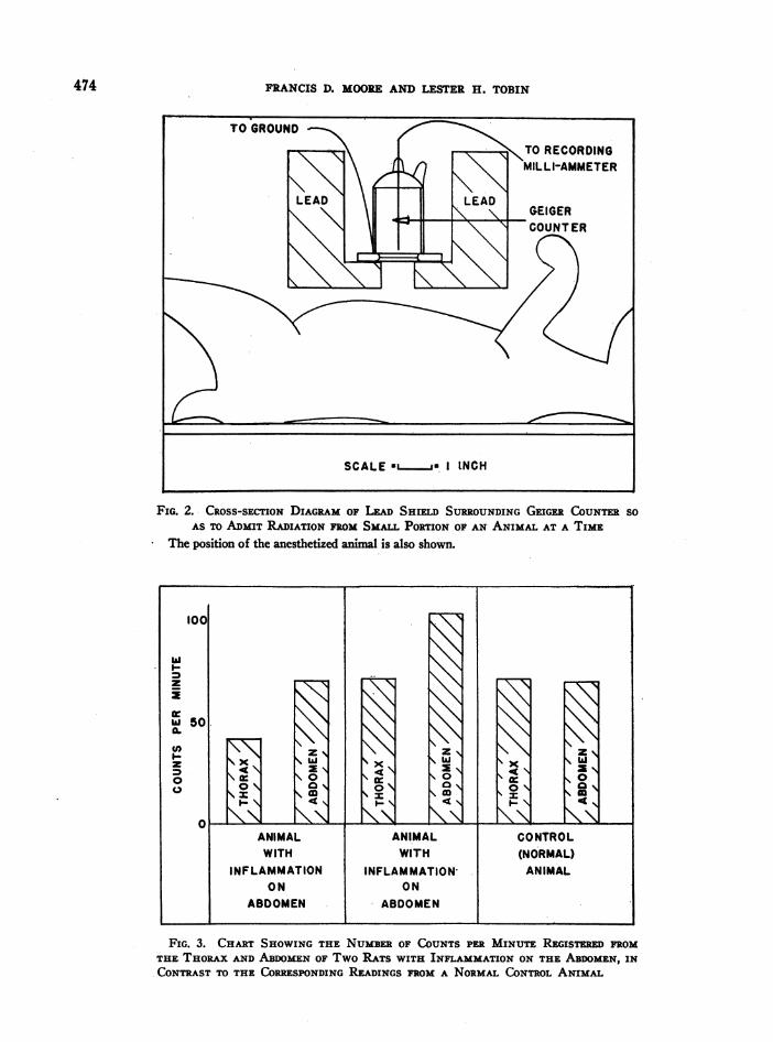
The detecting instrument is a Geiger counter, screenedwith lead so as to focus its registration on one portionof the animal at a time. This device and the amount oflead used are shown in Figure 2. The counter andcounting-rate meter are connected to a recording milli-ammeter, so that the number of counts per minute emanat-ing from any portion of an animal is integrated andrecorded. This counter has a background of counts fromcosmic rays and local radiation of other sources, of about50 counts per minute (C.P.M.). The background countis subtracted from all the readings taken from the animalto give the net C.P.M. for any specific area. With thedoses mentioned, readings from such an area on the bodyof the rabbit fall into the range of 500 to 2000 C.P.M.
The animal is anaesthetized so that it will lie quietly,and is then moved about under the counter to study theradioactivity in its various portions and in the abscess.The C.P.M. registered from the various points are en-tered on a standard diagram of the animal to make apermanent record. It can then be determined to whatextent the dye has accumulated in the abscess, and there-fore to what degree the dye accumulation indicates thelocation of the lesion.
RESULTS
1. In rats
The findings in the initial experiments on ratsare shown in Figure 3. The inflammatory proc-esses were on the abdomen, and roughly 50 percent more C.P.M. were registered from that regionthan from the thorax. The control animal, onthe other hand, showed fewer C.P.M. over theabdomen than over the thorax. This indicatedconcentration of the dye in the lesion to an ex-tent detectable with the counter, this finding beingcorrelated with the appearance of dye visually atthe site.
2. Distribution in normal rabbitsAn animal larger than the rat is desirable for
this work because it allows the making of a moredetailed study of the distribution of the radio-active dye in the intact animal. On the rabbit,10 or 12 readings may be made down the midlineof the abdomen with a counter of this type,
473
FRANCIS D. MOOREAND LESTER H. TOBIN
FIG. 2. CROSS-SECTION DIAGRAM OF LEAD SHIELD SURROUNDINGGEIGER COUNTERSOAS TO ADMIT RADIATION FROMSMALL PORTION OF AN ANIMAL AT A TIME
The position of the anesthetized animal is also shown.
INccNa
z Is
ax N
ANIMALWITH
INFLAMMATIONON
ABODOMEN
4Ni\0
'x\
7
KKaO0a
ANIMALWITH
INFLAMMATION-ON
.ABDOMEN
FIG. 3. CHARTSHOWINGTHE NUMBEROF COUNTSPER MINUTE REGISTRED FROMTHE THORAXANDABDOMENOF Two RATS WITH INFLAMMATIONON THE ABDOMEN,INCONTRASTTO THE CORRESPONDINGREADINGS FROMA NORMALCONTROLANIMAL
474
10c
w
z
iw
I-
z0
501.
xKx a '
4'
CONTROL(NORMAL)
ANIMAL

STUDIES WITH RADIOACTIVE DI-AZO DYES
FIG. 4. DiSTRIBUTION OF RADIOAcTIvE DI-BROM TRYPANBLUE IN A NORMALRABBIT
Figures are in counts per minute, as registered from the various points shown.
whereas in the rat there is room for only twoor three.
The distribution of radioactive di-brom trypanblue in the normal rabbit is shown in Figure 4.The bilateral symmetry of readings from variousregions of the body should be noted. This sym-metry is consistent within a variation of 10 to20 per cent. Readings from the two sides of thehead may vary to a greater degree than this be-cause it is difficult to place the head of theanaesthetized animal in precisely the same positionon the two sides. The same applies to the hindlegs, where variations up to 25 per cent betweennormal sides may be observed.
If the counter is first centered over the upperthorax, in the midline, and then slowly moveddown to the pelvis, taking readings each inch orso throughout its course, a set of readings areobtained which may be charted as shown in Fig-ure 5. This indicates that the maximal concen-tration of dye is found in the region of thexiphoid under which are found the liver, spleen,heart, lungs, and superior splanchnic circulation,all closely grouped and accounting for much ofthe blood volume of the animal. Insofar as the
dye stays in the blood stream, largely, for thefirst 6 hours after injection, it is to be expectedthat this area will contain most of the dye.Furthermore, as the dye leaves the circulation, itis taken up by the reticulo-endothelial system (5),and as this same region contains the liver andspleen, it will continue to contain much of thedye even after the dye leaves the blood stream.
It is to be noted in Figure 5 that as the counteris moved down the abdomen of the animal, awayfrom these viscera, the resultant chart is a straightline. That is, the decrease in C.P.M. is propor-tional to the distance from the xiphoid at whichthe count is taken.
These three points, then, bilateral symmetry,maximal counts over the xiphoid, and "linearityof decrease" of C.P.M. as the counter is movedtoward the pelvis, are the chief normal findings ofdistribution of the dye in the animal from 1 to 6hours after it is injected. The absolute valuesmay vary; the pitch or absolute height of theabdominal "line" may vary, but symmetry of dis-tribution, maximal counts over the xiphoid, and astraight line of decrease down the abdomen havebeen our findings in 53 observations on 15 control
475

FRANCIS D. MOOREAND LESTER H. TOBIN
FIG. 5. CHARTSHOWINGTHE READINGSTAKENFROMVARIOUS POINTS ON THE Mn-LINE OF A NORMALRABBIT AFTER THE INJECTION OF RADIOACTIVE DI-BROM TRYPANBLUE
The decline in readings as the counter is moved from the xiphoid to the pelvis approxi-mates a straight line.
animals. In this same group, the average differ-ence in C.P.M. between two normal sides was10.9 per cent, the mean variation was 22.25 percent, and the maximum (one instance) w-as 44.5per cent.
3. Results in rabbits with inflammatory lesionsOur first study was devoted to lesions well away
from the large blood-containing viscera in the up-per abdomen. These lesions were on the hind legswhere the accumulation of radioactivity due tothe lesion would be superimposed on a normallylow reading.
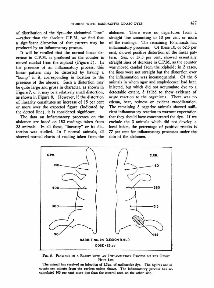
The findings in a rabbit with a hind-leg lesionare shown in Figure 6. The inflammatory lesionhas produced an increment of 105 per cent moreC.P.M. than registered from the contralateralnormal leg.
This indicates concentration of the radioactivedye in the lesion to an extent detectable with thecounter, and hence permitting diagnostic localiza-tion of the lesion by this technique.
This result can be produced with regularity.The average variation between normal sides, in acontrol group of 6 normal animals in which legreadings were taken, was 6.1 per cent. In an ex-perimental group of 9 animals with leg lesions theaverage increment produced by inflammation was91.0 per cent. There were no failures.
When the lesion is in the subcutaneous tissuesof the abdominal wall, a different problem pre-sents itself. The accumulation of radioactivityattendent upon the inflammatory process is super-imposed on the high concentration of radioactivitynormally found in the abdomen. Wecannot ex-pect a 90 per cent increment in C.P.M. due to aninflammatory process when that process is smalland localized near tissues which normally accumu-late a large proportion of the injected radioactivedye. In fact, the increase in C.P.M., due to aninflammatory process on the abdomen, may beonly in the range of 10 to 15 per cent over thenormal figure for that point on the animal.
However, if we turn our attention to the pattern
476
STUDIES WITH RADIOACTIVE DI-AZO DYES
of distribution of the dye-the abdominal "line"-rather than the absolute C.P.M., we find thata significant distortion of that pattern may beproduced by an inflammatory process.
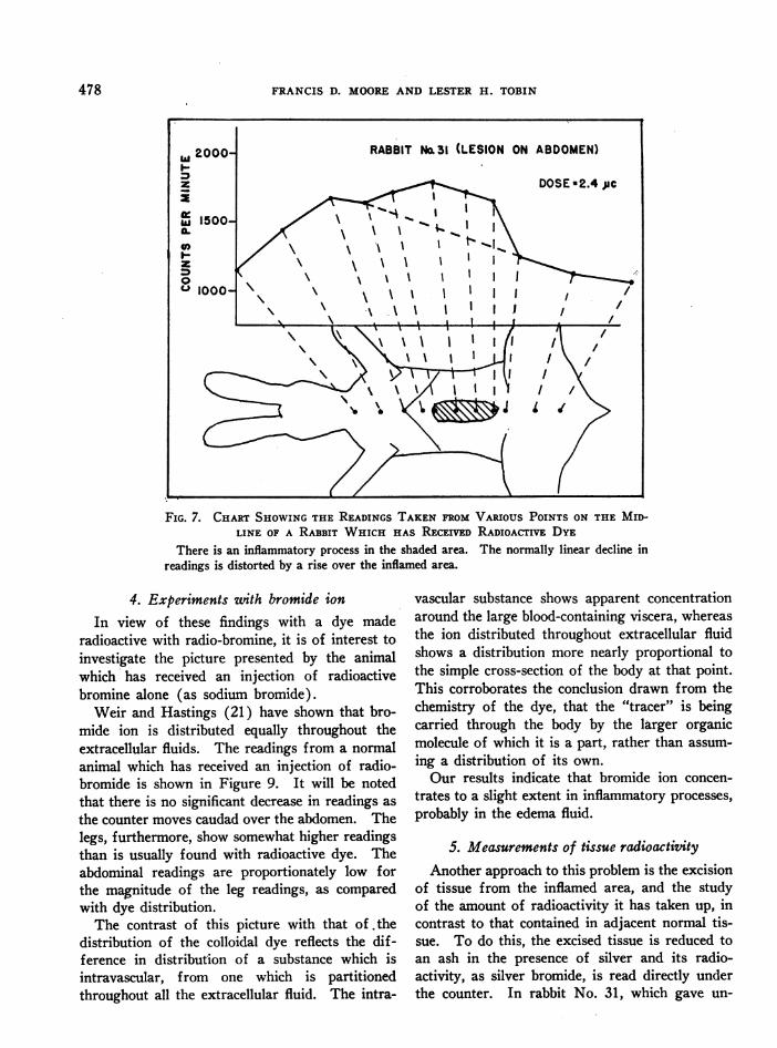
It will be recalled that the normal linear de-crease in C.P.M. is produced as the counter ismoved caudad from the xiphoid (Figure 5). Inthe presence of an inflammatory process, thislinear pattern may be distorted by having a"bump" in it, corresponding in location to thepresence of the abscess. Such a distortion maybe quite large and gross in character, as shown inFigure 7, or it may be a relatively small distortion,as shown in Figure 8. However, if the distortionof linearity constitutes an increase of 15 per centor more over the expected figure (indicated bythe dotted line), it is considered significant.
The data on inflammatory processes on theabdomen are based on 152 readings taken from23 animals. In all these, "linearity" or its dis-tortion was studied. In 7 normal animals, allshowed normal charts of reading taken from the
abdomen. There were no departures from astraight line amounting to 10 per cent or moreof the readings. The remaining 16 animals hadinflammatory processes. Of these 10, or 62.5 percent, showed positive distortion of the linear pat-tern. Six, or 37.5 per cent, showed essentiallystraight lines of decrease in C.P.M. as the counterwas moved caudad from the xiphoid; in 2 cases,the lines were not straight but the distortion overthe inflammation was inconsequential. Of the 6animals in whomagar and staphylococci had beeninjected, but which did not accumulate dye to adetectable extent, 3 failed to show evidence ofacute reaction to the organisms. There was noedema, heat, redness or evident vasodilatation.The remaining 3 negative animals showed suffi-cient inflammatory reaction to warrant expectationthat they should have concentrated the dye. If weexclude the 3 animals which did not develop alocal lesion, the percentage of positive results is77 per cent for inflammatory processes under theskin of the abdomen.
FIG. 6. FINDINGS IN A RABBIT WITH AN INFLAMMATORYPROCESSON THE RIGHTHIND LEG
The animal has received an injection of 1.3Uc. of radioactive dye. The figures are in
counts per minute from the various points shown. The inflammatory process has ac-
cumulated 105 per cent more dye than the control area on the other side.
RABBIT No. 24 (LESION RKL.)DOSE*1.3juc
477
FRANCIS D. MOOREAND LESTER H. TOBIN
FIG. 7. CHARTSHOWINGTHE READINGS TAKEN FROMVARIOUS POINTS ON THE MID-LINE OF A RABBIT WHICH HAS RECEIVED RADIOACTIVE DYE
There is an inflammatory process in the shaded area. The normally linear decline inreadings is distorted by a rise over the inflamed area.
4. Experiments wtith bromide ionIn view of these findings with a dye made
radioactive with radio-bromine, it is of interest toinvestigate the picture presented by the animalwhich has received an injection of radioactivebromine alone (as sodium bromide).
Weir and Hastings (21) have shown that bro-mide ion is distributed equally throughout theextracellular fluids. The readings from a normalanimal which has received an injection of radio-bromide is shown in Figure 9. It will be notedthat there is no significant decrease in readings asthe counter moves caudad over the abdomen. Thelegs, furthermore, show somewhat higher readingsthan is usually found with radioactive dye. Theabdominal readings are proportionately low forthe magnitude of the leg readings, as comparedwith dye distribution.
The contrast of this picture with that of.thedistribution of the colloidal dye reflects the dif-ference in distribution of a substance which isintravascular, from one which is partitionedthroughout all the extracellular fluid. The intra-
vascular substance shows apparent concentrationaround the large blood-containing viscera, whereasthe ion distributed throughout extracellular fluidshows a distribution more nearly proportional tothe simple cross-section of the body at that point.This corroborates the conclusion drawn from thechemistry of the dye, that the "tracer" is beingcarried through the body by the larger organicmolecule of which it is a part, rather than assum-ing a distribution of its own.
Our results indicate that bromide ion concen-trates to a slight extent in inflammatory processes,probably in the edema fluid.
5. Measurements of tissue radioactivityAnother approach to this problem is the excision
of tissue from the inflamed area, and the studyof the amount of radioactivity it has taken up, incontrast to that contained in adjacent normal tis-sue. To do this, the excised tissue is reduced toan ash in the presence of silver and its radio-activity, as silver bromide, is read directly underthe counter. In rabbit No. 31, which gave un-
478

STUDIES WITH RADIOACTIVE DI-AZO DYES
FIG. 8. CHARTSHOWINGTHE READINGS TAKEN FROMVARIOUS POINTS ON THE MID-LINE OF A RABBIT WHICHHAS RECEIVED RADIOACTrE DYE
There is an inflammatory process in the shaded area. The normally linear decline inreadings has been distorted by the inflammation but to a lesser extent than in the animalshown in Figure 7.
mistakable evidence of the presence of its abscessby the accumulation of radioactive dye (Figure7), approximately 110 per cent more radioactivityper gram of tissue was found in the area sur-rounding the inflammatory process than in theneighboring normal tissue. In another animal, inwhich the original counter readings gave littleevidence as to the whereabouts of the abscess, dis-section revealed a very quiescent process, and only40 per cent more radioactive dye per gram oftissue was found in the tissues around the agarnidus, than in adjacent normal tissue. Evidentlythe animal's resistance to the organism was suchas to preclude the appearance of an abscess at thesite of injection, and thus to prevent the accumu-lation there of enough dye to increase the counterreading.
6. DiscussionRadioactive di-brom trypan blue gives us a
means of diagnosing the location of abscesses onthe legs or in the subcutaneous tissues of the
abdominal wall of the rabbit. The leg lesionspresent a fairly simple problem, and so long asthere is any inflammation there, enough dye willaccumulate to make the lesion evident on thecounter. The abdominal lesions, on the otherhand, present a less favorable situation, and theresults are less gratifying, since in only 77 percent of the animals could a positive correlationbe made of the presence of a lesion and localiza-tion of a detectable amount of dye. In the caseof the abdominal lesions, the inflammatory processmust be quite intense in order to accumulateenough dye to be demonstrable from outside thebody.
The failure of some of the animals to concen-trate a detectable amount of dye in these lesions isattributable to three factors:
(1) There is a normal variation in the abilityof animals to concentrate the dye in inflammatorylesions, as noted by previous workers with dye(9a).
479

FRANCIS D. MOOREAND LESTER H. TOBIN
FIG. 9. CHARTSHOWINGTHE READINGS. TAKENFROMVARIOUS POINTS ON THE MID-LINE AND LEGS OF A RABBIT WHICH HAS RECEIVED AN INJECTION OF RADIOACTIVEBROMINEAS SODIUM BROMIDE
There is only a slight decrease in readings as the counter moves down the abdomen.The leg readings are relatively higher than those found using radioactive dye.
(2) Variations in virulence of the organism orresistance of the animal may lead to such a mildprocess that the dye is not accumulated.
(3) This particular dye does not seem to con-centrate as consistently in inflammatory lesions asdoes its non-brominated counterpart, trypan blue.
The first two of these factors are importantwhenever one attempts to simulate human lesionswith staphylococcic abscesses. Virulence and re-sistance cannot be closely enough controlled toproduce unfailing abscess-formation. Propertiesof the dye may also be an important factor in itsfailure to accumulate in lesions on some of theanimals. As previously pointed out, the molecularweight has been increased, the dye is less solublein water, and some particulate dye may be filteredout in lungs or liver and thus lost to the inflamma-tory area. Shortcomings such as these, inherentin the dye itself, can only be overcome by furtherstudy of this dye or of other similar dyes such as
its isomer, radioactive di-brom Evans blue. Suchstudy is in progress at present.
SUMMARY
(1) A study of the distribution of radioactivedi-brom trypan blue in normal and inflamed ani-mals is described.
(2) This radioactive colloidal dye concentratesin inflammatory lesions to an extent detectablefrom outside the intact animal with a suitablecounter.
(3) Using this radioactive dye, lesions in theperiphery of the body were detectable in all cases,whereas abdominal lesions were detectable in 77per cent of instances.
The authors wish to express their gratitude to Dr.Joseph C. Aub, Dr. Waldo E. Cohn and Dr. Austin M.Brues for their continued interest and valuable advice,and to Prof. Louis F. Fieser for his assistance in regardto the chemistry involved.
480-

STUDIES WITH RADIOACTIVE DI-AZO DYES
BIBLIOGRAPHYi. Goldmann, E. E., Die Aussere und Innere Sektretion
des Gesunden und Kranken Organismus im Lichteder "vitale Farbung." Beitr. z. Klin. Chir., 1909,64, 192. Quoted by Burrows (11).
2. Bowman, F. B., Winternitz, M. C., and Evans, H. M.,Ueber die vitale Farbung des Tuberkels. Centralb.f. Bakt., 1912, 65, 403.
3. Kline, R. S., and Winternitz, M. C., Studies uponexperimental pneumonia, in rabbits. VIII. Intra-vitam staining in experimental pneumonia and thecirculation in the pneumonic lung. J. Exper. Med.,1915, 21, 311.
4. MacCurdy, J. T., and Evans, H. M., ExperimentelleLasionen des Centralnervensystems, untersucht mitHilfe der vitalen Farbung. Berl. Klin. Wchnschr.,1912, 49, 1695.
5. Evans, H. M., The macrophages of mammals. Am.J. Physiol., 1915, 37, 243.
6. Evans, H. M., and Schulemann, W., The action ofvital stains belonging to the benzidine group. Sci-ence, 1914, 39, 443.
7. Duran-Reynals, F., A general permeability-increasingeffect of a factor from mammalian testicle onblood capillaries. Yale J. Biol. and Med., 1939,11, 601.
8. Menkin, V., Studies on inflammation. I. Fixation ofvital dyes in inflamed areas. J. Exper. Med., 1929,50, 171.
9. Menkin, V., The Dynamics of Inflammation. Mac-millan, New York, 1940.
9a. Menkin, V., Personal communication.10. Rigdon, R. H., Capillary permeability in the skin of
the rabbit. Proc. Soc. Exper. Biol. and Med.,1939, 42, 43.
11. Burrows, H., Some Factors in the Localization ofDisease in the Body. Wm. Wood and Co., NewYork, 1932.
12. Keith, N. M., Rowntree, L. G., and Geraghty, J. T.,A method for the determination of plasma andblood volume. Arch. Int. Med., 1915, 16, 547.
13. Gibson, J. G., Jr., Personal communication.14. Dawson, A. B., Evans, H. M., and Whipple, G. H.,
Blood volume studies. III. The behaviour of alarge series of dyes introduced into the circulatingblood. Am. J. Physiol., 1920, 51, 232.
15. Gregerson, M. I., Gibson, J. G., and Stead, E. A.,Plasma volume determinations with dyes; errorsin colorimetry; use of the blue dye T-1824. Am.J. Physiol., 1935, 113, 54.
16. Strauss, S. F., Neuwelt, F., Rovner, L., and Necheles,H., A new method for the detection of hiddenabscesses. Surgery, 1938, 4, 930.
17. Kroll, H. H., Strauss, S. F., and Necheles, H., Con-centration and detection of a dye in abscesses.Proc. Soc. Exper. Biol. and Med., 1940, 43, 228.
18. Kroll, H. H., Strauss, S. F., and Necheles, H.,Studies on the detection of abscesses and tumors.III. Concentration and detection of a radioactivesubstance in abscesses. J. Lab. and Clin. Med.,1941, 27, 50.
19. Seaborg, G. T., Artificial radioactivity. Chem. Rev.,1940, 27, 199.
20. Tobin, L. H., and Moore, F. D., Studies with radio-active di-azo dyes. III. The synthesis and proper-ties of radioactive di-brom trypan blue and radio-active di-brom Evans blue. In preparation.
21. Weir, E. G., and Hastings, A. B., The distribution ofbromide and chloride in tissues and body fluids.J. Biol. Chem., 1939, 129, 547.
481