Embed Size (px)
Citation preview
2017 Neutrophil Formylpeptide Receptor Single Nucleotide Polymorphism in Localized
and Generalized Aggressive Periodontitis
Porras Laura*, Pooja Maney†, Archontia Palaiologou ‡ Department of Periodontics Louisiana State University Health Science Center Center of Excellence in Oral and Craniofacial Biology School of Dentistry 1100 Florida Avenue New Orleans, LA 70119 Keywords: Aggressive Periodontitis, polymorphisms, Genetics, SNPs, FPR1, African American Abbreviations: Aggressive Periodontitis (AP), Aggressive Periodontitis Localized (LAP), Aggressive Periodontitis Generalized (GAP), Aggregatibacter actinomycetemcomintans (A.a) , Polymorphonuclear cells (PMNs) , Formylpeptide receptors (FPRs) , Single nucleotide polymorphisms (SNPs), Polymerase chain reaction (PCR) Abstract Background: Aggressive periodontitis (AP) is a destructive disease that affects around 2-10% of the population. There are two main sub-classifications of AP: Localized (LAP) and Generalized (GAP). However, very little is known about the etiologic differences between these two entities. Single nucleotide polymorphisms (SNPs) have been associated with AP in key genes. The objective of this study is to compare FPR1 SNPs between LAP and GAP in African Americans.
Methods: Blood samples were obtained from African American subjects in New Orleans (LAP n= 27, GAP n=17, and healthy controls n=20), and genomic DNA was isolated. Polymerase chain reaction (PCR) and sequencing (for analysis of SNPs) was performed on a highly polymorphic fragment of the FPR1 gene.
Results: No difference was found between subjects with LAP or GAP for 301 G>C, 348
* Department of Periodontics † Department of Periodontics ‡ Department of Periodontics
T>C, 546 C>A, 568 A>T, 576 T>C>G allele association. However, 546 C>A, 576 T>C>G FPR1 SNPs were associated with AP when compared to healthy controls. Specifically, 546 C and 576 G alleles were more frequently associated with GAP, and the 546 C allele was more frequently associated with LAP. None of the other three SNPs was detected, indicating no association with AP.
Conclusions: FPR1 SNP 546 C>A, 576 T>C>G FPR1 SNPs were associated with AP in African Americans. Individuals who are homozygous (?) for 546C and 576 T may have increased susceptibility to developing this condition.
Introduction
Aggressive periodontitis (AP) is a disease that is characterized by rapid
attachment loss and bone destruction in otherwise clinically healthy patients. In the
United States, AP is most prevalent in African Americans, affecting around 2-10% of the
population (Melvin, 1991) (Albander 1997). It primarily affects teenagers and young
adults below 35 years of age (Albandar, 2002). Tooth loss due to this disease can have
debilitating consequences for the future formation and function of the dentition.
Aggressive periodontitis can be sub-classified into localized (LAP) and
generalized (GAP). LAP appears to affect the first molar/incisor regions with
interproximal attachment loss on at least 2 permanent teeth, one of which is a first
molar, with involvement of no more than 2 teeth other than the first molars and incisors.
GAP patients have at least 3 permanent teeth affected by interproximal attachment loss
other than the first molars and incisors (Lang, 1999). Brown et al. found that 35% of
patients who initially were diagnosed with LAP progressed to GAP 6 years later.
Even though there is an established classification of these two entities based on
the different clinical presentations, to date it is still unclear if these two conditions have
different etiologies or contributing factors to explain their different presentations.
A classic finding of these diseases is the presence of a highly pathogenic biofilm,
such as Aggregatibacter actinomycetemcomintans (A.a) (Califano, 2003). A.
actinomycetemcomintans (A.a) serotype b strains have been more highly associated
with AP than A.a serotype a or c (Ebersole, 1991). Wilson et al. reported that a humoral
response to A.a includes the production of LPS-reactive IgG2 antibodies in patients with
LAP. Based on these studies, some have correlated the increased level IgG2 as a key
component to differentiate between LAP and GAP, and explained why LAP is limited to
affect first molar and incisors. However, others have mentioned that these IgG2
antibodies have poor opsonizing capability against this specific antigen and may limit
the humeral response from the host against A.a (Wilson, 1992). Very little is reported
regarding the increased or decreased presence of IgG2 in GAP to support or contradict
this theory. Overall AP is a complex multifactorial disease involving a complex interplay
between the microbial challenge and host response.
Polymorphonuclear cells (PMNs) are a vital component of this host response.
They are the first line of defense against bacterial infections. These cells rely upon high
affinity formylpeptide receptors (FPRs) found on their surface to help locate and
neutralize bacteria. Defects in these receptors can render the host more susceptible to
infection. As an example, mice devoid of FPRs exhibit significantly reduced resistance
to Listeria monocytogenes (Gao, 1999).
The most frequent PMN anomaly reported in AP has been altered fMLP-
mediated chemotaxis in African-American patients with the localized form of disease
(Perez, 1991). The etiology of this altered chemotaxis is still unknown, although both
innate genetic and acquired anomalies have been proposed. (Agarwal 1994) One
possibility suggests the inability of the PMN to detect FMLP caused by a receptor defect
(Van Dyke, 1891). In patients with AP, PMNs had a diminished number of high affinity
FPRs (Perez, 1991). An alternate explanation would suggest an intrinsic cellular defect,
causing a decrease in the number of FPR on the neutrophils surface. (Van Dyke, 1981)
AP tends to run in families, and its etiology is associated with a genetic
component that could modify host response. Single nucleotide Polymorphisms (SNPs)
are common genetic variations that occur when a single nucleotide in a specific position
of the genome is changed for another. Several studies have reported SNPs that have
been associated with AP in key genes (Vieira, 2014). Formyl peptide receptor is
encoded by the FPR1 gene, which is located on Chromosome 19.
The FPR1 gene is a highly polymorphic gene. Sahagun-Ruiz et al. (Sahagun-
Ruiz, 2001) systematically analyzed polymorphisms in the FPR1 open reading frame by
direct sequencing of cloned alleles from random North American blood donors. They
detected five common non-synonymous SNPs (c.32C>T, c.301G>C, c.568A>T,
c.576T>C>G and c.1037C>A) as well as two synonymous (silent) SNPs (c.348T>C and
c.546C>A) that do not encode an amino acid substitution. Previous reports have
suggested that AP is associated with specific patterns of single nucleotide
polymorphisms (SNPs) of the FPR1 gene, including SNPs c.568A>T and c.576T>C>G
in African-Americans (Gwinn, 1999) (Zhang, 2003). Exploratory research has been
conducted to investigate the relationship between FPR polymorphisms, defective FPR
expression, PMN chemotactic defects and susceptibility to AP. These studies of an
African-American population suggest that AP is associated with the silent SNP
c.348T>C in the gene encoding the FPR (FPR1) as well as defective PMN chemotaxis
(Maney, 2009). No study has directly analyzed the frequency of SNP’s between LAP
and GAP to determine if this may represent a difference in etiology, clinical presentation
and/ or progression of the disease.
The aim of this study is to compare FPR1 SNPs between LAP and GAP in an
African American population, as well as to healthy controls.
Materials and Methods
Subject recruitment
African American subjects (n=24), aged 13-35years old, diagnosed with AP at
LSUHSC School of Dentistry were recruited between January 2015 and February 2017
following an approved LSUNO-IRB protocol # 8796. Patients were diagnosed
according to the criteria of the 1999 International Workshop for a Classification of
Periodontal Diseases and Conditions (Armitage, 1999). Additionally, unpublished data
from a previous study of African American subjects diagnosed with AP (n=20) and
matched healthy controls (n=20) were included in this study. These patients were
previously recruited between January 2010 - January 2011. Written informed consent
for blood sampling and DNA analysis was obtained from each subject. Patients were
excluded if they had a history of prophylaxis within the last 3 months, any chronic
medical condition, history of recent antibiotic therapy, or history of smoking.
Clinical examination
Demographic information including birth date, sex, race/ethnicity, tobacco
exposure, and contact information was collected. All subjects received a full mouth
periodontal and radiographic examination. Periodontal examination consisted of probing
depth and attachment level measurements on 6 sites per tooth using a periodontal
probe§. Bleeding on probing and plaque levels were also recorded. These
measurements will be collected and be available for data exploration, but specific aims
requiring these measurements are not planned.
Patients were classified as LAP when they presented with 4mm or more of
attachment loss in at least two permanent first molars and incisors, including at least
one first molar, but not more than two permanent teeth other than the first molars and
incisors. GAP was diagnosed when at least three teeth other than first molars and
incisors had an attachment loss of 4 mm or more. The control subjects (n = 20) were
periodontally healthy. (Armitage 1999) (Tonetti 1999).
§ PCPUNC156, Hu-Friedy, Chicago.
Blood drawn and DNA isolation
Peripheral venous blood samples were drawn from each subject (approximately
5cc’s). Genomic DNA was purified from the whole blood samples using a blood DNA
purification kit according to the manufacturer’s instructions**
PCR and Sequencing of the FPR1 Coding Region
To detect FPR1 SNP, PCR amplification of a 439–base pair (bp) fragment of
FPR1 was performed as previously described (Maney, 2009). The forward and reverse
primers used were 5’TTCACCTCCACTTTGCCATT 3’ and
3’TGACAGCAACGATGGACATG5’, respectively. For every sample, PCR was carried
out for 35 cycles in a mixture of 45 µl PCR Supermix (Invitrogen), 1 µl of each of the two
primers, 1.5 µL of the DNA sample and 1.5 µl of water. Prior to initiating PCR, the
reaction mixture was heated at 95ºC for one minute to dissociate antibody from the Taq
polymerase. The parameters for one cycle of the PCR reaction include denaturation at
95ºC for one minute, annealing at 60ºC for one minute, and extension at 72ºC for one
minute. PCR products were then purified using a commercially available kit††.
** QIAamp DNA Blood Mini Kit, Qiagen, Valencia, CA. †† QIAquick PCR Purification Kit, Qiagen.
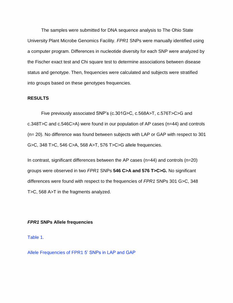
The samples were submitted for DNA sequence analysis to The Ohio State
University Plant Microbe Genomics Facility. FPR1 SNPs were manually identified using
a computer program. Differences in nucleotide diversity for each SNP were analyzed by
the Fischer exact test and Chi square test to determine associations between disease
status and genotype. Then, frequencies were calculated and subjects were stratified
into groups based on these genotypes frequencies.
RESULTS
Five previously associated SNP’s (c.301G>C, c.568A>T, c.576T>C>G and
c.348T>C and c.546C>A) were found in our population of AP cases (n=44) and controls
(n= 20). No difference was found between subjects with LAP or GAP with respect to 301
G>C, 348 T>C, 546 C>A, 568 A>T, 576 T>C>G allele frequencies.
In contrast, significant differences between the AP cases (n=44) and controls (n=20)
groups were observed in two FPR1 SNPs 546 C>A and 576 T>C>G. No significant
differences were found with respect to the frequencies of FPR1 SNPs 301 G>C, 348
T>C, 568 A>T in the fragments analyzed.
FPR1 SNPs Allele frequencies
Table 1.
Allele Frequencies of FPR1 5’ SNPs in LAP and GAP
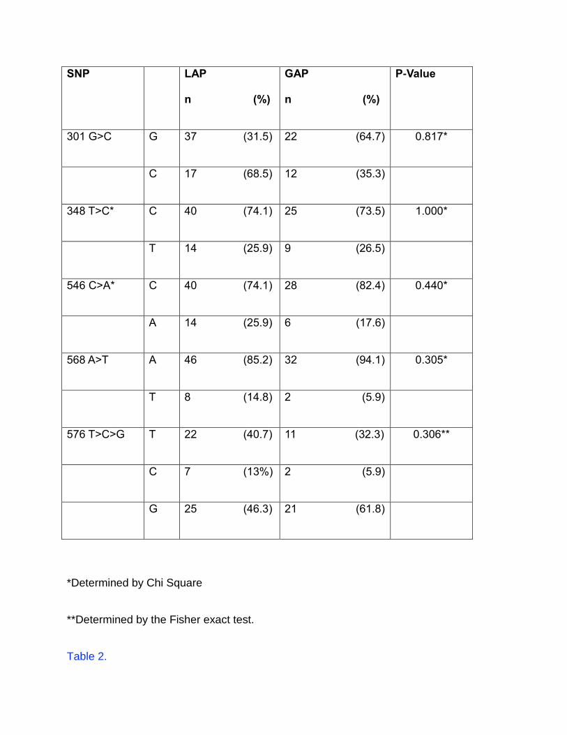
SNP LAP
n (%)
GAP
n (%)
P-Value
301 G>C G 37 (31.5) 22 (64.7) 0.817*
C 17 (68.5) 12 (35.3)
348 T>C* C 40 (74.1) 25 (73.5) 1.000*
T 14 (25.9) 9 (26.5)
546 C>A* C 40 (74.1) 28 (82.4) 0.440*
A 14 (25.9) 6 (17.6)
568 A>T A 46 (85.2) 32 (94.1) 0.305*
T 8 (14.8) 2 (5.9)
576 T>C>G T 22 (40.7) 11 (32.3) 0.306**
C 7 (13%) 2 (5.9)
G 25 (46.3) 21 (61.8)
*Determined by Chi Square
**Determined by the Fisher exact test.
Table 2.
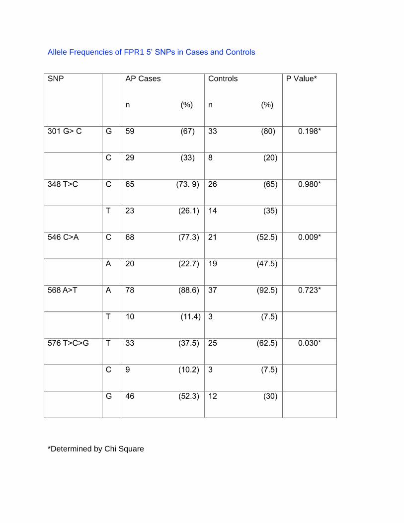
Allele Frequencies of FPR1 5’ SNPs in Cases and Controls
SNP AP Cases
n (%)
Controls
n (%)
P Value*
301 G> C G 59 (67) 33 (80) 0.198*
C 29 (33) 8 (20)
348 T>C C 65 (73. 9) 26 (65) 0.980*
T 23 (26.1) 14 (35)
546 C>A C 68 (77.3) 21 (52.5) 0.009*
A 20 (22.7) 19 (47.5)
568 A>T A 78 (88.6) 37 (92.5) 0.723*
T 10 (11.4) 3 (7.5)
576 T>C>G T 33 (37.5) 25 (62.5) 0.030*
C 9 (10.2) 3 (7.5)
G 46 (52.3) 12 (30)
*Determined by Chi Square
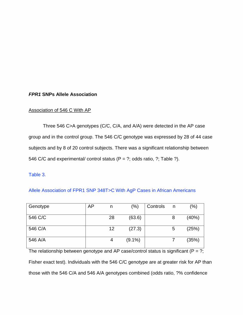
FPR1 SNPs Allele Association
Association of 546 C With AP
Three 546 C>A genotypes (C/C, C/A, and A/A) were detected in the AP case
group and in the control group. The 546 C/C genotype was expressed by 28 of 44 case
subjects and by 8 of 20 control subjects. There was a significant relationship between
546 C/C and experimental/ control status (P = ?; odds ratio, ?; Table ?).
Table 3.
Allele Association of FPR1 SNP 348T>C With AgP Cases in African Americans
Genotype AP n (%) Controls n (%)
546 C/C 28 (63.6) 8 (40%)
546 C/A 12 (27.3) 5 (25%)
546 A/A 4 (9.1%) 7 (35%)
The relationship between genotype and AP case/control status is significant (P = ?;
Fisher exact test). Individuals with the 546 C/C genotype are at greater risk for AP than
those with the 546 C/A and 546 A/A genotypes combined (odds ratio, ?% confidence
interval: ?).
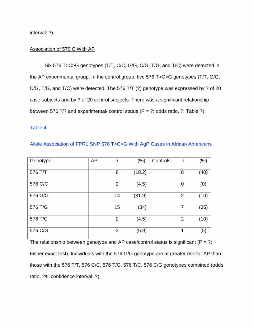
Association of 576 C With AP
Six 576 T>C>G genotypes (T/T, C/C, G/G, C/G, T/G, and T/C) were detected in
the AP experimental group. In the control group, five 576 T>C>G genotypes (T/T, G/G,
C/G, T/G, and T/C) were detected. The 576 T/T (?) genotype was expressed by ? of 20
case subjects and by ? of 20 control subjects. There was a significant relationship
between 576 ?/? and experimental/ control status (P = ?; odds ratio, ?; Table ?).
Table 4.
Allele Association of FPR1 SNP 576 T>C>G With AgP Cases in African Americans
Genotype AP n (%) Controls n (%)
576 T/T 8 (18.2) 8 (40)
576 C/C 2 (4.5) 0 (0)
576 G/G 14 (31.9) 2 (10)
576 T/G 15 (34) 7 (35)
576 T/C 2 (4.5) 2 (10)
576 C/G 3 (6.9) 1 (5)
The relationship between genotype and AP case/control status is significant (P = ?;
Fisher exact test). Individuals with the 576 G/G genotype are at greater risk for AP than
those with the 576 T/T, 576 C/C, 576 T/G, 576 T/C, 576 C/G genotypes combined (odds
ratio, ?% confidence interval: ?).
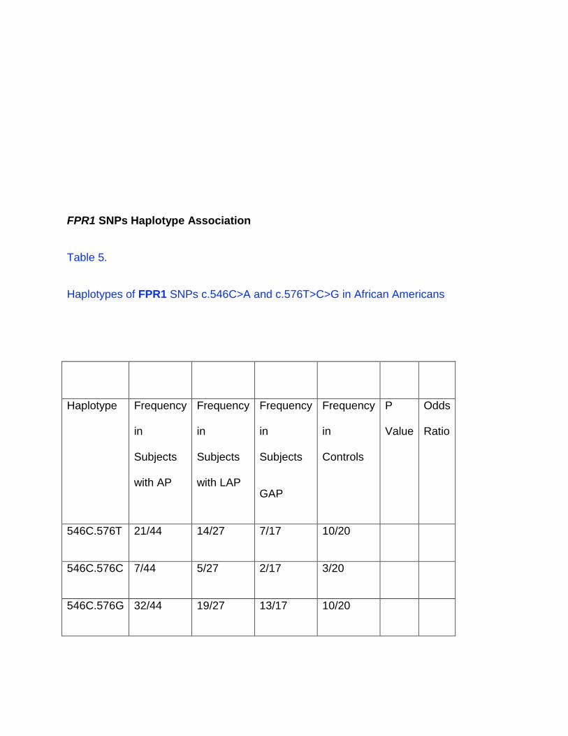
FPR1 SNPs Haplotype Association
Table 5.
Haplotypes of FPR1 SNPs c.546C>A and c.576T>C>G in African Americans
Haplotype Frequency
in
Subjects
with AP
Frequency
in
Subjects
with LAP
Frequency
in
Subjects
GAP
Frequency
in
Controls
P
Value
Odds
Ratio
546C.576T 21/44 14/27 7/17 10/20
546C.576C 7/44 5/27 2/17 3/20
546C.576G 32/44 19/27 13/17 10/20
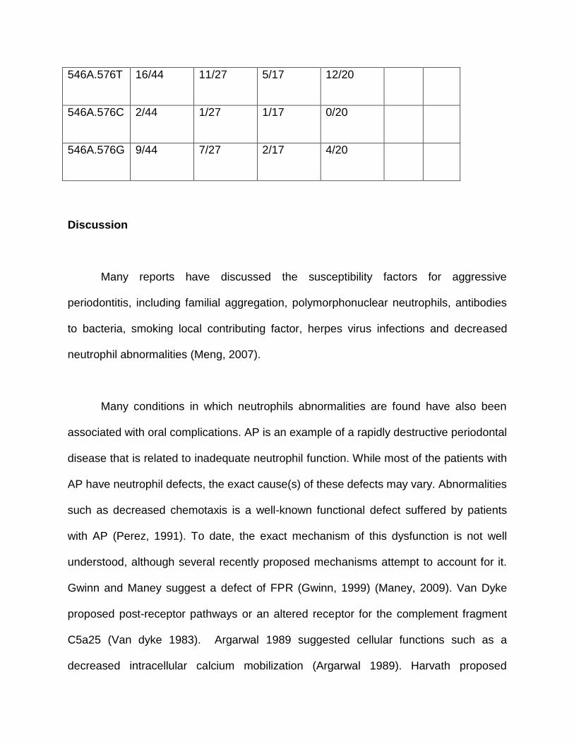
546A.576T 16/44 11/27 5/17 12/20
546A.576C 2/44 1/27 1/17 0/20
546A.576G 9/44 7/27 2/17 4/20
Discussion
Many reports have discussed the susceptibility factors for aggressive
periodontitis, including familial aggregation, polymorphonuclear neutrophils, antibodies
to bacteria, smoking local contributing factor, herpes virus infections and decreased
neutrophil abnormalities (Meng, 2007).
Many conditions in which neutrophils abnormalities are found have also been
associated with oral complications. AP is an example of a rapidly destructive periodontal
disease that is related to inadequate neutrophil function. While most of the patients with
AP have neutrophil defects, the exact cause(s) of these defects may vary. Abnormalities
such as decreased chemotaxis is a well-known functional defect suffered by patients
with AP (Perez, 1991). To date, the exact mechanism of this dysfunction is not well
understood, although several recently proposed mechanisms attempt to account for it.
Gwinn and Maney suggest a defect of FPR (Gwinn, 1999) (Maney, 2009). Van Dyke
proposed post-receptor pathways or an altered receptor for the complement fragment
C5a25 (Van dyke 1983). Argarwal 1989 suggested cellular functions such as a
decreased intracellular calcium mobilization (Argarwal 1989). Harvath proposed
elevation of TNF-α and IL-1 cytokines in the serum and presence of serum inhibitors of
chemotaxis (Harvath, 1982).
Specifically, there are two main sub-classifications for AP, LAP and GAP.
However, it remains unclear whether different etiologies or contributing factors explain
the different clinical presentations of these two subgroups.
Our study is mainly focused in detecting any significant frequency alteration in
FPR1 SNPs between LAP and GAP. Studying the human genome to determine DNA
variants as possible explanations to human diseases is well established; however, using
genetic factors for multifactorial conditions presents a major challenge due the fact that
both genetic and environment interact. The presence of a mutation or polymorphism in
a single gene may not be associated with a specific disease, but when this one is
combined with some variation in other genes, such changes may contribute to a
disease genotype. (Shafer 1998)
Specific genotypes associated with SNPs in genes encoding proteins that play
key roles in the pathogenesis of AP, have been reported to be associated with the
increased odds of detecting A actinomycetemcomitans, Tannerella forsythia, and
Porphyromonas gingivalis (Nibali, 2007).
Consistent with our hypothesis, subjects with the LAP and GAP showed no
difference with respect to 301 G>C, 348 T>C, 546 C>A\, 568 A>T, 576 T>C>G allele
associations. However, the present study is also consistent with previous studies’
observations that FPR1 SNP 576 T>C>G is associated with AP in African Americans.
Subjects with the 576 (?) genotype seem to have a significantly increased risk for
developing AP compared to those with the 576 ?/? or 576?/? genotype (odds ratio,
?.9?). These SNPs are localized to the second extracellular loop of FPR1 and are non-
synonymous, meaning they will cause a change in the amino acid sequence and,
therefore, the protein formed.
The above is consistent with findings of a previous study (Zhang, 2003) in which
SNPs 576 T>C> G were reported to cause a change in Asparagine to Lysine, altering
the amide characteristic from hydrophilic to basic. This amino acid variation would
affect ligand binding specificity and/or binding affinity that occurs in the extracellular
domain (Quehenberger, 1993). Zhang et al. concluded that this SNP is accordingly
significantly associated with AP phenotype in African Americans (Zhang, 2003)
The present study also identified synonymous SNP 546 c genotypes in the
diseased cohort compared to the control group. Subjects with the 546 (?) genotype
seem to have a significantly increased risk for developing AP compared to those with
the 546 ?/? or 546?/? genotype (odds ratio, ?.?).
Although 546 C is a synonymous SNP that does not alter the amino acid, it is
reasonable to hypothesize that it could be associated with other SNPs in the FPR1
promoter or enhancer regions. Some association of this same kind has been made in
other studies where they have a concluded multiple SNPs within specific haplotypes
containing variants of synonymous coding region SNPs and non-synonymous promoter
SNPs could affect the biologic phenotype (Drysdale, 2000) (Mummidi, 2000). Zhang et
al. did not report any associations of SNP 546 C>A with AP as our study shows. This
discrepancy may be related to the DNA diversity in the specific study populations.
Another possible explanation could be differences in the selection criteria of patients
with aggressive periodontitis. Zhang et al. studied a more heterogeneous population
that included African American, Brazilian and Turkish subjects, compared to our
population which is a more homogenous population of African Americans localized in
the New Orleans area.
Healthy controls were also analyzed to determine if these possible AP specific
alterations are also present in otherwise healthy matched controls. Healthy controls
showed no statistically significant alteration, suggesting that these proposed alterations
may be specific to AP.
Within the limitations of this study, it is possible to state that the difference in the
clinical presentations of LAP and GAP cannot be explained by an increase in the
frequency of the SNPs we studied, since our results failed to find a difference between
FPR1 polymorphisms in patients with these two entities. However, as shown in previous
studies, we found significant associations with SNP 546 and 576. It is possible that
these variations in the gene play a role in or may be a risk indicator for the disease.
Statistical analysis showed an association between these alterations and the presence
of the condition (P= 0.005)
In the long term, a better understanding of the etiology and pathogenesis of
aggressive periodontitis could help develop laboratory tests for individuals at risk for AP.
Targeted therapeutic intervention to eliminate pathogens prior to clinical expression of
AP could mitigate periodontal destruction, which may improve prognosis, reduce tooth
loss produced by AP, minimize functional and esthetic problems related to tooth loss
and reduce the overall cost of dental care.
Conclusion
No difference was found between subjects with LAP or GAP with respect to 301
G>C, 348 T>C, 546 C>A, 568 A>T, 576 T>C>G allele association. However, SNPs 576
G and 546 C allele were more frequently associated with GAP when compared to
controls. SNP546 C allele was more frequently associated with LAP. Considering the
small population of this study, our findings should be confirmed by large scale,
methodologically sophisticated studies.
References
1. Albandar JM, Tinoco EMB. Global epidemiology of periodontal diseases in chidren and
young persons. Periodontology 2000. 2002;29:153–76.
2. Agarwal S, Suzuki JB, Riccelli AE. Role of cytokines in the modulation of neutrophil
chemotaxis in localiz *
3. Agarwal S,Reynolds MA, Duckett LD, Suzuki JB. Altered free cytosolic calcium changes
and neutrophil chemotaxis in patients with juvenile periodontitis. J Periodontal
Res. 1989 Mar;24(2):149-54. *
4. Armitage GC. Development of a classification system for periodontal diseases and
conditions. Ann Peri- odontol 1999;4:1-6. *
5. Ebersole, J. Serum antibody in Actinobacillus Actinomycetencomitans infected patients
with periodontal disease. 1991*
6. Califano JV, Research, Science and Therapy Committee, American Academy of
Periodontology. Position paper: periodontal diseases of children and adolescents.
journal of periodontology. 2003. pp. 1696–704.
7. Christersson LA, Albini B, Zambon JJ, Wikesjö UM, Genco RJ. Tissue localization of
Actinobacillus actinomycetemcomitans in human periodontitis. I. Light,
immunofluorescence and electron microscopic studies. journal of periodontology. 1987
Aug;58(8):529–39.
8. Drysdale CM, McGraw DW, Stack CB, et al. Complex promoter and coding region b2-
adrenergic receptor haplotypes alter receptor expression and predict in vivo
responsiveness. Proc Natl Acad Sci USA 2000; 97:10483-10488.
9. Gao, J. L., Lee, E. J. & Murphy, P. M. (1999) Impaired antibacterial host defense in
mice lacking the N-formylpeptide receptor. The Journal of experimental medicine 189,
657-662.\
10. Genco RJ, Christersson LA, Zambon JJ. Juvenile periodontitis. Int Dent J 1986; 36:
168–176. *
11. Gwinn, M. R., Sharma, A. & De Nardin, E. (1999) Single nucleotide polymorphisms of
the N-formyl peptide receptor in localized juvenile periodontitis. J Periodontol 70, 1194-
1201. doi:10.1902/jop.1999.70.10.1194.
12. Han YW, Shi W, Huang G, Haake SK. Interactions between periodontal bacteria and
human oral epithelial cells: Fusobacterium nucleatum adheres to and invades epithelial
cells. Infection and immunity 2000.
13. Harvath L, Leonard EJ. Two neutrophil populations in human blood with different
chemotactic activities: separation and chemoattractant binding. Infect
Immun. 1982 May;36(2):443-9. *
14. Lamont RJ, Chan A, Belton CM, Izutsu KT, Vasel D, Weinberg A. Porphyromonas
gingivalis invasion of gingival epithelial cells. Infection and Immunity. 1995
Oct;63(10):3878–85.
15. Lang N, Bartold PM, Cullinan M, Jeffcoat M, Mombelli A, Murakami S, et al. Consensus
report: aggressive periodontitis. annals of periodontology / the American Academy of
Periodontology. Am Acad Periodontology; 1999;4(1):53–3.
16. Madianos PN, Papapanou PN, Nannmark U, Dahlén G, Sandros J. Porphyromonas
gingivalis FDC381 multiplies and persists within human oral epithelial cells in vitro.
Infection and Immunity. 1996 Feb;64(2):660–4.
17. Mah TF, O'Toole GA. Mechanisms of biofilm resistance to antimicrobial agents. Trends
Microbiol. 2001 Jan;9(1):34–9.
18. Maney, P. & Walters, J. D. (2009) Formylpeptide receptor single nucleotide
polymorphism 348T>C and its relationship to polymorphonuclear leukocyte chemotaxis
in aggressive periodontitis. J Periodontol 80, 1498-1505. doi:10.1902/jop.2009.090103.
19. Maney P, Emecen P, Mills JS, Walters JD. Neutrophil formylpeptide receptor single
nucleotide polymor- phism 348T>C in aggressive periodontitis. J Periodon- tol
2009;80:492-498.
20. Meng H, Xu L, Li Q, Han J, Zhao Y. Determinants of host susceptibility in aggressive
periodontitis. Periodontol 2000. 2007;43:133–159. *
21. Melvin WL, Sandifer JB, Gray JL (1991). The prevalence and sex ratio of juvenile
periodontitis in a young racially mixed population. J Periodontol 62:330-334.
22. Meyer DH, Lippmann JE. Invasion of epithelial cells by Actinobacillus
actinomycetemcomitans: a dynamic, multistep process. Infection and Immunity. 1996.
23. Mummidi S, Banshad M, Ahuja SS, et al. Evolution of human and non-human primate
CC chemokine re- ceptor 5 gene and mRNA. J Biol Chem 2000;275: 18946-18961. *
24. Nibali, L., Ready, D. R., Parkar, M., Brett, P. M., Wilson, M., Tonetti, M. S. & Griffiths, G.
S. (2007) Gene polymorphisms and the prevalence of key periodontal pathogens. J
Dent Res 86, 416-420.
25. Perez HD, Kelly E, Elfman F, Armitage G, Winkler J. Defective polymorphonuclear
leukocyte formyl peptide receptor(s) in juvenile periodontitis. J Clin Invest 1991; 87:
971–976. *
26. Quehenberger O, Prossnitz ER, Cavanagh SL, Cochrane CG, Ye RD. Multiple domains
of the N-formyl peptide receptor are required for high-affinity ligand binding.
Construction and analysis of chimeric N-formyl peptide receptors. J Biol Chem 1993;
268: 18167–18175. *
27. Quirynen M, De Soete M, Dierickx K, Van Steenberghe D. The intra‐oral translocation
of periodontopathogens jeopardises the outcome of periodontal therapy. Journal of
Clinical Periodontology. Munksgaard International Publishers; 2001;28(6):499–507
28. Rudney JD, Chen R, Sedgewick GJ. Actinobacillus actinomycetemcomitans,
Porphyromonas gingivalis, and Tannerella forsythensis are components of a
polymicrobial intracellular flora within human buccal cells. Journal of Dental Research.
SAGE Publications; 2005;84(1):59–63.
29. Saglie R, Newman MG, Carranza FA, Pattison GL. Bacterial invasion of gingiva in
advanced periodontitis in humans. journal of periodontology. 1982 Apr;53(4):217–22.
30. Sommer MO, Dantas G. Antibiotics and the resistant microbiome. Current Opinion in
Microbiology. Elsevier Ltd; 2011 Oct 1;14(5):556–63.
31. Tonetti MS, Mombelli A. Early-onset periodontitis. Ann Periodontol 1999;4:39-52. *
32. Van Dyke TE, Levine MJ, Tabak LA, Genco RJ. Juvenile periodontitis as a model for
neutrophil function: reduced binding of the complement chemotactic fragment, C5a. J
Dent Res. 1983 Aug;62(8):870-2. *
33. Vieira AR, Albandar JM. Role of genetic factors in the pathogenesis of aggressive
periodontitis. Periodontol 2000. 2014;65:92-106.
34. Zaura E, Keijser BJF, Huse SM, Crielaard W. Defining the healthy “core microbiome” of
oral microbial communities. BMC Microbiol. 2009;9(1):259.
35. Zhang, Y., Syed, R., Uygar, C., Pallos, D., Gorry, M. C., Firatli, E., Cortelli, J. R.,
VanDyke, T. E., Hart, P. S., Feingold, E. & Hart, T. C. (2003) Evaluation of human
leukocyte N-formylpeptide receptor (FPR1) SNPs in aggressive periodontitis patients.
Genes Immun 4, 22-29. doi:10.1038/sj.gene.6363900.