Embed Size (px)
Citation preview
1
Toxicity of Cobalt-Chromium nanoparticles released from a resurfacing hip implant 1
and Cobalt ions on primary human lymphocytes in vitro 2 Olga M Posada
1,I, R. J. Tate
2, and M. H. Grant
1 3
1Biomedical Engineering Department, University of Strathclyde, Wolfson Centre, Glasgow, 4
UK. 5 2Strathclyde Institute for Pharmacy & Biomedical Sciences, University of Strathclyde, 6
Glasgow, UK. G4 0RE, UK. 7
8 Running title: CoCr nanoparticle toxicity in lymphocytes 9
10
Abstract 11 Adverse tissue responses to prostheses wear particles and released ions are important 12
contributors to hip implant failure. In implant-related adverse reactions T-lymphocytes play a 13
prominent role in sustaining the chronic inflammatory response. To further understand the 14
involvement of lymphocytes in metal-on-metal (MoM) implant failure, primary human 15
lymphocytes were isolated and treated with CoCr wear debris and Co ions, individually, and 16
in combination, for 24, 48, and 120h. There was a significant increase in cell number where 17
debris was present, as measured by the Neutral Red assay. Interleukin 6 (IL-6), interferon γ 18
(IFNγ), and tumour necrosis factor α (TNFα) secretion levels significantly decreased in the 19
presence of metal particles, as measured by ELISA. Interleukin 2 (IL-2) secretion levels were 20
significantly decreased by both debris and Co ions. Flow cytometry analysis showed that the 21
metal nanoparticles induced a significant increase in apoptosis after 48h exposure. This 22
investigation showed that prolonged exposure (120h) to metal debris induces lymphocyte 23
proliferation, suggesting that activation of resting lymphocytes may have occurred. Although 24
cytokine production was affected mainly by metal debris, cobalt toxicity may also modulate 25
IL-2 secretion, and even Co ion concentrations below the MHRA guideline levels (7ppb) may 26
contribute to the impairment of immune regulation in vivo in patients with MoM implants. 27
28
Short abstract 29 In implant-related adverse reactions T-lymphocytes play a prominent role in sustaining the 30
chronic inflammatory response. Primary human lymphocytes were isolated and treated with 31
CoCr wear debris and Co ions, individually, and in combination, for 24, 48, and 120h. 32
Prolonged exposure to metal debris induced lymphocyte proliferation, suggesting that 33
activation of resting lymphocytes may have occurred. Furthermore, cobalt toxicity may 34
modulate IL-2 secretion, which may contribute to the impairment of immune regulation in 35
vivo in patients with MoM implants. 36
37 Key words: nanoparticles; metal wear debris; metal-on-metal hip replacement; implant 38
failure. 39
40
Corresponding Author details: Olga M Posada; Leeds Institute of Cardiovascular and 41
Metabolic Medicine (LICAMM), University of Leeds, LIGHT laboratories, Leeds LS2 9JT, 42
UK; [email protected]; TEL: +44 (0) 113 343 7747. 43
44
Introduction 45 Modern day metal-on-metal (MoM) total hip resurfacings were introduced in the 1990s 46
(Quesada et al., 2008). They represented approximately 10% of all hip arthroplasties in 47
developed countries between 1990 and 2010 (Corten and MacDonald, 2010; Jiang et al., 48
ICurrent address: LIGHT laboratories, University of Leeds, Leeds LS2 9JT, UK.
2
2011). MoM hip resurfacing bearings are made from high-carbon CoCr alloy (Amstutz and 49
Le Duff, 2006; Mahendra et al., 2009) . 50
51
The most common cause of failure of total hip arthroplasty is aseptic loosening of the implant 52
due predominantly to adverse tissue responses to prostheses wear particles (Luo et al., 2005). 53
Host response to a prosthesis or prosthetic debris results in the formation of a fibrous 54
synovial-like membrane surrounding the prosthesis (Wang et al., 1996). It is believed that 55
mononuclear phagocytic cells in the pseudomembrane surrounding the implant phagocytose 56
wear particles and become activated. This activation results in the release of pro-57
inflammatory cytokines, such as IL-6 and TNF-α, and inflammatory mediators, such as 58
PGE2, which stimulate osteoclastic bone resorption (Ingham et al., 2000). Lymphocytes are 59
known to be important regulators of macrophage function (Arora et al., 2003). T cells are 60
recognised as modulators of immune response pathways as a result of stimulation of either 61
the Th1 or Th2 pathway, which involves cell types and cytokines that may influence 62
loosening of total hip replacements (Cachinho et al., 2013). The Th1-cell response is crucial 63
to the activation of macrophages and cytotoxic T-lymphocytes and is involved in the cell-64
mediated immune response. On the other hand, the Th2-cell response is the most effective 65
activator of B-lymphocytes and is associated with humoral immunity (Cachinho et al., 2013). 66
67
T lymphocytes also play a prominent role in cell mediated type IV hypersensitivity reactions 68
sustaining the chronic inflammatory response. Cell-mediated type-IV hypersensitivity 69
reactions are characterised in vivo by vasculitis with perivascular and intramural lymphocytic 70
infiltration of the postcapillary venules, swelling of the vascular endothelium, recurrent 71
localised bleeding, and necrosis which has been reported following MoM hip replacements 72
(Willert et al., 2005). Lymphocyte infiltrates have also been reported in soft-tissue masses, 73
described as pseudo-tumours, following MoM resurfacing arthroplasty (Boardman et al., 74
2006; Pandit et al., 2008). 75
76
Metals modulate the activities of immunocompetent cells by a variety of mechanisms. The 77
outcome of this modulation depends on the particular metal, its concentration and biological 78
availability (Lawrence and McCabe, 2002). A variety of soluble metals, including Co2+
and 79
Cr3+
, at a range of concentrations between 0.05 and 5mM were found to induce Jurkat T-80
lymphocyte DNA damage, apoptosis, and/or direct necrosis in a metal-, and concentration-81
dependent manner (Caicedo et al., 2008). 82
83
Co corrodes faster than Cr under physiological conditions (Xia et al., 2011) and, contrary to 84
Cr, Co ions tend to remain mobile, which is reflected in the higher levels measured in blood, 85
allowing them to reach remote organs (Afolaranmi et al., 2012). Data from the seventh 86
annual report of the National Joint Registry for England and Wales showed high failure rates 87
for MoM hip prostheses, which led to the market recall of the DePuy ASRTM
, both the 88
Resurfacing and XL Systems in August 2010 (DePuy International Ltd, Leeds, UK) 89
(MDA/2010/069). Following this, the Medicines and Healthcare products Regulatory Agency 90
(MHRA) safety alert in September 2010 drew attention to the long term biological safety of 91
all types of MoM hip implants. The MHRA have suggested that combined whole blood Co 92
and Cr levels of greater than 7ppb (7µg/l or 0.1µM) are associated with significant soft-tissue 93
reactions and failed MoM hips (MDA/2010/069). However, there is still considerable debate 94
about the existence of a safe threshold. 95
96
In the present study, the effects of CoCr alloy wear debris and Co ions on primary human 97
lymphocytes were explored in terms of viability, proliferation, cytokine production, and 98
3
apoptosis. Release of Co and Cr ions from the CoCr debris was measured at physiological pH 99
of 7.4, and at the pH estimated to exist in inflammatory conditions (Mansson et al., 1990). 100
Cells were pretreated with Co ions before exposure to the CoCr wear debris in order to detect 101
any interactions between the ions and particle effects. 102
103
Methods 104 Preparation of wear debris 105
Co-Cr wear debris was donated by DePuy International (Leeds, UK). A high-carbon cast (≥ 106
0.2%) cobalt chrome (ISO 5832-12: Co Balance, Cr 26.0–30.0%, Mo 5.0–7.0%, Ni 1.0% 107
max., Si 1.0% max., Mn 1.0% max., Fe 0.75% max., C 0.35% max., N 0.25% max. 108
(Dearnley, 1999) hip resurfacing implant was worn on a multi-station hip joint simulator 109
using a non-standard protocol (personal communication, Dr C. Hardaker, DePuy 110
International, Leeds, UK). The wear debris was produced over 250000 cycles using distilled 111
water as the lubricating fluid. Wear debris produced by hip simulator under different 112
conditions has previously been shown to be of similar size and morphology to wear debris 113
produced in vivo (Brown et al., 2007), Once produced, the wear debris was centrifuged at 114
3500g for 20 minutes. The debris was heat-treated (180°C for 5h, 60kPa) in a vacuum oven 115
to destroy any endotoxin. The dry debris was then suspended in sterile phosphate buffered 116
saline (PBS; Invitrogen; Paisley, UK). Heat-treated wear debris was characterised with a 117
Field Emission Scanning Electron Microscope (FE-SEM) (Hitachi SU-6600, Hitachi; 118
Germany) at magnifications of 100-1000x. The sample was then transferred to a Scanning 119
Electron Microscope (SEM) (Hitachi TM-1000, Hitachi; Germany). Energy Dispersive X-ray 120
Spectroscopy (EDS) was used for quantitative analysis of elemental composition. Hitachi 121
TM-1000 and EDSwift-TM software was used to obtain the images and chemical spectra of 122
the wear debris. The sterility of the treated wear debris was tested as described elsewhere 123
(Akbar et al., 2012) by exposing dendritic cells (isolated from bone marrow of male BALB/c 124
(Harlan, UK) mouse femurs and tibias (Lutz et al., 1999) to the debris for 24h, in vitro, and 125
then assessing the expression of surface activation markers by flow cytometry. The debris 126
was found not to increase the surface expression of CD40, CD86, or MHC II on these cells, 127
and, therefore, the suspended debris was deemed sterile and endotoxin-free (data not shown). 128
129
ICP-MS analysis of metal ion release from CoCr nanoparticles 130
Experiments were carried out to determine the extent of metal ion release when wear debris 131
was incubated with cultured cells in vitro. In order to assess the effects of foetal calf serum 132
(FCS) and pH on the metal ion release, 2.5mg metal wear debris /1x106cells were incubated 133
for 24h in RPMI-1640 medium in the presence and absence of FCS and complete RPMI-1640 134
medium, pH 4. Controls of each condition with no metal debris were also present. Standards 135
were prepared by diluting Multielement Standard Solution 1 for ICP (Sigma-Aldrich (Fluka); 136
Dorset, UK) in RPMI-1640. Samples were analysed using an Agilent 7700x octopole 137
collision system ICP-MS (Agilent Technologies; Wokingham, UK) in helium gas mode using 138
scandium as internal standard. 139
140
Human lymphocyte isolation 141
Human buffy coat samples were collected from the Scottish Blood Transfusion Service 142
(SNBTS), Glasgow, UK) with ethical permission from the SNBTS Committee for the 143
Governance of Blood and Tissue Samples for Non-Therapeutic Use. All samples had been 144
donated by anonymous healthy donors no more than 5h before use. Peripheral blood 145
mononuclear cells (PBMCs) were isolated under sterile conditions from 60 ml of Buffy Coat 146
by density gradient centrifugation using Histopaque-1077 (Sigma-Aldrich, Cambridge, UK), 147
4
and lymphocyte enrichment performed as previously described (Martin-Romero et al., 148
2000). Briefly, PBMC (2.5×10⁶cells/ml) were incubated in a 75cm
2 culture flask (TPP, 149
Trasadingen, Switzerland) with complete RPMI-1640 for 1h at 37°C in a 5% (v/v) CO2 150
chamber. The medium with the non-adherent cell suspension was then transferred to another 151
culture flask and incubated for an additional 1h to further deplete the numbers of any 152
monocytes present in the population. Lymphocyte viability ≥90% and a mean lymphocyte 153
yield of 5.89x10⁶cells/ml (±0.61 SEM, n=3) was obtained. 154
155
Exposure of lymphocytes to wear debris and metal ions 156
Isolated peripheral human lymphocytes were exposed to metal wear debris and Co2+
in a 157
resting state. Lymphocytes were cultured (1x10⁵cells/well) in 96-well round-bottom plates 158
(100μl/well) with 5mg wear debris/1x106cells, 0.1μM of Co
2+ and 5mg wear 159
debris/1x106cells combined with 0.1μM of Co
2+ in complete RPMI-1640. Cultures were 160
carried out for 24, 48, and 120h at 37°C under 5% (v/v) CO2 air. For apoptosis analyses, 161
debris concentration was 2.5mg wear debris/1x106
cells, a lower concentration than for 162
cytotoxicity studies, in order to facilitate detection of early apoptosis. 163
164
Measurement of viability, proliferation and apoptosis 165
At 24 and 120h, cell viability was assessed by the neutral red (NR) and MTT assays as 166
described previously (Akbar et al, 2011). Proliferation was determined after 48 and 120h of 167
exposure to the treatments using a BrdU Cell Proliferation Immunoassay kit (kit number 168
QIA58, Merck Chemicals; Nottingham, UK), as suggested by the manufacturer. The 169
absorbance was measured using a Thermo Scientific Multiskan Ascent spectrophotometer 170
plate reader (Thermo Scientific; Hampshire, UK) at dual wavelengths of 450–540nm. At 24 171
and 48h post treatment, lymphocytes cells were collected by centrifugation and incubated for 172
15min with phycoerythrin-labelled annexin V and 7-aminoactinomycin D in the dark. The 173
samples were analysed by a FACSCanto flow cytometer (BD Bioscience, Oxford, UK), and 174
all data were analysed using FACSDiva software. 175
176
Cytokine secretion measured by ELISA 177
Cytokine levels were determined by collecting the supernatants from cell cultures at 24 and 178
120h following exposure to the treatments. The concentrations of tumor necrosis factor-alpha 179
(TNF-α), interferon-γ (IFNγ), interleukin-2 (IL-2), and interleukin-6 (IL-6) in the culture 180
media were determined from aliquots of cell-free isolates using Ready-Set-Go! ELISA kits 181
(eBioscience; Hatfield, UK) in accordance with the manufacturer’s instructions. Each of the 182
kits had a sensitivity level of 4pg/ml, and linear standard curves were generated between 0-183
500pg/ml for TNFα and IFNγ, 0-250pg/ml for IL-2, and 0-200pg/ml for IL-6. The presence 184
of high concentrations of metal ions did not interfere with detection of cytokines. 185
186
Statistics 187
Statistical analyses were carried out by a one-way analysis of variance (ANOVA), followed 188
by a Dunnett’s multiple comparison test. Significance was assigned where p values were 189
found to be < 0.05. 190
191
Results 192 The aim of this study was to assess the toxicity of CoCr nanoparticles released from a 193
resurfacing hip implant and Co ions on primary human lymphocytes. In order to achieve this, 194
viability, proliferation, cytokine production, and apoptosis were evaluated in lymphocytes 195
exposed to the ions and the CoCr wear debris particles. 196
197
5
Characterisation of heat treated wear debris 198
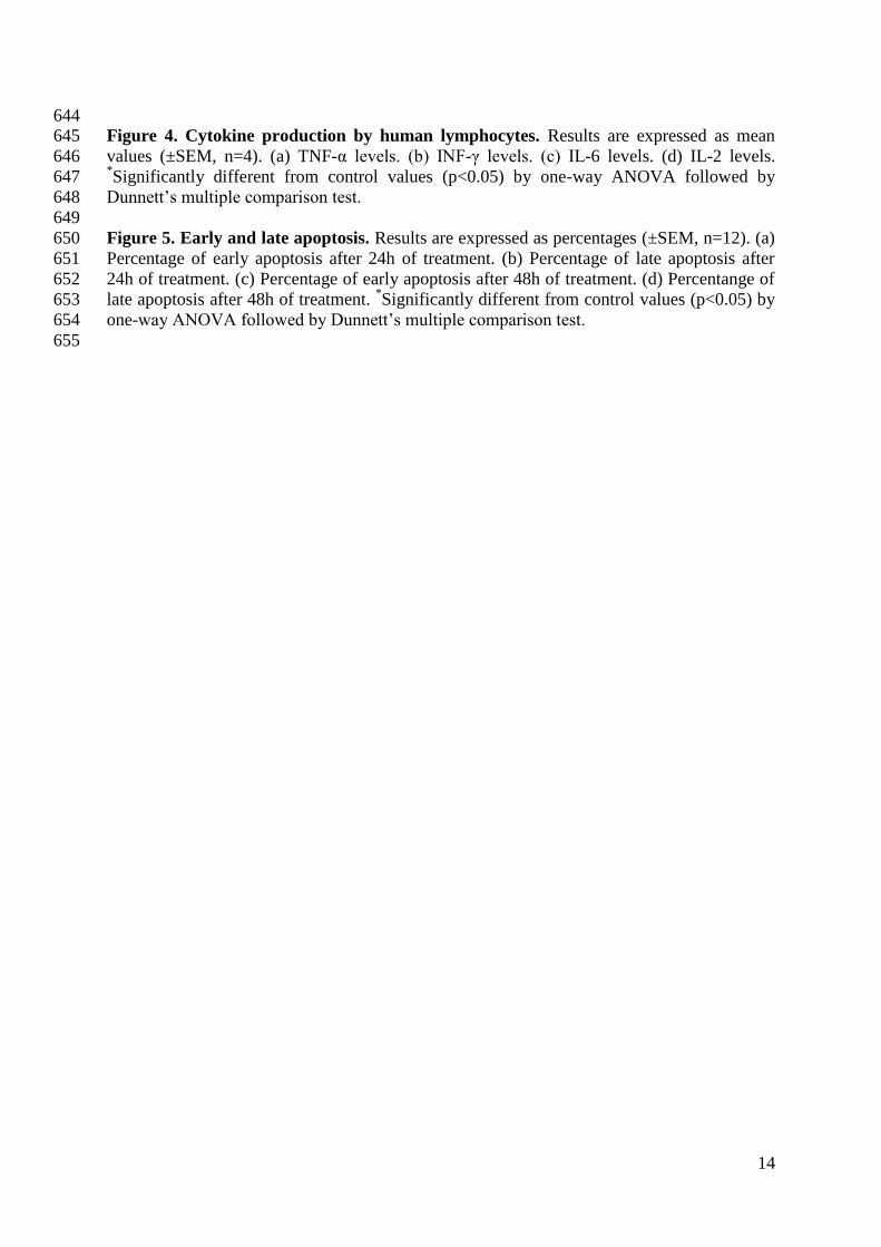
SEM images show irregular shapes and sizes varying from the nano to the micro scale (from 199
150nm to 6.5µm). The larger irregular shaped particles suggest that the debris aggregates 200
(Image 1), and this has been reported previously by Akbar and coworkers (2012). Doorn et 201
al(1998) isolated particles from MoM retrieval tissues that varied in size (51-116nm particles 202
to micrometre sized aggregates) and shape. Moreover, metal particles (0.1-3 microns in size) 203
have also been found in tissues post-mortem (Brown et al., 2013). EDS analysis indicated 204
that the wear debris is primarily composed of Co and Cr, which is in agreement with the alloy 205
composition (Singh and Dahotre, 2007). Analysis of 25 different particles indicated a mean 206
composition of 59.57% (±1.15%) Co and 40.43% (±1.25%) Cr, with a small content of Mo 207
which was below the quantification limit. 208
209
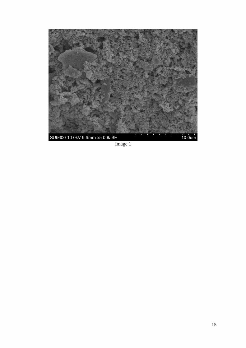
Metal ion release into cell culture medium 210
Metal debris was incubated, in the absence of cells, under different conditions. Analysis of 211
ICP-MS results found that CoCr wear debris releases metal ions into culture medium (Figure 212
1) was no significant difference (p>0.05) in ion release from metal debris in the presence and 213
absence of 10% (v/v) FCS. This concentration of FCS was used as it was the concentration 214
used when the Co-Cr wear debris was incubated with cells for up to 120h. In contrast to these 215
data, the acidic pH 4.0 had a considerable effect as seen in the significant increase (p<0.05) in 216
the levels of ion release compared with release in medium at normal physiological pH of 7.4. 217
Even though Co was the ion predominantly released in all cases, the change in pH seemed to 218
have a more pronounced effect on Cr ion release. 219
220
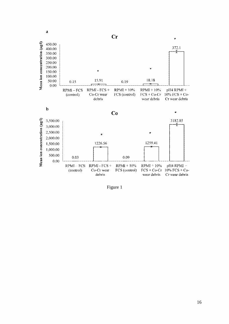
Effects of metal debris and ions on human primary lymphocyte cell viability, proliferation 221
and apoptosis. 222
The viability of primary human lymphocytes was tested after 24 and 120h of exposure to 223
5mg wear debris/1x106cells; 0.1μM of Co
2+; and 5mg wear debris/1x10
6cells combined with 224
0.1μM of Co2+
. In this study, the 5mg/1x106cells debris concentration was chosen to mimic 225
the local metallosis environment surrounding an implant. There was a significant increase in 226
cell number as indicated by the NR assay, measured both at 24 and 120h in the presence of 227
metal debris when compared to controls (Figure 2). Although there was an initial increase in 228
cell number (24h), there was no significant difference in the reduction of MTT in cells in the 229
presence of metal wear debris. Co ions on their own did not seem to have an effect on cell 230
number or cell metabolic activity. These findings suggest that the effects of the debris on the 231
lymphocytes are due to the synergistic action of the nanoparticles and the Co and Cr ions 232
released from the particles. 233
234
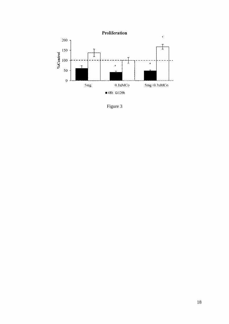
Effects on cell proliferation were assessed with the BrdU cell proliferation assay after 48 and 235
120h of treatment with 5mg wear debris/1x106cells; 0.1μM of Co
2+; and 5mg wear 236
debris/1x106cells combined with 0.1μM of Co
2+. At 48h, there is an initial decrease in cell 237
proliferation followed by an increase by 120h of treatment (Figure 3). These results suggest 238
an activation response of the cells to both debris and ions, where the cells overcome the 239
initial growth arrest effect. 240
241
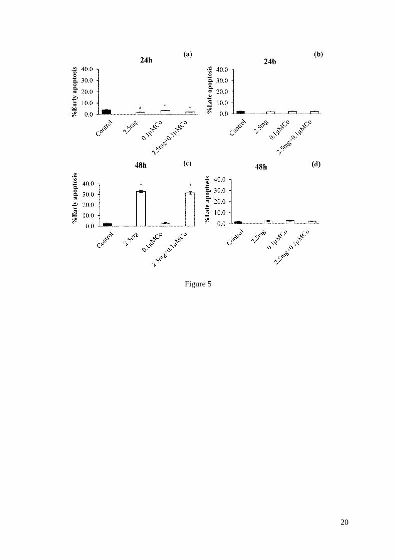
To determine the effects of metal ions on cell damage leading to apoptosis, flow cytometry 242
following Annexin V and 7-AAD staining at 24 and 48h of exposure was performed. To 243
avoid cytotoxic effects and the growth arrest effect observed after exposure to 5mg, a lower 244
debris concentration (2.5mg metal debris/1x106 cells) was used for apoptosis analysis in 245
order to facilitate the detection of the process at early stages (to measure both early apoptosis 246
and detect any repair). Apoptosis was not observed within 24h of exposure, but was evident 247
6
after 48h (Figure 5) where the debris caused apoptosis whereas the Co ions did not, and the 248
effect of the debris was unaltered by preincubation with Co ions. 249
250
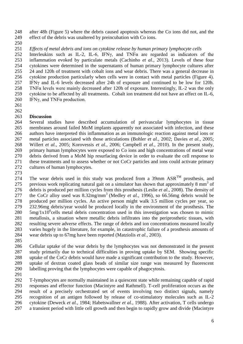
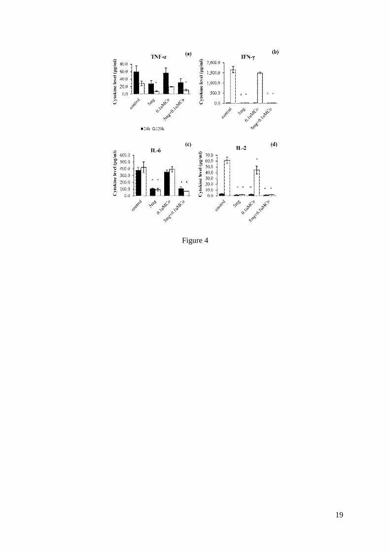
Effects of metal debris and ions on cytokine release by human primary lymphocyte cells 251
Interleukins such as IL-2, IL-6, IFNγ, and TNFα are regarded as indicators of the 252
inflammation evoked by particulate metals (Cachinho et al., 2013). Levels of these four 253
cytokines were determined in the supernatants of human primary lymphocyte cultures after 254
24 and 120h of treatment with cobalt ions and wear debris. There was a general decrease in 255
cytokine production particularly when cells were in contact with metal particles (Figure 4). 256
IFNγ and IL-6 levels decreased after 24h of exposure and continued to be low for 120h. 257
TNFα levels were mainly decreased after 120h of exposure. Interestingly, IL-2 was the only 258
cytokine to be affected by all treatments. Cobalt ion treatment did not have an effect on IL-6, 259
IFNγ, and TNFα production. 260
261
262
Discussion 263 Several studies have described accumulation of perivascular lymphocytes in tissue 264
membranes around failed MoM implants apparently not associated with infection, and these 265
authors have interpreted this inflammation as an immunologic reaction against metal ions or 266
metal particles associated with those articulations (Bohler et al., 2002; Davies et al., 2005; 267
Willert et al., 2005; Korovessis et al., 2006; Campbell et al., 2010). In the present study, 268
primary human lymphocytes were exposed to Co ions and high concentrations of metal wear 269
debris derived from a MoM hip resurfacing device in order to evaluate the cell response to 270
these treatments and to assess whether or not CoCr particles and ions could activate primary 271
cultures of human lymphocytes. 272
273
The wear debris used in this study was produced from a 39mm ASRTM
prosthesis, and 274
previous work replicating natural gait on a simulator has shown that approximately 8 mm3 of 275
debris is produced per million cycles from this prosthesis (Leslie et al., 2008). The density of 276
the CoCr alloy used was 8.32mg/mm3
(Medley et al., 1996), so 66.56mg debris would be 277
produced per million cycles. An active person might walk 3.5 million cycles per year, so 278
232.96mg debris/year would be produced locally in the environment of the prosthesis. The 279
5mg/1x106cells metal debris concentration used in this investigation was chosen to mimic 280
metallosis, a situation where metallic debris infiltrates into the periprosthetic tissues, with 281
resulting severe adverse effects. The range of debris and ion concentrations measured locally 282
varies hugely in the literature, for example, in catastrophic failure of a prosthesis amounts of 283
wear debris up to 67mg have been reported (Matziolis et al., 2003). 284
285
Cellular uptake of the wear debris by the lymphocytes was not demonstrated in the present 286
study primarily due to technical difficulties in proving uptake by SEM. Showing specific 287
uptake of the CoCr debris would have made a significant contribution to the study. However, 288
uptake of dextran coated glass beads of similar size range was measured by fluorescent 289
labelling proving that the lymphocytes were capable of phagocytosis. 290
291
T-lymphocytes are normally maintained in a quiescent state while remaining capable of rapid 292
responses and effector function (Macintyre and Rathmell). T-cell proliferation occurs as the 293
result of a precisely orchestrated set of events involving two distinct signals, namely 294
recognition of an antigen followed by release of co-stimulatory molecules such as IL-2 295
cytokine (Deweck et al., 1984; Habetswallner et al., 1988). After activation, T cells undergo 296
a transient period with little cell growth and then begin to rapidly grow and divide (Macintyre 297
7
and Rathmell). In this investigation, high concentrations of metal debris caused an increase in 298
cell number despite the initial decrease in cell proliferation suggesting that the particles may 299
exert a cell activation effect. It is proposed that this took place during the first 48h of 300
treatment reflected by the initial lower proliferation rates (24-48h) followed by the rapid 301
growth and division seen by 120h. In addition to this, a significant decrease in IL-2 302
production was observed after 24h of exposure to all the treatments. Since IL-2 is an 303
important molecule for lymphocyte activation and proliferation, its diminished production 304
could have contributed to the decrease in proliferation observed at 48h. 305
306
A state of reduced function in which a viable, antigen-specific T cell is unable to respond to 307
an immunogenic stimulus has been referred to as anergy (Zheng et al., 2008). Anergy can be 308
induced under a number of circumstances that can be categorised as resulting from either a 309
normal antigenic stimulus received in the absence of co-stimulation or from an altered and/or 310
chronic T-cell receptor stimulus (Wells, 2009). To the authors’ knowledge, anergy has not 311
been described as part of the biological reaction to metal debris and ions, and most studies 312
report implant-related hypersensitivity reactions, in particular type-IV delayed-type 313
hypersensitivity mediated by T lymphocytes. Nevertheless, results from this investigation 314
suggest that there may be an anergy-like response to high concentrations of metal debris. The 315
significant decrease in IL-2 production and proliferation observed here are hallmarks of T-316
cell anergy (Chappert and Schwartz, 2010; Kuklina, 2013). Moreover, defective production 317
of inflammatory cytokines such as IFNγ and TNFα is also a characteristic of anergy (Wells, 318
2009) and significant decreases in both cytokines were observed in the present study in the 319
presence of metal debris. 320
321
Metals corrode in vivo releasing metal ions (Hanawa, 2004). Such ions can potentially bind to 322
proteins, remain in solution, or disseminate into the surrounding tissues and bloodstream, and 323
thus reach remote organs. The microenvironment conditions surrounding the debris can 324
influence the rate of ion release (Cadosch et al., 2009). It is generally presumed that metal 325
ions facilitate cell activation and sensitization. However, depending on the concentration of 326
metal ions present, they may also be cytotoxic and suppressive. It has been shown that 327
production of TNFα and IL-6 by human peripheral blood mononuclear cells exposed to Cr (1, 328
5, and 10µM) significantly decreases (Villanueva et al., 2000). In the current investigation, 329
inhibition of cytokine production was observed in the presence of metal particles. IFNγ, 330
TNFα, and IL-6 levels did not seem to be affected by Co ions alone, whereas IL-2 levels were 331
decreased. Although the effects of Cr may be related to the regulation of TNFα and IL-6, IL-332
2 production is more likely to be modulated by Co. Additionally, high concentrations (5mg 333
debris/1x106cells) of metal debris were not cytotoxic to primary lymphocytes. However, a 334
marked increase in apoptosis was observed at a lower dose (2.5mg debris/1x106cells). These 335
findings suggest a dose dependent effect on cell damage pathways. Akbar et al. (2011) 336
exposed resting and activated lymphocytes to a range of Co and Cr ions. They found that 337
exposure to higher concentrations of Cr6+
(10 and 100μM), and Co2+
(100μM) significantly 338
decreased cell viability and increased apoptosis in both resting and activated lymphocytes at 339
24 and 48h of exposure. In their study metal ions were assessed independently. The effects 340
observed in the current study are the results of the concerted action of the particles and both 341
Co and Cr ions. It would have been interesting to pre-incubate the cells with Co ions and 342
subsequently treat them with the particles to identify if sensitisation occurs. In fact, priming 343
human monocyte-like U937 cells with Co ions for subsequent challenge with wear debris has 344
been investigated in our laboratory (Posada et al., 2014). Results from such investigation 345
showed that metal debris was more effective as an inducer of apoptosis and gene expression 346
when cells had been pre-treated with Co ions. However, this set of experiments could not 347
8
readily be performed with the primary lymphocytes due to difficulties in maintaining 348
prolonged culture. 349
350
Released metal ions can activate the immune system by forming metal-protein complexes 351
that are considered to be candidate antigens for eliciting hypersensitivity responses 352
(Korovessis et al., 2006). Upon recognition by lymphocytes, the metal-protein complexes 353
induce the production of proinflammatory cytokines and chemokines by various cell types 354
due to triggering of innate immune responses (Martin, 2004). According to this, it is thought 355
that high local metal ion and nanoparticles concentrations facilitate a T-lymphocyte mediated 356
inflammatory response resulting in the destruction seen around the prostheses (Davies et al., 357
2005; Willert et al., 2005; Boardman et al., 2006; Kwon et al., 2010). Three mechanisms 358
have been proposed by which metal–protein complexes can activate lymphocytes: 1. antigen-359
independent, 2. antigen-dependent, and 3. superantigen-like, which is a synergistic 360
combination of the first two mechanisms (Hallab et al., 2001b). Metals may act with serum 361
proteins to crosslink lymphocyte receptors (e.g., BV17 of CDR1 T cell receptor) without the 362
presence of an antigen-presenting cell leading to a superantigen enhancement of T cell 363
receptor-protein contact (Vollmer et al., 1997; Vollmer et al., 1999). In this circumstance, 364
proteins or peptides that would not otherwise be antigenic are able to provoke a response 365
(Hallab et al., 2001b). The lymphocyte reactions in the current investigation seem to be 366
consistent with such nonspecific mitogenic activation mechanisms, which could explain the 367
increase in cell proliferation despite the significant decrease in IL-2 production. 368
369
Implant failure is largely caused by aseptic loosening and osteolysis (Huber et al., 2010) in 370
response to accumulation of metal particles in the periprosthetic tissues, which also generates 371
inflammation, pain, and pseudotumours (Langton et al., 2011). Evidence of systemic effects 372
can also be found in multiple reports (Steens et al., 2006; Oldenburg et al., 2009; Rizzetti et 373
al., 2009; Ikeda et al., 2010; Tower, 2010; Machado et al., 2012; Pelclova et al., 2012; 374
Tower, 2012) describing patients with MoM implants who presented symptoms including 375
neurological symptoms such as auditory impairment/deafness, visual impairment/blindness, 376
peripheral neuropathy/dysesthesia of the extremities, poor concentration/cognitive decline, 377
cardiomyopathy and hypothyroidism. All patients had elevated cobalt and/or chromium 378
concentrations in their blood, serum, plasma, and/or urine, suggesting that these systemic 379
symptoms may be due to metal toxicity as a result of excessive implant wear. Polyzois et al 380
(2012), reviewed the evidence of local and systemic toxicity of wear debris from total hip 381
arthroplasty. They found extensive evidence and experimental data supporting the fact that 382
orthopaedic metals induce local immunological effects characterised by an unusual 383
lymphocytic infiltration and cell-mediated hypersensitivity. In terms of systemic toxicity, 384
there are in vivo and in vitro experimental, as well as a limited number of epidemiological 385
studies, where the systems most commonly involved are haematopeietic, immune, 386
hepatobiliary, renal, respiratory, nervous, cardiovascular, musculoskeletal, skin, and 387
endocrine and reproductive. Concern has been raised regarding a potential link between 388
metal wear debris and carcinogenesis. In an attempt to address this, Christian et al (2014) 389
used quantitative methods to evaluate the relationship between CoCr-containing hip implants 390
and increased cancer risk. They concluded that although the evidence suggests that such 391
implants are unlikely to be associated with an increased risk of systemic cancers, additional 392
research is warranted in this area. 393
394
The importance of Co ions in the inflammatory responses to CoCr particles has been 395
recognised, and chronic exposure to circulating levels of ions, plus high local concentrations 396
may act synergistically in vivo to trigger and promote implant loosening (Hallab et al., 2001a; 397
9
Caicedo et al., 2010; Hart et al., 2012). The present study has shown that high concentrations 398
of wear debris, derived from a CoCr MoM hip resurfacing, induce lymphocyte proliferation 399
and inhibit cytokine production after 120h exposure. The fact that IL-2 production was 400
affected by 0.1µM Co (5.9ppb or 5.9µg/L) suggests that even circulating blood metal ion 401
concentrations within the MHRA guideline levels of 7ppb or 7µg/L (MHRA) may contribute 402
to the impairment of immune regulation in patients with MoM implants. 403
404
Acknowledgements 405 This study was supported by an Overseas Research Studentship awarded to Olga M. Posada, 406
and by funds from University of Strathclyde. The authors are grateful to Dr C Hardaker 407
(DePuy International) who prepared the CoCr nanoparticles. 408
409
References 410 Afolaranmi, G.A., Akbar, M., Brewer, J., Grant, M.H., 2012. Distribution of metal released 411
from cobalt-chromium alloy orthopaedic wear particles implanted into air pouches in mice. J 412
Biomed Mater Res A. 100A, 1529-1538. doi: 10.1002/jbm.a.34091. 413
Akbar, M., Brewer, J.M., Grant, M.H., 2011. Effect of chromium and cobalt ions on primary 414
human lymphocytes in vitro. J Immunotoxicol. 8, 140-149. doi: 415
10.3109/1547691X.2011.553845. 416
Akbar, M., Fraser, A.R., Graham, G.J., Brewer, J.M., Grant, M.H., 2012. Acute inflammatory 417
response to cobalt chromium orthopaedic wear debris in a rodent air-pouch model. J R Soc 418
Interface. 9, 2109-2119. doi: 10.1098/rsif.2012.0006. 419
Amstutz, H.C., Le Duff, M.J., 2006. Background of metal-on-metal resurfacing. Proc Inst 420
Mech Eng H. 220, 85-94. 421
Arora, A., Song, Y., Chun, L., Huie, P., Trindade, M., Smith, R.L., Goodman, S., 2003. The 422
role of the TH1 and TH2 immune responses in loosening and osteolysis of cemented total hip 423
replacements. J Biomed Mater Res A. 64A, 693-697. 424
Boardman, D.R., Middleton, F.R., Kavanagh, T.G., 2006. A benign psoas mass following 425
metal-on-metal resurfacing of the hip. J Bone Joint Surg Br. Volume 88B, 402-404. 426
Bohler, M., Kanz, F., Schwarz, B., Steffan, I., Walter, A., Plenk, H., Knahr, K., 2002. 427
Adverse tissue reactions to wear particles form Co-alloy articulations, increased by alumina-428
blasting particle contamination from cementless Ti-based total hip implants - A report of 429
seven revisions with early failure. J Bone Joint Surg Br. Volume 84B, 128-136. 430
Brown, C., Lacharme-Lora, L., Mukonoweshuro, B., Sood, A., Newson, R.B., Fisher, J., 431
Case, C.P., Ingham, E., 2013. Consequences of exposure to peri-articular injections of micro- 432
and nano-particulate cobalt-Chromium alloy. Biomaterials 34, 8564-8580. doi: 433
10.1016/j.biomaterials.2013.07.073. 434
Brown, C., Williams, S., Tipper, J.L., Fisher, J., Ingham, E., 2007. Characterisation of wear 435
particles produced by metal on metal and ceramic on metal hip prostheses under standard and 436
microseparation simulation. J Mater Sci Mater Med. 18, 819-827. 437
Cachinho, S.C.P., Pu, F.R., Hunt, J.A., 2013. Cytokine secretion from human peripheral 438
blood mononuclear cells cultured in vitro with metal particles. J Biomed Mater Res A. 101A, 439
1201-1209. doi: 10.1002/jbm.a.34410. 440
Cadosch, D., Chan, E., Gautschi, O.P., Filgueira, L., 2009. Metal is not inert: Role of metal 441
ions released by biocorrosion in aseptic loosening-Current concepts. J Biomed Mater Res A. 442
91A, 1252-1262. doi: 10.1002/jbm.a.32625 443
Caicedo, M., Jacobs, J.J., Reddy, A., Hallab, N.J., 2008. Analysis of metal ion-induced DNA 444
damage, apoptosis, and necrosis in human (Jurkat) T-cells demonstrates Ni2+
, and V3+
are 445
more toxic than other metals: Al3+
, Be2+
, Co2+
, Cr3+
, Cu2+
, Fe3+
, Mo5+
, Nb5+
, Zr2+
. J Biomed 446
Mater Res A. 86A, 905-913. 447
10
Caicedo, M.S., Pennekamp, P.H., McAllister, K., Jacobs, J.J., Hallab, N.J., 2010. Soluble 448
ions more than particulate cobalt-alloy implant debris induce monocyte costimulatory 449
molecule expression and release of proinflammatory cytokines critical to metal-induced 450
lymphocyte reactivity. J Biomed Mater Res A. 93A, 1312-1321. doi: 10.1002/jbm.a.32627 451
Campbell, P., Ebramzadeh, E., Nelson, S., Takamura, K., De Smet, K., Amstutz, H.C., 2010. 452
Histological Features of Pseudotumor-like Tissues From Metal-on-Metal Hips. Clin Orthop 453
Relat Res. 468, 2321-2327. doi: 10.1007/s11999-010-1372-y. 454
Chappert, P., Schwartz, R.H., 2010. Induction of T cell anergy: integration of environmental 455
cues and infectious tolerance. Curr Opin Immunol. 22, 552-559. doi: 456
10.1016/j.coi.2010.08.005. 457
Christian, W.V., Oliver, L.D., Paustenbach, D.J., Kreider, M.L., Finley, B.L., 2014. 458
Toxicology-based cancer causation analysis of CoCr-containing hip implants: a quantitative 459
assessment of genotoxicity and tumorigenicity studies. J Appl Toxicol. 34, 939-967. doi: 460
10.1002/jat.3039. 461
Corten, K., MacDonald, S.J., 2010. Hip resurfacing data from national joint registries What 462
do they tell us? What do they not tell us? Clin Orthop Relat Res. 468, 351-357. doi: 463
10.1007/s11999-009-1157-3. 464
Davies, A.P., Willert, H.G., Campbell, P.A., Learmonth, I.D., Case, C.P., 2005. An unusual 465
lymphocytic perivascular infiltration in tissues around contemporary metal-on-metal joint 466
replacements. J Bone Joint Surg Am. Volume 87A, 18-27. 467
Dearnley, P.A., 1999. A review of metallic, ceramic and surface-treated metals used for 468
bearing surfaces in human joint replacements. Proc Inst Mech Eng H. 213, 107-135. 469
Deweck, A.L., Kristensen, F., Joncourt, F., Bettens, F., Walker, C., Wang, Y., 1984. Analysis 470
of lymphocyte-proliferation by cytofluorometric techniques in aging and various clinical 471
conditions. Asian Pac J Allergy Immunol. 2, 253-261. 472
Doorn, P.F., Campbell, P.A., Worrall, J., Benya, P.D., McKellop, H.A., Amstutz, H.C., 1998. 473
Metal wear particle characterization from metal on metal total hip replacements: 474
Transmission electron microscopy study of periprosthetic tissues and isolated particles. J 475
Biomed Mater Res. 42, 103-111. 476
Habetswallner, D., Pelosi, E., Bulgarini, D., Camagna, A., Samoggia, P., Montesoro, E., 477
Giannella, G., Lazzaro, D., Isacchi, G., Testa, U., Peschle, C., 1988. Activation and 478
proliferation of normal resting human lymphocytes-t in serum-free culture - role of IL-4 and 479
IL-6. Immunology 65, 357-364. 480
Hallab, N., Merritt, K., Jacobs, J.J., 2001a. Metal sensitivity in patients with orthopaedic 481
implants. J Bone Joint Surg Am. Volume 83A, 428-436. 482
Hallab, N.J., Mikecz, K., Vermes, C., Skipor, A., Jacobs, J.J., 2001b. Differential lymphocyte 483
reactivity to serum-derived metal-protein complexes produced from cobalt-based and 484
titanium-based implant alloy degradation. J Biomed Mater Res. 56, 427-436. 485
Hanawa, T., 2004. Metal ion release from metal implants. Mater Sci Eng C Mater Biol Appl 486
24, 745-752. doi: 10.1016/j.msec.2004.08.018. 487
Hart, A.J., Quinn, P.D., Lali, F., Sampson, B., Skinner, J.A., Powell, J.J., Nolan, J., Tucker, 488
K., Donell, S., Flanagan, A., Mosselmans, J.F.W., 2012. Cobalt from metal-on-metal hip 489
replacements may be the clinically relevant active agent responsible for periprosthetic tissue 490
reactions. Acta Biomater. 8, 3865-3873. doi: 10.1016/j.actbio.2012.05.003. 491
Huber, M., Reinisch, G., Zenz, P., Zweymueller, K., Lintner, F., 2010. Postmortem study of 492
femoral osteolysis associated with metal-on-metal articulation in total hip replacement an 493
analysis of nine cases. J Bone Joint Surg Am. Volume 92A, 1720-1731. doi: 494
10.2106/JBJS.I.00695. 495
11
Ikeda, T., Takahashi, K., Kabata, T., Sakagoshi, D., Tomita, K., Yamada, M., 2010. 496
Polyneuropathy caused by cobalt-chromium metallosis after total hip replacement. Muscle 497
Nerve 42, 140-143. doi: 10.1002/mus.21638. 498
Ingham, E., Green, T.R., Stone, M.H., Kowalski, R., Watkins, N., Fisher, J., 2000. Production 499
of TNF-alpha and bone resorbing activity by macrophages in response to different types of 500
bone cement particles. Biomaterials 21, 1005-1013. 501
Jiang, Y., Zhang, K., Die, J., Shi, Z., Zhao, H., Wang, K., 2011. A systematic review of 502
modern metal-on-metal total hip resurfacing vs standard total hip arthroplasty in active young 503
patients. J Arthroplasty. 26, 419-426. doi: 10.1016/j.arth.2010.07.008. 504
Korovessis, P., Petsinis, G., Repanti, M., Repantis, T., 2006. Metallosis after contemporary 505
metal-on-metal total hip arthroplasty - Five to nine-year follow-up. J Bone Joint Surg Am 506
Volume 88A, 1183-1191. 507
Kuklina, E.M., 2013. Molecular mechanisms of T-cell anergy. Biochemistry (Mosc). 78, 144-508
156. doi: 10.1134/S000629791302003X. 509
Kwon, Y.M., Glyn-Jones, S., Simpson, D.J., Kamali, A., McLardy-Smith, P., Gill, H.S., 510
Murray, D.W., 2010. Analysis of wear of retrieved metal-on-metal hip resurfacing implants 511
revised due to pseudotumours. J Bone Joint Surg Br. Volume 92B, 356-361. doi: 512
10.1302/0301-620X.92B3.23281. 513
Langton, D.J., Joyce, T.J., Jameson, S.S., Lord, J., Van Orsouw, M., Holland, J.P., Nargol, 514
A.V.F., De Smet, K.A., 2011. Adverse reaction to metal debris following hip resurfacing. 515
The influence of component type, orientation and volumetric wear. J Bone Joint Surg Br. 516
Volume 93B, 164-171. doi: 10.1302/0301-620X.93B2.25099. 517
Lawrence, D.A., McCabe, M.J., 2002. Immunomodulation by metals. Int Immunopharmacol 518
2, 293-302. 519
Leslie, I., Williams, S., Brown, C., Isaac, G., Jin, Z., Ingham, E., Fisher, J., 2008. Effect of 520
bearing size on the long-term wear, wear debris, and ion levels of large diameter metal-on-521
metal hip replacements - An in vitro study. J J Biomed Mater Res B Appl Biomater. 87B, 522
163-172. doi: 10.1002/jbm.b.31087. 523
Luo, L., Petit, A., Antoniou, J., Zukor, D.J., Huk, O.L., Liu, R.C.W., Winnik, F.M., Mwale, 524
F., 2005. Effect of cobalt and chromium ions on MMP-1 TIMP-1, and TNF-alpha gene 525
expression in human U937 macrophages: A role for tyrosine kinases. Biomaterials 26, 5587-526
5593. 527
Lutz, M.B., Kukutsch, N., Ogilvie, A.L.J., Rossner, S., Koch, F., Romani, N., Schuler, G., 528
1999. An advanced culture method for generating large quantities of highly pure dendritic 529
cells from mouse bone marrow. J Immunol Methods 223, 77-92. 530
Machado, C., Appelbe, A., Wood, R., 2012. Arthroprosthetic cobaltism and cardiomyopathy. 531
Heart Lung Circ. 21, 759-760. doi: 10.1016/j.hlc.2012.03.013. 532
Macintyre, A., Rathmell, J., Activated lymphocytes as a metabolic model for carcinogenesis. 533
Cancer Metab C7 - 5 1, 1-12. doi: 10.1186/2049-3002-1-5. 534
Mahendra, G., Pandit, H., Kliskey, K., Murray, D., Gill, H.S., Athanasou, N., 2009. Necrotic 535
and inflammatory changes in metal-on-metal resurfacing hip arthroplasties Relation to 536
implant failure and pseudotumor formation. Acta Orthop. 80, 653-659. doi: 537
10.3109/17453670903473016. 538
Mansson, B., Geborek, P., Saxne, T., Bjornsson, S., 1990. Cytidine deaminase activity in 539
synovial-fluid of patients with rheumatoid-arthritis - Relation to lactoferrin, acidosis, and 540
cartilage proteoglycan release. Ann Rheum Dis 49, 594-597. 541
Martin-Romero, C., Santos-Alvarez, J., Goberna, R., Sanchez-Margalet, V., 2000. Human 542
leptin enhances activation and proliferation of human circulating T lymphocytes. Cell 543
Immunol. 199, 15-24. 544
12
Martin, S.F., 2004. T lymphocyte-mediated immune responses to chemical haptens and metal 545
ions: Implications for allergic and autoimmune disease. Int Arch Allergy Immunol. 134, 186-546
198. 547
Matziolis, G., Perka, C., Disch, A., 2003. Massive metallosis after revision of a fractured 548
ceramic head onto a metal head. Arch Orthop Trauma Surg. 123, 48-50. 549
Medley, J.B., Chan, F.W., Krygier, J.J., Bobyn, J.D., 1996. Comparison of alloys and designs 550
in a hip simulator study of metal on metal implants. Clin Orthop Relat Res., S148-S159. 551
Oldenburg, M., Wegner, R., Baur, X., 2009. Severe cobalt intoxication due to prosthesis wear 552
in repeated total hip arthroplasty. J Arthroplasty. 24, 825.e815-820. doi: 553
10.1016/j.arth.2008.07.017. 554
Pandit, H., Glyn-Jones, S., McLardy-Smith, P., Gundle, R., Whitwell, D., Gibbons, C.L.M., 555
Ostlere, S., Athanasou, N., Gill, H.S., Murray, D.W., 2008. Pseudotumours associated with 556
metal-onmetal hip resurfacings. J Bone Joint Surg Br. Volume 90B, 847-851. doi: 557
10.1302/0301-620X.90B7.20213. 558
Pelclova, D., Sklensky, M., Janicek, P., Lach, K., 2012. Severe cobalt intoxication following 559
hip replacement revision: Clinical features and outcome. Clin Toxicol (Phila). 50, 262-265. 560
doi: 10.3109/15563650.2012.670244. 561
Polyzois, I., Nikolopoulos, D., Michos, I., Patsouris, E., Theocharis, S., 2012. Local and 562
systemic toxicity of nanoscale debris particles in total hip arthroplasty. J Appl Toxicol. 32, 563
255-269. doi: 10.1002/jat.2729. 564
Posada, O.M., Gilmour D., Tate R.J., Grant M.H., 2014. CoCr wear particles generated from 565
CoCr alloy metal-on-metal hip replacements, and cobalt ions stimulate apoptosis and 566
expression of general toxicology-related genes in monocyte-like U937 cells. Toxicol Appl 567
Pharmacol. 281, 125-135. doi: 10.1016/j.taap.2014.09.010 568
Quesada, M.J., Marker, D.R., Mont, M.A., 2008. Metal-on-metal hip resurfacing. J 569
Arthroplasty. 23, 69-73. doi: 10.1016/j.arth.2008.06.015. 570
Rizzetti, M.C., Liberini, P., Zarattini, G., Catalani, S., Pazzaglia, U., Apostoli, P., Padovani, 571
A., 2009. Loss of sight and sound. Could it be the hip? Lancet 373, 1052-1052. doi: 572
10.1016/S0140-6736(09)60490-6. 573
Singh, R., Dahotre, N.B., 2007. Corrosion degradation and prevention by surface 574
modification of biometallic materials. J Mater Sci Mater Med 18, 725-751. 575
Steens, W., von Foerster, G., Katzer, A., 2006. Severe cobalt poisoning with loss of sight 576
after ceramic-metal pairing in a hip- a case report. Acta Orthop. 77, 830-832. 577
Tower, S.S., 2010. Arthroprosthetic cobaltism: neurological and cardiac manifestations in 578
two patients with metal-on-metal arthroplasty. A case report. J Bone Joint Surg Am. Volume 579
92A, 2847-2851. doi: 10.2106/JBJS.J.00125. 580
Tower, S.S., 2012. Metal on metal hip implants. Arthroprosthetic cobaltism associated with 581
metal on metal hip implants. BMJ. 344e430. doi: 10.1136/bmj.e430. 582
Villanueva, M.B.G., Koizumi, S., Jonai, H., 2000. Cytokine production by human peripheral 583
blood mononuclear cells after exposure to heavy metals. J Health Sci 46, 358-362. 584
Vollmer, J., Fritz, M., Dormoy, A., Weltzien, H.U., Moulon, C., 1997. Dominance of the 585
BV17 element in nickel-specific human T cell receptors relates to severity of contact 586
sensitivity. Eur J Immunol. 27, 1865-1874. 587
Vollmer, J., Weltzien, H.U., Moulon, C., 1999. TCR reactivity in human nickel allergy 588
indicates contacts with complementarity-determining region 3 but excludes superantigen-like 589
recognition. J Immunol. 163, 2723-2731. 590
Wang, J.Y., Wicklund, B.H., Gustilo, R.B., Tsukayama, D.T., 1996. Titanium, chromium and 591
cobalt ions modulate the release of bone-associated cytokines by human 592
monocytes/macrophages in vitro. Biomaterials 17, 2233-2240. 593
13
Wells, A.D., 2009. New insights into the molecular basis of t cell anergy: Anergy factors, 594
avoidance sensors, and epigenetic imprinting. J Immunol. 182, 7331-7341. doi: 595
10.4049/jimmunol.0803917. 596
Willert, H.G., Buchhorn, G.H., Fayyazi, A., Flury, R., Windler, M., Koster, G., Lohmann, 597
C.H., 2005. Metal-on-metal bearings and hypersensitivity in patients with artificial hip joints 598
- A clinical and histomorphological study. J Bone Joint Surg Am. Volume 87A, 28-36. 599
Xia, Z.D., Kwon, Y.M., Mehmood, S., Downing, C., Jurkschat, K., Murray, D.W., 2011. 600
Characterization of metal-wear nanoparticles in pseudotumor following metal-on-metal hip 601
resurfacing. Nanomedicine 7, 674-681. doi: 10.1016/j.nano.2011.08.002. 602
Zheng, Y., Zha, Y.Y., Gajewski, T.F., 2008. Molecular regulation of T-cell anergy. EMBO 603
Rep. 9, 50-55. doi: 10.1038/sj.embor.7401138. 604
605
Web References 606 1. Australian Orthopaedic Association National Joint Replacement Registry, A., 2011. Hip 607
and Knee Arthroplasty Annual Report 2011 http://www.surfacehippy.info/pdf/australian-nat-608
reg-2011.pdf [last accesed 13/02/2014]. 609
2. Canadian Joint Replacement Registry, C., 2013. Title Hip and Knee Replacements in 610
Canada: Canadian Joint Replacement Registry 2013 Annual 611
Report. https://secure.cihi.ca/free_products/CJRR_2013_Annual_Report_EN.pdf [last 612
accesed 13/02/2014]. 613
3. National Joint Registry, N., 2012. National Joint Registry for England and Wales 9th 614
Annual Report. 615
http://www.njrcentre.org.uk/njrcentre/Portals/0/Documents/England/Reports/9th_annual_rep616
ort/NJR%209th%20Annual%20Report%202012.pdf [last accesed 13/02/2014]. 617
4. MDA/2010/069, Medical Device Alert. Ref: MDA/2010/069. Issues: 7 September 618
2010. http://www.mhra.gov.uk/home/groups/dts-619
bs/documents/medicaldevicealert/con093791.pdf [last accesed 13/02/2014]. 620
5. MHRA, Medical Device Alert. Ref: MDA/2010/033. Issues: 22 April 2010. 621
http://www.dhsspsni.gov.uk/mda-2010-033.pdf [last accesed 13/02/2014]. 622
Figure and Image legends 623
624
Image 1. Scanning Electron Microscopy images of simulator generated wear debris 625 from an ASR hip implant. Image taken at 5000X with a FE-SEM Hitachi SU-6600. 626
627
Figure 1. Metal ions in RPMI-1640 in the presence and absence of metal wear debris. 628 Results are expressed as mean values (±SEM, n=3). (a) Cr ion concentrations. (b) Co ion 629
concentrations. *Significantly different from control values (p<0.05) by one-way ANOVA 630
followed by Dunnett’s multiple comparison test. bSignificant difference between pH 7.4 and 631
pH 4.0. 632
633
Figure 2. Cell number (NR) and metabolic activity (MTT) of human lymphocytes. 634 Results are expressed as percentages (±SEM, n=12) where 100% represents control untreated 635
cells. (a) Effects on cell number as measured by NR. (b) Cell metabolic activity as measured 636
by MTT. *Significantly different from control values (p<0.05) by one-way ANOVA followed 637
by Dunnett’s multiple comparison test. 638
639
Figure 3. Proliferation of human lymphocytes measured by BrdU assay. Results are 640
expressed as percentages (±SEM, n=6) where 100% represents control untreated cells. 641 *Significantly different from control values (p<0.05) by one-way ANOVA followed by 642
Dunnett’s multiple comparison test. 643
14
644 Figure 4. Cytokine production by human lymphocytes. Results are expressed as mean 645
values (±SEM, n=4). (a) TNF-α levels. (b) INF-γ levels. (c) IL-6 levels. (d) IL-2 levels. 646 *Significantly different from control values (p<0.05) by one-way ANOVA followed by 647
Dunnett’s multiple comparison test. 648
649
Figure 5. Early and late apoptosis. Results are expressed as percentages (±SEM, n=12). (a) 650
Percentage of early apoptosis after 24h of treatment. (b) Percentage of late apoptosis after 651
24h of treatment. (c) Percentage of early apoptosis after 48h of treatment. (d) Percentange of 652
late apoptosis after 48h of treatment. *Significantly different from control values (p<0.05) by 653
one-way ANOVA followed by Dunnett’s multiple comparison test. 654
655
15
Image 1
16
Figure 1
17
Figure 2
18
Figure 3
19
Figure 4
20
Figure 5