Embed Size (px)
Citation preview

Molecular Diagnostics Fundamentals, Methods and Clinical ApplicationsSecond Edition
Copyright © 2012 F.A. Davis Company
Analysis and Characterization of Nucleic Acids and Proteins
Chapter 6
Molecular Diagnostics Fundamentals, Methods and Clinical ApplicationsSecond Edition
Copyright © 2012 F.A. Davis Company
Objectives Describe how restriction enzyme sites are mapped on DNA.
Construct a restriction enzyme map of a DNA plasmid or fragment.
Diagram the Southern blot procedure. Define hybridization, stringency, and meltingtemperature.
Calculate the melting temperature of a given sequence of dsDNA.
Describe comparative genomic hybridization (CGH).

Molecular Diagnostics Fundamentals, Methods and Clinical ApplicationsSecond Edition
Copyright © 2012 F.A. Davis Company
Restriction Enzyme Mapping
Clinical and forensic analyses require characterization of specific genes or genomic regions at the molecular level. Because of their sequence‐specific activity, restriction endonucleases provide a convenient tool for molecular characterization of DNA

Molecular Diagnostics Fundamentals, Methods and Clinical ApplicationsSecond Edition
Copyright © 2012 F.A. Davis Company
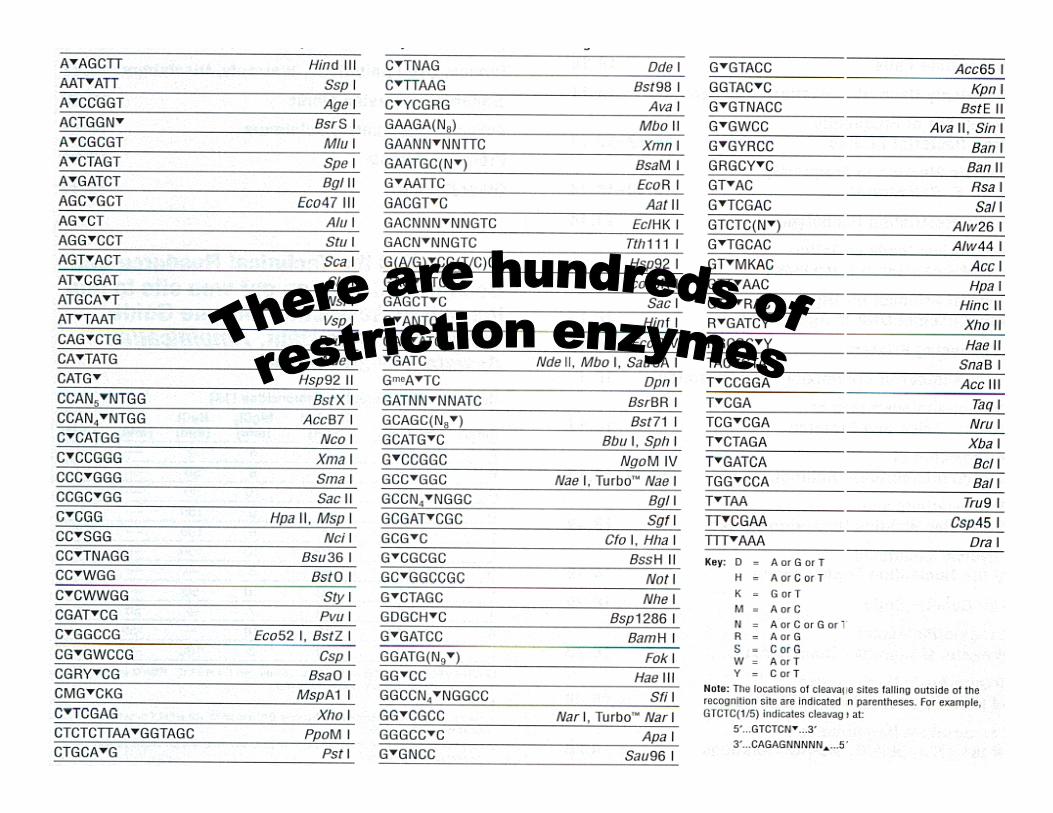
Restriction EndonucleasesEnzyme Isolated From Recognition
SequenceEco RI E. coli, strain R,
1st enzymeGν AATTC
Eco RV E. coli, strain R, 5th enzyme
Gv ATATC
Hind III H. influenzae, strain d, 3rd enzyme
Av AGCTT

Molecular Diagnostics Fundamentals, Methods and Clinical ApplicationsSecond Edition
Copyright © 2012 F.A. Davis Company
Restriction Enzymes Type I methylation/cleavage (3 subunits)
>1000 bp from binding sitee.g., Eco AI GAGNNNNNNNGTCA
Type II cleavage at specific recognition sites
Type III methylation/cleavage (2 subunits)
24–26 bp from binding sitee.g., Hinf III CGAAT

Molecular Diagnostics Fundamentals, Methods and Clinical ApplicationsSecond Edition
Copyright © 2012 F.A. Davis Company
G AATTCCTTAA G
Eco R15′ overhang
5′
5′
CTGCA GG ACGTC
PstI3′ overhang
5′
5′
CCC GGGGGG CCC
Sma1blunt
5′
5′
Restriction enzymes cut DNA at specific sites in one of three ways.
Restriction Enzymes, Type II

Molecular Diagnostics Fundamentals, Methods and Clinical ApplicationsSecond Edition
Copyright © 2012 F.A. Davis Company
Sticky ends must match (be complementary) for optimal religation.
Sticky ends can be converted to blunt ends with nuclease or polymerase.Blunt ends can be converted to sticky ends by ligating to synthetic adaptors.
Blunt ends can be religated with less efficiency than sticky ends.

Molecular Diagnostics Fundamentals, Methods and Clinical ApplicationsSecond Edition
Copyright © 2012 F.A. Davis Company
Restriction Enzyme Mapping
Digest DNA with a restriction enzyme.
Resolve the fragments by gel electrophoresis.
The number of bands indicates the number of restriction sites.
The size of the bands indicates the distance between restriction sites.

Molecular Diagnostics Fundamentals, Methods and Clinical ApplicationsSecond Edition
Copyright © 2012 F.A. Davis Company
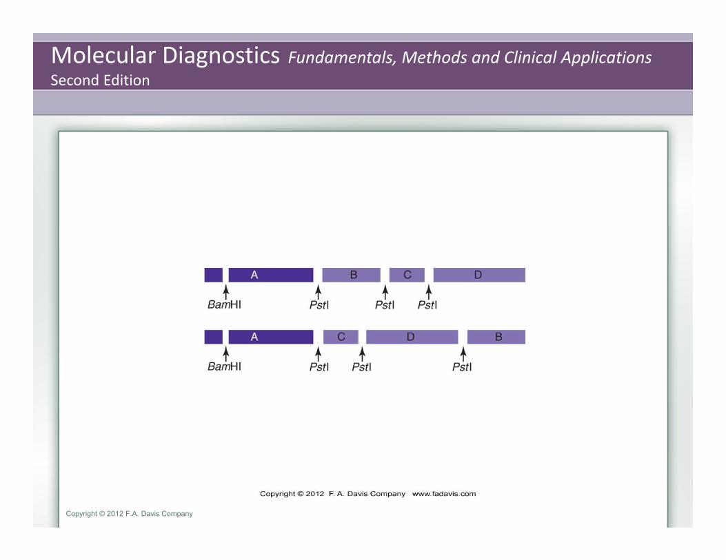
Restriction mapping of a linear DNA fragment (top green bar). The fragment is first cut with the enzyme PstI. Four fragments result, as deter‐mined by agarosegel electrophoresis, indicating that there are three PstI sites in the linear fragment. The size of the pieces indicates the distance between the restriction sites. A second cut with BamHI(bottom) yields two fragments, indicating one site. Since one BamHIfragment (E) is very small, the BamHI site must be near one end of the fragment. Cutting with both enzymes indicates that the BamHI site is in the PstI fragment A.
Molecular Diagnostics Fundamentals, Methods and Clinical ApplicationsSecond Edition
Copyright © 2012 F.A. Davis Company

Molecular Diagnostics Fundamentals, Methods and Clinical ApplicationsSecond Edition
Copyright © 2012 F.A. Davis Company
Restriction Enzyme Mapping

Molecular Diagnostics Fundamentals, Methods and Clinical ApplicationsSecond Edition
Copyright © 2012 F.A. Davis Company
Under nonstandard conditions, some restriction enzymes will bind to and cut sequences other than their defined recognition sequence. This altered specificity is called star activity. The propensity for star activity varies among enzymes. Thus, the nature and degree of star activity depends on the enzyme and the reaction conditions.
Reaction conditions that induce star activity include suboptimal buffer, contamination with solvents or high concentrations of glycerol, prolonged reaction time, high concentration of enzymes, and divalent cationimbalance.
Restriction fragment length polymorphisms (RFLPs). RFLPs were the basis of the first molecular‐based human identification and mapping methods.

Molecular Diagnostics Fundamentals, Methods and Clinical ApplicationsSecond Edition
Copyright © 2012 F.A. Davis Company
Hybridization Technologies Procedures performed in the clinical molecular laboratory are aimed at specific targets in genomic
DNA. This requires visualization or detection of a particular gene or region of DNA in the backdrop of all other genes. There are several ways to find a target region of DNA.
Hybridization Technologies Hybridization Method Target Probe Purpose Southern blot DNA Nucleic acid Gene structure Northern blot RNA Nucleic acid Transcript structure, processing,
gene expression Western blot Protein Protein Protein processing, gene
expression Southwestern blot Protein DNA DNA binding proteins, gene
regulation Eastern blot Protein Protein Modification of Western blot
using enzymatic detection(PathHunterTM); also, detection of specific agriculturally important proteins
Far‐eastern blot Lipids (None) Transfer of HPLC‐separated lipids to PVDF membranes for analysis by mass spectometry
Molecular Diagnostics Fundamentals, Methods and Clinical ApplicationsSecond Edition
Copyright © 2012 F.A. Davis Company
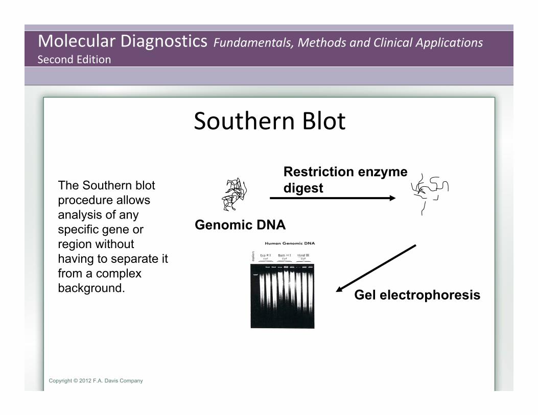
Southern Blot
Restriction enzymedigest
Gel electrophoresis
Genomic DNA
The Southern blot procedure allows analysis of any specific gene or region without having to separate it from a complex background.

Molecular Diagnostics Fundamentals, Methods and Clinical ApplicationsSecond Edition
Copyright © 2012 F.A. Davis Company
Southern Blots Restriction Enzyme Cutting and Resolution
appropriate restriction enzyme complete cutting of all sites electrophoresis
Preparation of Resolved DNA for Blotting (Transfer) Depurination
larger fragments (>500 bp) are more efficiently denatured if they are depurinated before denaturation.
the gel is first soaked in dilute hydrogen chloride (HCl) solution, a process that removes purine bases from the sugar‐phosphate backbone. This will “loosen up” the larger fragments for more complete denaturation.
Denaturation DNA is denatured by exposing it in the gel to the strong base sodium hydroxide (NaOH)
Blotting (Transfer) 10X SSC: 1 .5 M NaCl, 0.1 5 M Na citrate

Molecular Diagnostics Fundamentals, Methods and Clinical ApplicationsSecond Edition
Copyright © 2012 F.A. Davis Company

Molecular Diagnostics Fundamentals, Methods and Clinical ApplicationsSecond Edition
Copyright © 2012 F.A. Davis Company
An apurinic site in double‐stranded DNA. Loss of the guanine (right) leaves an open site but does not break the sugarphosphatebackbone of the DNA
A/G
Molecular Diagnostics Fundamentals, Methods and Clinical ApplicationsSecond Edition
Copyright © 2012 F.A. Davis Company
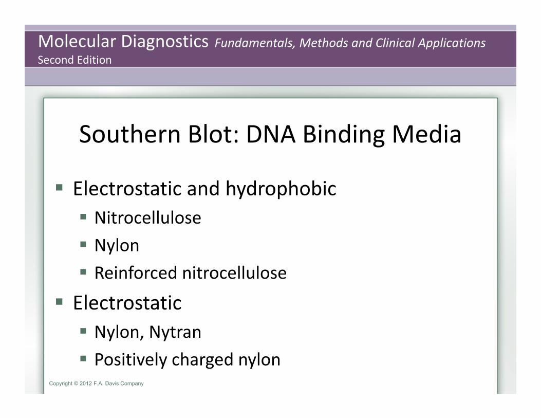
Southern Blot: DNA Binding Media
Electrostatic and hydrophobic Nitrocellulose Nylon Reinforced nitrocellulose
Electrostatic Nylon, Nytran Positively charged nylon
Molecular Diagnostics Fundamentals, Methods and Clinical ApplicationsSecond Edition
Copyright © 2012 F.A. Davis Company
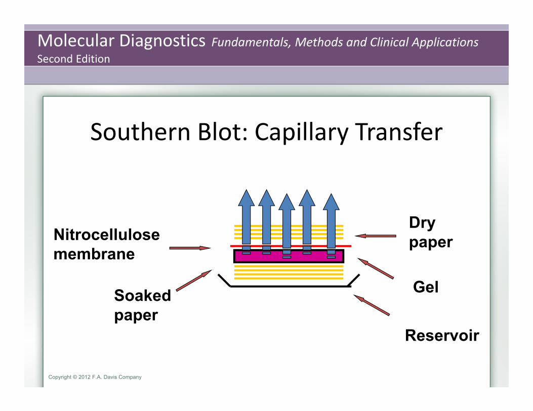
Southern Blot: Capillary Transfer
Drypaper Nitrocellulose
membrane
Gel Soakedpaper
Reservoir

Molecular Diagnostics Fundamentals, Methods and Clinical ApplicationsSecond Edition
Copyright © 2012 F.A. Davis Company
Southern Blot: Electrophoretic Transfer
- +
Buffer Buffer
Whatmanpaper Nitrocellulose filter
Gel Glass plates

Molecular Diagnostics Fundamentals, Methods and Clinical ApplicationsSecond Edition
Copyright © 2012 F.A. Davis Company
Southern Blot: Vacuum Transfer
Nitrocellulose filter
Porous plate
Gel Recirculatingbuffer
Vacuum

Molecular Diagnostics Fundamentals, Methods and Clinical ApplicationsSecond Edition
Copyright © 2012 F.A. Davis Company
Immobilization and Prehybridization After transfer, the cut, denatured DNA is avidly bound to the membrane.
The DNA can be permanently immobilized to the membrane by baking in a vacuum oven (80°C, 30–60 minutes) or by uv cross‐linking, that is, covalently attaching the DNA to the nitrocellulose using UV light energy
Following immobilization of the DNA, a prehybridization step is required to prevent the probe from binding to nonspecific sites on the membrane surface, which will cause high background noise. Prehybridization involves incubating the membrane in the same buffer in
which the probe will subsequently be introduced or in a specially formulated prehybridization buffer. At this point, the buffer does not contain probe.
Blocking agents as Denhardt solution (Ficoll, polyvinyl pyrrolidane, bovine serum albumin) and salmon sperm DNA. Sodium dodecyl sulfate (SDS, 0.01 %) may also be included, along with formamide, the latter especially for RNA probes.
Molecular Diagnostics Fundamentals, Methods and Clinical ApplicationsSecond Edition
Copyright © 2012 F.A. Davis Company
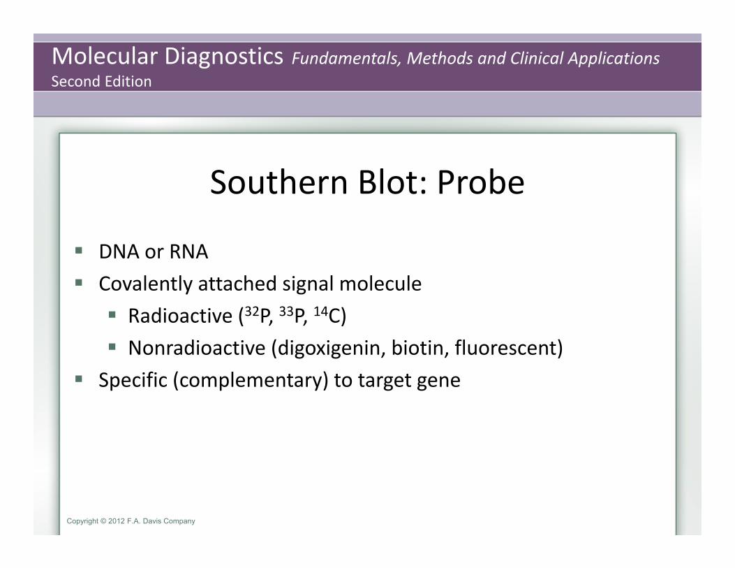
Southern Blot: Probe
DNA or RNA Covalently attached signal molecule Radioactive (32P, 33P, 14C) Nonradioactive (digoxigenin, biotin, fluorescent)
Specific (complementary) to target gene
Molecular Diagnostics Fundamentals, Methods and Clinical ApplicationsSecond Edition
Copyright © 2012 F.A. Davis Company
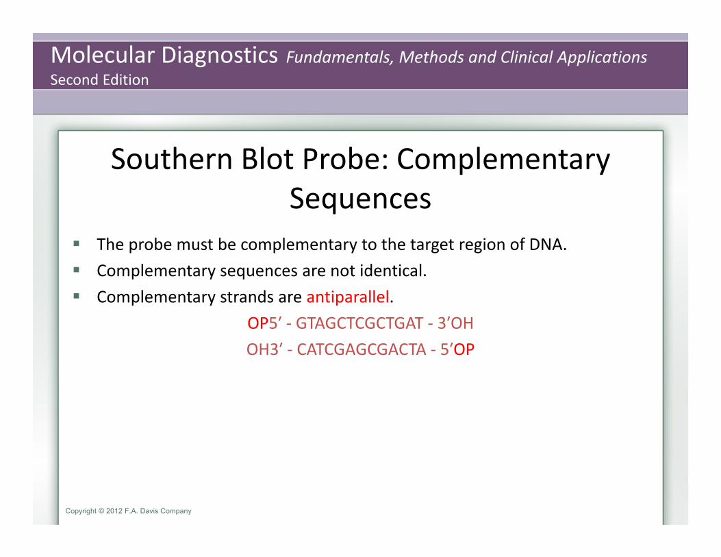
Southern Blot Probe: Complementary Sequences
The probe must be complementary to the target region of DNA. Complementary sequences are not identical. Complementary strands are antiparallel.
OP5′ ‐ GTAGCTCGCTGAT ‐ 3′OHOH3′ ‐ CATCGAGCGACTA ‐ 5′OP

Molecular Diagnostics Fundamentals, Methods and Clinical ApplicationsSecond Edition
Copyright © 2012 F.A. Davis Company
Southern Blot: Probe
The probe determines what fragments are seen on the blot.

Molecular Diagnostics Fundamentals, Methods and Clinical ApplicationsSecond Edition
Copyright © 2012 F.A. Davis Company
Melting Temperature (Tm)
The temperature at which 50% of a nucleic acid is hybridized to its complementary strand.
DS
DS = SS
SS
Tm
Increasing temperature
Molecular Diagnostics Fundamentals, Methods and Clinical ApplicationsSecond Edition
Copyright © 2012 F.A. Davis Company
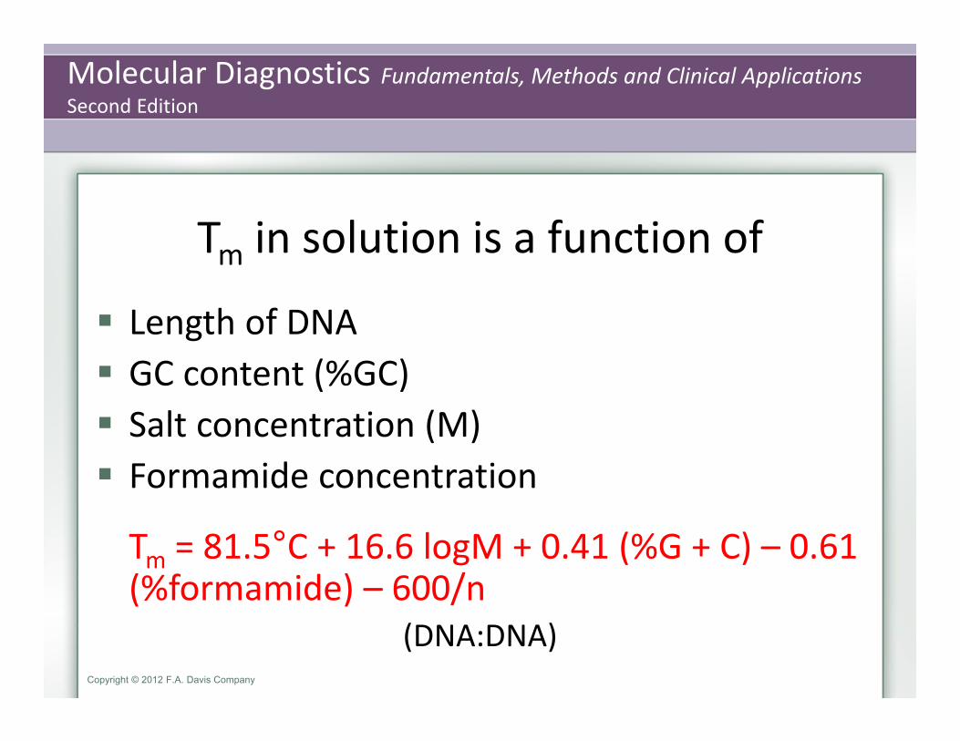
Tm in solution is a function of
Length of DNA GC content (%GC) Salt concentration (M) Formamide concentration
Tm = 81.5°C + 16.6 logM + 0.41 (%G + C) – 0.61 (%formamide) – 600/n
(DNA:DNA)

Molecular Diagnostics Fundamentals, Methods and Clinical ApplicationsSecond Edition
Copyright © 2012 F.A. Davis Company
Tm
For short (14–20 bp) oligomers:Tm = 4° (GC) + 2° (AT)
Molecular Diagnostics Fundamentals, Methods and Clinical ApplicationsSecond Edition
Copyright © 2012 F.A. Davis Company
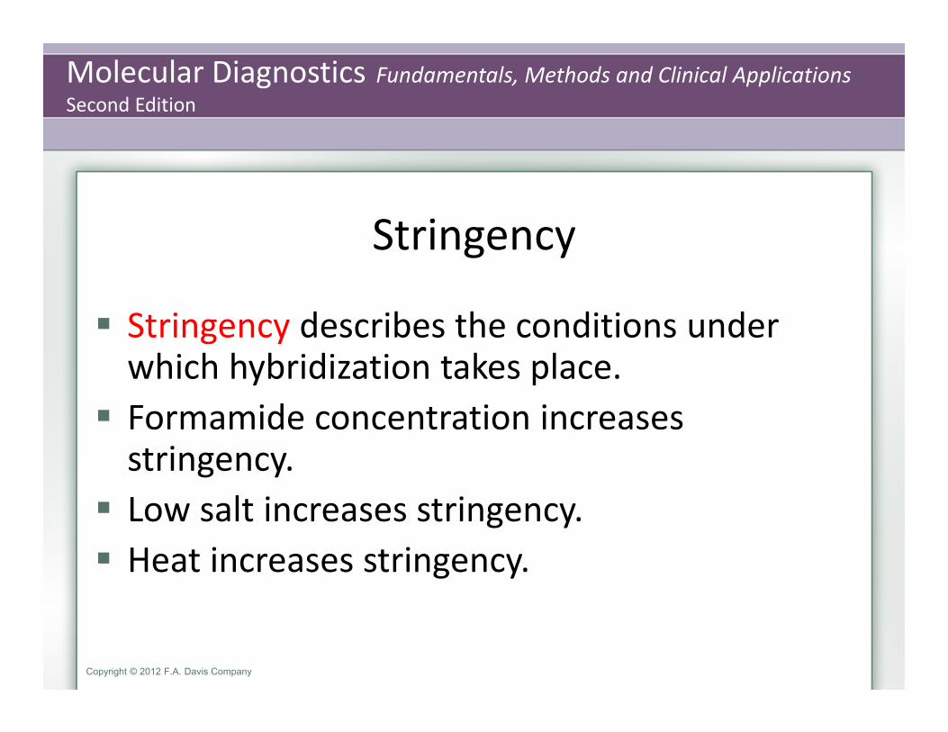
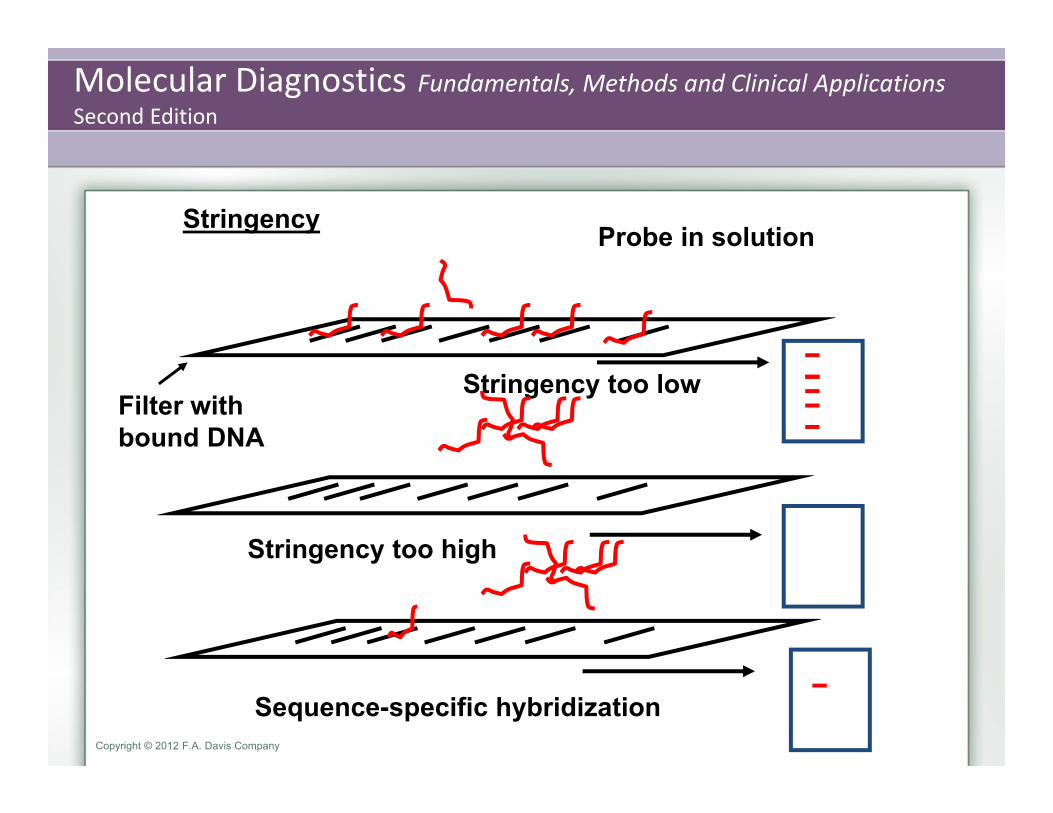
Stringency
Stringency describes the conditions under which hybridization takes place. Formamide concentration increases stringency. Low salt increases stringency. Heat increases stringency.
Molecular Diagnostics Fundamentals, Methods and Clinical ApplicationsSecond Edition
Copyright © 2012 F.A. Davis Company
Stringency Probe in solution
Filter with bound DNA
Stringency too high
Sequence-specific hybridization
Stringency too low
Molecular Diagnostics Fundamentals, Methods and Clinical ApplicationsSecond Edition
Copyright © 2012 F.A. Davis Company
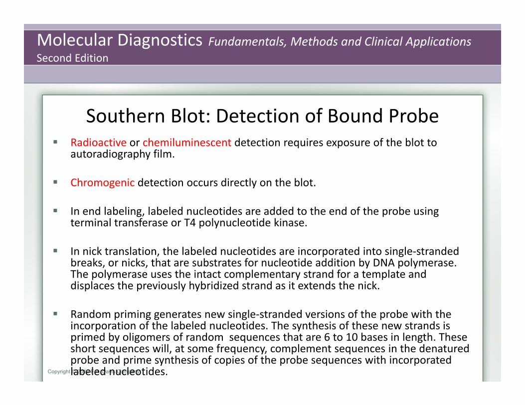
Southern Blot: Detection of Bound Probe Radioactive or chemiluminescent detection requires exposure of the blot to
autoradiography film.
Chromogenic detection occurs directly on the blot.
In end labeling, labeled nucleotides are added to the end of the probe using terminal transferase or T4 polynucleotide kinase.
In nick translation, the labeled nucleotides are incorporated into single‐stranded breaks, or nicks, that are substrates for nucleotide addition by DNA polymerase. The polymerase uses the intact complementary strand for a template and displaces the previously hybridized strand as it extends the nick.
Random priming generates new single‐stranded versions of the probe with the incorporation of the labeled nucleotides. The synthesis of these new strands is primed by oligomers of random sequences that are 6 to 10 bases in length. These short sequences will, at some frequency, complement sequences in the denatured probe and prime synthesis of copies of the probe sequences with incorporated labeled nucleotides.

Molecular Diagnostics Fundamentals, Methods and Clinical ApplicationsSecond Edition
Copyright © 2012 F.A. Davis Company
Radioactive isotope
Probe
Filter with bound DNA
Radioactive Signal Detection
Molecular Diagnostics Fundamentals, Methods and Clinical ApplicationsSecond Edition
Copyright © 2012 F.A. Davis Company
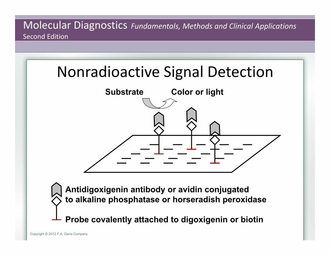
Nonradioactive Signal Detection
Antidigoxigenin antibody or avidin conjugatedto alkaline phosphatase or horseradish peroxidase
Probe covalently attached to digoxigenin or biotin
Substrate Color or light

Molecular Diagnostics Fundamentals, Methods and Clinical ApplicationsSecond Edition
Copyright © 2012 F.A. Davis Company
Southern Blot Results
Radioactive orchemiluminescent detection
(autoradiography film)
Chromogenic detection
(nitrocellulose membrane)
Molecular Diagnostics Fundamentals, Methods and Clinical ApplicationsSecond Edition
Copyright © 2012 F.A. Davis Company
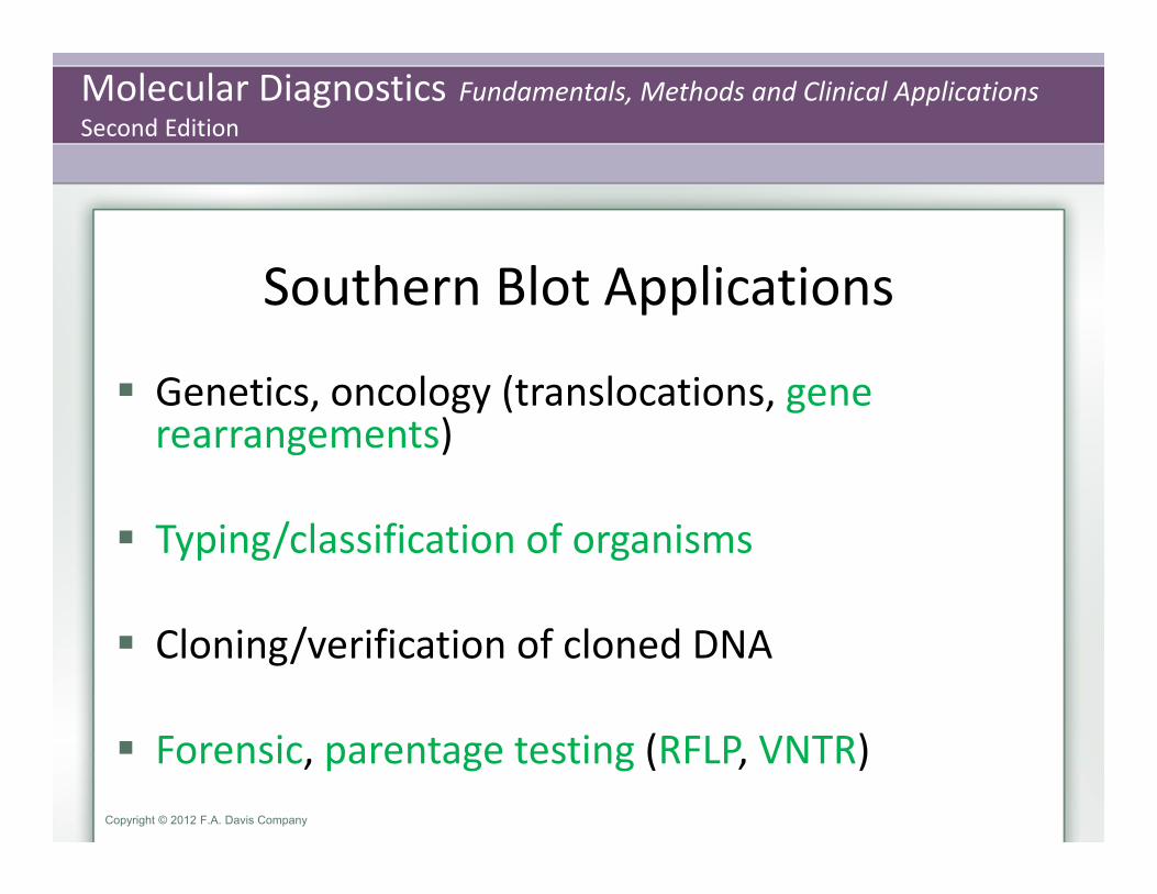
Southern Blot Applications
Genetics, oncology (translocations, gene rearrangements)
Typing/classification of organisms
Cloning/verification of cloned DNA
Forensic, parentage testing (RFLP, VNTR)

Molecular Diagnostics Fundamentals, Methods and Clinical ApplicationsSecond Edition
Copyright © 2012 F.A. Davis Company
Northern Blot
RNA structure and quantity
Similar rationale as Southern blot except RNA target rather than DNA No restriction digestion required Probe must be designed as complement to the single‐stranded RNA Provides measure of relative expression of genes normalized to internal control

Molecular Diagnostics Fundamentals, Methods and Clinical ApplicationsSecond Edition
Copyright © 2012 F.A. Davis Company
(up to approximately 30 µg total RNA or 0.5–3.0 g polyARNA
Gel electrophoresis of RNA must be carried out under denaturing conditions
Denaturant such as formaldehyde must be removed from the gel before transfer because it inhibits binding of the RNA to nitrocellulose. This is accomplished by rinsing the gel in deionized water

Molecular Diagnostics Fundamentals, Methods and Clinical ApplicationsSecond Edition
Copyright © 2012 F.A. Davis Company
Western Blot Serum, cell lysate, or protein extract is separated on SDS‐polyacrylamide gels (SDS‐PAGE) or isoelectric focusing gels (IEF). The former resolves proteins according to molecular weight, and the latter according to charge.
Samples are treated with denaturant, such as mixing 1:1 with 0.04 M Tris HCl, pH 6.8, 0.1% SDS. 1 –50 µg of protein is loaded per well
5%–20% polyacrylamide gels
Molecular Diagnostics Fundamentals, Methods and Clinical ApplicationsSecond Edition
Copyright © 2012 F.A. Davis Company
Western Blot Proteins may be renatured before blotting to optimize antibody
(probe)‐epitope binding.
Proteins are blotted to membranes by capillary or electrophoretic transfer.
Probes are specific binding proteins, polyclonal antibodies, or monoclonal antibodies.
Nitrocellulose has high affinity for proteins and is easily treated with detergent (0.1% Tween 20 in 0.05 M Tris and 0.15 M sodium chloride, pH 7.6) to prevent binding of the primary antibody probe to the membrane itself (blocking) before hybridization.

Molecular Diagnostics Fundamentals, Methods and Clinical ApplicationsSecond Edition
Copyright © 2012 F.A. Davis Company
Western Blot Signal Detection
Target protein
Primaryantibody(probe)
Secondaryantibody
Label
Molecular Diagnostics Fundamentals, Methods and Clinical ApplicationsSecond Edition
Copyright © 2012 F.A. Davis Company
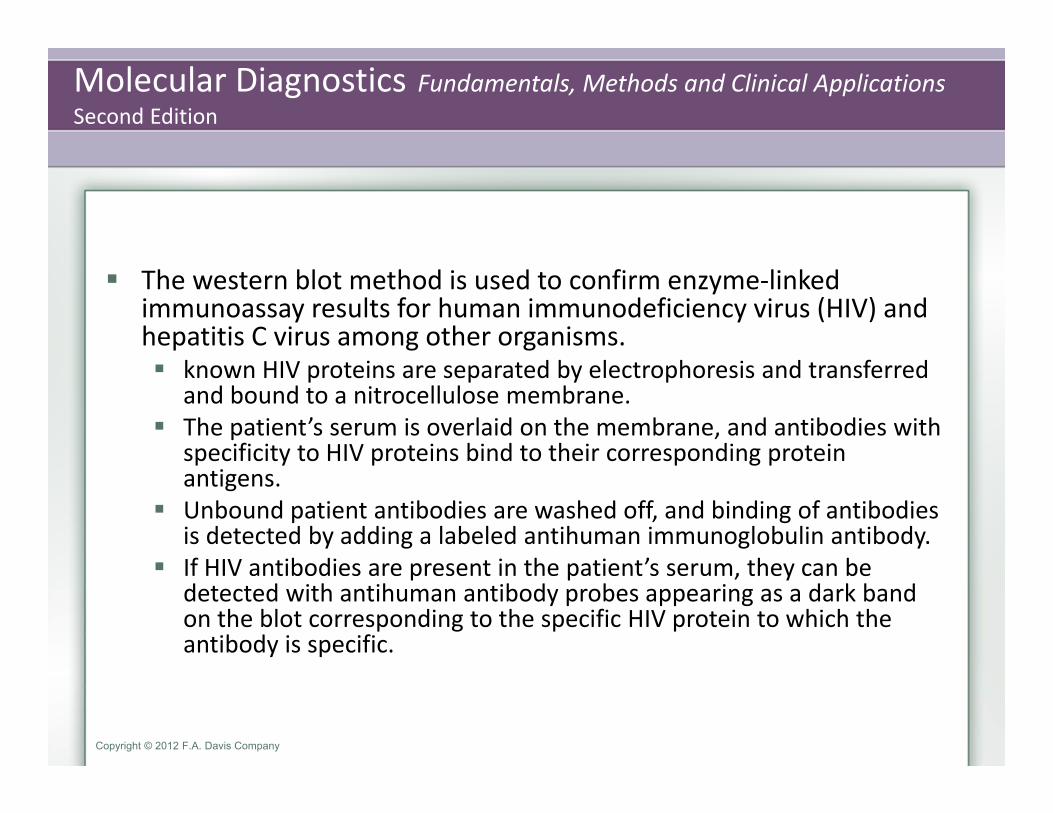
The western blot method is used to confirm enzyme‐linked immunoassay results for human immunodeficiency virus (HIV) and hepatitis C virus among other organisms. known HIV proteins are separated by electrophoresis and transferred
and bound to a nitrocellulose membrane. The patient’s serum is overlaid on the membrane, and antibodies with
specificity to HIV proteins bind to their corresponding protein antigens.
Unbound patient antibodies are washed off, and binding of antibodies is detected by adding a labeled antihuman immunoglobulin antibody.
If HIV antibodies are present in the patient’s serum, they can be detected with antihuman antibody probes appearing as a dark band on the blot corresponding to the specific HIV protein to which the antibody is specific.

Molecular Diagnostics Fundamentals, Methods and Clinical ApplicationsSecond Edition
Copyright © 2012 F.A. Davis Company
Polyclonal antibodies are products of a generalized response to a specific antigen, usually a peptide or protein.
Polyclonal antibodies are comprised of a mixture of immunoglobulinsdirected at more than one epitope (molecular structure) on the antigen
Monoclonal antibodies are more difficult to produce
In western blot technology, polyclonal antibodies can give a more robust signal, especially if the target epitopes are partially lost during electrophoresis and transfer. Monoclonal antibodies are more specific and may give less background noise; however, if the targeted epitope is lost, these antibodies do not bind and no signal is generated.

Molecular Diagnostics Fundamentals, Methods and Clinical ApplicationsSecond Edition
Copyright © 2012 F.A. Davis Company
Filter‐Based Hybridization Technologies
Target ProbeSouthern blot DNA nucleic acid
Northern blot RNA nucleic acid
Western blot protein protein
Southwestern blot protein DNAEastern blot protein lectin, protein
Molecular Diagnostics Fundamentals, Methods and Clinical ApplicationsSecond Edition
Copyright © 2012 F.A. Davis Company
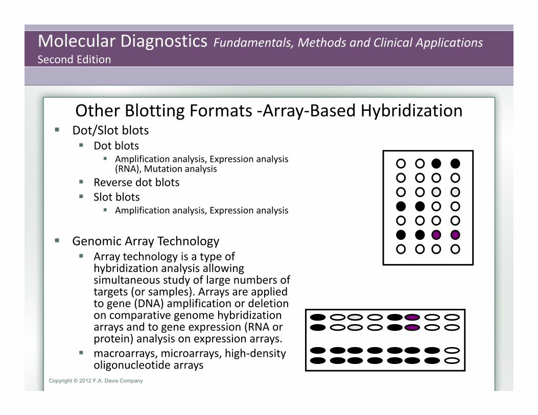
Other Blotting Formats ‐Array‐Based Hybridization Dot/Slot blots
Dot blots Amplification analysis, Expression analysis
(RNA), Mutation analysis Reverse dot blots Slot blots
Amplification analysis, Expression analysis
Genomic Array Technology Array technology is a type of
hybridization analysis allowing simultaneous study of large numbers of targets (or samples). Arrays are applied to gene (DNA) amplification or deletion on comparative genome hybridization arrays and to gene expression (RNA or protein) analysis on expression arrays.
macroarrays, microarrays, high‐density oligonucleotide arrays

Molecular Diagnostics Fundamentals, Methods and Clinical ApplicationsSecond Edition
Copyright © 2012 F.A. Davis Company
Macroarrays are reverse dot blots of up to several thousand targets on nitrocellulose membranes.
Microarray ‐ Tens of thousands of targets can be screened simultaneously in a very small area by miniaturizing the deposition of droplets
High‐density oligonucleotide arrays and are used for mutation analysis, single nucleotide polymorphism analysis, and sequencing.
Sample preparation for array analysis requires fluorescent labeling of the test sample, as microarrays and other high‐density arrays are read by automated fluorescent detection systems. The most frequent labeling method used for RNA is synthesis of cDNA or RNA copies with incorporation of labeled nucleotides. For DNA, random priming or nick translation is used.

Molecular Diagnostics Fundamentals, Methods and Clinical ApplicationsSecond Edition
Copyright © 2012 F.A. Davis Company

Molecular Diagnostics Fundamentals, Methods and Clinical ApplicationsSecond Edition
Copyright © 2012 F.A. Davis Company
Expression arrays measure transcript or protein production relative to a reference control isolated from untreated or normal specimens.
Comparative genome hybridization (array CGH) is designed to test DNA. This method is used to screen the genome or specific genomic loci for deletions and amplifications.

Molecular Diagnostics Fundamentals, Methods and Clinical ApplicationsSecond Edition
Copyright © 2012 F.A. Davis Company
Comparative Genomic Hybridization (CGH)
Immobilized, denatured normal chromosomes
Test and reference DNA are labeled by incorporation of nucleotides covalently attached to fluorescent dyes.
(Test) (Reference)

Molecular Diagnostics Fundamentals, Methods and Clinical ApplicationsSecond Edition
Copyright © 2012 F.A. Davis Company
Comparative Genomic Hybridization (CGH) The labeled DNA is hybridized to the normal
chromosomes on a microscope slide.
Differences between normal and reference will be revealed. Amplification: test color dominates. Deletion: reference color dominates.
Normal reference DNA
Test sample DNA
(Amplification at this locus)
(Deletion at this locus)
Molecular Diagnostics Fundamentals, Methods and Clinical ApplicationsSecond Edition
Copyright © 2012 F.A. Davis Company
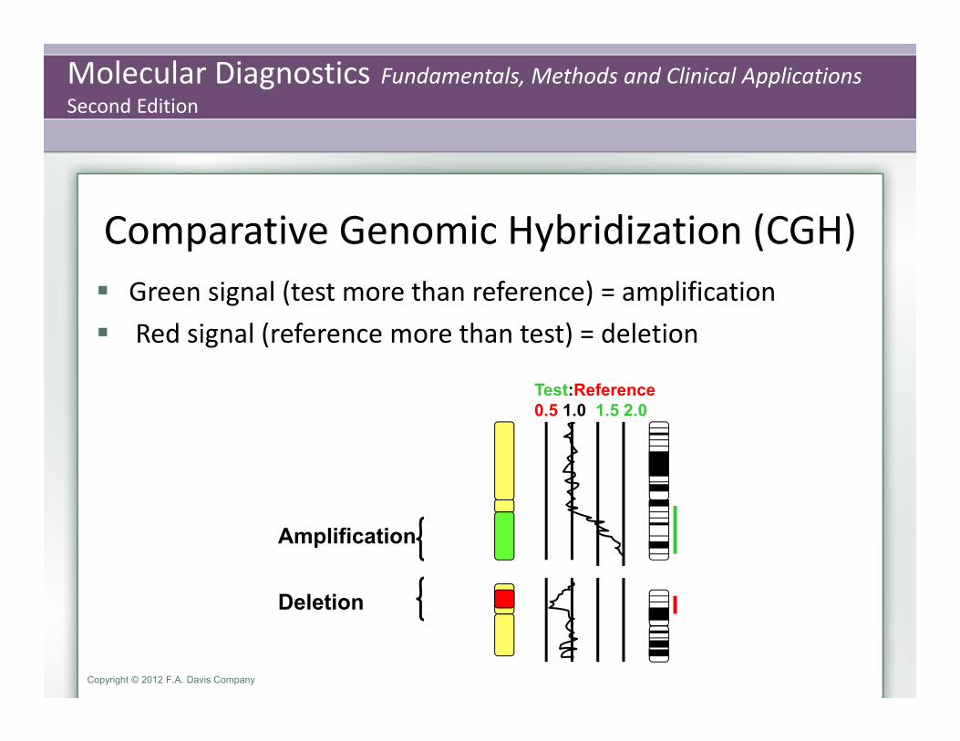
Comparative Genomic Hybridization (CGH) Green signal (test more than reference) = amplification Red signal (reference more than test) = deletion
Amplification
Deletion
Test:Reference0.5 1.0 1.5 2.0
Molecular Diagnostics Fundamentals, Methods and Clinical ApplicationsSecond Edition
Copyright © 2012 F.A. Davis Company
Bead Array Technology The probes may also be immobilized on beads, allowing
hybridization of the targets in the bead suspension
In order to distinguish specific probes carried on different beads, the beads are color‐coded with a fluorescent dye. The sample is then labeled with a different dye so that the combination of the target and bead fluorescent signals indicates the presence or absence of a specific target. This technology can be used for protein as well as nucleic acid targets. Clinical tests using Luminex systems are available for infectious diseases and tissue typing.
Molecular Diagnostics Fundamentals, Methods and Clinical ApplicationsSecond Edition
Copyright © 2012 F.A. Davis Company
Solution hybridization In solution hybridization, neither the probe nor the target is
immobilized. Probes and targets bind in solution, followed by resolution of the bound products.
With the increasing interest in short interfering RNAs (siRNAs) and microRNAs (miRNAs), which are conveniently analyzed by this type of hybridization analysis, solution methods may come into more frequent use.
Solution hybridization has been used to measure mRNA expression, especially when there are low amounts of target RNA. One version of the method is called RNase protection, or S1 mapping, for the S1 singlestrand–specific nuclease.
Molecular Diagnostics Fundamentals, Methods and Clinical ApplicationsSecond Edition
Copyright © 2012 F.A. Davis Company
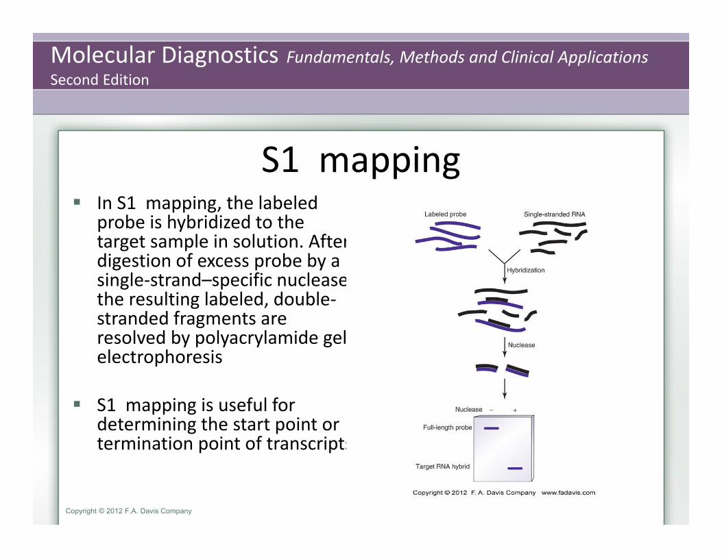
S1 mapping In S1 mapping, the labeled
probe is hybridized to the target sample in solution. After digestion of excess probe by a single‐strand–specific nuclease, the resulting labeled, double‐stranded fragments are resolved by polyacrylamide gel electrophoresis
S1 mapping is useful for determining the start point or termination point of transcripts

Molecular Diagnostics Fundamentals, Methods and Clinical ApplicationsSecond Edition
Copyright © 2012 F.A. Davis Company
Gel mobility shift assay
Solution hybridization can also be applied to the analysis of protein‐protein interactions and to nucleic acid–binding proteins using a gel mobility shift assay
After mixing the labeled DNA or protein with the test material, such as a cell lysate, a change in mobility, usually a shift to slower migration, indicates binding of a component in the test material to the probe protein or nucleic acid

Molecular Diagnostics Fundamentals, Methods and Clinical ApplicationsSecond Edition
Copyright © 2012 F.A. Davis Company
Summary Restriction enzymes cut DNA at specific recognition sequences.
DNA can be characterized by restriction enzyme mapping.
Specific DNA regions in a complex mixture are characterized using Southern blot.
Specific proteins in a complex mixture are characterized using western blot.
Regions of genomic amplification or deletion are characterized using comparative genomic hybridization.