Embed Size (px)
Citation preview

Anatomy & Physiology
Tissue: The Living Fabric
Anatomy & Physiology
Tissue: The Living Fabric
ObjectivesIdentify and describe the functions of the 4 main types of body tissues
Describe the various types and functions of epithelia
Explain the properties and functions of different types of connective tissue
Identify the major types of muscle tissue
Describe the basic types and functions of nerve tissue
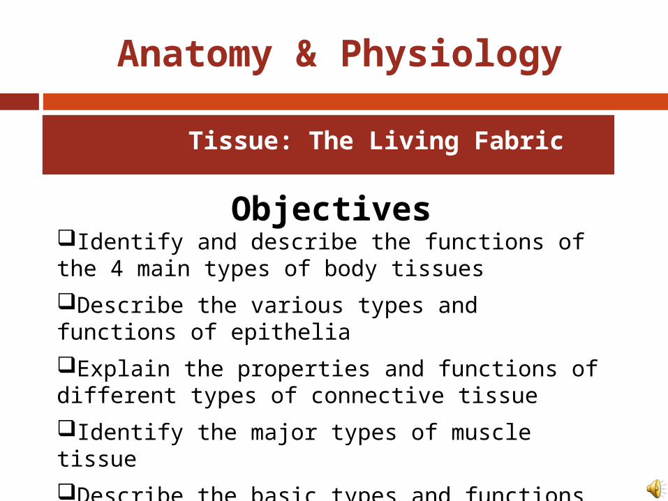
Tissues
Groups of cells similar in structure and function
Types of tissues Epithelial tissue Connective tissue Muscle tissue Nerve tissue

Figure 4.1
Nervous tissue: Internal communication• Brain, spinal cord, and nerves
Muscle tissue: Contracts to cause movement• Muscles attached to bones (skeletal)• Muscles of heart (cardiac)• Muscles of walls of hollow organs (smooth)
Epithelial tissue: Forms boundaries between different environments, protects, secretes, absorbs, filters• Skin surface (epidermis)• Lining of GI tract organs and other hollow organs
Connective tissue: Supports, protects, bindsother tissues together• Bones• Tendons• Fat and other soft padding tissue
Intro to tissues

Epithelial Tissue (Epithelium) Two main types (by location):
1. Covering and lining epithelia On external and internal surfaces
2. Glandular epithelia Secretory tissue in glands
Functions of Epithelial Tissue 6 main functions of epithelium
Protection (skin) Absorption (digestive tract, kidneys) Filtration (digestive tract, kidneys) Excretion (digestive tract, kidneys) Secretion (glands, kidneys) Sensory reception (skin)

Homeostatic Imbalance
Epithelial Tissue An important characteristic of cancerous
epithelial cells is their failure to respect the basement membrane boundary. (85 out of 100 cancers are of epithelial cells) They penetrate it and invade the tissues
beneath.

Characteristics of Epithelial Tissue
1. Cells have polarity- apical (upper, free) and basal (lower, attached) surfaces- cell regions near the apical surface differ in structure and function from cell regions in the basal surface.
Apical surfaces may have: Microvilli: finger-like
extensions of the plasma membrane that increase surface area.
Lining of the intestine Cilia: tiny hair like
projections that propel substances along their free surface.
Lining of the trachea

Characteristics of Epithelial Tissue
2. Are composed of closely packed cells to form continuous sheets.
3. Supported by a connective tissue reticular lamina (under the basal lamina)
A layer of extracellular material containing a fine network of collagen and protein fibers.
4. Avascular (contains no blood vessels) but innervated (supplied by nerve fibers).
Epithelial cells are nourished by substances diffusing from blood vessels in the underlying connective tissue.
5. High rate of regeneration Reproduce rapidly to replace lost cells due to hostile
substances in the external environment.

Classification of Epithelia
Ask two questions:1. How many layers?
1 layer = simple epithelium
Very thin most concerned with absorption, secretion, and filtration.
2 or more layers = stratified epithelium
More durable, major role is protection.
Stratified
Simple
Apical surface
Basal surface
Apical surface
Basal surface
(a) Classification based on number of cell layers.
Figure 4.2a

Squamous
Cuboidal
Columnar(b) Classification based on cell shape.
Classification of Epithelia
2. What type of cell? Squamous
Flattened and scale-like Cuboidal
Box-like, tall as they are wide Columnar
Tall and column shaped
(If stratified, name according to apical (top) layer of cells)
Figure 4.2b

Epithelia: Simple Squamous
Thin, often permeable cells
Found where filtration and exchange of substances by rapid diffusion is a priority
KidneysLungsBlood vessels

Epithelia: Simple Squamous
Two noteworthy names Endothelium:
“innercovering” slick-friction reducing lining The lining of lymphatic
vessels, blood vessels, and heart
Mesothelium: “middlecovering” The epithelium of serous
membranes in the ventral body cavity

Figure 4.3a
(a) Simple squamous epithelium
Description: Single layer of flattenedcells with disc-shaped central nucleiand sparse cytoplasm; the simplestof the epithelia.
Function: Allows passage ofmaterials by diffusion and filtrationin sites where protection is notimportant; secretes lubricatingsubstances in serosae.
Location: Kidney glomeruli; air sacsof lungs; lining of heart, bloodvessels, and lymphatic vessels; liningof ventral body cavity (serosae).
Photomicrograph: Simple squamous epitheliumforming part of the alveolar (air sac) walls (125x).
Air sacs oflung tissue
Nuclei ofsquamousepithelialcells

Figure 4.3b
(b) Simple cuboidal epithelium
Description: Single layer ofcubelike cells with large,spherical central nuclei.
Function: Secretion andabsorption.
Location: Kidney tubules;ducts and secretory portionsof small glands; ovary surface.
Photomicrograph: Simple cuboidalepithelium in kidney tubules (430x).
Basementmembrane
Connectivetissue
Simplecuboidalepithelialcells

Figure 4.3c
(c) Simple columnar epithelium
Description: Single layer of tall cells with round to oval nuclei; some cells bear cilia; layer may contain mucus-secreting unicellular glands (goblet cells).
Function: Absorption; secretion of mucus, enzymes, and other substances; ciliated type propels mucus (or reproductive cells) by ciliary action.
Location: Nonciliated type lines most of the digestive tract (stomach to anal canal),gallbladder, and excretory ducts of someglands; ciliated variety lines small bronchi, uterine tubes, and some regionsof the uterus.
Photomicrograph: Simple columnar epitheliumof the stomach mucosa (860X).
Simplecolumnarepithelialcell
Basementmembrane

Figure 4.3d
(d) Pseudostratified columnar epithelium
Description: Single layer of cells ofdiffering heights, some not reachingthe free surface; nuclei seen atdifferent levels; may contain mucus-secreting cells and bear cilia.
Function: Secretion, particularly ofmucus; propulsion of mucus byciliary action.
Location: Nonciliated type in male’ssperm-carrying ducts and ducts oflarge glands; ciliated variety linesthe trachea, most of the upperrespiratory tract.
Photomicrograph: Pseudostratified ciliatedcolumnar epithelium lining the human trachea (570x).
Trachea
Cilia
Pseudo-stratifiedepitheliallayer
Basementmembrane
Mucus ofmucous cell

Figure 4.3e
(e) Stratified squamous epithelium
Description: Thick membranecomposed of several cell layers;basal cells are cuboidal or columnarand metabolically active; surfacecells are flattened (squamous); in thekeratinized type, the surface cells arefull of keratin and dead; basal cellsare active in mitosis and produce thecells of the more superficial layers.
Function: Protects underlyingtissues in areas subjected to abrasion.
Location: Nonkeratinized type formsthe moist linings of the esophagus,mouth, and vagina; keratinized varietyforms the epidermis of the skin, a drymembrane.
Photomicrograph: Stratified squamous epitheliumlining the esophagus (285x).
Stratifiedsquamousepithelium
Nuclei
Basementmembrane
Connectivetissue

Epithelia: Stratified Cuboidal Quite rare in body Found in some sweat and mammary
glands Typically two cell layers thick
Sweat Duct

Epithelia: Stratified Columnar Limited distribution in body Small amounts in pharynx, male urethra,
and lining some glandular ducts Also occurs at transition areas between
two other types of epithelia

Figure 4.3f
(f) Transitional epithelium
Description: Resembles both stratified squamous and stratified cuboidal; basal cells cuboidal or columnar; surface cells domeshaped or squamouslike, depending on degree of organ stretch.
Function: Stretches readily and permits distension of urinary organ by contained urine.
Location: Lines the ureters, urinary bladder, and part of the urethra.
Photomicrograph: Transitional epithelium lining the urinary bladder, relaxed state (360X); note the bulbous, or rounded, appearance of the cells at the surface; these cells flatten and become elongated when the bladder is filled with urine.
BasementmembraneConnectivetissue
Transitionalepithelium

Answer the following questions and turn in!!
Explain what is meant by epithelial tissue being avascular but innervated.
What structure can be noted on the apical surface of the cells in this image?What is the name of this tissue type?A multilayered epithelium with cuboidal basal cells and flat cells at its surface would be classified as ________?

Answer the following questions and turn in!!
Explain what is meant by epithelial cells having polarity
What is significant about the cells closes to the basement membrane of this tissue type?What is the name of this tissue type?A multilayered epithelium with cuboidal basal cells and columnar cells at its surface would be classified as ________?What types of organs would you find transitional epithelium in?
.

Glandular Epithelia
Objectives: Define gland Differentiate between exocrine and
endocrine glands, and differentiate between multicellular and unicellular glands
Describe how multicellular exocrine glands are classified structurally and functionally

Glandular Epithelia
A gland is one or more cells that makes and secretes an aqueous fluid.
Glandular cells obtain needed substances from blood and transform them chemically into a product that is then released from the cell.
Classified by: Site of product release—endocrine (internally
secreting) or exocrine (externally secreting) Relative number of cells forming the gland—
unicellular (e.g., goblet cells) or multicellular (typically ducted)

Endocrine Glands
Ductless glands Secrete hormones that travel through
lymph or blood to target organs

Exocrine Glands
More numerous than endocrine glands Secrete products into ducts Secretions released onto body surfaces
(skin) or into body cavities Examples include mucous, sweat, oil,
and salivary glands

Microvilli
Secretoryvesiclescontainingmucin
Golgiapparatus
Rough ER
Nucleus
Unicellular Exocrine Glands
The only important unicellular gland are mucous cells and goblet cells. (scattered) Goblet cells- look like a glass
with a stem due to the accumulation of mucin at the top of the cell.
Both produce mucin that dissolves in water when secreted and forms mucous- a slimy protective and lubricating coating found within the human body.
Goblet Cell

Multicellular Exocrine Glands Multicellular exocrine glands are
composed of a duct and a secretory unit Classified according to:
Duct type Simple: unbranched duct Compound: branched duct
Structure of their secretory units Tubular: secretory cells form tubes. Alveolar (acinar): secretory cells form small sacs. Tubuloalveolar: have both types of secretory
units.

Figure 4.5
Compound duct structure(duct branches)
Simple tubular
ExampleIntestinal glands
Simple branchedtubular
ExampleStomach (gastric)glands
Compound tubular
ExampleDuodenal glands of small intestine
Compound alveolar
ExampleMammary glands
Simplealveolar
ExampleNo importantexample in humans
Simple branchedalveolar
ExampleSebaceous (oil)glands
Compoundtubuloalveolar
ExampleSalivary glands
Tubularsecretorystructure
Alveolarsecretorystructure
Surface epithelium Duct Secretory epithelium
Simple duct structure(duct does not branch)

Modes of Secretion
Merocrine Products are secreted by
exocytosis Examples: pancreas,
sweat and salivary glands.
Holocrine Products are secreted by
rupture of gland cells Example: sebaceous
glands

Answer the following questions and turn in!!
What is a gland? Explain the difference between endocrine and
exocrine glands and provide an example of each
What type of an exocrine gland is a goblet cell and what does it produce?
Draw and label the various structural presentations for multicellular exocrine glands.
What is the primary difference between the way merocrine and halocrine glands secrete their products?

Connective Tissue
Objectives:Identify common characteristics of connective tissue, and list and describe its common structural elements
Describe the common types of connective tissue found in the body and indicated their particular functions

Connective Tissue
Most abundant and widely distributed tissue type
Four classes Connective tissue proper Cartilage Bone tissue Blood

Table 4.1

Major Functions of Connective Tissue Binding and support Protection Insulation Transportation (blood)

Characteristics of Connective Tissue Connective tissues have:
Mesenchyme (embryonic tissue) as their common tissue of origin
Varying degrees of vascularity (supply of blood vessels)
Cells separated by nonliving extracellular matrix (ground substance and fibers) This is what enables the
connective tissue to bear weight, withstand great tension, and endure physical trauma.
Areolar Connective Tissue
Mesenchymal Cells

Structural Elements of Connective Tissue Ground substance
Unstructured material that fills the space between the cells and contains the fibers
Functions as a molecular sieve through which nutrients and other dissolved substances diffuse between blood capillaries and cells.
Components: Interstitial fluid Adhesion proteins (“glue”) Proteoglycans
Protein core + large polysaccharides (chrondroitin sulfate and hyaluronic acid)
Trap water in varying amounts, affecting the thickness of the ground substance

Structural Elements of Connective Tissue Fibers provide support. Three types of fibers
Collagen (white fibers) Strongest and most abundant type Provides high tensile strength
Elastic Networks of long, thin, elastin fibers that allow
for stretch Reticular
Short, fine, highly branched collagenous fibers

Structural Elements of Connective Tissue Cells
Mitotically active and secretory cells = “blasts” Mature cells = “cytes”
Fibroblasts in connective tissue proper Chondroblasts and chondrocytes in cartilage Osteoblasts and osteocytes in bone Hematopoietic stem cells in bone marrow Fat cells, white blood cells, mast cells, and
macrophages

Figure 4.7
Macrophage
Fibroblast
Lymphocyte
Fat cell
Mast cell
Neutrophil
Capillary
Cell types Extracellular matrix
Fibers• Collagen fiber• Elastic fiber• Reticular fiber
Ground substance
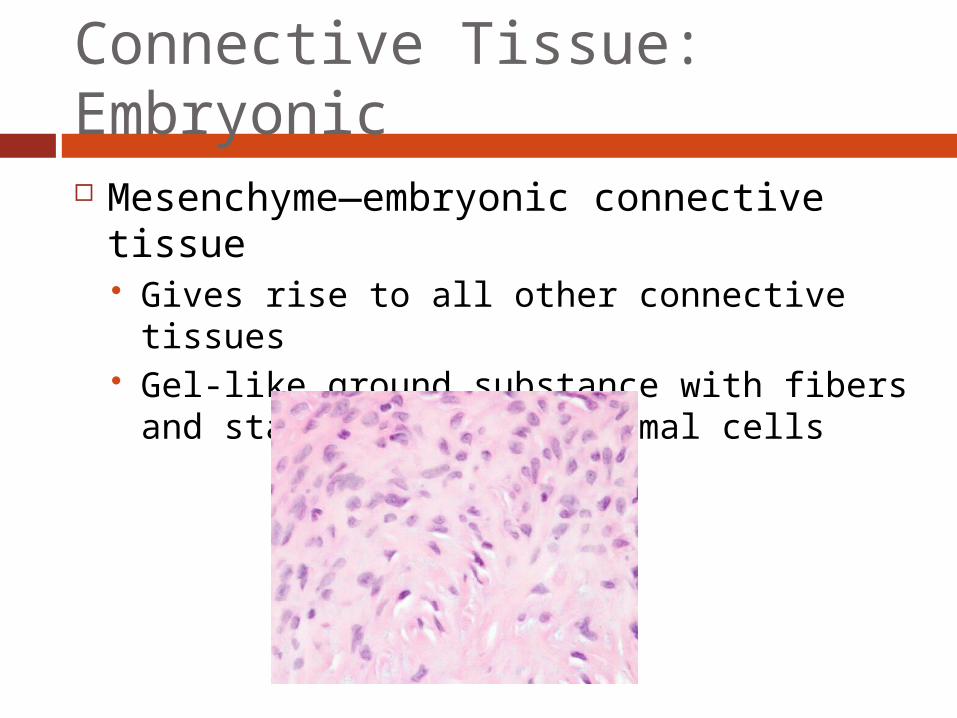
Connective Tissue: Embryonic Mesenchyme—embryonic connective
tissue Gives rise to all other connective tissues Gel-like ground substance with fibers and
star-shaped mesenchymal cells

Answer the following questions and turn in!!
What are the 4 classes of connective tissue?
What 3 types of fibers provide support for connective tissue?
What are 4 functions of connective tissue? What function(s) does adipose serve?

Connective Tissue Proper
Types: Loose connective
tissue Areolar Adipose Reticular
Dense connective tissue Dense regular Dense irregular Elastic
The name connective tissue proper is used to designate the connective tissue that fills interstitial spaces as opposed to the specialized connective tissues (blood, bones, cartilage, etc…).

(a) Connective tissue proper: loose connective tissue, areolar
Description: Gel-like matrix with allthree fiber types; cells: fibroblasts,macrophages, mast cells, and somewhite blood cells.
Function: Wraps and cushionsorgans; its macrophages phagocytizebacteria; plays important role ininflammation; holds and conveystissue fluid.
Location: Widely distributed underepithelia of body, e.g., forms laminapropria of mucous membranes;packages organs; surroundscapillaries.
Photomicrograph: Areolar connective tissue, asoft packaging tissue of the body (300x).
Epithelium
Laminapropria
Fibroblastnuclei
Elasticfibers
Collagenfibers
Figure 4.8a

Figure 4.8b
(b) Connective tissue proper: loose connective tissue, adipose
Description: Matrix as in areolar,but very sparse; closely packedadipocytes, or fat cells, havenucleus pushed to the side by largefat droplet.
Function: Provides reserve foodfuel; insulates against heat loss;supports and protects organs.
Location: Under skin in thehypodermis; around kidneys andeyeballs; within abdomen; in breasts.
Photomicrograph: Adipose tissue from thesubcutaneous layer under the skin (350x).
Nucleus offat cell
Vacuolecontainingfat droplet
Adiposetissue
Mammaryglands

Figure 4.8c
(c) Connective tissue proper: loose connective tissue, reticular
Description: Network of reticularfibers in a typical loose groundsubstance; reticular cells lie on thenetwork.
Function: Fibers form a soft internalskeleton (stroma) that supports othercell types including white blood cells,mast cells, and macrophages.
Location: Lymphoid organs (lymphnodes, bone marrow, and spleen).
Photomicrograph: Dark-staining network of reticularconnective tissue fibers forming the internal skeletonof the spleen (350x).
Spleen
White bloodcell(lymphocyte)
Reticularfibers

Figure 4.8d
(d) Connective tissue proper: dense connective tissue, dense regular
Description: Primarily parallelcollagen fibers; a few elastic fibers;major cell type is the fibroblast.
Function: Attaches muscles tobones or to muscles; attaches bonesto bones; withstands great tensilestress when pulling force is appliedin one direction.
Location: Tendons, mostligaments, aponeuroses.
Photomicrograph: Dense regular connectivetissue from a tendon (500x).
Shoulderjoint
Ligament
Tendon
Collagenfibers
Nuclei offibroblasts

Figure 4.8e
(e) Connective tissue proper: dense connective tissue, dense irregular
Description: Primarilyirregularly arranged collagenfibers; some elastic fibers;major cell type is the fibroblast.
Function: Able to withstandtension exerted in manydirections; provides structuralstrength.
Location: Fibrous capsules oforgans and of joints; dermis ofthe skin; submucosa ofdigestive tract.
Photomicrograph: Dense irregularconnective tissue from the dermis of theskin (400x).
Collagenfibers
Nuclei offibroblasts
Fibrousjointcapsule

Figure 4.8f
(f) Connective tissue proper: dense connective tissue, elastic
Description: Dense regularconnective tissue containing a highproportion of elastic fibers.
Function: Allows recoil of tissuefollowing stretching; maintainspulsatile flow of blood througharteries; aids passive recoil of lungsfollowing inspiration.
Location: Walls of large arteries;within certain ligaments associatedwith the vertebral column; within thewalls of the bronchial tubes.
Elastic fibers
Aorta
HeartPhotomicrograph: Elastic connective tissue inthe wall of the aorta (250x).

Connective Tissue: Cartilage Three types of cartilage:
Hyaline cartilage Elastic cartilage Fibrocartilage

Homeostatic Imbalance
Cartilage Aging cartilage cells lose their ability to
divide injure cartilages heal slowly. During later life cartilages tend to calcify or
ossify (become bony).

Figure 4.8g
(g) Cartilage: hyaline
Description: Amorphous but firmmatrix; collagen fibers form animperceptible network; chondroblastsproduce the matrix and when mature(chondrocytes) lie in lacunae.
Function: Supports and reinforces;has resilient cushioning properties;resists compressive stress.
Location: Forms most of theembryonic skeleton; covers the endsof long bones in joint cavities; formscostal cartilages of the ribs; cartilagesof the nose, trachea, and larynx.
Photomicrograph: Hyaline cartilage from thetrachea (750x).
Costalcartilages
Chondrocytein lacuna
Matrix
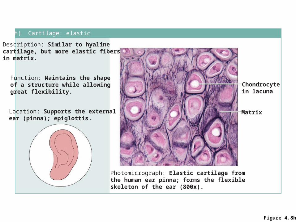
Figure 4.8h
(h) Cartilage: elastic
Description: Similar to hyalinecartilage, but more elastic fibersin matrix.
Function: Maintains the shapeof a structure while allowinggreat flexibility.
Location: Supports the externalear (pinna); epiglottis.
Photomicrograph: Elastic cartilage fromthe human ear pinna; forms the flexibleskeleton of the ear (800x).
Chondrocytein lacuna
Matrix

Figure 4.8i
(i) Cartilage: fibrocartilage
Description: Matrix similar tobut less firm than that in hyalinecartilage; thick collagen fiberspredominate.
Function: Tensile strengthwith the ability to absorbcompressive shock.
Location: Intervertebral discs;pubic symphysis; discs of kneejoint.
Photomicrograph: Fibrocartilage of anintervertebral disc (125x). Special stainingproduced the blue color seen.
Intervertebraldiscs
Chondrocytesin lacunae
Collagenfiber
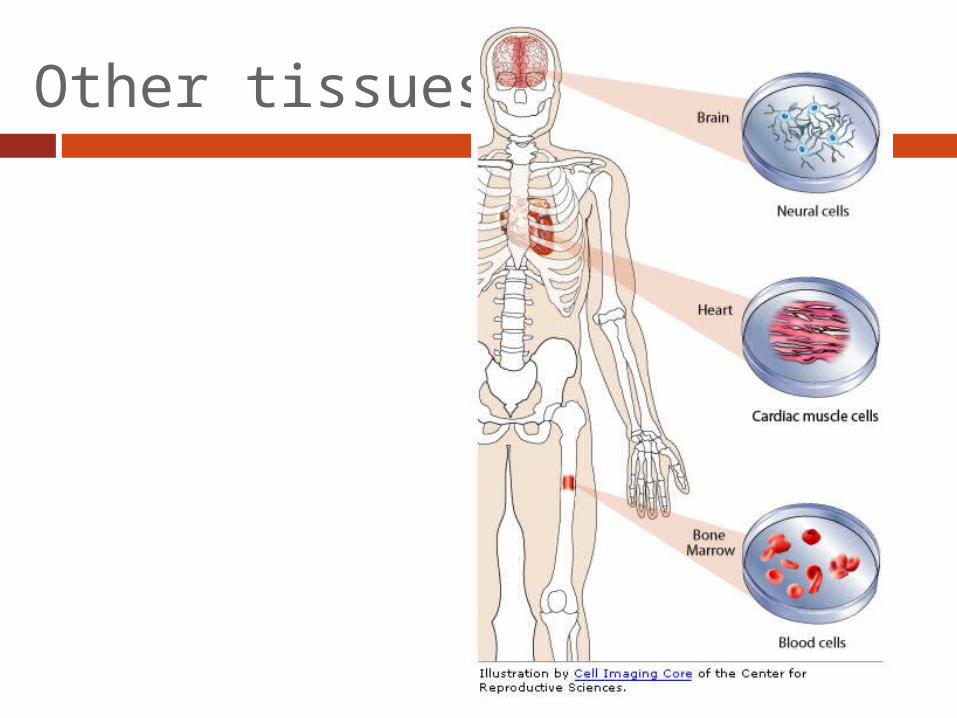
Other tissues…

Figure 4.8j
(j) Others: bone (osseous tissue)
Description: Hard, calcifiedmatrix containing many collagenfibers; osteocytes lie in lacunae.Very well vascularized.
Function: Bone supports andprotects (by enclosing);provides levers for the musclesto act on; stores calcium andother minerals and fat; marrowinside bones is the site for bloodcell formation (hematopoiesis).Location: Bones
Photomicrograph: Cross-sectional viewof bone (125x).
Lacunae
Lamella
Centralcanal

Figure 4.8k
(k) Others: blood
Description: Red and whiteblood cells in a fluid matrix(plasma).
Function: Transport ofrespiratory gases, nutrients,wastes, and other substances.
Location: Contained withinblood vessels.
Photomicrograph: Smear of human blood (1860x); twowhite blood cells (neutrophil in upper left and lymphocytein lower right) are seen surrounded by red blood cells.
Neutrophil
Red bloodcells
Lymphocyte
Plasma

Nervous Tissue
Nervous system
Photomicrograph: Neurons (350x)
Function: Transmit electricalsignals from sensory receptorsand to effectors (muscles andglands) which control their activity.Location: Brain, spinalcord, and nerves.
Description: Neurons arebranching cells; cell processesthat may be quite long extend fromthe nucleus-containing cell body;also contributing to nervous tissueare nonirritable supporting cells(not illustrated).
Dendrites
Neuron processes Cell body
Axon
Nuclei ofsupportingcells
Cell bodyof a neuron
Neuronprocesses
Nervous tissue
Figure 4.9

Muscle Tissue(a) Skeletal muscle
Description: Long, cylindrical,multinucleate cells; obviousstriations.
Function: Voluntary movement;locomotion; manipulation of theenvironment; facial expression;voluntary control.
Location: In skeletal musclesattached to bones oroccasionally to skin.
Photomicrograph: Skeletal muscle (approx. 460x).Notice the obvious banding pattern and thefact that these large cells are multinucleate.
Nuclei
Striations
Part ofmuscle fiber (cell)
Figure 4.10a

Muscle Tissue
Figure 4.10b
(b) Cardiac muscle
Description: Branching, striated, generally uninucleate cells that interdigitate atspecialized junctions (intercalated discs).
Function: As it contracts, it propels blood into the circulation; involuntary control.Location: The walls of the heart.
Photomicrograph: Cardiac muscle (500X);notice the striations, branching of cells, andthe intercalated discs.
Intercalateddiscs
Striations
Nucleus

Muscle Tissue
Figure 4.10c
(c) Smooth muscle
Description: Spindle-shapedcells with central nuclei; nostriations; cells arranged closely to form sheets.
Function: Propels substancesor objects (foodstuffs, urine,a baby) along internal passage-ways; involuntary control.Location: Mostly in the wallsof hollow organs.
Photomicrograph: Sheet of smooth muscle (200x).
Smoothmusclecell
Nuclei

Scab
Blood clot inincised wound
Epidermis
Vein
Inflammatorychemicals
Inflammation sets the stage:• Severed blood vessels bleed and inflammatory chemicals are
released.• Local blood vessels become more permeable, allowing white
blood cells, fluid, clotting proteins and other plasma proteinsto seep into the injured area.
• Clotting occurs; surface dries and forms a scab.
Migrating whiteblood cell
Artery
1
Steps in Tissue Repair

Steps in Tissue RepairRegeneratingepithelium
Area ofgranulationtissueingrowth
Fibroblast
Macrophage
Organization restores the blood supply:• The clot is replaced by granulation tissue, which restores
the vascular supply.• Fibroblasts produce collagen fibers that bridge the gap.• Macrophages phagocytize cell debris.• Surface epithelial cells multiply and migrate over the
granulation tissue.
2

Steps in Tissue Repair
Regeneratedepithelium
Fibrosedarea
Regeneration and fibrosis effect permanent repair:• The fibrosed area matures and contracts; the epitheliumthickens.• A fully regenerated epithelium with an underlying area ofscar tissue results.
3

Homeostatic Imbalance
Scar Tissue Scar tissue that forms in any
muscular organ- heart or urinary bladder can severely impair the function of that organ.
Scars reduce the internal volume of the organ and block movement of substances through the hollow organ.
Can hamper the muscle’s ability to contract. Heart = progressive heart failure. Visceral organs = adhesions
connect organs together and prevent normal shifting about ex. Intestines: adhesions obstruct the flow of foodstuffs.
Tissue Damage-Snake Bite

The End!